


Abstract
This study evaluates the mechanobiological responses of MC3T3-E1 cells to fluid shear stress utilizing a coupled CFD-CPM mesoscale framework. Computational fluid dynamics was utilized to calculate the distribution of fluid shear stress within the culture chamber, which was subsequently mapped onto a discrete system of lattices. The cellular Potts model was employed to simulate behaviors of the cells governed by rules for proliferation, migration, contact inhibition, and osteogenic differentiation. To accurately reflect developmental stages, the computational workflow dictated that the cells complete the phase of growth prior to the initiation of differentiation. Evaluations demonstrated that the culture region formed a relatively uniform plateau of shear stress. Within an optimal range, fluid shear stress accelerates the transition of these cells into mature osteoblasts. Furthermore, staining for alkaline phosphatase revealed responses of osteogenic differentiation strictly correlated with the local distribution of fluid shear stress. Ultimately, this study establishes a visualized framework of mesoscale modeling to analyze the collective behavior of osteoblasts under mechanical stimulation in microfluidic environments, demonstrating the feasibility of predicting subsequent extracellular matrix mineralization and providing valuable insights into the dynamic evolution of bone remodeling.
1. Introduction
Osteoblasts (OBs) maintain bone homeostasis by secreting proteins to synthesize the matrix during remodeling [1,2,3]. This process is highly mechanosensitive. Mechanical loading deforms trabecular cavities, generating pressure gradients and interstitial fluid flow that produce fluid shear stress (FSS) [4]. Physiological FSS activates osteogenic signaling pathways and promotes OB-mediated bone formation. Understanding this mechanism provides critical insights into osteoporosis and bone remodeling [5,6,7].
Traditionally, macroscopic continuum models have been widely applied to describe collective dynamics in active biological systems [8,9,10,11]. However, they struggle to capture individual heterogeneity and discrete interactions, particularly under low-density conditions. Additionally, solving their nonlinear equations is computationally expensive [12]. Relying solely on in vitro experiments is also challenging due to complex interacting variables and condition-dependent accuracy [13].
Consequently, discrete models like cellular automata (CA) offer computationally efficient alternatives for simulating multicellular tissue dynamics [14]. CA models have been effectively utilized to simulate diverse biological processes, including cellular differentiation [15], thymocyte migration [16], skeletal muscle stem cell regeneration [17], microfluidic organ-on-chip simulations [18], and fibroblast density variations in diseases [19]. Collectively, these studies demonstrate the efficacy of CA as a robust tool for investigating in vitro cell population dynamics.
However, conventional CA models struggle with coupled continuous physical fields. The cellular Potts model (CPM) addresses this by integrating energy constraints into the CA lattice [20]. CPM naturally incorporates local pressure and maps external continuous fields onto discrete lattices. This enables direct simulations of mechanobiological behaviors like embryogenesis and tumor growth [21,22].
In parallel, microfluidic organ-on-chip systems have become indispensable for reconstructing in vitro organ models. In bone research, microfluidic chips have successfully simulated bone cellular microenvironments [23] and quantified OB calcium dynamics under FSS [24]. These devices streamline mechanical stimulation experiments, providing valuable platforms for investigating OB differentiation and migration.
Although individual-based discrete models have been widely used in cell dynamics, existing computational models rarely integrate continuous macroscopic computational fluid dynamics (CFD) fields with the microscopic CPM.
In this study, a CPM coupled with FSS effects was developed to investigate the influence of this mechanical stimulation on osteoprogenitor cells. Model parameters were derived from literature and CFD analyses, and the framework was experimentally validated using a microfluidic platform. This computational approach provides a mesoscale mechanistic explanation for FSS-regulated cell proliferation, differentiation, migration, and bone mineralization, overcoming the limitations of conventional experimentally intensive methods.
2. Materials and Methods
2.1. Design and Fabrication of Microfluidic Chips
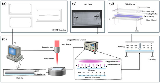
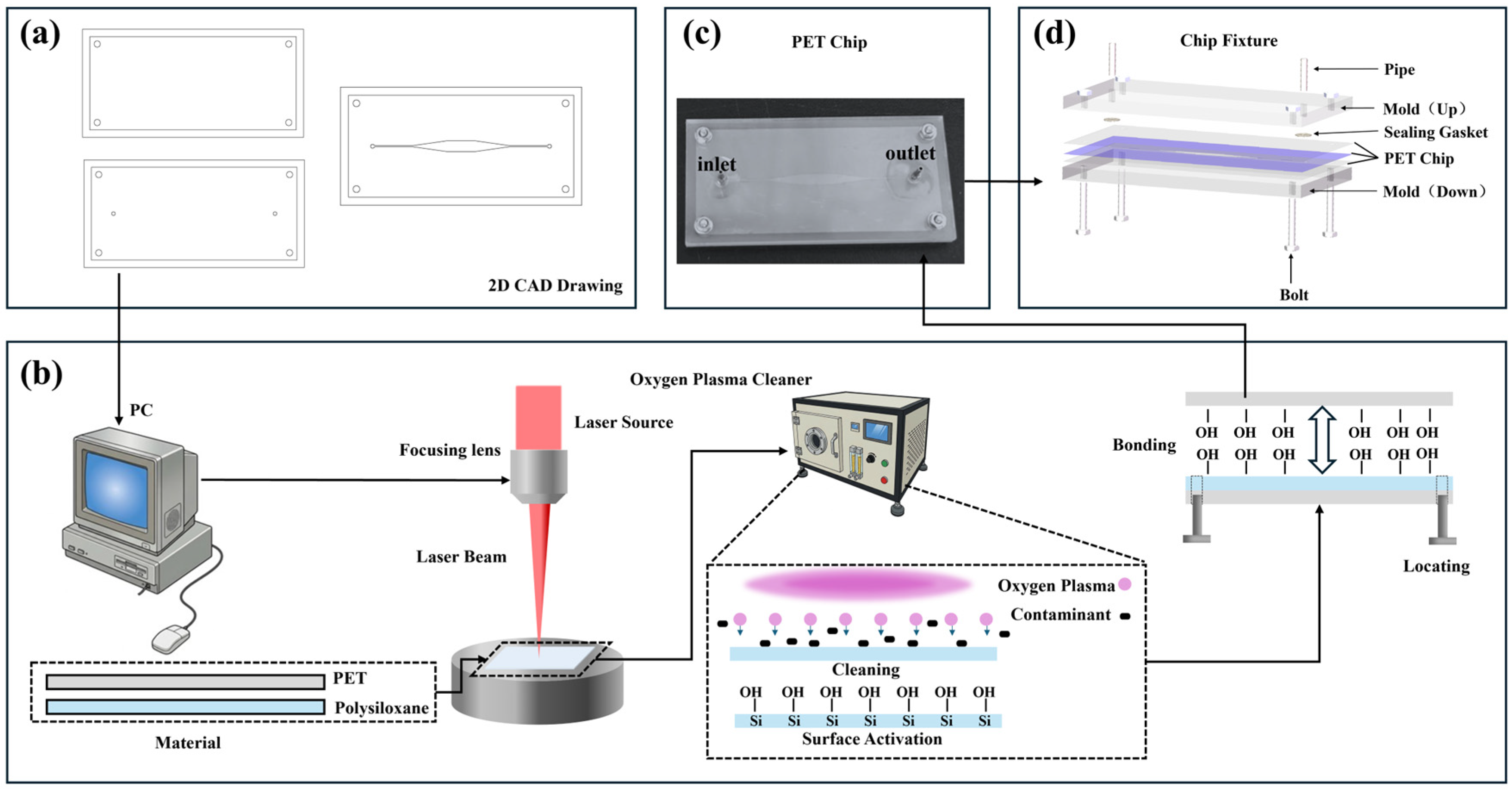
The microfluidic chip was fabricated using a thin-film manufacturing process (Figure 1a). The upper and lower polyethylene terephthalate (PET) films were designed with functional holes for fluidic access and alignment. The intermediate polysiloxane layer (Dudao United Chemical Company, Shenzhen, China) was patterned to form the core microchannel structure, comprising a culture chamber 4 mm in width and 50 μm in height.
Figure 1.
(a) Microfluidic chip design. (b) Chip fabrication process including laser machining and bonding. (c) Image of fabricated and sterilized microfluidic chip. (d) Exploded view of chip and fixture.
Following the fabrication procedure depicted in Figure 1b, the engineering design files were imported into the processing software of an ultraviolet laser cutting machine (TH-UV200A, Suzhou Tianhong; 10 W, 355 nm laser source, Suzhou, China). The designated regions of the material were ablated to define the chip pattern. The processed layers were subsequently treated in an oxygen plasma cleaner (PDC-002, Harrick Plasma, Ithaca, NY, USA) for 3 min to remove surface contaminants and organic residues, while simultaneously introducing functional groups onto the material surfaces. Alignment pins were inserted through the circular holes located at the four corners of the three layers to ensure precise alignment. The layers were then bonded together to form a robust interface, thereby ensuring the sealing integrity of the internal microchannels. The fabricated chip is shown in Figure 1c. Finally, the chip was mounted into a fixture and secured with bolts to complete the device assembly (Figure 1d).
2.2. CFD Analysis of Flow Field in Microchannels
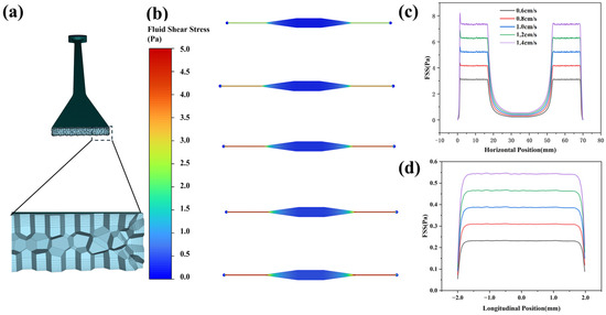
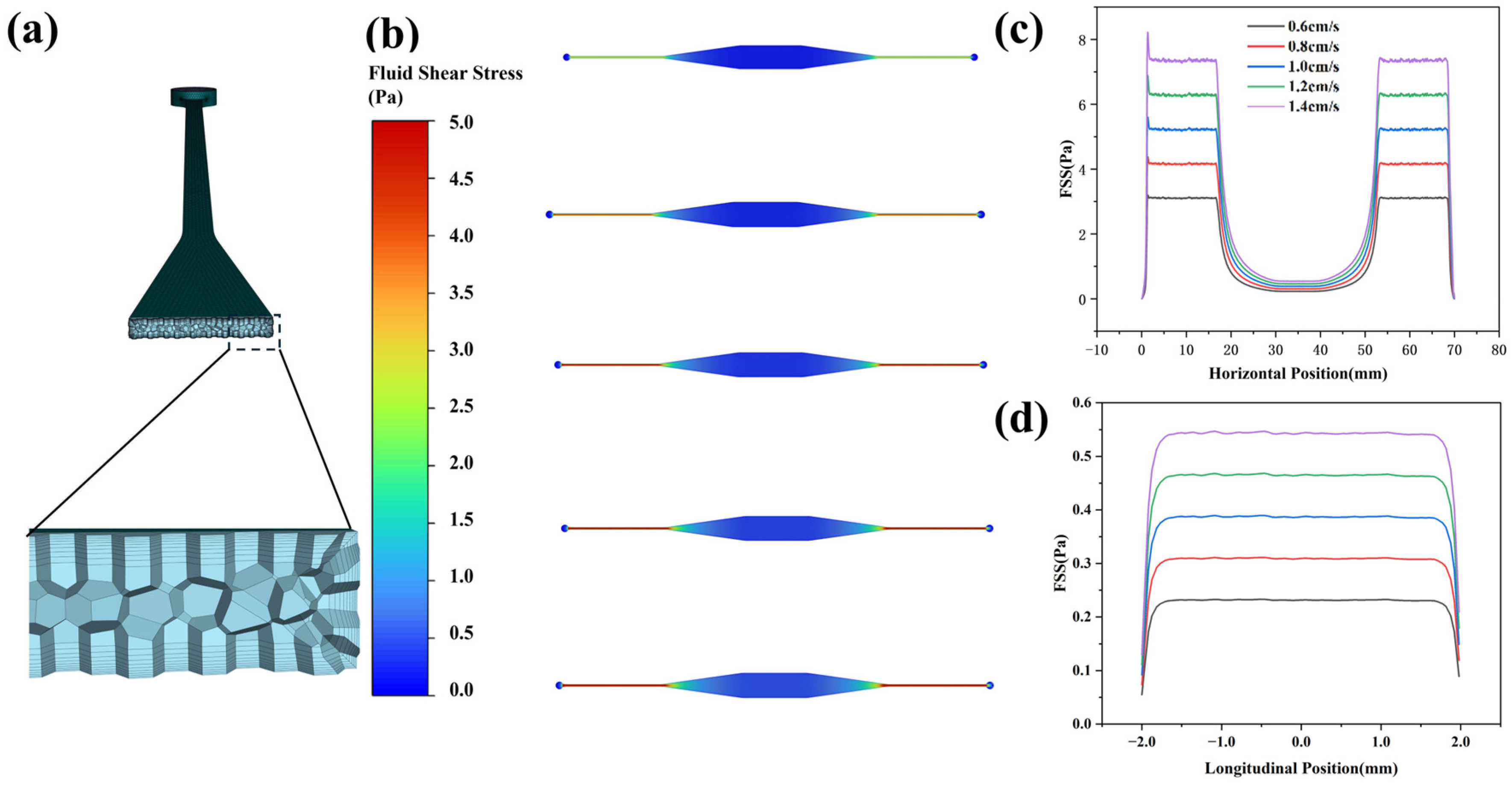
To investigate the distribution of FSS and ensure an appropriate mechanical environment for the cells, a CFD analysis was conducted. The fluid was modeled as an incompressible, steady-state flow, with a density of 1.009 g/cm3 and a dynamic viscosity of 0.93 mPa·s. Boundary conditions included an inlet velocity of 0.6 cm/s, an outlet pressure of 101.325 kPa (standard atmospheric pressure), and a no-slip condition at the walls. The maximum calculated Reynolds number within the channel was 4.05, confirming the presence of a typical laminar flow regime. The computational domain was discretized using a triangular mesh with element sizes ranging from 0.2 to 6 μm. Furthermore, an eight-layer boundary layer mesh was generated near the inner walls to capture the steep velocity gradients (Figure 2a). The steady-state flow field was obtained by solving the Navier–Stokes equations using a fully coupled approach (Figure 2b). The results demonstrated that although relatively high shear stress (>2 Pa) occurred within the narrow inlet and outlet channels, an exceptionally broad and uniform shear stress plateau (0.5–1.2 Pa) formed in the central region of the culture chamber along the flow direction (Figure 2c). Moreover, the transverse distribution perpendicular to the flow direction exhibited a highly uniform profile (Figure 2d). This uniform distribution falls within the optimal range for stimulating osteogenic differentiation [5,25,26].
Figure 2.
(a) Mesh and boundary layer. (b) Nephogram of bottom shear stress in the chip at 0.6–1.4 cm/s. (c) Shear stress distribution along the bottom centerline of the chip along the flow direction. (d) Fluid shear stress (FSS) distribution along the chip bottom centerline perpendicular to the flow direction.
2.3. Microfluidic Cell Culture and Osteogenic Evaluation
The osteoprogenitor cells (MC3T3-E1) used in this study were provided by the Institute of Stomatology at Nanjing Medical University. Only cells between passages 5 and 10 were used. The cells were routinely cultured in 60 mm dishes containing a complete growth medium composed of α-MEM supplemented with 10% FBS and 1% penicillin-streptomycin solution. The culture medium was replaced every three days. The fluid perfusion system, consisting of a commercial syringe pump, disposable sterile syringes, silicone tubing, and a buffer reservoir, was sterilized prior to cell seeding. Specifically, the microfluidic chip was sterilized via injection of a 75% ethanol solution, followed by exposure to ultraviolet radiation for 30 min and a 12 h undisturbed incubation period. Prior to use, the assembled chip was repeatedly rinsed with phosphate-buffered saline (PBS) and coated with a 0.1 mg/mL poly-L-lysine (PLL) solution for 1 h to enhance cell adhesion.
Upon reaching approximately 80% confluence, the MC3T3-E1 cells were detached using a 0.25% trypsin-EDTA solution and diluted to a concentration of 1 × 10⁶ cells/mL. The cell suspension was injected into the microfluidic chip and incubated overnight at 37 °C in a humidified atmosphere containing 5% CO2 to ensure sufficient cell adhesion. After a three-day culture period to allow for cell growth, the culture medium was replaced with an osteogenic induction medium. This medium was supplemented with 50 μg/mL ascorbic acid, 10 mM β-glycerophosphate sodium, and 10 nM dexamethasone to induce osteogenic differentiation. Intermittent dynamic perfusion was applied at a flow rate of 7.2 μL/min for 1 h per day over a 7-day period. A conventional static culture control group without FSS stimulation was concurrently established in 60 mm dishes for 7 days.
Following the induction period, the differentiated cells were evaluated using an ALP staining kit. The chip was rinsed with PBS, fixed with a 4% paraformaldehyde solution for 30 min, and subsequently rinsed with Tris-buffered saline. The staining solution was added, and the chip was incubated at 37 °C for 2 h until clear color development was observed.
2.4. Model Design
2.4.1. FSS-Coupled CPM Framework
To simulate the evolution of cells under FSS, a cross-scale coupled CPM strategy was adopted. The physical space of the system is discretized into a regular lattice. Each lattice site is assigned a specific state: medium (0), wall (1), osteoprogenitor cell (2), osteoblast (3), or mineralized matrix (4). Apoptosis or cell detachment induced by excessive FSS is modeled as a state transition to the culture medium (0). The morphological evolution of the cells is driven by the minimization of an effective Hamiltonian energy (H), which represents constraints of perimeter (Hp), constraints of area (Hs), contact energy (Hc), and specific external energy (He) [20]:
where Hp, Hs, Hc and He denote the perimeter constraint, area constraint, interfacial contact energy, and specific external energy, respectively. Their specific forms are:
where λp and λs are stiffness coefficients; p(σ) and s(σ) are the current perimeter and cell area, respectively; P(σ) and S(σ) are their target values; J(σi,σj) is the contact energy coefficient matrix between neighboring lattice sites; and δσi,σj represents the Kronecker delta function, which ensures that the interfacial contact energy is calculated exclusively at cell boundaries.
The external energy term incorporates the fluid-induced force, enabling the cells to respond to local shear stress. Subsequent state updates of the lattice are governed by the Metropolis criterion, where the transition probability P is determined based on the total energy change (ΔH) in the system:
where h denotes the yield energy threshold for state transitions, and T represents the activity of membrane edge fluctuations, reflecting the intrinsic motility of the cells.
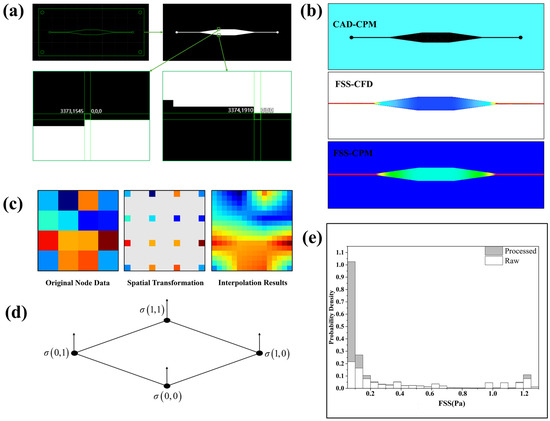
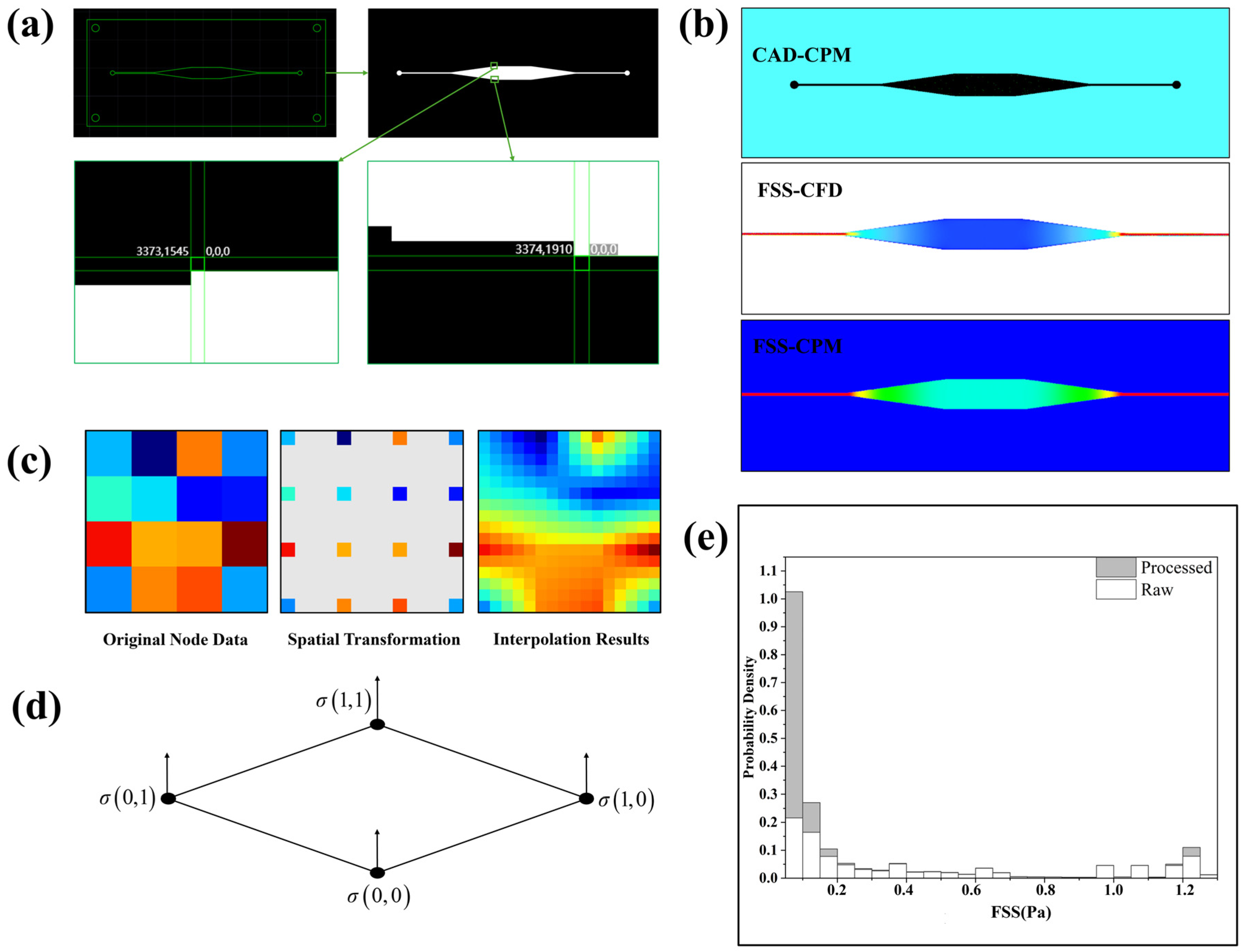
To transform continuous biophysical processes into a computational model, the spatiotemporal domain of the simulation region was discretized by extracting pixel coordinates from binarized mask images of the chip (Figure 3a). Periodic boundaries were applied in the flow direction to ensure flow field continuity, whereas the physical channel walls were treated as frozen boundaries (Figure 3b). To address the resolution mismatch between the CFD mesh and the CPM lattice, a bilinear interpolation algorithm was employed to resample the flow field (Figure 3c,d) [27]. As shown in Figure 3e, the global FSS distribution became significantly smoother following interpolation, thereby providing a reliable data basis for accurate lattice-based dynamic evolution simulations.
Figure 3.
(a) Binary image of the chip. (b) Mapping result in CPM. (c) Pixels of different colors contain data from different nodes. The initial node is mapped to a high-resolution grid coordinate system containing numerous gaps to be filled. Using the values of the four surrounding known points, the value at the gap is calculated using a bilinear formula based on distance-weighted interpolation. (d) Bilinear interpolation surface. (e) Global distribution characteristics of FSS before and after interpolation.
2.4.2. Dynamic Evolution Model of Osteoblasts Under FSS
In the computational model, FSS was not applied continuously. To accurately replicate intermittent physiological loading and align with in vitro experimental conditions, an intermittent loading paradigm was explicitly programmed into the CPM. By mapping the temporal scale such that 1440 Monte Carlo steps (MCS) represent a 24 h physiological day, the dynamic simulation applied the FSS field for 60 MCS (equivalent to 1 h), followed by a 1380 MCS (23 h) static resting phase for each simulated day.
The global simulation parameters and cellular morphology constraints are summarized in Table 1 and Table 2.
Table 1.
Global Simulation Parameters.
Table 2.
Cell Morphological Constraints.
2.4.3. Mechanobiological Rules for Cellular Evolution in CPM
Proliferation and Migration: The assumption of a constant cell division rate was discarded. Instead, the proliferation probability for each cell is jointly determined by its intrinsic basal probability, a spatial limitation factor (ψs), and a shear-stress regulatory factor (ψτ):
where N and K denote the current local cell density and the maximum carrying capacity, respectively; τ represents the local FSS, with τl and τh defining its lower and upper thresholds; and C0, A, B, α, and β are empirical coefficients shaping the response curve.
When the population density approaches K, ψs imposes a physical constraint reflecting contact inhibition. Concurrently, ψτ employs hyperbolic tangent functions to accurately capture the nonlinear, biphasic “stimulation-damage” response to FSS. Furthermore, an adaptive migration-driving force field (Ff) was constructed to propel the centroid of undifferentiated cells:
where FM represents the maximal driving force magnitude, τM is the shear-stress saturation threshold, ta denotes the cellular mechanosensory adaptation time, and is the unit vector of the local flow direction.
Differentiation: Osteogenic differentiation is an irreversible process. To simulate the transition of cells that have completed their growth phase and entered differentiation under mechanical regulation, a nonlinear response function based on the Michaelis-Menten equation was adopted:
where αd represents the maximum differentiation enhancement coefficient, τc is the FSS activation threshold, and Ks denotes the Michaelis constant corresponding to the half-maximal response. Furthermore, the initiation of differentiation requires the macroscopic spatial confluence of the system to reach a predefined threshold, thereby simulating contact inhibition.
Mineralization: Following differentiation, the osteoblasts enter the matrix secretion and mineralization stages. A two-stage decoupled mechanism was constructed. Initially, a local shear-stress regulatory coefficient, ψm(τ), was introduced:
where αm denotes the maximum enhancement amplitude for matrix secretion. Subsequently, cells with an osteoblastic phenotype secrete a collagen matrix into the surrounding lattice with a specific probability (Pm):
where P0 is the basal secretion probability, Θ denotes the Heaviside step function, and ti(τ) represents the shear-dependent onset time of matrix secretion. Finally, terminal mineralization occurs exclusively when the local collagen concentration reaches a specific threshold and a nucleation region exists in the neighborhood. This process is governed by a strict logic gate:
where Cc and C* denote the local and threshold concentrations of the collagen matrix, respectively; tm is the maturation time required for mineralization; and Ωn represents the nucleation factor determined by the presence of existing mineralization cores in the local neighborhood. The mineralized regions exhibit significantly increased stiffness, thereby restricting further migration and deformation of surrounding cells.
3. Results and Discussion
3.1. Osteogenic Staining
The foundation of this mechanobiological response lies in the precise characterization of the mechanical microenvironment. Our CFD analysis demonstrated a broad, uniform FSS plateau (0.5–1.2 Pa) inside the microfluidic chip (Figure 2c), which falls well within the optimal range known to promote osteoprogenitor cell activity [25,26,32].
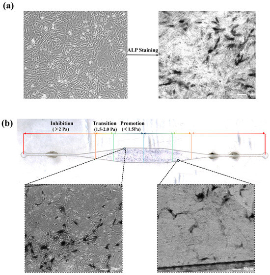
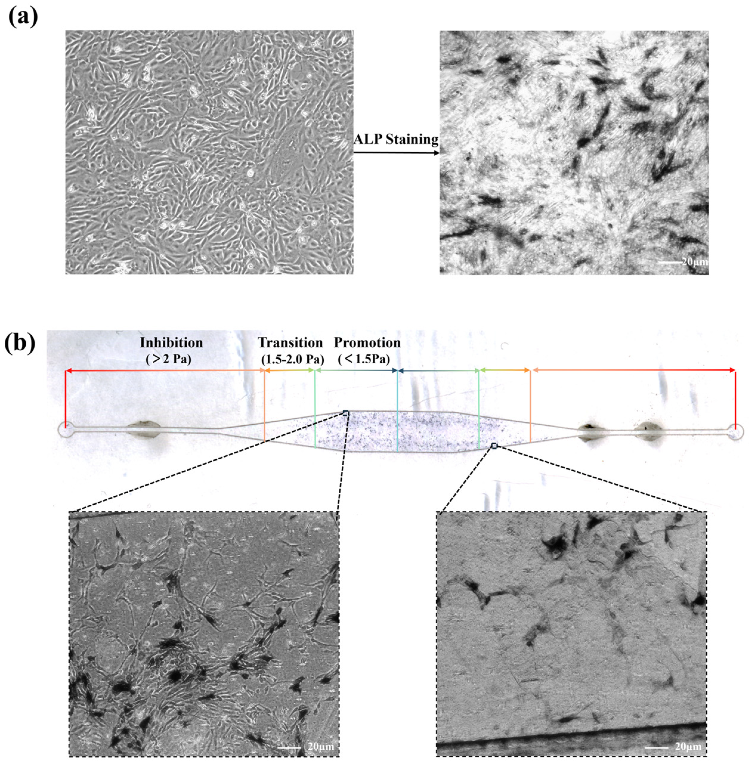
Driven by this mechanical microenvironment, the conventional static culture exhibited a uniform, baseline expression of ALP, as shown in Figure 4a. In contrast, dynamic perfusion within the microfluidic chip induced a pronounced, spatially heterogeneous ALP distribution strongly correlated with the local FSS gradient (Figure 4b). Extensive purple ALP-positive regions and a high density of mature osteoblasts were observed in the central chamber, where the shear stress remained within the optimal range (<1.5 Pa).
Figure 4.
Staining outcomes of static culture and FSS groups: (a) ALP staining for osteogenic differentiation in the static group. (b) ALP staining for osteogenic differentiation within the microfluidic chip, the purple regions represent ALP-positive areas, indicating active osteogenic differentiation, and local microscopic images under different shear stresses were also obtained, where dark areas correspond to osteoblasts. The transition of the arrow colors from red to blue indicates a spatial decrease in fluid shear stress from high to low across the different regions of the chip.
This spatial correlation quantitatively aligns with the biphasic “stimulation-damage” theory of cellular mechanotransduction reported in previous independent studies, which demonstrated that moderate FSS promotes MC3T3-E1 proliferation and osteogenic marker expression, whereas stresses exceeding physiological limits trigger cell detachment and cell cycle arrest [5,6,29,33,34]. Consistent with these findings, our results demonstrated that both ALP staining intensity and the number of adherent cells were markedly reduced in the transition zones (1.5–2.0 Pa) and the narrow inlet and outlet channels (>2.0 Pa), corroborating that excessive shear stress induces mechanical fatigue rather than differentiation.
Furthermore, the successful induction of this targeted osteogenesis demonstrates that this rigid PET-based microfluidic architecture offers a highly reliable alternative to polydimethylsiloxane (PDMS), which is commonly used in organ-on-a-chip systems [23,24,35,36]. While PET chip fabrication relies on layer-by-layer ablation, making it potentially less versatile than PDMS for constructing highly complex 3D organ-on-a-chip structures [37], it remains entirely adequate for the requirements of the current study.
3.2. Cellular Dynamic Evolution Simulation Under FSS
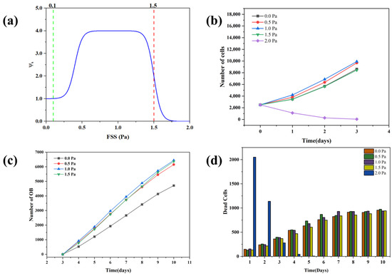
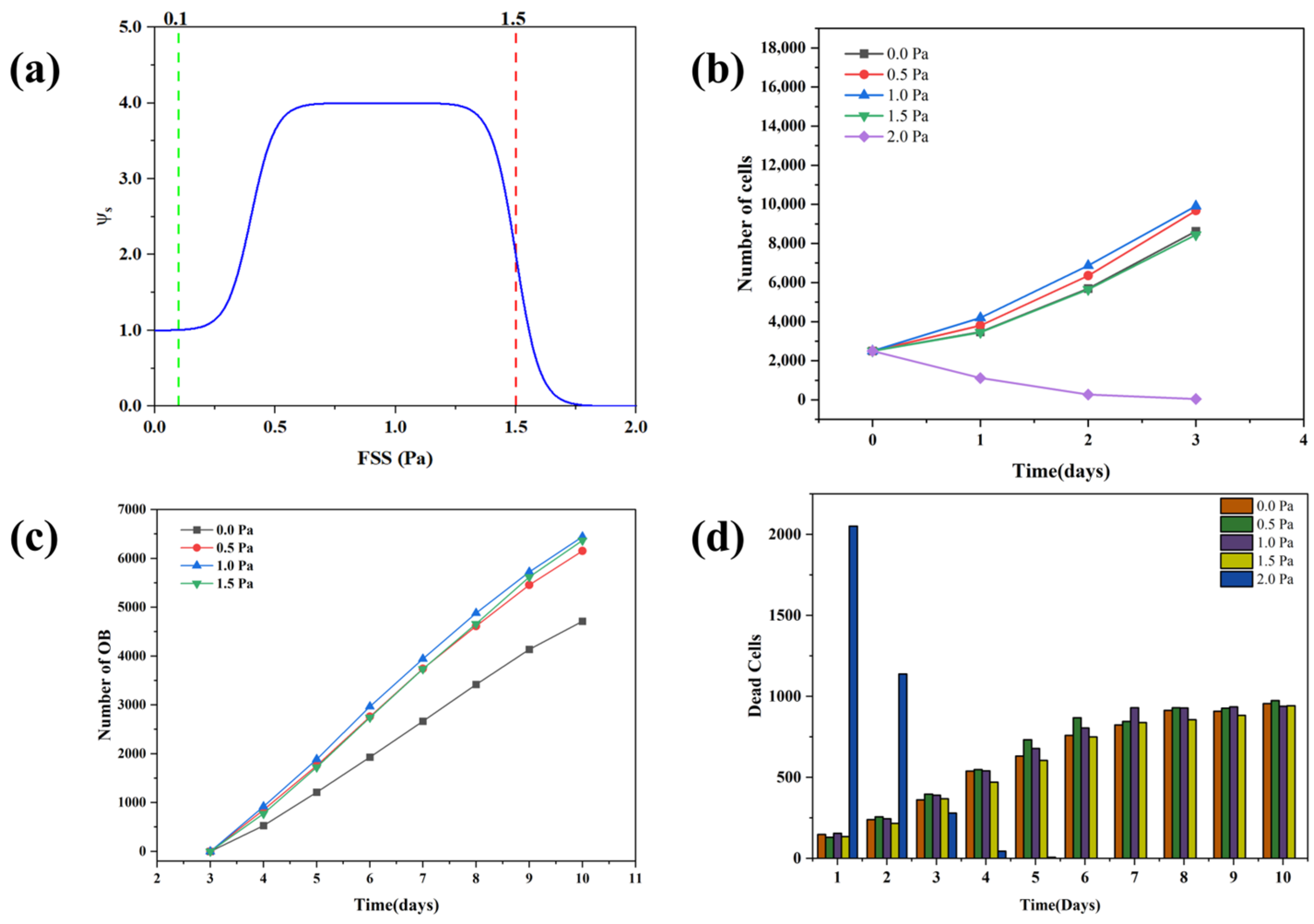
To accurately govern the mechanobiological response of the cells, the CPM incorporates a mathematically defined, nonlinear biphasic “stimulation-damage” function (Figure 5a). This regulatory curve dictates that moderate FSS yields a maximal biological gain plateau, whereas shear stress exceeding a critical threshold triggers a steep suppressive penalty. Driven by this underlying mechanism, the population evolution of MC3T3-E1 cells under five FSS levels (0.0–2.0 Pa) was simulated over a 10-day period. During the initial proliferation stage (days 0–3), FSS between 0.5 and 1.5 Pa promoted cellular growth compared to the ideal static control (0.0 Pa), peaking at an optimal stimulation of 1.0 Pa (Figure 5b). This aligns with experimental observations that FSS at approximately 0.8–1.2 Pa maximally upregulates osteogenic marker expression and proliferation in MC3T3-E1 cells [5,6,26]. Conversely, an excessive shear stress of 2.0 Pa induced severe growth inhibition and massive early apoptosis (Figure 5d), computationally corroborating prior reports that supra-physiological FSS triggers cellular damage, membrane compromise, and cell cycle arrest in osteoblasts [29,33,34].
Figure 5.
Growth simulation of MC3T3-E1 cells under 5 FSS levels: (a) The “stimulation–damage” curve regulated by FSS. (b) Overall cell apoptosis within 10 days. (c) Number of osteogenic differentiated cells during differentiation from day 3 to day 10. Cell differentiation under 2.0 Pa FSS was not analyzed, as most cells failed to survive to this stage. (d) Cell phase diagrams on day 10 under five shear stress intensities.
Overall, the biphasic regulation of cellular behavior became highly apparent, echoing the dose-dependent “stimulation-inhibition” pattern observed in in vitro osteoblast cultures [34]. Our simulations successfully demonstrated that an appropriate FSS range (0.5–1.5 Pa) significantly enhanced proliferation [25,36], whereas excessive shear stress induced injury and cell cycle arrest [33,34].
Following the growth phase, the simulation transitioned to induced differentiation (days 3–10), excluding the non-viable 2.0 Pa group. The generation of mature osteoblasts exhibited a strong dependence on shear intensity; stimulation at 1.0 Pa yielded the maximum number of differentiated cells, while 0.5 Pa and 1.5 Pa provided moderate enhancements over the static baseline (Figure 5c). This shear-intensity-dependent differentiation profile agrees with experimental studies showing that moderate FSS maximally induces osteogenic marker expression and mineralization in osteoblasts [5,26,38], and that optimizing flow rates is critical for maximizing mineralized matrix production [39].
3.3. Spatio-Temporal Evolution of Cellular Morphology and Proliferation
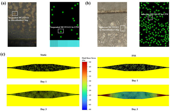
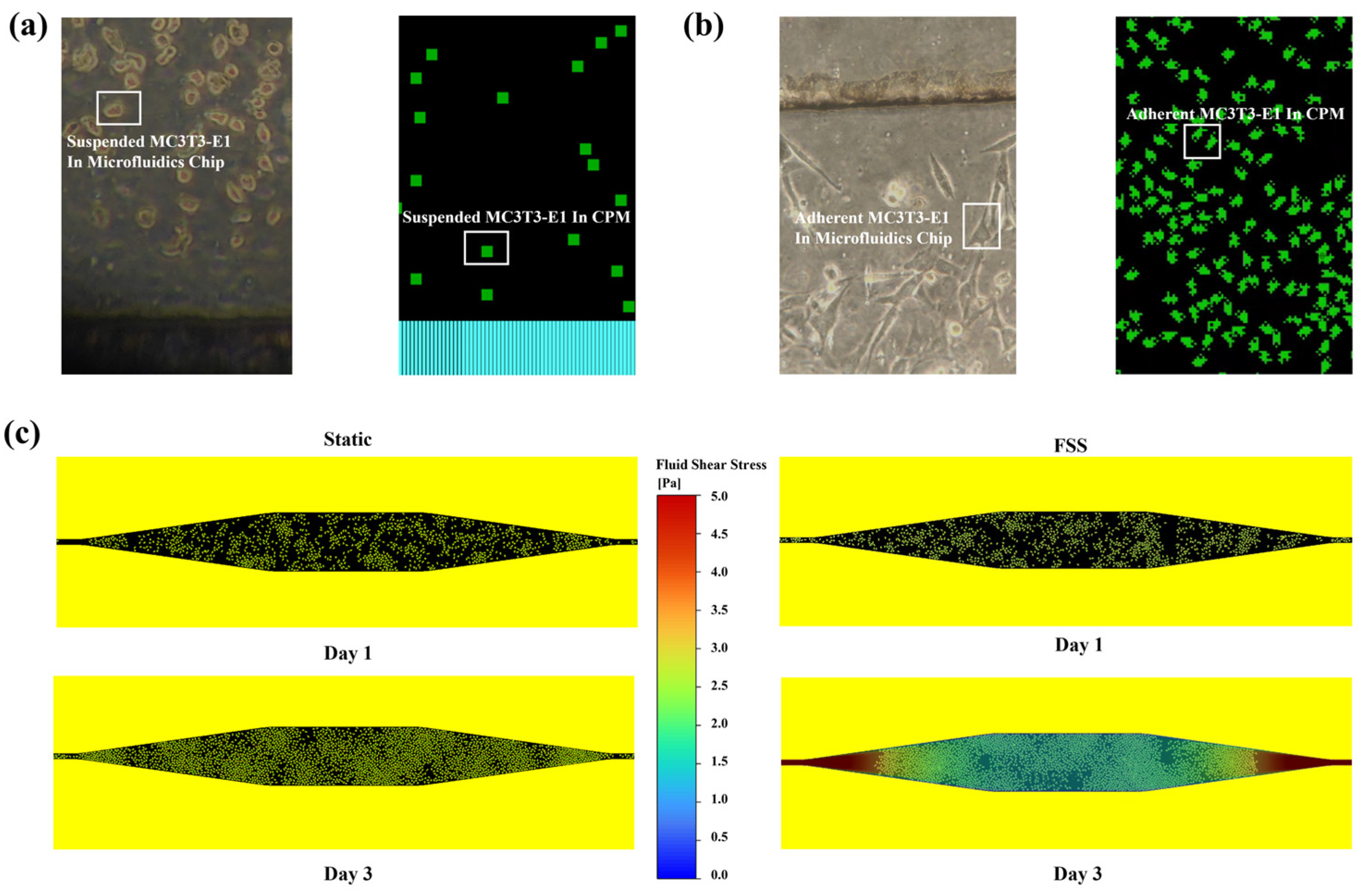
To establish a rigorous correspondence between the computational framework and actual physiological time, the simulation mapped 1440 Monte Carlo steps (MCS) to a 24 h cycle, of which 60 MCS corresponded to the 1 h daily FSS exposure and the remaining 1380 MCS represented the static incubation period, consistent with the intermittent perfusion protocol (1 h/day) used in the in vitro experiments [5,26,32]. The spatiotemporal evolution during the early proliferation phase (days 1–3) revealed that population dynamics were highly dependent on the macroscopic mechanical field. Specifically, the static control group (0.0 Pa) was computationally configured to exclude mechanical disturbances associated with medium exchange. This established an idealized baseline that consequently exhibited uniform spatial growth (Figure 6c).
Figure 6.
FSS-driven cell proliferation fate decision model and early spatio-temporal evolution characteristics in microfluidic chip: (a) Captured images of cells in the chip at suspended state immediately after seeding; the initial suspended cell morphology in the corresponding CPM on the right is represented by quadrilaterals. (b) The left image shows cells in adherent state inside the chip, presenting a fusiform shape with actin-based pseudopodia. The MC3T3-E1 cells imposed with major axis constraints in the right CPM possess analogous pseudopodial structures. (c) Spatio-temporal evolution snapshots of the cell population during the early proliferation stage.
In contrast, the spatial distribution under dynamic FSS displayed an FSS-dependent state transition, consistent with experimental observations from multi-shear microfluidic platforms demonstrating that osteoblast responses are spatially heterogeneous under varying FSS magnitudes [24,36]. In the central chamber, where the fluid field formed a uniform plateau within the optimal stimulation regime, osteoprogenitor cells maintained a robust proliferative state [5,6]. These local aggregates continued to divide and expand until reaching a volumetric limit, which was regulated by the spatial dimensions of the microchamber and the algorithm’s contact inhibition constraints [21,40]. Conversely, when the local FSS exceeded the critical tolerance threshold (>2.0 Pa), a distinct fate transition occurred. The osteoprogenitor cells located in the narrow inlet and outlet channels accumulated mechanical damage, rapidly transitioning from a proliferative state into one characterized by apoptosis and substrate detachment. This heterogeneous spatial distribution indicates that maintaining FSS within an appropriate physiological window is important for the sustainable growth and functional population expansion of osteoblasts in microfluidic systems.
3.4. Osteogenic Differentiation Simulation
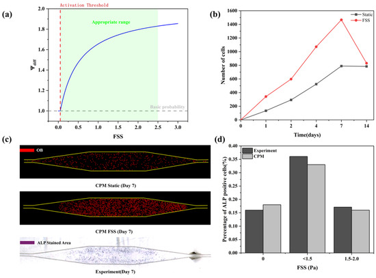
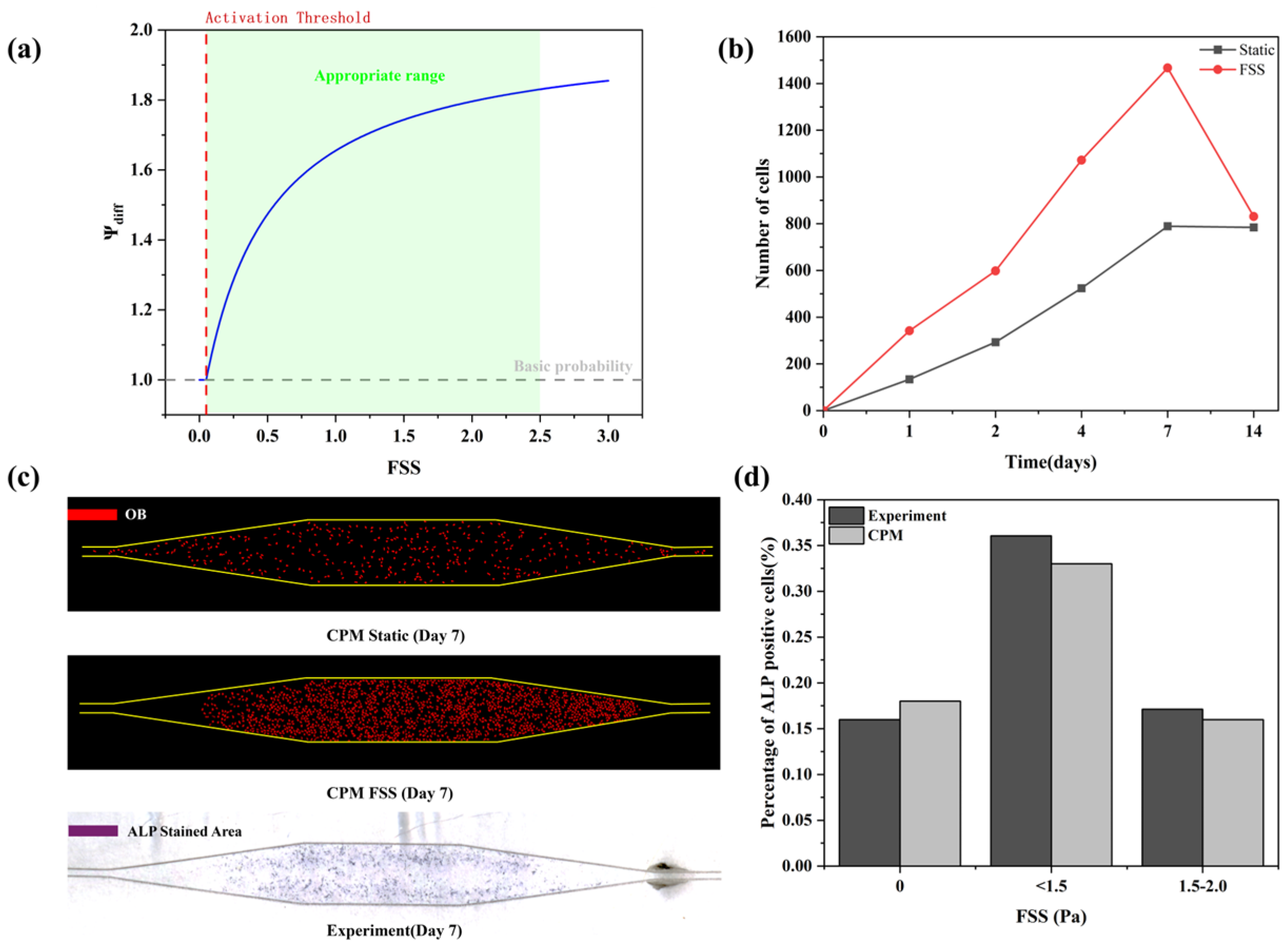
Following the initial growth phase, the transition of osteoprogenitor cells into mature osteoblasts was simulated over a 14-day period, governed by the differentiation gain factor, ψdiff (Figure 7a). To model this irreversible transition, a Michaelis-Menten-based nonlinear response function under local mechanical stimulation was employed. Within the CPM lattice, the local fluid shear stress τ acting on individual cells modulates their basal differentiation probability. Once this mechanical stimulus exceeds a predefined activation threshold, the cellular differentiation rate follows a saturation curve, reflecting the limited receptor-mediated mechanotransduction capacity inherent to biological systems. This mathematical formulation aligns with the multiphase modeling framework proposed by Pearson [31], who applied Michaelis-Menten-type kinetics to simulate nonlinear cellular fate decisions under dynamic fluid flow.
Figure 7.
Differentiation period: (a) Differentiation gain factor. (b) Number of progenitor cells within 14 days after induced differentiation. (c) Comparison of CPM simulation images and experimental staining results on day 7 of induced differentiation under static state (without FSS) and FSS loading. Undifferentiated precursor cells were masked to better observe osteogenic differentiation. (d) Proportion of ALP-positive cells in regions with varying fluid shear stress levels.
The temporal dynamics of the progenitor population (Figure 7b) indicated that while the static group exhibited a gradual trajectory, the FSS-loaded group peaked at day 7. Driven by local flow gradients, regions experiencing higher shear stress exhibited accelerated differentiation. This trend is consistent with the well-documented temporal progression of MC3T3-E1 differentiation, in which progenitor cells are progressively depleted as they commit to the osteoblast lineage [41].
The spatial heterogeneity of this transition is illustrated in the day 7 CPM phase diagrams (Figure 7c). With undifferentiated precursors masked, the static simulation displayed a sparse, random distribution of mature osteoblasts (red). In comparison, the FSS-driven simulation demonstrated an accumulation of osteoblasts concentrated within the optimal shear regions of the central chamber. This simulated spatial pattern is comparable to our experimental results; the macroscopic distribution of osteoblasts predicted by the CPM closely corresponds to the ALP-positive regions observed in the microfluidic chip.
To evaluate the predictive accuracy of the computational model, a quantitative comparison of ALP-positive cell proportions across varying FSS zones was conducted (Figure 7d). The computational predictions showed strong agreement with the in vitro experimental data. Specifically, both the simulation and the experiment indicated that the proportion of differentiated cells peaked within the optimal stimulation regime (<1.5 Pa), while differentiation efficiency was markedly reduced in the transition zones (1.5–2.0 Pa) and the static control group (0 Pa). This quantitative concordance suggests that the constructed spatial Michaelis-Menten rules effectively capture the mechanosensitivity and fate decision mechanisms of osteoprogenitor populations.
3.5. Terminal Mineralization Simulation
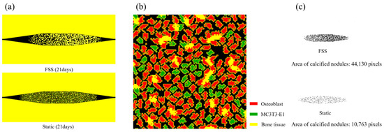
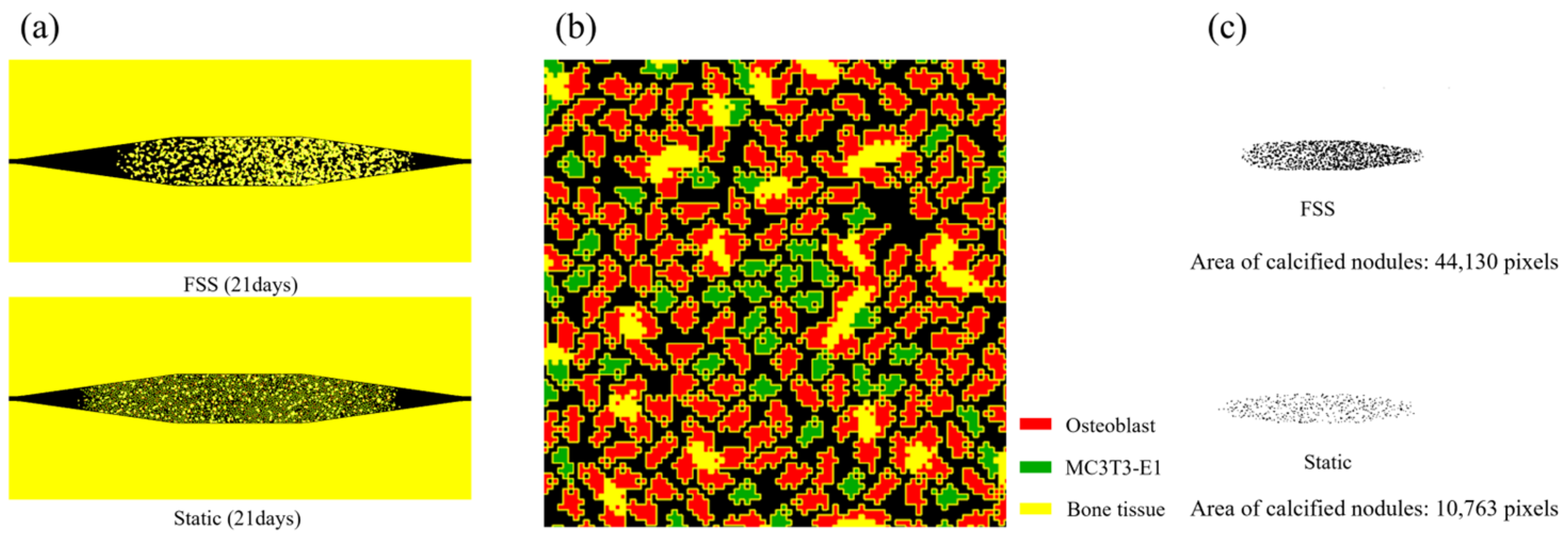
To evaluate the spatial distribution of calcified nodules, a CPM analysis was conducted at day 21. By extrapolating the mechanobiological rules calibrated from the 7-day early differentiation experiments, the model enables the predictive simulation of long-term terminal mineralization, thereby circumventing the technical bottlenecks typically associated with prolonged 21-day microfluidic perfusion cultures. As shown in Figure 8a, macroscopic bone-like tissue structures were formed in both dynamic and static simulations. However, the FSS-driven simulation exhibited enhanced matrix deposition. Following image binarization (Figure 8c), the total calcified nodule area in the FSS group reached 44,130 pixels, notably exceeding the 10,763 pixels observed in the idealized static control group. This approximately fourfold enhancement is consistent with in vitro perfusion bioreactor studies: Bancroft et al. [42] reported that all flow conditions yielded significantly higher mineralized matrix production than static controls, with calcium content increasing in a dose-dependent manner with flow rate, while Sikavitsas et al. [43] demonstrated that increasing fluid shear forces—while holding chemotransport constant—directly increased mineral deposition. Datta et al. [44] further showed that FSS and bone-like extracellular matrix act synergistically, producing an up to 75-fold increase in calcium content. The strong agreement between our simulation results and these experimental findings suggests that the mechanobiological rules captured by the CPM adequately represent the osteoinductive effect of shear stress on terminal mineralization.
Figure 8.
Characteristics of terminal mineralization distribution and morphological evolution during bone formation: (a) Comparison of global spatial distribution of calcified nodules in microfluidic culture regions between FSS group and static group. (b) Local lattice state snapshots in the core region of CPM simulation. (c) Macroscopic contours of actual mineralized nodules after binarization extraction.
Unlike previous cellular automata models that were limited to static cultures without mechanical cues [13,45,46], our model introduces FSS as an active inducing factor for terminal bone matrix formation [4,38,44]. Under this framework, regions with dynamic, uniformly distributed shear stress achieved quantitatively higher and more stable matrix deposition [47] with a more uniform spatial distribution [39,43]. This is consistent with perfusion bioreactor studies showing that FSS enhances both the amount and the spatial distribution of the mineralized matrix, whereas static culture produces heterogeneous, surface-localized deposition [42,43]. Local lattice snapshots (Figure 8b) further reveal that mature bone tissue (yellow) accumulated around differentiated osteoblasts (red), while undifferentiated MC3T3-E1 cells (green) were confined to peripheral, low-shear regions. This spatial segregation mirrors the stage-dependent compartmentalization of MC3T3-E1 cultures, in which proliferating cells remain ALP-negative and non-mineralizing until growth arrest initiates matrix deposition [41]. Furthermore, it aligns with bioreactor observations demonstrating that dynamic perfusion drives cell differentiation into the scaffold interior, while static conditions leave undifferentiated cells at the periphery [48].
4. Conclusions
This study establishes a multiscale computational framework integrating fluid mechanics with a cellular Potts model to simulate the mechanobiological evolution of osteoprogenitor cells. While existing approaches have advanced mechanobiology, they generally lack the integration of computational fluid dynamics with the CPM. Our framework addresses this methodological gap, establishing a direct mechanistic link between complex shear stress fields and cellular evolution. The integrated platform successfully translates the mesoscale perception of physical forces into macroscopic biological responses, accurately reproducing the nonlinear biophysical dynamics where controlled FSS guides cell proliferation and differentiation, and predicting spatial matrix mineralization. The strong agreement between the in silico spatial patterns and in vitro microfluidic staining validates the model’s robust capability to capture the mechanically driven self-organization mechanism inherent to early bone remodeling.
While the long-term mineralization predicted by the model is well-corroborated by established literature, direct in vitro validation (such as Alizarin Red staining) was not performed in this study. Specifically, although the thin-film PET chip employed facilitates rapid fabrication and reliable early differentiation assessment, incorporating such endpoint assays would further enhance the biological fidelity of the model by providing direct physicochemical confirmation of matrix mineralization. Maintaining stable long-term perfusion cultures poses inherent technical challenges compared to mature PDMS-based platforms. Consequently, optimizing the microfluidic device for prolonged dynamic culture and completing the terminal experimental validation should be performed in future investigations. Furthermore, a comprehensive understanding of bone homeostasis requires extending this description beyond isolated monocultures toward a framework that accounts for 3D architectural constraints and multi-cellular crosstalk. We emphasize that the representation of complex in vivo bone microenvironments within this in vitro framework remains at a preliminary stage of development. Direct simulation evidence incorporating full 3D osteoclast-osteoblast coupling is still evolving, and our current 2D mesoscale approach should therefore be seen as a robust foundation that complements future comprehensive tissue-level digital twin models.
Author Contributions
Conceptualization, D.J. and M.L.; methodology, D.J., Y.L., X.Q., L.L. and L.X.; software, Y.L.; validation, X.Q. and L.L.; investigation, D.J. and Y.L.; resources, M.L., L.X. and B.Y.; data curation, Y.L.; writing—original draft preparation, Y.L.; writing—review and editing, D.J.; visualization, Y.L.; supervision, L.X. and B.W.; project administration, D.J. and B.Y.; funding acquisition, D.J., B.W., L.X. and B.Y. All authors have read and agreed to the published version of the manuscript.
Funding
This work was funded by the National Natural Science Foundation of China (82571135, 82371000 and 82571165) and the Key R&D Program of Jiangsu Province (BE2023836).
Data Availability Statement
The datasets in this article are available from the corresponding author upon reasonable request.
Conflicts of Interest
The authors declare no conflicts of interest.
References
- Ducy, P.; Schinke, T.; Karsenty, G. The osteoblast: A sophisticated fibroblast under central surveillance. Science 2000, 289, 1501–1504. [Google Scholar] [CrossRef] [Scilit] [PubMed]
- Mizoguchi, T.; Ono, N. The diverse origin of bone-forming osteoblasts. J. Bone Miner. Res. 2021, 36, 1432–1447. [Google Scholar] [CrossRef] [Scilit] [PubMed]
- Hadjidakis, D.J.; Androulakis, I.I. Bone remodeling. Ann. N. Y. Acad. Sci. 2006, 1092, 385–396. [Google Scholar] [CrossRef] [Scilit] [PubMed]
- Ma, C.; Du, T.; Niu, X.; Fan, Y. Biomechanics and mechanobiology of the bone matrix. Bone Res. 2022, 10, 59. [Google Scholar] [CrossRef] [Scilit] [PubMed]
- Pei, T.; Su, G.; Yang, J.; Gao, W.; Yang, X.; Zhang, Y.; Ren, J.; Shen, Y.; Liu, X. Fluid shear stress regulates osteogenic differentiation via AnnexinA6-mediated autophagy in MC3T3-E1 cells. Int. J. Mol. Sci. 2022, 23, 15702. [Google Scholar] [CrossRef] [Scilit] [PubMed]
- Wang, X.; He, J.; Wang, H.; Zhao, D.; Geng, B.; Wang, S.; An, J.; Wang, C.; Han, H.; Xia, Y. Fluid shear stress regulates osteoblast proliferation and apoptosis via the lncRNA TUG1/miR-34a/FGFR1 axis. J. Cell Mol. Med. 2021, 25, 8734–8747. [Google Scholar] [CrossRef] [Scilit] [PubMed]
- Middleton, K.; Al-Dujaili, S.; Mei, X.; Günther, A.; You, L. Microfluidic co-culture platform for investigating osteocyte-osteoclast signalling during fluid shear stress mechanostimulation. J. Biomech. 2017, 59, 35–42. [Google Scholar] [CrossRef] [Scilit] [PubMed]
- Wensink, H.H.; Dunkel, J.; Heidenreich, S.; Drescher, K.; Goldstein, R.E.; Löwen, H.; Yeomans, J.M. Meso-scale turbulence in living fluids. Proc. Natl. Acad. Sci. USA 2012, 109, 14308–14313. [Google Scholar] [CrossRef] [Scilit] [PubMed]
- Dunkel, J.; Heidenreich, S.; Drescher, K.; Wensink, H.H.; Bär, M.; Goldstein, R.E. Fluid dynamics of bacterial turbulence. Phys. Rev. Lett. 2013, 110, 228102. [Google Scholar] [CrossRef] [Scilit] [PubMed]
- Li, H.; Shi, X.Q.; Huang, M.; Chen, X.; Xiao, M.; Liu, C.; Chaté, H.; Zhang, H. Data-driven quantitative modeling of bacterial active nematics. Proc. Natl. Acad. Sci. USA 2019, 116, 777–785. [Google Scholar] [CrossRef] [Scilit] [PubMed]
- Cai, A.Q.; Landman, K.A.; Hughes, B.D. Multi-scale modeling of a wound-healing cell migration assay. J. Theor. Biol. 2007, 245, 576–594. [Google Scholar] [CrossRef] [Scilit] [PubMed]
- Chuang, Y.L.; D’Orsogna, M.R.; Marthaler, D.; Bertozzi, A.L.; Chayes, L.S. State transitions and the continuum limit for a 2D interacting, self-propelled particle system. Phys. D Nonlinear Phenom. 2007, 232, 33–47. [Google Scholar] [CrossRef] [Scilit]
- Scoy, G.K.V.; George, E.L.; Asantewaa, F.O.; Kerns, L.; Saunders, M.M.; Prieto-Langarica, A. A cellular automata model of bone formation. Math. Biosci. 2017, 286, 58–64. [Google Scholar] [CrossRef] [Scilit] [PubMed]
- Osborne, J.M.; Fletcher, A.G.; Pitt-Francis, J.M.; Maini, P.K.; Gavaghan, D.J. Comparing individual-based approaches to modelling the self-organization of multicellular tissues. PLoS Comput. Biol. 2017, 13, e1005387. [Google Scholar] [CrossRef] [Scilit] [PubMed]
- Silva, H.S.; Martins, M.L. A cellular automata model for cell differentiation. Phys. A Stat. Mech. Its Appl. 2003, 322, 555–566. [Google Scholar] [CrossRef] [Scilit]
- Souza-e-Silva, H.; Savino, W.; Feijóo, R.A.; de Vasconcelos, A.T.R. A cellular automata-based mathematical model for thymocyte development. PLoS ONE 2009, 4, e8233. [Google Scholar] [CrossRef] [Scilit] [PubMed]
- Garijo, N.; Manzano, R.; Osta, R.; Perez, M.A. Stochastic cellular automata model of cell migration, proliferation and differentiation: Validation with in vitro cultures of muscle satellite cells. J. Theor. Biol. 2012, 314, 1–9. [Google Scholar] [CrossRef] [Scilit] [PubMed]
- Ballesteros Hernando, J.; Ramos Gómez, M.; Díaz Lantada, A. Modeling living cells within microfluidic systems using cellular automata models. Sci. Rep. 2019, 9, 14886. [Google Scholar] [CrossRef] [Scilit] [PubMed]
- Dupin, I.; Eyraud, E.; Maurat, É.; Sac-Épée, J.M.; Vallois, P. Probabilistic cellular automata modelling of intercellular interactions in airways: Complex pattern formation in patients with chronic obstructive pulmonary disease. J. Theor. Biol. 2023, 564, 111448. [Google Scholar] [CrossRef] [Scilit] [PubMed]
- Graner, F.; Glazier, J.A. Simulation of biological cell sorting using a two-dimensional extended Potts model. Phys. Rev. Lett. 1992, 69, 2013–2016. [Google Scholar] [CrossRef] [Scilit] [PubMed]
- Li, J.F.; Lowengrub, J. The effects of cell compressibility, motility and contact inhibition on the growth of tumor cell clusters using the Cellular Potts model. J. Theor. Biol. 2014, 343, 79–91. [Google Scholar] [CrossRef] [Scilit] [PubMed]
- Merks, R.M.H.; Brodsky, S.V.; Goligorksy, M.S.; Newman, S.A.; Glazier, J.A. Cell elongation is key to in silico replication of in vitro vasculogenesis and subsequent remodeling. Dev. Biol. 2006, 289, 44–54. [Google Scholar] [CrossRef] [Scilit] [PubMed]
- Conceição, F.; Araújo-Gomes, N.; Husch, J.F.A.; Becker, M.; van den Beucken, J.J.J.P.; Leijten, J.; Moreira Teixeira, L. Engineering osteoclast resorption units via sacrificial microgels in a bone-on-chip platform. Lab. Chip 2026, 26, 331–344. [Google Scholar] [CrossRef] [Scilit] [PubMed]
- Kou, S.; Pan, L.; van Noort, D.; Meng, G.; Wu, X.; Sun, H.; Xu, J.; Lee, I. A multishear microfluidic device for quantitative analysis of calcium dynamics in osteoblasts. Biochem. Biophys. Res. Commun. 2011, 408, 350–355. [Google Scholar] [CrossRef] [Scilit] [PubMed]
- Li, Y.; Luo, Y.; Xie, Z.; Xing, J.; Lin, M.; Yang, L.; Wang, Y.; Huang, K. The optimal combination of substrate chemistry with physiological fluid shear stress. Colloids Surf. B Biointerfaces 2013, 112, 51–60. [Google Scholar] [CrossRef] [Scilit] [PubMed]
- Mai, Z.; Peng, Z.; Wu, S.; Zhang, J.; Chen, L.; Liang, H.; Bai, D.; Yan, G.; Ai, H. Single bout short duration fluid shear stress induces osteogenic differentiation of MC3T3-E1 cells via integrin β1 and BMP2 signaling cross-talk. PLoS ONE 2013, 8, e61600. [Google Scholar] [CrossRef] [Scilit] [PubMed]
- Kim, K.H.; Shim, P.S.; Shin, S. An alternative bilinear interpolation method between spherical grids. Atmosphere 2019, 10, 123. [Google Scholar] [CrossRef] [Scilit]
- Arisaka, Y.; Hakariya, M.; Iwata, T.; Masuda, H.; Yoda, T.; Tamura, A.; Yui, N. Surface-tethering of methylated polyrotaxanes with 4-vinylbenzyl groups onto poly (ether ether ketone) substrates for improving osteoblast compatibility. J. Dent. Mater. J. 2021, 40, 813–819. [Google Scholar] [CrossRef] [Scilit] [PubMed]
- Li, Y.; Wang, J.; Xing, J.; Wang, Y.; Luo, Y. Surface chemistry regulates the sensitivity and tolerability of osteoblasts to various magnitudes of fluid shear stress. J. Biomed. Mater. Res. Part A 2016, 104, 2978–2991. [Google Scholar] [CrossRef] [Scilit] [PubMed]
- Riehl, B.D.; Lee, J.S.; Ha, L.; Kwon, I.K.; Lim, J.Y. Flowtaxis of osteoblast migration under fluid shear and the effect of RhoA kinase silencing. PLoS ONE 2017, 12, e0171857. [Google Scholar] [CrossRef] [Scilit] [PubMed]
- Pearson, N.C.; Waters, S.L.; Oliver, J.M. Multiphase modelling of the effect of fluid shear stress on cell yield and distribution in a hollow fibre membrane bioreactor. Biomech. Model. Mechanobiol. 2015, 14, 387–402. [Google Scholar] [CrossRef] [Scilit] [PubMed]
- Ogasawara, A.; Arakawa, T.; Kaneda, T.; Takuma, T.; Sato, T.; Kaneko, H.; Kumegawa, M.; Hakeda, Y. Fluid shear stress-induced cyclooxygenase-2 expression is mediated by C/EBP β, cAMP-response element-binding protein, and AP-1 in osteoblastic MC3T3-E1 cells. J. Biol. Chem. 2001, 276, 7048–7054. [Google Scholar] [CrossRef] [Scilit] [PubMed]
- Horikawa, A.; Okada, K.; Sato, K.; Sato, M. Morphological changes in osteoblastic cells (MC3T3-E1) due to fluid shear stress: Cellular damage by prolonged application of fluid shear stress. Tohoku J. Exp. Med. 2000, 191, 127–137. [Google Scholar] [CrossRef] [Scilit] [PubMed]
- Zhang, B.; Pan, J.; Wang, Y. The physiological response of osteoblasts to pulsatile fluid flow shear stress in vitro. J. Biomed. Eng. 2008, 25, 845–848. [Google Scholar] [CrossRef]
- Park, S.H.; An, H.J.; Kim, H. Contribution of osteoblast and osteoclast supernatants to bone formation: Determination using a novel microfluidic chip. Int. J. Mol. Sci. 2024, 25, 6605. [Google Scholar] [CrossRef] [Scilit] [PubMed]
- Yu, W.; Qu, H.; Hu, G. A microfluidic-based multi-shear device for investigating the effects of low fluid-induced stresses on osteoblasts. PLoS ONE 2014, 9, e89966. [Google Scholar] [CrossRef] [Scilit] [PubMed]
- Bahmaee, H.; Owen, R.; Boyle, L.; Perrault, C.M.; Garcia-Granada, A.A.; Reilly, G.C.; Claeyssens, F. Design and evaluation of an osteogenesis-on-a-chip microfluidic device incorporating 3D cell culture. Front. Bioeng. Biotechnol. 2020, 8, 557111. [Google Scholar] [CrossRef] [Scilit] [PubMed]
- Delaine-Smith, R.M.; Sittichokechaiwut, A.; Reilly, G.C. Primary cilia respond to fluid shear stress and mediate flow-induced calcium deposition in osteoblasts. FASEB J. 2014, 28, 430–439. [Google Scholar] [CrossRef] [Scilit] [PubMed]
- Zhao, F.; van Rietbergen, B.; Ito, K.; Hofmann, S. Flow rates in perfusion bioreactors to maximise mineralisation in bone tissue engineering in vitro. J. Biomech. 2018, 79, 232–237. [Google Scholar] [CrossRef] [Scilit] [PubMed]
- Holley, R.; Kiernan, J. “Contact Inhibition” of cell division in 3T3 cells. Proc. Natl. Acad. Sci. USA 1968, 60, 300–304. [Google Scholar] [CrossRef] [Scilit] [PubMed]
- Quarles, L.D.; Yohay, D.A.; Lever, L.W.; Caton, R.; Wenstru, R.J. Distinct Proliferative and Differentiated Stages of Murine MC3T3-E1 Cells in Culture: An in Vitro Model of Osteoblast Development. J. Bone Miner. Res. 1992, 7, 683–692. [Google Scholar] [CrossRef] [Scilit] [PubMed]
- Bancroft, G.N.; Sikavitsas, V.I.; van den Dolder, J.; Sheffield, T.L.; Ambrose, C.G.; Jansen, J.A.; Mikos, A.G. Fluid Flow Increases Mineralized Matrix Deposition in Three-Dimensional Perfusion Culture of Marrow Stromal Osteoblasts in a Dose-Dependent Manner. Proc. Natl. Acad. Sci. USA 2002, 99, 12600–12605. [Google Scholar] [CrossRef] [Scilit]
- Sikavitsas, V.I.; Bancroft, G.N.; Holtorf, H.L.; Jansen, J.A.; Mikos, A.G. Mineralized Matrix Deposition by Marrow Stromal Osteoblasts in 3D Perfusion Culture Increases with Increasing Fluid Shear Forces. Proc. Natl. Acad. Sci. USA 2003, 100, 14683–14688. [Google Scholar] [CrossRef] [Scilit]
- Datta, N.; Pham, Q.P.; Sharma, U.; Sikavitsas, V.I.; Jansen, J.A.; Mikos, A.G. In vitro generated extracellular matrix and fluid shear stress synergistically enhance 3D osteoblastic differentiation. Proc. Natl. Acad. Sci. USA 2006, 103, 2488–2493. [Google Scholar] [CrossRef] [Scilit]
- Tovar, A.; Niebur, G.L.; Sen, M.; Renaud, J.E.; Sanders, B. Bone Structure Adaptation As a Cellular Automaton Optimization. In Proceedings of the 45th AIAA/ASME/ASCE/AHS/ASC Structures, Structural Dynamics & Materials Conference, Palm Springs, CA, USA, 19–22 April 2004. [Google Scholar] [CrossRef] [Scilit]
- Czarnecki, J.S.; Blackmore, M.; Jolivet, S.; Lafdi, K.; Tsonis, P.A. Bone Growth on Reticulated Vitreous Carbon Foam Scaffolds and Implementation of Cellular Automata Modeling As a Predictive Tool. Carbon 2014, 79, 135–148. [Google Scholar] [CrossRef] [Scilit]
- Li, T.; Chen, Z.; Gao, Y. Fluid-solid coupling numerical simulation of trabecular bone under cyclic loading in different directions. J. Biomech. 2020, 111, 109912. [Google Scholar] [CrossRef] [Scilit]
- Pereira, A.R.; Lipphaus, A.; Ergin, M.; Salehi, S.; Gehweiler, D.; Rudert, M.; Hansmann, J.; Herrmann, M. Modeling of the Human Bone Environment: Mechanical Stimuli Guide Mesenchymal Stem Cell–Extracellular Matrix Interactions. Materials 2021, 14, 4431. [Google Scholar] [CrossRef] [Scilit]

Disclaimer/Publisher’s Note: The statements, opinions and data contained in all publications are solely those of the individual author(s) and contributor(s) and not of MDPI and/or the editor(s). MDPI and/or the editor(s) disclaim responsibility for any injury to people or property resulting from any ideas, methods, instructions or products referred to in the content. |
© 2026 by the authors. Licensee MDPI, Basel, Switzerland. This article is an open access article distributed under the terms and conditions of the Creative Commons Attribution (CC BY) license.