


Abstract
Sarcopenia is an age-related condition characterized by a decline in skeletal muscle mass and function, leading to impaired mobility and an increased risk of adverse health outcomes. However, the neuromuscular mechanisms underlying gait dysfunction in sarcopenia remain incompletely understood. In this study, individuals with sarcopenia and age-matched healthy controls were recruited. Gait parameters were assessed using a motion capture system and quantified through spatiotemporal analysis, muscle activity was evaluated using surface electromyography (sEMG) with phase-specific activation metrics, and cortical activity was measured using electroencephalography (EEG) and further analyzed using spectral analysis and partial directed coherence (PDC)-based graph-theoretical measures to assess frequency-specific functional connectivity. Individuals with sarcopenia exhibited significantly reduced gait speed and shorter step length, along with prolonged loading response and pre-swing phases. Among the recorded muscles, the tibialis anterior (TA) showed significant alterations, characterized by an increased and earlier first activation peak and a reduced and delayed second peak during the gait cycle. Phase-specific analysis revealed increased TA activity during the loading response phase and decreased activity during the pre-swing phase. EEG analysis revealed beta-band-specific alterations, with increased node strength and node degree in the frontal and central regions and elevated node strength in the parietal region, while no significant differences were observed in the delta, theta, alpha, or gamma bands. These findings suggest that sarcopenia is associated with neuromuscular alterations. The coexistence of increased beta-band functional connectivity strength and persistent gait impairment may reflect inefficient compensation, in which increased neural recruitment does not fully restore gait function. These results highlight the importance of targeting neuromuscular coordination in rehabilitation.
1. Introduction
Sarcopenia is a progressive age-related condition characterized by the loss of skeletal muscle mass and function, and is strongly associated with impaired mobility, increased risk of falls, and reduced quality of life [1,2,3]. With the rapid aging of the global population, sarcopenia has emerged as a major clinical and public health concern [4]. Among its functional consequences, gait impairment is one of the most critical manifestations, as it directly reflects motor performance and is closely linked to adverse outcomes such as frailty, disability, and hospitalization [5,6,7]. However, despite extensive research on muscle loss, the mechanisms underlying gait dysfunction in sarcopenia remain incompletely understood, particularly from an integrated neuromuscular perspective.
Gait function involves coordinated interactions between peripheral muscle activity and central nervous system control [8,9]. During walking, motor commands originate in cortical regions, are transmitted via corticospinal pathways to lower-limb muscles, and are continuously refined by sensory feedback [10,11]. Traditionally, gait abnormalities in sarcopenia have been primarily attributed to peripheral muscular weakness [12,13]. However, increasing evidence suggests that age-related motor dysfunction may also involve altered cortical involvement rather than being solely driven by peripheral deficits. For example, Sha et al. reported that sarcopenia may be associated with an increased risk of cognitive impairment [14]. Furthermore, advancing age is associated with profound remodeling of the central nervous system, including the degradation of white matter integrity and a reduction in neurotransmitter efficiency, which inevitably disrupts the temporal precision of descending motor drives [15,16]. This indicates that the pathophysiology of sarcopenia extends beyond the boundaries of muscular atrophy, encompassing a systemic decline in the neural networks responsible for locomotion. Nevertheless, the specific neural mechanisms underlying gait dysfunction in sarcopenia remain unclear.
To address this gap, it is important to examine both peripheral muscle activation and cortical activity during gait. Movement requires dynamic and coordinated activation of specific lower-limb muscle groups to maintain postural stability, weight-bearing capacity, and forward propulsion during gait. Among these muscles, the tibialis anterior (TA), the primary ankle dorsiflexor, plays a critical role during heel strike and the swing phase by controlling foot clearance and ankle stability. In contrast, proximally located muscles such as the rectus femoris and biceps femoris are primarily responsible for hip and knee joint stabilization as well as lower-limb flexion and extension. The gastrocnemius muscle (GL) is associated with ankle propulsion during the single stance and terminal stance phase. Disruptions in the timing or magnitude of activation of these muscles may directly impair gait stability and locomotor efficiency [17]. Surface electromyography (EMG) provides insights into neuromuscular control strategies, particularly in muscles that are critical for gait stability and phase-specific control [18]. Age-related muscle loss predominantly affects the lower limbs, contributing to impaired locomotor function and increased fall risk [19,20].
Meanwhile, electroencephalography (EEG) enables the assessment of cortical activity during movement [21]. Importantly, investigating central or peripheral mechanisms in isolation fails to capture the dynamic, bidirectional “corticomuscular coupling” that governs human walking. The central nervous system constantly adapts to peripheral muscular changes by reorganizing cortical networks, a phenomenon known as neural plasticity or compensation [22,23]. Conversely, primary age-related muscle decline can destabilize the precision of these descending neural drives. Therefore, concurrent and multimodal recording of EEG and sEMG during actual ambulation is indispensable to unravel how the aging brain communicates with compromised effectors in sarcopenic states. Among different frequency bands, beta-band activity has been consistently linked to corticospinal drive and sensorimotor integration during motor tasks [24]. Alterations in beta-band activity have been reported in motor-related conditions and may reflect changes in cortical involvement during movement [25]. Therefore, integrating lower-limb muscle activation with beta-band cortical activity may provide a more comprehensive understanding of neuromuscular alterations underlying gait dysfunction in sarcopenia.
The aim of this study was to investigate gait dysfunction in sarcopenia from a neuromuscular perspective using an integrated approach combining gait analysis, surface EMG, and EEG measurements in older adults aged 65 years and older. We examined changes in gait parameters, muscle activation patterns, and frequency-specific cortical functional connectivity. To isolate the specific impact of sarcopenia on gait and neuro-motor control, potential individual confounding factors—including gender distribution, age matching, and body weight—were rigorously balanced between the cohorts, while individuals with confounding neurological or musculoskeletal comorbidities were systematically excluded. We hypothesized that sarcopenia is associated with altered peripheral muscle activation (particularly in ankle-modulating muscles) and a concurrent increase in beta-band cortical involvement, which may reflect a central compensatory response to impaired peripheral motor control. Understanding this specific pattern of neuromuscular alteration may provide new insights into the pathophysiology of sarcopenia and support the development of objective, neural-targeted strategies for rehabilitation.
2. Materials and Methods
2.1. Subjects
This study was approved by the Ethics Committee of Shanghai Sixth People’s Hospital (Approval No.: 2023-075-(1), Clinical Registration No. ChiCTR2500102516). A total of 28 participants, including 13 healthy controls (HCs) and 15 patients with sarcopenia (SAR), were recruited from the Department of Geriatrics at Shanghai Sixth People’s Hospital. Due to the exploratory nature of this preliminary pilot trial and the high technical complexity involved in continuous, synchronized ambulatory EEG-sEMG recordings in geriatric populations, a convenience sample based on clinical availability was utilized. All participants provided written informed consent prior to data collection.
Inclusion criteria were as follows:
- Patients diagnosed with sarcopenia based on the criteria of the Asian Working Group on sarcopenia (AWGS 2019), combined with clinical manifestations;
- Elderly people aged 65 years and above, regardless of gender;
- Informed consent: voluntarily participated in the study and provided written informed consent prior to enrollment.
Exclusion criteria were as follows:
- A history of neurological disorders (including stroke, Parkinson’s disease, or vestibular vertigo) or severe orthopedic conditions (such as severe knee osteoarthritis) that significantly alter normal gait, posture, or balance;
- Presence of cognitive dysfunction or severe speech impediments that prevent the participant from understanding experimental instructions;
- Active malignancy or a history of cancer that severely impacts physical function or muscle metabolism;
- A history of lower-limb fracture or major lower-limb orthopedic surgery within the past 10 years.
2.2. Assessment of Sarcopenia
Sarcopenia was diagnosed adhering to the Asian Working Group for Sarcopenia (AWGS 2019) criteria. Appendicular skeletal muscle mass was evaluated via multi-frequency bioelectrical impedance analysis (InBody S10, InBody Co., Ltd., Seoul, Republic of Korea) and normalized by height squared to calculate the ASMI. Low muscle mass was defined as ASMI < 7.0 kg/m2 for men and <5.7 kg/m2 for women. Maximum handgrip strength was recorded using a digital dynamometer (Camry EH101, Camry Electronic Co., Ltd., Zhongshan, China) after three standing/sitting trials, with muscle weakness defined as <28 kg for men and <18 kg for women. Physical performance was assessed using a 6 m usual gait speed test (cutoff < 1.0 m/s). According to the AWGS 2019 consensus, participants were stratified based on phenotype combinations: “Sarcopenia” was defined as low muscle mass plus either low handgrip strength or slow gait speed [13].
2.3. Experimental Protocol
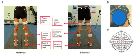
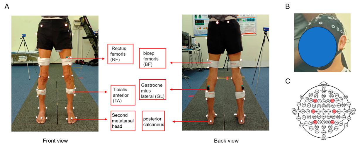
All experiments were conducted in the Sino-UK Human Performance Laboratory at Shanghai Jiao Tong University. Participants were instructed to walk along a 10 m pathway at their self-selected comfortable speed. Although a total of 20 walking trials were initially collected for each participant, only 8 trials with the most consistent walking velocities were retained for subsequent analysis to ensure data stationarity. Specifically, trials were ranked by average walking speed, and the 8 trials with the smallest deviation from the median speed were selected. For each subject, four infrared-reflexive markers were placed on the heel and the second toe positions of the feet, which were used to get the gait events including heel strike and toe off. The trajectories of these four markers were collected by an optical motion capture system (Vicon Nexus 1.8.5, Oxford Metrics, Oxford, UK) with eight cameras in a sampling frequency of 100 Hz. Eight surface electromyography (sEMG) sensors (Trigno Wireless System, Delsys Inc., Boston, MA, USA) were placed over the muscle bellies of the tibialis anterior (TA), gastrocnemius lateralis (GL), rectus femoris (RF), and biceps femoris (BF) bilaterally. The sEMG signals were acquired using the Trigno Control Utility software (version 3.6.0, Delsys Inc., MA, USA) at a sampling rate of 2000 Hz. All sensors were placed based on the SENIAM guidelines [26]. EEG signals were recorded using a 64-channel wireless system (NSW64, Neuracle Technology Co., Ltd., Changzhou, China) at 1000 Hz. Conductive gel was applied to ensure electrode impedance <10 kΩ (Figure 1). The EEG, EMG, and 3D motion capture systems were synchronized via an event trigger: a timing pulse sent at trial onset enabled precise temporal alignment of all signals during post-processing.
Figure 1.
(A) Placement of reflective markers and surface electromyography (EMG) electrodes. (B) Participant wearing the electroencephalography (EEG) cap. (C) Schematic diagram of the EEG channel layout.
2.4. Data Processing
EMG and EEG were processed using Python 3.13. Gait events including heel strike (HS) and toe off (TO) were calculated based on the trajectory data of the infrared reflective markers of the heel and the second toe [27]. Gait cycle was divided into four phases: loading response phase, single stance phase, pre-swing phase and swing phase. Spatiotemporal parameters including cadence (steps/min), step length (m), step time (s), gait phase time (s), walking speed (m/s) and step width (m) were calculated.
Since sEMG signals were collected bilaterally from homologous muscle pairs (TA, GL, RF, and BF), the averaged values of the left and right limbs were calculated and used for all subsequent analyses. This approach was adopted to represent symmetric lower-limb muscle activation patterns during steady-state walking and to reduce potential side-related variability. Raw sEMG signals were visually inspected to exclude trials contaminated by artifacts or signal corruption. Valid signals then underwent detrending and DC removal, followed by band-pass filtering using a 4th-order zero-lag Butterworth filter (20–500 Hz). sEMG envelopes were extracted using a moving RMS procedure with a 100 ms sliding window. Specifically, signals were squared and smoothed using a moving-average window corresponding to 100 ms. To reduce inter-subject variability and facilitate comparison of temporal activation profiles, sEMG amplitudes were normalized within each gait cycle by dividing each time point of the sEMG envelope by the mean envelope amplitude of the corresponding gait cycle (within-cycle mean normalization). After normalization, each gait cycle was resampled to 100 points representing 0–100% of the gait cycle. Consequently, normalized sEMG values were dimensionless and expressed in arbitrary units (a.u.).
EEG preprocessing and artifact rejection: Raw EEG data were preprocessed using MNE-Python. The pipeline included: (1) automatic bad channel detection followed by spherical interpolation; (2) band-pass filtering (0.5–50 Hz) combined with a 50 Hz notch filter to suppress line noise; and (3) Independent Component Analysis (FastICA algorithm) for source decomposition. ICA components were manually screened based on standardized neurophysiological criteria. Ocular artifacts were identified by frontal-polar scalp topographies and time courses synchronized with blink or saccade activity. Gait-related motion and muscle artifacts were identified by widespread or focal topographies, particularly over temporal and occipital regions, elevated high-frequency spectral content (>30 Hz), and transient large-amplitude deflections phase-locked to heel strikes. Components meeting these criteria were excluded prior to signal reconstruction. On average, approximately 4–6 ICA components per participant were removed, with the majority corresponding to ocular and gait-related motion artifacts. Following ICA reconstruction, EEG data were segmented into epochs time-locked to consecutive left heel strikes (LHS), representing individual gait cycles. Finally, sensorimotor cortical activity was extracted from electrodes F3, F4, C3, C4, P3, and P4 for subsequent analysis.
To investigate cortical dynamics during gait in older adults with sarcopenia, electroencephalographic (EEG) signals were analyzed using a multi-modal approach encompassing spectral and functional connectivity features. Power spectral density (PSD) was estimated using Welch’s method, an improved periodogram approach that reduces spectral variance through signal segmentation, windowing, and averaging. At the same time, differences in skull thickness, electrode impedance, and brain structure among individuals could lead to significant variations in absolute PSD levels; therefore, we used the widely accepted Theta–Alpha Ratio (TAR) as a sensitive biomarker reflecting cognitive function [28,29,30].
Partial directed coherence (PDC) was used to assess directional information flow between brain regions in the frequency domain during walking. PDC is based on a multivariate autoregressive (MVAR) model of order :
where is the EEG signal vector at time , is the coefficient matrix at lag , and is the residual noise. The frequency-domain representation is:
PDC from channel to channel at frequency is defined as:
where is the element of , and the denominator normalizes the influence of source across all targets. PDC values range from 0 to 1, reflecting the relative strength of directed connectivity.
To eliminate spurious connections arising from volume conduction or random fluctuations, a significance test was performed on the PDC network. Phase-randomized surrogate data were generated by applying a fast Fourier transform (FFT) to each original time series, preserving the amplitude spectrum while replacing the phase with uniformly distributed random values, followed by an inverse FFT to reconstruct the surrogate signals. Given that PDC quantifies directed coupling based on inter-signal phase relationships, genuine effective connectivity should significantly exceed the connection strengths estimated from the surrogate data. Consequently, only PDC values that passed the significance threshold were retained for subsequent graph-theoretical analysis, with non-significant connections set to zero. The thresholded network was subsequently analyzed as a weighted network.
Based on the PDC-weighted functional networks, four graph-theoretical metrics were extracted to evaluate brain network organization at local levels:
- (1)
- Node Strength: the sum of connection weights (PDC values) linked to a node, indicating its overall involvement in information exchange.
- (2)
- Node Degree: the number of significant connections to a node, reflecting its topological centrality.
- (3)
- Clustering Coefficient: measures the extent to which a node’s neighbors are interconnected, reflecting local functional segregation.
- (4)
- Local Efficiency: the efficiency of information transfer within a node’s neighborhood, indicating network resilience.
2.5. Statistical Analysis
Statistical analyses were performed using SPSS software (version 25.0; IBM Corp., Chicago, IL, USA) and GraphPad Prism software (version 10.4.1; GraphPad Software, San Diego, CA, USA). Continuous variables—including demographic profiles, spatiotemporal gait parameters, sEMG root-mean-square (RMS) envelopes, and EEG frequency values—were first evaluated for normality using the Shapiro–Wilk test. Formally, normally distributed data were expressed as Mean ± SD and analyzed using an independent samples Student’s t-test to evaluate differences between the Sarcopenia (SAR) and Healthy Control (HC) groups. Non-normally distributed parameters were evaluated utilizing the non-parametric Mann–Whitney U test. Categorical clinical characteristics, including gender distribution and comorbidity prevalence (hypertension, non-lower limb osteoarthritis, and medication profiles), were presented as frequencies and percentages [n (%)] and compared using the Chi-square test or Fisher’s exact test. For all statistical verifications, the significance threshold was strictly set at p < 0.05.
3. Results
3.1. Baseline Characteristics of Participants
At baseline, no significant between-group differences were observed regarding gender (p = 0.937), age (p = 0.102), or height (p = 0.377). However, compared to healthy controls, individuals with sarcopenia exhibited significantly lower body weight (p = 0.013), reduced ASMI (p < 0.005), and impaired handgrip strength (p < 0.005). Regarding clinical baselines, the primary comorbidities across the cohort were hypertension and non-lower limb osteoarthritis, with antihypertensive agents and non-steroidal anti-inflammatory drugs (NSAIDs) being the most prevalent medications; notably, the distribution of both comorbidities and concomitant medications was statistically comparable between the two groups (all p > 0.05) (Table 1).
Table 1.
Baseline characteristics of participants.
3.2. Gait Spatiotemporal Parameters
The analysis of gait spatiotemporal parameters showed that the step length and walking speed of patients with sarcopenia were significantly lower than those of the healthy control group (p = 0.01, p < 0.005). Compared with the healthy control group, the loading response period and pre-swing period of patients with sarcopenia were significantly prolonged (p = 0.043, p = 0.036) (Table 2).
Table 2.
Gait spatiotemporal parameters.
3.3. Muscle Activity Results
Surface EMG analysis revealed that among the four recorded muscles (gastrocnemius lateralis, rectus femoris, biceps femoris, and tibialis anterior (TA)), only the TA exhibited significant changes, while no consistent differences were observed in the other muscles between groups.
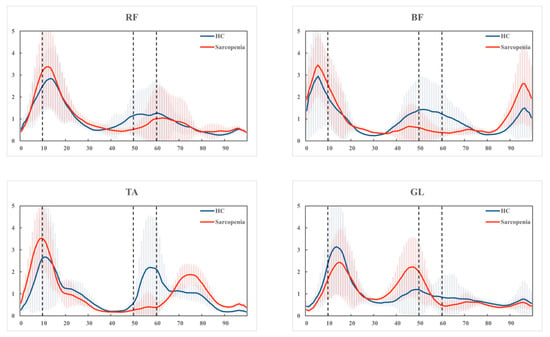
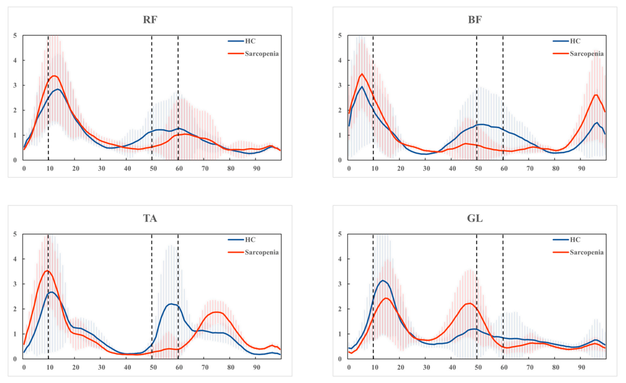
The TA muscle demonstrated a characteristic biphasic activation pattern during the gait cycle. In the sarcopenia group, the first activation peak was significantly increased (p = 0.009) and occurred earlier compared to the control group (p < 0.001). In contrast, the second peak was reduced (p = 0.004) and showed a delayed timing (p < 0.016). These findings indicate a disruption in the temporal coordination of TA activation (Figure 2) (Table 3).
Figure 2.
The sEMG profiles of the four muscles—specifically the gastrocnemius lateralis (GL), rectus femoris (RF), biceps femoris (BF), and tibialis anterior (TA)—are plotted as a percentage of the normalized gait cycle (0% to 100%). sEMG amplitudes were normalized to the mean envelope amplitude of each gait cycle; therefore, y-axis values represent normalized RMS EMG amplitudes in arbitrary units (a.u.). The blue solid lines and shaded areas represent the Mean ± SD of the Healthy Control group (HC, n = 13), while the red solid lines and shaded areas represent the Sarcopenia group (SAR, n = 15). Vertical dashed lines delineate the four core gait phases: Loading Response (LR), Single Stance (SS), Pre-Swing (PS), and Swing Phase (SW).
Table 3.
sEMG Peak parameters in SAR patients and HC.
Further analysis across gait phases revealed phase-specific differences in TA activity. The root mean square (RMS) value of TA was significantly increased during the loading response (LR) phase (p = 0.001), whereas it was significantly decreased during the pre-swing (PS) phase in the sarcopenia group (p = 0.001). No significant differences were observed in the single stance (SS) and swing (SW) phases (Table 4).
Table 4.
sEMG parameters in SAR patients and HC.
3.4. EEG Analysis
No significant differences were observed in node strength, node degree, clustering coefficient, or local efficiency across delta, theta, and alpha bands in the frontal, central, and parietal regions between the sarcopenia and control groups. Further analysis revealed no significant differences were observed in the theta/alpha ratio (TAR) across frontal, central, and parietal regions between the sarcopenia and control groups. These findings suggest that global cortical activity and general cognitive state were comparable between groups (Table 5).
Table 5.
EEG parameters in SAR patients and HC.
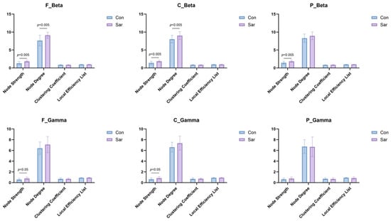
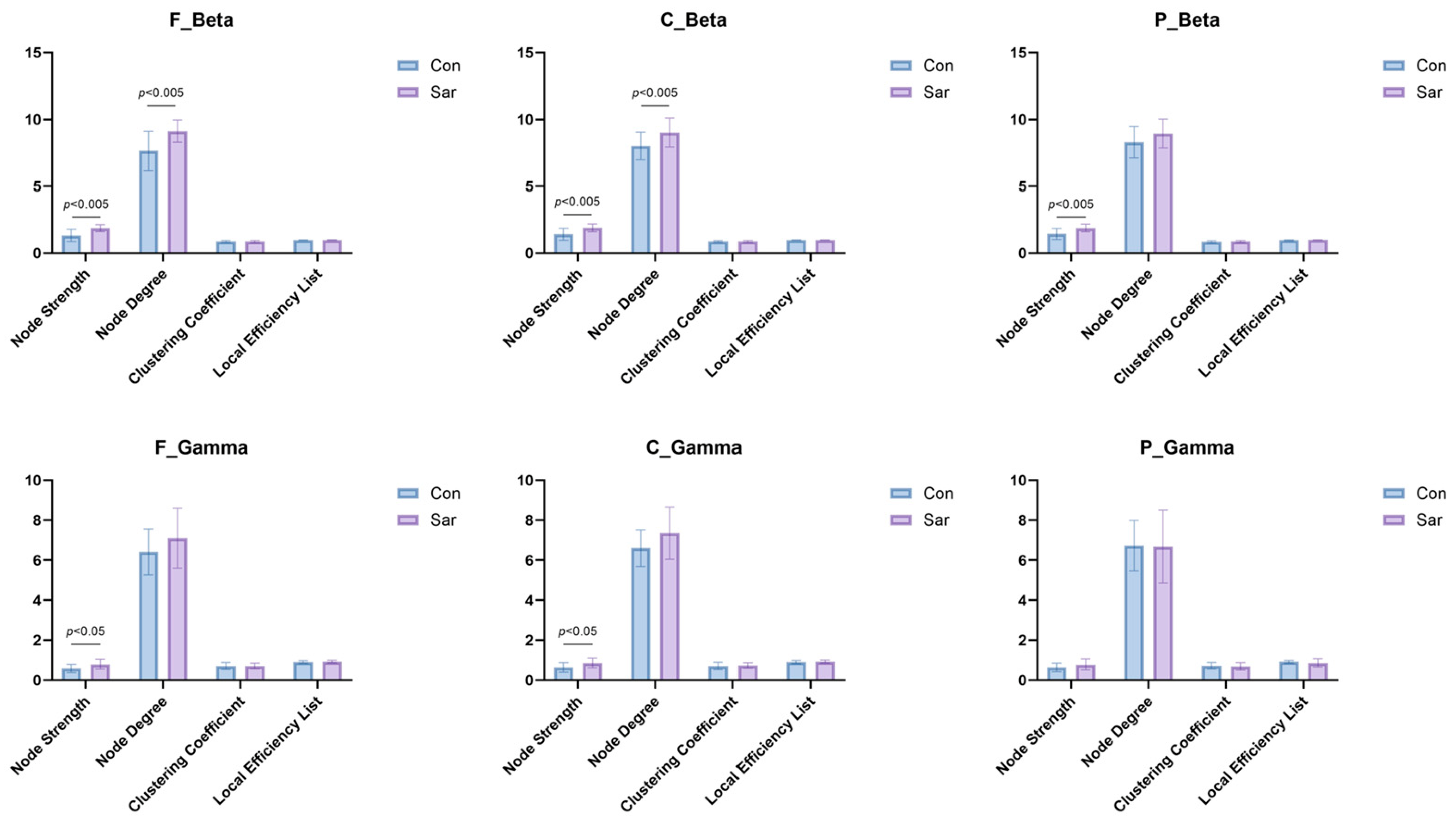
In the beta band (13–30 Hz), significant increases in node strength and node degree were observed in the sarcopenia group compared with controls. Specifically, both node strength and node degree were significantly elevated in the frontal and central regions, while the parietal region showed a significant increase in node strength. No significant differences were found in clustering coefficient or local efficiency between groups, indicating that network alterations were primarily reflected in connectivity strength rather than global network topology. In the gamma band (30–50 Hz), a significant increase in node strength was observed in the frontal and central regions, while other network metrics remained unchanged (Figure 3).
Figure 3.
Comparison of EEG power spectral parameters in the beta and gamma bands across distinct cortical regions during gait. The bar plots illustrate the localized neurophysiological alterations between the Healthy Control group (HC, n = 13, represented by blue bars) and the Sarcopenia group (SAR, n = 15, represented by purple bars).
3.5. Correlation Between Cortical Connectivity and Gait Impairment
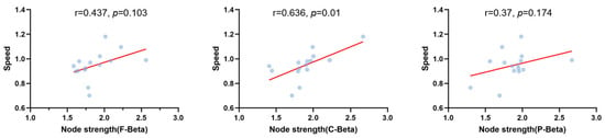
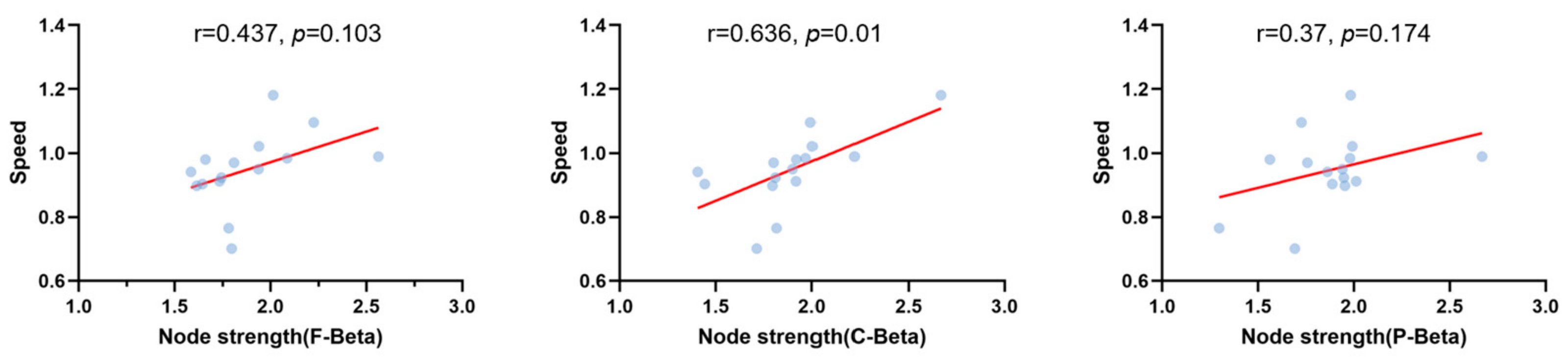
To elucidate the clinical correlation between neurophysiological alterations and motor decline, a within-group correlation analysis was conducted exclusively within the sarcopenia group. The results revealed that the beta-band node strength in the central region was significantly positively correlated with patients’ walking speed (r = 0.636, p = 0.01). From an individual perspective, patients with higher node strength exhibited superior gait preservation. In contrast, the correlations within the frontal and parietal cortical areas fell short of statistical significance, yet consistently maintained parallel positive trajectories (Figure 4).
Figure 4.
Intra-group correlation trajectories between beta-band cortical node strength and walking speed within the sarcopenia group.
4. Discussion
In this study, we assessed gait characteristics, muscle activity, and cortical functional connectivity in individuals with sarcopenia compared to healthy controls using motion capture, surface EMG, and EEG analysis. Compared with controls, individuals with sarcopenia showed reduced walking speed and shorter step length, consistent with previous studies [13,31,32,33]. Surface EMG analysis revealed that only the tibialis anterior exhibited significant alterations, characterized by disrupted activation timing and phase-dependent changes in RMS. In addition, EEG analysis demonstrated increased beta-band functional connectivity strength in frontal, central, and parietal regions, while network topology remained unchanged. Taken together, these findings suggest that sarcopenia is associated with concurrent alterations in gait performance, muscle activation patterns, and beta-band functional connectivity strength.
A notable finding of this study is that gait impairment in sarcopenia was not limited to global spatiotemporal decline, but also involved phase-specific alterations. The prolongation of loading response and pre-swing suggests that gait dysfunction in sarcopenia may reflect impaired weight acceptance and reduced transition efficiency from stance to swing [34,35]. Many gait disorders are characterized by an extended double-support phase in the early stage, which is aimed at enhancing walking stability, patterns commonly interpreted as a more cautious and less efficient walking strategy [36,37]. In this context, prolonged early and late stance subphases may indicate that individuals with sarcopenia require more time to stabilize the body after initial contact and to generate adequate propulsion before limb advancement.
At the peripheral level, the most distinctive EMG finding was the selective alteration of tibialis anterior activity, whereas the other recorded muscles (RF, BF, GL) did not show consistent between-group differences. This muscle-specific pattern is important, because the tibialis anterior plays a central role in controlling ankle dorsiflexion during limb advancement and in modulating foot positioning at initial contact [38,39,40]. In the present study, the earlier and enhanced first peak, together with the delayed and attenuated second peak, indicates a disruption in the normal temporal coordination of tibialis anterior recruitment. The phase-specific RMS pattern further supports this interpretation: increased activity during loading response may reflect a greater demand for stabilization during early stance, whereas reduced activity during pre-swing may indicate impaired preparation for limb transition. Together, these findings suggest that sarcopenia-related gait impairment may be linked to altered phase-dependent activation of the tibialis anterior, which may in turn affect ankle control during walking [41,42,43], rather than reflecting a generalized alteration of all lower-limb muscles.
The cortical findings provide an additional and clinically meaningful layer of interpretation. No significant differences were observed in delta, theta, alpha, or TAR measures, suggesting that neural alterations in sarcopenia are unlikely to reflect global cortical changes [44,45]. Instead, the most robust differences emerged in the beta band, where node strength and node degree increased mainly in frontal and central regions, with additional increases in parietal strength. This result aligns with previous work showing that beta oscillatory activity has been repeatedly linked to corticospinal contributions to walking and motor output [46,47]. In contrast, only limited and isolated differences were observed in the gamma band, suggesting that high-frequency activity may not represent a dominant neural feature in this context. Furthermore, the absence of group differences in clustering coefficient and local efficiency indicates that the overall network topology remained relatively preserved. Thus, the principal neural change in sarcopenia in this dataset was not a wholesale reorganization of network architecture, but rather an increase in the strength and extent of functional interactions within a largely preserved network scaffold.
Importantly, these peripheral and cortical findings should not be interpreted in isolation. To bridge the gap between supraspinal alterations and behavioral deficits, we performed a direct correlation analysis, which unraveled that beta-band network node strength in the central region was significantly and positively coupled with usual walking velocity within the sarcopenia cohort (r = 0.636, p < 0.05). This quantitative brain–behavior relationship provides direct evidence that altered cortical connectivity is intertwined with actual ambulatory performance. However, given the cross-sectional, observational design of this study, caution is mandatory against drawing definitive causal inferences. The coexistence of phase-specific gait slowing and increased beta-band connectivity presents a clear statistical correlation, which may be interpreted as a framework of inefficient compensation. Increased cortical involvement may represent an adaptive neural correlate to offset declining peripheral motor control; however, this increased neural recruitment did not normalize gait performance. Importantly, we cannot rule out the alternative hypothesis that this hyper-connectivity fundamentally reflects age-related motor network degradation or a pathological, inefficient scattering of synchronized neural resources, rather than a purposeful compensatory effort. Ultimately, our observational data capture a robust neural engagement concomitant with gait dysfunction, but tracking whether this central shift represents a top-down survival strategy or a symptom of systemic neuromuscular decay requires longitudinal or interventional validation. Previous studies have shown that older adults frequently rely more on supraspinal resources during walking and balance, particularly when automatic motor control becomes less efficient [48,49].
From a translational perspective, the cortical–behavioral coupling unraveled in this exploratory study offers preliminary insights for future neurorehabilitation design. Our findings imply that future clinical interventions might benefit from transitioning from conventional peripheral training toward central–peripheral modulation strategies. For therapists, exploring innovative motor–sensory integration protocols could be a promising avenue to optimize neural resource mobilization and mitigate potential central overloads. For patients, structured coordination exercises might serve as a potential approach to facilitate active top-down cortical compensation. Nevertheless, these conceptual frameworks represent early-stage theoretical paths that require systematic verification in future large-scale trials.
Several limitations should be acknowledged in this study. First, the sample size was relatively small, and the cross-sectional design precludes causal inference. Crucially, due to the exploratory nature and restricted sample size of this pilot trial, a statistically powered gender-stratified sub-analysis could not be performed, although gender was stringently matched at baseline to control for potential confounding effects. Second, only a limited number of muscles and network metrics were analyzed, which may not fully capture the complete profile of neuromuscular coordination. Future studies should incorporate larger, sex-balanced cohorts and explore multi-frequency brain–muscle relationship metrics to further clarify the role of beta-band connectivity in functional outcomes and refine targeted neurorehabilitation protocols.
5. Conclusions
This study suggests that sarcopenia-related gait dysfunction is associated with peripheral motor impairment alongside increased beta-band cortical involvement. While functional connectivity strength was elevated, network topology remained preserved, indicating increased neural engagement without structural reorganization. Importantly, gait performance remains impaired despite enhanced cortical involvement, supporting the presence of inefficient compensation. These findings highlight a mismatch between neural effort and motor function and suggest that improving neuromuscular efficiency may be critical for intervention. Nevertheless, given the limited sample size of this exploratory cohort, these preliminary insights must be interpreted with caution, as they cannot be widely generalized prior to large-scale clinical validation.
Author Contributions
Z.W. aided in conceptualization, data curation, investigation, formal analysis, and writing—original draft. B.W. aided in data curation, formal analysis, visualization, and software. X.Z. contributed to methodology, supervision, validation, and writing—review & editing. D.G. contributed to conceptualization, funding acquisition, methodology, supervision, validation, and writing—review & editing. All authors have read and agreed to the published version of the manuscript.
Funding
This study was funded by the National Nature Science Foundation of China (11972233) and the National Key Research and Development Program of China (2022YFC2009502).
Data Availability Statement
The datasets used and/or analyzed during the current study are available from the corresponding author on request.
Conflicts of Interest
The authors declare that they have no known competing financial interests or personal relationships that could have appeared to influence the work reported in this paper.
References
- Hu, Y.; Peng, W.; Ren, R.; Wang, Y.; Wang, G. Sarcopenia and mild cognitive impairment among elderly adults: The first longitudinal evidence from CHARLS. J. Cachexia Sarcopenia Muscle 2022, 13, 2944–2952. [Google Scholar] [CrossRef] [Scilit]
- Cho, M.R.; Lee, S.; Song, S.K. A Review of Sarcopenia Pathophysiology, Diagnosis, Treatment and Future Direction. J. Korean Med. Sci. 2022, 37, e146. [Google Scholar] [CrossRef] [Scilit]
- Liu, D.; Wang, S.; Liu, S.; Wang, Q.; Che, X.; Wu, G. Frontiers in sarcopenia: Advancements in diagnostics, molecular mechanisms, and therapeutic strategies. Mol. Asp. Med. 2024, 97, 101270. [Google Scholar] [CrossRef] [Scilit]
- Yuan, S.; Larsson, S.C. Epidemiology of sarcopenia: Prevalence, risk factors, and consequences. Metabolism 2023, 144, 155533. [Google Scholar] [CrossRef] [Scilit]
- Widajanti, N.; Ichwani, J.; Dharmanta, R.S.; Firdausi, H.; Haryono, Y.; Yulianti, E.; Kandinata, S.G.; Wulandari, M.; Widyasari, R.; Adyanita, V.A.; et al. Sarcopenia and Frailty Profile in the Elderly Community of Surabaya: A Descriptive Study. Acta Med. Indones. 2020, 52, 5–13. [Google Scholar] [PubMed]
- Dodds, R.; Sayer, A.A. Sarcopenia and frailty: New challenges for clinical practice. Clin. Med. 2015, 15, s88–s91. [Google Scholar] [CrossRef] [Scilit]
- Jang, J.Y.; Kim, D.; Kim, N.D. Pathogenesis, Intervention, and Current Status of Drug Development for Sarcopenia: A Review. Biomedicines 2023, 11, 1635. [Google Scholar] [CrossRef] [Scilit] [PubMed]
- Tharawadeepimuk, K.; Limroongreungrat, W.; Pilanthananond, M.; Nanbancha, A. Auditory Cue Effects on Gait-Phase-Dependent Electroencephalogram (EEG) Modulations during Overground and Treadmill Walking. Sensors 2024, 24, 1548. [Google Scholar] [CrossRef] [Scilit] [PubMed]
- Gramigna, V.; Palumbo, A.; Perri, G. Advancing Gait Analysis: Integrating Multimodal Neuroimaging and Extended Reality Technologies. Bioengineering 2025, 12, 313. [Google Scholar] [CrossRef] [Scilit]
- Seynaeve, M.; Mantini, D.; de Beukelaar, T.T. Electrophysiological Approaches to Understanding Brain-Muscle Interactions During Gait: A Systematic Review. Bioengineering 2025, 12, 471. [Google Scholar] [CrossRef] [Scilit]
- Roeder, L.; Breakspear, M.; Kerr, G.K.; Boonstra, T.W. Dynamics of brain-muscle networks reveal effects of age and somatosensory function on gait. iScience 2024, 27, 109162. [Google Scholar] [CrossRef] [Scilit] [PubMed]
- Fan, Y.; Zhang, B.; Huang, G.; Zhang, G.; Ding, Z.; Li, Z.; Sinclair, J.; Fan, Y. Sarcopenia: Body Composition and Gait Analysis. Front. Aging Neurosci. 2022, 14, 909551. [Google Scholar] [CrossRef] [Scilit]
- Jiang, Z.; Hu, P.; Cheng, R.; Wang, H.; Zhang, Q.; Ma, S.; Tsai, T.Y. Quantitative analysis of gait dysfunction in sarcopenia patients: Based on spatiotemporal parameters and kinematic performance. Gait Posture 2025, 118, 108–114. [Google Scholar] [CrossRef] [Scilit]
- Sha, T.; Zhang, Y.; Wei, J.; Li, C.; Zeng, C.; Lei, G.; Wang, Y. Sarcopenia and Risk of Cognitive Impairment: Cohort Study and Mendelian Randomization Analysis. JMIR Aging 2025, 8, e66031. [Google Scholar] [CrossRef] [Scilit] [PubMed]
- Groh, J.; Simons, M. White matter aging and its impact on brain function. Neuron 2025, 113, 127–139. [Google Scholar] [CrossRef] [Scilit] [PubMed]
- Ahn, K.; Lee, S.J.; Mook-Jung, I. White matter-associated microglia: New players in brain aging and neurodegenerative diseases. Ageing Res. Rev. 2022, 75, 101574. [Google Scholar] [CrossRef] [Scilit]
- Khattak, Z.K.; Jiao, X.; Hu, T.; Shao, Q.; Sun, X.; Zhao, X.; Gu, D. Investigation of gait and balance function in cervical spondylotic myelopathy patients using wearable sensors. Spine J. 2023, 23, 1127–1136. [Google Scholar] [CrossRef] [Scilit]
- Martín-Fuentes, I.; Oliva-Lozano, J.M.; Muyor, J.M. Electromyographic activity in deadlift exercise and its variants. A systematic review. PLoS ONE 2020, 15, e0229507. [Google Scholar] [CrossRef] [Scilit]
- Cabrolier-Molina, J.; Martín-Rodríguez, A.; Clemente-Suárez, V.J. The Effects of Exercise Intervention in Older Adults with and Without Sarcopenia: A Systematic Review. Sports 2025, 13, 152. [Google Scholar] [CrossRef] [Scilit]
- Park, T.S.; Shin, M.J. Comprehensive Assessment of Lower Limb Function and Muscle Strength in Sarcopenia: Insights from the Sit-to-Stand Test. Ann. Geriatr. Med. Res. 2024, 28, 1–8. [Google Scholar] [CrossRef] [Scilit]
- Bronstein, A.M.; Mirdamadi, J.L.; Ellmers, T.J. The cortical vestibular system: Insights from electroencephalography. Curr. Opin. Neurol. 2026, 39, 92–98. [Google Scholar] [CrossRef] [Scilit] [PubMed]
- Borzuola, R.; Giombini, A.; Torre, G.; Campi, S.; Albo, E.; Bravi, M.; Borrione, P.; Fossati, C.; Macaluso, A. Central and Peripheral Neuromuscular Adaptations to Ageing. J. Clin. Med. 2020, 9, 741. [Google Scholar] [CrossRef] [Scilit]
- Gennaro, F.; Maino, P.; Kaelin-Lang, A.; Bock, K.; Bruin, E.D. Corticospinal Control of Human Locomotion as a New Determinant of Age-Related Sarcopenia: An Exploratory Study. J. Clin. Med. 2020, 9, 720. [Google Scholar] [CrossRef] [Scilit]
- Siegel, M.; Donner, T.H.; Engel, A.K. Spectral fingerprints of large-scale neuronal interactions. Nat. Rev. Neurosci. 2012, 13, 121–134. [Google Scholar] [CrossRef] [Scilit]
- Peng, J.; Zikereya, T.; Shao, Z.; Shi, K. The neuromechanical of Beta-band corticomuscular coupling within the human motor system. Front. Neurosci. 2024, 18, 1441002. [Google Scholar] [CrossRef] [Scilit]
- Hermens, H.J.; Freriks, B.; Disselhorst-Klug, C.; Rau, G. Development of recommendations for SEMG sensors and sensor placement procedures. J. Electromyogr. Kinesiol. 2000, 10, 361–374. [Google Scholar] [CrossRef] [Scilit]
- Short, M.R.; Damiano, D.L.; Kim, Y.; Bulea, T.C. Children with Unilateral Cerebral Palsy Utilize More Cortical Resources for Similar Motor Output During Treadmill Gait. Front. Hum. Neurosci. 2020, 14, 36. [Google Scholar] [CrossRef] [Scilit] [PubMed]
- Baik, K.; Jung, J.H.; Jeong, S.H.; Chung, S.J.; Yoo, H.S.; Lee, P.H.; Sohn, Y.H.; Kang, S.W.; Ye, B.S. Implication of EEG theta/alpha and theta/beta ratio in Alzheimer’s and Lewy body disease. Sci. Rep. 2022, 12, 18706. [Google Scholar] [CrossRef] [Scilit] [PubMed]
- Tarasova, I.V.; Trubnikova, O.A.; Barbarash, O.L. EEG and Clinical Factors Associated with Mild Cognitive Impairment in Coronary Artery Disease Patients. Dement. Geriatr. Cogn. Disord. 2018, 46, 275–284. [Google Scholar] [CrossRef] [Scilit]
- Babiloni, C.; Carducci, F.; Lizio, R.; Vecchio, F.; Baglieri, A.; Bernardini, S.; Cavedo, E.; Bozzao, A.; Buttinelli, C.; Esposito, F.; et al. Resting state cortical electroencephalographic rhythms are related to gray matter volume in subjects with mild cognitive impairment and Alzheimer’s disease. Hum. Brain Mapp. 2013, 34, 1427–1446. [Google Scholar] [CrossRef] [Scilit]
- Bhasin, S.; Travison, T.G.; Manini, T.M.; Patel, S.; Pencina, K.M.; Fielding, R.A.; Magaziner, J.M.; Newman, A.B.; Kiel, D.P.; Cooper, C.; et al. Sarcopenia Definition: The Position Statements of the Sarcopenia Definition and Outcomes Consortium. J. Am. Geriatr. Soc. 2020, 68, 1410–1418. [Google Scholar] [CrossRef] [Scilit] [PubMed]
- Mori, K.; Murata, S.; Goda, A.; Kikuchi, Y.; Shiraiwa, K.; Horie, J.; Nakano, H. Gait Characteristics of Dynapenia, Sarcopenia, and Presarcopenia in Community-Dwelling Japanese Older Women: A Cross-Sectional Study. Healthcare 2022, 10, 1905. [Google Scholar] [CrossRef] [Scilit] [PubMed]
- Manabe, T.; Tsuchida, W.; Kobayashi, T.; Fujimoto, M.; Inai, T.; Kido, K.; Kudo, S.; Fukunaga, K.; Saheki, T.; Yoshimura, T.; et al. Spatiotemporal and kinematic gait characteristics in older patients with type 2 diabetes mellitus with and without sarcopenia. Sci. Rep. 2025, 15, 18000. [Google Scholar] [CrossRef] [Scilit]
- Cao, Y.; Zhuang, H.; Zhang, X.; Guo, R.; Pang, H.; Zheng, P.; Xu, H. Impact of foot progression angle on spatiotemporal and plantar loading pattern in intoeing children during gait. Sci. Rep. 2024, 14, 14402. [Google Scholar] [CrossRef] [Scilit] [PubMed]
- Wei, P.; Zhang, J.; Wei, P.; Wang, B.; Hong, J. Different sEMG and EEG Features Analysis for Gait phase Recognition. Annu. Int. Conf. IEEE Eng. Med. Biol. Soc. 2020, 2020, 1002–1006. [Google Scholar] [CrossRef] [Scilit]
- Kobayashi, T.; Koh, M.W.P.; Jor, A.; Hisano, G.; Murata, H.; Ichimura, D.; Hobara, H. Ground reaction forces during double limb stances while walking in individuals with unilateral transfemoral amputation. Front. Bioeng. Biotechnol. 2022, 10, 1041060. [Google Scholar] [CrossRef] [Scilit]
- Jiao, X.; Hu, T.; Li, Y.; Wang, B.; Acquah, M.E.E.; Wang, Z.; Chen, Q.; Gan, Y.; Gu, D. Association between Elastic Modulus of Foot Soft Tissues and Gait Characteristics in Young Individuals with Flatfoot. Bioengineering 2024, 11, 728. [Google Scholar] [CrossRef] [Scilit]
- Maharaj, J.N.; Cresswell, A.G.; Lichtwark, G.A. Tibialis anterior tendinous tissue plays a key role in energy absorption during human walking. J. Exp. Biol. 2019, 222, jeb.191247. [Google Scholar] [CrossRef] [Scilit]
- Titova, A.; Bilyalov, A.; Filatov, N.; Perepechenov, S.; Kupriyanova, D.; Brovkin, S.; Shestakov, D.; Bodunova, N.; Gusev, O. Muscle Aging Heterogeneity: Genetic and Structural Basis of Sarcopenia Resistance. Genes 2025, 16, 948. [Google Scholar] [CrossRef] [Scilit]
- Li, Y.; Wang, B.; Jiao, X.; Esi Acquah, M.E.; Zhu, Y.; Jin, J.; Zhang, X.; Gu, D. Muscle activation of lower limb during walking in elderly individuals with sarcopenia: A pilot study. Annu. Int. Conf. IEEE Eng. Med. Biol. Soc. 2024, 2024, 1–4. [Google Scholar] [CrossRef] [Scilit]
- Li, Q.; Chen, L. Effect of tibialis anterior muscle resistance training on ankle and foot dorsum extension function in hypertensive cerebral hemorrhage hemiplegia patients: A randomized controlled trial. Medicine 2023, 102, e33827. [Google Scholar] [CrossRef] [Scilit]
- Massot, C.; Guyot, M.A.; Donze, C.; Simoneau, E.; Gillet, C.; Leteneur, S. Ankle dysfunction in multiple sclerosis and the effects on walking. Disabil. Rehabil. 2021, 43, 2454–2463. [Google Scholar] [CrossRef] [Scilit] [PubMed]
- Amiridis, I.G.; Kannas, T.; Sahinis, C.; Negro, F.; Trypidakis, G.; Kellis, E.; Enoka, R.M. More Variability in Tibialis Anterior Function during the Adduction of the Foot than Dorsiflexion of the Ankle. Med. Sci. Sports Exerc. 2024, 56, 851–859. [Google Scholar] [CrossRef] [Scilit]
- Kaushik, P.; Shrivastava, P.K. Remediation of Learning Difficulty Utilizing School-Based Cognitive Behavioral Intervention Measured by EEG Theta-Alpha and Theta-Beta Ratio During Resting and Cognitive Task Performance Conditions. Clin. EEG Neurosci. 2024, 55, 426–444. [Google Scholar] [CrossRef] [Scilit]
- Trammell, J.P.; MacRae, P.G.; Davis, G.; Bergstedt, D.; Anderson, A.E. The Relationship of Cognitive Performance and the Theta-Alpha Power Ratio Is Age-Dependent: An EEG Study of Short Term Memory and Reasoning during Task and Resting-State in Healthy Young and Old Adults. Front. Aging Neurosci. 2017, 9, 364. [Google Scholar] [CrossRef] [Scilit]
- Quivira-Lopesino, A.; Sevilla-García, M.; Cuesta, P.; Pusil, S.; Bruña, R.; Fiedler, P.; Cebolla, A.M.; Cheron, G.; Funke, M.; Maestu, F. Changes of EEG beta band power and functional connectivity during spaceflight: A retrospective study. Sci. Rep. 2025, 15, 13399. [Google Scholar] [CrossRef] [Scilit]
- Khanjari, Y.; Arabameri, E.; Shahbazi, M.; Tahmasebi, S.; Bahrami, F.; Mobaien, A. The simultaneous changes in motor performance and EEG patterns in beta band during learning dart throwing skill in dominant and non-dominant hand. Comput. Methods Biomech. Biomed. Eng. 2023, 26, 127–137. [Google Scholar] [CrossRef] [Scilit] [PubMed]
- Clark, D.J. Automaticity of walking: Functional significance, mechanisms, measurement and rehabilitation strategies. Front. Hum. Neurosci. 2015, 9, 246. [Google Scholar] [CrossRef] [Scilit] [PubMed]
- Yogev-Seligmann, G.; Hausdorff, J.M.; Giladi, N. The role of executive function and attention in gait. Mov. Disord. 2008, 23, 329–342. [Google Scholar] [CrossRef] [Scilit]

Disclaimer/Publisher’s Note: The statements, opinions and data contained in all publications are solely those of the individual author(s) and contributor(s) and not of MDPI and/or the editor(s). MDPI and/or the editor(s) disclaim responsibility for any injury to people or property resulting from any ideas, methods, instructions or products referred to in the content. |
© 2026 by the authors. Licensee MDPI, Basel, Switzerland. This article is an open access article distributed under the terms and conditions of the Creative Commons Attribution (CC BY) license.