


Abstract
Polyploid giant cancer cells (PGCCs) are stress-associated tumor cells implicated in therapeutic resistance, recurrence, and disease progression. Their heterogeneous physical and biological characteristics present challenges for isolation and characterization, highlighting the need for integrated experimental approaches. This review examines conventional and microfluidic strategies for PGCC induction, physical separation, and downstream analysis, emphasizing demonstrated applications and their complementary capabilities. Microfluidic drug-gradient platforms have enabled the controlled generation of PGCC/polyaneuploid cancer cell (PACC)-associated resistant populations, while inertial microfluidic devices have achieved efficient, label-free enrichment of large cancer cells for subsequent biological characterization. Image-guided laser-induced microbubble detachment has further enabled the selective separation of PGCC-like large cells from normal-sized drug-resistant cells, revealing differences in drug sensitivity. In addition, fluorescence-assisted sorting combined with microwell-based single-cell transcriptomics has demonstrated molecular profiling of selected PGCC populations. Proteomic and functional studies have provided complementary insights into PGCC formation, progeny development, cellular plasticity, and therapeutic resistance. Beyond these demonstrated applications, filtration, sedimentation, field-flow fractionation, acoustic separation, and dielectrophoresis offer additional physical separation principles for workflow development. We discuss how cellular properties, recovery performance, and downstream analytical requirements guide the selection and integration of these approaches. Collectively, these advances establish a practical foundation for investigating PGCC heterogeneity and support the further development of integrated platforms connecting controlled induction, selective recovery, and molecular or functional analysis.
1. Introduction
Polyploid giant cancer cells (PGCCs) are enlarged cancer cells with increased DNA content that are found in tumors and can arise in response to chemotherapy, hypoxia, radiation, and other cellular stresses [1,2,3,4]. They were initially described as enlarged, non-dividing cancer cells, but subsequent studies established that polyploid giant-cell states can arise transiently under stress and can generate viable progeny through different forms of ploidy reduction or asymmetric division [1,2]. Experimental studies have further linked these cells to stem-like phenotypes [5], altered secretory programs, tumorigenic capacity, and resistance to adverse conditions [1,6,7]. PGCCs can arise through several mechanisms, including endoreplication, cell fusion, cytokinesis failure, and mitotic slippage [2,3]. Overlapping cell populations have also been described under the term polyaneuploid cancer cells (PACCs) [8]. Coward and Harding provided an influential framework for assessing polyploid tumor-cell populations, highlighted the potential biological significance of elevated genomic content, and noted that polyploid cells accumulate within carboxyfluorescein succinimidyl ester (CFSE) label-retaining, infrequently cycling subpopulations [9]. That contribution is an important foundation for PGCC research; however, label retention reflects division history rather than ploidy or cell size, and quantifying or identifying a polyploid fraction is distinct from selecting, recovering, and validating viable cells for a specified downstream assay.
Clinical and experimental studies have associated PGCC/PACC abundance with disease-related features in prostate, breast, ovarian, and colorectal (including rectal) cancers, such as recurrence, tumor grade, invasion, metastasis, and treatment response [10,11,12,13,14]. Comparable polyploid or giant-cell states have also been described in virally induced tumors [15]. Stress-responsive pathways, including AMP-activated protein kinase (AMPK)–mechanistic target of rapamycin (mTOR) signaling, and exposure to DNA-damaging or targeted agents have been implicated in the formation or persistence of these populations [16,17]. Therapeutic strategies directed toward the PGCC state have begun to be explored [18].
Interpreting these biological properties at the molecular or functional level requires populations that are sufficiently enriched, and for many applications, remain viable after separation. However, obtaining such populations is challenging because PGCCs are physically heterogeneous, and no validated PGCC-specific surface marker has been established in the literature reviewed here for preparative live-cell isolation. Reported PGCC characterization and candidate separation criteria therefore rely primarily on physical or optical properties, including cell size [19,20], DNA content [21,22], morphology [23,24], and deformability [25,26]. Earlier studies used filtration [25], centrifugation [27,28,29], or flow-cytometric sorting [30,31], but often did not report the combination of physical enrichment, recovery, viability, and ploidy characterization required for direct comparison. A central challenge is therefore to determine how a physical selection criterion relates to the biological definition of a PGCC and how reliably that criterion can produce a population suitable for downstream analysis.
Microfluidic approaches offer engineering solutions to this challenge by converting cellular physical properties into controlled transport, retention, trapping, or retrieval responses without requiring a validated surface marker [32,33,34,35]. Channel geometry, hydrodynamic forces, confinement, and applied fields can be designed to produce continuous or spatially resolved separation, while optical interrogation can enable the targeted recovery of individually identified cells [20,36]. These approaches could facilitate physical enrichment with controlled cell handling and downstream characterization. Representative applications to PGCC-containing or large-cell populations include the spiral inertial separation of doxorubicin-resistant and patient-derived PGCC-containing populations [19] and image-guided detachment of optically selected cells using laser-induced microbubbles [20]. These studies demonstrate the feasibility of micro-engineered platforms, but also highlight the importance of understanding how cellular physical properties influence separation behavior and how recovered cells are subsequently verified.
This review focuses on microfluidic approaches to PGCC separation and organizes them according to how cellular physical properties are translated into measurable device-level responses. Five categories are considered: cell size, morphology, deformability, density, and field-responsive properties. These properties are linked to separation observables, including light scattering, hydrodynamic positioning, trapping, retention, and field-driven displacement. Importantly, physical enrichment does not establish PGCC identity and must be distinguished from biological verification based on ploidy, morphology, or other independent criteria. Separation strategies are also evaluated in relation to downstream analytical requirements, including cell recovery, viability, and access to individual cells (Figure 1). Throughout the review, direct evidence from PGCC/PACC studies is distinguished from findings in related large, polyploid, or mechanically heterogeneous cell populations. Conventional separation methods are introduced as baselines, followed by microfluidic approaches and their integration with downstream single-cell, bulk, and functional analyses. In this sequence, induction defines the biological input, separation determines which cells are recovered, and the downstream endpoint determines the required sampling unit and acceptable handling. The purpose is to provide an engineering-oriented framework for method selection rather than to imply that an end-to-end PGCC workflow has already been validated.
Figure 1.
Conceptual map of PGCC-related separation workflows and downstream study scale. (a) Representative workflows are positioned by an illustrative processing scale and the type of information shown. The rows denote readout categories, not numerical validation grades. Green and blue symbols denote conventional and microfluidic methods, respectively; filled symbols indicate direct PGCC/PACC-associated studies and open symbols indicate related-cell studies. The dashed outline groups workflows positioned at both higher reported-information rows and higher illustrative throughput; it is a conceptual grouping, not a validated performance threshold. Assay timing and sampling units are specified in Section 6 and Section 7. (b) Colored bands illustrate possible study scales qualitatively; band color is used for visual grouping only and does not follow the symbol colors in (a). Their positions and widths are schematic, with no numerical cell-count endpoints or universal minimum-input interpretation. Input, recovered, and profiled cells or nuclei must be distinguished for each workflow (Section 9.1). DEP, dielectrophoresis; DLD, deterministic lateral displacement; FACS, fluorescence-activated cell sorting; FFF, field-flow fractionation; PACC, polyaneuploid cancer cell; PGCC, polyploid giant cancer cell.
PGCC-associated populations should nevertheless be considered within the broader spectrum of therapy-associated cellular states. Treatment persistence and failure in solid tumors are multifactorial processes involving several coexisting stress-adapted or tumor-repopulating populations and cannot be attributed exclusively to PGCCs. Accordingly, this review does not propose PGCCs as the sole or dominant drivers of therapeutic failure. Instead, it examines the engineering requirements for physically enriching and recovering PGCC/PACC-associated populations while considering how separation mechanisms, potential selection biases, and subsequent biological verification influence the interpretation of recovered cells.
2. Defining PGCCs for Physical Separation
2.1. PGCC Definitions and Their Limitations
Most PGCC criteria are based on cell or nuclear dimensions or genomic content (Table 1). Zhang et al. defined a PGCC in cell culture as a cancer cell at least three times larger than its parental cancer cells [1]. This is a relative whole-cell-size description, not a threefold nuclear-diameter threshold. In contrast, subsequent clinicopathological studies described nuclei at least three times larger than the surrounding diploid nuclei on histological sections [11,12,13,14]. These cell-size and nuclear-size descriptions should not be combined or automatically converted to a common diameter, area, or volume threshold. Other studies have used absolute cell-size thresholds in combination with characteristic nuclear morphology [19], whereas cytometric approaches have relied on DNA content exceeding the normal diploid cell-cycle range [1,22]. The PACC concept also includes the absence of division while cells occupy the PACC state, rather than a permanent inability to generate progeny [8,25]. Clinical histological applications [10] should be distinguished from live-culture observation of this dynamic state.
Table 1.
Reported PGCC/PACC identification criteria and complementary verification readouts.
These criteria are not necessarily equivalent. Relative nuclear size depends on the surrounding reference population, cell dimensions can be affected by cell spreading and adhesion state, and DNA-content measurements capture a different biological feature from morphological criteria. A cell meeting one criterion may not meet another. More importantly, physical enrichment criteria may identify distinct cell populations without confirming their PGCC identity.
PGCCs have been discussed within the genome-chaos framework, which describes extensive numerical and structural genome reorganization [37]. Ye et al. classify PGCC formation as a numerical subtype within this framework [38]. The resulting cellular heterogeneity can be reflected in both cell dimensions and nuclear organization. Reported PGCC-associated cells range from moderately enlarged to highly enlarged or massive cells, with nuclear morphologies including a single enlarged nucleus, multiple nuclei of different sizes, and micronucleated or fragmented nuclear structures [23,24,37,38]. These morphological features may coexist with micronucleus formation, a nuclear abnormality, and with chromothripsis-associated rearrangements, a distinct type of genomic alteration; however, neither feature alone establishes PGCC identity. Similarly, senescence-associated features may occur in a subset of PGCCs, but senescence and PGCC identity are not interchangeable; senescent cancer cells do not necessarily exhibit giant-cell morphology or increased genomic content [7]. Stress-tolerant persister cells may also share selected stress-response or survival features with PGCCs without necessarily being enlarged or polyploid. These categories therefore represent distinct, potentially overlapping cellular features or states whose relationships may depend on the biological model and time point. Their identification requires appropriate morphological, genomic, senescence, or functional persistence assays rather than morphology alone. Accordingly, this review considers the broader spectrum of genome-chaos-associated and therapy-related cellular states when interpreting enrichment outcomes, without assuming that physical separation can resolve all of them.
The terms PGCC and PACC are used in closely related, partially overlapping contexts in the literature, particularly when describing therapy-associated cells with increased genomic content and altered cell-cycle behavior [8,19,25]. However, operational definitions and identification criteria vary among studies; the terms should therefore not be treated as universally interchangeable. In this review, the terminology used by each source study is retained, and PGCC/PACC-associated populations are interpreted according to the criteria actually measured.
2.2. Physical Properties Relevant to PGCC Separation
In the absence of a validated PGCC-specific surface marker, separation strategies rely on measurable cellular properties, including size, morphology, deformability, density, and responses to applied fields. Among these, size-related properties have been most extensively explored for the label-free enrichment of viable large-cell populations. However, cell size alone does not establish PGCC identity, and biological verification remains necessary. These physical properties may vary with PGCC induction methods, recovery time, and cell-handling conditions. Differences in cell-size distribution, morphology, deformability, and target-cell frequency can influence separation efficiency and recovery. Characterizing the incoming cell population is therefore important for interpreting and comparing separation performance across experimental systems.
3. Induction Strategies and Their Implications for Separation
A range of chemical and physical stresses have been used to generate PGCC- or PACC-associated populations. Reported conditions include cisplatin or docetaxel treatment of PC3 prostate cancer and MDA-MB-231 breast cancer models [23,24], doxorubicin exposure [19,30], poly(ADP-ribose) polymerase (PARP) inhibition [17], triptolide treatment [39], carboplatin-associated polyploidization [40], ionizing radiation [14], and hypoxia-mimetic conditions such as cobalt chloride (CoCl2) [41]. CoCl2 has been reported to induce polyploid populations with stem-associated and chemoresistance-related features [41] and to alter erythroid-associated programs [42], while subsequent changes associated with hypoxia-inducible factor 1α (HIF-1α) have been linked to increased invasion of daughter cells [43]. These approaches differ in stress type, exposure duration, and the criteria used to characterize the resulting populations.
Microfluidic platforms can complement conventional culture methods by providing spatial and temporal control over drug exposure and other environmental stresses. Controlled gradients and localized exposure regions allow heterogeneous populations to develop within a defined microenvironment. Induction should first reflect the biological question; the separator should then be selected or adapted to the resulting population. Reporting the stressor, dose, exposure duration, post-treatment culture, and harvest time provides the context needed to compare the physical properties of input samples.
In our previous study, we described a Cancer Drug Resistance Accelerator (CDRA) chip comprising 444 hexagonal microchambers to generate doxorubicin-resistant cancer cell populations under a controlled drug gradient (Figure 2) [30]. This platform induced large doxorubicin-resistant (L-DOXR) cells exhibiting PACC-associated characteristics, which were subsequently isolated by FACS and subjected to molecular and functional characterization. The CDRA-based induction approach was also employed in our subsequent study integrating the inertial microfluidic separation of PGCC-enriched populations [19]. Together, these studies demonstrate the feasibility of linking controlled microfluidic induction with physical separation and downstream biological analysis.
Figure 2.
Doxorubicin-gradient induction of a PGCC/PACC-associated large resistant population. (a) Schematic of the microchamber array, showing the opposing medium and doxorubicin reservoirs, the central cell-seeding hole, and the resulting high-, mid-, and low-drug regions (DOX, doxorubicin); (b) bright-field image of the microchamber array, in which a large doxorubicin-resistant (L-DOXR) cell is outlined by the red dotted line (scale bar, 100 µm); (c) spatial distribution of live-cell counts in each microchamber on days 0, 5, 8, and 11, where the color bar indicates the number of cells per chamber and the magenta chambers indicate those containing L-DOXR cells on day 11. Adapted from Figure 2B,D,E of Lim et al. [30], eLife; published by eLife Sciences Publications, 2024, under the terms of the Creative Commons Attribution (CC BY) 4.0 License.
Induction history provides the starting conditions for method selection. Gradient and bolus exposure, harvest time, adhesion state, and nuclear organization determine which population enters the separator. For example, cisplatin-surviving prostate cells have been described as predominantly mononucleated with a distinct nucleolar phenotype [23]. Measuring the induced population at the intended harvest time therefore allows channel dimensions, collection windows, and imaging criteria to be matched to that population. The induction model should remain tied to the biological question, with any changes in dose or timing documented as changes to the model.
4. Design Criteria for PGCC Separation
4.1. Selecting Thresholds from Continuous Size Distributions
Size-based separation is most useful when the input distribution contains a large-cell fraction that can be enriched to meet the intended assay’s needs. In chemotherapy-treated prostate and breast cancer models, surviving cells exhibited increased nuclear diameter and cell size [24]. The schematic distributions in Figure 3a illustrate how overlap between target and parental populations influences the choice of separation threshold. This makes the collection threshold an adjustable design choice. Input distributions provide the basis for locating that threshold and for estimating which part of the heterogeneous population will be recovered.
Figure 3.
Biological inputs to the design of microfluidic PGCC separation. (a) Overlapping projected-area distributions illustrate the purity–recovery trade-off when choosing a collection threshold. (b) Schematic of deformability-dependent cell transit through a constriction. (c) The overlap between diploid G2/M and tetraploid G1 at approximately 4C DNA content motivates complementary cell-cycle or chromosome-copy assessment. The panels are schematic design considerations.
Raising a threshold can improve purity at the expense of target recovery; lowering it can retain a broader population. A candidate threshold should therefore be evaluated using size-resolved input and outlet counts, then selected according to the purity and recovered-cell number needed downstream. Calibration particles help establish device geometry and focusing behavior, while the induced cell population establishes the biological recovery profile. Reporting both makes the choice of threshold reproducible without treating a particular diameter as a universal PGCC definition.
4.2. Matching Confinement and Operating Conditions to Cell Mechanics
Mechanical measurements inform the choice of confinement and operating pressure. Magnetic twisting cytometry revealed greater peripheral deformability in PACCs than in parental cells, whereas atomic force microscopy showed no significant difference in perinuclear Young’s modulus [25]. These mechanical features, described as a “hyper-elastic” phenotype, were associated with increased vimentin expression, motility, and directional migration. PACCs also migrated through 10 µm and 20 µm invasion channels despite their substantially larger nominal diameters [25], indicating that cell size alone does not predict passage through confined structures. Figure 3b schematically illustrates the general principle of deformability-dependent passage through a constriction.
Mechanical behavior is not uniform across PGCC models or measurement methods. In an MDA-MB-231 model, Xuan et al. reported increased cytoplasmic and nuclear stiffness using particle-tracking microrheology, together with altered actin organization and migratory persistence [44]. These measurements probe different cellular regions and mechanical responses from the peripheral magnetic twisting and perinuclear indentation measurements described above [25]. The findings therefore support model- and measurement-dependent mechanics, rather than a universal assumption that PGCCs are either softer or stiffer than parental cells.
For a confinement-based device, these results motivate testing pore or gap size together with pressure or flow rate using matched aliquots. Recovery and damage across that operating range indicate whether a collection window preserves the desired large-cell fraction. Where intact passage is poor, wider geometries or non-constriction recovery formats become alternatives to evaluate. Mechanical measurements should be made under conditions relevant to the chosen transport regime, because active migration and pressure-driven transit probe different responses.
4.3. DNA Content and Ploidy in Separation and Verification
DNA-content measurement supports two distinct tasks: selecting live cells and characterizing recovered populations. Hoechst 33342 permits live-cell staining, although dye efflux should be evaluated because it can reduce fluorescence in efflux-competent populations [21]. Propidium iodide (PI) is commonly used on fixed and permeabilized material for quantitative DNA-content analysis. The intended endpoint therefore determines whether to use a live-cell sorting assay or reserve a separate aliquot for fixed-cell characterization.
DNA-content measurements also cannot always distinguish ploidy from the cell-cycle state. For example, a tetraploid cell in G1 and a diploid cell in G2/M can both contain approximately 4C DNA (Figure 3c). This overlap has been identified as a problem in endoreduplicating populations and can be resolved analytically by combining Hoechst 33342 DNA staining with cyclin B1 and p105 measurements in formaldehyde–methanol-fixed cells [22]. Because such verification is generally performed on fixed material, it is more appropriately regarded as a characterization procedure than as a universal live-cell preparative criterion. DNA-content measurements may nevertheless support preparative sorting in selected workflows when compatible with cell viability and downstream objectives. Their utility therefore depends on the balance among measurement accuracy, perturbation of the cells, recovery, and the intended downstream analysis.
As an example of complementary verification, Zhang et al. combined flow-cytometric cell-cycle analysis with X-chromosome fluorescence in situ hybridization in individual PGCCs [1]. Increased X-chromosome copy signals provide chromosome-specific evidence beyond a DNA-content histogram, but a single-chromosome assay does not establish a complete genome-wide karyotype. Morphological selection, DNA-content measurement, and chromosome-copy assessment should therefore be reported as distinct readouts.
Together, these criteria translate the input population into three design decisions: select a collection threshold from the measured size distribution, match confinement and flow to cell mechanics, and choose biological readouts compatible with the downstream endpoint. The following sections link those decisions to the operating principles and recovery formats of conventional and microfluidic methods.
5. Physical Properties and Microfluidic Transduction of PGCCs
5.1. Cell Size and Volume
Cell size and volume provide the most direct geometric basis for label-free PGCC enrichment and are among the most commonly exploited physical properties in microfluidic separation. Cell diameter and volume can influence optical scattering, passage through confined structures, sedimentation behavior, and size-dependent hydrodynamic transport (Figure 4a). Because PGCC-containing populations can span a broad and overlapping size range [19,24], the key engineering question is not whether PGCCs are large in absolute terms, but whether their size distribution can be converted into a sufficiently distinct and reproducible device-level response.
Figure 4.
Physical properties and device-level observables relevant to PGCC separation. (a) Cell diameter and volume as geometric inputs; (b) morphology and shape; (c) deformability-dependent passage and transport; (d) buoyant density and sedimentation behavior; and field-responsive properties, including (e) acoustic and (f) dielectrophoretic responses. The panels illustrate the physical property and the corresponding device-level response in each case rather than the behavior expected for PGCCs; in particular, the separation depicted in (d) is schematic, because concurrent increases in genome content and cell volume may limit the buoyant-density contrast actually available for PGCC enrichment.
Geometric size should consequently be regarded as an underlying cellular property rather than as a universal separation measurement. In optical systems, size may be transduced into a scattering signal or image-derived dimension rather than measured directly as a physical diameter. In hydrodynamic microfluidic systems, size can be converted into a trajectory or equilibrium position [32,33], whereas confined structures can convert size into a passage or retention condition that is further modified by deformability [26,45]. Thus, cells with similar physical dimensions can generate different observables depending on the separation architecture, and the same nominal cell diameter does not necessarily correspond to the same separation behavior across devices.
Cell volume also differs conceptually from projected area. Three-dimensional volume can influence transport and field-induced responses, whereas two-dimensional projected area depends on orientation, spreading, and adhesion state. This distinction becomes particularly important for PGCCs because their enlarged dimensions are often accompanied by substantial morphological heterogeneity. Accordingly, microfluidic strategies should distinguish the geometric property being exploited from the observable through which that property is actually selected.
5.2. Cell Morphology
Morphology influences how cells are detected and transported in imaging and microfluidic systems. Non-spherical cells exhibit orientation-dependent dimensions, while shape anisotropy and nuclear organization may affect optical appearance and interactions with confined structures. These features can support image-based identification, sorting, or targeted retrieval, although morphology alone does not establish cell volume or genomic content.
Projected area is particularly sensitive to cell spreading and adhesion. A spread cell may occupy a large projected area without a proportional increase in volume, mass, or DNA content, whereas a rounded cell of similar volume may present a smaller two-dimensional footprint (Figure 4b). This distinction is especially relevant to image-guided approaches, where selection depends on optical appearance. Morphology may also affect entry into constrictions and interactions with channel walls, making its contribution to separation dependent on both imaging conditions and device geometry. For image-guided selection, suspension imaging can reduce spreading-related variation, but projected area is not a universal proxy for volume in irregular or differently oriented PGCCs. A useful validation step is to standardize the imaging state and compare the selected projected-area range with an independently calibrated size or volume measurement.
5.3. Deformability
Deformability describes the ability of a cell to change shape in response to confinement, pressure, or hydrodynamic forces (Figure 4c). It becomes particularly important when a microfluidic device contains pores, microsieves, constrictions, narrow channels, or other confined interfaces [26,45,46,47]. Under these conditions, the effective passage dimension can differ substantially from the nominal cell diameter.
This effect can decouple geometric size from separation behavior. A deformable cell may pass through a structure smaller than its undeformed diameter, whereas a mechanically stiffer cell may be retained at a larger opening. Nuclear morphology and mechanical properties may become additional limiting factors when confinement approaches the dimensions of the nucleus. Deformability may also modify hydrodynamic trajectories or focusing behavior even when cells have similar nominal dimensions.
Deformability is useful for selecting gap dimensions and operating pressure in confinement-based systems and for interpreting shifts in hydrodynamic trajectories. For a chosen device, the relevant comparison is the passage or focusing response of target and background cells under the same flow conditions. This can identify a candidate collection window or indicate that a wider channel or image-guided recovery is a better starting point. The proposed use is a design strategy grounded in transport behavior, rather than a claim of an established deformability-only PGCC sorter.
5.4. Density
Density represents the relationship between cellular mass and volume and provides a physical basis for buoyancy- and sedimentation-based separation (Figure 4d). Unlike geometric size, density is not determined by cell dimensions alone but also depends on cellular composition and on the properties of the surrounding medium.
Density is most informative when target and background populations differ in mass relative to volume. A large change in cell dimensions can coexist with a small density contrast if mass and volume increase together. Measuring the density distributions in a common medium therefore indicates whether density should serve as a primary fractionation variable or a complementary variable alongside size.
Sedimentation [27,48] and density-gradient centrifugation [28,29] provide experimental precedents for exploiting density differences. Settling also depends on cell size, viscosity, and the applied gravitational or centrifugal field. For PGCC-containing samples, an initial fractionation step can be evaluated by measuring the target content and volume in each recovered fraction. The useful outcome is a fraction that improves the next sorting or analytical step while retaining sufficient target cells.
5.5. Acoustic Contrast and Dielectric Polarizability
Acoustic and electrical separation exploit cell responses to applied fields, which depend on both cellular properties and operating conditions. In acoustophoresis, radiation force depends on cell volume and the acoustic contrast factor determined by density and compressibility (Figure 4e) [35]. In dielectrophoresis, force depends on cell volume and the frequency-dependent Clausius–Mossotti factor, which reflects membrane capacitance, cytoplasmic conductivity, and intracellular organization (Figure 4f) [49].
Field-based manipulation in related cell systems [36,49,50,51,52,53,54] motivates candidate approaches when size alone provides insufficient contrast. For an acoustic device, measured acoustic response and size can guide tests of drive amplitude and flow. For dielectrophoresis (DEP), frequency-dependent response in the chosen medium can guide frequency and conductivity selection. Comparing target and background response distributions identifies candidate windows for testing, after which recovered-cell composition, viability, and yield determine their value.
Across the five categories, cellular properties should be distinguished from the device-level observables used for separation. Size and volume can produce changes in scattering, passage, sedimentation, or hydrodynamic position; morphology affects projected area, optical recognition, and orientation-dependent transport; deformability influences passage and trajectory; density determines buoyant fractionation and sedimentation; and field-responsive properties generate acoustic displacement or dielectrophoretic migration. DNA content remains a biological characterization or labeled-sorting variable rather than a general label-free microfluidic observable.
5.6. Implications for Separation Performance
The same cellular property can support different recovery formats. Size can generate an optical signal for individual sorting, a hydrodynamic position for continuous collection, or a settling velocity for bulk fractionation [19,55]. Elutriation of primary megakaryocytes [29] and comparisons with unit-gravity sedimentation in murine fibrosarcoma cells [48] illustrate the last option in related systems. Table 2 connects each property to a device response and a practical selection decision.
Table 2.
Physical properties underlying PGCC separation and their corresponding device-level observables, with PGCC-specific considerations. DEP, dielectrophoresis.
Performance comparisons begin with the study’s target definition. An optical or size-defined purity measures the selected physical population, whereas a DNA-content fraction characterizes a biological readout within that output. Reporting both shows what was recovered and how it was characterized. The same distinction applies to total input cell concentration, the input PGCC fraction, and a DNA-content fraction measured after sorting.
A meaningful comparison of separation methods therefore requires reporting the selection criterion, operating conditions, biological verification method, recovery, and viability. These considerations apply to both conventional and microfluidic approaches (Table 2), particularly when recovered cells are intended for downstream functional or molecular analysis.
For quantitative reporting, purity is the target-cell fraction within the study-defined counted output population; the denominator must specify whether all cells or only nucleated cells were counted. Recovery is the number of target cells retrieved divided by the number introduced. Fractional enrichment is the output target fraction divided by the input target fraction. A source-defined capture efficiency or outlet distribution is retained under its original name when its denominator differs from recovery. Viability is a separate live-cell fraction at a stated assay time. Sample flow, dilution, processing cycles, and target definition accompany these metrics; dispersion terms are retained as reported, and standard deviation (SD), standard error, and between-sample variability are not treated as interchangeable.
6. Conventional Separation as Physical Baselines for Microfluidic Design
Conventional separation methods provide operating precedents and practical starting options for PGCC workflows. Optical sorting offers individual-cell selection, filtration offers retention-based collection, and sedimentation or elutriation offers bulk fractionation. Direct PGCC/PACC-associated applications and related-cell experiments are identified separately in Table 3. The older studies are retained for the physical behavior they establish and the recovery formats they illustrate.
6.1. Size as an Optical Cross-Section: FACS
FACS enables cell sorting based on optical scattering and fluorescence signals (Figure 5a). Scatter-based gating permits label-free enrichment of viable cells, whereas DNA-content staining can provide additional discrimination of polyploid populations. However, forward scatter is influenced by cell size, shape, refractive-index contrast, and intracellular structure rather than diameter alone [56,57]. The heterogeneous morphology of PGCCs may therefore complicate scatter-based gating and the discrimination of single cells from aggregates [58].
Figure 5.
Conventional separation baselines according to the physical selection variable. (a) FACS using light-scattering response or DNA-content fluorescence; (b) membrane filtration using a minimum passable cross-section; (c) density-gradient centrifugation using buoyant density; and (d) centrifugal elutriation, in which transport velocity under a centrifugal field is balanced against an opposing counterflow; unit-gravity sedimentation relies on the same dependence on cell size and buoyant density under gravity alone. The panels illustrate the selection principle of each method rather than the behavior expected for PGCCs.
For live large-cell sorting, nozzle size and sheath pressure can be evaluated against the measured upper end of the cell-size distribution. Larger nozzles and lower pressures are candidate settings where supported by the instrument. Choosing among them requires aggregate discrimination, intact-cell recovery, and viability measurements, so that the operating condition is matched to the intended downstream assay.
FACS is a useful starting option when fluorescence-assisted identification is acceptable and individual-cell collection is required. Its optical selection principle also provides a baseline for on-chip sorting. Direct PGCC-associated applications show how selected cells can be linked to subsequent analysis.
A direct PGCC example is provided by Zhou et al., who used Hoechst 33342 and viability staining to sort breast cancer populations for molecular and functional analysis [31]. For single-cell RNA sequencing, the top 1% Hoechst-high and bottom 5% Hoechst-low fractions were selected as PGCC and non-PGCC groups, respectively, and visually checked after sorting. The study also assessed ploidy by G-banded metaphase analysis of sorted populations. This fluorescence-assisted workflow is distinct from scatter-only, label-free enrichment; its stringent fluorescence gates select specific subpopulations rather than defining all PGCCs. A second PGCC/PACC-associated example is the L-DOXR population from the gradient chip in Section 3 [30]. Following off-chip resistant-cell selection, live Hoechst 33342 staining and FACS were used to isolate the large fraction, which was characterized by PI DNA-content analysis, drug-response assays, xenograft experiments, and bulk RNA sequencing. The 40% fraction above the study’s 4N gate describes this selected population, not sorting purity. This staged workflow connects controlled induction, cell recovery, and molecular or functional characterization; Table 3 records the characterization endpoint separately from unreported sorting-performance metrics.
6.2. Size and Deformability at a Constriction: Membrane Filtration
Membrane filtration is a practical starting point for retention-based enrichment when the target population contains large cells. It implements size-dependent exclusion and provides a baseline for microfluidic microsieves and constriction arrays (Figure 5b) [45,46,47]. Pore size, applied pressure, membrane thickness, and cell deformability jointly determine the effective passage threshold. These variables can be adjusted together to collect a target-rich retained fraction.
A 15 µm membrane was used as a purification step for induced PACC cultures [25] (Table 3), providing a direct use case rather than only a proposed application. The same study’s channel-transit observations emphasize that nominal cell diameter and passage dimensions differ. Related studies identify pressure-dependent whole-cell deformation and nucleus-limited passage in narrow gaps [26,59,60], informing how pore geometry and pressure should be evaluated together.
This established filtration-based approach is an important precedent and should not be regarded as inadequate in itself. Rather, its suitability depends on the biological model, the size and deformability distributions of the input cells, the required recovery and viability, and the downstream analytical endpoint. A single membrane-retention condition may be appropriate for bulk enrichment in a particular model, but its selectivity and cell recovery may differ across PGCC/PACC populations with different size and deformability distributions. Complementary approaches are therefore considered not as replacements for filtration, but as options when the target population, relevant physical property, or downstream sampling requirement differs. For example, image-guided retrieval can preserve a link to an individually identified cell, whereas hydrodynamic or field-based methods may provide continuous or tunable fractionation when the relevant physical contrast has been established.
A useful pilot comparison measures target cells in both retained and passed fractions across a pressure range and at a defined sample load. Recording viability and the ease of recovering retained cells then establishes whether the preparation suits a bulk assay or a subsequent single-cell selection step. This links the operating condition to usable output rather than nominal pore size alone.
From a microfluidic design perspective, filtration provides a basic example of how a geometric interface can convert cell size and deformability into a binary or quasi-binary passage response. Microfabricated microsieves, constriction arrays, and deterministic pore structures can provide more controlled geometries than conventional membranes, while integration with imaging or downstream collection can enable the direct characterization of retained and passed populations.
6.3. Density-Gradient Centrifugation
Density-gradient centrifugation produces bulk fractions according to buoyant density (Figure 5c). A related circulating-tumor-cell workflow combined sedimentation with size-dependent filtration [28], illustrating a preparative role ahead of a more selective collection step.
For a PGCC-containing sample with substantial background material, the analogous candidate workflow is initial density fractionation followed by size-based or image-guided recovery. Comparing target-cell counts, background burden, and viability before and after the density step establishes whether it improves the downstream preparation. Selection is based on the measured fraction composition rather than on the assumption that giant cells have a distinct density.
6.4. Elutriation and Unit-Gravity Sedimentation
Sedimentation-based methods fractionate cells according to their settling behavior under gravitational or centrifugal forces (Figure 5d). Counterflow centrifugal elutriation uses an opposing fluid flow to separate cells, whereas unit-gravity sedimentation relies on differences in settling velocity without centrifugation. Both methods depend on cell size and buoyant density, rather than either property alone.
Elutriation has been used to separate near-diploid and near-tetraploid fractions in CEM T-cell populations [27] and to enrich naturally polyploid megakaryocytes [29]. Studies involving carcinogen-treated rat liver have also separated hepatocytes into diploid and polyploid fractions [55,61], with reported fraction purities of 88 ± 3% and 84 ± 3%, respectively [61]. These related-cell experiments establish the feasibility of recovering size- and ploidy-associated fractions and provide starting protocols for evaluating an initial PGCC fractionation stage.
Sedimentation or elutriation is consequently a candidate when bulk fraction collection is the immediate goal and individual-cell identification can follow. Input and fraction counts, collected volumes, and viability reveal which fraction contains enough target material for the next step and whether an additional concentration step is needed (Table 3). This is a proposed preparative role, distinct from the directly demonstrated applications in the related systems above.
Table 3.
Conventional separation and characterization workflows. Selection-stage information is separated from measurements on recovered cells. Quantitative purity refers to each study’s stated target definition. FACS, fluorescence-activated cell sorting; L-DOXR, large doxorubicin-resistant.
7. Microfluidic Separation and Targeted Recovery
Microfluidic methods provide three useful recovery formats: continuous outlet collection, reversible trapping with release, and serial retrieval of identified cells. Their value depends on the required sample scale and downstream endpoint. Table 4 distinguishes direct PGCC/PACC-associated evidence from related-cell demonstrations and separates physical actuation from fluorescent identification or immunoaffinity preparation. In the sections below, related-cell results motivate candidate uses; PGCC applicability is evaluated through the input and recovered populations defined in Section 2 and Section 5.6.
7.1. Inertial Microfluidic Separation
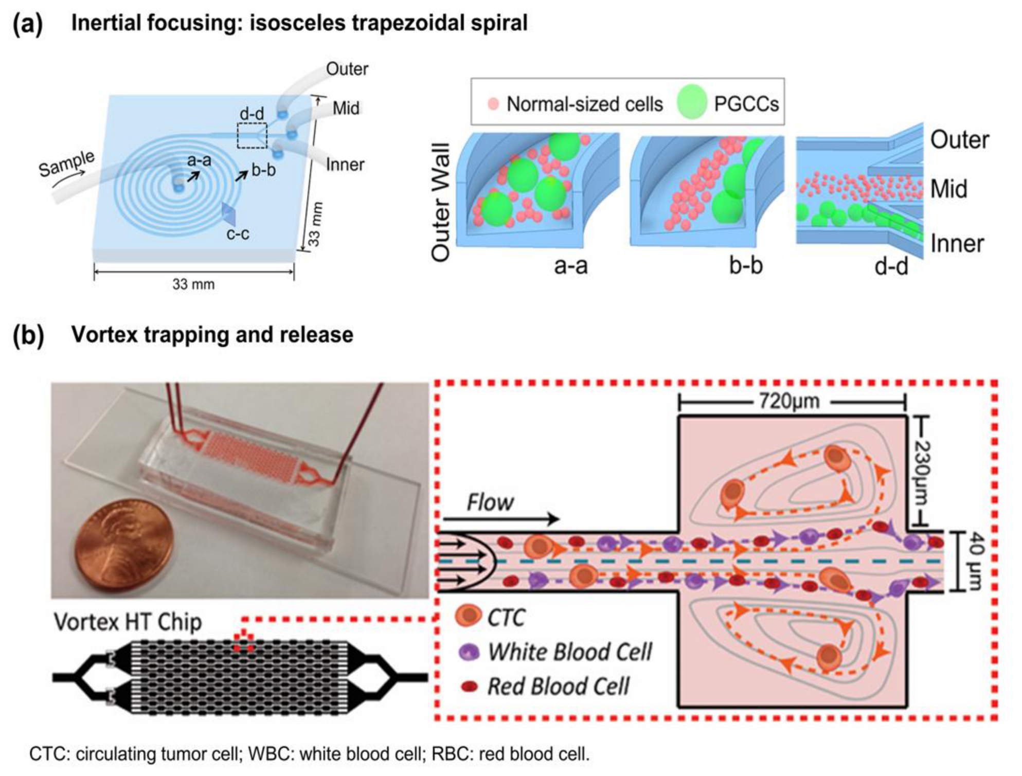
Inertial microfluidics separates cells through size-dependent lateral migration and focusing under controlled flow conditions (Figure 6a) [32,33]. In curved channels, the balance between inertial lift and Dean drag influences the focusing position while particle shape and orientation can further modify trajectories, as shown for chromosomes separated by size and shape in an inertial focusing device [62]. Channel geometry, flow rate, and cell deformability therefore affect the resulting separation performance.
Various architectures, including expansion structures, micro-obstacle arrays, cascaded focusing stages, and modified channel cross-sections, have been developed to improve focusing and separation [63,64,65,66,67]. Spiral channels exploit curvature-induced secondary flows to facilitate continuous cell recovery [68,69], while viscoelastic approaches introduce additional elastic forces that can modify focusing behavior [70,71,72]. Channel scaling and tests on large-cell inputs provide starting points for matching these architectures to PGCC-containing populations [73,74].
In our previous study, we developed a microbore-tubing spiral that enriched large cells from doxorubicin-resistant MDA-MB-231 cultures at 330 µL/min [74]. For the study’s approximately 36 µm large-cell criterion, two-pass purity was 28% without additive and 39% with 0.1% (w/w) poly(ethylene glycol) (PEG)-8000. Viability exceeded 90% after one pass, whereas the study’s Results section reports approximately 80% after two passes. This example shows how carrier formulation and repeated processing can improve enrichment while creating a trade-off with viable output. Table 4 retains the experiment-specific conditions and distinguishes cell results from bead tests.
In our subsequent study, we developed an isosceles trapezoidal spiral microchannel (ITSµC) that achieved over 90% purity, yield, and viability in separating PGCCs from doxorubicin-resistant (DOXR) MDA-MB-231 cells and patient-derived triple-negative breast cancer cells at optimized flow rates of 4 and 3 mL/min, respectively [19]. The recovered cells were further characterized by DNA-content analysis and increased expression of stemness- and resistance-associated markers, including CD133, SOX2, and IL-6. Together with the microbore-tubing spiral study [74], these findings demonstrate the potential of inertial microfluidics for efficient, label-free enrichment of PGCC-containing populations and their subsequent biological characterization.
Figure 6.
Inertial focusing and vortex trapping. (a) Isosceles trapezoidal spiral microchannel geometry and cross-sectional cell positions. Adapted from Figure 1A,C of Ref. [19]. Used with permission of the Royal Society of Chemistry, from “Efficient separation of large particles and giant cancer cells using an isosceles trapezoidal spiral microchannel”, Park, C. et al., Analyst, 149(17), 4496–4505, 2024; permission conveyed through Copyright Clearance Center, Inc. Copyright © 2024 Royal Society of Chemistry. Panel (a) is not covered by this article’s Creative Commons license. (b) High throughput vortex chip (Vortex HT) device and size-dependent trapping region; adapted and rearranged from Ref. [75] under the Creative Commons Attribution (CC BY) License. Panel headings and abbreviation text were added for readability; source experimental images, curves, and scale bars were retained.
7.2. Vortex-Based Microfluidic Trapping
Vortex-based microfluidic devices capture larger cells within recirculating flows, while smaller cells preferentially remain in the main flow (Figure 6b) [75,76]. Capture depends on cell size and inertial behavior, with shape and deformability potentially influencing retention. Unlike membrane filtration, vortex trapping does not require cells to pass through a fixed aperture.
Vortex HT illustrates reversible collection from diluted blood: up to approximately 84% cumulative capture was obtained after seven processing cycles for spiked MCF-7 human breast cancer cells, while a separate two-cycle experiment maintained purity above 80% [75]. Patient samples processed twice yielded 19.8 ± 13.9% purity among recovered nucleated cells. The 83.9 ± 4.0% viability result came from separate MCF-7 tests. Table 4 keeps these populations, processing cycles, and denominators distinct.
The useful feature for a candidate PGCC workflow is reversible trapping with optical access [75,76]. Cavity dimensions and flow can be tested against the input size distribution, with morphology documented while cells are trapped and biological characterization performed after release. This format is attractive for collecting an observable fraction before downstream analysis. Table 4 distinguishes the nominal total flow from the sample-inlet flow; repeated cycles increase processing time and must be included when comparing usable output.
7.3. Field-Flow Fractionation
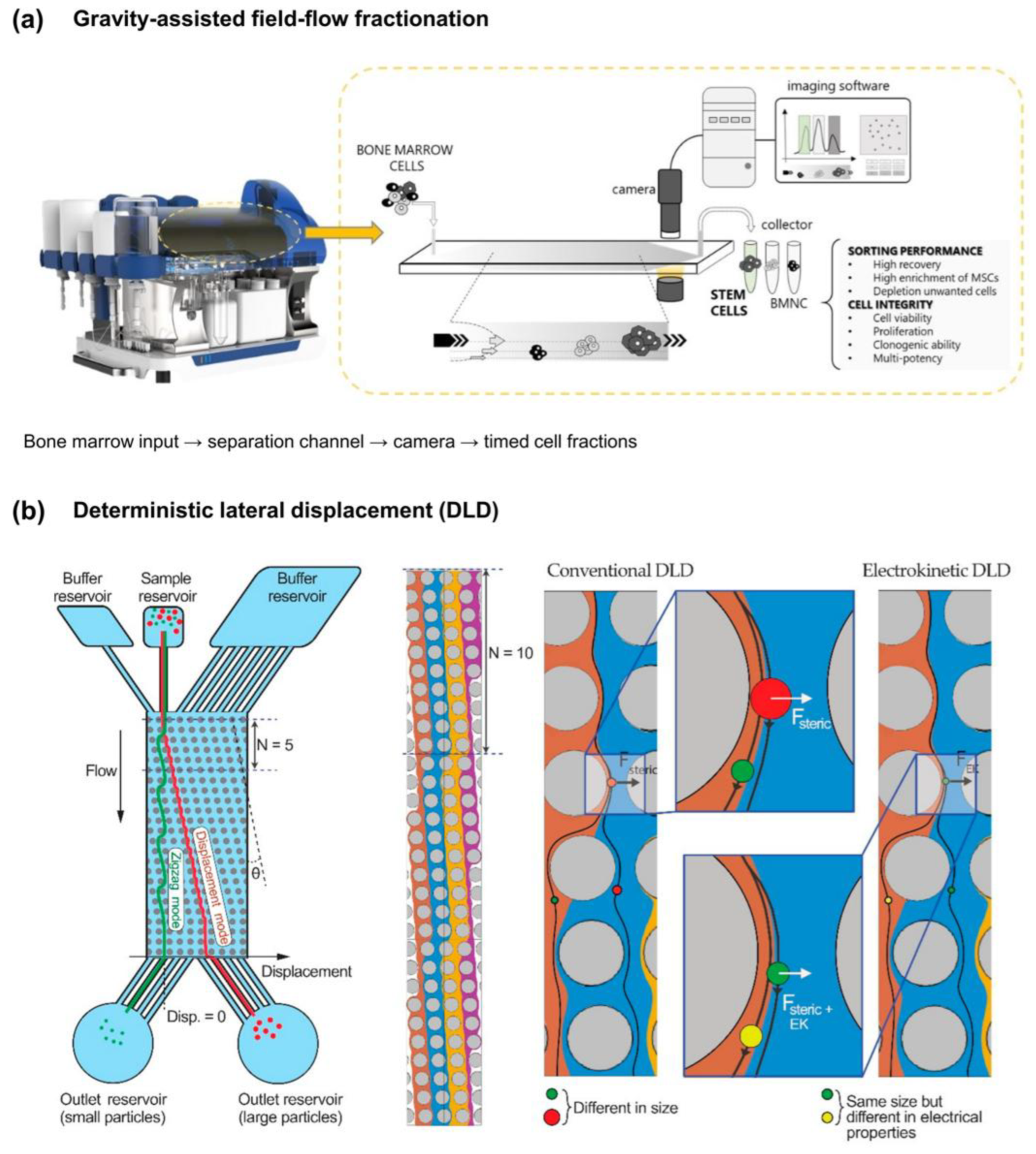
Field-flow fractionation (FFF) separates cells through differences in retention and elution within a thin flow channel (Figure 7a). Depending on the applied field and operating conditions, separation can be influenced by cell size, density, and hydrodynamic interactions. In steric or hyperlayer regimes, cell–wall interactions and hydrodynamic lift can modify elution behavior, producing size-dependent retention patterns that differ from simple sedimentation predictions [77,78].
Channel geometry, applied fields, and flow programming have been studied using both cells and model particles [79,80,81,82]. The red-blood-cell experiment [79] supplies cell-based evidence, while the latex studies [80,81,82] isolate engineering variables. Gravity-assisted sorting of clinical bone-marrow cells yielded successive fractions differing in composition and surface appearance [83]. Sedimentation FFF has also recovered cells from diosgenin-differentiated human erythroleukemia cultures, an induced megakaryocytic model [84], and glioblastoma subpopulations subsequently examined by ultrahigh-frequency DEP [85]. For a heterogeneous PGCC input, these precedents motivate the collection of several elution windows for parallel morphological, DNA-content, and molecular analysis. The advantage is access to multiple characterized fractions rather than a single imposed cutoff. Fraction selection can then follow the downstream endpoint using measured recovery and viability (Table 4).
7.4. Deterministic Lateral Displacement (DLD)
DLD separates cells through size-dependent interactions with ordered arrays of microposts (Figure 7b) [34,86,87]. Cells above a geometry-dependent critical size undergo lateral displacement, while smaller cells follow different trajectories. Unlike membrane filtration, DLD enables continuous separation without requiring cells to pass through a single fixed aperture. Cell shape, orientation, and deformability can also influence the effective displacement threshold. An electrokinetic variant superimposes an applied electric field, so that particles of the same size can be displaced according to differences in their electrical properties rather than size alone [86].
DLD offers a geometry-defined collection threshold and is a candidate when target and background passage sizes differ. Operating rates are device-specific: circular-post examples reviewed by McGrath et al. commonly operated at or below approximately 1 µL/min [34], whereas a triangular-post design processed diluted, cell-spiked blood at 10 mL/min [88]. In the inertially prefocused, sheath-free DLD system, MCF-7 cells in 500-fold-diluted healthy-donor blood were processed at 5.0 mL/h, with 99.0% capture efficiency based on outlet counts and 17.9% purity [87]. Table 4 states the counted populations and sample conditions. To evaluate giant-cell inputs, the critical diameter should be chosen from the target and background distributions, while the channel depth and gaps should accommodate the upper size range. This separates the choice of sorting threshold from the space needed for intact passage.
Figure 7.
Field-flow fractionation and deterministic lateral displacement (DLD). (a) Gravity-assisted fractionation of bone-marrow cells, with optical monitoring, timed fractions, and source-labeled sorting-performance and cell-integrity assessments; adapted and rearranged from Ref. [83]. (b) Conventional and electrokinetic DLD, showing size-dependent trajectories and additional electrically mediated displacement; adapted and rearranged from Ref. [86]. Both sources are used under the Creative Commons Attribution (CC BY) 4.0 License. Headings and selected labels were typeset for readability. These related-cell examples illustrate separation principles, not validated PGCC performance.
7.5. Acoustophoresis and Dielectrophoresis
Acoustophoresis provides field-controlled focusing or separation according to cell size and acoustic response (Figure 8a) [35,53]. Medium composition and operating conditions determine how that response is expressed [50,51,52,89]. Related-cell studies demonstrated preserved survival or function near 100 µL/min [50], affinity-bead-assisted CD8+ enrichment at 91 ± 8% purity with 63% median separation efficiency [51], and stromal-subset separation with 87–98% viability [52]. These examples motivate acoustic processing where field-controlled displacement and viable recovery are priorities; Table 4 retains their distinct populations and conditions.
Figure 8.
Acoustic and dielectrophoretic separation. (a) Alternating-frequency acoustofluidic workflow, including collection and immunofluorescence detection; adapted and rearranged from Ref. [53]. In (a), the chip schematic labels the sheath and sample inlets, the acoustic pressure node, the frequency-switching signal, and the collection and waste outlets; its legend distinguishes cancer cells from peripheral blood mononuclear cells (PBMCs). (b) Slanted-electrode channel and field-guided trajectories for negative DEP; adapted and rearranged from Ref. [54]. The separator consists of four slanted electrodes, labeled (i)–(iv) (with (i) and (iii) indicated in the trajectory schematic. In the source schematic, WBC denotes the leukocyte-sized population; the experiments used THP-1 cells and mouse erythrocytes. Both sources are used under the Creative Commons Attribution (CC BY) 4.0 License. Method headings and selected labels were added. The source micrograph, trajectory schematic, and scale bar are retained. These related-cell examples illustrate the separation principles discussed in the text.
DEP uses non-uniform electric fields to convert frequency-dependent cellular polarizability into motion (Figure 8b) [90]. In dielectrophoretic/gravitational field-flow fractionation (DEP/G-FFF), Yang et al. combined electrical levitation with gravity to separate MDA-MB-435 cells from blood cells [49]. Although described historically as a breast cancer model, MDA-MB-435 was subsequently authenticated as M14 melanoma-derived [91]. The study introduced approximately 50,000 cells in 10 µL, used a separate 5 min equilibration period, and achieved separation in under 8 min at 2 mL/min. At 10 kHz and 1 mL/min, the selected MDA-MB-435 and erythrocyte windows contained >98% and >99% of the respective eluted populations. More than 99% of collected cells excluded trypan blue [49]. The example illustrates how frequency and flow can be used to choose collection windows for further analysis.
Image-guided DEP cages offer a complementary format when individual-cell recovery is the goal. Peeters et al. combined CellSearch enrichment with DEPArray isolation of tumor cells spiked into healthy-donor blood, followed by mutation and transcription analyses of recovered single cells or small groups [36]. The reported 100% purity concerned the selected recovered cells; approximately 40% loss occurred during loading. The complete workflow used fluorescent identification and immunoaffinity enrichment. Its practical contribution is the connection between the selection of an individual cell and molecular analysis. For PGCC evaluation, accommodation of the input cells and the measured dielectric response define the candidate operating range, while recovered-cell identity and yield determine analytical suitability.
In our previous study, we demonstrated an integrated experimental workflow combining CDRA chip-based induction of doxorubicin-resistant MDA-MB-231 cells with image-guided isolation of individual PGCC-like large cells using laser-induced microbubbles [20]. Individual cells were characterized by their projected areas, while DAPI staining revealed enlarged or multiple nuclei characteristic of PGCCs. Selective detachment enabled the separation of large PGCC-like cells from normal-sized drug-resistant cells, allowing for subsequent comparison of their doxorubicin sensitivity. The large-cell population exhibited greater drug resistance than its normal-sized counterpart, demonstrating the utility of linking microfluidic induction, single-cell-level physical separation, and downstream functional analysis to investigate cancer cell heterogeneity.
Collectively, these studies demonstrate the utility of microfluidic approaches for enriching PGCC-containing populations and selectively isolating individual large cancer cells while preserving their suitability for subsequent biological and functional analyses. The availability of both population-level enrichment and targeted single-cell retrieval provides complementary strategies for investigating PGCC heterogeneity and selecting appropriate downstream analytical workflows. Table 4 summarizes sample sources, reported input volumes, total cell concentrations and target fractions, operating conditions, and separation outcomes for the reviewed microfluidic methods.
Table 4.
Microfluidic fractionation, trapping, and targeted-recovery workflows. Sample origin, preparation, reported input volume and concentration, operating conditions, and performance are kept study-specific. NR, not reported for the specified metric in the checked article; NS, not summarized here, rather than evidence that the source lacks the metric. Purity and recovery use the source definitions described in Section 5.6. CK, cytokeratin; DAPI, 4′,6-diamidino-2-phenylindole; DEP/G-FFF, dielectrophoretic/gravitational field-flow fractionation; DLD, deterministic lateral displacement; FFF, field-flow fractionation; ITSµC, isosceles trapezoidal spiral microchannel; PBPC, peripheral blood progenitor cell; PDMS, polydimethylsiloxane; PEG, poly(ethylene glycol); qRT-PCR, quantitative reverse-transcription PCR; RBC, red blood cell; SD, standard deviation; SdFFF, sedimentation field-flow fractionation; WBC, white blood cell.
8. Downstream Analysis After PGCC Enrichment
Downstream analysis of enriched PGCC populations requires the consideration of cell size, nuclear organization, and the condition of recovered cells. In particular, multinucleated PGCCs introduce a distinction between cell-based and nucleus-based measurements: a single cell may contribute multiple nuclear profiles, potentially affecting estimates of population composition and heterogeneity.
8.1. Droplet-Based Single-Cell Analysis
Droplet-based platforms enable high-throughput single-cell molecular profiling by encapsulating cells with barcoded beads or reagents [92,93,94]. However, conventional droplet geometries may not accommodate very large PGCCs. As a geometric illustration, increasing the spherical droplet diameter from 20 to 60 µm increases volume 27-fold, potentially affecting reagent consumption, partitioning efficiency, and processing throughput. These diameters are an illustrative calculation, not a universal specification of the cited platforms.
For an intact-cell endpoint, droplet dimensions, passage through the inlet, and molecular capture should be evaluated together. Where the target-cell dimensions are poorly matched to the available geometry, imaging-compatible microwells or individual-well deposition provide alternatives for evaluation (Section 8.2 and Section 8.3). The sampling unit and handling conditions can therefore be chosen before committing the recovered cells to a particular platform.
8.2. Combinatorial Indexing, Microwells, and Nucleus-Based Workflows
Combinatorial indexing offers an alternative to conventional droplet encapsulation by assigning molecular barcodes through repeated partitioning and pooling steps [95,96]. This approach may reduce geometric constraints associated with large intact cells, although compatibility with PGCCs and fixation-dependent processing requirements must be considered. Imaging-compatible microwell platforms may additionally support the association of cell morphology with subsequent molecular measurements.
Direct PGCC single-cell profiling has been demonstrated with microwells. Zhou et al. sorted and visually checked PGCC and non-PGCC fractions from SUM159, MDA-MB-231, and Vari068 cells, then paired cells with barcoded beads in microwells for transcriptome analysis [31]. Reads were processed with the Drop-seq analysis pipeline, while capture itself was microwell-based. This provides a demonstrated sorting-to-transcriptome route for the selected populations. This study demonstrated the feasibility of combining fluorescence-assisted PGCC enrichment with microwell-based single-cell transcriptomic analysis, supported by morphological assessment and population-level ploidy characterization [31].
Nucleus-based sequencing avoids many constraints associated with intact-cell dimensions but introduces an important analytical distinction. Multinucleated PGCCs can contribute multiple nuclear profiles, while dissociation removes information about which nuclei originated from the same cell. Consequently, cell-based and nucleus-based datasets may represent different biological sampling units and should not be interpreted interchangeably.
8.3. Non-Droplet, Bulk, and Functional Analysis
Non-droplet approaches, including plate-based Smart-seq2 and microfluidic C1 platforms, provide alternatives for single-cell molecular profiling [97,98]. Beyond transcriptomics, proteomic studies have provided important insights into PGCC biology. Zhang et al. employed iTRAQ-based LC–MS/MS to identify distinct protein expression patterns in PGCCs and their budding progeny in ovarian cancer [99]. Xu et al. used co-immunoprecipitation coupled with LC–MS/MS to investigate proteins associated with PGCC formation, survival, and cytoplasmic SIRT1-mediated paclitaxel resistance [100]. Zheng et al. further investigated Cdc42-associated cytoskeletal regulation in progeny derived from CoCl2-induced PGCCs using proteomic approaches [101]. More recently, Suriya et al. combined proteomic and transcriptomic analyses of PARP inhibitor-induced, PGCC-enriched dormant ovarian cancer populations, identifying redox-dependent vulnerabilities and demonstrating the therapeutic potential of disulfiram [102].
Together, these studies demonstrate the value of molecular and proteomic analyses for understanding PGCC-associated cellular plasticity, therapeutic resistance, and progeny formation. Complementary functional assays, including clonogenic growth and drug-response measurements, further enable assessment of the biological properties of recovered cells. Integrating physical PGCC separation with these molecular and functional approaches offers opportunities to characterize distinct PGCC populations and investigate their contributions to cancer progression and treatment resistance.
8.4. Separation and Computational Biases
Physical enrichment can preferentially recover PGCC subpopulations according to size, deformability, or adhesion, potentially altering the composition of the analyzed sample [74]. Subsequent computational processing may introduce additional bias because conventional quality-control and doublet-detection methods may not adequately account for large, polyploid, or multinucleated cells [103].
These effects should be considered separately. Comparing input and recovered populations can help assess selection introduced during separation, while appropriate validation of computational thresholds is necessary to avoid excluding genuine PGCC-derived profiles. Together, these considerations emphasize the importance of matching separation methods and downstream analytical strategies to the biological characteristics of PGCCs.
9. Integrating Microfluidic Separation with Downstream Requirements
9.1. Selecting a Workflow for the Analytical Endpoint
Method selection starts with the analytical endpoint and the recovery format it requires (Table 5). Bulk assays require a representative population with sufficient usable RNA or protein; single-cell profiling requires identifiable cells that can be transferred into the capture format; functional assays additionally require viable recovery. Within these requirements, the reviewed studies support several concrete starting routes.
Table 5.
Downstream sampling units, count-reporting stages, and implications for upstream enrichment. This table is qualitative workflow guidance, not a table of minimum cell-input requirements. Loaded cells, recovered cells, successfully profiled cells, nuclei, and spatially indexed objects are different denominators. Required counts must be determined for the specific assay, target abundance, recovery, and intended statistical analysis. iTRAQ, isobaric tags for relative and absolute quantitation; MS, mass spectrometry; RPPA, reverse-phase protein array.
For label-free population enrichment, ITSµC recovered large-cell fractions from DOXR and patient-derived cultures. The DOXR-derived fraction underwent DNA-content analysis, immunostaining, and bulk qRT-PCR [19]. Inertial collection is therefore an evidence-supported starting point when the intended target is a large-cell fraction and the assay can use pooled material. The microbore study [74] shows why pass number and carrier formulation should be chosen against viable output as well as purity. Extending either preparation to MS would require assay-specific validation; the reported iTRAQ study used CoCl2 selection instead [99].
For transcriptome profiling of selected PGCC-associated cells, Hoechst-assisted sorting followed by microwell capture has already been demonstrated [31]. This is a practical route when fluorescent selection is acceptable and the recovered cells fit the capture geometry. When inspection of a particular cell is the priority, image-guided detachment [20] or DEP-cage retrieval [36] offers a different starting format; the latter connected recovered cells to molecular assays in a related tumor-cell model. These examples distinguish a demonstrated PGCC sorting-to-transcriptome workflow from candidate morphology-guided extensions.
For studies of stress adaptation, the CDRA example links microfluidic induction to off-chip selection, FACS, and molecular or functional characterization of a PACC-like L-DOXR population [30]. This demonstrates that a useful staged workflow does not require all operations to occur on one chip. Sedimentation, elutriation, or FFF can be evaluated as initial fractionation steps when multiple phenotypic fractions are needed; targeted retrieval can follow when individual identity is the endpoint. In a new combination, the decision to retain each stage rests on its added enrichment, retained target number, and compatibility with the next assay.
9.2. Factors Influencing Enrichment Requirements
The need for upstream enrichment depends on PGCC abundance, recovery efficiency, and the specific endpoint. Table 5 distinguishes the biological sampling unit from the stages at which material is loaded, recovered, and successfully analyzed. The qualitative bands in Figure 1b have no numerical cell-count endpoints. The cited workflows do not establish a universal minimum number of PGCCs for each analytical category, so generic numerical ranges are not assigned. When PGCCs are rare, enrichment may increase their availability for subsequent assays; when target cells are sufficiently abundant or directly identifiable, extensive enrichment may be unnecessary.
Platform geometry and sampling unit further narrow the choice. For intact large-cell profiling, compare droplet, microwell, and well-deposition formats against the input dimensions. For nucleus-based analysis, define nuclei as the sampling units and account for multinucleation. Functional assays prioritize viable recovery, while spatial analyses favor retaining cells and their local context. These requirements determine which enrichment output is useful.
Ultimately, enrichment should be optimized for the intended analytical endpoint rather than maximized independently. The most suitable separation method is one that provides an appropriate balance of target-cell recovery, viability, processing capacity, and compatibility with downstream analysis.
Complementary measurements can inform separation-method selection before a new workflow is assembled. Impedance cytometry characterizes frequency-dependent electrical responses [104,105,106,107], quantitative phase imaging provides phase-derived mass or optical readouts [108,109,110], and real-time deformability cytometry measures mechanical responses under defined flow [111,112,113,114]. These approaches require verification of their usable size range for giant cells; acoustic contrast and buoyant density must be measured with appropriate modalities. Patient-derived models and tumor-on-chip systems provide additional contexts for testing whether the selected preparation and analytical stages remain compatible under more complex biological conditions [115,116,117,118].
10. Conclusions
This review highlights the substantial progress made in linking PGCC induction, physical separation, and downstream biological characterization through conventional and microfluidic approaches. Microfluidic drug-gradient platforms, including the Cancer Drug Resistance Accelerator (CDRA) chip, have enabled the controlled induction of PGCC/PACC-associated drug-resistant populations and their subsequent molecular and functional characterization [30]. Inertial microfluidic approaches, including microbore spiral devices and isosceles trapezoidal spiral microchannels, have demonstrated label-free enrichment of large cancer cells, with recovered populations subjected to DNA-content analysis, immunostaining, and molecular characterization [19,74]. Furthermore, image-guided laser-induced microbubble detachment has enabled the selective separation of PGCC-like large cells and normal-sized drug-resistant cells following CDRA-based induction, revealing differences in their doxorubicin sensitivity [20]. These findings demonstrate that microfluidic induction and physical separation can be successfully linked to downstream biological investigations, providing experimentally supported strategies for studying PGCC-associated heterogeneity and therapeutic resistance.
The reviewed separation methods offer complementary capabilities for different experimental objectives. Size- and flow-based approaches, including filtration, sedimentation, field-flow fractionation, and inertial microfluidics, provide strategies for population-level enrichment, whereas image-guided and field-controlled methods offer opportunities for more selective cell handling and retrieval. Importantly, downstream analytical studies have already established the biological value of characterizing PGCC-associated populations. Hoechst-assisted sorting combined with microwell-based single-cell transcriptomics has enabled the molecular investigation of selected PGCC populations at the single-cell level [31]. Proteomic studies have identified molecular pathways associated with PGCC formation, survival, and progeny development, including SIRT1-associated paclitaxel resistance and Cdc42-mediated cytoskeletal regulation [99,100,101]. More recently, integrated transcriptomic, proteomic, and functional analyses of PARP inhibitor-induced, PGCC-enriched dormant populations have revealed redox-dependent vulnerabilities relevant to therapeutic resistance and cellular recovery [102]. Together, these studies demonstrate the complementary value of physical separation, single-cell profiling, proteomics, and functional assays for understanding PGCC biology.
Building on these achievements, further advances will depend on selecting and integrating separation strategies according to PGCC characteristics and downstream analytical objectives. Quantitative characterization of input and recovered populations, appropriate biological verification, and evaluation of recovery efficiency and viability can strengthen reproducibility and facilitate comparisons across platforms. Rather than requiring a universal separation method, the diversity of available technologies provides opportunities to develop flexible workflows for population-level enrichment, targeted cell retrieval, and molecular or functional characterization. Continued integration of microfluidic induction and separation with advanced single-cell and proteomic analyses is expected to facilitate more precise investigations of PGCC heterogeneity, cellular plasticity, and treatment resistance. Overall, these approaches provide a strong experimental foundation for advancing PGCC research and developing more comprehensive strategies to investigate the roles of PGCCs in cancer progression and therapeutic response.
These engineering advances should be interpreted within the broader complexity of therapy-associated tumor states. PGCCs/PACCs are one biologically relevant component of this landscape, but they are not synonymous with senescent cells or stress-tolerant persisters, and their isolation alone cannot explain the multifactorial failure of solid-tumor therapy. Reliable physical enrichment coupled with independent biological verification can instead provide a more rigorous basis for identifying the recovered PGCC/PACC-associated populations and investigating their specific contributions alongside other tumor-persistent states.
Author Contributions
Conceptualization, C.P. and S.P.; investigation, C.P.; writing—original draft preparation, C.P. and S.P.; writing—review and editing, Y.J.O. and P.H.; visualization, S.B.; supervision, S.P.; project administration, S.P.; funding acquisition, S.P. All authors have read and agreed to the published version of the manuscript.
Funding
This research was supported by the Technology Innovation Program (RS-2025-02307623) and the Human Resource Development Program for Industrial Innovation (Global) (P0008746, through the Korea Institute for Advancement of Technology [KIAT]), both funded by the Ministry of Trade, Industry & Energy (MOTIE, Korea); and a grant (RS-2025-02263957) of the Korea Health Technology R&D Project through the Korea Health Industry Development Institute (KHIDI), funded by the Ministry of Health & Welfare.
Institutional Review Board Statement
Not applicable.
Informed Consent Statement
Not applicable.
Data Availability Statement
No new data were created or analyzed in this study. All data discussed are available in the cited publications.
Conflicts of Interest
The authors declare no conflicts of interest.
Abbreviations
The following abbreviations are used in this manuscript:
| AMPK | AMP-activated protein kinase |
| CDRA | Cancer Drug Resistance Accelerator |
| CK | cytokeratin |
| CTC | circulating tumor cell |
| DAPI | 4′,6-diamidino-2-phenylindole |
| DEP | dielectrophoresis |
| DEP/G-FFF | dielectrophoretic/gravitational field-flow fractionation |
| DLD | deterministic lateral displacement |
| DOX | doxorubicin |
| DOXR | doxorubicin-resistant |
| FACS | fluorescence-activated cell sorting |
| FFF | field-flow fractionation |
| HEL | human erythroleukemia |
| HIF-1α | hypoxia-inducible factor 1α |
| iTRAQ | isobaric tags for relative and absolute quantitation |
| ITSµC | isosceles trapezoidal spiral microchannel |
| LC–MS/MS | liquid chromatography–tandem mass spectrometry |
| L-DOXR | large doxorubicin-resistant |
| MS | mass spectrometry |
| mTOR | mechanistic target of rapamycin |
| NR | not reported |
| NS | not summarized |
| PACC | polyaneuploid cancer cell |
| PARP | poly(ADP-ribose) polymerase |
| PBMC | peripheral blood mononuclear cell |
| PBPC | peripheral blood progenitor cell |
| PDMS | polydimethylsiloxane |
| PEG | poly(ethylene glycol) |
| PGCC | polyploid giant cancer cell |
| PI | propidium iodide |
| qRT-PCR | quantitative reverse-transcription PCR |
| RBC | red blood cell |
| RPPA | reverse-phase protein array |
| scRNA-seq | single-cell RNA sequencing |
| SD | standard deviation |
| SdFFF | sedimentation field-flow fractionation |
| WBC | white blood cell |
References
- Zhang, S.; Mercado-Uribe, I.; Xing, Z.; Sun, B.; Kuang, J.; Liu, J. Generation of cancer stem-like cells through the formation of polyploid giant cancer cells. Oncogene 2014, 33, 116–128, Erratum in Oncogene 2014, 33, 134. https://doi.org/10.1038/onc.2013.318. [Google Scholar] [CrossRef] [Scilit] [PubMed]
- Amend, S.R.; Torga, G.; Lin, K.C.; Kostecka, L.G.; De Marzo, A.; Austin, R.H.; Pienta, K.J. Polyploid giant cancer cells: Unrecognized actuators of tumorigenesis, metastasis, and resistance. Prostate 2019, 79, 1489–1497. [Google Scholar] [CrossRef] [Scilit] [PubMed]
- Zhou, X.; Zhou, M.; Zheng, M.; Tian, S.; Yang, X.; Ning, Y.; Li, Y.; Zhang, S. Polyploid giant cancer cells and cancer progression. Front. Cell Dev. Biol. 2022, 10, 1017588. [Google Scholar] [CrossRef] [Scilit] [PubMed]
- Liu, P.; Wang, L.; Yu, H. Polyploid giant cancer cells: Origin, possible pathways of formation, characteristics, and mechanisms of regulation. Front. Cell Dev. Biol. 2024, 12, 1410637. [Google Scholar] [CrossRef] [Scilit] [PubMed]
- Richards, J.S.; Candelaria, N.R.; Lanz, R.B. Polyploid giant cancer cells and ovarian cancer: New insights into mitotic regulators and polyploidy. Biol. Reprod. 2021, 105, 305–316. [Google Scholar] [CrossRef] [Scilit] [PubMed]
- Song, Y.; Zhao, Y.; Deng, Z.; Zhao, R.; Huang, Q. Stress-induced polyploid giant cancer cells: Unique way of formation and non-negligible characteristics. Front. Oncol. 2021, 11, 724781. [Google Scholar] [CrossRef] [Scilit] [PubMed]
- Bharadwaj, D.; Mandal, M. Senescence in polyploid giant cancer cells: A road that leads to chemoresistance. Cytokine Growth Factor Rev. 2020, 52, 68–75. [Google Scholar] [CrossRef] [Scilit] [PubMed]
- Pienta, K.J.; Hammarlund, E.U.; Austin, R.H.; Axelrod, R.; Brown, J.S.; Amend, S.R. Cancer cells employ an evolutionarily conserved polyploidization program to resist therapy. Semin. Cancer Biol. 2022, 81, 145–159. [Google Scholar] [CrossRef] [Scilit] [PubMed]
- Coward, J.; Harding, A. Size does matter: Why polyploid tumor cells are critical drug targets in the war on cancer. Front. Oncol. 2014, 4, 123. [Google Scholar] [CrossRef] [Scilit] [PubMed]
- Trabzonlu, L.; Pienta, K.J.; Trock, B.J.; De Marzo, A.M.; Amend, S.R. Presence of cells in the polyaneuploid cancer cell (PACC) state predicts the risk of recurrence in prostate cancer. Prostate 2023, 83, 277–285. [Google Scholar] [CrossRef] [Scilit] [PubMed]
- Fei, F.; Zhang, D.; Yang, Z.; Wang, S.; Wang, X.; Wu, Z.; Wu, Q.; Zhang, S. The number of polyploid giant cancer cells and epithelial-mesenchymal transition-related proteins are associated with invasion and metastasis in human breast cancer. J. Exp. Clin. Cancer Res. 2015, 34, 158, Correction in J. Exp. Clin. Cancer Res. 2024, 43, 221. https://doi.org/10.1186/s13046-024-03148-y. [Google Scholar] [CrossRef] [Scilit] [PubMed]
- Lv, H.; Shi, Y.; Zhang, L.; Zhang, D.; Liu, G.; Yang, Z.; Li, Y.; Fei, F.; Zhang, S. Polyploid giant cancer cells with budding and the expression of cyclin E, S-phase kinase-associated protein 2, stathmin associated with the grading and metastasis in serous ovarian tumor. BMC Cancer 2014, 14, 576. [Google Scholar] [CrossRef] [Scilit] [PubMed]
- Zhang, D.; Yang, X.; Yang, Z.; Fei, F.; Li, S.; Qu, J.; Zhang, M.; Li, Y.; Zhang, X.; Zhang, S. Daughter cells and erythroid cells budding from PGCCs and their clinicopathological significances in colorectal cancer. J. Cancer 2017, 8, 469–478. [Google Scholar] [CrossRef] [Scilit] [PubMed]
- Fei, F.; Zhang, M.; Li, B.; Zhao, L.; Wang, H.; Liu, L.; Li, Y.; Ding, P.; Gu, Y.; Zhang, X.; et al. Formation of polyploid giant cancer cells involves in the prognostic value of neoadjuvant chemoradiation in locally advanced rectal cancer. J. Oncol. 2019, 2019, 2316436. [Google Scholar] [CrossRef] [Scilit] [PubMed]
- Herbein, G.; Nehme, Z. Polyploid giant cancer cells, a hallmark of oncoviruses and a new therapeutic challenge. Front. Oncol. 2020, 10, 567116. [Google Scholar] [CrossRef] [Scilit] [PubMed]
- You, B.; Xia, T.; Gu, M.; Zhang, Z.; Zhang, Q.; Shen, J.; Fan, Y.; Yao, H.; Pan, S.; Lu, Y.; et al. AMPK-mTOR-mediated activation of autophagy promotes formation of dormant polyploid giant cancer cells. Cancer Res. 2022, 82, 846–858. [Google Scholar] [CrossRef] [Scilit] [PubMed]
- Zhang, X.; Yao, J.; Li, X.; Niu, N.; Liu, Y.; Hajek, R.A.; Peng, G.; Westin, S.; Sood, A.K.; Liu, J. Targeting polyploid giant cancer cells potentiates a therapeutic response and overcomes resistance to PARP inhibitors in ovarian cancer. Sci. Adv. 2023, 9, eadf7195. [Google Scholar] [CrossRef] [Scilit] [PubMed]
- Adibi, R.; Moein, S.; Gheisari, Y. Zoledronic acid targets chemo-resistant polyploid giant cancer cells. Sci. Rep. 2023, 13, 419. [Google Scholar] [CrossRef] [Scilit] [PubMed]
- Park, C.; Lim, W.; Song, R.; Han, J.; You, D.; Kim, S.; Lee, J.E.; van Noort, D.; Mandenius, C.-F.; Lee, J.; et al. Efficient separation of large particles and giant cancer cells using an isosceles trapezoidal spiral microchannel. Analyst 2024, 149, 4496–4505. [Google Scholar] [CrossRef] [Scilit] [PubMed]
- Lim, W.; Lee, S.; Park, S.; Baac, H.W. Differential detachment of intact and viable cells of different sizes using laser-induced microbubbles. Biomed. Opt. Express 2019, 10, 4919–4930. [Google Scholar] [CrossRef] [Scilit] [PubMed]
- Krishan, A. Effect of drug efflux blockers on vital staining of cellular DNA with Hoechst 33342. Cytometry 1987, 8, 642–645. [Google Scholar] [CrossRef] [Scilit] [PubMed]
- Sramkoski, R.M.; Wormsley, S.W.; Bolton, W.E.; Crumpler, D.C.; Jacobberger, J.W. Simultaneous detection of cyclin B1, p105, and DNA content provides complete cell cycle phase fraction analysis of cells that endoreduplicate. Cytometry 1999, 35, 274–283. [Google Scholar] [CrossRef] [Scilit]
- Kim, C.J.; Gonye, A.L.K.; Truskowski, K.; Lee, C.F.; Cho, Y.K.; Austin, R.H.; Pienta, K.J.; Amend, S.R. Nuclear morphology predicts cell survival to cisplatin chemotherapy. Neoplasia 2023, 42, 100906. [Google Scholar] [CrossRef] [Scilit] [PubMed]
- Schmidt, M.J.; Naghdloo, A.; Prabakar, R.K.; Kamal, M.; Cadaneanu, R.; Garraway, I.P.; Lewis, M.; Aparicio, A.; Zurita-Saavedra, A.; Corn, P.; et al. Polyploid cancer cells reveal signatures of chemotherapy resistance. Oncogene 2025, 44, 439–449. [Google Scholar] [CrossRef] [Scilit] [PubMed]
- Mallin, M.M.; Kim, N.; Choudhury, M.I.; Lee, S.J.; An, S.S.; Sun, S.X.; Konstantopoulos, K.; Pienta, K.J.; Amend, S.R. Cells in the polyaneuploid cancer cell (PACC) state have increased metastatic potential. Clin. Exp. Metastasis 2023, 40, 321–338. [Google Scholar] [CrossRef] [Scilit] [PubMed]
- Xia, Y.; Wan, Y.; Hao, S.; Nisic, M.; Harouaka, R.A.; Chen, Y.; Zou, X.; Zheng, S. Nucleus of circulating tumor cell determines its translocation through biomimetic microconstrictions and its physical enrichment by microfiltration. Small 2018, 14, 1802899. [Google Scholar] [CrossRef] [Scilit] [PubMed]
- Distelhorst, C.W.; Benutto, B.M.; Bergamini, R.A. Centrifugal elutriation of human lymphoid cells: Synchronization and separation of aneuploid cells into diploid and tetraploid populations. Cell. Immunol. 1983, 79, 68–80. [Google Scholar] [CrossRef] [Scilit] [PubMed]
- Park, J.M.; Lee, J.Y.; Lee, J.G.; Jeong, H.; Oh, J.M.; Kim, Y.J.; Park, D.; Kim, M.S.; Lee, H.J.; Oh, J.H.; et al. Highly efficient assay of circulating tumor cells by selective sedimentation with a density gradient medium and microfiltration from whole blood. Anal. Chem. 2012, 84, 7400–7407. [Google Scholar] [CrossRef] [Scilit] [PubMed]
- Berkow, R.L.; Straneva, J.E.; Bruno, E.; Beyer, G.S.; Burgess, J.S.; Hoffman, R. Isolation of human megakaryocytes by density centrifugation and counterflow centrifugal elutriation. J. Lab. Clin. Med. 1984, 103, 811–818. [Google Scholar] [PubMed]
- Lim, W.; Hwang, I.; Zhang, J.; Chen, Z.; Han, J.; Jeon, J.; Koo, B.-K.; Kim, S.; Lee, J.E.; Kim, Y.; et al. Exploration of drug resistance mechanisms in triple negative breast cancer cells using a microfluidic device and patient tissues. eLife 2024, 12, RP88830. [Google Scholar] [CrossRef] [PubMed]
- Zhou, M.; Ma, Y.; Chiang, C.-C.; Rock, E.C.; Butler, S.C.; Anne, R.; Yatsenko, S.; Gong, Y.; Chen, Y.-C. Single-cell morphological and transcriptome analysis unveil inhibitors of polyploid giant breast cancer cells in vitro. Commun. Biol. 2023, 6, 1301. [Google Scholar] [CrossRef] [Scilit] [PubMed]
- Di Carlo, D. Inertial microfluidics. Lab Chip 2009, 9, 3038–3046. [Google Scholar] [CrossRef] [Scilit] [PubMed]
- Martel, J.M.; Toner, M. Inertial focusing in microfluidics. Annu. Rev. Biomed. Eng. 2014, 16, 371–396. [Google Scholar] [CrossRef] [Scilit] [PubMed]
- McGrath, J.; Jimenez, M.; Bridle, H. Deterministic lateral displacement for particle separation: A review. Lab Chip 2014, 14, 4139–4158. [Google Scholar] [CrossRef] [Scilit] [PubMed]
- Lenshof, A.; Magnusson, C.; Laurell, T. Acoustofluidics 8: Applications of acoustophoresis in continuous flow microsystems. Lab Chip 2012, 12, 1210–1223. [Google Scholar] [CrossRef] [Scilit] [PubMed]
- Peeters, D.J.E.; De Laere, B.; Van den Eynden, G.G.; Van Laere, S.J.; Rothé, F.; Ignatiadis, M.; Sieuwerts, A.; Lambrechts, D.; Rutten, A.; Van Dam, P.; et al. Semiautomated isolation and molecular characterisation of single or highly purified tumour cells from CellSearch enriched blood samples using dielectrophoretic cell sorting. Br. J. Cancer 2013, 108, 1358–1367. [Google Scholar] [CrossRef] [Scilit] [PubMed]
- Heng, J.; Heng, H.H. Genome chaos: Creating new genomic information essential for cancer macroevolution. Semin. Cancer Biol. 2022, 81, 160–175. [Google Scholar] [CrossRef] [Scilit] [PubMed]
- Ye, J.C.; Horne, S.; Zhang, J.Z.; Jackson, L.; Heng, H.H. Therapy induced genome chaos: A novel mechanism of rapid cancer drug resistance. Front. Cell Dev. Biol. 2021, 9, 676344. [Google Scholar] [CrossRef] [Scilit] [PubMed]
- Wang, X.; Zheng, M.; Fei, F.; Li, C.; Du, J.; Liu, K.; Li, Y.; Zhang, S. EMT-related protein expression in polyploid giant cancer cells and their daughter cells with different passages after triptolide treatment. Med. Oncol. 2019, 36, 82. [Google Scholar] [CrossRef] [Scilit] [PubMed]
- Rohnalter, V.; Roth, K.; Finkernagel, F.; Adhikary, T.; Obert, J.; Dorzweiler, K.; Bensberg, M.; Müller-Brüsselbach, S.; Müller, R. A multi-stage process including transient polyploidization and EMT precedes the emergence of chemoresistent ovarian carcinoma cells with a dedifferentiated and pro-inflammatory secretory phenotype. Oncotarget 2015, 6, 40005–40025. [Google Scholar] [CrossRef] [Scilit] [PubMed]
- Lopez-Sánchez, L.M.; Jimenez, C.; Valverde, A.; Hernandez, V.; Peñarando, J.; Martinez, A.; Lopez-Pedrera, C.; Muñoz-Castañeda, J.R.; De la Haba-Rodríguez, J.R.; Aranda, E.; et al. CoCl2, a mimic of hypoxia, induces formation of polyploid giant cells with stem characteristics in colon cancer. PLoS ONE 2014, 9, e99143. [Google Scholar] [CrossRef] [Scilit] [PubMed]
- Zhang, S.; Mercado-Uribe, I.; Liu, J. Generation of erythroid cells from fibroblasts and cancer cells in vitro and in vivo. Cancer Lett. 2013, 333, 205–212. [Google Scholar] [CrossRef] [Scilit] [PubMed]
- Zheng, M.; Tian, S.; Zhou, X.; Yan, M.; Zhou, M.; Yu, Y.; Zhang, Y.; Wang, X.; Li, N.; Ren, L.; et al. MITF regulates the subcellular location of HIF1α through SUMOylation to promote the invasion and metastasis of daughter cells derived from polyploid giant cancer cells. Oncol. Rep. 2024, 51, 63. [Google Scholar] [CrossRef] [Scilit] [PubMed]
- Xuan, B.; Ghosh, D.; Cheney, E.M.; Clifton, E.M.; Dawson, M.R. Dysregulation in actin cytoskeletal organization drives increased stiffness and migratory persistence in polyploidal giant cancer cells. Sci. Rep. 2018, 8, 11935. [Google Scholar] [CrossRef] [Scilit] [PubMed]
- Adams, D.L.; Zhu, P.; Makarova, O.V.; Martin, S.S.; Charpentier, M.; Chumsri, S.; Li, S.; Amstutz, P.; Tang, C.-M. The systematic study of circulating tumor cell isolation using lithographic microfilters. RSC Adv. 2014, 4, 4334–4342. [Google Scholar] [CrossRef] [Scilit] [PubMed]
- Fan, X.; Jia, C.; Yang, J.; Li, G.; Mao, H.; Jin, Q.; Zhao, J. A microfluidic chip integrated with a high-density PDMS-based microfiltration membrane for rapid isolation and detection of circulating tumor cells. Biosens. Bioelectron. 2015, 71, 380–386. [Google Scholar] [CrossRef] [Scilit] [PubMed]
- Chen, C.L.; Mahalingam, D.; Osmulski, P.; Jadhav, R.R.; Wang, C.M.; Leach, R.J.; Chang, T.-C.; Weitman, S.D.; Kumar, A.P.; Sun, L.; et al. Single-cell analysis of circulating tumor cells identifies cumulative expression patterns of EMT-related genes in metastatic prostate cancer. Prostate 2013, 73, 813–826. [Google Scholar] [CrossRef] [Scilit] [PubMed]
- Sigdestad, C.P.; Grdina, D.J. Velocity sedimentation of tumor cells: A comparison of methods. Experientia 1981, 37, 501–502. [Google Scholar] [CrossRef] [Scilit] [PubMed]
- Yang, J.; Huang, Y.; Wang, X.-B.; Becker, F.F.; Gascoyne, P.R.C. Cell separation on microfabricated electrodes using dielectrophoretic/gravitational field-flow fractionation. Anal. Chem. 1999, 71, 911–918. [Google Scholar] [CrossRef] [Scilit] [PubMed]
- Burguillos, M.A.; Magnusson, C.; Nordin, M.; Lenshof, A.; Augustsson, P.; Hansson, M.J.; Elmér, E.; Lilja, H.; Brundin, P.; Laurell, T.; et al. Microchannel acoustophoresis does not impact survival or function of microglia, leukocytes or tumor cells. PLoS ONE 2013, 8, e64233. [Google Scholar] [CrossRef] [Scilit] [PubMed]
- Urbansky, A.; Lenshof, A.; Dykes, J.; Laurell, T.; Scheding, S. Affinity-bead-mediated enrichment of CD8+ lymphocytes from peripheral blood progenitor cell products using acoustophoresis. Micromachines 2016, 7, 101. [Google Scholar] [CrossRef] [Scilit] [PubMed]
- Olm, F.; Lim, H.C.; Schallmoser, K.; Strunk, D.; Laurell, T.; Scheding, S. Acoustophoresis enables the label-free separation of functionally different subsets of cultured bone marrow stromal cells. Cytom. Part A 2021, 99, 476–487. [Google Scholar] [CrossRef] [Scilit] [PubMed]
- Zhang, Y.; Zhang, Z.; Zheng, D.; Huang, T.; Fu, Q.; Liu, Y. Label-free separation of circulating tumor cells and clusters by alternating frequency acoustic field in a microfluidic chip. Int. J. Mol. Sci. 2023, 24, 3338. [Google Scholar] [CrossRef] [Scilit] [PubMed]
- Yasukawa, T.; Yamada, J.; Shiku, H.; Matsue, T.; Suzuki, M. Microfluidic separation of blood cells based on the negative dielectrophoresis operated by three dimensional microband electrodes. Micromachines 2020, 11, 833. [Google Scholar] [CrossRef] [Scilit] [PubMed]
- Wanson, J.C.; Bernaert, D.; Penasse, W.; Mosselmans, R.; Bannasch, P. Separation in distinct subpopulations by elutriation of liver cells following exposure of rats to N-nitrosomorpholine. Cancer Res. 1980, 40, 459–471. [Google Scholar] [PubMed]
- Keller, H.U.; Fedier, A.; Rohner, R. Relationship between light scattering in flow cytometry and changes in shape, volume, and actin polymerization in human polymorphonuclear leukocytes. J. Leukoc. Biol. 1995, 58, 519–525. [Google Scholar] [CrossRef] [Scilit] [PubMed]
- Sloot, P.M.; Hoekstra, A.G.; Figdor, C.G. Osmotic response of lymphocytes measured by means of forward light scattering: Theoretical considerations. Cytometry 1988, 9, 636–641. [Google Scholar] [CrossRef] [Scilit] [PubMed]
- Wersto, R.P.; Chrest, F.J.; Leary, J.F.; Morris, C.; Stetler-Stevenson, M.A.; Gabrielson, E. Doublet discrimination in DNA cell-cycle analysis. Cytometry 2001, 46, 296–306. [Google Scholar] [CrossRef] [Scilit] [PubMed]
- McGregor, A.L.; Hsia, C.R.; Lammerding, J. Squish and squeeze—The nucleus as a physical barrier during migration in confined environments. Curr. Opin. Cell Biol. 2016, 40, 32–40. [Google Scholar] [CrossRef] [Scilit] [PubMed]
- Cao, X.; Moeendarbary, E.; Isermann, P.; Davidson, P.M.; Wang, X.; Chen, M.B.; Burkart, A.K.; Lammerding, J.; Kamm, R.D.; Shenoy, V.B. A chemomechanical model for nuclear morphology and stresses during cell transendothelial migration. Biophys. J. 2016, 111, 1541–1552. [Google Scholar] [CrossRef] [Scilit] [PubMed]
- Höhne, M.W.; Zieroth, S.; Veser, U.; Kahl, G.F.; Schwarz, L.R. Carcinogen-induced diploid hepatocytes: Sensitive target cells for transformation by mutated c-Ha-ras oncogene. Mol. Carcinog. 1993, 7, 180–189. [Google Scholar] [CrossRef] [Scilit] [PubMed]
- Feng, H.; Hockin, M.; Capecchi, M.; Gale, B.; Sant, H. Size and shape based chromosome separation in the inertial focusing device. Biomicrofluidics 2020, 14, 064109. [Google Scholar] [CrossRef] [Scilit] [PubMed]
- Gou, Y.; Zhang, S.; Sun, C.; Wang, P.; You, Z.; Yalikun, Y.; Tanaka, Y.; Ren, D. Sheathless inertial focusing chip combining a spiral channel with periodic expansion structures for efficient and stable particle sorting. Anal. Chem. 2020, 92, 1833–1841. [Google Scholar] [CrossRef] [Scilit] [PubMed]
- Shen, S.; Tian, C.; Li, T.; Xu, J.; Chen, S.W.; Tu, Q.; Yuan, M.-S.; Liu, W.; Wang, J. Spiral microchannel with ordered micro-obstacles for continuous and highly-efficient particle separation. Lab Chip 2017, 17, 3578–3591. [Google Scholar] [CrossRef] [Scilit] [PubMed]
- Abdulla, A.; Liu, W.; Gholamipour-Shirazi, A.; Sun, J.; Ding, X. High-throughput isolation of circulating tumor cells using cascaded inertial focusing microfluidic channel. Anal. Chem. 2018, 90, 4397–4405. [Google Scholar] [CrossRef] [Scilit] [PubMed]
- Mihandoust, A.; Razavi Bazaz, S.; Maleki-Jirsaraei, N.; Alizadeh, M.; Taylor, R.A.; Ebrahimi Warkiani, M. High-throughput particle concentration using complex cross-section microchannels. Micromachines 2020, 11, 440. [Google Scholar] [CrossRef] [Scilit] [PubMed]
- Razavi Bazaz, S.; Mashhadian, A.; Ehsani, A.; Saha, S.C.; Krüger, T.; Ebrahimi Warkiani, M. Computational inertial microfluidics: A review. Lab Chip 2020, 20, 1023–1048. [Google Scholar] [CrossRef] [Scilit] [PubMed]
- Xiang, N.; Ni, Z. High-throughput blood cell focusing and plasma isolation using spiral inertial microfluidic devices. Biomed. Microdevices 2015, 17, 110. [Google Scholar] [CrossRef] [Scilit] [PubMed]
- Nathamgari, S.S.P.; Dong, B.; Zhou, F.; Kang, W.; Giraldo-Vela, J.P.; McGuire, T.; McNaughton, R.; Sun, C.; Kessler, J.A.; Espinosa, H.D. Isolating single cells in a neurosphere assay using inertial microfluidics. Lab Chip 2015, 15, 4591–4597. [Google Scholar] [CrossRef] [Scilit] [PubMed]
- Yuan, D.; Zhang, J.; Yan, S.; Pan, C.; Alici, G.; Nguyen, N.T.; Li, W.H. Dean-flow-coupled elasto-inertial three-dimensional particle focusing under viscoelastic flow in a straight channel with asymmetrical expansion-contraction cavity arrays. Biomicrofluidics 2015, 9, 044108. [Google Scholar] [CrossRef] [Scilit] [PubMed]
- Xiang, N.; Zhang, X.; Dai, Q.; Cheng, J.; Chen, K.; Ni, Z. Fundamentals of elasto-inertial particle focusing in curved microfluidic channels. Lab Chip 2016, 16, 2626–2635. [Google Scholar] [CrossRef] [Scilit] [PubMed]
- Zhou, Y.; Ma, Z.; Ai, Y. Dynamically tunable elasto-inertial particle focusing and sorting in microfluidics. Lab Chip 2020, 20, 568–581. [Google Scholar] [CrossRef] [Scilit] [PubMed]
- Moloudi, R.; Oh, S.; Yang, C.; Teo, K.L.; Lam, A.T.; Ebrahimi Warkiani, M.; Naing, M.W. Scaled-up inertial microfluidics: Retention system for microcarrier-based suspension cultures. Biotechnol. J. 2019, 14, e1800674. [Google Scholar] [CrossRef] [Scilit] [PubMed]
- Pasitka, L.; van Noort, D.; Lim, W.; Park, S.; Mandenius, C.-F. A microbore tubing based spiral for multistep cell fractionation. Anal. Chem. 2018, 90, 12909–12916. [Google Scholar] [CrossRef] [Scilit] [PubMed]
- Che, J.; Yu, V.; Dhar, M.; Renier, C.; Matsumoto, M.; Heirich, K.; Garon, E.B.; Goldman, J.; Rao, J.; Sledge, G.W.; et al. Classification of large circulating tumor cells isolated with ultra-high throughput microfluidic Vortex technology. Oncotarget 2016, 7, 12748–12760. [Google Scholar] [CrossRef] [Scilit] [PubMed]
- Khojah, R.; Stoutamore, R.; Di Carlo, D. Size-tunable microvortex capture of rare cells. Lab Chip 2017, 17, 2542–2549. [Google Scholar] [CrossRef] [Scilit] [PubMed]
- Giddings, J.C.; Moon, M.H.; Williams, P.S.; Myers, M.N. Particle size distribution by sedimentation/steric field-flow fractionation: Development of a calibration procedure based on density compensation. Anal. Chem. 1991, 63, 1366–1372. [Google Scholar] [CrossRef] [Scilit] [PubMed]
- Shin, S.Y.; Seo, J.W.; Kim, J.Y.; Williams, P.S.; Moon, M.H. Flow field-flow fractionation with a thickness-tapered channel. Anal. Chem. 2022, 94, 14460–14466. [Google Scholar] [CrossRef] [Scilit] [PubMed]
- Rasouli, S.; Assidjo, E.; Chianéa, T.; Cardot, P.J. Experimental design methodology applied to the study of channel dimensions on the elution of red blood cells in gravitational field flow fractionation. J. Chromatogr. B Biomed. Sci. Appl. 2001, 754, 11–21. [Google Scholar] [CrossRef] [Scilit] [PubMed]
- Kassab, J.R.; Cardot, P.J.P.; Zahoransky, R.A.; Battu, S. Fast “hyperlayer” separation development in sedimentation field flow fractionation. J. Chromatogr. B 2005, 826, 8–16. [Google Scholar] [CrossRef] [Scilit] [PubMed]
- Ratanathanawongs, S.K.; Giddings, J.C. Dual-field and flow-programmed lift hyperlayer field-flow fractionation. Anal. Chem. 1992, 64, 6–15. [Google Scholar] [CrossRef] [Scilit] [PubMed]
- Park, M.R.; Kang, D.Y.; Chmelik, J.; Kang, N.; Kim, J.S.; Lee, S. Different elution modes and field programming in gravitational field-flow fractionation. Effect of channel angle. J. Chromatogr. A 2008, 1209, 206–211. [Google Scholar] [CrossRef] [Scilit] [PubMed]
- Zia, S.; Cavallo, C.; Vigliotta, I.; Parisi, V.; Grigolo, B.; Buda, R.; Marrazzo, P.; Alviano, F.; Bonsi, L.; Zattoni, A.; et al. Effective label-free sorting of multipotent mesenchymal stem cells from clinical bone marrow samples. Bioengineering 2022, 9, 49. [Google Scholar] [CrossRef] [Scilit] [PubMed]
- Léger, D.Y.; Battu, S.; Liagre, B.; Beneytout, J.L.; Cardot, P.J.P. Megakaryocyte cell sorting from diosgenin-differentiated human erythroleukemia cells by sedimentation field-flow fractionation. Anal. Biochem. 2006, 355, 19–28. [Google Scholar] [CrossRef] [Scilit] [PubMed]
- Saydé, T.; Manczak, R.; Saada, S.; Bégaud, G.; Bessette, B.; Lespes, G.; Le Coustumer, P.; Gaudin, K.; Dalmay, C.; Pothier, A.; et al. Characterization of glioblastoma cancer stem cells sorted by sedimentation field-flow fractionation using an ultrahigh-frequency range dielectrophoresis biosensor. Anal. Chem. 2021, 93, 12664–12671. [Google Scholar] [CrossRef] [Scilit] [PubMed]
- Ho, B.D.; Beech, J.P.; Tegenfeldt, J.O. Cell sorting using electrokinetic deterministic lateral displacement. Micromachines 2021, 12, 30. [Google Scholar] [CrossRef] [Scilit] [PubMed]
- Tottori, N.; Nisisako, T. Particle/cell separation using sheath-free deterministic lateral displacement arrays with inertially focused single straight input. Lab Chip 2020, 20, 1999–2008. [Google Scholar] [CrossRef] [Scilit] [PubMed]
- Loutherback, K.; D’Silva, J.; Liu, L.; Wu, A.; Austin, R.H.; Sturm, J.C. Deterministic separation of cancer cells from blood at 10 mL/min. AIP Adv. 2012, 2, 042107. [Google Scholar] [CrossRef] [Scilit] [PubMed]
- Deshmukh, S.; Brzozka, Z.; Laurell, T.; Augustsson, P. Acoustic radiation forces at liquid interfaces impact the performance of acoustophoresis. Lab Chip 2014, 14, 3394–3400. [Google Scholar] [CrossRef] [Scilit] [PubMed]
- Zhu, W.; Wang, J.; Luo, H.; Luo, B.; Li, X.; Liu, S.; Li, C. Electrical characterization and analysis of single cells and related applications. Biosensors 2023, 13, 907. [Google Scholar] [CrossRef] [Scilit] [PubMed]
- Rae, J.M.; Creighton, C.J.; Meck, J.M.; Haddad, B.R.; Johnson, M.D. MDA-MB-435 cells are derived from M14 melanoma cells—A loss for breast cancer, but a boon for melanoma research. Breast Cancer Res. Treat. 2007, 104, 13–19. [Google Scholar] [CrossRef] [Scilit] [PubMed]
- Macosko, E.Z.; Basu, A.; Satija, R.; Nemesh, J.; Shekhar, K.; Goldman, M.; Tirosh, I.; Bialas, A.R.; Kamitaki, N.; Martersteck, E.M.; et al. Highly parallel genome-wide expression profiling of individual cells using nanoliter droplets. Cell 2015, 161, 1202–1214. [Google Scholar] [CrossRef] [Scilit] [PubMed]
- Klein, A.M.; Mazutis, L.; Akartuna, I.; Tallapragada, N.; Veres, A.; Li, V.; Peshkin, L.; Weitz, D.A.; Kirschner, M.W. Droplet barcoding for single-cell transcriptomics applied to embryonic stem cells. Cell 2015, 161, 1187–1201. [Google Scholar] [CrossRef] [Scilit] [PubMed]
- Zheng, G.X.Y.; Terry, J.M.; Belgrader, P.; Ryvkin, P.; Bent, Z.W.; Wilson, R.; Ziraldo, S.B.; Wheeler, T.D.; McDermott, G.P.; Zhu, J.; et al. Massively parallel digital transcriptional profiling of single cells. Nat. Commun. 2017, 8, 14049. [Google Scholar] [CrossRef] [Scilit] [PubMed]
- Cao, J.; Packer, J.S.; Ramani, V.; Cusanovich, D.A.; Huynh, C.; Daza, R.; Qiu, X.; Lee, C.; Furlan, S.N.; Steemers, F.J.; et al. Comprehensive single-cell transcriptional profiling of a multicellular organism. Science 2017, 357, 661–667. [Google Scholar] [CrossRef] [Scilit] [PubMed]
- Rosenberg, A.B.; Roco, C.M.; Muscat, R.A.; Kuchina, A.; Sample, P.; Yao, Z.; Graybuck, L.T.; Peeler, D.J.; Mukherjee, S.; Chen, W.; et al. Single-cell profiling of the developing mouse brain and spinal cord with split-pool barcoding. Science 2018, 360, 176–182. [Google Scholar] [CrossRef] [Scilit] [PubMed]
- Picelli, S.; Faridani, O.R.; Björklund, A.K.; Winberg, G.; Sagasser, S.; Sandberg, R. Full-length RNA-seq from single cells using Smart-seq2. Nat. Protoc. 2014, 9, 171–181. [Google Scholar] [CrossRef] [Scilit] [PubMed]
- Wu, A.R.; Neff, N.F.; Kalisky, T.; Dalerba, P.; Treutlein, B.; Rothenberg, M.E.; Mburu, F.M.; Mantalas, G.L.; Sim, S.; Clarke, M.F.; et al. Quantitative assessment of single-cell RNA-sequencing methods. Nat. Methods 2014, 11, 41–46. [Google Scholar] [CrossRef] [Scilit] [PubMed]
- Zhang, S.; Mercado-Uribe, I.; Hanash, S.; Liu, J. iTRAQ-based proteomic analysis of polyploid giant cancer cells and budding progeny cells reveals several distinct pathways for ovarian cancer development. PLoS ONE 2013, 8, e80120. [Google Scholar] [CrossRef] [Scilit] [PubMed]
- Xu, H.; Zeng, S.; Wang, Y.; Yang, T.; Wang, M.; Li, X.; He, Y.; Peng, X.; Li, X.; Qiao, Q.; et al. Cytoplasmic SIRT1 promotes paclitaxel resistance in ovarian carcinoma through increased formation and survival of polyploid giant cancer cells. J. Pathol. 2023, 261, 210–226. [Google Scholar] [CrossRef] [Scilit] [PubMed]
- Zheng, M.; Chen, L.; Fu, J.; Yang, X.; Chen, S.; Fu, W.; Li, Y.; Zhang, S. Cdc42 regulates the expression of cytoskeleton and microtubule network proteins to promote invasion and metastasis of progeny cells derived from CoCl2-induced polyploid giant cancer cells. J. Cancer 2023, 14, 1920–1934. [Google Scholar] [CrossRef] [Scilit] [PubMed]
- Suriya, S.; Jamnongsong, S.; Jamyuang, C.; Therasakvichya, S.; Sampattavanich, S. PARP inhibition induces a redox-dependent dormant reservoir vulnerable to disulfiram in high-grade serous ovarian cancer. Biomed. Pharmacother. 2026, 202, 119821. [Google Scholar] [CrossRef] [Scilit] [PubMed]
- Wolock, S.L.; Lopez, R.; Klein, A.M. Scrublet: Computational identification of cell doublets in single-cell transcriptomic data. Cell Syst. 2019, 8, 281–291.e9. [Google Scholar] [CrossRef] [Scilit] [PubMed]
- Chen, J.; Xue, C.; Zhao, Y.; Chen, D.; Wu, M.H.; Wang, J. Microfluidic impedance flow cytometry enabling high-throughput single-cell electrical property characterization. Int. J. Mol. Sci. 2015, 16, 9804–9830. [Google Scholar] [CrossRef] [Scilit] [PubMed]
- Zhu, S.; Zhang, X.; Zhou, Z.; Han, Y.; Xiang, N.; Ni, Z. Microfluidic impedance cytometry for single-cell sensing: Review on electrode configurations. Talanta 2021, 233, 122571. [Google Scholar] [CrossRef] [Scilit] [PubMed]
- Zhong, J.; Yang, D.; Zhou, Y.; Liang, M.; Ai, Y. Multi-frequency single cell electrical impedance measurement for label-free cell viability analysis. Analyst 2021, 146, 1848–1858. [Google Scholar] [CrossRef] [Scilit] [PubMed]
- Feng, Y.; Huang, L.; Zhao, P.; Liang, F.; Wang, W. High-efficiency single-cell electrical impedance spectroscopy. Methods Mol. Biol. 2023, 2644, 81–97. [Google Scholar] [CrossRef] [Scilit] [PubMed]
- Kastl, L.; Isbach, M.; Dirksen, D.; Schnekenburger, J.; Kemper, B. Quantitative phase imaging for cell culture quality control. Cytom. Part A 2017, 91, 470–481. [Google Scholar] [CrossRef] [Scilit] [PubMed]
- Su, Y.; Fu, R.; Du, W.; Yang, H.; Ma, L.; Luo, X.; Wang, R.; Lin, X.; Jin, X.; Shan, X.; et al. Label-free and quantitative dry mass monitoring for single cells during in situ culture. Cells 2021, 10, 1635. [Google Scholar] [CrossRef] [Scilit] [PubMed]
- Aknoun, S.; Savatier, J.; Bon, P.; Galland, F.; Abdeladim, L.; Wattellier, B.; Monneret, S. Living cell dry mass measurement using quantitative phase imaging with quadriwave lateral shearing interferometry: An accuracy and sensitivity discussion. J. Biomed. Opt. 2015, 20, 126009. [Google Scholar] [CrossRef] [Scilit] [PubMed]
- Otto, O.; Rosendahl, P.; Mietke, A.; Golfier, S.; Herold, C.; Klaue, D.; Girardo, S.; Pagliara, S.; Ekpenyong, A.; Jacobi, A.; et al. Real-time deformability cytometry: On-the-fly cell mechanical phenotyping. Nat. Methods 2015, 12, 199–202. [Google Scholar] [CrossRef] [Scilit] [PubMed]
- Bashant, K.R.; Toepfner, N.; Day, C.J.; Mehta, N.N.; Kaplan, M.J.; Summers, C.; Guck, J.; Chilvers, E.R. The mechanics of myeloid cells. Biol. Cell 2020, 112, 103–112. [Google Scholar] [CrossRef] [Scilit] [PubMed]
- Mietke, A.; Otto, O.; Girardo, S.; Rosendahl, P.; Taubenberger, A.; Golfier, S.; Ulbricht, E.; Aland, S.; Guck, J.; Fischer-Friedrich, E. Extracting cell stiffness from real-time deformability cytometry: Theory and experiment. Biophys. J. 2015, 109, 2023–2036. [Google Scholar] [CrossRef] [Scilit] [PubMed]
- Urbanska, M.; Rosendahl, P.; Kräter, M.; Guck, J. High-throughput single-cell mechanical phenotyping with real-time deformability cytometry. Methods Cell Biol. 2018, 147, 175–198. [Google Scholar] [CrossRef] [Scilit] [PubMed]
- Goenka, A.; Khan, F.; Verma, B.; Sinha, P.; Dmello, C.C.; Jogalekar, M.P.; Gangadaran, P.; Ahn, B. Tumor microenvironment signaling and therapeutics in cancer progression. Cancer Commun. 2023, 43, 525–561. [Google Scholar] [CrossRef] [Scilit] [PubMed]
- Ahn, J.; Sei, Y.J.; Jeon, N.L.; Kim, Y. Tumor microenvironment on a chip: The progress and future perspective. Bioengineering 2017, 4, 64. [Google Scholar] [CrossRef] [Scilit] [PubMed]
- Wang, H.F.; Liu, Y.; Wang, T.; Yang, G.; Zeng, B.; Zhao, C.X. Tumor-microenvironment-on-a-chip for evaluating nanoparticle-loaded macrophages for drug delivery. ACS Biomater. Sci. Eng. 2020, 6, 5040–5050. [Google Scholar] [CrossRef] [Scilit] [PubMed]
- Tian, C.; Zheng, S.; Liu, X.; Kamei, K.I. Tumor-on-a-chip model for advancement of anti-cancer nano drug delivery system. J. Nanobiotechnol. 2022, 20, 338. [Google Scholar] [CrossRef] [Scilit] [PubMed]
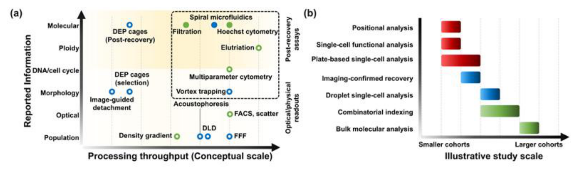
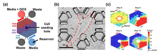
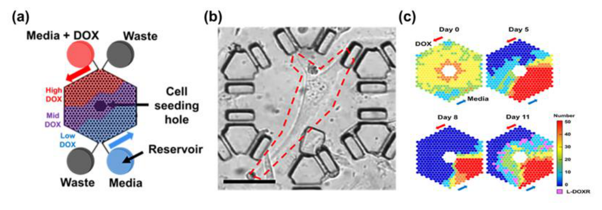
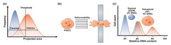
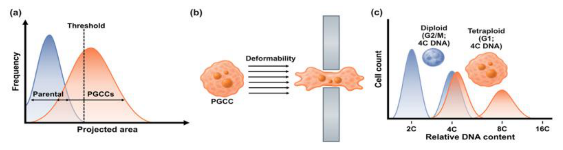
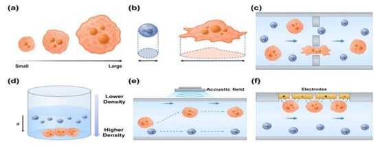
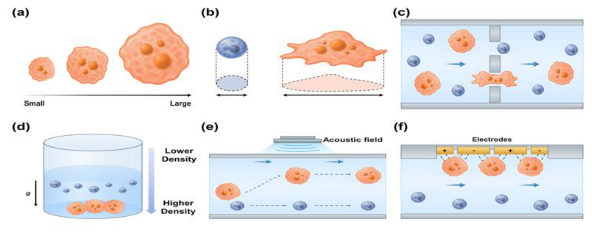
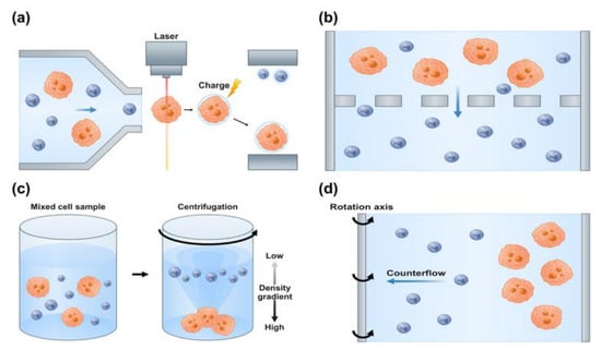
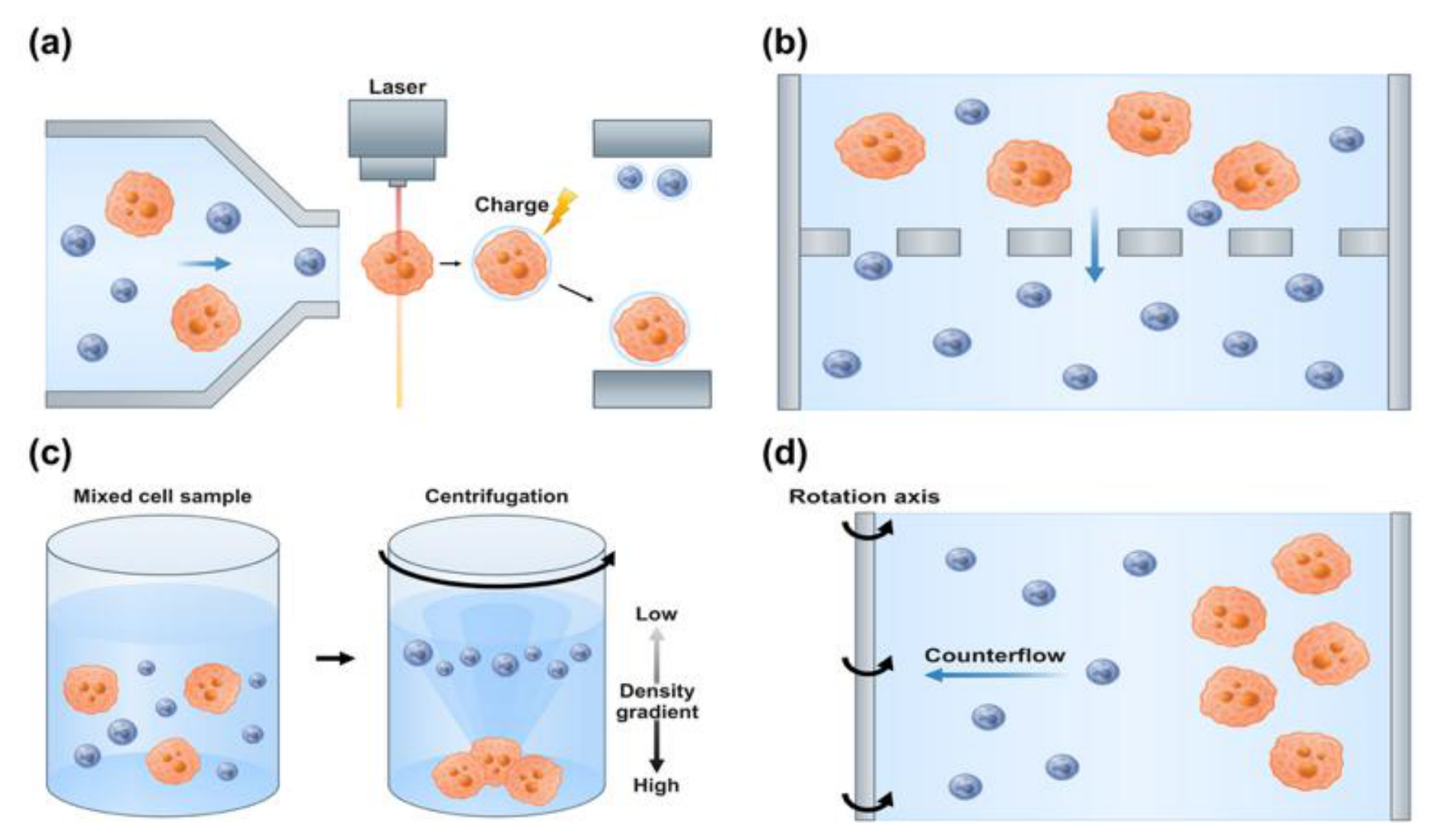
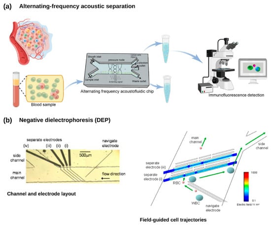
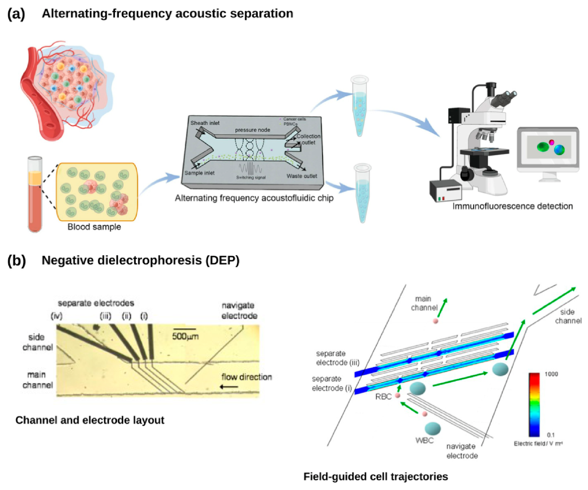

Disclaimer/Publisher’s Note: The statements, opinions and data contained in all publications are solely those of the individual author(s) and contributor(s) and not of MDPI and/or the editor(s). MDPI and/or the editor(s) disclaim responsibility for any injury to people or property resulting from any ideas, methods, instructions or products referred to in the content. |
© 2026 by the authors. Licensee MDPI, Basel, Switzerland. This article is an open access article distributed under the terms and conditions of the Creative Commons Attribution (CC BY) license.