


Abstract
Subcutaneous adipose tissue loss from trauma, congenital deformity, aging, or weight loss can compromise facial contour, symmetry, and soft-tissue support, making volume restoration a central target in soft-tissue reconstruction. Current approaches include autologous fat grafting, dermal fillers, and decellularized adipose matrices, each having distinct limitations related to tissue harvesting, material composition, durability, or absence of adipocyte structure. To address these limitations, dermaClaeTM was developed as a ready-to-use, minimally manipulated nonviable human structural adipose allograft for small volume applications in delicate areas, such as the face. This study characterizes the structural and compositional properties of dermaClae using histology, scanning electron microscopy (SEM), and biochemical assays to evaluate tissue structure, composition, and extracellular matrix (ECM) components relevant to tissue integration and remodeling. Histological and SEM analyses demonstrated adipocytes surrounded by collagen-rich connective tissue. Adipocyte cross-sectional area demonstrated overlapping distributions with native adipose tissue. Total collagen, laminin α4, proteoglycans, and elastin levels were all measured in dermaClae at concentrations higher than native adipose tissue. Retained adipocyte structures contribute to tissue bulk, cushioning, and support, while the ECM provides a structural framework relevant to incorporation and remodeling. Together, these characteristics may position dermaClae as a potential natural alternative to current approaches for facial reconstruction.
1. Introduction
Restoration of soft-tissue volume after loss of subcutaneous adipose tissue remains a challenge in reconstructive surgery. Such loss can result from trauma, congenital deformity, oncologic surgery, aging, or substantial weight loss and can alter contour, symmetry, and soft-tissue support [1,2,3]. Current approaches for volume restoration include autologous fat grafting, injectable dermal fillers, and tissue-derived biomaterials, each of which addresses volume loss differently [4]. The clinical challenge is not only to restore volume, cushioning, and support at the time of treatment, but also to achieve predictable volume retention over time.
Autologous fat grafting is widely regarded as the gold standard for soft-tissue volume restoration because it replaces lost tissue with the patient’s own adipose tissue and provides immediate volume, cushioning, and support [1,2,5,6,7]. Systematic reviews of facial fat grafting report high patient satisfaction and approximately 50% to 60% volume retention at one year [6,8]. However, retention remains variable. A separate meta-analysis reported facial fat retention ranging from 26% to 83%, with a pooled retention rate of 47% [7,9]. This variability is influenced by the viability of transferred adipocytes, harvesting and processing methods, injection technique, and perfusion at the recipient site [7,10,11]. Inadequate perfusion and delivery of oxygen and nutrients can compromise adipocyte survival and contribute to resorption and loss of correction [12,13,14]. Fat grafting also requires a surgical tissue-harvesting procedure and is limited by available adipose tissue volume at the donor site, and may result in donor-site pain, contour irregularities, and other morbidity [1,14,15,16]. As a result, fat autografting may lead to additional corrective procedures to achieve the intended outcome.
Injectable dermal fillers offer a less invasive option for localized volume restoration. These materials differ in composition and duration, with hyaluronic acid (HA) commonly used for office-based applications without tissue harvesting [17,18,19,20]. Because HA is gradually degraded, it commonly requires maintenance treatment, as do longer-lasting fillers such as calcium hydroxylapatite and poly-L-lactic acid, which may extend correction through host collagen deposition, with poly-L-lactic acid typically requiring a series of applications [21]. Longer-lasting fillers are available, but they do not replace lost adipose tissue or recreate its organized structure, which provides volume, cushioning, and support.
These limitations highlight the need for adipose-derived approaches that avoid surgical tissue harvest and provide sustained soft-tissue volume restoration, cushioning and support. Native adipose tissue provides more than volume; adipocytes account for most of its bulk, while connective tissue surrounds and organizes the adipocytes and contains the ECM, a noncellular framework of proteins and proteoglycans [22]. The ECM microstructure and interfacial properties can influence cell adhesion, morphology, and other cell-material interactions [23,24]. The ECM provides sites for host–cell attachment and migration into an applied material, enabling these cells to participate in remodeling [25,26]. Endothelial-cell migration and organization contribute to vascular ingrowth, which supplies oxygen and nutrients, while host progenitor cells may contribute to adipogenesis as remodeling progresses [27]. These processes have been associated with adipose graft integration and longer-term volume retention [12,13].
Many adipose-derived biomaterials used for facial soft-tissue reconstruction are decellularized and do not retain adipocytes [28]. Although these materials retain the ECM and can support host-mediated remodeling, they do not provide the immediate bulk volume and cushioning characteristic of native adipose tissue at the time of application. Instead, restoration of adipose volume occurs over time as host-derived adipocytes develop during remodeling [27,29,30]. An allograft retaining adipocytes with ECM-containing connective tissue could provide immediate volume and cushioning while preserving an ECM framework for subsequent host-mediated integration and remodeling. Developing such a shelf-stable allograft has been a challenge in tissue engineering [26]. Recently, a novel engineering approach was developed that enabled the production of a ready-to-use and off-the-shelf nonviable human adipose allograft, described in Fanniel et al., that retained both components [5]. In athymic mice, this allograft demonstrated site retention and host–cell infiltration, supporting host-mediated vascularization and formation of new adipocytes over six months [5].
Because that allograft was developed for larger-volume body applications that are not applicable for the face, the tissue engineering principles of the approach were applied in the development of dermaClae, a distinct nonviable human adipose allograft with a fine consistency that may provide volume and structural support for potential use in facial soft-tissue reconstruction. The present study evaluated the bulk appearance and composition, tissue structure, and key ECM components and found that dermaClae retained structural and compositional characteristics of native adipose tissue relevant to volume restoration, cushioning, and support. These retained characteristics provide a foundation for future investigation of how dermaClae may support cell attachment, angiogenesis, adipogenesis, and volume retention, all of which are relevant to successful soft-tissue reconstructive outcomes.
2. Materials and Methods
2.1. Production of Human-Derived Adipose Allografts
Adipose allografts were derived from 7 donated cadaveric human adipose tissues screened and recovered by FDA-registered tissue banks in accordance with the Association for Advancing Tissue and Biologics (AATB) standards and FDA regulations (21 CFR Part 1271). The donors for this study included both males and females, aged 18–79 years. Prior to processing, samples of native adipose tissue were collected for each donor and retained for selected assays defined in this study. The donor tissue was processed using a proprietary procedure in which the tissue was subjected to a freeze/thaw cycle, debrided, and processed through a series of rinses as previously described in Fanniel et al. [4], with additional mechanical size-reduction steps to produce an allograft capable of passing through a 0.66 mm diameter for finer applications. The allograft was generated using aseptic technique and underwent terminal sterilization using low-dose gamma-irradiation in compliance with ISO-11137 [31].
Following processing, the resulting dermaClae product was maintained at ambient temperature until evaluation. Native adipose tissue and the corresponding remaining processed allograft were evaluated using the assays described below. Native adipose tissue from one donor was insufficient to complete all assay-specific requirements; therefore, depending on sample availability, analyses included native tissue from 6 or 7 donors and processed allograft tissue samples from all 7 donors. The number of donors included is reported for each analysis.
2.2. Free Lipid Quantification
A method adapted from Fanniel et al. and An et al. [5,32] was used to quantify free lipids. Approximately 3 g of allografts were transferred to pre-weighed microcentrifuge tubes, and the total weight was recorded. Allografts were centrifuged at 3200× g for 3 min, after which the upper oil layer was removed and weighed. The percentage of free lipids was calculated as the proportion of the weight to the total sample weight. Analysis was conducted on 7 distinct donors, with 3 representative allografts.
2.3. Proportional Weight Composition
Approximately 3 g of native tissue and dermaClae allografts were dehydrated in an oven (COSORI, Orange, CA, USA, Cat. No. CP267-FD) set at 37 °C for 24 h. Post-dehydration, the weight loss was recorded for water content calculations. Samples were then soaked in 99% isopropyl alcohol (IPA) (VWR, Radnor, PA, USA, Cat. No. 89370-086) for 24 h on an orbital shaker (ThermoFisher Scientific, Waltham, MA, USA, Cat. No. 88861023) to extract lipids twice, with an IPA replacement between washes. Following lipid extraction, samples were air-dried for 24 h, and the final weight was recorded to determine lipid and connective tissue content. Analysis was conducted on 7 distinct donors, with 3 representative allografts.
2.4. Histological Analysis
Both native tissue and dermaClae allografts were fixed in 10% NBF (StatLab, McKinney, TX, USA, Cat. No. 28600-5). The cross-sections were embedded in paraffin, sectioned, and stained with Hematoxylin and Eosin (H&E) and Masson’s trichrome for histological analysis by the Precision Pathology Laboratory (San Antonio, TX, USA) in accordance with standard procedures. H&E staining was conducted on 7 matched native adipose tissue and dermaClae donors, each with 3 samples or allografts. Masson’s trichrome staining (MTS) was conducted on 7 distinct dermaClae donors, each with 3 allografts. Imaging was performed using a MoticEasyScan Pro 6 Microscope (Universal City, TX, USA).
2.5. Adipocyte Cross-Sectional Area and Analysis
Adipocyte size and distribution were assessed from whole-slide H&E cross-sections acquired from Section 2.4 using ImageJ Fiji’s (Version 2.18.0) (National Institutes of Health, Bethesda, MD, USA) Adiposoft (Version 1.16) extension. Five representative images were captured at 5× magnification per cross-section using SlideViewer (Version 2.9.0) (3DHISTECH, Budapest, Hungary), from the center of each section and four surrounding regions and exported as TIFF for analysis. Images were analyzed through Adiposoft using a minimum and maximum diameter of 25 µm and 130 µm, respectively, based on previously published methodology [33,34]. The minimum diameter cutoff is designed to exclude cellular components that are unlikely to be adipocytes, as described in Ramalho et al. [33,35]. The maximum diameter cutoff was used to exclude cellular clusters that can naturally occur during histological tissue preparation to ensure only individual adipocytes were measured [33]. Analysis was conducted on 6 donors of native adipose tissue and dermaClae allografts, with 3 representative samples or allografts per donor, each sample prepared in triplicate embedded tissue blocks for each sample or allograft, with 5 representative images captured per cross-section.
For cross-sectional area analysis, individual cell measurements were averaged within each tissue sample or allograft, and these sample-level averages were then averaged within each donor and tissue condition. This yielded one donor-level mean per tissue group (native or dermaClae), with donors representing the biological replicates. Native adipose tissue and dermaClae were compared using a paired t-test on these donor-level means (n = 6 matched donor pairs). Individual cells and samples from the same donor were not treated as independent biological replicates.
2.6. Scanning Electron Microscopy
Allograft samples were fixed in a 5 mL conical tube containing 4 mL of Karnovsky’s fixative solution (Electron Microscopy Sciences, Morgantown, PA, USA, Cat. No. 15720) and sent to the University of Texas Health Science Center San Antonio (UTHSCSA) for processing and imaging. Samples were rinsed with 0.1 M phosphate buffer before being placed in 1% Zetterquist’s osmium tetroxide for one hour. The samples were sequentially dehydrated in 70% ethanol for three cycles of 15 min, followed by 95% ethanol for three cycles of 15 min, and 100% ethanol for two cycles of 20 min. After dehydration, the samples were treated with hexamethyldisilazane (HMDS) for five minutes and allowed to air-dry in a desiccator. Once dried, they were mounted and sputter-coated with gold/palladium. Imaging was performed using a JEOL 6610 scanning electron microscope. Images were captured at 30 Kv voltage, SS50 spot size, and 10 mm working distance for each sample. Imaging was conducted on 7 distinct donors of dermaClae, with 3 representative allografts per donor.
2.7. Total Collagen Quantification
Collagen levels were determined using the QuickZyme Total Collagen Assay (QuickZyme Biosciences, Leiden, The Netherlands, Cat. No. QZBtotcol2), following the manufacturer’s instructions. Approximately 25 mg of tissue samples were digested in 1 mL of 6 M HCl (Sigma-Aldrich, Burlington, MA, USA, Cat. No. H1758-100ML) at 95 °C for 20 h. Post-digestion, the solution was centrifuged at 13,000× g for 10 min, and the supernatant was diluted to 4 M HCl. Collagen standards and allografts were mixed with an assay buffer, incubated at room temperature (RT) for 20 min on an orbital shaker at 16 RPM, then treated with detection reagent and incubated at 60 °C for 60 min. Absorbance was measured at 570 nm after cooling to RT. Analysis was conducted on 7 donors of native adipose tissue and dermaClae allografts with 3 representative samples or allografts per donor for each group in technical triplicate. Samples producing absorbance values above the upper range of the collagen standard curve were excluded from quantitative analysis because their concentrations could not be reliably interpolated, and this occurred for one native tissue sample from each of 3 donors.
2.8. Laminin Alpha-4 ELISA
Tissue samples (50–75 mg) were homogenized using a BeadBlaster 24R Tissue Homogenizer (Benchmark, Tempe, AZ, USA, Cat. No. D2400-R) in RIPA buffer (VWR, Radnor, PA, USA, Cat. No. AAJ63306-AP) supplemented with Halt Protease Inhibitor (ThermoFisher Scientific, Waltham, MA, USA, Cat. No. 78439) undergoing three cycles at 5 m/s for 30 s with 30 s rest intervals, followed by centrifugation at 7000× g for 15 min at 4 °C. Laminin α4 levels were assessed using the Human Laminin α4 ELISA Kit (ThermoScientific, Waltham, MA, USA, Cat. No. EH293RB), following the manufacturer’s instructions. Optical density values were analyzed for interpolation with a second-degree polynomial line best fit to generate standard curves. All samples’ absorbance values fell within the standard curve. Analysis was conducted on 6 donors of native adipose tissue and dermaClae with 3 representative samples or allografts per donor for each group in technical triplicate.
2.9. Total Proteoglycan Quantification
Proteoglycan content was assessed using the Blyscan-sulfated Glycosaminoglycan (sGAG) Assay Kit (BioColor, Belfast, UK, Cat. No. B100), following the manufacturer’s protocol. Briefly, tissue (40–50 mg) was digested in 1 mL of Papain Extraction Reagent (US Pharmacopeia, Rockville, MD, USA, Cat. No. 1495005) at 55 °C for 3 h. Post-digestion, the samples were centrifuged at 10,000× g for 10 min, and the supernatant was mixed with 1 mL of the Blyscan Dye Reagent. Samples were incubated in the thermomixer at 300 RPM for 30 min, then centrifuged at 13,000× g for 10 min, and again for 5 min. The supernatant was discarded, and the pellet was solubilized in 0.5 mL of the Dissociation Reagent. Absorbance was measured at 656 nm. All samples’ absorbance values fell within the standard curve. Analysis was conducted on 7 donors of native adipose tissue and dermaClae with 3 representative samples or allografts per donor for each group in technical triplicate.
2.10. Total Elastin Quantification
Elastin content was assessed using the Fastin Elastin Assay Kit (BioColor, Belfast, UK, Cat. No. F4000), following the manufacturer’s protocol. Briefly, tissue (25–35 mg) was hydrolyzed in 0.75 mL of 0.25 M oxalic acid for 1 h at 100 °C. Tissue samples were centrifuged at 13,000× g for 10 min to collect the supernatant; then, elastin was precipitated and centrifuged again to discard the supernatant. The precipitate was dyed, mixed, and incubated before the final centrifugation to remove unbound dye. The bound dye was dissolved and quantified at 513 nm. All samples’ absorbance values fell within the standard curve. Analysis was conducted on 6 donors of native adipose tissue and 7 donors of dermaClae with 3 representative samples or allografts per donor for each group in technical triplicate.
2.11. Data Analysis and Handling
All data were analyzed using GraphPad Prism (Version 10.2.3) (GraphPad Software, Boston, MA, USA). Comparative analyses were performed using a mixed-effects model, with tissue group (native or dermaClae) entered as the column factor and individual donor entered as the row factor. Three separate native tissue samples or dermaClae allografts were entered for each donor under each tissue condition. Donors represented the biological replicates, and the measurements from three separate samples or allografts within each donor and group represented technical replicates.
For all quantitative ECM-based assays, values falling outside the established assay-specific standard range were excluded from analysis because they could not be reliably quantified. No statistical outliers were identified using the ROUT method (Q = 1%). p-values less than 0.05 were considered statistically significant (* p < 0.05, ** p < 0.01, *** p < 0.001, **** p < 0.0001, ns = not significant). Exact p-values are reported when available; when GraphPad Prism reports only a significance threshold, the corresponding threshold is provided (e.g., p < 0.0001). Plots show the mean and range (minimum and maximum) representative at the donor level.
3. Results
3.1. dermaClaeTM Retains Key Bulk Characteristics of Adipose Tissue
Human donor-derived dermaClae allografts were evaluated to characterize the gross appearance, free lipid content, and proportional weight composition.
3.1.1. dermaClaeTM Retains the Natural Structural Appearance of Adipose Tissue
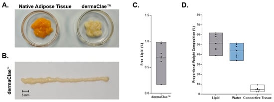
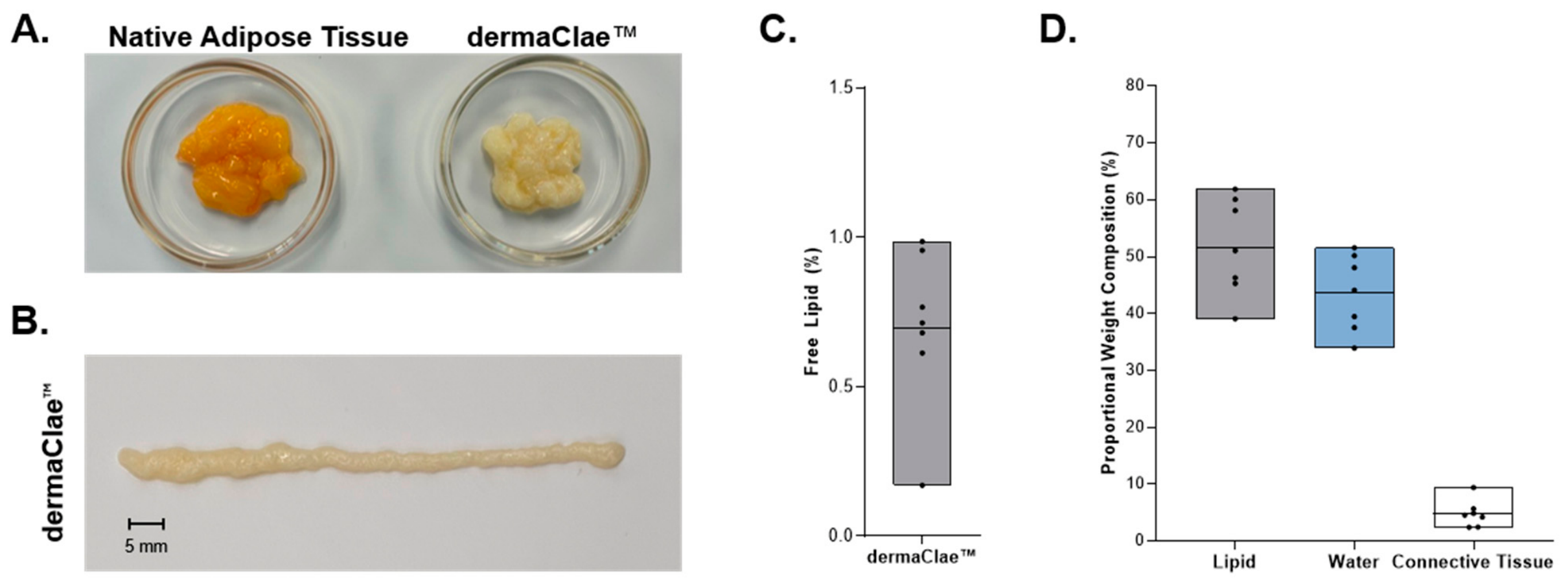
Macroscopic comparison demonstrated close visual comparability between dermaClae and native, unprocessed adipose tissue (Figure 1A). The donor-derived allografts retained the overall bulk form and gross structural appearance of native adipose tissue, with no discernible difference in composite organization. dermaClae exhibited lighter coloration, which is consistent with the removal of residual blood and free lipid from the donor tissue debris during debridement and serial rinsing. Figure 1B shows a representative extrusion of dermaClae after manual passage through a 0.66 mm diameter opening, where the allograft visibly maintained bulk appearance and overall structure of adipose tissue.
Figure 1.
Physical appearance and compositional analysis of dermaClaeTM. (A) Representative images of native adipose tissue alongside processed dermaClae. (B) Representative extrusion line of dermaClae after passage through a 0.66 mm diameter opening. (C) Mean percent free lipid content of dermaClae following centrifugation from 7 dermaClae donors, each with 3 independent allografts. (D) Proportional weight composition of dermaClae, based on analysis from 7 dermaClae donors, each with 3 independent allografts. Data are presented as floating bar graphs showing the minimum and maximum values, with a solid line indicating the mean across donors. Each individual data point represents a single donor.
3.1.2. Free Lipid Content Is Low in dermaClaeTM
Adipocytes store lipids within a single intracellular lipid droplet [36]. Disruption of adipocytes can release this lipid into the surrounding tissue, where it exists as a natural, unrestricted component of the native adipose tissue milieu. Quantification of free lipid therefore provides a measure of lipid present outside adipocytes and is commonly used to characterize fat lipoaspirate preparations used for fat autografting procedures [32,37]. Reported free lipid levels in lipoaspirates can be up to 15% for standard decanting techniques and 2% to 9% for wash and filtration systems [37,38,39,40].
Free lipid was separated from the bulk tissue by centrifugation without mechanical disruption of the allograft, as described in Section 2.2. The separated lipid was quantified relative to the initial allograft weight. This approach isolated the readily separable lipid fraction without releasing additional intracellular lipid during the analysis.
The minimally manipulated allograft exhibited an exceptionally low mean free lipid content of 0.70%, with individual values ranging from 0.05% to 1.69% (Figure 1C). Free lipid remained below 2% of the allograft weight in every sample evaluated. These findings demonstrate that free lipid constituted a minor proportion of dermaClae and remained below the ranges previously reported for lipoaspirate.
3.1.3. Macro Compositional Analysis: Establish the Bulk Composition of dermaClaeTM
To characterize the bulk composition of dermaClae, relative contributions of total lipid, aqueous, and connective tissue components were determined by weight. This analysis provided a more comprehensive assessment of total lipid content than the free lipid analysis, which quantified the unrestricted, extracellular lipid content separated from the bulk tissue by centrifugation.
The aqueous content of dermaClae was estimated from the change in allograft weight before and after dehydration and ranged from 32.18% to 57.38% of the initial weight. Total lipid, which includes free lipid, was estimated from the change in tissue weight before and after lipid extraction and ranged from 31.27% to 63.86%. The remaining tissue was dried and weighed and represented the connective tissue component, which accounted for 2.11% to 11.35% of the initial allograft weight (Figure 1D). For this analysis, connective tissue was defined as the remaining nonaqueous, nonlipid tissue. Collectively, these findings define the weight-based composition of dermaClae and the relative contributions of aqueous solution, total lipid, and connective tissue.
3.2. Structural Analysis Demonstrates Retention of Adipocyte and Connective Tissue in dermaClaeTM
Histological and ultrastructural imaging was performed to characterize the adipocyte and connective tissue structural integrity of donor-derived dermaClae allografts relative to native, unprocessed adipose tissue.
3.2.1. H&E Analysis Demonstrates the Retention of Adipocytes and Connective Tissue in dermaClaeTM
H&E staining was used to evaluate the overall tissue structure of dermaClae, focusing on the presence, organization, and cellular appearance of two major components of adipose tissue: adipocytes and connective tissue.
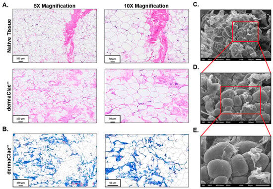
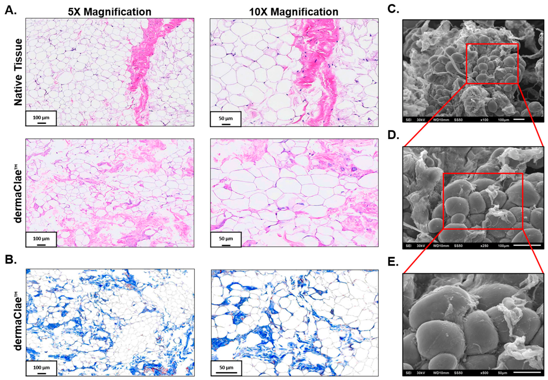
H&E-stained sections of dermaClae retained the characteristic honeycomb-like organization of native adipose tissue, with adipocyte morphology and continuous cellular margins distributed throughout the evaluated sections (Figure 2A). Eosin-stained connective tissue was present, interspersed across the adipocytes, maintaining the spatial organization and structural framework of adipose tissue. Compared with native adipose tissue, the overall tissue structure of dermaClae was visually comparable.
Figure 2.
Histological and ultrastructural assessments of dermaClaeTM. (A) Representative H&E images were selected from 7 matched native adipose tissue and dermaClae donors, each with 3 samples or allografts (n = 21 per group), with 5 representative fields of view per cross-section. (B) Representative Masson’s Trichrome-stained cross-section images of dermaClae were selected from 7 dermaClae donors, each with 3 allografts (n= 21), with 5 representative fields of view per cross-section. Scale bar: 100 µm (5× magnification) and 50 µm (10× magnification). (C–E) Representative SEM images of dermaClae showing adipocyte surface morphology and the connective tissue network at (C) 100×, (D) 250×, and (E) 500× magnification. Scale bar, 100 µm (100× and 250× magnification) and 50 µm (500× magnification). Red boxes in panels C and D indicated the regions shown at higher maginification in panels D and E, respectively. Representative images from 7 dermaClae donors, each with 3 independent allografts (n = 21), with 5 representative fields of view.
These findings demonstrate that dermaClae retained adipocytes and connective tissue structure characteristic of native adipose tissue and confirm that the lighter color of dermaClae is not associated with the removal or loss of adipocyte structure.
3.2.2. Collagen-Rich Connective Tissue Is Distributed Throughout dermaClaeTM
Collagen is a primary component of the adipose tissue connective tissue and contributes to its structural organization [41]. Although H&E staining demonstrated the presence of connective tissue within dermaClae, MTS was used to visualize the collagen-rich regions and distribution of connective tissue throughout the allograft.
Blue-stained collagen-rich connective tissue was observed surrounding the adipocyte structure across the evaluated sections of dermaClae (Figure 2B). The staining was evenly distributed in the allograft sections rather than concentrated within discrete regions. These findings demonstrate that collagen-rich connective tissue was retained as a broadly distributed network throughout dermaClae.
3.2.3. Scanning Electron Microscopy Demonstrates Adipocyte Morphology and Organized Connective Tissue Networks in dermaClaeTM
Histologic analysis provides a two-dimensional view of tissue cross-sections but offers limited visualization of surface features and connective tissue organization. SEM was therefore used to examine adipocyte surface characteristics and the structural arrangement of connective tissue within dermaClae at higher resolution with topographic depth of view.
SEM images revealed a distribution of spherical adipocyte structures characterized by smooth, continuous-appearing surface morphologies, consistent with previously reported SEM images of native adipose tissue (Figure 2C–E) [42,43]. Connective tissue extended throughout the allograft and appeared as both web-like and organized sheet-like structures. The observed distribution of these structures is consistent with the connective tissue distribution seen in the two-dimensional histological cross-sections (Figure 2A).
These findings provide higher-resolution evidence of adipocyte surface morphology and connective tissue network within dermaClae. Together with histological analysis, these findings demonstrate that dermaClae retains key structural features characteristic of adipose tissue.
3.3. dermaClaeTM Retains the Native Dimensions and Size Distributions of Adipocytes
Adipocyte size varies within native adipose tissue [44], and comparison of average values alone may not identify differences in the relative frequency of smaller and larger adipocytes. Adipocyte size was therefore evaluated to determine whether dermaClae retained the range and overall dimensions of adipocytes observed in native, unprocessed adipose tissue. Adipocyte cross-sectional area was quantified from H&E-stained sections using the Adiposoft plugin in ImageJ Fiji for the image-analysis method described in Section 2.5.
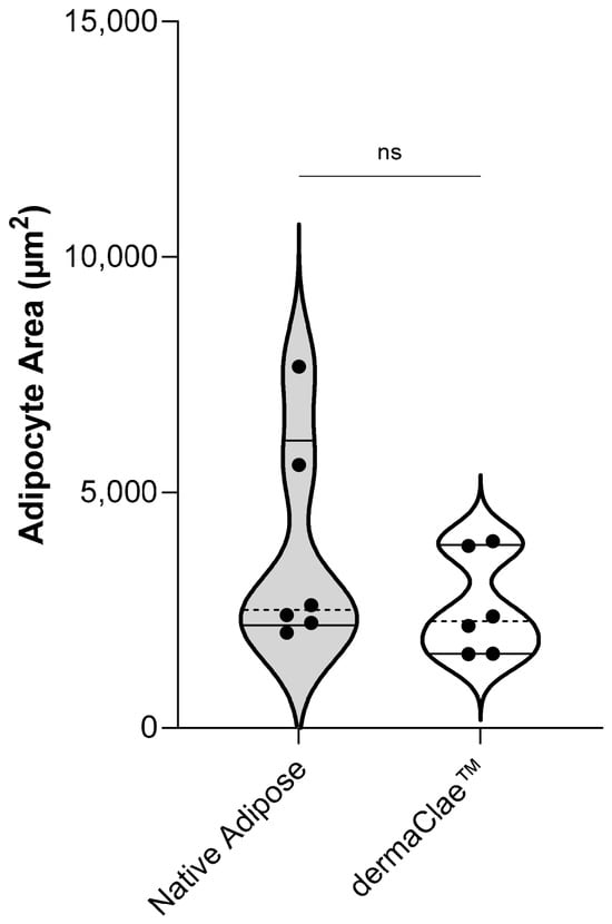
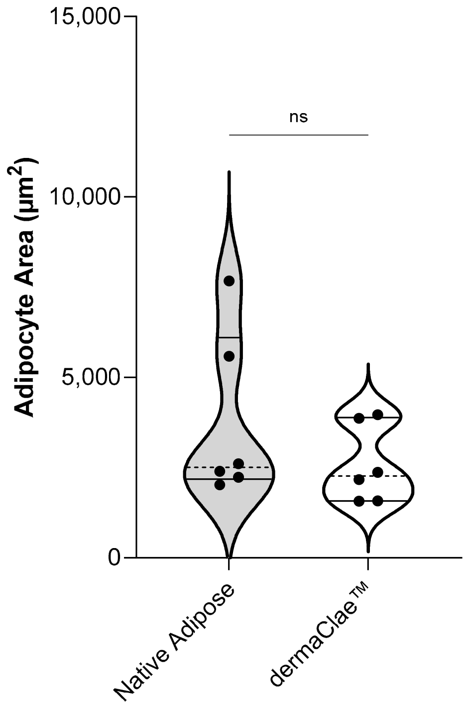
Mean adipocyte cross-sectional area was 2584 ± 1082 µm2 for dermaClae and 3743 ± 2336 µm2 for native adipose tissue (Figure 3), with no statistically significant difference detected between the two groups (p = 0.0764). The violin plots show overlap in the distributions of adipocyte cross-sectional area measurements, particularly within the range extending up to the 75th percentile of adipose native tissue. Native adipose tissue exhibited a broader distribution extending toward larger cross-sectional areas, with higher donor-level mean values observed in a subset of donors, consistent with inherent donor-to-donor variability. The medians of donor-level means were 2270 µm2 for dermaClae and 2504 µm2 for native adipose, showing smaller differences between groups than observed for the means. Collectively, these data demonstrate similarities in the adipocyte size distribution profiles of dermaClae and native adipose tissue while also capturing the broader variability present in native tissue.
Figure 3.
Adipocyte cross-sectional measurements in native adipose and dermaClaeTM. Cross-sectional area (µm2) of adipocytes measured with 5 representative fields of view examined per cross-section matched native adipose tissue and dermaClae. Representative data are from 6 native adipose tissue and dermaClae donors (not statistically significant; p = 0.0764), each with 3 independent samples or allografts. Adipocyte segmentation was performed using the Adiposoft plugin in Fiji ImageJ. Data are presented as a violin plot; dotted lines indicate the median of representative surface area values per donor and solid lines indicate the interquartile range. Each data point represents the donor-level mean (n = 6 donors per group).
3.4. dermaClaeTM Retains Key Extracellular Matrix Components Native to Adipose Tissue
Histological and ultrastructural analyses demonstrated that connective tissue was distributed throughout dermaClae. Because the ECM forms the non-cellular structural component of connective tissue, additional analyses were performed to define its composition and determine whether key ECM components were retained in dermaClae. Total collagen, laminin α4, total proteoglycans, and total elastin were selected because they are established components of adipose tissue ECM and are commonly evaluated in tissue-derived biomaterials due to their relevance to tissue integration and remodeling [45,46].
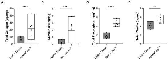
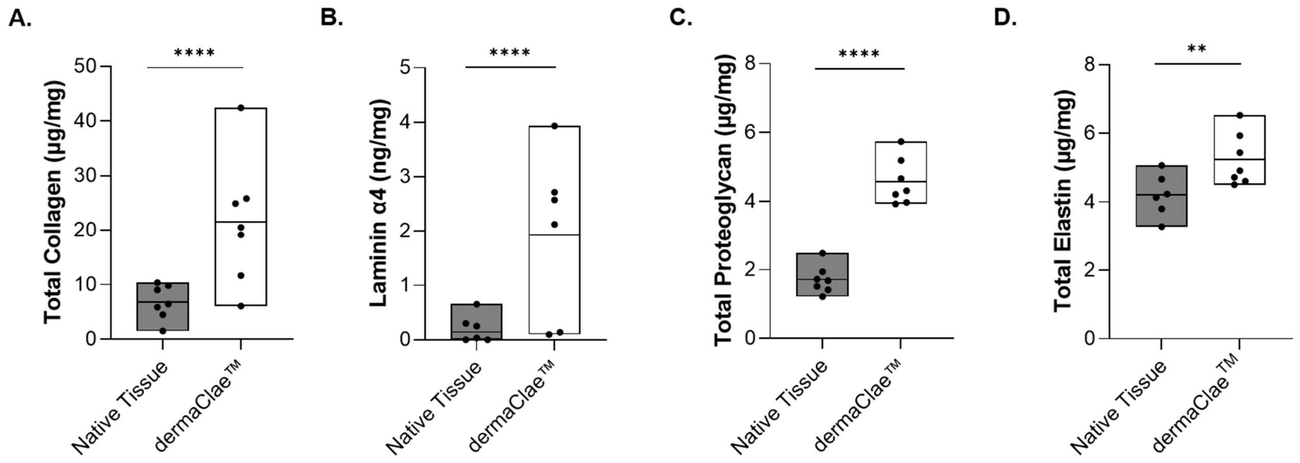
ECM components were quantified relative to the initial wet sample weight analyzed for both dermaClae and native adipose tissue. Mean ± standard deviation total collagen, laminin α4, and proteoglycan content were all measured on a wet-tissue-weight basis in dermaClae and native adipose tissue. Mean collagen concentration was 20.60 ± 8.31 µg/mg in dermaClae versus 6.54 ± 4.06 µg/mg in native adipose tissue (Figure 4A), mean laminin α4 at 1.69 ± 1.46 ng/mg versus 0.21 ± 0.31 ng/mg (Figure 4B), mean proteoglycan content at 4.65 ± 0.71 µg/mg versus 2.08 ± 0.41 µg/mg (Figure 4C), and mean elastin content at 5.24 ± 0.92 µg/mg and native adipose tissue with 4.16 ± 0.81 µg/mg (Figure 4D).
Figure 4.
Extracellular matrix protein analysis in native adipose tissue and dermaClaeTM. (A) Total collagen (µg/mg) (****; p < 0.0001), (B) laminin α4 (ng/mg) (****; p < 0.0001), (C) total proteoglycan (µg/mg) (****; p < 0.0001), (D) total elastin (µg/mg) (**; p = 0.0046) in native adipose tissue and dermaClae. Total collagen and proteoglycan representative data from 7 native adipose tissue and dermaClae donors, evaluated in technical triplicate. Laminin α4 and total elastin representative data from 6 native adipose tissue and 7 dermaClae donors, evaluated in technical triplicate. Data are presented as floating bar graphs showing the minimum and maximum values, with a solid line indicating the mean across donors. Each individual data point represents a single donor.
All four ECM components were detected in dermaClae, demonstrating their retention within the final allograft. Together with the histological and ultrastructural findings, these results demonstrate that the connective tissue component of dermaClae contains multiple ECM components characteristic of adipose tissue. The differences in measured concentrations further characterize the relative abundance of these components within the final allograft compared with native adipose tissue.
4. Discussion
This study characterizes dermaClae, a nonviable human adipose allograft, and demonstrates that the final allograft retains several of the hallmark structural and compositional characteristics of native adipose tissue. This evaluation consisted of bulk appearance, including after extrusion through a 0.66 mm diameter, composition, histological and ultrastructural organization, quantitative analysis of adipocytes, and quantitation of key ECM components. These assessments characterized the structural and compositional features of dermaClae that are relevant to providing volume, cushioning, and structural support. Characterization of these features provides a foundation for subsequent preclinical evaluation of the allograft for tissue-grafting applications, including facial reconstruction, where restoration of localized soft-tissue volume and support is clinically relevant.
One of the most notable findings was that dermaClae exhibited compositional and morphological characteristics consistent with native adipose tissue. Proportional-weight analysis confirmed a compositional profile comprising water, lipid, and connective tissue, which are major constituents of native adipose tissue [41]. Free lipids averaged 0.7%, indicating that most measured lipids in the proportional weight analysis remained associated with the bulk tissue. Separately, histological and SEM analyses confirmed the presence of adipocytes and connective tissue, and quantitative image analysis established their relative surface-area distribution within tissue cross-sections. These findings demonstrate retention of the bulk appearance, structural organization, and major constituents of the source tissue following processing.
Unlike conventional dermal fillers and matrix-only adipose allografts, dermaClae retains adipocytes and connective tissue with water. Adipocytes contribute to tissue bulk and cushioning, while connective tissue forms a continuous framework that organizes the adipocytes and helps maintain the overall form of the tissue [22,47]. These characteristics provide a structural basis for the potential use of dermaClae for restoration of soft-tissue volume and form.
Previous studies have demonstrated the importance of adipose tissue organization [48]. In one preclinical study, structurally organized fat lipoaspirate demonstrated greater volume retention and vascularization, with less fibrosis, than unstructured lipoaspirate [49]. A separate study evaluated an inactivated adipose tissue model in which nearly all cells were nonviable while spatial structure remained intact [50]. The investigators concluded that intact adipose tissue structure provided a local framework for adipogenesis and angiogenesis following application [50]. These studies suggest that native adipose tissue structure organization may contribute to host-mediated remodeling and maintenance of grafted tissue.
The connective tissue retained in dermaClae was further characterized through analysis of its ECM composition. The ECM is the non-cellular network of proteins and proteoglycans that surrounds and organizes cells within adipose tissue [22]. Total collagen, laminin α4, total proteoglycan, and total elastin were quantified to determine whether these components remained in the final allograft. Biochemical analysis confirmed retention of all four components. These findings demonstrate that key ECM components characteristic of native adipose tissue are retained in dermaClae and that some may occur at higher concentrations per unit tissue weight relative to native adipose tissue.
The ECM profile retained in dermaClae is important for the collective contribution of its individual components to the tissue microenvironment. Importantly, ECM function is influenced not only by its biochemical composition, but also by its physicochemical properties and microstructural organization. Features such as geometry, topography, and mechanical characteristics can influence cell adhesion, morphology, cytoskeletal organization, and subsequent cellular responses [24]. These structural and biochemical features of the ECM are also associated with host–cell attachment and infiltration, vascular ingrowth, and progressive tissue remodeling [24,25]. These host-mediated processes are fundamental to successful long-term graft retention [12,13]. Collagen is a major structural component of adipose connective tissue and contains physical sites available for host–cell attachment [51,52]. MTS showed that collagen-rich connective tissue was distributed throughout dermaClae rather than concentrated in discrete regions, demonstrating a broadly distributed connective tissue framework. Studies of collagen-based matrices used for soft-tissue augmentation have reported host–cell and blood-vessel ingrowth together with maintenance of augmented tissue volume [53]. Within this connective tissue framework, laminin α4 is a component of the basement membranes surrounding adipocytes and blood vessels [54]. These basement membranes provide physical attachment sites for endothelial and other host cells [55], and loss of laminin α4 has been associated with disruption of vascular basement membranes and impaired microvessel maturation [54]. Laminin-containing basement membranes are also characteristic of the adipocyte microenvironment and undergo reorganization during adipose tissue development and remodeling [56].
Elastin and proteoglycans provide additional properties of the native adipose ECM. Elastin provides flexibility and elasticity to soft tissue [57] and, in a canine soft-tissue augmentation model, elastin within a collagen-elastin matrix persisted for up to 90 days and became incorporated into newly formed connective tissue [52]. The retention of these proteins in dermaClae is relevant to host–cell attachment and vascularization; however, their contribution to the mechanical or biological behavior of dermaClae was not directly evaluated in the present study. Proteoglycans contribute to the hydrated state of connective tissue through their glycosaminoglycan chains, including sulfated glycosaminoglycans (sGAGs), which have a high capacity to bind water [58,59]. This water-binding property contributes to the hydrated physical state and bulk of adipose tissue [58]. Further, sGAGs interact with ECM proteins and bind signaling molecules involved in cell migration, vascular remodeling, and tissue repair [58,59,60]. Collectively, collagen, laminin α4, elastin, and proteoglycans comprise complementary structural and compositional elements of native adipose ECM that define the tissue environment in which host-mediated incorporation and remodeling occur.
Studies of adipose-derived allografts demonstrate the relationship between retained ECM components, host-mediated tissue remodeling, and volume retention [26,61]. In a rodent model, higher volume retention was associated with greater CD-31-positive angiogenesis and adipogenesis. Retention increased from 25% to 60% at 8 weeks, while adipose tissue area increased from 12% to 51% [61]. Decellularized human adipose allografts retaining key ECM components, including collagen and elastin, have also demonstrated material retention through 24 weeks in mice and volume retention through 16 weeks in a human dorsal wrist study within the range described for autologous fat grafting, with histologic evidence of perilipin-positive adipocytes and vascularity [27]. Importantly, a related nonviable adipose allograft intended for larger-volume body applications demonstrated retention, host–cell infiltration, vascularization, integration, and formation of new adipocytes through six months in athymic mice [5]. Collectively, these findings provide compelling evidence that the composition and organization of adipose-derived allografts could support the same host-mediated processes associated with soft tissue volume maintenance.
The present study establishes that dermaClae retains the bulk appearance and major structural and compositional characteristics of adipose tissue, including adipocyte morphology organized in a honeycomb-like network supported by connective tissue and key ECM components. The broader tissue matrices literature indicates that ECM composition, microstructure, and material interfaces can influence cell-material interactions and subsequent cellular responses, processes associated with successful long-term soft-tissue reconstructive outcomes [5,28,30], providing rationale for further investigation of these characteristics in dermaClae. This study is limited to the bulk appearance, tissue morphology, and extracellular matrix composition only. Subsequent preclinical studies are needed to determine how these characteristics translate to biological and reconstructive performance, including host tissue response, long-term tissue integration, quantitative injectability, and delivery performance.
5. Conclusions
This study characterized the bulk appearance, tissue morphology, and extracellular matrix composition of dermaClae, a newly developed, off-the-shelf, nonviable human adipose allograft. The findings demonstrated that dermaClae retains the bulk appearance and major structural and compositional characteristics of adipose tissue, including adipocyte morphologies distributed in a honeycomb-like network supported by connective tissue and key ECM components. By preserving these components, the allograft retains the physical components responsible for immediate volume restoration, cushioning, and structural support, together with ECM components relevant to host-mediated integration and remodeling. Its ready-to-use, off-the-shelf configuration and fine consistency may allow for application in areas, such as the face and hands, positioning dermaClae as a promising alternative to currently available technologies and biomaterials for soft tissue reconstruction. Because this study was limited to profile data, functional outcomes such as cell attachment, angiogenesis, adipogenesis, integration, remodeling, and long-term volume retention remain to be evaluated in future preclinical and clinical outcomes.
6. Patents
The subject matter of this manuscript is patent-pending and the intellectual property of RegenTX Partners, LLC.
Author Contributions
Conceptualization, S.H. and J.M.; methodology, H.B., V.F., S.H., J.M., P.S. and K.T.M.; formal analysis, H.B., V.F., P.S. and K.T.M.; investigation, H.B., V.F., A.R.K., P.S., K.T.M. and D.V.C.; resources, H.B., S.H. and J.M.; data curation, H.B., V.F., P.S. and K.T.M.; writing—original draft preparation, S.H., A.R.K., J.M. and D.V.C.; writing—review and editing, H.B., V.F., S.H., J.M., P.S. and K.T.M.; visualization, S.H.; supervision, H.B. and S.H.; project administration, S.H. and J.M.; funding acquisition, S.H. and J.M. All authors have read and agreed to the published version of the manuscript.
Funding
This research was financially supported by RegenTX Partners, LLC.
Institutional Review Board Statement
Ethical review and approval were waived for human tissue as the protocol followed did not involve clinical investigation per 21 CFR 56.102, nonclinical laboratory study per 21 CFR 58.3, biologic, drug, or device. Therefore, the information gathered during the examination of the adipose allograft under development did not require having an IRB or IND review and approval.
Informed Consent Statement
The allografts were recovered and manufactured in accordance with 21 CFR 1271 and meet the criteria regulated in Section 361 of the Public Health Service Act. The protocol was performed utilizing human tissue from cadaveric donation with authorization permitting use in research, in accordance with the AATB Standards, 15th edition, Section B5.100 Release for Research. The protocol followed did not involve clinical investigation per 21 CFR 56.102, Nonclinical laboratory study, per 21 CFR 58.3, Biologic, drug, or device.
Data Availability Statement
The data presented in this study are available upon request from the corresponding author.
Acknowledgments
We extend our appreciation to Ihab Atawneh, Jonathan Savoie, Cristina Rodriguez, Analaura Villarreal Berain and Jeffrey Harris for their technical support, and to Tracy Bui for coordinating study materials. We acknowledge the work of Precision Pathology Laboratory for their histological services and imaging core facility at the University of Texas Health Science Center at San Antonio for SEM imaging. We also extend our gratitude to the entire team at RegenTX Partners, LLC. for their collective support and dedication.
Conflicts of Interest
V.F., J.M., P.S., K.T.M., and S.H. are employed by RegenTX Partners, LLC. H.B., A.R.K., and D.V.C. are not employed by RegenTX Partners, LLC.
Abbreviations
The following abbreviations are used in this manuscript:
| AATB | Association for Advancing Tissue and Biologics |
| CFR | Code of Federal Regulations |
| ECM | Extracellular Matrix |
| ELISA | Enzyme-Linked Immunosorbent Assay |
| FDA | Federal Drug Administration |
| H&E | Hematoxylin and Eosin |
| HA | Hyaluronic Acid |
| HCl | Hydrochloric Acid |
| HMDS | Hexamethyldisilane |
| IPA | Isopropyl Alcohol |
| mg | Milligram |
| MTS | Masson’s Trichrome Staining |
| NBF | Neutral Buffered Formalin |
| ng | Nanogram |
| ns | Not Significant |
| RIPA | Radioimmunoprecipitation Assay Buffer |
| ROUT | Robust Regression and Outlier Removal |
| RPM | Rotations Per Minute |
| SEM | Scanning Electron Microscopy |
| sGAG | Sulfated Glycosaminoglycan |
| Tiff | Tagged Image File Format |
| UTHSCSA | University of Texas Health Science Center San Antonio |
| µg | Microgram |
| µm | Micrometer |
References
- El-Mallah, J.C.; Wen, C.; Waldron, O.; Jikaria, N.R.; Asgardoon, M.H.; Schlidt, K.; Goldenberg, D.; Horchler, S.; Landmesser, M.E.; Park, J.H.; et al. Current Modalities in Soft-Tissue Reconstruction and Vascularized Adipose Engineering. Biomolecules 2025, 15, 780. [Google Scholar] [CrossRef] [Scilit] [PubMed]
- Mahoney, C.M.; Imbarlina, C.; Yates, C.C.; Marra, K.G. Current Therapeutic Strategies for Adipose Tissue Defects/Repair Using Engineered Biomaterials and Biomolecule Formulations. Front. Pharmacol. 2018, 9, 507. [Google Scholar] [CrossRef] [Scilit] [PubMed]
- Yang, J.; Zhou, C.; Fu, J.; Yang, Q.; He, T.; Tan, Q.; Lv, Q. In situ Adipogenesis in Biomaterials Without Cell Seeds: Current Status and Perspectives. Front. Cell Dev. Biol. 2021, 9, 647149. [Google Scholar] [CrossRef] [Scilit] [PubMed]
- Gold, M.; Biesman, B.; Cohen, J.; Day, D.; Goldberg, D.; Guenin, S.; Lain, T.; Schlesinger, T.; Shamban, A.; Chilukuri, S. Real-World Clinical Experience With an Allograft Adipose Matrix for Replacing Volume Loss in Face, Hands, and Body. J. Cosmet. Dermatol. 2024, 23, 1–9. [Google Scholar] [CrossRef] [Scilit] [PubMed]
- Fanniel, V.; Atawneh, I.; Savoie, J.; Izaguirre-Ramirez, M.; Marquez, J.; Khorsandi, C.; Hill, S. Advancing Soft Tissue Reconstruction with a Ready-to-Use Human Adipose Allograft. Bioengineering 2025, 12, 612. [Google Scholar] [CrossRef] [Scilit] [PubMed]
- Krastev, T.K.; Beugels, J.; Hommes, J.; Piatkowski, A.; Mathijssen, I.; van der Hulst, R. Efficacy and Safety of Autologous Fat Transfer in Facial Reconstructive Surgery: A Systematic Review and Meta-analysis. JAMA Facial Plast. Surg. 2018, 20, 351–360. [Google Scholar] [CrossRef] [Scilit] [PubMed]
- Acharya, P.; Mohammed, C.; Desai, A.; Rojas Gomez, M.C.; Sunil, G.; Duran, S.P.; Kocaekiz, S.; Thottakurichi, A.A.; Munoz, I.J.G.; Chavez-Alvarez, L.A.; et al. Maximizing the Longevity and Volume Retention of Fat Grafts: Advances in Clinical Practice. Cureus 2025, 17, e88493. [Google Scholar] [CrossRef] [Scilit] [PubMed]
- Groen, J.W.; Krastev, T.K.; Hommes, J.; Wilschut, J.A.; Ritt, M.; van der Hulst, R. Autologous Fat Transfer for Facial Rejuvenation: A Systematic Review on Technique, Efficacy, and Satisfaction. Plast. Reconstr. Surg. Glob. Open 2017, 5, e1606. [Google Scholar] [CrossRef] [Scilit] [PubMed]
- Lv, Q.; Li, X.; Qi, Y.; Gu, Y.; Liu, Z.; Ma, G.E. Volume Retention After Facial Fat Grafting and Relevant Factors: A Systematic Review and Meta-analysis. Aesthetic Plast. Surg. 2021, 45, 506–520. [Google Scholar] [CrossRef] [Scilit] [PubMed]
- Sommer, B.; Sattler, G. Current concepts of fat graft survival: Histology of aspirated adipose tissue and review of the literature. Dermatol. Surg. 2000, 26, 1159–1166. [Google Scholar] [CrossRef]
- Varghese, J.; Griffin, M.; Mosahebi, A.; Butler, P. Systematic review of patient factors affecting adipose stem cell viability and function: Implications for regenerative therapy. Stem Cell Res. Ther. 2017, 8, 45. [Google Scholar] [CrossRef] [Scilit] [PubMed]
- Eto, H.; Kato, H.; Suga, H.; Aoi, N.; Doi, K.; Kuno, S.; Yoshimura, K. The fate of adipocytes after nonvascularized fat grafting: Evidence of early death and replacement of adipocytes. Plast. Reconstr. Surg. 2012, 129, 1081–1092. [Google Scholar] [CrossRef] [Scilit] [PubMed]
- Landau, M.J.; Birnbaum, Z.E.; Kurtz, L.G.; Aronowitz, J.A. Review: Proposed Methods to Improve the Survival of Adipose Tissue in Autologous Fat Grafting. Plast. Reconstr. Surg. Glob. Open 2018, 6, e1870. [Google Scholar] [CrossRef] [Scilit] [PubMed]
- Homer, N.A.; Patel, A.S.; Epstein, A.; Durairaj, V.D.; Nakra, T. Autologous Fat Transfer Harvest-site Complications: Incidence, Risk Factors, and Management. Ophthalmic Plast. Reconstr. Surg. 2022, 38, 65–67. [Google Scholar] [CrossRef] [Scilit] [PubMed]
- Karen, B.; Makayla, M.; Tanuj, N. Complications of facial autologous fat grafting. Plast. Aesthetic Res. 2024, 11, 7. [Google Scholar] [CrossRef] [Scilit]
- Wederfoort, J.L.M.; Hebels, S.A.; Heuts, E.M.; van der Hulst, R.; Piatkowski, A.A. Donor site complications and satisfaction in autologous fat grafting for breast reconstruction: A systematic review. J. Plast. Reconstr. Aesthet. Surg. 2022, 75, 1316–1327. [Google Scholar] [CrossRef] [Scilit] [PubMed]
- de Castro Costa, M.; Andrade, C.A.; Dantas, R.V.F.; Germani, M.; Buzalaf, M.A.R.; Soares, D.G. Clinical Durability of Hyaluronic Acid-Based Dermal Fillers for Facial Application: A Systematic Review. Aesthetic Plast. Surg. 2026, 50, 1971–1993. [Google Scholar] [CrossRef] [Scilit] [PubMed]
- de Vries, C.G.; Geertsma, R.E. Clinical data on injectable tissue fillers: A review. Expert Rev. Med. Devices 2013, 10, 835–853. [Google Scholar] [CrossRef] [Scilit] [PubMed]
- Sanchez-Carpintero, I.; Candelas, D.; Ruiz-Rodriguez, R. Dermal fillers: Types, indications, and complications. Actas Dermosifiliogr. 2010, 101, 381–393. [Google Scholar] [CrossRef] [Scilit] [PubMed]
- Kaufman-Janette, J.; Taylor, S.C.; Cox, S.E.; Weinkle, S.H.; Smith, S.; Kinney, B.M. Efficacy and safety of a new resilient hyaluronic acid dermal filler, in the correction of moderate-to-severe nasolabial folds: A 64-week, prospective, multicenter, controlled, randomized, double-blind and within-subject study. J. Cosmet. Dermatol. 2019, 18, 1244–1253. [Google Scholar] [CrossRef] [Scilit] [PubMed]
- Huang, Y.; Ma, X.; Guo, J. Calcium Hydroxylapatite for Facial Rejuvenation: Properties, Mechanisms, and Clinical Applications. Tissue Eng. Part B Rev. 2026. [Google Scholar] [CrossRef] [Scilit] [PubMed]
- Divoux, A.; Clement, K. Architecture and the extracellular matrix: The still unappreciated components of the adipose tissue. Obes. Rev. 2011, 12, e494–e503. [Google Scholar] [CrossRef] [Scilit] [PubMed]
- Guilak, F.; Cohen, D.M.; Estes, B.T.; Gimble, J.M.; Liedtke, W.; Chen, C.S. Control of stem cell fate by physical interactions with the extracellular matrix. Cell Stem Cell 2009, 5, 17–26. [Google Scholar] [CrossRef] [Scilit] [PubMed]
- Cramer, M.C.; Badylak, S.F. Extracellular Matrix-Based Biomaterials and Their Influence Upon Cell Behavior. Ann. BioMed. Eng. 2020, 48, 2132–2153. [Google Scholar] [CrossRef] [Scilit] [PubMed]
- Badylak, S.F. The extracellular matrix as a biologic scaffold material. Biomaterials 2007, 28, 3587–3593. [Google Scholar] [CrossRef] [Scilit] [PubMed]
- Flynn, L.E. The use of decellularized adipose tissue to provide an inductive microenvironment for the adipogenic differentiation of human adipose-derived stem cells. Biomaterials 2010, 31, 4715–4724. [Google Scholar] [CrossRef] [Scilit] [PubMed]
- Kokai, L.E.; Sivak, W.N.; Schilling, B.K.; Karunamurthy, A.; Egro, F.M.; Schusterman, M.A.; Minteer, D.M.; Simon, P.; D’Amico, R.A.; Rubin, J.P. Clinical Evaluation of an Off-the-Shelf Allogeneic Adipose Matrix for Soft Tissue Reconstruction. Plast. Reconstr. Surg. Glob. Open 2020, 8, e2574. [Google Scholar] [CrossRef] [Scilit] [PubMed]
- Anderson, A.E.; Wu, I.; Parrillo, A.J.; Wolf, M.T.; Maestas, D.R., Jr.; Graham, I.; Tam, A.J.; Payne, R.M.; Aston, J.; Cooney, C.M.; et al. An immunologically active, adipose-derived extracellular matrix biomaterial for soft tissue reconstruction: Concept to clinical trial. npj Regen. Med. 2022, 7, 6. [Google Scholar] [CrossRef] [Scilit] [PubMed]
- Gold, M.H.; Kinney, B.M.; Kaminer, M.S.; Rohrich, R.J.; D’Amico, R.A. A multi-center, open-label, pilot study of allograft adipose matrix for the correction of atrophic temples. J. Cosmet. Dermatol. 2020, 19, 1044–1056. [Google Scholar] [CrossRef] [Scilit] [PubMed]
- Ostadi, Y.; Khanali, J.; Tehrani, F.A.; Yazdanpanah, G.; Bahrami, S.; Niazi, F.; Niknejad, H. Decellularized Extracellular Matrix Scaffolds for Soft Tissue Augmentation: From Host-Scaffold Interactions to Bottlenecks in Clinical Translation. Biomater. Res. 2024, 28, 0071. [Google Scholar] [CrossRef] [Scilit] [PubMed]
- ISO 11137-2; Sterilization of Health Care Products–Radiation–Part 2: Establishing the Sterilization Dose. International Organization for Standardization: Geneva, Switzerland, 2013.
- An, Y.; Panayi, A.C.; Mi, B.; Fu, S.; Orgill, D.P. Comparative Analysis of Two Automated Fat-processing Systems. Plast. Reconstr. Surg. Glob. Open 2020, 8, e2587. [Google Scholar] [CrossRef] [Scilit] [PubMed]
- Ramalho, A.; Gauthier, M.-F.; Maltais-Payette, I.; Ostinelli, G.; Hould, F.; Biertho, L.; Tchernof, A. High-throughput measurement of adipocyte size with open-source software using whole-slide adipose tissue images. Adipocyte 2025, 14, 2528437. [Google Scholar] [CrossRef] [Scilit] [PubMed]
- Galarraga, M.; Campion, J.; Munoz-Barrutia, A.; Boque, N.; Moreno, H.; Martinez, J.A.; Milagro, F.; Ortiz-de-Solorzano, C. Adiposoft: Automated software for the analysis of white adipose tissue cellularity in histological sections. J. Lipid Res. 2012, 53, 2791–2796. [Google Scholar] [CrossRef] [Scilit] [PubMed]
- Yang Loureiro, Z.; Solivan-Rivera, J.; Corvera, S. Adipocyte Heterogeneity Underlying Adipose Tissue Functions. Endocrinology 2022, 163, bqab138. [Google Scholar] [CrossRef] [Scilit] [PubMed]
- Chu, M.; Sampath, H.; Cahana, D.Y.; Kahl, C.A.; Somwar, R.; Cornea, A.; Roberts, C.T., Jr.; Varlamov, O. Spatiotemporal dynamics of triglyceride storage in unilocular adipocytes. Mol. Biol. Cell 2014, 25, 4096–4105. [Google Scholar] [CrossRef] [Scilit] [PubMed]
- Fang, C.; Patel, P.; Li, H.; Huang, L.T.; Wan, H.; Collins, S.; Connell, T.L.; Xu, H. Physical, Biochemical, and Biologic Properties of Fat Graft Processed via Different Methods. Plast. Reconstr. Surg. Glob. Open 2020, 8, e3010. [Google Scholar] [CrossRef] [Scilit] [PubMed]
- Gabriel, A.; Kabaria, N.; Fang, C.H.; Lombardi, J.A.; Stec, E.; Huang, L.T.; Li, H.; Sandor, M. In Vitro Characterization of Fat Grafts Processed Using the REVOLVE ENVI System versus Decantation. Plast. Reconstr. Surg. Glob. Open 2024, 12, e5615. [Google Scholar] [CrossRef] [Scilit] [PubMed]
- Zhu, M.; Cohen, S.R.; Hicok, K.C.; Shanahan, R.K.; Strem, B.M.; Yu, J.C.; Arm, D.M.; Fraser, J.K. Comparison of three different fat graft preparation methods: Gravity separation, centrifugation, and simultaneous washing with filtration in a closed system. Plast. Reconstr. Surg. 2013, 131, 873–880. [Google Scholar] [CrossRef] [Scilit] [PubMed]
- Shridharani, S.M.; Kabaria, N.; Fang, C.; Lombardi, J.; Stec, E.; Huang, L.-T.; Li, H.; Gardocki-Sandor, M. Evaluating In Vitro Properties and Tissue Quality of Fat Grafts From 2 Lipoaspirate-Processing Techniques Compared With Standard Decantation. Aesthetic Surg. J. Open Forum 2025, 7, ojaf068. [Google Scholar] [CrossRef] [Scilit]
- McKee, T.J.; Perlman, G.; Morris, M.; Komarova, S.V. Extracellular matrix composition of connective tissues: A systematic review and meta-analysis. Sci. Rep. 2019, 9, 10542. [Google Scholar] [CrossRef] [Scilit] [PubMed]
- Mizu Mizukoshi, K.; Kurosumi, M.; Hamanaka, Y. Age-related changes in the fiber structure around adipocytes in the subcutaneous fat layer and their association with skin viscoelasticity. Skin. Res. Technol. 2024, 30, e13566. [Google Scholar] [CrossRef] [Scilit] [PubMed]
- Cinti, S. Transdifferentiation properties of adipocytes in the adipose organ. Am. J. Physiol. Endocrinol. Metab. 2009, 297, E977–E986. [Google Scholar] [CrossRef] [Scilit] [PubMed]
- Honecker, J.; Weidlich, D.; Heisz, S.; Lindgren, C.M.; Karampinos, D.C.; Claussnitzer, M.; Hauner, H. A distribution-centered approach for analyzing human adipocyte size estimates and their association with obesity-related traits and mitochondrial function. Int. J. Obes. 2021, 45, 2108–2117. [Google Scholar] [CrossRef] [Scilit] [PubMed]
- Choi, J.S.; Kim, B.S.; Kim, J.Y.; Kim, J.D.; Choi, Y.C.; Yang, H.J.; Park, K.; Lee, H.Y.; Cho, Y.W. Decellularized extracellular matrix derived from human adipose tissue as a potential scaffold for allograft tissue engineering. J. BioMed. Mater. Res. A 2011, 97, 292–299. [Google Scholar] [CrossRef] [Scilit] [PubMed]
- Major, G.; Simcock, J.; Kumar, A.; Kleffmann, T.; Woodfield, T.B.F.; Lim, K.S. Comprehensive Matrisome Profiling of Human Adipose Tissue for Soft Tissue Reconstruction. Adv. Biol. 2024, 8, e2300448. [Google Scholar] [CrossRef] [Scilit] [PubMed]
- Alkhouli, N.; Mansfield, J.; Green, E.; Bell, J.; Knight, B.; Liversedge, N.; Tham, J.C.; Welbourn, R.; Shore, A.C.; Kos, K.; et al. The mechanical properties of human adipose tissues and their relationships to the structure and composition of the extracellular matrix. Am. J. Physiol. Endocrinol. Metab. 2013, 305, E1427–E1435. [Google Scholar] [CrossRef] [Scilit] [PubMed]
- Xiaonan, Y.; Francesco, M.E.; Taraneh, J.W.; Vincent, N.; Michael, Y.; Jeffrey, A.G.J.; Peter, R.; Lauren, E.K. Comparison of adipose particle size on autologous fat graft retention in a rodent model. Plast. Aesthetic Res. 2020, 7, 8. [Google Scholar] [CrossRef] [Scilit]
- Zhao, J.; Bi, X.; Wu, W.; Lin, Z.; Li, Y.; Chen, Y.; Lu, F.; Dong, Z. Three-Dimensional Culture Promotes Secretion of Extracellular Matrix Structure Fat Flap with Lipoaspirates in Vitro. Tissue Eng. Part A Res. Adv. 2022, 28, 929–939. [Google Scholar] [CrossRef] [Scilit] [PubMed]
- Yuan, Y.; Zhang, S.; Gao, J.; Lu, F. Spatial structural integrity is important for adipose regeneration after transplantation. Arch. Dermatol. Res. 2015, 307, 693–704. [Google Scholar] [CrossRef] [Scilit] [PubMed]
- Knight, C.G.; Morton, L.F.; Peachey, A.R.; Tuckwell, D.S.; Farndale, R.W.; Barnes, M.J. The Collagen-binding A-domains of Integrins α1β1 and α2β1 Recognize the Same Specific Amino Acid Sequence, GFOGER, in Native (Triple-helical) Collagens *. J. Biol. Chem. 2000, 275, 35–40. [Google Scholar] [CrossRef] [Scilit] [PubMed]
- Caballé-Serrano, J.; Zhang, S.; Sculean, A.; Staehli, A.; Bosshardt, D.D. Tissue Integration and Degradation of a Porous Collagen-Based Scaffold Used for Soft Tissue Augmentation. Materials 2020, 13, 2420. [Google Scholar] [CrossRef] [Scilit] [PubMed]
- Ferrantino, L.; Bosshardt, D.; Nevins, M.; Santoro, G.; Simion, M.; Kim, D. Tissue Integration of a Volume-Stable Collagen Matrix in an Experimental Soft Tissue Augmentation Model. Int. J. Periodontics Restor. Dent. 2016, 36, 807–815. [Google Scholar] [CrossRef] [Scilit] [PubMed]
- Petäjäniemi, N.; Korhonen, M.; Kortesmaa, J.; Tryggvason, K.; Sekiguchi, K.; Fujiwara, H.; Sorokin, L.; Thornell, L.-E.; Wondimu, Z.; Assefa, D.; et al. Localization of Laminin α4-Chain in Developing and Adult Human Tissues. J. Histochem. Cytochem. 2002, 50, 1113–1130. [Google Scholar] [CrossRef] [Scilit] [PubMed]
- Bailey, J.L.; Burk, D.H.; Burke, S.J.; Reed, S.D.; Ghosh, S.; Elks, C.M. Adipocyte-Specific Laminin Alpha 4 Deletion Preserves Adipose Tissue Health despite Increasing Adiposity. Biomedicines 2022, 10, 2077. [Google Scholar] [CrossRef] [Scilit] [PubMed]
- Hallmann, R.; Horn, N.; Selg, M.; Wendler, O.; Pausch, F.; Sorokin, L.M. Expression and function of laminins in the embryonic and mature vasculature. Physiol. Rev. 2005, 85, 979–1000. [Google Scholar] [CrossRef] [Scilit] [PubMed]
- Jiang, F.; Tohgasaki, T.; Kami, M.; Sanuki, R.; Nakata, Y.; Kondo, S.; Chen, X. Influence of aging on dermal elastin fiber architecture and skin firmness assessed by finite element modeling. Sci. Rep. 2025, 15, 28598. [Google Scholar] [CrossRef] [Scilit] [PubMed]
- Menezes, R.; Vincent, R.; Osorno, L.; Hu, P.; Arinzeh, T.L. Biomaterials and tissue engineering approaches using glycosaminoglycans for tissue repair: Lessons learned from the native extracellular matrix. Acta Biomater. 2023, 163, 210–227. [Google Scholar] [CrossRef] [Scilit] [PubMed]
- Jiang, M.; Yang, S.; Ren, Q.; Hu, D.; Lu, Z.; Liu, Z.; Li, Z. Glycosaminoglycans in hard tissue repair and regeneration: A group of re-emerging natural carbohydrates. Carbohydr. Polym. 2025, 366, 123867. [Google Scholar] [CrossRef] [Scilit] [PubMed]
- Forsten-Williams, K.; Chu, C.L.; Fannon, M.; Buczek-Thomas, J.A.; Nugent, M.A. Control of growth factor networks by heparan sulfate proteoglycans. Ann. BioMed. Eng. 2008, 36, 2134–2148. [Google Scholar] [CrossRef] [Scilit] [PubMed]
- Ziegler, M.E.; Sorensen, A.M.; Banyard, D.A.; Sayadi, L.R.; Chnari, E.; Hatch, M.M.; Tassey, J.; Mirzakhanyan, Y.; Gershon, P.D.; Hughes, C.C.W.; et al. Deconstructing Allograft Adipose and Fascia Matrix: Fascia Matrix Improves Angiogenesis, Volume Retention, and Adipogenesis in a Rodent Model. Plast. Reconstr. Surg. 2023, 151, 108–117. [Google Scholar] [CrossRef] [Scilit] [PubMed]
Disclaimer/Publisher’s Note: The statements, opinions and data contained in all publications are solely those of the individual author(s) and contributor(s) and not of MDPI and/or the editor(s). MDPI and/or the editor(s) disclaim responsibility for any injury to people or property resulting from any ideas, methods, instructions or products referred to in the content. |
© 2026 by the authors. Licensee MDPI, Basel, Switzerland. This article is an open access article distributed under the terms and conditions of the Creative Commons Attribution (CC BY) license.