


Abstract
Synthetic calcium phosphates have been tested in randomised trials of sinus augmentation and ridge preservation but rarely in ridge augmentation. To our knowledge, the augmented volume of biphasic calcium phosphate (BCP) mixed with autogenous bone has not been measured clinically in non-contained ridge defects. We grafted four such maxillary defects with BCP (RESORBA Synthetic Bone, hydroxyapatite/β-tricalcium phosphate 60/40; S1, S2) or porcine xenogeneic mineral (RESORBA Xenogenic Bone Graft; X1, X2), each mixed 50:50 with scraper-harvested autogenous chips, under a native collagen membrane (PARASORB RESODONT Forte). Cone-beam computed tomography volumetry gave the net radiodense volume (new bone and residual graft) in a fixed 10 × 10 mm window, with measurement envelopes over analysis choices. All four sites healed uneventfully and received implants, with the graft (S1, X1) or at re-entry (S2, X2), S2 with 2 mm of buccal hard tissue despite loose particles outside the contour. All were uneventful for 13–65 months after grafting, apart from one resolved sinus tract in X1. Volumes (mm3, envelopes in brackets) were 99.7 (91.8–112.3) in S1 and 137.0 (127.6–150.8) in X1, their envelopes overlapping before the post hoc changes, and 95.4 (82.3–99.5) in S2 and 267.1 (261.4–272.3) in X2, apart before and after them. As each volume difference is also one between patients and sites, none is attributed to the substitute.
1. Introduction
Guided bone regeneration restores the volume of deficient alveolar ridges, providing a foundation for implant-prosthetic treatment. Its predictability depends on biologic principles established more than twenty years ago: primary wound closure, angiogenesis, space maintenance and stability of the wound [1]. For primary closure, Burkhardt and Lang [2] found that flap tension above 0.1 N raised dehiscence from 10% to at least 40%. In a meta-analysis by Garcia et al. [3], sites with membrane exposure regenerated less bone than covered sites. Moreover, a stable closure protects the blood clot and its revascularisation [4], and this angiogenesis supplies the blood and mesenchymal cells needed for bone formation [1].
To support angiogenesis, autogenous bone chips are often mixed into a bone substitute. In porcine bone, Miron et al. [5] found that scraper-harvested chips held more viable cells and expressed more bone morphogenetic protein-2 and vascular endothelial growth factor than chips from drilling or piezosurgery. In membrane-covered defects in minipigs, Buser et al. [6] found more new bone with autografts than with other fillers at four weeks, although the difference had gone by twelve weeks. In a randomised split-mouth trial of two bovine mineral–autogenous bone mixtures in lateral ridge augmentation, Mordenfeld et al. [7] found that the one with more autogenous bone lost less graft width, whereas the proportion of new bone did not differ. With a 1:1 mixture of autogenous bone and bovine mineral under a collagen membrane, prospective series by Urban et al. [8] and Meloni et al. [9] reported mean ridge-width gains of 5.7 and 5.0 mm, respectively. In contrast, Aludden et al. [10] found in another trial that adding 10% particulate autogenous bone to deproteinised bovine mineral changed neither bone formation, width, nor volume at ten months. In a systematic review by Meza-Mauricio et al. [11], adding autogenous bone to an allogeneic substitute did not significantly increase regenerated bone.
A non-contained defect lies outside the ridge contour, with no buccal wall to hold a particulate graft. Therefore, space and stability are difficult to maintain, and the particles can move. Mertens et al. [12] showed in vitro that wound closure displaced particulate grafts. Mir-Mari et al. [13] found the same in vitro in pig mandibles, where fixation pins reduced the displacement. In a secondary observation from a randomised trial of biphasic calcium phosphate at peri-implant dehiscences, Benic et al. [14] found vertical defect fill at six months incomplete in 61.9% of non-contained and 7.1% of contained defects.
Synthetic calcium-phosphate substitutes have been tested in randomised trials of maxillary sinus augmentation and alveolar ridge preservation [15,16], but systematic reviews find them rarely in lateral or vertical ridge augmentation [17,18,19]. In non-contained ridge defects, synthetic substitutes have been used without autogenous bone as in situ-hardening granules [20,21], as carriers for cells [22,23], and as custom blocks [24], but reports combining them with autogenous bone are few (PubMed and Europe PMC, searched 14 September 2026).
Two multicentre trials, by Lee et al. [25] and Cha et al. [26], compared a biphasic calcium phosphate of the same 60/40 hydroxyapatite/β-tricalcium phosphate ratio used here with bovine xenograft at peri-implant dehiscences, and neither found the synthetic inferior, although only Cha et al. formally tested non-inferiority, meeting its margin. Cha et al. [26] treated non-contained defects, the morphology closest to our cases, in 44 of 49 patients. However, neither trial added autogenous bone, and both measured linear defect dimensions rather than augmented volume and re-entered within six months. Human trials of adding autogenous bone to a substitute in lateral augmentation have used xenogeneic or allogeneic substitutes [7,10,11], and we found no equivalent comparison for a synthetic substitute.
To the best of our knowledge, and in the same search, the only related clinical report is a human autopsy case by Cha et al. [27], in which autogenous bone combined with a synthetic substitute at a peri-implant dehiscence did not maintain the augmented volume over 5.5 years. No clinical report has measured the augmented volume of a biphasic calcium phosphate mixed with autogenous bone in non-contained ridge defects by cone-beam computed tomography (CBCT). The closest reports mixed a synthetic carbonate apatite 1:1 with scraper-harvested autogenous bone at ridge defects grafted with or before implant placement and measured linear CBCT widths [28,29], or layered a biphasic calcium phosphate over autogenous bone at dehiscences [30].
Superimposing pre- and post-operative CBCT scans measures the augmented volume without re-entry [31,32]. In the cases reported here, we measured all radiodense tissue gained (the net radiodense volume), new bone and residual graft alike, only within a fixed 10 × 10 mm window anchored on pre-operative landmarks, so that the same region was read before and after.
Here, we describe four non-contained ridge defects grafted with either a synthetic biphasic calcium phosphate (hydroxyapatite/β-tricalcium phosphate 60/40) or a porcine xenogeneic substitute, each mixed 50:50 with scraper-harvested autogenous chips. The cases form two pairs, one with an implant in the healed scan, S1 (synthetic) and X1 (xenogeneic), and one without, S2 (synthetic) and X2 (xenogeneic). We report each clinical course and read the within-pair volume differences against per-case measurement envelopes. Every site was later reopened, so each augmentation was also confirmed surgically. The four cases are not a controlled comparison of efficacy.
2. Materials and Methods
This retrospective case series reports four patients from routine care in a private specialist practice. The synthetic substitute was also used in other patients, but S1 and S2 were the only ones with a non-contained defect grafted with the substitute mixed 50:50 with autogenous chips, the ratio used with the routine xenogeneic substitute, and both were included. For each, the operating surgeon chose the most similar xenogeneic case with complete records (pre-operative and healed CBCT, photographs of the surgical steps). Similarity was judged on the site, defect morphology, soft-tissue condition, flap design, muscle pull on the flap, membrane and autogenous component, and sequence of surgical steps. Muscle pull had been assessed during surgery and was visible on the photographs. In each pair, the xenogeneic case chosen was the one whose surgical steps came closest to those of the synthetic case. The operating surgeon matched the cases knowing how each had healed but before any measurement had been attempted. The matching gave two pairs, S1 with X1 and S2 with X2. All were adults treated by lateral ridge augmentation at a maxillary site (FDI 15, 24, 14 and 24 in S1, S2, X1 and X2) after a vertical root fracture. The operating surgeon judged a defect non-contained on the pre-operative CBCT when the planned implant site lay outside the residual bony contour, with no buccal wall to hold a particulate graft (Table 1; Figure 1 and Figure 2), as in the Class 3 defects that Cha et al. [26] graded after implant placement. The operating surgeon verified this classification at flap elevation. The defects were not measured clinically. On the pre-operative scan, we measured the residual ridge width at mid-span, 1, 3 and 5 mm apical to the crest, and the span between the bounding teeth at their cemento-enamel junctions (Table 1, assessor A). We placed the implants with the graft in the implant pair (S1, X1) and at re-entry, after the healed scan, in the no-implant pair (S2, X2), so the pairs may also differ in surgical indication.
Table 1.
Patient, defect, procedure, imaging and clinical course per case. S1 and X1 form the implant pair, S2 and X2 the no-implant pair. All four defects followed a vertical root fracture and were non-contained. Each graft was covered with a native collagen membrane, deep horizontal mattress sutures (5-0) were placed apically, and the incision lines were closed with 6-0 monofilament sutures (RESOPREN, or GLYCOLON in S2).
Figure 1.
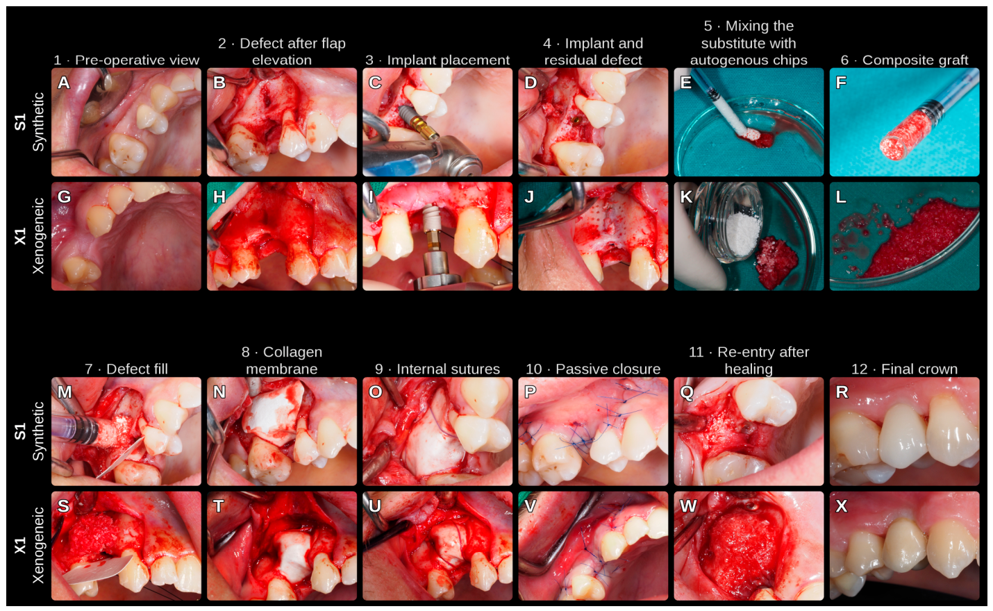
Implant pair, clinical sequence; the implants were placed with the graft. S1 in (A–F,M–R) and X1 in (G–L,S–X). (A,G) Pre-operative view; (B,H) defect after flap elevation; (C,I) implant placement; (D,J) implant and residual defect; (E,K) mixing the substitute with autogenous chips; (F,L) composite graft; (M,S) defect fill; (N,T) collagen membrane; (O,U) internal sutures; (P,V) passive closure; (Q,W) re-entry after healing; (R,X) final crown. S1: synthetic biphasic calcium phosphate, hydroxyapatite/β-tricalcium phosphate 60/40 (RESORBA Synthetic Bone); X1: porcine anorganic (deproteinised) bone mineral (RESORBA Xenogenic Bone Graft); each mixed 50:50 by volume with autogenous chips. Membrane (step 8), native equine collagen (PARASORB RESODONT Forte), fixed with titanium tacks in X1; internal sutures (step 9), GLYCOLON 5-0; closure (step 10), RESOPREN 6-0 (RESORBA Medical GmbH, Nuremberg, Germany).
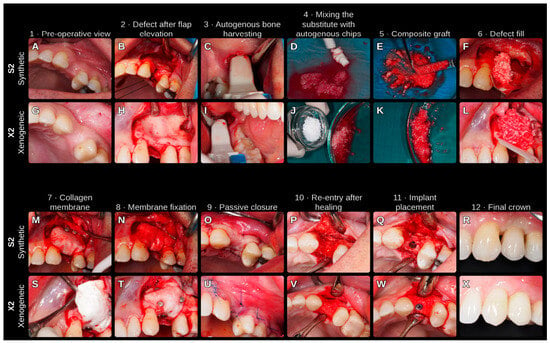
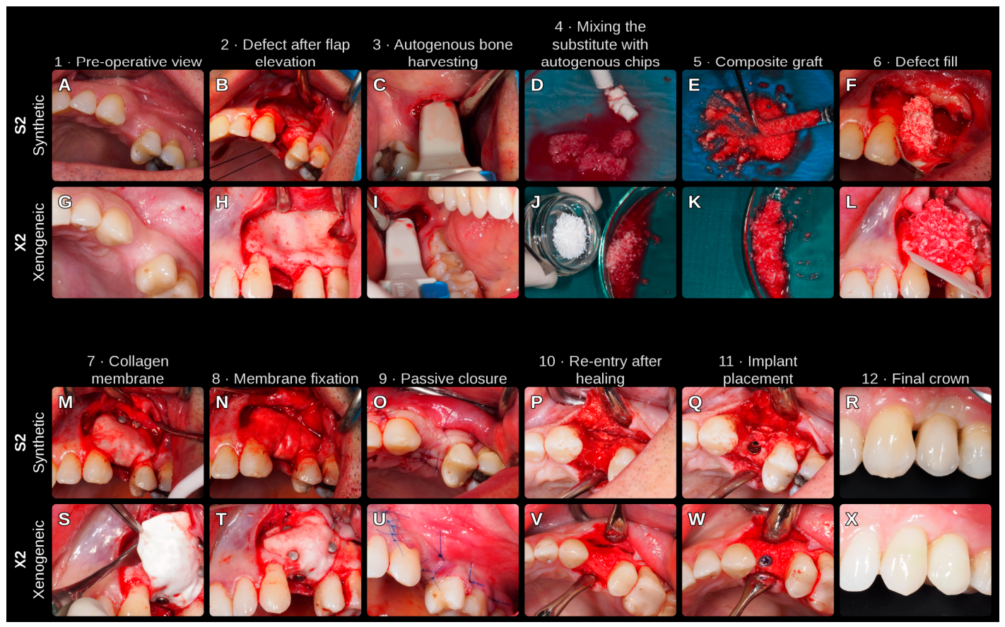
Figure 2.
No-implant pair, clinical sequence; the implants were placed at re-entry. S2 in (A–F,M–R) and X2 in (G–L,S–X). (A,G) Pre-operative view; (B,H) defect after flap elevation; (C,I) autogenous bone harvesting; (D,J) mixing the substitute with autogenous chips; (E,K) composite graft; (F,L) defect fill; (M,S) collagen membrane; (N,T) membrane fixation; (O,U) passive closure; (P,V) re-entry after healing; (Q,W) implant placement; (R,X) final crown. S2: synthetic biphasic calcium phosphate, hydroxyapatite/β-tricalcium phosphate 60/40 (RESORBA Synthetic Bone, earlier formulation); X2: porcine anorganic (deproteinised) bone mineral (RESORBA Xenogenic Bone Graft); each mixed 50:50 by volume with autogenous chips. Membrane (step 7), native equine collagen (PARASORB RESODONT Forte), fixed with titanium tacks in both (step 8; panel N shows the GLYCOLON 5-0 internal suture in S2); closure (step 9), GLYCOLON 6-0 in S2 and RESOPREN 6-0 in X2.
The study was approved by the Ethics Committee of the Faculty of Dental Medicine and Health, University of Osijek (Institutional Review Board Statement), and is reported following the CARE guideline [33,34] (Supplementary Table S1). All patients gave written informed consent for the surgical treatment and for publication of their clinical photographs and radiographic images. The study was not preregistered.
We grafted every defect with one of two particulate substitutes, mixed 50:50 by volume with scraper-harvested autogenous chips:
- Synthetic, S1 and S2: a biphasic calcium phosphate, hydroxyapatite/β-tricalcium phosphate 60/40, in a hydrogel (RESORBA Synthetic Bone; Advanced Medical Solutions (Nantes) SAS, formerly Biomatlante, Vigneux-de-Bretagne, France), with granules of 0.5–1.0 mm in S1 (current formulation) and 0.25–0.5 mm in S2 (an earlier formulation, no longer made);
- Xenogeneic, X1 and X2: a porcine anorganic (deproteinised) bone mineral (RESORBA Xenogenic Bone Graft, 0.25–1.0 mm granules; Collagen Matrix Inc., Oakland, NJ, USA).
We raised a rectangular full-thickness mucoperiosteal flap. We covered the graft, whose packed volume was not measured, with a native (not chemically cross-linked) equine collagen membrane (PARASORB RESODONT Forte; RESORBA Medical GmbH, Nuremberg, Germany), held with titanium tacks, internal sutures or both. At the end of the procedure, just before closure, we released the periosteum microsurgically so that the flap closed passively. Internal sutures (GLYCOLON USP 5-0; RESORBA Medical GmbH) and deep horizontal mattress sutures (RESOPREN USP 5-0; RESORBA Medical GmbH, or GLYCOLON USP 5-0 in S2), placed apically, preceded closure of the incision lines with USP 6-0 monofilament sutures, non-absorbable polyvinylidene fluoride (RESOPREN) in S1, X1 and X2 and absorbable poly(glycolic acid-co-ε-caprolactone) (GLYCOLON) in S2. We prescribed amoxicillin with clavulanic acid 1 g twice daily for seven days from the day before surgery, chlorhexidine rinses twice daily from three days before to two weeks after surgery and ibuprofen 600 mg three times daily from before surgery to the third post-operative day. At re-entry, we assessed each grafted site clinically for consolidation and loose particles.
Each case had a pre-operative and a healed CBCT scan. We exported all scans from DICOM to NIfTI, which carries no patient information, and coded them. The healed scans of X1 and X2 came from a different CBCT unit than their pre-operative scans, whereas each synthetic case was scanned twice on the same unit, with the same voxel size and reconstructed volume (Table 1). We therefore calibrated every scan to a common grey scale. Each scan was rescaled linearly so that its own most frequent grey values for air and for soft tissue read −1000 and +60, as both tissues are present in every scan and unchanged by surgery. With no immediate post-operative scan, the outcome is the net radiodense volume at healing, that is, the graft placed, less the graft resorbed, plus the new bone formed. CBCT cannot separate these terms, and no biopsy was taken, so the measurement shows how much radiodense tissue the site held at healing, not how much of it was bone.
We superimposed (registered) each healed scan on its pre-operative scan in SimpleITK 2.5.5 [35,36], using the bone of the upper jaw, with the surgical site masked out by a box built on the landmarks (below). The registration also allowed for a small difference in scale, because the healed X2 scan, taken on another CBCT unit, was about 0.7% larger when tooth–crown distances were compared. In S2, a rule set before fitting chose a separate stretch for each axis. We adopted this registration after the material key was opened (post hoc), when a review of X2, prompted by its large volume, showed that the original one, which used all bone in the scan, including the lower jaw, and a misplaced site box, had inflated its volume. It was applied identically to all four cases.
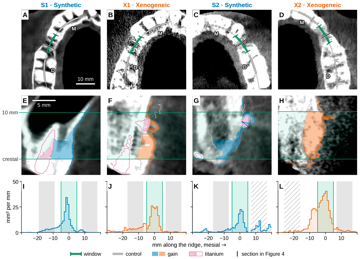
Six landmarks, placed on the pre-operative scan on the two teeth bounding the defect and on the mid-span crest, oriented each case. From them, the software set a fixed 10 × 10 mm measurement window between the bounding teeth, extending apically from a crestal plane through the mid-span buccal crest (Figure 3). We fixed the window because a region drawn around each augmentation would be shaped by the gain it measured. It therefore read a ridge area of the same size, at the same position relative to the bounding teeth, in every case, whatever the size of the augmentation. Tissue built coronal to this plane lay outside the window by design. Tissue outside the window is described in Section 3.2.
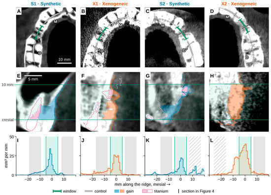
Figure 3.
Measurement window in each case (assessor A); implant pair left, no-implant pair right. (A–D) Pre-operative scan from above; M, mesial; D, distal; the black tick marks the cross-section shown in (E–H) and in Figure 4. (E–H) The same cross-section on the healed scan. The tinted gain between the two green lines, the window, is counted, and gain coronal to it, including the ridge top, is not. Dashed white line, pre-operative bone surface; coloured line, healed surface. (I–L) New radiodense tissue along the ridge, per 1 mm, titanium not subtracted; green, the window. Grey, control windows; dashed in (C,D) and hatched in (K,L), a control window left out under the rules in Section 2. Blue, synthetic graft; orange, xenogeneic graft; each mixed 50:50 with autogenous chips.
Registration and differences between scans can make even untouched bone appear to move. We therefore measured this apparent movement in two 10 mm control windows of untouched bone, one on each side of the site. They lay at the same height as the measurement window (post hoc), and their mean movement was subtracted from the gain as a background offset. An error of the grey-scale calibration at the threshold would also move the surface of untouched bone. The offset therefore removed such an error as well, but only as far as the error was the same at the control windows and at the site. A control window was used only if its apparent movement stayed within 0.90 mm, a limit raised from 0.75 mm before the material key was opened, and if no voxel in it reached metal density (post hoc). We counted as metal density a calibrated grey value above 7000 or the scanner’s maximum. Two control windows failed these rules and were left out, so S2 and X2 each kept one. In S2, the buccal bone of the mesial window had itself changed after surgery, and in X2, the distal window lay over root-filled teeth with large composite fillings, where some voxels reached metal density on both scans.
We then extracted the pre-operative and healed bone surfaces at a calibrated grey value of 450 and, on a 0.2 mm raster across the window, summed the radiodense tissue gained between them, less the background offset. This grey value was set in July 2026, when the method was developed on S1, and was kept unchanged for the other three cases. We subtracted the implant’s titanium between the two surfaces, a term biased upward by metal bloom. After the material key was opened, we added a pin correction (post hoc, the fourth rule tried). It subtracted the metal of fixation pins (the titanium tacks) within 3.0 mm of the healed surface and, where an automatic test found the healed surface bulging over a pin head, flattened it, so that this metal artefact was not counted. It could only lower a volume, and it changed nothing in S1, which had no tacks.
Two assessors, the operating surgeon (assessor A, primary) and a second dentist among the authors (assessor B), placed the landmarks separately on the anonymised pre-operative scans, which showed neither graft nor implant. In S1, assessor B re-placed the root apex of the distal molar after the two sets were compared, as the assessors had chosen different roots. This one landmark is therefore not an independent repeat, although it was re-placed before the material key was opened. The tooth numbers were shown, and the operating surgeon knew which material each case had received, whereas assessor B did not. Both landmark sets went through the same measurement. The volumes were computed by software with no access to the material key, which the operating surgeon held, and the first values were frozen on 7 September 2026, before the pin correction, the revised registration and the control-window rules were added. We refer to these first values as the original analysis and to the final ones as the revised analysis. The key was opened on 13 September 2026, when the materials were entered into the analysis. Keeping the key from the software until then was not blinding, because the operating surgeon, who placed the primary landmarks and directed the analysis, knew the material of every case throughout. Every rule and analysis defined after the key was opened was therefore proposed with the materials known and is declared post hoc, and we claim only that each was applied identically to all four cases by code that did not read the material.
We also report the volume-to-surface ratio (net volume per measured area) and the inward shift of the native surface (where the healed surface lies inside the pre-operative one), which is not netted off the volume [31]. Mean horizontal gain is the bucco-lingual thickness of radiodense gain on the pin-corrected surfaces, less the background and without titanium subtraction, averaged across the window at 1, 2, 3, 5 and 7 mm apical to its crestal plane.
With only two cases per material, we did not test differences statistically. Instead, we asked whether a difference held when the volume was computed in other reasonable ways. For each case, we recomputed the volume with one analysis choice changed at a time, using only choices available in all four cases, and the range of these results is its measurement envelope (Table 2 note). An envelope is not a confidence interval. It shows only how far the volume of one case moved with our own analysis choices, each varied one at a time. When the envelopes of two cases stayed apart overall, for each assessor alone and with S1’s extra checks, no single one of these choices, in either case, reversed their order, which says nothing about the cause of the difference. Otherwise, we describe them as overlapping. The envelopes do not cover differences between patients, sites or CBCT units, the amount of graft placed, the inter-scan interval or the segmentation threshold, and they cover registration only in part. The envelope rules and the rule for calling two envelopes apart are post hoc, and the envelopes of the original analysis, built only from values stored before the key was opened, are given beside them (Table 2).
Table 2.
Net radiodense volume in the 10 × 10 mm window, arranged by pair, on the revised analysis and on the original one, frozen on 7 September 2026. Cells: assessor A/assessor B, who differed only in landmark placement; ranges, the measurement envelopes.
We checked the measurement on a computer-generated test object, a flat bone surface to which a smooth dome of exactly known volume was added, and against an independent implementation in CloudCompare 2.13.2 [37]. As a human reference, the operating surgeon outlined the bone cross-section on both scans at five stations per case (Figure 4), and in S1 the ratio of the traced gain to the software’s gave an operator-scaled volume. Sensitivity analyses varied the specification, raster step, segmentation threshold, background rule, gain apical to the window, earlier pin rules and the window itself, re-anchored on the surgeon’s traced crest or re-oriented. After the material key was opened, we also varied the segmentation threshold from 300 to 800 in steps of 50, measuring the background again in the same control windows at each step and, for the implant pair, also keeping it at its value at 450. We also checked the calibration on untouched maxillary bone in each case, by how far its surface shifted between the two scans and by how much the volume changed when the healed scan’s threshold was reset to remove that shift or to match the grey values inside uniform untouched tissue. The analysis code, written in Python 3.12.3 (NumPy 2.5.1, SciPy 1.18.0, scikit-image 0.26.0), is openly available (Data Availability Statement), and when we ran it again from the coded scans, it reproduced every value of the revised analysis, and from the stored records every value of the original analysis. One complete run by assessor B, on a separate computer and without any AI tool, also reproduced every value of the revised analysis. Figure 5 was drawn in R 4.5.3 [38] with ggplot2 4.0.3 [39].
We orchestrated the analysis pipeline, and a generative AI tool (Claude; Anthropic, San Francisco, CA, USA) wrote the code under our guidance, proposed analysis rules and produced the figures and tables. The tool also assisted with the literature search and review and with the wording of the text. We reviewed and verified every step and take full responsibility for the content, and the tool placed no landmark, drew no outline and supplied no clinical information.
3. Results
3.1. Case Presentation
No patient had a relevant medical history, and Figure 1B,H and Figure 2B,H show the non-contained defects after flap elevation. Apart from a resolved sinus tract in X1, two months after prosthetic loading (below), all four implants were uneventful. At six-monthly recalls, S2, X1 and X2 showed no detectable bone loss and probing depths of at most 5 mm, whereas neither was assessed in S1, whose recall was the shortest. Bone levels were checked on periapical radiographs, and no implant in S2, X1 or X2 showed bleeding on probing or suppuration. Table 1 summarises the course of each case. The volumes below describe each site at the healed scan, and all later follow-up is clinical.
S1: synthetic composite, implant pair. A 39-year-old woman, a non-smoker who had discontinued vaping four months before surgery, was treated at site FDI 15. We placed the implant with the graft, healing was uneventful, and at re-entry, the grafted site appeared consolidated and the graft incorporated (Figure 1Q).
X1: xenogeneic composite, implant pair. A 28-year-old man was treated at site FDI 14, and a lower third molar was removed by alveolectomy at the same surgery. We placed the implant with the graft, healing was uneventful, and at re-entry, the grafted site appeared consolidated, and the graft incorporated (Figure 1W). Two months after the crown was fitted, a sinus tract with serous exudate developed, and at reopening, no bone loss was found, the site was irrigated and the tacks removed. The tract did not recur, and follow-up, 52 months after grafting, was otherwise uneventful.
S2: synthetic composite, no-implant pair. A 56-year-old man, a non-smoker, was treated at site FDI 24. Healing was uneventful, although muscle pull on the flap was obvious. At re-entry, eight months after grafting, part of the graft particles outside the ridge contour were still loose (Figure 2P), and muscle pull had left an obvious indentation on the buccal aspect. We placed the implant at the same visit, with 2 mm of augmented buccal hard tissue (Figure 2Q).
X2: xenogeneic composite, no-implant pair. A 31-year-old woman, an occasional smoker who had stopped four months before surgery, was treated at site FDI 24. Healing was uneventful, although muscle pull on the flap was obvious. At re-entry, nine months after grafting, the grafted site appeared consolidated and the graft largely incorporated, with only a few loose particles and part of the gain outside the ridge contour, although muscle pull had left an obvious indentation on its buccal aspect (Figure 2V). We placed the implant at the same visit (Figure 2W).
3.2. Measurement Window and Quality
Unless stated otherwise, every volume reported here is the net radiodense volume inside the 10 × 10 mm window, the region of interest, not that of the whole augmentation. It counts new bone and residual graft together. Each difference between two cases is also one between two patients, so none of the readings below compares the substitutes. In the window, it was 99.7, 137.0, 95.4 and 267.1 mm3 in S1, X1, S2 and X2 (assessor A; Table 2). We then extended the window, with every correction unchanged, along the ridge until the healed scan showed little new radiodense tissue and apically until gain matched that of undisturbed bone (14 mm in X2, 10 mm in the others; Figure 3). The extended volume was 135.5, 158.2, 95.4 and 476.4 mm3, so the window held 74, 87, 100 and 56% of it. In S1 and X1, the change outside the window lay mostly distal, in a region where the native surface had also shifted inward, which the extended volume does not subtract. Their extended values may therefore be too high, and the share held by the window too low, as may X2’s, whose extended region reached towards root-filled teeth. Extending the window, a post hoc and descriptive step, widened the difference between S2 and X2 and narrowed that between S1 and X1. Moving the window’s crestal plane to the surgeon’s traced crest, or tilting its apical axis, changed no pair reading, although the tilted window was tested on the original registration only.
Our measurement read the volume of the test object 1.8–1.9% too high. Computed the same way as in CloudCompare, on the original registration and before the background, titanium and pin corrections, it differed from CloudCompare by at most 3.7%. Assessor B’s landmarks gave volumes that differed from assessor A’s by 0.2–12.4%, and each case’s envelope spanned 4–21% of its volume (Table 2). Registering without the size factor (rigid registration) on the same bone changed the volumes by −3.1, +2.0, +2.0 and +9.0 mm3 in S1, X1, S2 and X2 (assessor A, before the pin correction). Varying the size factor within its 95% confidence interval moved no envelope. Before the revised registration and control-window rules, but with the pin correction, the window volumes were 105.6, 139.5, 95.7 and 306.3 mm3 in S1, X1, S2 and X2 (assessor A). The first values, frozen on 7 September 2026 before the material key was opened, were 105.6, 139.5, 119.9 and 316.1 mm3, without the pin correction (Table 2; each step, for both assessors, in Table S2). On the original and the revised analysis, and for both assessors, S1 measured less than X1 and S2 less than X2.
On untouched maxillary bone, the bone surface shifted by −0.009, −0.131, −0.056 and +0.012 mm between the two calibrated scans of S1, X1, S2 and X2. Taking the healed surface at the grey value at which that bone did not shift, instead of at 450, changed the window volumes by −0.1, +15.3, +1.8 and −3.0 mm3 (assessor A; −0.1, +11.6, +1.4 and −1.8 mm3 for B). Matching the healed threshold to the grey values inside uniform untouched tissue gave larger changes, up to +62.3 mm3 in X1 and +68.5 mm3 in X2 (assessor A), but that method overstated the shift at the bone edge, where the surface was taken. X2’s pre-operative scan was the only one with a third grey-value peak, and taking that peak as air lowered X2’s volume by 32.0 mm3, to 235.1 mm3 (assessor A). Between grey values of 400 and 800, where every control window met its limit, X1’s volume changed most, from 118.2 to 194.3 mm3, and X2’s least, from 262.6 to 279.0 mm3 (assessor A). X1’s healed scan covered the least of the jaw, although the grafted site itself lay inside it (Figure 3J). Only 44.8% of the upper-jaw bone seen before surgery also lay in the healed scan, against 88.0–97.8% in the others, so its superimposition rested on the least bone. Moreover, both bone surfaces could be measured over only 88.7% of its window.
3.3. Implant Pair: S1 (Synthetic) and X1 (Xenogeneic)
S1 measured 99.7 mm3 in the window and X1 137.0 mm3 (assessor A; Table 2; Figure 4A–F and Figure 5A), a difference of 37.3 mm3. Both values excluded the implant titanium lying between the two surfaces, 28.8 mm3 in S1 and 11.9 mm3 in X1 (of 82.7 and 127.5 mm3 visible on the scans). Their envelopes did not overlap, but S1’s highest value, with its extra checks, lay only 7.4 mm3 below X1’s lowest. Four findings leave the size and even the direction of this difference uncertain. First, S1’s superimposition was the least stable of the four, as superimposing it without the size factor lowered its volume by 3.1 mm3 with assessor A’s landmarks and by 17.5 mm3 with assessor B’s. Second, at grey values of 300 and 350, X1’s mesial control window moved more than the 0.90 mm limit, so the background could not be measured within our rules. With the background kept at its value at 450, S1 measured more than X1 at these two grey values. From 400 to 800, where every control window met its limit, S1 measured less than X1 at every grey value, by 20.5–79.0 mm3 (assessor A; 28.0–66.7 mm3 for B). Third, on the original analysis their envelopes overlapped by 5.2 mm3 (Table 2), and when the revised registration was combined with the analysis choices of the original envelope, they still overlapped, by 3.8 mm3. Fourth, the order depended on the background offset, the apparent movement of untouched bone in the control windows that is subtracted from the gain. Without it, S1 measured more than X1 in three of the four combinations of analysis and assessor, and less only for assessor B on the revised analysis.
Figure 4.
CBCT cross-sections, one per case at the outlined station with the largest traced change: pre-operative (A,D,G,J), healed (B,E,H,K) and both outlines superimposed (C,F,I,L); implant pair top, no-implant pair bottom. Blue, synthetic graft; orange, xenogeneic graft; each mixed 50:50 with autogenous chips. Bone outlined by the operating surgeon, who knew the material: dashed white, pre-operative; solid, healed. Grey ticks mark the window’s limits; the tint is solid inside the window, where it is counted, and faint outside it (mainly coronal in S1 and X1, partly implant; mainly apical in S2 and X2). “Window”, net radiodense volume in the window (assessor A). Sections are rotated so the window lies level; grey scale −400 to 1400.
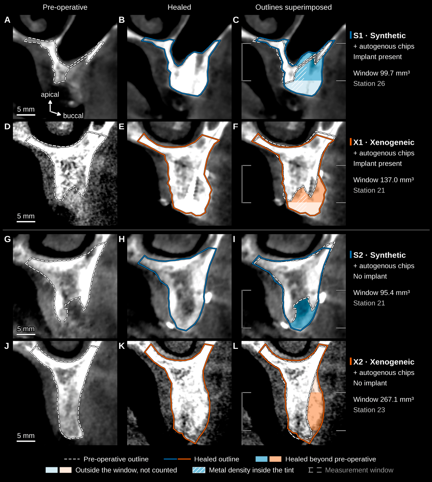
Figure 5.
(A) Net radiodense volume in the 10 × 10 mm window, titanium subtracted: filled dots, assessor A (primary); open dots, assessor B; bars, each assessor’s envelope (combined in Table 2); triangles, two of S1’s extra checks, which set the ends of its wider envelope (Table 2 note). The implant-pair envelopes lay only 7.4 mm3 apart and overlapped on the original analysis (Table 2). (B) Mean horizontal gain at five depths apical to the window’s crestal plane, titanium not subtracted: grey bars, the 0.2 mm depth bands; solid lines, assessor A; dashed, assessor B; half-filled markers, incomplete bands. Blue, synthetic graft; orange, xenogeneic graft; each mixed 50:50 with autogenous chips. Envelopes are not confidence intervals, and with one case per material in each pair, no difference between materials is inferred.
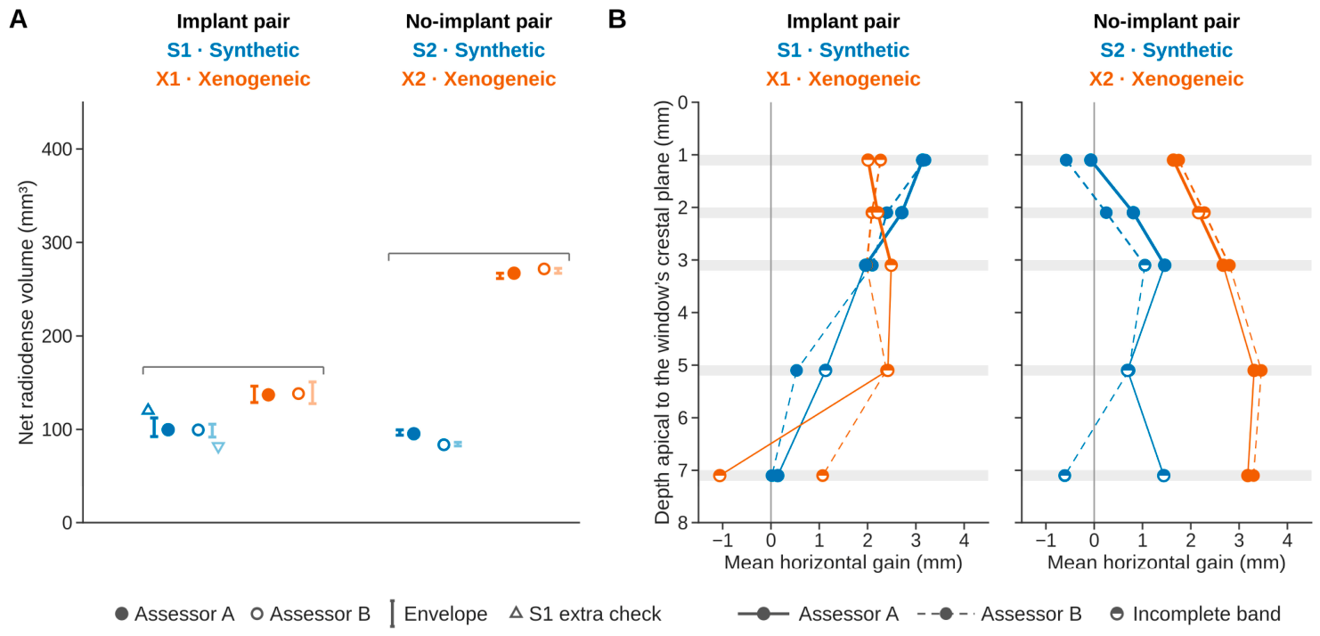
The volume-to-surface ratio was 1.00 mm3/mm2 in S1 and 1.54 in X1, and the inward shift of the native surface measured 3.8 and 79.8 mm3. Mean horizontal gain ranged from 0.14 to 3.14 mm in S1 and from −1.06 to 2.49 mm in X1 (assessor A; Figure 5B). Gain coronal to the window was not counted in the volumes. In Figure 4’s sections, the operating surgeon’s outlines placed 30.5 mm2 of gain coronal to the window in S1 and 23.8 mm2 in X1, about a quarter of it on implant metal.
3.4. No-Implant Pair: S2 (Synthetic) and X2 (Xenogeneic)
S2 measured 95.4 mm3 in the window and X2 267.1 mm3 (assessor A; Table 2; Figure 4G–L and Figure 5A), a difference of 171.7 mm3. S2’s highest value lay 161.9 mm3 below X2’s lowest, and on the original analysis, their envelopes lay 160.5 mm3 apart (Table 2). S2 measured less than X2 at every grey value from 300 to 800, by 157.9–189.5 mm3 (assessor A; 170.8–194.9 mm3 for B). The augmentation extended 7 mm along the arch in S2 and 22 mm in X2 (assessor A; Figure 3I–L), and in X2, it continued apical to the window, so its window value is a lower bound. Deepened to 14 mm, with the background offset re-measured over the same depth, the window measured 316.0 mm3 in X2. The post hoc pin correction lowered S2 from 117.4 to 95.4 mm3 and X2 from 274.8 to 267.1 mm3, by flattening surface bulges over two pin heads in S2 and subtracting pin metal in both (6.5 and 7.7 mm3). For assessor A, the bulge over one X2 pin head could not be confirmed and was left in place, whereas for assessor B it was flattened (3.1 mm3).
The volume-to-surface ratio was 0.97 mm3/mm2 in S2 and 2.68 in X2, and the inward shift of the native surface measured 33.9 and 0.6 mm3. Mean horizontal gain ranged from −0.07 to 1.46 mm in S2 and from 1.64 to 3.31 mm in X2 (assessor A; Figure 5B). In Figure 4’s sections, the outlines placed only 1.3 mm2 of gain coronal to the window in S2 and 5.1 mm2 in X2.
3.5. Cases with the Same Substitute
The two synthetic cases overlapped, by 17.2 mm3 with S1’s extra checks, and the post hoc pin correction reversed their order, whereas the two xenogeneic cases lay 110.6 mm3 apart (Figure 5A). On the original analysis (Table 2), the synthetic cases also overlapped, and the xenogeneic cases also lay apart.
4. Discussion
We presented four non-contained maxillary ridge defects, two grafted with a synthetic biphasic calcium phosphate and two with a porcine xenogeneic mineral, each mixed 50:50 with scraper-harvested autogenous chips. All four cases received implants, two with the graft and two at re-entry. The envelopes of S2 and X2 lay apart on both the original and the revised analysis, whereas those of S1 and X1 overlapped on the original analysis, so the implant-pair difference is uncertain. With one case per combination of substitute and implant status, the series cannot test efficacy or show that either composite is better.
However, biphasic calcium phosphate and xenogeneic mineral have been compared in randomised trials. In sinus floor augmentation, a network meta-analysis by Somngam et al. [15], pooling 11 trials of biphasic calcium phosphate against other substitutes, found that biphasic calcium phosphate formed 4.14 percentage points more new bone than xenogeneic mineral and left less residual graft. In a split-mouth sinus trial by Mordenfeld et al. [40], implants in biphasic calcium phosphate and bovine mineral survived alike over five years of loading, and graft height fell by 6.6 and 5.8% over six years, respectively. At peri-implant dehiscences, the two trials of a 60/40 formulation used alone found no disadvantage against bovine mineral [25,26]. Their comparator was bovine, whereas ours was porcine, and the two did not differ histomorphometrically or radiographically in a systematic review, though only in sinus floor augmentation and ridge preservation [41].
Van Assche et al. [30] layered a biphasic calcium phosphate over autogenous bone at dehiscences, and at re-entry, it left a residual defect no different from that left by bovine mineral. Our no-implant pair resembled that result clinically, as both ridges allowed implant placement at re-entry. In S2, however, part of the graft particles outside the ridge contour were still loose, and the gain beyond the contour was smaller than in X2, where only a few particles were loose. Muscle pull on the flap, a strain on the stability of the wound (Section 1), was obvious in both cases during healing. With one patient per substitute in this pair, we cannot tell whether the difference between S2 and X2 came from the substitute, its formulation, the defect or the patient. Nor does the radiodense volume show how much of either graft became bone, as it counts residual granules together with new bone and, in S2, possibly loose granules that lay inside the window. The volumetric CBCT data we found for biphasic calcium phosphate in atrophic ridges come mainly from a 20/80 formulation carrying expanded mesenchymal stem cells, without bone chips, so they cannot be set beside ours [22,23].
Dimensional stability depends on the substitute and its formulation. After socket grafting, a biphasic calcium phosphate lost less ridge width than bovine mineral [16], whereas after contour augmentation at implants, a 60/40 formulation lost more facial thickness than a 70/30 one [42]. In minipig mandibular defects, bone formation and degradation of the substitute appeared to fall as the hydroxyapatite fraction rose, and the 60/40 and 80/20 formulations performed much like deproteinised bovine bone mineral [43]. Our net radiodense volume counts residual granules and new bone alike, so it shows the net result of the graft placed, the graft resorbed and the new bone formed, without separating them.
Both synthetic cases received implants, S1 with the graft and S2 at re-entry. When the sites were reopened, S1’s graft appeared incorporated, whereas in S2, part of the graft particles outside the ridge contour were still loose. Granule size, 0.5–1.0 mm in S1 and 0.25–0.5 mm in S2 (a formulation no longer commercially available), may have favoured S1 over S2. In rat calvarial defects, however, the same two size ranges of biphasic calcium phosphate formed similar amounts of bone [44]. Moreover, the S1–S2 order was fragile, reversing with the post hoc pin correction. The two synthetic cases received different formulations, and the xenogeneic granules, 0.25–1.0 mm, spanned both of their size ranges. These cases therefore cannot separate an effect of the substitute from one of its formulation or granule size.
Ogata et al. [28] and Shido et al. [29], whose composites are the closest to ours (Table 3), measured CBCT widths referenced differently from our gains, so we did not compare the values. Unlike our fixed 10 × 10 mm window, the region of Palkovics et al. [31] follows the augmentation, so their volumes are not like-for-like with ours. Stricker et al. [32] likewise outlined the graft itself on each scan, so their graft volumes are not like-for-like with ours either.
Table 3.
Closest published clinical evidence, as reported by each study.
Beyond these four cases, we see three ways in which a fixed window may help to assess guided bone regeneration. First, its position is set from landmarks on the pre-operative scan, so it does not depend on the healed scan. A region drawn around the gain would take in, at its edge, noise that looks like gain, and such selection can distort even descriptive results [45]. Second, it reads a ridge area of the same size and position at the implant site in every case. Third, control windows of untouched bone show how far superimposition and imaging alone move a bone surface, so a gain can be read against that background rather than taken at face value. The trade-off is that the window misses tissue outside it. It held all of the extended volume in S2 but only 56% in X2, so the extent of each augmentation should be reported beside it. Linear widths [28,29] and whole-augmentation volumes [31,32] answer other questions, and the window adds to them rather than replacing them.
The implant-pair difference is uncertain, for the four reasons given in the Results. S1’s background offset, 0.330 mm with assessor A’s landmarks, came mostly from its superimposition. Superimposed without the size factor, it was 0.055 mm, whereas the calibration moved S1’s untouched bone by only −0.009 mm. Moreover, three further influences, not fully covered by the envelopes, pull in opposite directions. First, the implant’s metal bloom makes the subtracted titanium too large. More titanium was subtracted in S1 (28.8 mm3) than in X1 (11.9 mm3), so S1 may be underestimated and the difference between the two overstated. Second, superimposing without the size factor lowered S1 and raised X1, which would enlarge the difference. Third, setting X1’s healed threshold so that its untouched bone did not shift raised its volume by 15.3 mm3, more than the 7.4 mm3 gap between the envelopes, which would also enlarge the difference. Their net effect cannot be established from these data.
The S2–X2 difference is a single-case observation and cannot be credited to the substitute. The two synthetic cases differed by 4.3 mm3 and the two xenogeneic cases by 130.1 mm3. In both comparisons, one case received its implant with the graft and the other at re-entry. Even so, they show that large differences can arise between patients with the same substitute. A larger radiodense volume also does not mean that more bone formed, as residual granules and new bone are counted together. S2’s augmentation was shorter along the arch than X2’s, which also extended beyond the window, so X2’s window value is a lower bound. X2’s distal control window lay over two root-filled teeth and was excluded, so its background offset rests on the mesial window alone. Only the two xenogeneic cases changed CBCT unit between scans, so substitute and unit change coincide. Voxel size also differed, from 0.16 to 0.30 mm (Table 1), and the coarser voxels of S2 may blur thin or particulate tissue at a fixed threshold.
Without a space-maintaining device, graft displacement at wound closure [12,13] may also have differed between cases, as S1’s membrane was held by sutures alone. The inward shift of the native surface may reflect bed preparation, flap elevation, imaging near metal or registration, so it is not interpreted clinically.
A strength of this series is that one fixed window and one set of rules were applied to all four cases, with two assessors’ landmark sets and every analysis record kept (Data Availability Statement). The main limitation is the design, since every difference between our cases is also one between patients, sites and defects, and may reflect the CBCT unit and inter-scan interval. We selected the cases retrospectively in one practice, when the healed scans already existed, so the series cannot be assumed to represent the range of outcomes with either composite. Moreover, the operating surgeon chose the xenogeneic cases knowing how each had healed, so the choice could favour cases that healed well. The operating surgeon alone classified the defects, on the pre-operative CBCT and at flap elevation, so the classification rests on a single observer. The substitute’s composition is taken from the manufacturer’s declaration, and S1 and S2 received different formulations (above). An immediate post-operative CBCT is not part of our standard of care, as an exposure to radiation must bring the patient a net benefit and be kept as low as reasonably achievable (ALARA) [46], and in this retrospective analysis, only scans taken for clinical reasons were available. Without an immediate post-operative scan or histology, the net radiodense volume supports no statement about resorption or about how much bone formed. Grey-scale calibration was checked at the grey values of bone only after the material key was opened, and it was least accurate in X1. No calibration phantom was scanned, so the grey scales could be checked only against each other, not against a reference. The segmentation threshold was varied only outside the envelopes. The volumes were computed without the material key only until it was opened, and the revised registration, the control-window rules, the pin correction, the envelope rules, the rule for calling two envelopes apart, the threshold sweep and the sensitivity analyses were all defined afterwards. X2’s size factor differed by up to about 1% between estimators, more than its confidence interval, but fixing it at 1.012 moved its volume by 1.3 mm3. The operating surgeon, who knew the material used in each case, placed the primary landmark set, but on pre-operative scans, which show neither graft nor implant. The operating surgeon’s outlines, used as a human reference, were traced once, unblinded, and the region compared was set afterwards. Follow-up was shortest in S1, 13 months after grafting (Table 1), so its clinical course is the least established. The volumes were measured once, at healing, and all later follow-up was clinical, so they show nothing of the long-term stability of either graft. As the implants were placed at different stages, the clinical courses of the two pairs also cannot be set side by side. The analysis has not been repeated by an independent group. Our own re-runs of the public code, including assessor B’s on a separate computer, are not independent. Comparing the composites would need randomisation, one CBCT unit, pins outside the measured window and verified substitute composition, with a sample size not derived from these cases [47].
5. Conclusions
This case series provided three observations on a biphasic calcium phosphate (hydroxyapatite/β-tricalcium phosphate 60/40) mixed 50:50 with scraper-harvested autogenous chips in non-contained ridge defects. First, both synthetic cases, like both xenogeneic ones, received implants, and all four implants were uneventful apart from one resolved sinus tract in X1. Second, judged against per-case measurement envelopes, S2 and X2 lay apart on both the original and the revised analysis, whereas S1 and X1 overlapped on the original analysis, so their difference was uncertain. As each difference was also one between patients and sites, none was attributed to the substitute, and the series cannot show that either composite is better. Third, we found no earlier report that measured the radiodense volume of this composite graft, and these cases complement the randomised evidence for the substitute used alone. These findings should be interpreted as single-case observations and warrant evaluation in larger, controlled studies.
Supplementary Materials
The following supporting information can be downloaded at: https://www.mdpi.com/article/10.3390/bioengineering13111286/s1, Table S1: CARE checklist: each item of the CARE guideline and where it is reported in this article. Cases are labelled S1 and X1 (implant pair) and S2 and X2 (no-implant pair); S, synthetic composite; X, xenogeneic composite; Table S2: Original and revised analyses. Net radiodense volume in the 10 × 10 mm window, mm3, assessor A/assessor B: (a) Step by step, from the values frozen on 7 September 2026 to the revised analysis; (b) The four rules tried for the fixation pins (titanium tacks), each on the original registration.
Author Contributions
Conceptualization, J.B.; methodology, J.B.; investigation, J.B. and B.M.; validation, B.M., S.S. and M.T.; data curation, J.B.; writing—original draft preparation, J.B.; writing—review and editing, J.B. and D.G.; supervision, J.B., S.S. and M.T. All authors have read and agreed to the published version of the manuscript.
Funding
This research received no external funding. The article processing charge (APC) was funded by Advanced Medical Solutions Group plc, through its subsidiary RESORBA Medical GmbH (Nuremberg, Germany). The funder had no role in the design of the study; in the collection, analyses, or interpretation of data; in the writing of the manuscript; or in the decision to publish the results, and was not granted access to the manuscript at any stage prior to its acceptance and publication.
Institutional Review Board Statement
The study was conducted in accordance with the Declaration of Helsinki and approved by the Ethics Committee of the Faculty of Dental Medicine and Health, University of Osijek (Class: 602-01/26-12/07; Reg. No.: 2158/97-97-10-26-89; date of approval: 8 July 2026).
Informed Consent Statement
Written informed consent for the surgical treatment was obtained from all patients. Written informed consent for publication of this paper, including clinical photographs and radiographic images, was obtained from all patients.
Data Availability Statement
The data supporting the findings of this case series are contained within the article. The analysis code is openly available at https://github.com/jbrozovic-fdmz/ridge-graft-cbct-volumetry (accessed on 27 October 2026). The analysis records, including each assessor’s landmark set, the operating surgeon’s outlines, the result files and the material key, are available from the corresponding authors on reasonable request. In these files, X1 and X2 carry their analysis codes, S3 and S4.
Acknowledgments
During the preparation of this study, the authors used Claude (Opus 4.8, Opus 5 and Opus 5.5; Anthropic, San Francisco, CA, USA), through Claude Code, as an assistant under the authors’ direction, for the purposes of writing the analysis code, proposing analysis rules, producing the figures and tables, and assisting with the literature search and review and with the wording of the text. The authors have reviewed and edited the output and take full responsibility for the content of this publication.
Conflicts of Interest
The article processing charge for this publication was funded by Advanced Medical Solutions Group plc, through its subsidiary RESORBA Medical GmbH (Nuremberg, Germany), which supplied the bone substitutes, membrane and sutures used in the reported cases. These materials were used as part of routine surgical care, of which the funder had no knowledge. The funder had no role in the design or conduct of the reported cases; in the collection, analysis, or interpretation of data; in the writing of the manuscript; or in the decision to publish, and had no access to the manuscript at any stage prior to its acceptance and publication. Mention of the products does not constitute an endorsement. The authors declare no other conflicts of interest.
References
- Wang, H.-L.; Boyapati, L. “PASS” Principles for Predictable Bone Regeneration. Implant Dent. 2006, 15, 8–17. [Google Scholar] [CrossRef] [Scilit] [PubMed]
- Burkhardt, R.; Lang, N.P. Role of Flap Tension in Primary Wound Closure of Mucoperiosteal Flaps: A Prospective Cohort Study. Clin. Oral Implant. Res. 2010, 21, 50–54. [Google Scholar] [CrossRef] [Scilit] [PubMed]
- Garcia, J.; Dodge, A.; Luepke, P.; Wang, H.-L.; Kapila, Y.; Lin, G.-H. Effect of Membrane Exposure on Guided Bone Regeneration: A Systematic Review and Meta-Analysis. Clin. Oral Implant. Res. 2018, 29, 328–338. [Google Scholar] [CrossRef] [Scilit] [PubMed]
- Burkhardt, R.; Lang, N.P. Fundamental Principles in Periodontal Plastic Surgery and Mucosal Augmentation—A Narrative Review. J. Clin. Periodontol. 2014, 41, S98–S107. [Google Scholar] [CrossRef] [Scilit] [PubMed]
- Miron, R.J.; Gruber, R.; Hedbom, E.; Saulacic, N.; Zhang, Y.; Sculean, A.; Bosshardt, D.D.; Buser, D. Impact of Bone Harvesting Techniques on Cell Viability and the Release of Growth Factors of Autografts. Clin. Implant Dent. Relat. Res. 2013, 15, 481–489. [Google Scholar] [CrossRef] [Scilit] [PubMed]
- Buser, D.; Hoffmann, B.; Bernard, J.-P.; Lussi, A.; Mettler, D.; Schenk, R.K. Evaluation of Filling Materials in Membrane-Protected Bone Defects. A Comparative Histomorphometric Study in the Mandible of Miniature Pigs. Clin. Oral Implant. Res. 1998, 9, 137–150. [Google Scholar] [CrossRef] [Scilit] [PubMed]
- Mordenfeld, A.; Johansson, C.B.; Albrektsson, T.; Hallman, M. A Randomized and Controlled Clinical Trial of Two Different Compositions of Deproteinized Bovine Bone and Autogenous Bone Used for Lateral Ridge Augmentation. Clin. Oral Implant. Res. 2014, 25, 310–320. [Google Scholar] [CrossRef] [Scilit] [PubMed]
- Urban, I.A.; Nagursky, H.; Lozada, J.L.; Nagy, K. Horizontal Ridge Augmentation with a Collagen Membrane and a Combination of Particulated Autogenous Bone and Anorganic Bovine Bone-Derived Mineral: A Prospective Case Series in 25 Patients. Int. J. Periodontics Restor. Dent. 2013, 33, 299–307. [Google Scholar] [CrossRef] [Scilit] [PubMed]
- Meloni, S.M.; Jovanovic, S.A.; Urban, I.; Baldoni, E.; Pisano, M.; Tallarico, M. Horizontal Ridge Augmentation Using GBR with a Native Collagen Membrane and 1:1 Ratio of Particulate Xenograft and Autologous Bone: A 3-Year after Final Loading Prospective Clinical Study. Clin. Implant Dent. Relat. Res. 2019, 21, 669–677. [Google Scholar] [CrossRef] [Scilit] [PubMed]
- Aludden, H.; Starch-Jensen, T.; Dahlin, C.; Sdik, J.; Cederlund, A.; Mordenfeld, A. Histological and Radiological Outcome after Horizontal Guided Bone Regeneration with Bovine Bone Mineral Alone or in Combination with Bone in Edentulous Atrophic Maxilla: A Randomized Controlled Trial. Clin. Oral Implant. Res. 2024, 35, 396–406. [Google Scholar] [CrossRef] [Scilit] [PubMed]
- Meza-Mauricio, J.; Furquim, C.P.; dos Reis, L.D.; Maximiano, M.M.; Mendoza-Azpur, G.; Muniz, F.W.; Rasperini, G.; Faveri, M. How Efficacious Is the Combination of Substitute Bone Graft with Autogenous Bone Graft in Comparison with Substitute Bone Graft Alone in the Horizontal Bone Gain? A Systematic Review and Meta-Analysis. J. Clin. Exp. Dent. 2022, 14, e678–e688. [Google Scholar] [CrossRef] [Scilit] [PubMed]
- Mertens, C.; Braun, S.; Krisam, J.; Hoffmann, J. The Influence of Wound Closure on Graft Stability: An in Vitro Comparison of Different Bone Grafting Techniques for the Treatment of One-Wall Horizontal Bone Defects. Clin. Implant Dent. Relat. Res. 2019, 21, 284–291. [Google Scholar] [CrossRef] [Scilit] [PubMed]
- Mir-Mari, J.; Wui, H.; Jung, R.E.; Hämmerle, C.H.F.; Benic, G.I. Influence of Blinded Wound Closure on the Volume Stability of Different GBR Materials: An in Vitro Cone-Beam Computed Tomographic Examination. Clin. Oral Implant. Res. 2016, 27, 258–265. [Google Scholar] [CrossRef] [Scilit] [PubMed]
- Benic, G.I.; Bienz, S.P.; Song, Y.W.; Cha, J.-K.; Hämmerle, C.H.F.; Jung, U.-W.; Jung, R.E. Randomized Controlled Clinical Trial Comparing Guided Bone Regeneration of Peri-Implant Defects with Soft-Type Block versus Particulate Bone Substitutes: Six-Month Results of Hard-Tissue Changes. J. Clin. Periodontol. 2022, 49, 480–495. [Google Scholar] [CrossRef] [Scilit] [PubMed]
- Somngam, C.; Samartkit, S.; Kanchanasurakit, S.; Strietzel, F.P.; Khongkhunthian, P. New Bone Formation of Biphasic Calcium Phosphate Bone Substitute Material: A Systematic Review and Network Meta-Analysis of Randomized Controlled Trials (RCTs). Int. J. Implant Dent. 2025, 11, 47. [Google Scholar] [CrossRef] [Scilit] [PubMed]
- Mardas, N.; Chadha, V.; Donos, N. Alveolar Ridge Preservation with Guided Bone Regeneration and a Synthetic Bone Substitute or a Bovine-Derived Xenograft: A Randomized, Controlled Clinical Trial. Clin. Oral Implant. Res. 2010, 21, 688–698. [Google Scholar] [CrossRef] [Scilit] [PubMed]
- Thoma, D.S.; Bienz, S.P.; Figuero, E.; Jung, R.E.; Sanz-Martín, I. Efficacy of Lateral Bone Augmentation Performed Simultaneously with Dental Implant Placement: A Systematic Review and Meta-Analysis. J. Clin. Periodontol. 2019, 46, 257–276. [Google Scholar] [CrossRef] [Scilit] [PubMed]
- Urban, I.A.; Montero, E.; Monje, A.; Sanz-Sánchez, I. Effectiveness of Vertical Ridge Augmentation Interventions: A Systematic Review and Meta-Analysis. J. Clin. Periodontol. 2019, 46, 319–339. [Google Scholar] [CrossRef] [Scilit] [PubMed]
- Roca-Millan, E.; Jané-Salas, E.; Marí-Roig, A.; Jiménez-Guerra, Á.; Ortiz-García, I.; Velasco-Ortega, E.; López-López, J.; Monsalve-Guil, L. The Application of Beta-Tricalcium Phosphate in Implant Dentistry: A Systematic Evaluation of Clinical Studies. Materials 2022, 15, 655. [Google Scholar] [CrossRef] [Scilit] [PubMed]
- Troedhan, A.; Wainwright, M.; Kurrek, A.; Schlichting, I. Biomechanical Stability of Dental Implants in Augmented Maxillary Sites: Results of a Randomized Clinical Study with Four Different Biomaterials and PRF and a Biological View on Guided Bone Regeneration. BioMed Res. Int. 2015, 2015, 850340. [Google Scholar] [CrossRef] [Scilit] [PubMed]
- Kakar, A.; Kakar, K.; Sripathi Rao, B.H.; Lindner, A.; Nagursky, H.; Jain, G.; Patney, A. Lateral Alveolar Ridge Augmentation Procedure Using Subperiosteal Tunneling Technique: A Pilot Study. Maxillofac. Plast. Reconstr. Surg. 2018, 40, 3. [Google Scholar] [CrossRef] [Scilit] [PubMed]
- Gjerde, C.; Mustafa, K.; Hellem, S.; Rojewski, M.; Gjengedal, H.; Yassin, M.A.; Feng, X.; Skaale, S.; Berge, T.; Rosen, A.; et al. Cell Therapy Induced Regeneration of Severely Atrophied Mandibular Bone in a Clinical Trial. Stem Cell Res. Ther. 2018, 9, 213. [Google Scholar] [CrossRef] [Scilit] [PubMed]
- Sanz, M.; Gjerde, C.; Gjertsen, B.T.; Ortiz-Vigón, A.; Sanchez, N.; Hoornaert, A.; Caballe-Serrano, J.; Giralt-Hernando, M.; Gaultier, F.; Reinald, N.; et al. Bone Augmentation of Atrophic Alveolar Ridges Using a Synthetic Bone Substitute with Mesenchymal Stem Cells: A Randomized, Controlled Clinical Trial. Clin. Oral Implant. Res. 2025, 36, 1498–1514. [Google Scholar] [CrossRef] [Scilit] [PubMed]
- Mangano, C.; Luongo, G.; Luongo, F.; Lerner, H.; Margiani, B.; Admakin, O.; Mangano, F. Custom-Made Computer-Aided-Design/ Computer-Assisted-Manufacturing (CAD/CAM) Synthetic Bone Grafts for Alveolar Ridge Augmentation: A Retrospective Clinical Study with 3 Years of Follow-Up. J. Dent. 2022, 127, 104323. [Google Scholar] [CrossRef] [Scilit] [PubMed]
- Lee, J.-H.; An, H.-W.; Im, J.-S.; Kim, W.-J.; Lee, D.-W.; Yun, J.-H. Evaluation of the Clinical and Radiographic Effectiveness of Treating Peri-Implant Bone Defects with a New Biphasic Calcium Phosphate Bone Graft: A Prospective, Multicenter Randomized Controlled Trial. J. Periodontal Implant Sci. 2023, 53, 306–317. [Google Scholar] [CrossRef] [Scilit] [PubMed]
- Cha, J.-K.; Jung, U.-W.; Montero-Solis, E.; Sanz-Sánchez, I.; Sanz-Alonso, M. Guided Bone Regeneration at Dehiscence Comparing Synthetic Bone Substitute versus Bovine Bone Mineral: A Multicenter, Noninferiority, Randomized Trial. Clin. Implant Dent. Relat. Res. 2024, 26, 1233–1244. [Google Scholar] [CrossRef] [Scilit] [PubMed]
- Cha, J.-K.; Sanz, M.; Jung, U.-W. Human Autopsy Study of Peri-Implant Dehiscence Defects with Guided Bone Regeneration: A Case Report. Int. J. Periodontics Restor. Dent. 2019, 39, 517–524. [Google Scholar] [CrossRef] [Scilit] [PubMed]
- Ogata, K.; Ohba, S.; Sumita, Y.; Ashahina, I. Safety and Feasibility Assessment of Biodegradable Poly (l-Lactic Acid/ε-Caprolactone) Membrane for Guided Bone Regeneration: A Case Series of First-in-Human Pilot Study. J. Dent. Sci. 2022, 17, 368–376. [Google Scholar] [CrossRef] [Scilit] [PubMed]
- Shido, R.; Ohba, S.; Tominaga, R.; Sumita, Y.; Asahina, I. A Prospective Study of the Assessment of the Efficacy of a Biodegradable Poly(l-Lactic Acid/ε-Caprolactone) Membrane for Guided Bone Regeneration. J. Clin. Med. 2023, 12, 5994. [Google Scholar] [CrossRef] [Scilit] [PubMed]
- Van Assche, N.; Michels, S.; Naert, I.; Quirynen, M. Randomized Controlled Trial to Compare Two Bone Substitutes in the Treatment of Bony Dehiscences. Clin. Implant Dent. Relat. Res. 2013, 15, 558–568. [Google Scholar] [CrossRef] [Scilit] [PubMed]
- Palkovics, D.; Solyom, E.; Somodi, K.; Pinter, C.; Windisch, P.; Bartha, F.; Molnar, B. Three-Dimensional Volumetric Assessment of Hard Tissue Alterations Following Horizontal Guided Bone Regeneration Using a Split-Thickness Flap Design: A Case Series. BMC Oral Health 2023, 23, 118. [Google Scholar] [CrossRef] [Scilit] [PubMed]
- Stricker, A.; Jacobs, R.; Maes, F.; Fluegge, T.; Vach, K.; Fleiner, J. Resorption of Retromolar Bone Grafts after Alveolar Ridge Augmentation—Volumetric Changes after 12 Months Assessed by CBCT Analysis. Int. J. Implant Dent. 2021, 7, 7. [Google Scholar] [CrossRef] [Scilit] [PubMed]
- Gagnier, J.J.; Kienle, G.; Altman, D.G.; Moher, D.; Sox, H.; Riley, D.; CARE Group. The CARE Guidelines: Consensus-Based Clinical Case Report Guideline Development. J. Clin. Epidemiol. 2014, 67, 46–51. [Google Scholar] [CrossRef] [Scilit] [PubMed]
- Riley, D.S.; Barber, M.S.; Kienle, G.S.; Aronson, J.K.; von Schoen-Angerer, T.; Tugwell, P.; Kiene, H.; Helfand, M.; Altman, D.G.; Sox, H.; et al. CARE Guidelines for Case Reports: Explanation and Elaboration Document. J. Clin. Epidemiol. 2017, 89, 218–235. [Google Scholar] [CrossRef] [Scilit] [PubMed]
- Lowekamp, B.C.; Chen, D.T.; Ibáñez, L.; Blezek, D. The Design of SimpleITK. Front. Neuroinform. 2013, 7, 45. [Google Scholar] [CrossRef] [Scilit] [PubMed]
- Yaniv, Z.; Lowekamp, B.C.; Johnson, H.J.; Beare, R. SimpleITK Image-Analysis Notebooks: A Collaborative Environment for Education and Reproducible Research. J. Digit. Imaging 2018, 31, 290–303. [Google Scholar] [CrossRef] [Scilit] [PubMed]
- CloudCompare, Version 2.13.2. 2024. Available online: http://www.cloudcompare.org/ (accessed on 9 October 2026).
- R Core Team. R: A Language and Environment for Statistical Computing; R Foundation for Statistical Computing: Vienna, Austria, 2026; Available online: https://www.R-project.org/ (accessed on 9 October 2026).
- Wickham, H. ggplot2: Elegant Graphics for Data Analysis; Springer: New York, NY, USA, 2016; ISBN 978-3-319-24277-4. [Google Scholar]
- Mordenfeld, A.; Lindgren, C.; Hallman, M. Sinus Floor Augmentation Using Straumann® BoneCeramic™ and Bio-Oss® in a Split Mouth Design and Later Placement of Implants: A 5-Year Report from a Longitudinal Study. Clin. Implant Dent. Relat. Res. 2016, 18, 926–936. [Google Scholar] [CrossRef] [Scilit] [PubMed]
- Wang, K.; Zhang, J.; Ding, M.; Xie, Y.; Wang, Y.; Jin, C.; Yan, M.; Liu, L.; Ding, C.; Chen, X. Comparative Evaluation of Porcine and Bovine Bone Xenografts in Bone Grafting: A Systematic Review and Meta-Analysis. Int. J. Implant Dent. 2025, 11, 45. [Google Scholar] [CrossRef] [Scilit] [PubMed]
- Arunjaroensuk, S.; Thunyakitpisal, P.; Nampuksa, K.; Monmaturapoj, N.; Mattheos, N.; Pimkhaokham, A. Stability of Guided Bone Regeneration with Two Ratios of Biphasic Calcium Phosphate at Implant Sites in the Esthetic Zone: A Randomized Controlled Clinical Trial. Clin. Oral Implant. Res. 2023, 34, 850–862. [Google Scholar] [CrossRef] [Scilit] [PubMed]
- Jensen, S.S.; Bornstein, M.M.; Dard, M.; Bosshardt, D.D.; Buser, D. Comparative Study of Biphasic Calcium Phosphates with Different HA/TCP Ratios in Mandibular Bone Defects. A Long-Term Histomorphometric Study in Minipigs. J. Biomed. Mater. Res. B Appl. Biomater. 2009, 90B, 171–181. [Google Scholar] [CrossRef] [Scilit] [PubMed]
- de Oliveira Junior, J.M.; Montagner, P.G.; Carrijo, R.C.; Martinez, E.F. Physical Characterization of Biphasic Bioceramic Materials with Different Granulation Sizes and Their Influence on Bone Repair and Inflammation in Rat Calvaria. Sci. Rep. 2021, 11, 4484. [Google Scholar] [CrossRef] [Scilit] [PubMed]
- Kriegeskorte, N.; Simmons, W.K.; Bellgowan, P.S.F.; Baker, C.I. Circular Analysis in Systems Neuroscience: The Dangers of Double Dipping. Nat. Neurosci. 2009, 12, 535–540. [Google Scholar] [CrossRef] [Scilit] [PubMed]
- Harris, D.; Horner, K.; Gröndahl, K.; Jacobs, R.; Helmrot, E.; Benic, G.I.; Bornstein, M.M.; Dawood, A.; Quirynen, M.E.A.O. Guidelines for the Use of Diagnostic Imaging in Implant Dentistry 2011. A Consensus Workshop Organized by the European Association for Osseointegration at the Medical University of Warsaw. Clin. Oral Implant. Res. 2012, 23, 1243–1253. [Google Scholar] [CrossRef] [Scilit] [PubMed]
- Kraemer, H.C.; Mintz, J.; Noda, A.; Tinklenberg, J.; Yesavage, J.A. Caution Regarding the Use of Pilot Studies to Guide Power Calculations for Study Proposals. Arch. Gen. Psychiatry 2006, 63, 484–489. [Google Scholar] [CrossRef] [Scilit] [PubMed]

Disclaimer/Publisher’s Note: The statements, opinions and data contained in all publications are solely those of the individual author(s) and contributor(s) and not of MDPI and/or the editor(s). MDPI and/or the editor(s) disclaim responsibility for any injury to people or property resulting from any ideas, methods, instructions or products referred to in the content. |
© 2026 by the authors. Licensee MDPI, Basel, Switzerland. This article is an open access article distributed under the terms and conditions of the Creative Commons Attribution (CC BY) license.