


Abstract
Background: Sagittal root position (SRP) influences immediate implant planning in the maxillary anterior region. Although previous studies have reported straight screw-channel (SSC) and angulated screw-channel (ASC) frequencies, their relationship with SRP classification remains insufficiently characterized. Aim: The aim was to evaluate the association between SRP classification and projected restorative type in virtually planned immediate implants. Materials and Methods: This retrospective cross-sectional study included 300 selected CBCT scans from Imam Abdulrahman Bin Faisal University, Saudi Arabia, comprising 1800 maxillary anterior teeth. SRP classification, buccal wall thickness, virtual implant eligibility, implant dimensions, and restorative categories were recorded using predefined planning criteria. Mixed-effects models accounted for within-patient clustering, with significance set at p < 0.05. Results: The mean buccal wall thickness was 0.86 ± 0.30, 1.09 ± 0.46, and 0.88 ± 0.49 mm at 0, 2, and 4 mm from the crest, respectively. SRP Class I predominated (76.4%). Overall, 1436 sites (79.8%) fulfilled virtual implant planning criteria. Among the eligible sites, 82.5% were classified as ASC, 13.7% as SSC, and 3.8% as cement-retained. ASC frequencies among all sites within SRP Classes I, II, and III were 75.6%, 56.4%, and 52.4%, respectively. After adjustment for tooth type and patient clustering, eligible Class II/III sites had higher odds of SSC classification than Class I sites (OR = 2.60; 95% CI: 1.65–4.09). Conclusions: In this selected CBCT sample, SRP classification was associated with projected restorative requirements. ASC correction expanded the proportion of virtually eligible sites theoretically compatible with screw-retained restorations. These findings describe planning feasibility rather than demonstrated clinical outcomes.
1. Introduction and Aim
The technique of immediate implant placement in fresh extraction sockets was first reported in the 1970s [1]. Later, in the 1990s, Wöhrle reported the successful use of immediate implant placement and provisionalization in the esthetic zone [2]. Since then, it has been accepted as a well-established and successful treatment modality addressing non-restorable single maxillary anterior dentition [3,4,5]. Unlike delayed implant placement protocol in the healed alveolar ridge, which potentially mandates additional surgical interventions and prolonged overall treatment time, immediate implant placement minimizes the overall treatment period with comparable survival and success rates [6].
For successful immediate implant placement in the challenging esthetic zone, a comprehensive analysis of morphological and anatomical tooth characteristics, the quality and quantity of available bone permissive for the engagement of an implant, and the careful planning of implant position and trajectory are of prime importance [7,8,9,10,11,12,13].
The assessment of alveolar housing using cone beam computed tomography (CBCT) is critical in immediate implant treatment planning [7,8,9,10,11,12,13,14]. In 2011, Kan et al. classified the sagittal root position of maxillary anterior teeth in relation to its alveolar housing using CBCT and reported that 81% of anterior teeth roots had a tendency towards engaging the labial cortical plate [9]. Consequently, this common root position is associated with a 20% risk of buccal bone fenestration if an immediate implant was planned in a cingulum position [15]. Similarly, subsequent virtual implant planning studies reported that only 20–26% of immediate implants can be positioned in a straight screw-retained abutment position without risking buccal plate perforation [10].
Recent evidence advocates the use of screw-retained prosthesis over the cement-retained alternatives whenever clinically feasible [16,17] so as to avoid the biological complications usually associated with cement-retained restorations [18,19]. From there, the concept of angulated screw-channel (ASC) abutment was utilized, allowing for up to 25 degrees of correction of the access hole to a more palatal (cingulum) position, mimicking the natural tooth emergence profile [11,20,21,22]. Although previous CBCT studies have reported the frequency of straight and angulated screw-channel restorations in virtually planned immediate implants [10,11,22], the association between sagittal root position classification and projected restorative type remains insufficiently characterized. The current cross-sectional study aims to evaluate the feasibility of using straight or angulated screw-channel abutments in virtually planned, immediate implants in maxillary anterior teeth and to assess the frequency of using each abutment type according to sagittal root position classification.
2. Objectives
- (1)
- To report the frequency of sites feasible for immediate implant placement in maxillary anterior teeth.
- (2)
- To evaluate the probability of using either straight or angulated screw-channel screw-retained abutments in virtually planned, immediate implants in maxillary anterior teeth.
- (3)
- To evaluate the frequency of using straight or angulated screw-channel screw-retained abutments according to the sagittal root position classification.
3. Materials and Methods
This study was approved by the Institutional Review Board (IRB) of Imam Abdulrahman Bin Faisal University (IRB-PGS-2025-02-0175). The study was conducted using CBCT images retrieved from the radiographic database of Imam Abdulrahman Bin Faisal University, Dammam, Saudi Arabia. Due to the retrospective nature of the study and the use of anonymized radiographic records, the requirement for informed consent was waived by the IRB. Additionally, the study followed the STROBE criteria checklist for Strengthening the Reporting of Observational Studies in Epidemiology.
All radiographic images were acquired using a Carestream unit (Carestream CS 9300, Carestream Health Inc., Kodak, Rochester, NY, USA) from 2016 to 2024. The scan settings were 70 kVp, 10 mA and 14.3 s scan time according to the manufacturer and vendor recommendations, with a large field of view of 16.5 cm in height by 13 cm in width.
3.1. Sample Size
Based on previous studies with comparable methodologies reporting a 75% prevalence of ASC abutment use in immediate implant placement [10,11], and assuming a 5% margin of error with a 95% confidence level, the minimum required sample size was calculated to be 289 CBCT scans. To account for potential exclusions and incomplete records, the sample size was rounded up to 300 scans.
3.2. Inclusion and Exclusion Criteria
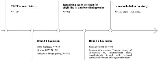
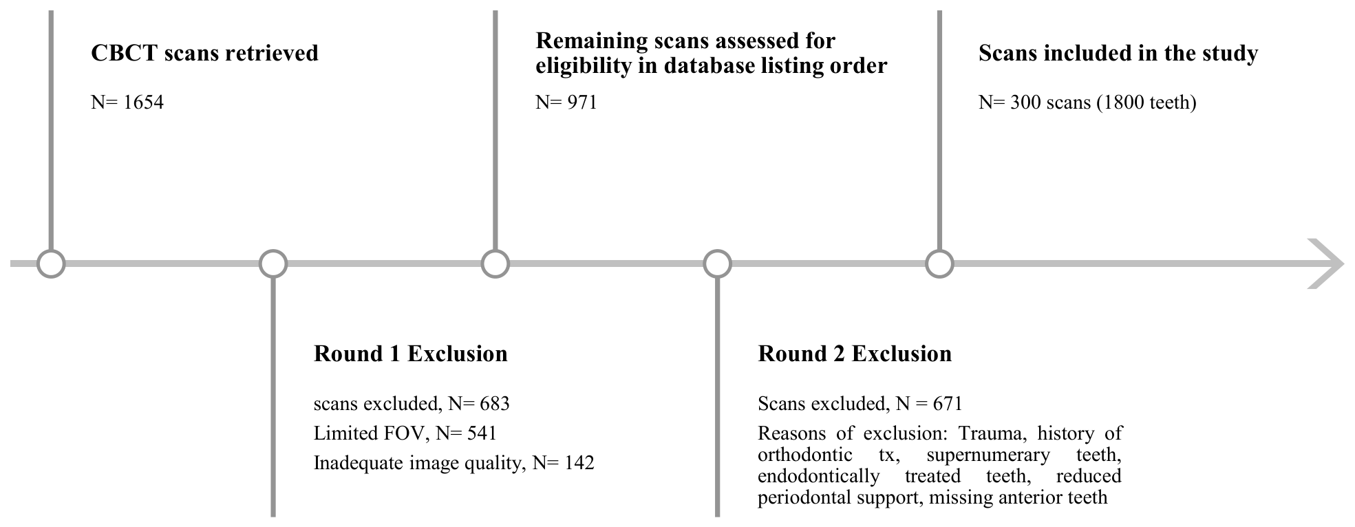
Figure 1 summarizes the scan screening process, exclusions, and final sample selection. A total of 1654 CBCT scans were retrieved from the institutional radiographic database for 2016–2024. After excluding 541 scans with a limited field of view and 142 with inadequate image quality, all remaining 971 scans were assessed against the eligibility criteria in database listing order. Of these, 671 were excluded based on the specified inclusion and exclusion criteria, leaving 300 eligible scans comprising 1800 maxillary anterior teeth. No scans remained unassessed.
Figure 1.
Flow diagram of CBCT scan screening, exclusions, and final sample selection. FOV: field of view; CBCT: cone beam computed tomography.
Exclusion criteria comprised mixed dentition, a history of trauma, previous or current orthodontic treatment, supernumerary teeth, endodontically treated or restored maxillary anterior teeth, malformed or dilacerated roots, reduced periodontal support, and missing maxillary anterior teeth. Scans showing maxillary anterior teeth adjacent to missing posterior teeth or dental implants were also excluded.
These criteria standardized anatomical and restorative assessment and minimized potential confounding factors. Teeth with stable periodontal support and intact natural crowns were selected so that the crowns could serve as references for planned restorations and desired screw-access positions. The exclusion of restored teeth also minimized restoration-related imaging artifacts.
3.3. Examiner Calibration and Intra-Examiner Reliability
All scans were handled by a single examiner (EA) calibrated to two experienced implantologists (A.A., B.A.) for digital implant planning. To assess intra-examiner reliability, 5% of randomly selected scans were chosen for repeat measurements at 2-week intervals. Categorical variables were evaluated using Cohen’s kappa statistics, demonstrating excellent agreement (κ = 0.82–1.00), while continuous variables were assessed using the intraclass correlation coefficient, which showed excellent reliability for all measurements (ICC = 0.90–0.96). Calibration with the two implantologists was conducted before data collection; however, inter-examiner agreement was not formally quantified, and therefore only intra-examiner reliability is reported.
3.4. Sagittal Root Position Classification
All six maxillary anterior teeth of each scan were evaluated in axially corrected sagittal view and categorized according to Kan et al.’s classification [9], as follows:
Class I: The root is positioned against the labial cortical plate.
Class II: The root is centered in the middle of the alveolar bone without engagement to either labial or palatal cortical plates at the apical third of the root.
Class III: The root is positioned against the palatal cortical plate.
Class IV: At least two-thirds of the root is engaging both the labial and palatal cortical plates, and it is a contraindication for immediate implant placement.
3.5. Implant Planning
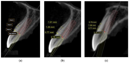
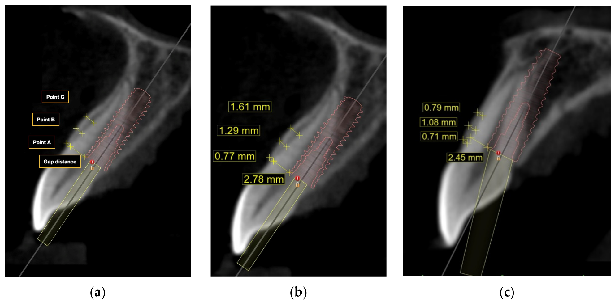
Figure 2 illustrates the measurements used during virtual implant planning and representative sites planned for straight and angulated screw-channel restorations. Implant planning was performed using BlueSky Bio planning software (Version 4.0.4.1; Blue Sky Bio LLC, Grayslake, IL, USA), where virtual implant placement was carried out using the digital library of Straumann Bone Level Tapered (BLT) implants (Straumann AG, Basel, Switzerland) with the following criteria of immediate implant placement [10,11]:
Figure 2.
Representative sagittal CBCT views illustrating virtual implant planning: (a) buccal wall thickness measurements at points A, B, and C located 0, 2, and 4 mm from the bone crest, respectively, and gap distance from implant platform to buccal crest; (b) a site planned for a straight screw-channel (SSC) restoration; (c) a site planned for an angulated screw-channel (ASC) restoration.
- The implant platform should be at labial bone level or measuring 2 mm apical to CEJ.
- There is a minimum gap distance of 1.5 mm between the implant and labial bone.
- At least 35% of the implant body should engage apical bone beyond the root.
SRP Class IV sites were classified as ineligible by protocol and were not subjected to virtual implant planning.
Restorative classification was based on the angular correction required to achieve screw access at the palatal cingulum of the anterior tooth. A straight screw-channel (SSC) restoration was indicated when no correction was required (0°), whereas an angulated screw-channel (ASC) restoration was indicated when correction of 1–25° achieved cingulum access. The maximum permitted correction was 25°, corresponding to the limit of the Straumann angled solution abutments. Sites requiring correction exceeding 25°, with screw access remaining outside the cingulum at the maximum permitted correction, were classified as requiring a cement-retained (CR) restoration.
3.6. Outcome Measures
For each tooth, the following measurements were acquired:
- Buccal wall thickness measured in three points perpendicular to long axis of the root.
- Whether immediate implant placement can be placed or not, with details related to implant diameter and length
- Whether screw-retained or cement-retained abutment can be used.
- The type of screw-retained abutment (straight or angulated) and exact angle of angulated abutment needed for the implant to be restored.
3.7. Statistical Analysis
Data was managed and summarized using SPSS (IBM SPSS Statistics, version 31, IBM Corp., Armonk, NY, USA). Descriptive statistics were calculated for all variables. Categorical variables, including eligibility for immediate implant placement and type of abutment used (straight screw-channel (SSC), angulated screw-channel (ASC), or cement-retained (CR)), were summarized as frequencies and percentages. Continuous variables, such as buccal wall thickness (mm) at the three reference points, horizontal gap distance (mm), and angulation required for angulated screw-channel abutments, were summarized as means and standard deviations.
Observations were clustered within patients and could not be assumed to be independent. All inferential analyses were therefore performed using mixed-effects models with a random intercept for patient (scan). Binary outcomes (SRP Class I, Class II, and Class III/IV versus the remaining classes, eligibility for immediate implant placement, SSC versus other abutment types, and implant length ≥ 14 mm) were analyzed with generalized linear mixed models (binomial distribution, logit link), and the results are presented as odds ratios (ORs) with 95% confidence intervals (CIs). Models of eligibility and abutment type according to SRP class were adjusted for tooth type. Cement-retained restorations were not modeled because no central incisor received a cement-retained restoration; this outcome showed quasi-complete separation by tooth type, which precludes stable estimation, and it is therefore reported descriptively. Continuous outcomes are presented with estimated mean differences, accompanied by 95% CIs. Model assumptions were examined through inspection of the histogram and a normal Q–Q plot of residuals, as well as a plot of residuals against the predicted values. The variance of the random intercept and the adjusted ICC were computed for each model to assess patient clustering, while the observation-level variance in logistic models was calculated using the delta method. Cramér’s V is reported as an indicator of the degree of association. Statistical inference is exclusively based on the results of mixed-effects models. The marginal R2 is presented for each model as the fraction of variance explained by the fixed effects. The analytic sample, number of contributing patients, model specifications, and the diagnostics of each model are provided in Supplementary Table S1.
4. Results
A total of 300 scans included with 1800 maxillary anterior teeth were evaluated (600 central incisors, 600 lateral incisors and 600 canines), with nearly equal gender distribution (51.3% females and 48.7% males). Sex was reported descriptively and was not evaluated as an explanatory variable.
4.1. Buccal Wall Thickness
Table 1 summarizes mean buccal wall thickness at three standardized reference points perpendicular to the long axis of the root (points A, B, and C at 0 mm, 2 mm, and 4 mm away from the bone crest, respectively). The mean buccal wall thickness was 0.86 ± 0.30 mm at point A, 1.09 ± 0.46 mm at point B, and 0.88 ± 0.49 mm at point C. Buccal wall thickness ≤ 1 mm was most frequently observed at point A (69.6%) and point C (60.7%). The complete absence of buccal bone at point A was identified in 3.7% of cases.
Table 1.
Mean buccal wall thickness.
4.2. SRP Classification
The frequency and percentage of SRP classification according to tooth type are summarized in Table 2. SRP classification was significantly associated with tooth type (Cramér’s V = 0.21), with canines most frequently associated with SRP Class I, lateral incisors with Classes III and IV, and central incisors with Class II. These associations remained significant after accounting for the clustering of teeth within patients.
Table 2.
Frequency (percentage) of Sagittal root position classification according to tooth type.
4.3. Eligibility for Immediate Implant Placement, Implant Diameter and Implant Length According to Tooth Type
Based on pre-set digital implant planning criteria, immediate implant placement was feasible in 1436 sites (79.8%), while 364 sites (20.2%) did not qualify for immediate placement protocol. Eligibility differed according to tooth type (Table 3), being higher in central incisors (86.3%) than in lateral incisors (76.7%) and canines (76.3%) (Cramér’s V = 0.12). The mean horizontal gap distance was 2.80 ± 0.61 mm. The largest mean gap was observed in canines (3.22 ± 0.64 mm), while the smallest gap was measured in central incisors (2.51 ± 0.47 mm). In the linear mixed model, gap distance differed significantly among tooth types (F(2, 1175) = 515.19, p < 0.001; marginal R2 = 0.25): compared with central incisors, the gap was 0.72 mm larger in canines (95% CI 0.67–0.78) and 0.17 mm larger in lateral incisors (95% CI 0.12–0.23), and it was 0.55 mm larger in canines than in lateral incisors (95% CI 0.49–0.61; all Bonferroni-adjusted p < 0.001). As expected, canines and central incisors that had the tendency to be planned for regular diameter implant length also differed according to tooth type (Cramér’s V = 0.30): implants ≥ 14 mm were planned in 72% of canine sites, 38.9% of lateral incisor sites, and 28.4% of central incisor sites, and this difference remained significant after accounting for clustering.
Table 3.
Frequency (percentage) of immediate implant placement, implant diameter and implant length according to tooth type.
4.4. Frequency of Immediate Implant Placement and Abutment Type According to Sagittal Root Position (SRP) Classification
Overall, ASC abutments were able to correct the angulation of 82.5% of immediately planned implants. SSC abutments were a feasible option only in 13.7% of the cases, while the remaining 3.8% had to be restored using a cement-retained crown (Table 4). When correlating abutment type to SRP position, ASC abutment was able to correct the angle of 75.6%, 56.4% and 52.4% of teeth classified as Classes I, II, and III, respectively (Table 5). Abutment type was significantly associated with both SRP class (Cramér’s V = 0.16) and tooth type (Cramér’s V = 0.19). After accounting for tooth type and clustering within patients, sites with SRP Classes II or III remained significantly more likely to be restorable with an SSC abutment than Class I sites. Cement-retained restorations occurred only in lateral incisors (n = 29) and canines (n = 26).
Table 4.
Frequency (percentage) of abutment types used in sites eligible for immediate implant placement; SSC: straight-screw channel abutment; ASC: angulated screw-channel abutment; CR: cement retained abutment. Percentages are calculated among sites eligible for immediate implant placement (n = 1436).
Table 5.
Virtual immediate implant eligibility and restorative category according to sagittal root position (SRP) classification; SSC: straight-screw channel abutment; ASC: angulated screw-channel abutment; CR: cement retained abutment. Percentages are calculated within each SRP class, including ineligible sites.
4.5. Mean ASC Angle According to Tooth Type and SRP Class
Among the 1184 sites restored with an ASC abutment, the mean correction angle was 14.1 ± 4.6° (range 4.4 to 25.0°). Angles were calculated only among sites requiring an ASC abutment; SSC sites, which require no angular correction, were not included. The maximum angle required was 25°, observed in five sites, and no site required correction beyond the 25° limit of the ASC system. Canines required the greatest mean angle (15.6 ± 4.4°) to relocate the screw access to the cingulum position, followed by lateral incisors (14.4 ± 4.6°) and central incisors (12.3 ± 4.1°); in the linear mixed model, these differences were significant (F(2, 974) = 100.02, p < 0.001; marginal R2 = 0.11), with canines requiring 3.61° (95% CI 2.99–4.24) and lateral incisors 2.49° (95% CI 1.86–3.12) more correction than central incisors, and canines requiring 1.12° (95% CI 0.50–1.74) more than lateral incisors (all Bonferroni-adjusted p < 0.001). The required angle also differed according to SRP class (p < 0.001): Class I sites required larger angles (14.5 ± 4.6°) than Class II sites (11.6 ± 3.7°; p < 0.001), whereas Class III sites (11.6 ± 4.5°; n = 11) did not differ significantly from either class.
4.6. Mixed-Effects Regression Analysis
Compared with central incisors, canines had approximately four times higher odds of presenting an SRP Class I root position (OR = 4.08, 95% CI 2.94–5.67; p < 0.001), whereas lateral incisors did not differ from central incisors (OR = 1.10; p = 0.492) (Table 6). Central incisors were the tooth type most likely to present a Class II root position, with markedly lower odds in lateral incisors (OR = 0.38, 95% CI 0.27–0.53) and canines (OR = 0.11, 95% CI 0.07–0.17). Conversely, lateral incisors had 3.6 times higher odds than central incisors of presenting an unfavorable Class III or IV position (OR = 3.57, 95% CI 2.34–5.43; p < 0.001), whereas canines did not differ significantly (p = 0.470).
Table 6.
Mixed-effects logistic regression models of root position, implant eligibility, implant length and abutment type, accounting for clustering of teeth within patients.
With regard to treatment planning, lateral incisors and canines had approximately half the odds of central incisors of being eligible for immediate implant placement (OR = 0.46 and 0.45, respectively; both p < 0.001). Among SRP Class I–III sites, and after adjustment for tooth type, the odds of eligibility decreased progressively from Class I to Class II (OR = 0.59, 95% CI 0.36–0.98; p = 0.039) and Class III (OR = 0.17, 95% CI 0.05–0.54; p = 0.003). Implants of 14 mm or longer were planned far more often in canines (OR = 16.32, 95% CI 11.29–23.57) and, to a lesser extent, in lateral incisors (OR = 1.97, 95% CI 1.41–2.75) than in central incisors (both p < 0.001).
When SRP class and tooth type were entered in the same model, sites with SRP Class II or III had 2.6 times higher odds of being restorable with an SSC abutment than Class I sites (OR = 2.60, 95% CI 1.65–4.09; p < 0.001), whereas lateral incisors (OR = 0.26, 95% CI 0.17–0.40) and canines (OR = 0.25, 95% CI 0.16–0.40) had about one quarter of the odds of central incisors (both p < 0.001).
5. Discussion
The present retrospective CBCT-based study provides a comprehensive three-dimensional assessment of maxillary anterior teeth and its relationship with surrounding alveolar housing, focusing on sagittal root position (SRP) classification and their implications on the timing of implant placement and type of abutment used as a final restoration.
5.1. Buccal Bone Thickness
The mean buccal bone thickness observed in this study confirms a predominantly thin facial bone wall in the maxillary anterior region. Similar CBCT-based investigations have reported that the buccal bone is ≤1 mm in most maxillary anterior sites, particularly at the most coronal/crestal level [23,24,25,26]. The high prevalence of thin buccal bone validates the inherent susceptibility of anterior maxilla to post-extraction ridge remodeling and esthetic complications following implant placement [13,27,28].
The complete absence of buccal plate was observed in 3.7% of cases, which is consistent with previous reports describing facial bone dehiscence in otherwise intact teeth [26,29]. While these findings support the necessity of CBCT-based evaluation prior to immediate implant placement to identify anatomical deficiencies requiring simultaneous augmentation interventions, it is worth noting that using large field of view CBCT scans may have affected the detection buccal plate, which is best measured in small FOV scans.
5.2. Sagittal Root Position and Tooth Type
A statistically significant association was observed between SRP classification and tooth type. Our findings align with the study by Kan et al. [9], which demonstrated the high prevalence of Class I root position, the most favorable root position for immediate implant protocol. The predominance of SRP Class I in canines observed in the present study may be attributed to their larger root diameter and the bucco–palatal positioning within the alveolar housing. Similar to Kan’s results, the least favorable root positions (Classes III and IV) for immediate implants were commonly encountered in lateral incisors, owing to their palatally positioned roots and reduced facial bone thickness, thereby complicating immediate implant placement [9,26,30].
5.3. Eligibility for Immediate Implant Placement
Based on our pre-defined eligibility criteria, approximately 80% of sites were deemed suitable for immediate implant placement. This proportion is comparable to previous CBCT-based planning studies reporting rates ranging between 65% and 85% [10,11,31]. The observed horizontal gap distances are consistent with previously reported values and support the frequent need for gap grafting, particularly in canine sites where larger jumping distances are commonly encountered [32].
5.4. Prosthetic Considerations
In the present study, only 13.7% of virtually planned immediate implants could be restored using a straight screw-channel (SSC) abutment, which is consistent with the 14% reported by Kan et al. [10] but lower than the 24% reported by Edmondson et al. [22]. In contrast, Radomsutthisarn et al. [33] reported a SSC frequency of only 3.5%. This discrepancy may be attributed to methodological differences, particularly the inclusion of 2.9-mm diameter implants, for which compatible dynamic or angulated screw-channel abutments are not manufactured. Consequently, a greater proportion of cases may have been classified as requiring cement-retained restorations, thereby reducing the reported SSC frequency. This finding may propose the idea of using narrow-diameter compared to regular-diameter implants to increase the likelihood of achieving straight screw-access owing to the restorative flexibility provided by a smaller platform diameter, a concept that warrants further research.
Angulated screw-channel (ASC) abutments were effective in correcting implant angulation in the majority of immediately planned implants. This finding aligns with recent studies demonstrating that ASC abutments allow for favorable screw access positioning while maintaining mechanical stability and minimizing the need for cement-retained restorations [11,12,20,34]. The greater angulation required for canines compared with central incisors reflects anatomical differences and has been similarly reported in prior prosthetically driven implant planning studies [11,20,22].
Although ASC abutments were indicated in 82.5% of virtually eligible sites, consistent with previous virtual implant-planning studies [10,11], concerns remain regarding the long-term mechanical performance of these restorations. Nevertheless, the current body of evidence suggests high survival rates for ASC restorations, with a relatively low incidence of mechanical complications [33,34,35,36]. The most frequently encountered complications were screw loosening (0–8%) and screw fracture (up to 2%), while ceramic chipping and loss of retention were reported less commonly [35,36]. From a biological perspective, multiple clinical studies have demonstrated comparable implant survival, marginal bone loss, and peri-implant tissue health between ASC and conventional screw-retained restorations. Collectively, these findings support the use of ASC abutments as a solution for managing implant angulation discrepancies in the esthetic zone while preserving the retrievability, maintenance advantages, and biological benefits associated with screw-retained prostheses.
5.5. Limitations
This retrospective CBCT study evaluated virtual planning feasibility and could not assess surgical, prosthetic, soft tissue, or long-term clinical outcomes. The selection criteria standardized anatomical assessment; however, the findings should be interpreted with caution when planning treatment for compromised sites. Furthermore, demographics such as age were unavailable, and systemic conditions and medications affecting bone metabolism were not systematically assessed.
Although predefined criteria guided SRP classification and virtual planning, both assessments were performed by the same examiner without blinding, and reliability testing was limited to intra-examiner repeatability. The relationship between SRP Class IV and ineligibility was determined by the study protocol rather than independently demonstrated through virtual implant planning. Mixed-effects models accounted for within-patient clustering, but the small numbers of SRP Class III sites and cement-retained restorations limited the precision of estimates for these categories.
Large-FOV CBCT introduces uncertainty when evaluating thin facial bone plates. Detection and measurement depend on spatial resolution, voxel size, reconstruction settings, and partial-volume effects rather than FOV alone. Lennholm et al. [37]. demonstrated variation in thin-bone visibility across imaging conditions, while Tanaka et al. [38]. reported limited agreement between CBCT and histologic measurements around maxillary anterior teeth. Very thin or apparently absent facial plates should therefore be interpreted cautiously. Without paired higher-resolution imaging or an independent anatomical reference, the magnitude and direction of measurement error and its influence on virtual eligibility classification could not be quantified.
Finally, planning was limited to Straumann BLT implants within specific implant planning software. Differences in implant geometry, restorative component compatibility, angulated screw-channel abutment correction limits, and planning workflows may yield different SSC/ASC frequencies, limiting generalizability to other systems.
6. Conclusions
Within this selected CBCT sample, 79.8% of maxillary anterior sites fulfilled the predefined virtual criteria for immediate implant placement, with the highest eligibility observed in central incisors (86.3%), followed by lateral incisors (76.7%) and canines (76.3%). SRP Class I showed the highest virtual eligibility, while the predominance of thin facial bone plates emphasized the importance of careful three-dimensional anatomical assessment. ASC restorations were indicated in 75.6%, 56.4%, and 52.4% of all sites classified as SRP Classes I, II, and III, respectively. Among the eligible sites, ASC correction expanded the proportion that was theoretically compatible with screw-retained restorations. These findings characterize the relationship between SRP and projected restorative requirements within virtual planning; clinical feasibility and long-term biological and mechanical outcomes require further investigation.
Supplementary Materials
The following supporting information can be downloaded at: https://www.mdpi.com/article/10.3390/dj14110709/s1, Table S1: Specification, analytical samples, clustering and estimated effects of the mixed-effects models.
Author Contributions
Conceptualization and study design: E.A. (Esraa Alabdurubalnabi), A.A.; Data collection: E.A. (Esraa Alabdurubalnabi); Virtual implant planning and measurements: E.A. (Esraa Alabdurubalnabi), A.A., B.A.; Data analysis and interpretation: E.A. (Esraa Alabdurubalnabi), M.A. (Mohammed Alsaati), F.E.A., M.A. (Mishali Alsharief), M.S.A., A.C.S., E.A. (Eman Aljoghaiman); Manuscript drafting: All authors; Critical revision of the manuscript: All authors. All authors have read and agreed to the published version of the manuscript.
Funding
This research received no external funding.
Institutional Review Board Statement
This retrospective study was approved by the Institutional Review Board of Imam Abdulrahman Bin Faisal University, Dammam, Saudi Arabia (IRB-PGS-2025-02-0175, approval date: 3 March 2025).
Informed Consent Statement
The requirement for informed consent was waived due to the use of anonymized CBCT radio-graphic records.
Data Availability Statement
The data presented in this study are available on reasonable request from the corresponding author, subject to institutional and ethical approval. The data are not publicly available due to patient privacy and confidentiality restrictions associated with clinical CBCT records.
Conflicts of Interest
The authors declare no conflicts of interest.
References
- Schulte, W.; Heimke, G. [The Tübinger immediate implant]. Quintessenz 1976, 27, 17–23. [Google Scholar] [PubMed]
- Wöhrle, P.S. Single-tooth replacement in the aesthetic zone with immediate provisionalization: Fourteen consecutive case reports. Pract. Periodontics Aesthetic Dent. PPAD 1998, 10, 1107–1114. [Google Scholar] [PubMed]
- Kan, J.Y.K.; Rungcharassaeng, K.; Lozada, J. Immediate placement and provisionalization of maxillary anterior single implants: 1-year prospective study. Int. J. Oral Maxillofac. Implants 2003, 18, 31–39. [Google Scholar]
- De Rouck, T.; Collys, K.; Cosyn, J. Immediate single-tooth implants in the anterior maxilla: A 1-year case cohort study on hard and soft tissue response. J. Clin. Periodontol. 2008, 35, 649–657. [Google Scholar] [CrossRef] [Scilit] [PubMed]
- Seyssens, L.; Eeckhout, C.; Cosyn, J. Immediate implant placement with or without socket grafting: A systematic review and meta-analysis. Clin. Implant Dent. Relat. Res. 2022, 24, 339–351. [Google Scholar] [CrossRef] [Scilit] [PubMed]
- Wittneben, J.; Molinero-Mourelle, P.; Hamilton, A.; Alnasser, M.; Obermaier, B.; Morton, D.; Gallucci, G.O.; Wismeijer, D. Clinical performance of immediately placed and immediately loaded single implants in the esthetic zone: A systematic review and meta-analysis. Clin. Oral Implants Res. 2023, 34, 266–303. [Google Scholar] [CrossRef] [Scilit] [PubMed]
- Kois, J.C.; Kan, J.Y. Predictable peri-implant gingival aesthetics: Surgical and prosthodontic rationales. Pract. Proced. Aesthetic Dent. PPAD 2001, 13, 691–692. [Google Scholar] [PubMed]
- Buser, D.; Martin, W.; Belser, U.C. Optimizing esthetics for implant restorations in the anterior maxilla: Anatomic and surgical considerations. Int. J. Oral Maxillofac. Implants 2004, 19, 43–61. [Google Scholar]
- Kan, J.Y.K.; Roe, P.; Rungcharassaeng, K.; Patel, R.D.; Waki, T.; Lozada, J.L.; Zimmerman, G. Classification of sagittal root position in relation to the anterior maxillary osseous housing for immediate implant placement: A cone beam computed tomography study. Int. J. Oral Maxillofac. Implants 2011, 26, 873–876. [Google Scholar]
- Kan, J.; Rungcharassaeng, K.; Lin, G.-H.; Zucchelli, G.; Kamolroongwarakul, P.; Matsuda, H.; Lozada, J. Incidence of Straight-Channel Screw-Retained Single Crown Following Immediate Implant Placement and Provisionalization in the Esthetic Zone: A CBCT Study. Int. J. Oral Maxillofac. Implants 2021, 36, 793–798. [Google Scholar] [CrossRef] [Scilit] [PubMed]
- Kan, J.Y.K.; Rungcharassaeng, K.; Kamolroongwarakul, P.; Lin, G.; Matsuda, H.; Yin, S.; Wang, H.; Tarnow, D.; Lozada, J.L. Frequency of screw-retained angulated screw channel single crown following immediate implant placement and provisionalization in the esthetic zone: A cone beam computed tomography study. Clin. Implant Dent. Relat. Res. 2023, 25, 789–794. [Google Scholar] [CrossRef] [Scilit] [PubMed]
- Swamidass, R.S.; Kan, J.Y.K.; Kattadiyil, M.T.; Goodacre, C.J.; Lozada, J. Abutment screw torque changes with straight and angled screw-access channels. J. Prosthet. Dent. 2021, 125, 675–681. [Google Scholar] [CrossRef] [Scilit] [PubMed]
- Chen, S.T.; Buser, D.; Sculean, A.; Belser, U.C. Complications and treatment errors in implant positioning in the aesthetic zone: Diagnosis and possible solutions. Periodontol. 2000 2023, 92, 220–234. [Google Scholar] [CrossRef] [Scilit] [PubMed]
- Xu, D.; Wang, Z.; Sun, L.; Lin, Z.; Wan, L.; Li, Y.; Lin, X.; Peng, W.; Zhang, Z.; Gao, Y. Classification of the Root Position of the Maxillary Central Incisors and its Clinical Significance in Immediate Implant Placement. Implant Dent. 2016, 25, 520–524. [Google Scholar] [CrossRef] [Scilit] [PubMed]
- Chan, H.-L.; Garaicoa-Pazmino, C.; Suarez, F.; Monje, A.; Benavides, E.; Oh, T.-J.; Wang, H.-L. Incidence of Implant Buccal Plate Fenestration in the Esthetic Zone: A Cone Beam Computed Tomography Study. Int. J. Oral Maxillofac. Implants 2014, 29, 171–177. [Google Scholar] [CrossRef] [Scilit] [PubMed]
- Staubli, N.; Walter, C.; Schmidt, J.C.; Weiger, R.; Zitzmann, N.U. Excess cement and the risk of peri-implant disease—A systematic review. Clin. Oral Implants Res. 2017, 28, 1278–1290. [Google Scholar] [CrossRef] [Scilit] [PubMed]
- Sailer, I.; Karasan, D.; Todorovic, A.; Ligoutsikou, M.; Pjetursson, B.E. Prosthetic failures in dental implant therapy. Periodontol. 2000 2022, 88, 130–144. [Google Scholar] [CrossRef] [Scilit] [PubMed]
- Wilson, T.G., Jr. The Positive Relationship Between Excess Cement and Peri-Implant Disease: A Prospective Clinical Endoscopic Study. J. Periodontol. 2009, 80, 1388–1392. [Google Scholar] [CrossRef] [Scilit] [PubMed]
- Quaranta, A.; Lim, Z.W.; Tang, J.; Perrotti, V.; Leichter, J. The Impact of Residual Subgingival Cement on Biological Complications Around Dental Implants. Implant Dent. 2017, 26, 465–474. [Google Scholar] [CrossRef] [Scilit] [PubMed]
- Cavallaro, J.; Greenstein, G. Angled Implant Abutments. J. Am. Dent. Assoc. 2011, 142, 150–158. [Google Scholar] [CrossRef] [Scilit] [PubMed]
- Garcia-Gazaui, S.; Razzoog, M.; Sierraalta, M.; Saglik, B. Fabrication of a screw-retained restoration avoiding the facial access hole: A clinical report. J. Prosthet. Dent. 2015, 114, 621–624. [Google Scholar] [CrossRef] [Scilit] [PubMed]
- Edmondson, E.K.; Trejo, P.M.; Soldatos, N.; Weltman, R.L. The ability to screw-retain single implant-supported restorations in the anterior maxilla: A CBCT analysis. J. Prosthet. Dent. 2022, 128, 443–449. [Google Scholar] [CrossRef] [Scilit] [PubMed]
- Braut, V.; Bornstein, M.M.; Belser, U.; Buser, D. Thickness of the anterior maxillary facial bone wall-a retrospective radiographic study using cone beam computed tomography. Int. J. Periodontics Restor. Dent. 2011, 31, 125–131. [Google Scholar]
- Januário, A.L.; Duarte, W.R.; Barriviera, M.; Mesti, J.C.; Araújo, M.G.; Lindhe, J. Dimension of the facial bone wall in the anterior maxilla: A cone-beam computed tomography study. Clin. Oral Implants Res. 2011, 22, 1168–1171. [Google Scholar] [CrossRef] [Scilit] [PubMed]
- Chappuis, V.; Engel, O.; Reyes, M.; Shahim, K.; Nolte, L.-P.; Buser, D. Ridge Alterations Post-extraction in the Esthetic Zone. J. Dent. Res. 2013, 92, 195S–201S. [Google Scholar] [CrossRef] [Scilit] [PubMed]
- Aljabr, A.A.; Almas, K.; Aljofi, F.E.; Aljabr, A.A.; Alzaben, B.; Alqanas, S. A CBCT Study of Labial Alveolar Bone Thickness in the Maxillary Anterior Region in a Teaching Hospital Population in the Eastern Province of Saudi Arabia. Biomedicines 2023, 11, 1571. [Google Scholar] [CrossRef] [Scilit] [PubMed]
- Kan, J.Y.K.; Rungcharassaeng, K.; Lozada, J.L.; Zimmerman, G. Facial gingival tissue stability following immediate placement and provisionalization of maxillary anterior single implants: A 2- to 8-year follow-up. Int. J. Oral Maxillofac. Implants 2011, 26, 179–187. [Google Scholar]
- Kan, J.Y.K.; Yin, S.; Rungcharassaeng, K.; Zucchelli, G.; Urban, I.; Lozada, J. Facial implant gingival level and thickness changes following maxillary anterior immediate tooth replacement with scarf-connective tissue graft: A 4–13-year retrospective study. J. Esthet. Restor. Dent. 2023, 35, 138–147. [Google Scholar] [CrossRef] [Scilit] [PubMed]
- Cook, D.R.; Mealey, B.L.; Verrett, R.G.; Mills, M.P.; Noujeim, M.E.; Lasho, D.J.; Cronin, R.J. Relationship between clinical periodontal biotype and labial plate thickness: An in vivo study. Int. J. Periodontics Restor. Dent. 2011, 31, 345–354. [Google Scholar]
- Zekry, A.; Wang, R.; Chau, A.C.M.; Lang, N.P. Facial alveolar bone wall width—A cone-beam computed tomography study in Asians. Clin. Oral Implants Res. 2014, 25, 194–206. [Google Scholar] [CrossRef] [Scilit] [PubMed]
- Funato, A.; Salama, M.A.; Ishikawa, T.; Garber, D.A.; Salama, H. Timing, positioning, and sequential staging in esthetic implant therapy: A four-dimensional perspective. Int. J. Periodontics Restor. Dent. 2007, 27, 313–323. [Google Scholar]
- Levine, R.A.; Dias, D.R.; Wang, P.; Araújo, M.G. Effect of the buccal gap width following immediate implant placement on the buccal bone wall: A retrospective cone-beam computed tomography analysis. Clin. Implant Dent. Relat. Res. 2022, 24, 403–413. [Google Scholar] [CrossRef] [Scilit] [PubMed]
- Radomsutthisarn, W.; Kretapirom, K.; Nakaparksin, P. Incidence of straight and angulated screw channel following immediate implant placement in esthetic zone: A simulated cone-beam computed tomography study. PLoS ONE 2024, 19, e0308158. [Google Scholar] [CrossRef] [Scilit] [PubMed]
- Wittneben, J.; Buser, D.; Salvi, G.E.; Bürgin, W.; Hicklin, S.; Brägger, U. Complication and Failure Rates with Implant-Supported Fixed Dental Prostheses and Single Crowns: A 10-Year Retrospective Study. Clin. Implant Dent. Relat. Res. 2014, 16, 356–364. [Google Scholar] [CrossRef] [Scilit] [PubMed]
- Pitman, J.; Van Craenenbroeck, M.; Glibert, M.; Christiaens, V. Screw loosening in angulation-correcting single implant restorations: A systematic review of in vitro studies. J. Prosthet. Dent. 2024, 132, 520–527. [Google Scholar] [CrossRef] [Scilit] [PubMed]
- Rasaie, V.; Abduo, J.; Falahchai, M. Clinical and Laboratory Outcomes of Angled Screw Channel Implant Prostheses: A Systematic Review. Eur. J. Dent. 2022, 16, 488–499. [Google Scholar] [CrossRef] [Scilit] [PubMed]
- Lennholm, C.; Andreasen, H.; Westerlund, A.; Lund, H. Visibility of alveolar bone thicknesses on CBCT images–a study on minimum bone requirements using various reconstruction techniques, viewing modes, and resolutions. Clin. Oral Investig. 2024, 28, 641. [Google Scholar] [CrossRef] [Scilit] [PubMed]
- Tanaka, Y.; Dutra, V.; Lin, W.-S.; Levon, J.; Hamada, Y. Evaluation of the accuracy of buccal bone thickness measurement from cone beam computed tomography compared with histologic analysis. J. Prosthet. Dent. 2023, 130, 68–73. [Google Scholar] [CrossRef] [Scilit] [PubMed]

Disclaimer/Publisher’s Note: The statements, opinions and data contained in all publications are solely those of the individual author(s) and contributor(s) and not of MDPI and/or the editor(s). MDPI and/or the editor(s) disclaim responsibility for any injury to people or property resulting from any ideas, methods, instructions or products referred to in the content. |
© 2026 by the authors. Licensee MDPI, Basel, Switzerland. This article is an open access article distributed under the terms and conditions of the Creative Commons Attribution (CC BY) license.