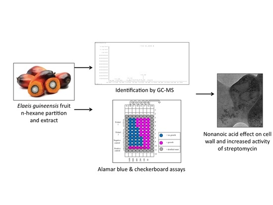
Abstract
Natural products with antimycobacterial adjuvant potential may be utilized to address the rise of multidrug-resistant tuberculosis (TB). The antioxidant-rich oil palm (Elaeis guineensis) fruit (OPF) was investigated for antimycobacterial activity against Mycobacterium tuberculosis (MTB) H37Ra using bio-guided fractionation techniques, followed by determination of fractional inhibition index (FIC) with first-line anti-TB drugs. In vitro screening using microplate Alamar blue assay showed n-hexane and chloroform partitions of OPF mesocarp had a minimum inhibitory concentration (MIC) of 400–800 µg/mL. The n-hexane fraction contained nonanoic acid (C9H18O2), decanoic acid (C10H20O2), and dodecanoic acid (C12H24O2), identified by gas chromatography-mass spectrometry, which all had an MIC of 50 µg/mL. Nonanoic and decanoic acids had additive effects when combined with streptomycin (FIC index: 0.625) and rifampicin (FIC index: 0.75), respectively. Isoniazid had a 16-fold increase in activity when combined with nonanoic acid and decanoic acid. The combination of nonanoic acid with streptomycin was bactericidal to 99.9% of MTB H37Ra by Day 7 of the time-kill assay, with structural damage of the cell wall observed using electron microscopy. Cytotoxicity assessment using Vero cells confirmed nonanoic acid had low toxicity with LC50 of > 200 µg/mL. The bio-guided fractionation of OPF shows the presence of fatty acids with anti-TB adjuvant potential.
1. Introduction
Mycobacterium tuberculosis (MTB) causes tuberculosis (TB), a major life-threatening respiratory disease. In 2019, 10 million people were infected with TB, resulting in 1.2 million deaths [1]. Although existing vaccines and diagnostics to control TB remain suboptimal [2], the disease is curable by a combination therapy of 2–3 first-line anti-TB drugs such as rifampicin, isoniazid, ethambutol, and pyrazinamide for at least six months [1]. However, the long treatment period and potential liver toxicity leads to poor adherence, incomplete treatment, and consequently, the emergence of drug-resistant TB, which further increases the difficulty of TB management [3]. The global increase in multidrug-resistant TB (MDR-TB) has become a serious threat to human health [1]. Although second-line drugs such as levofloxacin and bedaquiline can be administered as alternatives to the first-line anti-TB drugs, they are less effective and more toxic to the patients [3]. The discovery of new compounds with additive or synergistic interactions with existing drugs may reduce their required doses and toxicity and address this challenge [4]. Although both synergism and additivity can lower the minimum effective dose of antimicrobial agents, adjuvants with additive effects are less explored in combination therapy due to the presumption that they are less useful compared to adjuvants exerting synergistic effects. However, recent research suggests that compounds with additive effects may exert action on target cells that is similar to synergistic compounds [5].
Historically, fatty acids have shown promising antimycobacterial activity against a variety of mycobacteria [5,6,7]. Despite earlier promising results of antimycobacterial activity of fatty acids, such research was primarily conducted in the 1960–80s, without further pursuing the potential applications of these lipids to improve the existing treatment of TB. In particular, there is an opportunity to harness newer antimicrobial discovery methodologies such as broth microdilution [8] and prospect lipids from economical plant sources [9], such as oil palm (Elaeis guineensis) fruit (OPF). Abundantly grown and widely accessible in tropical countries with moderate to high TB prevalence, such as Malaysia and Indonesia [1,10], OPF contains compounds with antioxidant properties such as tocotrienols and carotenoids [11], which reportedly may prevent heart disease, moderate blood pressure, and suppress tumorigenesis [12]. Studies on pharmacologically active compounds of oil palm have previously focused on leaf extracts and phenolics for atheroprotective, anti-tumor, and neuroprotective effects [13]. Fewer studies have investigated the antimicrobial potential of the edible OPF. Although a study evaluating the antimicrobial activity of methanolic oil palm leaf extracts against a variety of microorganisms reported relatively high minimum inhibitory concentration (MIC) ranging from 6.25–12.5 mg/mL [14], linolenic acids in OPF have demonstrated inhibitory action against methicillin-resistant Staphylococcus aureus (MRSA) [15]. Despite this, the antimycobacterial and nutraceutical potential of OPF has not been previously evaluated. Thus, this study investigated n-hexane partitions of OPF containing several compounds with inhibitory action against MTB H37Ra, a TB surrogate species, using microplate Alamar blue assay (MABA) to determine MIC and minimum bactericidal concentration (MBC) and checkerboard assay to determine interactions with first-line anti-TB drugs. We report novel findings of additive interaction between nonanoic acid and decanoic acids with streptomycin and rifampicin, respectively.
2. Materials and Methods
2.1. Bacterial Culture
MTB H37Ra ATCC 25177 was purchased from American Type Culture Type (ATCC, Manassas, VA, USA). Stock cultures were maintained on Middlebrook 7H10 (M7H10) agar (Difco, Franklin Lakes, NJ, USA) at 4 °C and stored in Middlebrook 7H9 (M7H9) broth (Difco, Franklin Lakes, NJ, USA) with 20% glycerol at −20 °C. Before usage, the mycobacteria were subcultured on M7H10 agar to ensure the purity of the mycobacteria and to prevent contamination. Cultures were incubated for 10 days at 37 °C in 4% CO2, and freshly subcultured mycobacteria were used to standardize the inocula.
2.2. Plant Extraction
Oil palm fruit (OPF) was collected from United Oil Palm Industries Sdn Bhd, Jalan Bandar Baru, 878, Jalan Sungai Kechil, 14300, Nibong Tebal, Pulau Pinang, authenticated by the Herbarium Unit of School of Biological Sciences, Universiti Sains Malaysia (voucher numbers 11503 a, b, and c). OPF mesocarp was cut into small pieces and sun-dried for over one week, then ground into powder form, and macerated in 700 mL of 80% methanol (Qrec, Rawang, Malaysia) with 20% distilled water (dH2O) at room temperature (RT) (26–28 °C). Methanolic extracts were filtered by using filter paper no. 1 (Whatman, Maidstone, UK) and oven-dried at 40 °C. A new batch of 80% methanol was added to continue the maceration process, repeated for 4–5 times, and the pooled methanolic extracts were weighed and stored at 4 °C prior to usage. The dried crude methanolic extract of OPF mesocarp was partitioned using n-hexane, chloroform, ethyl acetate, and n-butanol (Qrec, Rawang, Malaysia). First, five grams of mesocarp crude methanolic extract was dissolved in 30 mL of 100% methanol and 170 mL dH2O was added into the mixture to give a total volume of 200 mL at 3:17 v/v ratio. A volume of 200 mL of n-hexane (first solvent) was added into the aqueous crude extract to produce a 1:1 ratio of mixture. The mixture was then transferred into a separation funnel and left overnight until two layers were formed. Then, the n-hexane extract was taken out and dried in a fume hood. Each solvent change was repeated three times. Dried extract partitions were weighed and stored in the refrigerator (4 °C) before screening.
2.3. Antimycobacterial Screening
2.3.1. MIC Assay
MABA was used to screen for OPF fractions, partitions, and compounds with antimycobacterial potential [8]. Briefly, 100 µL of M7H9 broth was added into all test and control wells. Next, 100 µL of each partition/fraction (diluted in 4% dimethyl sulfoxide (DMSO) (Sigma Aldrich, St. Louis, MO, USA) or drug were tested in triplicate at concentrations of 1600 µg/mL to 100 µg/mL and 10–0.0098 µg/mL, respectively. Then, 100 µL of MTB H37Ra inoculum was added, and the plates were incubated for five days at 37 °C in 4% CO2. Next, 50 µL of freshly prepared Alamar blue reagent in 10% Tween 80 was added and plates were re-incubated for 24 h. The MIC was determined based on the concentration of extract or antibiotic that prevented the color change from pink (growth) to blue (no growth).
2.3.2. MBC Assay
The MBC assay was conducted on compounds with the lowest MIC value and on the sixth day of incubation. Mycobacterial cultures from wells without growth on MABA were streaked on M7H10 agar plates in duplicate. The plates were incubated at 37 °C in 4% CO2 for 10 days and the presence of colonies was determined post-incubation. The MBC was recorded as the minimum concentration of the test agent required to kill 99% of bacterial cells.
2.4. Identification of Active Compound
2.4.1. Thin Layer Chromatography (TLC)
The n-hexane partition was selected for further fractionation using a series of different solvent systems on TLC. A line was drawn near the bottom of the TLC plate, and small amounts of dried partitions were dissolved in n-hexane and spotted repeatedly using a capillary glass tube on the line of the TLC plate containing silica gel F254 (Merck, Darmstadt, Germany) in 2 × 10 cm strips. After the plate was left to dry completely and placed in the chamber, the solvent was run towards the peak, and the plate was removed before the solvent reached the peak. The chromatogram was dried and observed under ultraviolet (UV) (245 nm) and visible (365 nm) light.
2.4.2. Column Chromatography
The n-hexane partition was fractionated using column chromatography with 20 × 2 cm (length/width) glass column packed with 30 g of silica gel 60, 230–400 mesh (40–63 microns) (Merck, Darmstadt, Germany). The silica gel was mixed with n-hexane: ethyl acetate solvent system at a ratio of 98:2 and poured and packed into the glass column. The sample was loaded by dissolving 0.5 g of sample in n-hexane mixed with 2 g of silica gel, which was left to dry then loaded into the packed column. A ratio of 98:2 of n-hexane: ethyl acetate was added into the column prior to the elution process, and then eluates were continuously collected using 100 mL beaker, before 100 mL of acetone and methanol were added to wash the column. Each fraction was dried and further analyzed by TLC for observation of collected compounds.
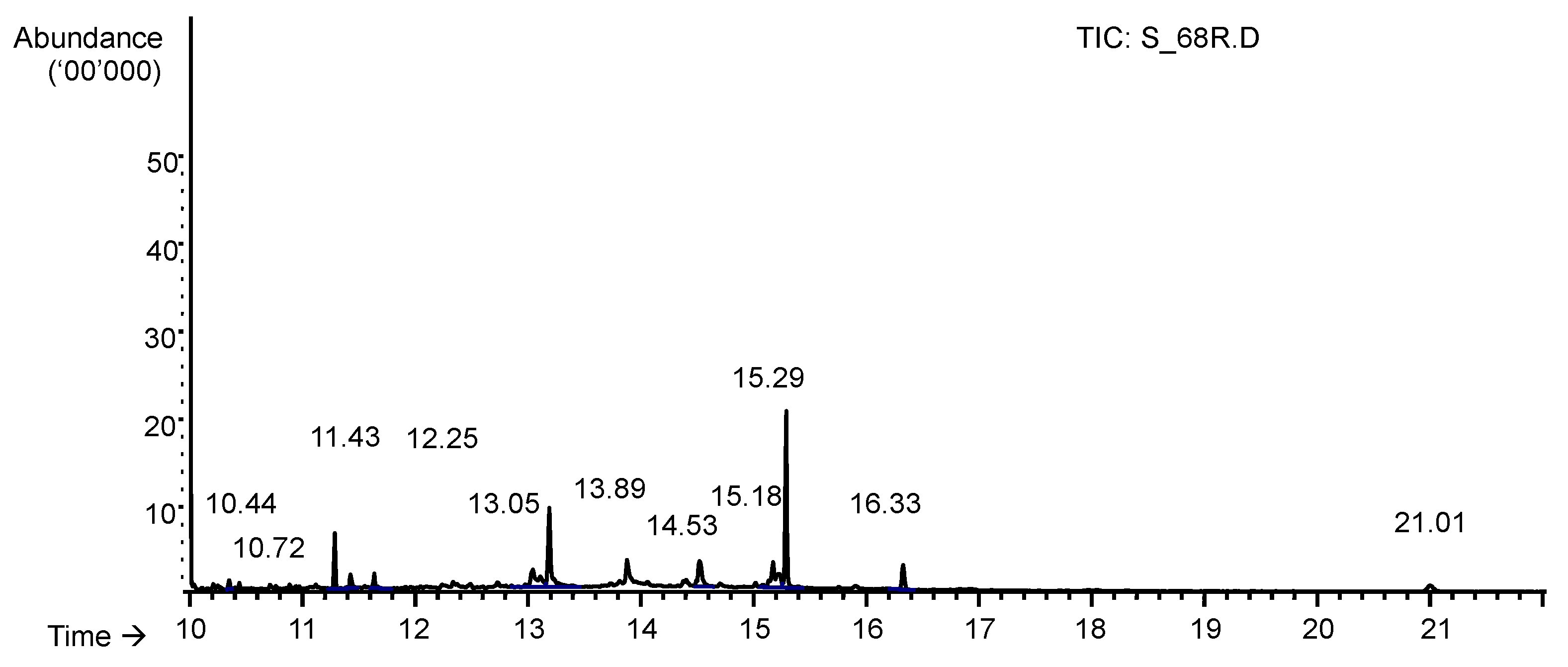
2.4.3. Gas Chromatography-Mass Spectrometry (GC-MS)
The fraction with the highest antimycobacterial activity (Fraction 8) was sorted, analyzed, and volatile compounds present were identified using GC-MS (outsourced to National Poison Centre, Universiti Sains Malaysia) on Agilent 7890 A GC system coupled with Agilent 5975C Inert XL EI/CI MSD with Triple-Axis Detector, equipped with an HP-5 cross-linked 5% phenyl methyl siloxane, and fused with silica capillary column. A constant flow rate of helium gas at 2 mL/min, 1 µL of injection volume, electron ionization system with 70 eV of ionizing energy, and inlet and detector temperature of 280 °C were maintained. Compounds were identified by mass spectral matching against databases of standard compounds in the National Institute Standard and Technology (NIST Version 0.8, Gaithersburg, MD, USA) and Wiley libraries.
2.5. Drug Interaction
2.5.1. Checkerboard Assay
The active compounds identified (nonanoic acid, decanoic acid, and dodecanoic acid) were obtained commercially (Sigma Aldrich, St. Louis, MO, USA) and combined with streptomycin, ethambutol, rifampicin, and isoniazid. Their interaction effects were assessed using an established checkerboard assay method (6 × 6 wells design in duplicate) with slight modifications [16]. The MIC of each drug was first determined against MTB H37Ra using the previously described MABA method, with working concentrations for compound and drug prepared four times higher than their MICs. The FIC index was calculated, and interactions were interpreted as such: ≤ 0.5 (synergism), 0.5 ≤ 0.75 (additive), 1 (indifferent), 1.0 ≤2.0 (transition), > 2.0 (antagonism).
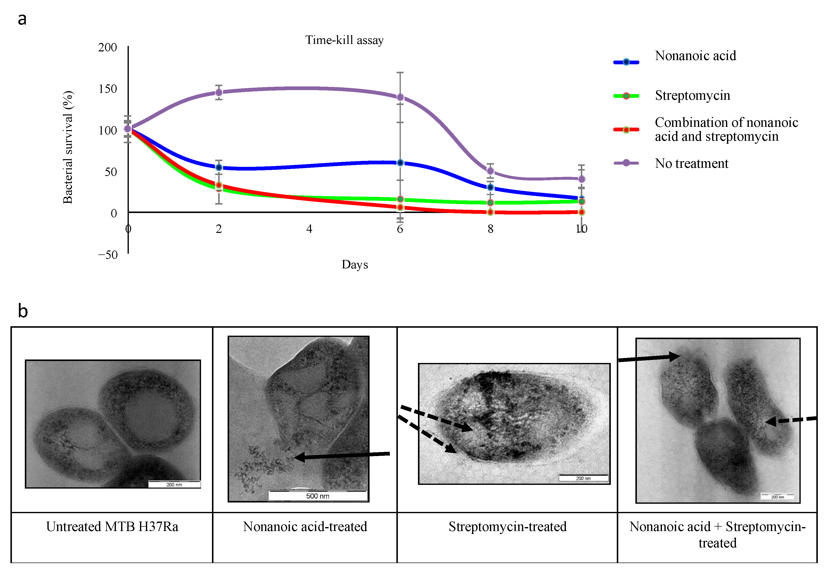
2.5.2. Time–Kill Assay
Test manipulates (growth control, compounds, drug control, and combination of compound with drug) were prepared in sterilized universal bottles in duplicates. Compounds, streptomycin, and rifampicin at their MIC concentrations were mixed thoroughly, 0.5 mL of the mycobacterial inoculum with a turbidity of McFarland No.3 standard was added amounting to 5 mL/bottle and incubated at 37 °C in 4% CO2 with swirling for 10 days. Each culture bottle was sampled (20 µL) on Days 0, 2, 4, 6, 8, and 10, transferred into 180 µL of phosphate buffer saline (PBS) on a 96-well microplate, serially diluted to 10−5 dilution. A 10 µL volume from each dilution factor was dropped onto each M7H10 agar plate in triplicate, air-dried, and incubated at 37 °C in 4% CO2 for 21 days. The colony numbers were recorded, and the CFU/ml were calculated. The interaction effects on bactericidal activity were interpreted as such: 100-fold increase (synergism), 10-fold increase (additive/indifferent), and 100 fold decrease (antagonism) [17].
2.6. Transmission Electron Microscopy
The pelleted bacterial cultures were resuspended and immersed in McDowell-Trump fixative solution for two h in 0.1 M phosphate buffer (pH 7.2), re-immersed in 0.1 M PBS, and centrifuged to obtain pellets (twice). Post-fixation, the pellets were suspended in 1% osmium tetroxide in PBS for one hour in a fume hood, the samples were centrifuged, and the pellets were re-washed twice with dH2O. Tubes containing fixed pellets were kept in a water bath while 3% agar solution was prepared and solidified in a test tube, and equilibrated to 45 °C. An agar drop was transferred into each pellet-containing tube, mixed well, poured onto a glass microscope slide, and cut into 1 mm3 cubes. The cubes were serially dehydrated with ethanol and 100% acetone. The mixture of acetone and Spurr’s mix resin (1:1) was used to infiltrate the cubes in a rotator for 15–30 min, infiltrated overnight with Spurr’s mix in a rotator, and changed daily for four days. The cubes were embedded and cured (60 °C, 48 h), and trimmed and sliced on an ultramicrotome machine (PowerTome XL, Tucson, AZ, USA). Ultra-thin (<1 µm) sectioned slices were placed on a copper grid for uranyl acetate and lead citrate staining (10 min), sequentially, and washed with dH2O pre- and post-staining, then observed under TEM (EFTEM Libra 120-Carl Zeiss, Oberkochen, Germany).
2.7. Cytotoxicity Evaluation
The cytotoxicity of nonanoic acid was tested against African Green Monkey (Vero) cell-line (ATCC, Manassas, VA, USA) using a modified 2D cytotoxicity assay [18]. Briefly, Vero cells were grown in Dulbecco’s modification of Eagle medium supplemented with 5% fetal bovine serum, 1% penicillin-streptomycin at 37 °C in 5% CO2. The cells were seeded at 1x 10⁴ cells/well and the culture plates were incubated for 24 h at 37 °C in 5% CO2 then tested with 100 µL nonanoic acid (25 µg/mL to 200 µg/mL) with 1% DMSO. The plate was incubated at 37 °C in 5% CO2 for five days, then 50 µL of 5 mg/mL MTT reagent (2.5 mg/mL 3-(4,5-dimethyl-2-thiazolyl)-2,5-diphenyl-2H-tetrazoliumbromide in PBS) was added. The absorbance was measured using a microplate spectrophotometer at 570 nm, and the cytotoxicity was determined by evaluating the viability of treated Vero cells compared to untreated controls [18].
3. Results and Discussion
3.1. Yield and Anti-Mycobacterial Activity of OPF Fractions and Partitions
A sequential partition of crude methanolic extract of OPF mesocarp was done using n-hexane, followed by chloroform, ethyl acetate, and butanol, consecutively. The MIC was evaluated using MABA. Of all partitions, the n-hexane partition had the highest yield, followed by aqueous, butanol, chloroform, and least of all, ethyl acetate partitions (Table 1). A preliminary screening of the six mesocarp partitions showed moderate activity with MIC values of 400 and 800 µg/mL, and weak activity with MIC of 1600 µg/mL [19]. Partitions of n-hexane and chloroform had MIC values of 800 and 400 µg/mL, respectively. Although the chloroform partition had a lower MIC, only the n-hexane partition was selected for further investigation due to the low yield of the chloroform partition.
Table 1.
Yield and in vitro anti-mycobacterial activity of oil palm fruit (OPF) mesocarp extract partitions against Mycobacterium tuberculosis (MTB) H37Ra.
Nine n-hexane fractions obtained through column chromatography using n-hexane: ethyl acetate solvent system and confirmed by thin-layer chromatography (TLC) were screened for antimycobacterial activity against MTB H37Ra using MABA. The fractions had MIC values in the range of 50–800 µg/mL, with Fraction 8 exhibiting the highest activity against MTB H37Ra with MIC of 50 µg/mL. After subjecting Fraction 8 to TLC and GC-MS analysis (Figure 1), ten compounds were identified through mass spectral matching (Figure S1). However, only nine compounds that were commercially available were further screened for antimycobacterial activity (Table 2). Among these, hexadecanoic acid was the most abundant (0.08%), followed by oleic acid (0.04%), and 9-octadecenamide (0.03%), and the remaining seven identified compounds each were 0.01% of the sample. The compounds identified amounted to ~0.23% of the sample, while the remaining constituent was the DMSO solvent. All 10 of the active compounds identified in Fraction 8 were fatty acids, likely because OPF is 50% oil [11,20]. However, this is limited by the fact that the other peaks detected were not identified or tested further in this study.
Figure 1.
The graph shows gas chromatography-mass spectrometry (GC-MS) chromatogram for Fraction 8, of which 9 out of 10 identified compounds were further investigated to determine antimycobacterial activity. Samples were analyzed using Agilent 7890 A GC system with constant flow rate of helium gas at 2 mL/min, 1 µL of injection volume, electron ionization system with 70 eV of ionizing energy, with inlet and detector temperature of 280 °C.
Table 2.
Characterization and antimycobacterial activity of active compounds detected in OPF n-hexane partition Fraction 8.
3.2. MIC and MBC Screening of GC-MS Identified Compounds in n-hexane Fractions
Among identified fatty acids, nonanoic (pelargonic) acid, decanoic acid (capric), and dodecanoic (lauric) acid had the lowest MIC of 50 µg/mL. Nonanoic acid and decanoic acid appeared to be bacteriostatic, while dodecanoic acid was likely bactericidal against MTB H37Ra, given that the MBC of the latter was less than four-fold its MIC value [21] (Table 2).
Plant extracts used as traditional therapy for TB or other respiratory diseases reportedly contain fatty acids [22], and the antimycobacterial potential of palmitic acid from citrus fruit extracts against MTB H37Rv and MTB M26 (clinical strain) [23] have been described. Our findings align with previous reports of nonanoic acid (C9H18O2) inhibiting M. smegmatis [24], and decanoic acid (C10H20O2) inhibiting MRSA strains [25]. The action of such volatile and semi-volatile fatty acids may be due to the fact that these compounds are able to traverse the lipophilic mycolic acid-rich mycobacterial cell wall more easily [26,27]. However, unlike earlier reports that suggest the bactericidal activity of fatty acids increases with carbon chain length [7], specifically the highly lethal effects of myristic acid (C14H28O2) on M. bovis and MTB H37Ra [6], our study finds that myristic acid had only modest MIC. No bactericidal effects were observed for other long-chain fatty acids.
3.3. Additive Interaction and Bactericidal Effect of Nonanoic Acid and Dodecanoic Acid with First-Line TB Drugs
Nonanoic acid, decanoic acid, and dodecanoic acid were further tested using checkerboard assays to determine possible interactions between the compounds and first-line anti-TB drugs, streptomycin, ethambutol, rifampicin, and isoniazid based on fractional inhibitory concentration (FIC) index. While no synergistic interactions were observed between the fatty acids and first-line anti-TB drugs (Table 3), we found that the combination of nonanoic acid and streptomycin produced an additive interaction (FIC index: 0.625), whereby the MIC of nonanoic acid and streptomycin was reduced from 50 to 6.25 µg/mL (8-fold increase in activity) and from 3.12 to 1.56 µg/mL (2-fold increase in activity), respectively. The combination of decanoic acid and rifampicin also exhibited additive interaction (FIC index: 0.75), whereby the MIC of decanoic acid and rifampicin were reduced from 50 to 25 µg/mL (2-fold increase in activity) and from 0.125 to 0.031 µg/mL (4-fold increase in activity), respectively. Additionally, the MIC of isoniazid was reduced from 0.156 to 0.0098 µg/mL (16-fold increase in activity) when combined with nonanoic acid and decanoic acid, albeit the compounds had unchanged activity, which has not been reported previously.
Table 3.
Interactions of compounds with first-line antituberculosis drugs against Mycobacterium tuberculosis H37Ra.
Anti-TB drugs such as isoniazid, which inhibits synthesis of the mycobacterial cell wall components [3], streptomycin, which inhibits protein synthesis by interrupting RNA translation, and rifampicin, which inhibits DNA-dependent RNA polymerase, are used in combination to enhance the antimicrobial activity of drugs, shorten the treatment period, reduce potential toxicity, and reduce the risk of drug resistance [28]. However, the current treatment regimen for first-line TB drugs still requires a lengthy duration and produces side effects [3]. Fatty acids are thought to inhibit mycolic acid production [29], interrupt the integrity of the mycobacterial cell wall, and thus, increase the permeability of the mycobacterial membrane to other drugs [30]. Thus, synergistic interactions between lipophilic compounds, such as oleanolic acid and rifampicin, isoniazid, and ethambutol, have been reported [31].
Finally, compared to the untreated control, which followed the typical bacterial growth curve showing clear lag, log, stationary, and death phases, streptomycin killed nearly 70% of the mycobacterial cells on Day 2, and gradually reduced the number of colonies by 87% on Day 10. The combination of nonanoic acid and streptomycin killed 99% and 100% of bacterial cells by Day 7 and Day 10 of the time-kill assay, respectively (Figure 2a). Under transmission electron microscope (TEM), there was clear evidence of mycobacterial cell wall damage and leakage of cytoplasm and cellular components when the MTB H37Ra was treated with nonanoic acid or nonanoic acid in combination with streptomycin (Figure 2b). When treated with streptomycin alone, there was apparent disorganization of subcellular components, but no cytoplasm leakage was observed. Thus, the combined nonanoic acid treatment may have increased cell wall permeability to streptomycin, enabling the drug to interrupt mRNA transcription, and subsequent protein translation, leading to cell death [32]. Given that compounds such as nonanoic acid are commercially available and inexpensive, their additive interaction and combined bactericidal effect with first-line TB drugs against MTB, which has not been reported previously, could be an opportunity to explore their potential use as cheap nutraceuticals.
Figure 2.
(a) Time–kill assay of Mycobacterium tuberculosis (MTB) H37Ra treated with nonanoic acid (50 µg/mL) and streptomycin (1.56 µg/mL). A total of 99% of bacterial cells were killed by a combination of nonanoic acid and streptomycin (red line) compared to 87% by streptomycin alone on Day 7. Each treatment condition contained 0.5 mL inoculum per 5 mL culture, and incubated at 37 °C in 4% CO2 with swirling for 10 days, and periodically sampled for plate counting; (b) Cross-sectional view of MTB H37Ra under a transmission electron microscope (TEM). TEM images show cross-sectional cell wall damage and cellular leakage of nonanoic acid-treated cells (12,500× magnification) and streptomycin-treated cells (25,000 × magnification); nonanoic acid- and streptomycin-treated cells (20,000× magnification) compared to untreated cells (10,000 × magnification). Notes: Treatments in the time-kill assay are based on their respective MIC values. Solid arrows indicate leakage of cellular components and dashed arrows indicate damaged cellular components and cell wall.
3.4. Nonanoic Acid Has Relatively Low Cytotoxicity
Based on the MTT tetrazolium assay using a 2D Vero cell-line culture model, the cytotoxicity of nonanoic acid was relatively low with an LC50 value of > 200 µg/mL. Cell viability ranged from 71.32% to 68.37% for all nonanoic acid concentrations tested, in the range of 25–200 µg/mL. The percentage of cell viability at MIC value of 50 µg/mL against MTB H37Ra was 71.17% (Table S1). These results are congruent with reports that nonanoic acid is toxic to L5178Y mouse lymphoma cell-line only at a concentration of 4000 µg/mL [33], which alludes to its high therapeutic potential. However, more experiments are needed to elucidate the effects of nonanoic acid in combination with other drugs on mammalian cells and in vivo.
4. Conclusions
This study identified fatty acids with antimycobacterial activity against MTB H37Ra (a TB surrogate organism) through bio-guided fractionation and sequential screening of OPF partitions, n-hexane fractions, and identification of active compounds using GC-MS. Although the inhibitory effects of fatty acids such as nonanoic and decanoic acids against mycobacteria have been described, the potential of these compounds are re-visited using newer methods such as microdilution and checkerboard assays and expanded to include investigations into their interaction with first-line TB drugs and potential use as anti-TB adjuvants. Importantly, we report novel observations of additive interaction of nonanoic and decanoic acids when used in combination with streptomycin and rifampicin, and 16-fold enhancement of isoniazid in the presence of these fatty acids. The findings of this study highlight the possible role of nonanoic and decanoic acids, present in OPF n-hexane fractions, as inexpensive and commercially available complementary agents with low toxicity, which may enhance the effectiveness of TB drug regimens and warrants further investigation.
Supplementary Materials
The following are available online at https://www.mdpi.com/2297-8739/8/2/19/s1, Table S1: In vitro cytotoxicity of nonanoic acid against Vero cell-line. Figure S1: GC-MS chromatograms for 10 compounds of Fraction 8.
Author Contributions
Conceptualization, S.M.; funding acquisition, S.M.; investigation, Z.Y.C.; methodology, Z.Y.C., S.S., and N.N.I.; supervision, K.M.H.; writing—original draft, K.M.H.; writing—review and editing, Z.Y.C. and K.M.H. All authors have read and agreed to the published version of the manuscript.
Funding
This study was supported by Ministry of Higher Education Malaysia Fundamental Research Grant Scheme (203/PBIOLOGI/671158) and Universiti Sains Malaysia Research University grant (1001/PBIOLOGI/811310). Z.Y.C. was supported by the Ministry of Higher Education Malaysia MyBrain15 (MyMaster) and Universiti Sains Malaysia Fellowship.
Institutional Review Board Statement
Not applicable.
Informed Consent Statement
Not applicable.
Data Availability Statement
Data contained within article, in supplement and additional information available upon request.
Acknowledgments
The authors would like to thank Pazilah Ibrahim, Thaigarajan Parumasivam, and Lai Ngit Shin for the materials and valuable discussions.
Conflicts of Interest
The authors have no conflict of interest to declare.
References
- World Health Organization. Global Tuberculosis Report 2020; World Health Organization: Geneva, Switzerland, 2020. [Google Scholar]
- Sulis, G.; Centis, R.; Sotgiu, G.; D’Ambrosio, L.; Pontali, E.; Spanevello, A.; Matteelli, A.; Zumla, A.; Migliori, G.B. Recent developments in the diagnosis and management of tuberculosis. NPJ Prim. Care. Respir. Med. 2016, 26, 16078. [Google Scholar] [CrossRef]
- Unissa, A.N.; Subbian, S.; Hanna, L.E.; Selvakumar, N. Overview on mechanisms of isoniazid action and resistance in Mycobacterium tuberculosis. Infect. Genet. Evol. 2016, 45, 474–492. [Google Scholar] [CrossRef]
- Bollenbach, T. Antimicrobial interactions: Mechanisms and implications for drug discovery and resistance evolution. Curr. Opin. Microbiol. 2015, 27, 1–9. [Google Scholar] [CrossRef]
- Yang, S.-K.; Yusoff, K.; Mai, C.-W.; Lim, W.-M.; Yap, W.-S.; Lim, S.-H.; Lai, K.S. Additivity vs Synergism: Investigation of the Additive Interaction of Cinnamon Bark Oil and Meropenem in Combinatory Therapy. Molecules 2017, 22, 1733. [Google Scholar] [CrossRef]
- Kondo, E.; Kanai, K. The relationship between the chemical structure of fatty acids and their mycobactericidal activity. Jpn. J. Med. Sci. Biol. 1977, 30. [Google Scholar] [CrossRef] [PubMed]
- Barker, R.M. The Bactericidal Action of Low Molecular Weight Compounds on Mycobacterium tuberculosis. J. Appl. Bacteriol. 1964, 27. [Google Scholar] [CrossRef]
- Balouiri, M.; Sadiki, M.; Ibnsouda, S.K. Methods for in vitro evaluating antimicrobial activity: A review. J. Pharm. Anal. 2016, 6, 71–79. [Google Scholar] [CrossRef] [PubMed]
- Barbieri, R.; Coppo, E.; Marchese, A.; Daglia, M.; Sobarzo-Sánchez, E.; Nabavi, S.F.; Nabavi, S.M. Phytochemicals for human disease: An update on plant-derived compounds antibacterial activity. Microbiol. Res. 2017, 196, 44–68. [Google Scholar] [CrossRef]
- Yew, V.W.C.; Awang, J.B.; Manaf, A.A.; Zaimah, R.; Nambiappan, B. The sustainability of oil palm industry in Malaysia: A comprehensive review. Int. J. Econ. Perspect. 2016, 10, 305–310. [Google Scholar]
- Sundram, K.; Sambanthamurthi, R.; Tan, Y. Palm fruit chemistry and nutrition. Asia Pac. J. Clin. Nutr. 2003, 12, 355–362. [Google Scholar]
- Ramanathan, N.; Tan, E.; Loh, L.J.; Soh, B.S.; Yap, W.N. Tocotrienol is a cardioprotective agent against ageing-associated cardiovascular disease and its associated morbidities. Nutr. Metab. 2018, 15, 6. [Google Scholar] [CrossRef]
- Syarifah-Noratiqah, S.-B.; Zulfarina, M.S.; Ahmad, S.U.; Fairus, S.; Naina-Mohamed, I. The Pharmacological Potential of Oil Palm Phenolics (OPP) Individual Components. Int. J. Med. Sci. 2019, 16. [Google Scholar] [CrossRef]
- Chong, K.; Zakaria, Z.; Sasidharan, S.; Kalnisha, P.; Lachimanan, Y.; Ramanathan, S. Antimicrobial Activity of Elaeis guineensis Leaf. Pharmacologyonline 2007, 3, 379–386. [Google Scholar]
- Ohta, S.; Shiomi, Y.; Kawashima, A.; Aozasa, O.; Nakao, T.; Nagate, T.; Kitamura, K.; Miyata, H. Antibiotic effect of linolenic acid from Chlorococcum strain HS-101 and Dunaliella primolecta on methicillin-resistant Staphylococcus aureus. J. Appl. Phycol. 1995, 7, 121–127. [Google Scholar] [CrossRef]
- Sopirala, M.M.; Mangino, J.E.; Gebreyes, W.A.; Biller, B.; Bannerman, T.; Balada-Llasat, J.-M.; Pancholi, P. Synergy testing by Etest, microdilution checkerboard, and time-kill methods for pan-drug-resistant Acinetobacter baumannii. Antimicrob. Agents Chemother 2010, 54, 4678–4683. [Google Scholar] [CrossRef] [PubMed]
- Van Vuuren, S.; Viljoen, A. Plant-Based Antimicrobial Studies-Methods and Approaches to Study the Interaction between Natural Products. Planta. Med. 2011, 77, 1168–1182. [Google Scholar] [CrossRef]
- Dzoyem, J.P.; Melong, R.; Tsamo, A.T.; Tchinda, A.T.; Kapche, D.G.W.F.; Ngadjui, B.T.; McGaw, L.J.; Eloff, J.N. Cytotoxicity, antimicrobial and antioxidant activity of eight compounds isolated from Entada abyssinica (Fabaceae). BMC Res. Notes. 2017, 10, 118. [Google Scholar] [CrossRef]
- Mohamad, S.; Ibrahim, P.; Sadikun, A. Susceptibility of Mycobacterium tuberculosis to isoniazid and its derivative, 1-isonicotinyl-2-nonanoyl hydrazine: Investigation at cellular level. Tuberculosis 2004, 84. [Google Scholar] [CrossRef] [PubMed]
- Basyuni, M.; Amri, N.; Agustina, L.; Syahputra, I.; Arifiyanto, D. Characteristics of Fresh Fruit Bunch Yield and the Physicochemical Qualities of Palm Oil during Storage in North Sumatra, Indonesia. Indones. J. Chem. 2017, 17, 182. [Google Scholar] [CrossRef]
- Levison, M.E.; Levison, J.H. Pharmacokinetics and pharmacodynamics of antibacterial agents. Infect. Dis. Clin. N. Am. 2009, 23, 791. [Google Scholar] [CrossRef] [PubMed]
- Luo, X.; Pires, D.; Aínsa, J.A.; Gracia, B.; Mulhovo, S.; Duarte, A.; Anes, E.; Ferreira, M.U. Antimycobacterial evaluation and preliminary phytochemical investigation of selected medicinal plants traditionally used in Mozambique. J. Ethnopharmacol. 2011, 137, 114–120. [Google Scholar] [CrossRef]
- Esquivel-Ferriño, P.C.; Clemente-Soto, A.F.; Ramírez-Cabriales, M.Y.; Garza-González, E.; Álvarez, L.; Camacho-Corona, M.R. Volatile Constituents Identified in Hexane Extract of Citrus sinensis Peel and Anti-Mycobacterial Tuberculosis Activity of Some of its Constituents. Rev. Soc. Química. Mex. 2014, 58, 431–434. [Google Scholar]
- Sahin, N.; Kula, İ.; Erdogan, Y. Investigation of antimicrobial activities of nonanoic acid derivatives. Fresenius. Environ. Bull. 2006, 15, 141–143. [Google Scholar]
- Kitahara, T.; Koyama, N.; Matsuda, J.; Aoyama, Y.; Hirakata, Y.; Kamihira, S.; Kohno, S.; Nakashima, M.; Sasaki, H. Antimicrobial Activity of Saturated Fatty Acids and Fatty Amines against Methicillin-Resistant Staphylococcus aureus. Biol. Pharm. Bull. 2004, 27, 1321–1326. [Google Scholar] [CrossRef] [PubMed]
- Brennan, P.J.; Nikaido, H. The Envelope of Mycobacteria. Annu. Rev. Biochem. 1995, 64, 29–63. [Google Scholar] [CrossRef]
- Chiaradia, L.; Lefebvre, C.; Parra, J.; Marcoux, J.; Burlet-Schiltz, O.; Etienne, G.; Tropis, M.; Daffé, M. Dissecting the mycobacterial cell envelope and defining the composition of the native mycomembrane. Sci. Rep. 2017, 7, 12807. [Google Scholar] [CrossRef]
- Dookie, N.; Rambaran, S.; Padayatchi, N.; Mahomed, S.; Naidoo, K. Evolution of drug resistance in Mycobacterium tuberculosis: A review on the molecular determinants of resistance and implications for personalized care. J. Antimicrob. Chemother. 2018, 73, 1138–1151. [Google Scholar] [CrossRef] [PubMed]
- Morbidoni, H.R.; Vilchèze, C.; Kremer, L.; Bittman, R.; Sacchettini, J.C.; Jacobs, W.R. Dual Inhibition of Mycobacterial Fatty Acid Biosynthesis and Degradation by 2-Alkynoic Acids. Chem. Biol. 2006, 13, 297–307. [Google Scholar] [CrossRef] [PubMed]
- Saito, H.; Tomioka, H.; Yoneyama, T. Growth of group IV mycobacteria on medium containing various saturated and unsaturated fatty acids. Antimicrob. Agents Chemother. 1984, 26, 164–169. [Google Scholar] [CrossRef]
- Ge, F.; Zeng, F.; Liu, S.; Guo, N.; Ye, H.; Song, Y.; Fan, J.; Wu, X.; Wang, X.; Deng, X.; et al. In vitro synergistic interactions of oleanolic acid in combination with isoniazid, rifampicin or ethambutol against Mycobacterium tuberculosis. J. Med. Microbiol. 2010, 59, 567–572. [Google Scholar] [CrossRef]
- Kohanski, M.A.; Dwyer, D.J.; Collins, J.J. How antibiotics kill bacteria: From targets to networks. Nat. Rev. Microbiol. 2010, 8, 423–435. [Google Scholar] [CrossRef] [PubMed]
- Johnson, W., Jr.; Heldreth, B.; Bergfeld, W.F.; Belsito, D.V.; Klaassen, C.D.; Hill, R.; Liebler, D.; Marks, J.G., Jr.; Shank, R.C.; Slaga, T.J.; et al. Final Report of the Cosmetic Ingredient Review Expert Panel on the Safety Assessment of Pelargonic Acid (Nonanoic Acid) and Nonanoate Esters. Int. J. Toxicol. 2011, 30, 228S–269S. [Google Scholar] [CrossRef] [PubMed]
Publisher’s Note: MDPI stays neutral with regard to jurisdictional claims in published maps and institutional affiliations. |
© 2021 by the authors. Licensee MDPI, Basel, Switzerland. This article is an open access article distributed under the terms and conditions of the Creative Commons Attribution (CC BY) license (http://creativecommons.org/licenses/by/4.0/).