


Abstract
Background: Functional Neurological Disorder (FND) and Postural Orthostatic Tachycardia Syndrome (POTS) are both complex, often debilitating conditions that frequently affect young women and involve high symptom burdens and psychiatric comorbidity. Although clinical overlap between these two disorders has long been observed, quantitative evidence assessing their co-occurrence has been lacking. Objective: The aim of this review was to determine the frequency of POTS in patients with FND and FND in patients with POTS through meta-analysis of the available literature, and to describe overlapping putative factors between the two conditions. Methods: A comprehensive literature search was conducted across PubMed and Google Scholar up to April 2025. The meta-analysis was conducted and reported in accordance with PRISMA guidelines. Studies reporting original data on the frequency of POTS in individuals with FND and FND in individuals with POTS were included. Meta-analyses were performed using random-effects models. Quality assessment was conducted using the JBI critical appraisal tools. Evidence relating to overlapping putative factors between POTS and FND was synthesised narratively. Results: From over 37,000 records, 11 studies comprising 1199 participants were included. The pooled proportion of POTS among individuals with FND was 14% (95% CI: 5–26%), with the highest rates seen in those with functional seizures (26%). Substantial heterogeneity (I2 = 94.6%) reflected differences in methodology, age group, setting, and diagnostic criteria. Evidence regarding FND in POTS remains limited. Overlapping putative factors included biopsychosocial predisposing factors, female gender, altered interoception, psychological trauma and stress response, immune dysregulation, neurohumoral factors and comorbidities. Conclusions: The findings suggest that the proportion of POTS among individuals with FND is higher than expected by chance alone. An overview of their overlap highlights potential shared biopsychosocial mechanisms and provides a framework for integrated clinical care. Recognising their co-occurrence and the need for careful differential diagnosis between these conditions may inform screening, diagnosis, and multidisciplinary management strategies. This perspective supports a non-dualistic, neuroscience-based approach and identifies priorities for future research.
1. Introduction
Postural Orthostatic Tachycardia Syndrome (POTS) and Functional Neurological Disorder (FND) are increasingly recognised as important causes of chronic disability, particularly among adolescents and young women. Although they are distinct disorders, they share notable similarities in predisposing vulnerabilities, precipitating events, perpetuating factors, and clinical sequelae. Patients with POTS frequently report dizziness, fatigue, cognitive impairment, tremor, weakness, and gait disturbance, many of which are also characteristic of FND [1]. Conversely, autonomic symptoms such as orthostatic intolerance, palpitations, light-headedness, and exercise intolerance are commonly reported by individuals with FND [2]. The convergence of these clinical features, together with emerging evidence of overlapping pathophysiological mechanisms [3,4], suggests that co-occurrence may be more common than currently appreciated.
Several factors provide a theoretical basis for this overlap. Both disorders disproportionately affect women and are commonly associated with precipitating events such as viral illness, physical injury, surgery, or other physiological stressors [4,5]. Emerging evidence also suggests shared mechanisms involving altered interoceptive processing, heightened bodily symptom awareness, central sensitisation, immune dysregulation, autonomic dysfunction, and abnormalities within brain networks involved in salience detection, self-monitoring, and bodily awareness [3,6]. Rather than representing entirely separate disease entities, POTS and FND may arise from interacting disturbances across autonomic, sensorimotor, cognitive, and affective domains. Delineating the relationship between these conditions has important clinical implications. Failure to recognise co-occurrence may contribute to diagnostic uncertainty, fragmented care pathways, unnecessary investigations, and suboptimal outcomes. Conversely, greater awareness of their overlap may facilitate more integrated, multidisciplinary approaches to assessment and management.
1.1. Functional Neurological Disorder (FND)
FND is a condition characterised by neurological symptoms that arise from dysfunction in the way the nervous system operates, rather than from identifiable structural damage within the nervous system, although FND commonly co-occurs in the setting of other diseases [7]. FND can be understood as a disorder of brain network functioning, presenting with a wide range of symptoms, including disturbances of motor, sensory, balance, speech processes, and functional seizures (FS), often comparable to symptoms seen in other neurological conditions.
A diagnosis of FND should be based on positive clinical signs [8], such as internal inconsistencies, rather than being a diagnosis of exclusion. The disorder may be triggered or influenced by various factors, such as psychological stress, physical injury, neurodivergence, and comorbid conditions like fatigue, migraine, sleep problems, chronic pain, depression or anxiety. The community incidence of FND is estimated to be 10–22 cases per 100,000 individuals annually. The lifetime prevalence in the general population ranges from 0.5% to 5.6%. FND is more common in women, with a peak age of onset between 35 and 50 years. It is a relatively frequent presentation in neurology, accounting for approximately 5–15% of new outpatient appointments, and is associated with substantial impacts on quality of life (QoL), mental health, and healthcare utilisation [9].
Historically, FND was conceptualised through Freudian theory of repression and conversion: unconscious psychological conflict is converted into symbolic somatic symptoms. Prior beliefs about illness play an important role in symptom presentation in conversion and somatization disorders, and individuals who somatise are more vigilant of bodily sensations [10]. Modern extensions of psychological theories include object relations, attachment theory, and illness behaviour models. Earlier biological perspectives, such as those proposed by Charcot, emphasised neurological vulnerability and introduced the concept of the “dynamic lesion”—a functional disturbance of the nervous system. Edwards and Adams et al. (2012) supported a shift toward models based on predictive coding and Bayesian inference in brain function and dysfunction [11]. They proposed that FND could result from aberrant top-down predictions (expectations) overriding (having greater precision) bottom-up sensory inputs from the periphery. This is based on the finding that patients with functional tremor misattribute the agency of voluntary movement so that they judge both the intent to move and the act of moving as occurring simultaneously in an aberrant attribution style [12]. Emerging evidence suggests that FND is associated with alterations in neurotransmitter and neuroplasticity-related pathways. Reported abnormalities include serotonergic signalling, stress-response genes, brain-derived neurotrophic factor (BDNF), and dopaminergic and glutamatergic systems, supporting the presence of measurable neurobiological changes. Genetic studies have linked serotonergic pathway genes to symptom severity and clinical outcomes, while biomarker studies have identified alterations in glutamate, dopamine, and BDNF levels. Although preliminary, these findings align with contemporary biopsychosocial and predictive-processing models that conceptualise FND as arising from interactions among neurobiological, psychological, and environmental factors [13,14]. A biopsychosocial model remains essential for formulation and treatment of FND. Studies show altered attentional processing and memory encoding, particularly in patients with a history of psychological trauma [15]. However, not all FND patients report psychological trauma, suggesting multiple etiological pathways [16]. The predictive coding model integrates cognitive behavioural, sensory and neuroendocrine mechanisms, emphasising that stress-induced cognitive changes, rather than psychological trauma alone, may predispose individuals to FND. Psychological trauma rates in studies vary from 14% to 100% among FND patients, with emotional, sexual and physical abuse and neglect being commonly reported [16]. Functional imaging reveals that FND is associated with distinct brain activation patterns, not seen in those simulating symptoms [17]. In motor FND, there is reduced activation in motor pathways and increased limbic activity, but no increased activation in areas typically linked to voluntary inhibition. Dysfunction in sensorimotor integration, particularly involving the supplementary motor area, prefrontal cortex, and parietal regions, is implicated in the loss of agency. Emotional factors also influence symptoms, with heightened amygdala activation and involvement of the ACC, OFC, insula, hypothalamus and PAG, suggesting maladaptive emotional processing contributes to symptom generation [18]. Psychiatric disorders, including anxiety, depression, PTSD, and somatic symptom disorders, are more common in FND and may act as predisposing, precipitating, or perpetuating factors in some patients. However, psychiatric comorbidity is not universal and is not required for the diagnosis of FND, supporting a multifactorial biopsychosocial model [19].
When assessing and treating FND, clinicians are encouraged to foster collaboration and trust, acknowledging physical, psychological, and social aspects equally seriously. In ICD 11, FND is recognised as a synonymous term for Dissociative Neurological Symptoms Disorder (DNSD). DSM-5 removed the requirement for psychological stressors and emphasised diagnosis being made based on positive clinical signs. For motor symptoms, signs like Hoover’s sign, give-way weakness, and co-contraction have high specificity [20]. For FS, while video-EEG remains the gold standard, there are numerous clinical signs and features which combine to provide high predictive accuracy with new criteria improving sensitivity and specificity [21,22].
1.2. Postural Orthostatic Tachycardia Syndrome (POTS)
POTS or orthostatic tachycardia or postural tachycardia has been referred by various names over the last two centuries: DaCosta’s syndrome, soldier’s heart, irritable heart, orthostatic intolerance and neurocirculatory asthenia [23]. POTS was first defined in 1993 by Schondorf and Low [24]. POTS is a common form of orthostatic intolerance (OI) defined by an aberrant and symptomatic heart rate increase of >30 beats per minute (BPM) with palpitations and dizziness within 10 min of orthostasis or head-up tilt [25], excluding other sinus tachycardia causes and for a 3-month period or longer. Unlike other forms of OI, such as syncope or neurogenic hypotension, POTS symptoms occur without orthostatic hypotension (a decrease of >20 mmHg in systolic blood pressure or >10 mmHg in diastolic blood pressure) (Box 1).
Box 1. Diagnostic criteria for POTS.
- Heart Rate Response: A sustained increase in heart rate of more than 30 BPM within 10 min of assuming an upright posture. For individuals younger than 20 years old, an increase of at least 40 BPM is required.
- Blood Pressure: Without a drop in BP (BP decrease of >20/10 mmHg) upon upright position.
- Clinical Symptoms: Presence of chronic symptoms associated with orthostatic intolerance, including but not limited to light-headedness, tremulousness, fatigue, blurred vision, generalised weakness and presyncope.
- Duration: Symptoms must be present for more than 3 months.
- Exclusion of Other Causes: Other medical conditions or medications that could account for the symptoms must be ruled out.
(NICE 2023 & Heart Rhythm Society) [26,27,28]
POTS presents primarily with symptoms of orthostatic intolerance (dizziness, light-headedness, tachycardia, palpitation, presyncope, headaches, weakness, exercise intolerance) [29]. However, people with POTS may also experience secondary symptoms related to the nervous, musculoskeletal, urogenital, gastrointestinal, and respiratory systems, as well as dermal and other general symptoms. Zadourian et al. (2018) reported that 66% of POTS patients have at least 10 other symptoms (Section 5.1) which can vary depending on factors such as hydration and stress [30]. It has also recently been reported that POTS symptoms can be triggered by postprandial hypotension, heat-related vasodilation and moderate exercise in some patients [31].
POTS commonly occurs in women of reproductive age. Studies report that approximately 80% of patients are female [32]. Although the reasons for this sex disparity remain incompletely understood, proposed mechanisms include the effects of sex hormones on autonomic and cardiovascular regulation, sex-related differences in immune function, and genetic susceptibility [33,34,35]. Psychosocial and environmental factors may also contribute, including greater exposure to adversity and psychological trauma that have been associated with autonomic dysregulation [36]. In addition, conditions that commonly co-occur with POTS, including anxiety disorders, migraine, fibromyalgia, chronic fatigue syndromes, and hypermobility spectrum disorders, are themselves more frequently diagnosed in women [37,38]. The interaction between biological susceptibility, comorbidity patterns, psychosocial factors, and healthcare-seeking behaviour may therefore contribute to the marked female predominance observed in POTS populations. The incidence is highest among individuals aged 15 to 45. POTS is most prevalent in the Caucasian population, accounting for over 90% of cases. This may reflect population bias in Western studies or underlying biological factors, such as greater genetic susceptibility to autoimmune disorders in people of European descent and differences in renal sodium handling via the renin–angiotensin–aldosterone pathway between African and Caucasian groups [39]. Prior to the COVID-19 pandemic, the prevalence of POTS in the general population was estimated at 0.2% [1]; however, it has since increased significantly. The condition is associated with significant impairment. A survey from the UK revealed that 37% of patients with POTS were forced to stop working, and 23% required the use of a wheelchair. Furthermore, 84% reported a reduced quality of life, and 50% of students were unable to attend school [40].
The pathophysiological mechanisms underlying POTS are complex (Box 2) and beyond the scope of this review but can be broadly categorised into hyperadrenergic, hypovolemic and neuropathic phenotypes. However, the prevailing mechanism that perpetuates POTS, regardless of trigger, appears to be related to baroreflex dysfunction, reduced plasma volume and/or reduction in heart size [23,41,42]. This cardiac atrophy is described as a physiological response to deconditioning rather than a pathological change [28]. Both factors contribute to a reduction in stroke volume which is exaggerated during orthostasis. Upon standing, approximately 400–800 mL of blood shifts from the thoracic cavity to the abdomen and lower extremities due to gravity. This venous pooling reduces return flow to the heart, resulting in an estimated 30% decrease in thoracic blood volume and a corresponding reduction in stroke volume [43]. This acute decrease in stroke volume triggers a compensatory increase in heart rate via the baroreflex mechanism, exacerbating symptomatology during orthostatic load. This baroreflex activation, which leads to the sympathetic nervous system (SNS) activation, causes various neurohumoral, peripheral nerve and effector organ changes that acutely trigger the typical symptoms of palpitations, shortness of breath, and anxiety, and if maintained over a chronic period, several other manifestations of chronic shift into adrenergic dominance.
The Heart Rhythm Society Expert Consensus Statement (2015) recommends that the diagnosis of POTS begin with a detailed history and physical examination, including assessment of orthostatic HR and BP [27] and an electrocardiogram (ECG). Blood tests, including full blood count (FBC), urea and electrolytes (U&Es) and thyroid function tests (TFTs) can be helpful in excluding alternative diagnoses such as anaemia and hyperthyroidism. When the diagnosis remains uncertain after initial assessment, head-up tilt (HUT) testing may be used to assess changes in HR and BP during orthostatic loading. In cases where paroxysmal tachycardia is suspected, a Holter monitor may be indicated. Ambulatory BP and HR monitor and activities of daily living (ADL) diary may be indicated to examine the factors that may provoke symptoms and aid diagnosis [44]. It is important to recognise that medications influencing autonomic nervous system regulation may precipitate or exacerbate POTS. Therefore, a comprehensive review of the patient’s current pharmacotherapy should constitute an essential initial step in the evaluation of individuals presenting with symptoms suggestive of POTS [1].
The treatment of POTS primarily focuses on addressing hypovolemia and physical deconditioning. Management includes patient education, avoiding exacerbating situations (e.g., alcohol, refined carbohydrates and heat), adequate hydration, increased salt intake, the use of compression stockings, carefully paced exercise, and psychophysiological therapies: One small study demonstrates slow deep abdominal breathing is a safe, zero-cost behavioural tool that can reduce heart rate and ease key symptoms in POTS during orthostatic stress [45]. In some cases, pharmacological treatment may be considered, including midodrine, propranolol, ivabradine, fludrocortisone and pyridostigmine. However, none of these medications have been evaluated in large randomised clinical trials, and their long-term effectiveness (e.g., beyond one year) or outcomes in placebo-controlled withdrawal studies remain unestablished [28,29,46].
Box 2. Overcoming the organic–functional dichotomy in POTS.
POTS is frequently misunderstood, and people often face significant diagnostic delays, averaging around five to seven years. Studies indicate that over 80% of patients are initially diagnosed with psychiatric conditions before receiving a POTS diagnosis. This mislabelling reflects a broader pattern observed in female-predominant conditions like FND or BPD, where clinical frustration with recalcitrant symptoms often leads to assumptions of psychogenic origin. However, the label “functional” does not imply a purely psychogenic origin; rather, it indicates a dysfunction without perceptible structural lesions that is under investigation and accommodates an underlying organic or undiagnosed cause. Indeed, sub-clinical psychiatric comorbidities such as anxiety, depression, somatic vigilance, and cognitive dysfunction (particularly during orthostasis) are frequently reported in POTS [47,48,49]. Also, psychosocial factors are associated with functional conditions and may modulate symptom expression. Historical associations between trauma and autonomic symptoms further influence medical and lay assumptions, although autonomic diagnostic criteria are rarely met by people with psychiatric disorders who report symptoms of orthostatic intolerance and POTS [50].
Several studies have identified physiological and anatomical abnormalities associated with POTS, particularly joint hypermobility syndrome [51]. One study found that nearly one-third of POTS patients exhibited adrenergic changes, while approximately one in seven cases had an autoimmune origin. Another study reported sudomotor dysfunction in the lower limbs. Fu et al. observed that 16% of POTS patients had a smaller heart mass and reduced blood volume [41]. However, many of these abnormalities may be secondary to deconditioning and chronic stress rather than primary etiological factors [28]. There is evidence that stress can significantly affect internal homeostasis and the autonomic nervous system. The reversal of reduced left ventricular mass with exercise demonstrates that deconditioning was the likely cause of the smaller heart size [41].
Recent research, such as that by Kaufmann et al. (2022), proposes that fear conditioning and anticipatory anxiety contribute to POTS symptoms, suggesting a cortical (top-down) mechanism which may be responsive to psychological interventions [52]. In this study, patients exhibited heightened heart rate responses merely in anticipation of standing, which correlated with anxiety and somatic vigilance scores. Microneurography studies (e.g., Muenter Swift et al., 2005) suggest that peripheral sympathetic activation during standing in POTS may involve mechanisms beyond simple hemodynamic changes, potentially implicating posture-related sympathoexcitation [53]. Masuki et al. removed the potential anticipatory anxiety trigger of HUT by using lower body negative pressure (LBNP) which simulates the allostasis stress of orthostasis by shunting blood from the thorax to the abdomen and legs without engaging muscle beds or central command [54]. During LBNP to −40 mmHg, POTS patients produced a tachycardic response but not during sham LBNP (5 mmHg). There was no HR increase during vacuum application without LBNP in POTS or healthy controls.
Neurohumoral studies show elevated epinephrine levels even at rest, indicating heightened sympathetic activity in patients with POTS [55]. Khurana (2020) observed involuntary emotional expressions, such as crying, in 22% of patients during HUT, highlighting a potential link between orthostasis and emotional processing [56]. Frohlich (1966) documented dramatic emotional shifts on standing in a patient with POTS [57]. These phenomena may arise from vestibular and baroreceptor signalling to higher-order brain regions—including the insular cortex and prefrontal cortex—which regulate both autonomic nervous system and emotional responses [58,59].
Deconditioning contributes to an exaggerated ventilatory response to modest hypoxic signals. Stewart et al. (2018) reported that hyperventilation in POTS takes the form of hyperpnoea without tachypnoea, which differs from anxiety-related hyperventilation [60]. This response is triggered by a rapid initial reduction in cardiac output and cerebral perfusion, leading to chemoreflex sensitization that further increases heart rate and blood pressure. Supporting this mechanism, Stick et al. (2024) demonstrated that slow, deep abdominal breathing reduced both heart rate and symptoms of POTS during orthostatic stress [45].
In conclusion, numerous pathophysiological mechanisms underscore the multiple manifestations of POTS, and we would not consider POTS a functional disorder. However, POTS can serve as a useful paradigm that challenges the binary distinction between “organic” and “functional” disorders. The condition demonstrates that disorders with measurable physiological abnormalities can still involve central processing and behavioural mechanisms that shape symptom expression. The management of POTS should include a biopsychosocial narrative grounded in patient education and validation. The confident yet empathetic communication style will enhance patient adherence to persist with conservative and lifestyle therapies which are the cornerstone of treatment for POTS, particularly as symptom improvement may require time to manifest.
Despite these clinical intersections, data regarding FND-POTS co-occurrence remains limited. To date, there has been no comprehensive synthesis of the frequency with which FND and POTS co-occur in the literature. This knowledge gap hinders both diagnostic accuracy and treatment planning, particularly in interdisciplinary settings such as neurology, cardiology, and neuropsychiatry. This study aimed to investigate the frequency of co-occurrence between FND and POTS, and the overlapping putative factors.
2. Methods
2.1. Study Design
This study combined a quantitative meta-analysis of studies reporting the frequency of POTS in individuals with FND and vice versa, with a narrative synthesis of evidence relating to overlapping putative factors between the two conditions. The review was conducted and reported in accordance with the PRISMA 2020 reporting guidelines. The study was registered with Prospero (Reg No: CRD420251036388).
2.2. Literature Search
Electronic searches were conducted in PubMed and Google Scholar from database inception to 30 April 2025. Additional records were identified through screening reference lists of included studies and grey literature obtained through direct communication with study authors.
The search strategy paired terms relating to POTS and related autonomic disorders with terms describing FND and its historical and clinical variants using the Boolean operator AND, with each possible pair searched individually (Table 1). Free-text searching was used because terminology describing FND has evolved considerably over time and historical publications are not consistently indexed using Medical Subject Headings (MeSH). Consequently, MeSH terms were not used to maximise retrieval of both historical and contemporary literature.
Table 1.
Search terms used.
Searches were limited to English-language publications. No publication date or study design filters were applied. Because Google Scholar displays only the first 1000 records for each search, only accessible records were screened. The complete search strategy, individual search combinations, filters, search yields, and record retrieval process are provided in Supplementary Tables S1 and S2.
2.3. Eligibility Criteria
Studies were eligible for inclusion if they reported original data on the frequency or prevalence of POTS in individuals with FND or the frequency or prevalence of FND in individuals with POTS. Observational studies, cohort studies, cross-sectional studies, and case–control studies were considered. Review articles, editorials, conference abstracts without sufficient data, and studies not reporting relevant outcomes were excluded. There were no date restrictions, but non-English studies, reviews, opinions and book chapters were excluded. Case series with fewer than 10 participants were excluded. The analysis included studies from various settings (inpatient, outpatient, community).
2.4. Study Selection and Data Extraction
Records were managed using EndNote 20 (Clarivate Analytics). Duplicate records were removed. Titles were initially screened, followed by title-and-abstract screening of potentially relevant records and full-text assessment of eligible articles. Two reviewers (BT and OF) independently screened full-text articles and extracted data using a pre-piloted standardised data extraction form. Extracted variables included citation details, study design, study setting, participant characteristics, eligibility criteria, sample size, diagnostic methods for FND and POTS, prevalence of co-occurrence, demographic characteristics, and relevant clinical findings. Data relating to potential overlapping mechanisms between POTS and FND were also extracted for narrative synthesis. Disagreements were resolved through discussion and, when necessary, consultation with a third reviewer. Authors of included studies were contacted when clarification or additional data were required.
2.5. Quality Assessment
Methodological quality was assessed using the Joanna Briggs Institute (JBI) critical appraisal tools appropriate to the study design. Domains assessed included participant selection, diagnostic ascertainment, measurement validity, management of confounding factors, and statistical methodology.
2.6. Quantitative Synthesis and Statistical Analysis
Meta-analyses were performed for studies reporting the frequency of POTS in individuals with FND. Analyses were conducted using MetaAnalysisOnline [61]. Pooled proportions and corresponding 95% confidence intervals were estimated using an inverse-variance random-effects model. Between-study variance (τ2) was estimated using the Restricted Maximum Likelihood (REML) estimator with the Knapp–Hartung adjustment. Individual-study 95% confidence intervals for proportions were calculated using the Clopper–Pearson exact binomial method. Proportions were stabilised using the Freeman–Tukey double arcsine transformation, which was selected to reduce variance instability when pooling prevalence estimates, particularly when event rates were close to 0 or 1. No included study reported zero events; therefore, no continuity correction or other special handling of zero-event studies was required. Statistical heterogeneity was assessed using Cochran’s Q statistic, τ2, and the I2 statistic. Prediction intervals were also calculated to reflect the expected range of prevalence estimates in future comparable studies. Consistent with the registered PROSPERO protocol, subgroup analyses were undertaken where sufficient data were available to explore potential sources of clinical heterogeneity. Because some subgroups contained only a single study, these subgroup findings were interpreted cautiously. Small-study effects were explored using funnel plots and Egger’s regression test.
2.7. Narrative Synthesis
The narrative synthesis of overlapping putative factors was undertaken to provide clinical and mechanistic context for the meta-analytic findings. This component focused on themes identified during review of the literature, including demographic characteristics, biopsychosocial factors, interoception, psychological trauma and stress-response systems, immune dysregulation, neurohumoral mechanisms, psychiatric comorbidity, and other shared clinical features. Themes were identified inductively from the included studies. To contextualise and elaborate on these findings, selected external studies were incorporated into the narrative synthesis, particularly when discussing putative mechanisms and areas where evidence from the included studies was limited. These additional references were not included in the formal review dataset and were used to support interpretation rather than theme generation.
3. Results
A total of 37,279 records were identified, comprising 36,526 records from Google Scholar, 721 from PubMed, and 32 additional records identified through reference screening and grey literature. Because Google Scholar limits retrieval to the first 1000 records per search, 19,546 records were not accessible for screening and were excluded before screening. Following removal of duplicate records, 3908 unique records underwent title screening (Figure 1). After title screening, 694 records underwent title-and-abstract screening, of which 536 were excluded. Full-text articles were sought for 158 studies, all of which were successfully retrieved and assessed for eligibility. Following full-text review, 147 reports were excluded because they were irrelevant, reported duplicate participant samples, were letters, book chapters, case reports, or included fewer than 10 participants. Consequently, 11 studies met the eligibility criteria and were included in the systematic review and quantitative synthesis (Table 2). Among the studies by Rai et al., 2022 [61], Charney et al., 2024 [62], Kozlowska et al., 2023 [63], Chung et al. (2023) [64] and Cummins et al. (2025) [65], only one study (Cummins et al.) was included to avoid overlapping participant data. The included study was selected in consultation with the principal investigator of those studies to ensure inclusion of all participants.
Figure 1.
PRISMA flow diagram of studies screened and selected for the quantitative synthesis of co-occurrence between POTS and FND. Qualitative themes were identified from the included studies, while selected external studies were used to contextualise findings and elaborate on putative overlapping factors between POTS and FND. These supplementary studies were not included in the PRISMA process.
Table 2.
Summary of included studies. FS—Functional Seizures, FS*—Functional Syncope.
The PRISMA flow diagram (Figure 1) reflects the identification, screening, and selection of studies eligible for the quantitative synthesis. Additional references cited within the narrative synthesis were used to contextualise and interpret the meta-analytic findings and were not included in the formal study selection process.
Summary of Quality Assessment
The methodological quality of the included studies was assessed using the appropriate Joanna Briggs Institute (JBI) critical appraisal checklists according to study design (Supplementary Table S3). The checklists evaluated key methodological domains relevant to each study design, including participant selection, exposure measurement, identification and management of confounding factors, outcome assessment, follow-up, and statistical analysis. Each criterion was rated as Yes or No, with No including criteria that were not met or were unclear. Where a criterion was only partially met, an overall reviewer judgement was made based on the predominant evidence. For descriptive purposes, the number of criteria rated Yes is presented in Supplementary Table S3; however, overall methodological quality was interpreted according to the complete pattern of responses across individual domains rather than the numerical total alone. Overall, the included studies demonstrated acceptable methodological quality, although important methodological limitations were identified in several domains. The most common concerns related to participant selection, diagnostic ascertainment, identification and control of confounding, and completeness of follow-up in cohort studies.
A total of eleven studies met the inclusion criteria for this systematic review, encompassing diverse geographical regions including the United Kingdom (UK), United States (US), Australia, and Japan. These studies varied in design, with most adopting retrospective cohort or observational methodologies across a range of clinical and community settings, such as tertiary care centres, specialist clinics, and online surveys. Sample sizes ranged from 26 to 527 participants, with a predominance of female participants across all cohorts. Of the eleven studies included, five focused exclusively on paediatric or adolescent populations, and three included young adult groups aged primarily between 17 and 30 years. The remaining three studies spanned a broader age range. This distribution reflects the prevalence of POTS and FND across the lifespan, with a notable emphasis on youth and young adult populations. The studies assessed a variety of FND subtypes, including functional seizures (FS), functional dystonia (FD), and functional tic-like behaviours (FTLB). The prevalence of POTS among individuals with FND demonstrated considerable variability, ranging from as low as 1.9% to as high as 38.3%, reflecting differences in study populations, condition studied and settings.
4. FND
Four studies directly examined the co-occurrence of POTS within populations diagnosed with FND. Ducroizet et al. (2023) [5] conducted a large cross-sectional online survey of 527 individuals with self-reported FND, primarily female (87.8%), recruited through FND charities and social media. Among this cohort, 1.9% (10/527) reported a formal diagnosis of POTS by a healthcare professional. The study’s reliance on self-report and online recruitment introduces potential selection and diagnostic biases. Cummins et al. (2025) [65] carried out a retrospective cohort study of 158 paediatric and adolescent inpatients diagnosed with FND at a tertiary mind–body rehabilitation programme in Australia. This study found a markedly higher co-occurrence rate, with 15.2% (24/158) of participants diagnosed with POTS. The inpatient context might offer stronger diagnostic reliability compared to self-reported data. Nagata et al. (2023) [66] reported the highest co-occurrence rate in their retrospective, cross-sectional study of 59 hospitalised neurology inpatients with FND in Japan, where 20.3% (12/59) were diagnosed with POTS. This study benefited from standardised clinical assessments, though the small sample size and cross-sectional design limit generalisability. The only prospective study identified was conducted by Knoop et al. (2023) [74], who investigated symptom profiles in 149 adult patients referred to a UK POTS clinic. In this cohort, 2% (3/149) were identified as having a co-occurring diagnosis of FND. Although the primary aim was to validate symptom measures for POTS, the inclusion of FND data (acquired via author correspondence) adds unique prospective insight. This clinic population would not have been seen by a neurologist with an interest in FND, so there is risk of underdiagnosis. Together, these studies suggest a variable but significant prevalence of POTS among patients with FND, ranging from 1.9% to 20.3%, depending on the study design, population characteristics, and diagnostic rigour.
4.1. Functional Seizures (FSs) and Functional Syncope (FS*)
Five studies reported on the co-occurrence of functional seizures—also referred to in the literature as dissociative seizures or psychogenic non-epileptic seizures (PNESs)—and POTS. Functional syncope or psychogenic pseudosyncope is a form of transient loss of consciousness in which individual appears to faint, but there is no actual decrease in cerebral blood flow or evidence of neurological dysfunction [75]. Muldowney et al. (2025) [67] conducted a retrospective cohort study of 327 patients with refractory syncope referred to a specialist autonomic dysfunction centre. Among these, 23% (75/327) were diagnosed with functional syncope, and notably, 35% (26/75) of this subgroup also met criteria for POTS. Patients with functional syncope were generally younger (mean age 37 ± 15 years), and were more often female (88% vs. 72%; p = 0.03). Similarly, Jarjour et al. (2025) [68] reviewed the medical records of 26 paediatric patients (88.5% female) diagnosed with functional syncope in a neurology clinic setting, identifying a notably higher POTS prevalence of 23% (6/26). In a paediatric context, Vasquez et al. (2023) [69] retrospectively reviewed medical records of 26 children and adolescents (mean age 13.9 years; 69% female) diagnosed with FS at a quaternary referral centre. Of the 18 patients followed longitudinally, only one (5.6%) was subsequently diagnosed with POTS. Scheurich et al. (2024) [70] examined the outcomes of 60 youths with FS undergoing an intensive interdisciplinary pain treatment (IIPT) programme at a day hospital in US. Among this cohort, 38.3% (23/60) had a concurrent diagnosis of POTS, indicating a high burden of autonomic symptoms in this population. In contrast, Staples et al. (2020) [73] analysed records from a paediatric autonomic disorders centre and identified only four cases of FS among 134 children diagnosed with POTS. Together, these studies reveal significant heterogeneity in reported POTS prevalence among patients with FS or functional syncope, ranging from 5.6% to 38.3%, likely influenced by differences in study design, patient age, clinical setting, diagnostic criteria and active case finding.
4.2. Functional Tic-like Behaviours (FTLB)
Cavanna et al. (2025) [71] conducted a retrospective chart review to compare clinical characteristics between patients with Tourette syndrome (TS) and those with FTLB, with a specific focus on the potential association between POTS and FND. In their analysis, the prevalence of POTS among patients with FTLB was 2.3% (4/177), whereas no cases of POTS were identified among those with TS (0/638). Notably, all four individuals with co-occurring FTLB and POTS were female. This study was the only one identified that specifically addressed the co-occurrence of POTS and FTLB.
4.3. Functional Dystonia (FD)
Stephen et al. (2021) [72] performed a retrospective case–control study aimed at identifying clinical predictors of FD and developing a diagnostic algorithm to assist clinicians. The study included 99 individuals with FD, the majority of whom were female (86.9%) and Caucasian (88.9%). Within this cohort, 7.1% (7/99) had a confirmed diagnosis of POTS. This was the sole study identified in the literature that examined the co-occurrence of POTS in individuals with functional dystonia.
4.4. Meta-Analysis
Eleven studies met the inclusion criteria for the systematic review. Of these, nine studies reporting the proportion of POTS in individuals with FND were included in the meta-analysis. The remaining two studies examined the prevalence of FND in individuals with POTS and were included in the narrative synthesis; however, given the very limited number of studies, a separate meta-analysis was not performed.
A meta-analysis was conducted incorporating data from nine studies reporting the proportion of POTS in FND, comprising a total of 1199 participants (Figure 2). The primary objective was to estimate the pooled proportion across studies using a random-effects model, applying the inverse variance method alongside the Freeman–Tukey double arcsine transformation to stabilise variances (Figure 2). The overall pooled proportion was estimated at 0.14, with a 95% confidence interval (CI) ranging from 0.05 to 0.26. Between-study heterogeneity was substantial (I2 = 94.6%; τ2 = 0.0354). The 95% prediction interval ranged from 0% to 57%, indicating that the prevalence in a future comparable study could vary widely, consistent with the marked clinical and methodological heterogeneity observed across studies. Subgroup analyses were performed to explore potential sources of heterogeneity: The FND subgroup (three studies; n = 744) yielded a pooled proportion of 0.10 (95% CI: 0.00 to 0.49), with high heterogeneity (I2 = 96%, p < 0.0001). The FS (functional seizure and functional syncope) subgroup (four studies; n = 179) demonstrated a significantly higher pooled proportion of 0.26 (95% CI: 0.06 to 0.52), with moderate heterogeneity (I2 = 69.3%, p = 0.0207). The FTLB and FD subgroups each included one study, reporting proportions of 0.02 and 0.07, respectively.
Figure 2.
Forest plot of the proportion of co-occurrence across diagnostic subgroups. Red squares represent the prevalence estimate for each study, and the size of the square is proportional to the study weight. Horizontal lines indicate the 95% confidence intervals (CIs). Black diamonds represent the pooled prevalence estimate for each subgroup and the overall analysis, with the width of each diamond indicating the corresponding 95% CI. The vertical dotted line indicates the overall pooled prevalence estimate [5,65,66,67,68,69,70,71,72].
Significant heterogeneity was observed in the overall analysis (τ2 = 0.0354, I2 = 94.6%, p < 0.0001), indicating substantial between-study heterogeneity. Exploratory subgroup analyses identified statistically significant differences between subgroups (χ2 = 19.51, df = 3, p = 0.0002). However, these findings should be interpreted cautiously because of the small number of included studies and the presence of subgroups represented by a single study.
A leave-one-out sensitivity analysis was performed by sequentially omitting each study and repeating the meta-analysis (Table S4 & Figure S1). Exclusion of individual studies produced pooled proportion estimates ranging from 11% to 16%, with overlapping 95% confidence intervals. The overall estimate remained similar to the primary analysis (14%, 95% CI 5%–26%), indicating that no single study exerted an undue influence on the pooled proportion estimate.
Small-study effects were explored using a funnel plot constructed from the Freeman–Tukey double arcsine transformed proportions (Figure 3). The funnel plot showed mild asymmetry, with a slightly greater concentration of studies on the right side of the funnel. Egger’s regression test did not demonstrate statistically significant evidence of small-study effects (p = 0.167). However, because only nine studies were included and the analysis involved pooled proportions, both the funnel plot and Egger’s regression test have limited ability to detect publication bias. Therefore, these findings should be interpreted cautiously and should not be considered evidence for the absence of publication bias.
Figure 3.
Funnel plot assessing publication bias in included studies.
4.5. Qualitative Synthesis of Overlapping Themes
Beyond the quantitative evidence of co-occurrence, the included studies identified several recurring factors that may contribute to the overlap between POTS and FND (Table 3). The overlapping themes include shared clinical manifestations, precipitating factors, biopsychosocial vulnerabilities, female predominance, deconditioning, trauma-related factors, interoceptive dysfunction, immune-related mechanisms, neurohumoral abnormalities, neurotransmitter disturbance, cellular stress pathway and shared comorbidities. Commonly reported comorbidities included chronic pain, fatigue, migraine, sleep disturbance, neurodevelopmental disorders, and psychiatric disorders.
Table 3.
Overlapping themes.
5. Discussion
This study is the first systematic review to quantify the proportion of POTS in FND, a relationship previously suggested by clinical observation but rarely measured. Our meta-analysis of 1199 participants across nine studies found that approximately 14% of individuals with FND also have POTS. Although estimates varied across subgroups, this finding indicates a meaningful clinical overlap. The proportion differed by FND subtype, with the highest rate in FS at 26%. This aligns with observations that both conditions disproportionately affect young women and share features including symptom complexity, autonomic dysregulation, and psychiatric comorbidity. Emerging mechanistic perspectives also suggest links between orthostatic intolerance, altered arousal, and dissociative processes seen in FS [76]. One possible hypothesis is that POTS may contribute to the emergence of functional symptoms in some vulnerable individuals. A similar pattern is described in epilepsy, where FS may occur alongside epileptic seizures [77]. Recurrent physical symptoms may increase attention to bodily sensations and influence behavioural responses, potentially providing a context in which functional neurological symptoms, such as functional syncope or functional seizures, can develop [76,78].
The rates of POTS were lower in FTLB and FD (2.3% and 7.1%, respectively), suggesting that overlap with POTS may vary across FND subtypes. The high heterogeneity (I2 = 94.6%) likely reflects differences in diagnostic criteria, study design, recruitment sources, and demographics.
FND and POTS are two distinct conditions with different diagnostic frameworks and pathophysiological models. FND is conceptualised primarily as a disorder of brain network functioning and altered brain–body signalling, whereas POTS is characterised by orthostatic tachycardia and autonomic dysregulation with identifiable physiological correlates. Nevertheless, emerging evidence suggests that the two conditions may overlap in their clinical presentation, symptom profiles, biopsychosocial factors and patient populations. Both disorders are commonly seen in young women, often present with a range of disabling symptoms, and characterised by high rates of psychiatric comorbidities and diagnostic delay. While mechanisms underlying the two disorders remain distinct, several factors—including autonomic dysfunction, altered interoceptive processing, neuroimmune mechanisms, trauma-related stress responses, and psychosocial influences—have been implicated in one or both conditions and may contribute to symptom overlap or comorbidity.
Beyond the quantitative findings of this meta-analysis, the following sections explore potential contributing factors—including gender effects, deconditioning, trauma, attachment patterns, interoceptive dysfunction, immune and neurohumoral mechanisms, and comorbidities—based on findings from the existing literature, to consider how FND and POTS may share overlapping clinical and mechanistic features.
5.1. Clinical Manifestation
In POTS, the baroreflex activation, which leads to the sympathetic nervous system (SNS) activation, causes various neurohumoral, peripheral nerve and effector organ changes that acutely trigger the typical symptoms of palpitations, shortness of breath, and anxiety, and if maintained over a chronic period, several other manifestations of chronic shift into adrenergic dominance (Table 4). The chronic presentation of POTS also encompasses a broader constellation of chronic, multisystem symptoms. Over time, many individuals develop persistent symptoms that extend beyond orthostatic intolerance, including chronic fatigue, exercise intolerance, cognitive impairment (“brain fog”), headaches, sleep disturbance, pain, gastrointestinal dysmotility, bladder dysfunction, temperature dysregulation, and reduced functional capacity affecting education, employment, and social participation (Table 4). Notably, several of these chronic manifestations, including fatigue, headache, cognitive dysfunction, weakness, tremor, gait difficulties, sensory hypersensitivity, pain, exercise intolerance, bladder and bowel symptoms and functional disability, are also frequently encountered in FND [5,79]. This substantial overlap in symptom burden and functional impairment may suggests that co-occurrence of these conditions may be more common than currently recognised.
Table 4.
Summary of acute and chronic manifestation of POTs (adapted from Bryarly et al., 2019, Eccles et al., 2015, and Shaw et al., 2019 [28,80,81]). Prevalence of symptoms given in %.
5.2. Precipitating Factors
Both POTS and FND are commonly preceded by identifiable physiological or psychological stressors, although no single precipitating factor is present in all cases. In FND, symptom onset frequently follows acute illness, injury, surgery, pain, psychological stress, or other significant life events, although many patients report no clear precipitant [82]. Similarly, in POTS, a substantial proportion of cases are preceded by a triggering event that appears to disrupt autonomic homeostasis.
Boris et al. report almost one in four POTS cases are triggered by infection [83]. Concussion, surgery and other forms of physical trauma have also been reported as potential triggers in a minority of cases; however, the evidence remains limited. In terms of infectious triggers, viral illness is the main trigger, particularly EBV and COVID-19. COVID-19 infection has increasingly been reported as a post-infectious precipitant of POTS, particularly in patients with long COVID or post-acute sequelae of COVID-19. Proposed mechanisms include autonomic nervous system dysfunction, immune dysregulation, neuroinflammation, and altered cardiovascular regulation [84,85,86]. A common feature across several reported triggers may be a period of reduced physical activity or inactivity, which has been proposed as a contributing factor to deconditioning in some patients with POTS. Observational data suggest that many individuals diagnosed with POTS were previously highly active, such as dancers, athletes, or military personnel, indicating that even relative reductions in physical activity, rather than complete bed rest alone, may play a contributory role in the onset of POTS [87].
5.3. Overlapping Biopsychosocial Factors
POTS and FND share a range of biological, psychological, and social factors that may increase vulnerability to symptom onset and persistence (Table 5). Biologically, both conditions occur predominantly in women and are frequently associated with fatigue, sleep disturbance, pain, neurodevelopmental disorders, autoimmune disease, migraine, joint hypermobility, and other chronic health conditions [88,89]. Psychological factors, including heightened distress, anxiety, depression, post-traumatic stress symptoms, somatic symptom amplification, catastrophising, adverse childhood experiences, and maladaptive illness beliefs, have also been described in subsets of patients, although neither condition can be explained solely by psychological factors [90]. Social determinants may further influence symptom expression and disability, including family dysfunction, social isolation, educational or occupational difficulties, stressful life events, limited social support, and negative experiences with healthcare systems. Collectively, these overlapping factors suggest that biological, psychological, and environmental influences may interact to shape symptom expression and disability in both disorders. However, this should not be taken to imply a shared aetiology or pathophysiology, as POTS and FND are distinct clinical entities with different underlying mechanisms.
Table 5.
Biopsychosocial factors associated with POTS and FND [88,89,90,91].
5.4. Female Gender
The prevalence of both FND and POTS is significantly higher in females. Shaw et al. (2019) analysed data from approximately 5000 patients and found that POTS is most common among females of reproductive age (94%) [81]. Symptoms typically begin around age 14, often around puberty. Notably, about 80% of female patients report a worsening of symptoms around menstruation, and in some cases, pregnancy may trigger the onset of POTS. Six out of ten women with POTS were also found to have a high prevalence of gynaecological conditions, including menorrhagia in one-third of cases, dysmenorrhea in another third, and endometriosis in one in five cases [92]. FND also disproportionately affects women, with studies indicating that approximately 70% of those diagnosed are female. Similarly, POTS raises multiple feminist concerns, as described by McLoughlin et al. in their study on FND (2023), given its predominance in women and the ongoing issue of under-recognition [93]. Diagnostic delays and frequent misdiagnoses are common, possibly influenced by gender bias. Many female patients with FND or POTS report being dismissed, told their symptoms were “all in your head”, and feel subjected to blame, shame, and humiliation [83,94].
Studies employing physiological indicators of emotional arousal and attention indicate that women tend to be more responsive to emotional stimuli than men [95,96,97,98]. Contemporary theories of emotion often emphasise the role of autonomic nervous system (ANS) activity as a central element of emotional responses [99]. Baseline muscle sympathetic nerve activity (MSNA) tends to be lower in young women compared to young men, with women typically exhibiting around 15–20 bursts per minute, while men show approximately 20–30 bursts per minute. Notably, this sex difference in MSNA occurs despite women often having similar or even lower resting arterial pressure than men. During orthostatic stress, women generally exhibit a blunted sympathetic response relative to men, and commensurate dampening of vasoconstriction [100]. This physiological difference likely contributes to the greater incidence of orthostatic intolerance observed in women. Furthermore, sex hormones such as oestrogen and progesterone play a key role in modulating both vascular reactivity and sympathetic nervous system activity. In women, MSNA varies across the menstrual cycle, with higher MSNA and vascular resistance observed during the luteal phase and lower MSNA during the follicular phase [101]. These cyclical hormonal influences further emphasise the complexity of autonomic regulation in women. Seeley et al. (2025) criticised the current diagnostic criteria for POTS, noting that they were established over three decades ago and do not account for gender-specific physiological parameters [102]. They argue that the current heart rate criteria may inadvertently overlook variations in autonomic responses between females and males, leading to underdiagnosis in women. There is a call for the development of sex-specific diagnostic guidelines.
5.5. Limitations of Current POTS Diagnostic Criteria
Growing criticism has emerged regarding the potential overdiagnosis of POTS in women, particularly due to concerns about the validity of the 30 BPM heart rate threshold. There remains a lack of large-scale, population-based prevalence studies to support current diagnostic standards. Critics argue that the POTS diagnostic criteria suffer from low specificity and sensitivity [46,83,102,103]. Several normative studies have shown that heart rate increases of 30 BPM or more upon standing are common in healthy individuals, especially among adolescents and young adults. Moreover, many patients who present with clear symptoms of orthostatic intolerance (OI) do not meet the heart rate criteria for POTS [104]. This has led to questions about the clinical utility and reliability of a diagnosis based solely on heart rate increase. For instance, studies demonstrated that diagnostic status often fluctuates across sessions, highlighting the poor reproducibility of single-time HUT assessments [105]. Similarly, Boris et al. (2020) found that the 40 BPM threshold still misclassifies a significant proportion of healthy adolescents [106,107]. In the context of post-viral fatigue syndromes such as post-COVID19 syndrome, over-diagnosis has been considered possible due to overly liberal criteria [108]. The correlation between heart rate increases and symptom severity has also been shown to be weak. Boris et al. (2020) reported that symptom burden and QoL were not predicted by heart rate changes, further challenging the diagnostic value of the current criteria [106]. Reflecting this perspective, Raj et al. (2022) recommended a shift toward symptom-based and functional criteria, with less reliance on fixed numerical cutoffs for heart rate [109].
5.6. Deconditioning
Physical deconditioning refers to a decline in strength, endurance, and mobility resulting from inactivity or avoidance of movement. Physical deconditioning is relatively common in patients with FND and can occur due to an initial illness or injury, physical disability associated with FND or as a consequence of comorbid conditions such as CFS, fibromyalgia, chronic pain, or joint hypermobility. Deconditioning has also been proposed as a contributing mechanism in the development of POTS [110]. In this context, it reflects an impaired compensatory response that hinders the body’s ability to maintain hemodynamic stability in a physically unfit state. In many POTS patients, deconditioning manifests with hypovolemia and cardiac atrophy, which are generally considered secondary consequences of physical deconditioning rather than primary causes of the condition [28]. This link is supported by the observation that POTS is often triggered by periods of inactivity following events such as infections, trauma, surgery, or general anaesthesia. Some studies have shown that individuals with POTS exhibit persistent tachycardia during upright exercise compared to controls, a response typically attributed to reduced stroke volume rather than altered autonomic regulation [111]. In both FND and POTS, there is evidence that structured exercise training can help improve symptoms [112,113].
5.7. Trauma and Stress Response
The connection between trauma and symptoms resembling POTS has historical roots. The first report of such symptoms came from Da Costa, who studied 300 soldiers and described what was later referred to as “soldier’s heart”. Psychological trauma and PTSD are commonly reported in military populations, and increasing evidence suggests that PTSD significantly elevates cardiovascular disease risk. It is proposed that autonomic dysfunction and dysregulation of the HPA axis contribute to this increased cardiovascular risk. A similar pattern of risk is observed in FND. Up to 90% of patients with FS report a history of traumatic experiences [16]. A recent study by Fox and Mishra (2024) found that individuals with FS are at increased risk of developing cardiovascular disease [114].
PTSD frequently manifests with aberrant sympathoexcitation, impaired baroreceptor function, abnormal cardiovascular response to stress/posture, and sleep-related autonomic dysregulation [115]. These are core dysautonomic features of POTS, suggesting that PTSD may contribute to or exacerbate OI symptoms in some people via shared autonomic pathophysiology, though in POTS, they cause greater functional impairment and impact QoL. Chronic stress and elevated catecholamines in PTSD stimulate β2-adrenergic receptors on immune cells, increasing proinflammatory cytokine production. PTSD is associated with elevated levels of cytokines like IL-1β, IL-2, IL-6, TNF-α, and reduced anti-inflammatory cytokines like IL-4. Immune cells in PTSD patients produce more proinflammatory markers than in healthy individuals, indicating a shift toward a proinflammatory state. Proinflammatory cytokines may contribute to POTS through several pathways. Cytokine-mediated neuroinflammation can affect both central and peripheral autonomic networks, alter neurotransmitter signalling, and disrupt blood–brain barrier integrity. These processes may impair autonomic regulation, contribute to sympathetic overactivity through activation of microglia, modulation of catecholaminergic signalling, and altered HPA axis function [47]. This immune imbalance, likely linked to HPA axis dysregulation and poor sleep quality, contributes to neuroinflammation, neuronal disruption, and impaired fear extinction. Additionally, angiotensin II (ANG II) exacerbates brain inflammation by activating microglia [116].
5.8. Attachment Problems and Fear Conditioning
Kozlowska et al. report that the ANS is substantially influenced by attachment relationships, with attachment figures acting as biopsychosocial regulators of a child’s physiological and emotional states. This regulation promotes the development of adaptive autonomic responses to both physical and psychological stressors, such as worry and fear, while dysregulation in the attachment figure can disrupt the child’s ANS functioning [117]. Interestingly, Porges et al. (2019) reported touch, vocal connection, eye contact and emotional communication helps children with autonomic regulation [118]. Kozlowska et al. report persistent autonomic dysregulation is a pervasive feature of functional disorders in children [117]. Autonomic dysregulation can manifest in a variety of symptoms; when these are recognisable and common, we assign diagnostic labels such as irritable bowel syndrome and POTS. Studies have found that insecure attachment style in people with childhood adverse experience and trauma increase the risk of developing FND. In addition, attachment problems alter fear learning. There is a significant positive association between avoidant and anxious attachment styles and alexithymia [119]. Bogdanov’s study found that alexithymia is associated with heightened autonomic arousal at the onset of emotional experiences and slower habituation, indicating sustained contextual SNS arousal [120].
Khurana (2020) found that 22% of individuals with POTS in his study displayed involuntary incongruent (subjective) negative emotional valence, including crying, during HUT testing [56]. Similarly, Frohlich et al. (1966) described a patient with a hyperactive beta-adrenergic response, characteristic of POTS, whose emotional state shifted dramatically from calm to emotional upheaval merely upon standing [57]. HUT alters the body’s orientation relative to gravity, thereby activating vestibular reflexes. The vestibular nuclei send signals downward to autonomic centres in the brainstem and upward to higher regions such as the parabrachial nucleus and rostral brain networks, forming a neural substrate for potential emotional responses [59,121]. In addition to engaging baroreflexes that regulate blood pressure via brainstem nuclei, baroreceptor signals also project to higher-order brain regions including the thalamus, insular cortex, and prefrontal cortex, where they modulate emotional processing [58,122,123].
Kaufmann observed that anticipatory tachycardia was positively associated with state anxiety (R2 = 0.314, p = 0.024) and somatic vigilance scores (R2 = 0.367, p = 0.018), in line with the neurovisceral phenotype hypothesis [80]. Additionally, the degree of anticipatory tachycardia predicted both the severity of hyperventilation (R2 = −0.352, p < 0.022) and the peak heart rate during tilt testing (R2 = 0.470, p = 0.001), the latter of which was linked to epinephrine release (p = 0.003) [52]. It is relevant that body vigilance increases anxiety and attentional orienting to perceived threat [124], for example, compared to healthy controls, POTS patients produce greater stimulus intensity orienting responses, to pleasant and particularly unpleasant emotional stimuli during HUT [125]. Yet the stimulus detection orienting response in POTS patients is normal, suggesting the cause of the increased responses of emotional intensity in POTS during dysautonomic symptom provocation is interoceptive rather than exteroceptive, thus predisposing to response-focused emotion regulation (ER) strategies when symptomatic, as excessive ANS arousal prohibits the employment of more effective antecedent-focused ER. FS patients record significantly lower sensory gating (via p50) and disturbed attentional processing, potentially leading to aberrant perception of stressful events that cognitively overload the individual’s capacity to cope and providing a neurological predisposition to be overwhelmed by stressors [126]. This reaction may equally be provoked by an interoceptive threat, such as symptomatic POTS, and further impacted by their impaired interoceptive accuracy (see Interoceptive dysfunction).
Dorris and Plisco conducted a study with 337 females with POTS and found that early relational trauma (ERT) significantly exacerbates both emotional and physical symptom severity in females with POTS, beyond the effects of general childhood adversity. ERT, including emotional abuse, neglect, and unstable attachment, emerged as a strong predictor of internalising problems (anxiety, depression, stress) and long-term health issues through mechanisms of chronic autonomic dysregulation and increased allostatic load [127].
5.9. Interoceptive Dysfunction
People with FND often have altered perception or awareness of their body. This aligns with research suggesting that interoceptive accuracy and interoceptive attention are disrupted in FND [128]. Modern theories of FND suggest that symptoms may result from a mismatch between predicted and actual bodily sensations. Since interoception is central to bodily awareness, impaired interoceptive prediction may lead to symptoms like weakness, seizures, or sensory loss. Interoception is also tied to emotional awareness. In FND, emotional dysregulation or unrecognised emotional distress might be channelled into physical symptoms, a process possibly mediated by interoceptive dysfunction [128]. Brain regions involved in interoception (like the insula, anterior cingulate cortex, and somatosensory cortices) are also implicated in FND. Neuroimaging studies often show altered activity or connectivity in these areas. Studies have identified autonomic dysfunction in individuals with FND [129]. Murphy and Viding suggest that women are particularly vulnerable to altered interoception because of ongoing physiological changes across the lifespan, including menstruation, pregnancy, childbirth, lactation, and menopause. These repeated disruptions to internal bodily states may contribute to high trait interoceptive prediction error (TIPE), which could contribute to the higher prevalence of conditions like FND and POTS in women [130].
Functional gastrointestinal problems and JHS/EDSIII are over-represented in POTS and JHS/EDSIII is also associated functional disorders [131,132,133,134,135]. It could be proposed that the combined autonomic, cognitive–affective and rheumatological profile of POTS may predispose to aberrant responses to being symptomatic, particularly pre-diagnosis. Functional disability in POTS relates to catastrophising and somatic hypervigilance [136], another common anxiety trait in POTS [54,137,138]. It may be noteworthy that catastrophic cognitions of interoceptive feedback are more commonly applied to cardiac sensations in anxious individuals [139].
It has been proposed that predictions of experienced versus expected interoceptive signals can be a physiological source of anxiety [140] and, as previously stated, patients with functional tremor judge both the intent and the act of moving as simultaneous in an aberrant attribution style [12]. It could be argued that it is even easier to adopt such an attribution style if one were experiencing recurrent ANS arousal, such as POTS, particularly pre-diagnosis. Owens et al. (2017) investigated somatic hypervigilance and interoception in individuals with conditions of ANS overexcitation (POTS, vasovagal syncope [VVS], essential hyperhidrosis [EH]) and healthy controls [138]. The study involved 84 participants who completed heartbeat interoceptive tasks during resting conditions and clinical ANS testing protocols. All clinical groups exhibited elevated body vigilance, with POTS patients showing the highest scores. Across all groups, participants underestimated their heartbeats, reflecting interoceptive prediction errors. Notably, POTS patients demonstrated significantly greater interceptive prediction errors under SNS arousal exercises, supporting the hypothesis that POTS affective and cognitive symptoms may result from and lead to altered processing of internal bodily signals, rather than solely from cardiovascular dysfunction.
Prior beliefs (a key Bayesian factor) about illness and disease play a significant role [10] in the presentation of somatised symptoms. Owens et al. (2018) applied Bayesian predictive coding and the “free-energy principle” to interoception—interoceptive inference [141]—to examine the relationship between higher order cardiac interoception and lower order interoceptively driven autonomic cardiac control (HRV) in healthy controls and individuals with POTS and to determine if this relationship was sensitive to increased interoceptive prediction error in POTS patients during HUT/symptom provocation. Cardiac interoceptive accuracy was reduced in the POTS cohort and healthy controls’ interoceptive sensibility positively correlated with supine HRV. POTS patients’ interoceptive awareness (metacognitive measure of the degree to which interoceptive accuracy relates to interoceptive sensibility) negatively correlated with HRV during HUT.
These results support previous findings [138] that POTS may involve a central underlying interoceptive deficit in which patients do not appropriately modulate the precision or gain of ascending interoceptive prediction errors, which is reinforced/exacerbated by the concomitant failure to engage typical autonomic reflexes during HUT. The authors offer an explanation for this paradoxical reduced interoceptive accuracy yet increased body vigilance by proposing that when interoceptive prediction errors cannot be resolved with autonomic reflexes (as they typically are), they ascend the cortical hierarchy for preconscious or conscious resolution. This higher order load to appropriately attend to afferent inputs may contribute to the reported cognitive difficulties in POTS and potentially FND symptoms, particularly during POTS provocation.
Beyond interoceptive abnormalities, disturbances in body schema and bodily self-representation may contribute to symptom generation in FND [142]. In POTS, recurrent orthostatic sensations such as tachycardia, dizziness, breathlessness, tremulousness, presyncope, and fatigue may increase attention to internal bodily signals and contribute to altered interoceptive processing, particularly in individuals with high symptom vigilance or autonomic arousal [141]. Body schema refers to the brain’s dynamic representation of the body, which is continuously updated through the integration of sensory, proprioceptive, motor, and interoceptive information. Altered agency, self-monitoring, and body representation have been described in FND and may contribute to abnormal symptom perception and bodily experiences, while in POTS, repeated orthostatic autonomic signals may shape bodily awareness and illness perception [143]. Some authors have also proposed a role for hemispheric asymmetries in attentional, emotional, and self-referential processing in FND, although evidence remains limited and inconsistent [142]. Further research is needed in this area.
5.10. Autoimmune and Mast Cell Activation
The emerging understanding of the involvement of autoimmunity and mast cell activation in POTS has been reported, although it remains controversial. Dahan et al. 2016 suggest an autoimmune mechanism as a causal mechanism for POTS in some cases due to frequent findings of autoantibodies [144]. Ganglionic alpha 3 acetylcholine receptor antibodies are found in at least one in seven patients with POTS [144]. Other notable autoantibodies include those targeting β1 and β2 adrenergic receptors, M2 and M3 muscarinic receptors, ganglionic nicotinic acetylcholine receptor (gAChR), the alpha-1 adrenergic receptor (α1AR), beta-adrenergic receptors and angiotensin II type 1 receptor (AT1R). Antinuclear antibodies are present in about 25% of cases, and around 20% of patients report having other autoimmune diseases like Hashimoto’s thyroiditis, rheumatoid arthritis, or Sjögren’s syndrome. In a group of 38 POTS patients with gastrointestinal motility issues, 76% tested positive for antiphospholipid antibodies, and 42% had novel Sjögren antibodies. Other antibodies found at elevated rates in POTS include those against NMDA receptors and the thyroid gland. Nagata et al. (2023) examined the link between serum anti-gAChR antibodies and autonomic symptoms in patients with FND [66]. Among 59 patients, 88.1% had autonomic disturbances and 27.1% tested positive for the antibodies. Anti-gAChR antibody-positive patients showed a significantly higher rate of cardiovascular autonomic dysfunction compared to those without the antibody. Although both POTS and orthostatic hypotension were more frequent in the antibody-positive group, only the increase in POTS was statistically significant (50.0% vs. 9.3%, p = 0.002). However, given that these results have not been replicated in independent larger cohorts, their interpretation warrants significant caution.
Mast cells are white blood cells which play roles in allergic reactions, inflammation, wound healing and defence. Their intracellular granules contain histamine, heparin, various enzymes and other vasoactive mediators. When activated, they degranulate to release these chemicals. Mast cell activation syndrome (MCAS) is an immunological syndrome with an estimated prevalence of 17% in the general population in which mast cells release chemical mediators inappropriately, a process that proponents relate to a range of chronic symptoms [145]. Psychological stress or trauma has been found to trigger mast cell activation in various tissues [146]. Weinstock et al. (2023) suggested that neuropsychiatric disorders linked to MCAS, including FND, may result either from abnormal mast cells within the central and/or peripheral nervous system, or indirectly through circulating mast cell mediators that trigger inflammation in the nervous system [147]. Mast cells can influence the ANS through multiple mechanisms, including activating microglia and astrocytes, disrupting the blood–brain barrier, affecting cardiac pacemaker activity, altering the circadian rhythm, causing coronary artery constriction, regulating the HPA axis, and modulating neuroinflammation, neural sensitisation, and vascular tone. Some evidence suggest that mast cell activation may play a role in the development of POTS in particular cases. In one study, 42% (29 out of 69) of patients initially diagnosed with POTS showed symptoms and at least one elevated biochemical marker indicative of mast cell activation syndrome. There are important limitations to this proposed association, as few studies have explored it and most POTS cohort studies suffer from major methodological weaknesses while failing to meet consensus MCAS diagnostic criteria. Well-designed, prospective controlled studies, using strict MCAS criteria (the 20% + 2 tryptase rule), standardised sampling, and demonstrating that mast-cell–targeted therapy can improve orthostatic tachycardia across typical POTS populations are needed.
5.11. Neurohumoral Factors
Neurohumoral factors are biological mediators, such as neurotransmitters, hormones, or peptides, that participate in regulating physiological processes through neural and circulating chemical signalling. There is growing evidence that ANS dysregulation plays a role in FND. Patients with FS often show elevated baseline heart rate, indicating heightened adrenergic activity and diminished parasympathetic regulation. fMRI studies reveal areas of the central autonomic network, particularly the amygdala, insula, and anterior cingulate cortex, are abnormally active in FND. The amygdala, which assesses emotional salience, projects to the hypothalamus and brainstem, initiating autonomic responses. One of the key neurotransmitters implicated is norepinephrine (NE). In the PNS, NE is released by the adrenal glands to produce fight-or-flight responses. In the CNS, NE is synthesised by the locus coeruleus (LC) and supports cognitive functions such as attention, memory, perception, emotion, and arousal. The LC-NE system also activates the amygdala, which further stimulates the ANS response via the hypothalamus. Core FND symptoms, like seizures, movement disorders, sensory deficits, as well as pain, fatigue, sleep issues, and cognitive impairments, closely align with known effects of NE dysfunction [21]. Many FND symptoms may derive from or be exacerbated by norepinephrine system dysregulation.
Elevated plasma NE levels accompanied by increased systolic blood pressure during HUT testing have been observed in some patients with POTS [35]. Additionally, an exaggerated response to the Valsalva manoeuvre has been noted in some phenotypes. Several studies have reported a general increase in muscle sympathetic nerve activity in certain POTS patients [41]. One study found higher NE levels in the venous return of the heart even when POTS patients are supine [148]. These findings support the theory that POTS may have a hyperadrenergic component driven by humoral mechanisms. Furthermore, dysfunction of the norepinephrine transporter has been implicated as a potential cause of hyperadrenergic POTS.
It is known that individuals with FND exhibit a flattened cortisol awakening response (CAR). This diminished morning cortisol surge was strongly linked to the duration and severity of emotional neglect experienced during childhood. It is also reported that the longer and more severe the early emotional neglect, the flatter the CAR, indicating that prolonged psychosocial stress in early life can disrupt the HPA axis [149]. Such disruption is interpreted as a maladaptive habituation of the stress system: over time, the HPA axis may become downregulated, resulting in the blunted CAR observed in adulthood. This persistent blunting of the morning stress hormone surge may, in turn, impair stress resilience and serve as a key mechanism connecting early-life trauma to the later development of FND.
The sympatho-excitatory effects of ANG II are well established. Psychological stress can activate the RAS, leading to increased production of ANG II. Elevated levels of circulating and brain-derived RAS components are associated with heightened stimulation of the HPA axis and increased secretion of corticotropin-releasing hormone (CRH), ultimately contributing to the dysregulation of CRH and cortisol levels. Khoury et al. reported that individuals with PTSD who were treated with angiotensin receptor blockers or angiotensin-converting enzyme inhibitors (ACE-Is) exhibited fewer symptoms compared to those receiving other classes of antihypertensive medications [150]. Inhibition of the RAS has also been shown to reduce SNS activity and improve baroreceptor sensitivity (BRS), a function often impaired in individuals with PTSD. Mathias et al. (2021) further highlight the role of the renin–angiotensin–aldosterone system (RAAS) in hypovolemia in POTS [44]. In these individuals, RAAS dysfunction may contribute to increased overnight urine output and altered hormonal or renal regulation of fluid balance. Especially, some treatment-resistant POTS patients respond favourably to intravenous saline or erythropoietin therapy, underscoring the potential role of impaired intravascular volume and red cell mass regulation in the pathophysiology of the condition.
5.12. Role of Neurotransmitters
Emerging evidence suggests that FND and POTS may involve distinct but potentially intersecting neurochemical and neurohumoral mechanisms. In FND, current pathophysiological models emphasise altered central network function, synaptic plasticity, and abnormal integration of motor, sensory, interoceptive, and affective signals [151]. Magnetic resonance spectroscopy studies have reported alterations in glutamate–glutamine (Glx) and GABA-related neurometabolism in children and adolescents with FND [62], while peripheral biomarker studies in functional movement disorders have demonstrated lower circulating levels of glutamate, brain-derived neurotrophic factor (BDNF), and dopamine compared with healthy controls [13]. Genetic studies have also identified associations between serotonergic pathway variants, including TPH1 and TPH2 polymorphisms, and FND symptom severity and clinical outcome, although these findings require replication in larger cohorts [14].
By contrast, POTS is characterised primarily by autonomic and cardiovascular dysregulation, with recognised central nervous system components [152]. A subset of patients exhibit a hyperadrenergic phenotype, characterised by elevated upright plasma norepinephrine levels [153]. Altered monoaminergic and neurohumoral signalling may therefore contribute to symptom expression in some patients, although these mechanisms likely vary across POTS subtypes. Emerging evidence also implicates gaseous signalling pathways. Nitric oxide (NO) plays a key role in vascular tone and cerebral blood flow regulation, and impaired NO-mediated vascular responses have been reported in POTS, potentially contributing to reduced cerebral perfusion and cognitive symptoms [154]. Hydrogen sulphide (H2S), another gasotransmitter involved in cardiovascular homeostasis, has also been implicated in orthostatic intolerance, with elevated plasma H2S levels reported in paediatric patients with POTS compared with healthy controls [155]. Collectively, these findings suggest that FND may be more closely associated with alterations in central network function, neurotransmitter systems, and neuroplasticity-related mechanisms, whereas POTS is characterised predominantly by autonomic, vascular, and neurohumoral dysregulation. However, the evidence remains preliminary.
5.13. Cellular Stress Pathways
An additional area warranting investigation is the potential role of cellular stress pathways in POTS and FND. Chronic inflammation, cytokine imbalance, autonomic dysregulation, and altered tissue perfusion may promote oxidative stress through the excessive production of reactive oxygen species, leading to cellular dysfunction and impaired neuronal signalling [156,157]. Persistent inflammatory signalling may also adversely affect mitochondrial function and cellular energy metabolism, potentially contributing to fatigue, cognitive dysfunction, exercise intolerance, and reduced physiological resilience, which are commonly reported in both conditions [158,159]. In POTS, immune activation, autoimmunity, and post-infectious inflammatory processes have been increasingly recognised, particularly in post-COVID and other post-viral presentations [159,160]. Such mechanisms may contribute to oxidative stress, endothelial dysfunction, impaired cerebral perfusion, and altered autonomic regulation. In FND, emerging evidence implicates abnormalities in neurotransmitter systems, neuroplasticity-related pathways, and neuroimmune signalling, raising the possibility that chronic cellular stress may influence neural network function and symptom persistence [13,62,151]. Although direct evidence for apoptosis, neurodegeneration, or progressive neuronal loss in either POTS or FND is currently lacking, chronic inflammatory and oxidative stress states have the potential to affect both neuronal and glial cell function, alter synaptic plasticity, and impair adaptive neural responses. Consequently, oxidative stress, mitochondrial dysfunction, and impaired energy metabolism may represent plausible biological pathways linking immune dysregulation, autonomic dysfunction, and persistent symptom burden across both disorders. These mechanisms remain speculative and require further investigation.
5.14. Comorbidities
FND and POTS share many common comorbidities (Table 6). Both conditions show significant overlap with connective tissue disorders such as Ehlers–Danlos Syndrome type III (EDS)/joint hypermobility syndrome (JHS). They are also frequently associated with chronic pain, migraines, fatigue, and sleep disturbances. Neurodevelopmental disorders, including ADHD and ASD, as well as psychiatric conditions like anxiety, depression, and PTSD, are markedly more prevalent in both conditions. Additionally, autoimmune disorders occur at substantially higher rates in both populations. This overlap in comorbidities may reflect shared risk factors, overlapping symptom profiles, or common modifiers of illness expression. However, it does not by itself establish a shared pathophysiology, and the mechanisms underlying these associations remain to be elucidated.
Table 6.
Comparison of comorbid conditions in POTS and FND (NA—not available) (references are in Table S5).
5.14.1. Depression and Anxiety
Many POTS patients experience mild to moderate symptoms of depression and anxiety. Some studies found up to 87% of adult POTS patients had clinician-rated depression, with symptoms often correlating with the severity of orthostatic intolerance, including fatigue, palpitations, and concentration issues. Some symptoms of POTS (e.g., palpitations, dizziness, nausea) overlap significantly with those of anxiety disorders, especially somatic anxiety symptoms. Studies suggest that anxiety-like symptoms in POTS are often driven by physiological responses rather than primary psychological conditions [47]. For example, POTS patients often react strongly to orthostatic or adrenergic stimuli with somatic symptoms, not necessarily reporting psychological fear or panic. Similarly, functional seizures may represent a dissociative response to acute physiological arousal, where the dissociation “protects” patients from consciously experiencing anxiety (“panic attack without panic”) [161].
It remains unclear whether depression and anxiety are secondary to living with a debilitating condition like POTS or if they are part of the syndrome’s underlying pre-existing vulnerability and/or pathophysiology (and this is not mutually exclusive). There is evidence that cognitive dysfunction (e.g., brain fog, poor attention, and memory issues) is linked to anxiety and depression levels, and these do not always correlate with changes in heart rate or norepinephrine levels [48]. People with POTS often have reduced quality of life, isolation, and functional disability. Suicidal ideation and behaviour are more common among POTS patients than the general population [162], especially in those experiencing sleep disturbances and poor social support.
Up to 95% of adults with FND have at least one psychiatric disorder. Depression is the most frequently diagnosed condition, affecting 30–48% of individuals with FS and 19–49% of those with functional movement disorders (FMD). Generalised anxiety disorder affects 21–35% of adults with FS and up to 75% of those with FMD. Anxiety and depression often present with somatic symptoms, such as fatigue, dizziness, or palpitations, that mimic or exacerbate FND symptoms. Insecure attachment styles and early-life adversity, including trauma, maltreatment, or family dysfunction, are prevalent in FND and are also well-established risk factors for depression and anxiety. The presence of anxiety or depression in FND patients is linked to worse quality of life, higher healthcare utilisation, and increased risk of suicidal ideation and self-harm [163]. In veterans, suicidal ideation was reported in up to 63% of those with FS, highlighting the severity of comorbid psychiatric burden [164].
5.14.2. Neurodevelopmental Disorders (ASD and ADHD)
ADHD-like symptoms, particularly inattention, are common in POTS patients, possibly due to shared abnormalities in the norepinephrine transporter [137]. This overlap implicates a common neurochemical basis involving dysregulated norepinephrine signalling. Atomoxetine, a non-stimulant ADHD medication that selectively inhibits norepinephrine reuptake, targets this pathway to improve attention and reduce impulsivity. However, unlike ADHD, POTS patients typically do not show childhood hyperactivity, and attentional symptoms may improve over time due to adaptation or treatment. Hyperactivity, when present in POTS, is likely secondary to anxiety or heightened interoceptive awareness rather than a core feature of the condition [137]. Recent research indicates a significant connection between ADHD and FND [165]. Findings show that up to 22% of individuals with FND may display symptoms consistent with ADHD, particularly among those with co-occurring tic disorders [166]. Both conditions commonly involve challenges with executive functioning, emotion regulation, and mental flexibility. They also share links to developmental vulnerabilities, traumatic experiences, and sensory processing difficulties. These overlapping characteristics raise the possibility that ADHD may influence the clinical presentation, symptom burden, or course of FND in some individuals, although the mechanisms underlying this association remain uncertain.
Studies suggest a strong link between ASD and autonomic dysfunction, particularly POTS. Individuals with ASD often exhibit enhanced sympathetic activity, such as elevated heart rate and sweating [167]. In one study, (n = 28) 80% of ASD participants had an autonomic condition; nine had POTS, and four had POTS and VVS [167]. Heart rate variability (HRV) refers to the variation in time intervals between successive heartbeats (typically the R–R intervals on ECG). HRV is a sensitive indicator of autonomic nervous system function. In ASD, POTS, and FND, reduced HRV has been reported and is interpreted as evidence of diminished vagal (parasympathetic) regulation and reduced autonomic flexibility [168,169,170].
On the other hand, the co-occurrence of ASD and FND is also increasingly recognised, with recent studies estimating a significantly higher-than-expected overlap; around 10% of individuals with FND also have ASD [171,172]. However, this figure is likely an underestimate. A significant proportion of individuals with FND exhibit clinically significant autistic traits, and in the general population, up to half of autistic individuals remain undiagnosed [173]. This link may be attributed to shared features across neurocognitive, perceptual, and emotional domains. Both conditions commonly involve sensory processing anomalies, alexithymia, and interoceptive differences. Additionally, attachment difficulties, trauma histories, and cognitive rigidity are prevalent in both, potentially contributing to symptom development.
5.14.3. Joint Hypermobility (JH)
There are several studies reporting the co-occurrence of POTS and JH, particularly in individuals with hypermobile Ehlers–Danlos syndrome (hEDS). Studies indicate that a significant proportion of patients with POTS also exhibit features of joint hypermobility. Joint hypermobility is present in about 30% of POTS patients [174]. Rowe et al. (1999) proposed that in these cases, collagen abnormalities may lead to poor vascular elasticity, causing venous pooling and orthostatic intolerance, which triggers compensatory sympathetic overactivity [175]. However, no empirical evidence has since confirmed this hypothesis.
Recent research highlights a significant association between JH and FND. In a cross-sectional study, 74% of individuals with FND met criteria for JH, compared to 45% of controls [176]. JH was found to be a stronger predictor of FND than anxiety or depression, and it notably moderated the impact of anxiety, having a greater influence when anxiety symptoms were mild. Given that JH typically predominates in childhood, it may act as a predisposing factor for FND through pathways such as altered interoception, pain and joint instability, or autonomic dysregulation, suggesting a possible biological vulnerability underlying FND development.
5.14.4. Functional Gastrointestinal Disorders (FGIDs)
There is a significant association between FGIDs and POTS, both of which involve dysregulation of the ANS. Up to 80% of children with POTS report abdominal pain, and 60% experience recurrent nausea and vomiting [177]. Gastro-oesophageal reflux disease (GORD), another common comorbidity, has also been linked to impaired autonomic regulation. The enteric nervous system, often referred to as the “third branch” of the ANS, plays a key role in gastrointestinal function and interacts closely with both sympathetic and parasympathetic pathways. Children with FGIDs frequently exhibit signs of autonomic dysfunction, with studies indicating that more than half also meet diagnostic criteria for POTS [178]. Symptoms such as abdominal pain, nausea, and vomiting are highly prevalent in POTS, suggesting a shared underlying pathophysiology. Potential contributing factors include low-grade inflammation, mast cell activation, and altered vagal tone, which may lead to visceral hypersensitivity and abnormal gastric motility, such as tachygastria. Conditions like GORD have been specifically associated with reduced parasympathetic tone, further highlighting the role of autonomic imbalance. Overall, these findings support a bidirectional relationship between FGIDs and POTS, likely mediated by disrupted neural, immune, and hormonal communication within the autonomic nervous system [1].
FND and FGIDs frequently co-occur and are associated with heightened interoceptive awareness, autonomic dysregulation, and stress-related physiological responses. Stone et al. reported that approximately 20–35% of patients with functional limb weakness also had IBS [179]. Similarly, Ducroizet et al. found self-reported IBS rates of 25–30% in their FND cohort, significantly higher than both general population estimates (~10%) and neurological control groups [5]. Patients with FND often report gastrointestinal symptoms such as abdominal pain, nausea, and altered bowel habits, which mirror those seen in FGIDs like IBS or functional dyspepsia [180]. These symptoms may result from maladaptive top-down processing, where the brain misinterprets or amplifies bodily sensations, leading to chronic distress and dysfunction without identifiable structural abnormalities.
5.14.5. Functional Urological Disorder (FUD)
Functional urological symptoms, such as urinary urgency, frequency, incontinence, and retention, often occur in the absence of identifiable structural abnormalities. These symptoms are thought to arise from disruptions in CNS and ANS of bladder function. Patients with FND frequently report urological complaints, which may be part of a broader pattern of functional somatic symptoms. Proposed mechanisms contributing to both FND and FUD include heightened stress responses, a history of trauma, and abnormal interoception [181].
Also, there is a connection between POTS and FUD, particularly lower urinary tract (LUT) dysfunction. Both conditions are heavily influenced by the ANS, and POTS can affect bladder control. Research shows that individuals with POTS frequently experience urinary symptoms, including urgency, frequency, and incomplete emptying, often linked to detrusor overactivity [182,183]. These symptoms may result from altered sympathetic and parasympathetic signalling or disrupted coordination between the brain and spinal centres involved in urination, such as the pontine micturition centre (PMC). Additionally, overlapping mechanisms, like abnormal C-fibre activation, volume dysregulation, and excessive sympathetic drive, may underlie both POTS and urinary disorders. POTS involves excessive activation of the sympathetic nervous system, leading to issues such as vasoconstriction and fluid imbalance. Individuals with POTS often experience ongoing symptoms like lower urinary tract problems such as detrusor overactivity (DO). The (often possibly negative) impact of medications on bladder function in POTS and FND has been insufficiently studied, but is likely a significant contributing factor.
Learning from POTS: Implications for FND
Although POTS has identifiable autonomic mechanisms, it offers useful insights into how physiological signals interact with symptom experience. POTS is defined by objective features—most notably orthostatic tachycardia and broader autonomic dysfunction—making it a valuable model for studying how disturbances in autonomic signalling influence interoception, symptom perception, and behavioural responses to bodily sensations. Importantly, the magnitude of physiological abnormalities in POTS does not consistently predict symptom severity. Individuals with similar orthostatic heart rate responses may report very different levels of dizziness, fatigue, cognitive difficulty, and functional impairment. This variability suggests that symptom expression reflects not only physiological dysregulation but also processes such as attention to bodily signals, threat appraisal, illness expectations, and learned responses to recurrent physiological states.
Central sensitization syndrome (CSS) may contribute to this variability. Recent research indicates that a large proportion of individuals with POTS meet criteria for CSS [184], and those who do experience greater symptom burden despite similar levels of autonomic dysfunction. This raises the possibility that amplified central processing of bodily signals may play an important role in determining symptom severity. Central sensitization involves increased responsiveness of neural circuits involved in sensory, pain, and autonomic processing. Heightened central processing may amplify sensations such as tachycardia, dizziness, or dyspnoea, increasing their perceived salience and promoting ongoing monitoring of bodily states. These conceptual frameworks overlap with contemporary neurocognitive models of FND [185,186], which emphasise altered brain–body communication and the role of attention and expectation in symptom generation. From this perspective, POTS illustrates how physiological disturbances and central processing mechanisms can interact to shape symptom experience, challenging a strict divide between “organic” and “functional” disorders.
Clinical Implications
The proportion of POTS in FND appears to be higher than by chance alone. Notably, there is considerable overlap in clinical features and comorbid conditions between the two disorders. This overlap highlights the importance of comprehensive clinical assessment and holistic, patient-centred care. In patients with FND who present with persistent and disabling symptoms suggestive of autonomic dysfunction, it may be appropriate to consider evaluation for POTS (Table 7). However, autonomic symptoms alone should not be equated with a diagnosis of POTS, which requires objective haemodynamic confirmation using established diagnostic criteria. Alternative explanations for orthostatic symptoms, including medication effects and other medical conditions, should also be carefully considered. Importantly, there are limited data regarding the diagnosis of POTS in patients taking medications that may influence autonomic function or heart rate.
Similarly, considering FND as a potential contributor for symptoms in individuals presenting with POTS may offer access to evidence-based interventions informed by the FND framework, such as those employed in Physio4FMD and the CODES trial. At the same time, applying an additional diagnostic label requires thoughtful consideration, particularly in presentations involving transient functional neurological symptoms. Diagnostic labels can be helpful in both research and clinical communication, especially when symptom patterns recur across patients, even if the symptoms represent manifestations of another underlying condition. However, it is equally important to reflect on whether assigning an additional label is ultimately beneficial for the patient, particularly when symptoms might be more appropriately understood within the context of one dominant disorder (in this case FND or POTS, or another, e.g., migraine).
In individuals presenting with symptoms that may be attributed to either POTS, FND, or a combination of both, it is important to consider the possibility of other coexisting conditions. For example, migraine [179] and sleep disorders [187] are frequently under-recognised and undertreated in the FND population, and there are limited data on these conditions in POTS. These conditions may not only contribute significantly to the symptom burden but could also be more appropriate and accessible initial treatment targets. Addressing these comorbidities early in the clinical pathway may help clarify the diagnostic picture and improve outcomes, potentially reducing the need to apply additional or overlapping diagnostic labels prematurely. Likewise, when symptoms occur in the context of multiple potential contributors, such as polypharmacy, co-occurring mood disorders, low fluid intake, or physical deconditioning, there is value in first addressing these mechanistic and modifiable factors before introducing an additional diagnosis. The process of diagnostic labelling should therefore be undertaken judiciously. While it can validate patient experiences and guide treatment pathways, misapplied or premature labels may risk overshadowing other relevant explanations or lead to unintended harms. A balanced, stepwise diagnostic and therapeutic approach remain essential to ensure that care is both individualised and grounded in a comprehensive understanding of the patient’s presentation.
Although our findings suggest that the co-occurrence of POTS in FND occurs more frequently than has previously been recognised, the available evidence does not permit identification of which patients are more likely to develop both conditions. The included studies were heterogeneous, largely retrospective, and were not designed to evaluate predictors of co-occurrence. Nevertheless, several clinical characteristics were repeatedly reported across the literature in patients with overlapping presentations. These included female sex, younger age, functional seizures or functional syncope, preceding infection or other physiological stressors, deconditioning, altered interoception, psychiatric comorbidity, chronic pain, fatigue, migraine, hypermobility spectrum disorders, and neurodevelopmental conditions. Whether these features are associated with an increased likelihood of co-occurrence, or simply reflect characteristics common to each condition individually, remains uncertain. Future prospective longitudinal studies are needed to determine whether specific clinical characteristics, or combinations of characteristics, are associated with co-occurrence, and whether their recognition could support more targeted screening and integrated multidisciplinary care. Such studies may ultimately inform clinically useful approaches to risk stratification.
Furthermore, the multifaceted and likely interrelated pathophysiological mechanisms implicated in both conditions reinforce the value of a multidisciplinary approach to care, involving collaboration among cardiologists, neurologists, and psychiatrists. In managing POTS, especially when comorbid with FND or other psychiatric manifestations, psychological interventions may be beneficial as an integral component of treatment, alongside established strategies such as hydration and graded physical reconditioning (Table 7 and Box 3).
Table 7.
POTS bedside screening test and management tips.
Box 3. Management principles for patients with a dual diagnosis of FND and POTS.
- Positive diagnosis rather than diagnosis of exclusion for both diagnoses.
- Communication with empathy and a non-challenging narrative.
- Education: validation, explanation, and trigger identification for both diagnoses.
- Anchoring the biopsychosocial model in understanding contributing factors and instilling a management plan.
- Encouraging non-pharmacological measures as cornerstone treatment which should be consistently maintained. In some cases, pharmacological therapy for POTS may be needed; however, there is also often a role for regular medication review and deprescribing where appropriate.
5.15. Strengths and Limitations
This study examines the co-occurrence between POTS and FND. Its key strengths include a broad and systematic search strategy, rigorous methodological design, and the integration of multidisciplinary perspectives. The use of the JBI critical appraisal tool reinforces the methodological robustness of the review. Additionally, the consideration of overlapping putative mechanisms enhances the clinical relevance and interpretive depth of the findings. Nevertheless, several limitations must be acknowledged. Despite initial screening of over 37,000 records, only nine studies met the criteria for inclusion in the meta-analysis, and these exhibited substantial heterogeneity (I2 = 94.6%), limiting generalisability. Most included studies did not primarily investigate FND–POTS co-occurrence and varied widely in design, setting, age range, and diagnostic rigor, introducing risks of selection, referral, and information bias. Many relied on retrospective data and some on self-reported diagnoses, increasing the potential for misclassification. Inconsistencies in the operational definitions and identification methods for both POTS and FND further limit the cross-study comparability and may have influenced pooled prevalence estimates. Samples were predominantly drawn from highly specialised tertiary services and Western populations, restricting applicability to broader and more diverse clinical contexts. Small subgroup sample sizes, particularly for functional tic-like behaviours and functional dystonia, reduce estimate precision. Although formal testing did not demonstrate significant publication bias, the small number of studies constrains confidence in this finding. In addition, few studies have examined the prevalence of FND among individuals with POTS, creating an asymmetrical evidence base that limits conclusions about the bidirectional relationship between these conditions.
Given the known female predominance of both FND and POTS, an important limitation is the inability to evaluate sex-specific associations, as most included studies did not provide sex-stratified POTS data. Substantial heterogeneity was observed across studies, with reported rates of POTS in FND ranging from 1.9% to 20.3%. This variability likely reflects differences in study design, patient populations, recruitment settings, and diagnostic methods. Some studies also relied on self-reported diagnoses or online recruitment. For example, the study by Ducroizet et al [5]. used an online survey design, which may be more susceptible to selection bias and uncertainty regarding diagnostic confirmation. Accordingly, the pooled prevalence estimates should be interpreted cautiously and considered preliminary pending larger prospective studies using standardised diagnostic criteria.
An additional limitation is the relatively small number of eligible studies identified despite a comprehensive search strategy. This likely reflects the fact that POTS and FND are not routinely or systematically assessed within studies of the other condition. Consequently, the available literature may not fully capture the true prevalence of co-occurrence, and pooled estimates should be interpreted with caution. Furthermore, studies that specifically investigated both conditions may differ systematically from those that did not, introducing potential selection bias. While meta-analysis remains valuable for synthesising the currently available evidence and quantifying the extent of the evidence gap, the limited number of studies and their methodological heterogeneity underscore the need for prospective research employing systematic screening and standardised diagnostic criteria for both POTS and FND.
6. Conclusions
This systematic review and meta-analysis provide the first quantitative synthesis of proportion of POTS in individuals with FND, estimating a pooled proportion of approximately 14% in clinical populations. However, this estimate should be interpreted cautiously due to study heterogeneity and methodological limitations. Evidence on the prevalence of FND in individuals with POTS remains limited. Overall, the findings indicate a clinically meaningful overlap between the conditions. Both disproportionately affect young women and share features including autonomic dysregulation, psychiatric comorbidity, and vulnerabilities such as trauma exposure, interoceptive dysfunction, and neuroimmune abnormalities. However, these observed associations should not be interpreted as evidence of a shared pathophysiology, as POTS and FND remain distinct clinical entities with different diagnostic frameworks and underlying disease models. Rather, the findings highlight the importance of comprehensive assessment, consideration of coexisting diagnoses where clinically appropriate, interdisciplinary collaboration, and patient-centred care. The review highlights the need for improved diagnostic frameworks, interdisciplinary collaboration, and integrated biopsychosocial care, challenging the traditional distinction between ”organic” and “functional” illness. Future research should prioritise prospective mechanistic studies to clarify causality and underlying neurobiology, with greater attention to gender and population diversity.
Supplementary Materials
The following supporting information can be downloaded at https://www.mdpi.com/article/10.3390/sanpp176020010/s1, Table S1: Presents the number of records retrieved for every individual search combination in PubMed and Google Scholar, together with the number of accessible Google Scholar records; Table S2: Search terms used; Table S3: Methodological quality assessment of included studies using Joanna Briggs Institute (JBI) Critical Appraisal Checklists; Table S4: Leave one-out sensitivity analysis pooled proportion of POTS in FND; Table S5: Comparison of comorbid conditions in POTS and FND; Figure S1: Leave one-out sensitivity analysis pooled proportion of POTS in FND; File S1: PRISMA Checklist.
Author Contributions
B.T. and O.F. conceived the study. B.T. conducted the literature search. B.T. and O.F. screened studies, extracted data, and performed quality assessment. B.T. performed the statistical analysis and drafted the manuscript. J.C., N.A., A.P.O., P.B.L., H.M. and N.P. contributed to interpretation of findings and critical revision of the manuscript. N.P. and N.A. supervised the study. All authors have read and agreed to the published version of the manuscript.
Funding
HM receives funding from the Wellcome Trust.
Data Availability Statement
The datasets generated and/or analysed during the current study are available from the corresponding author on reasonable request.
Conflicts of Interest
Andrew P. Owens is employed by Human Sciences and Human Factors Research, Jaguar Land Rover, Coventry, UK. This affiliation did not influence the conduct or reporting of this research. The remaining authors declare no conflicts of interest.
Abbreviations
The following abbreviations are used in this manuscript:
| ACE-Is | Angiotensin-Converting Enzyme Inhibitors |
| ADHD | Attention-Deficit/Hyperactivity Disorder |
| ANG II | Angiotensin II |
| ANS | Autonomic Nervous System |
| ASD | Autism Spectrum Disorder |
| BRS | Baroreceptor Sensitivity |
| BP | Blood Pressure |
| CFS | Chronic Fatigue Syndrome |
| CI | Confidence Interval |
| CNS | Central Nervous System |
| CRH | Corticotropin-Releasing Hormone |
| DSM | Diagnostic and Statistical Manual of Mental Disorders, 5th Edition |
| ECG | Electrocardiogram |
| EDS | Ehlers–Danlos Syndrome |
| FBC | Full Blood Count |
| FD | Functional Dystonia |
| FMD | Functional Movement Disorder |
| FND | Functional Neurological Disorder |
| FS | Functional Seizures |
| FTLB | Functional Tic-Like Behaviour |
| FUD | Functional Urological Disorder |
| FGID | Functional Gastrointestinal Disorder |
| GORD | Gastro-Oesophageal Reflux Disease |
| HPA axis | Hypothalamic–Pituitary–Adrenal Axis |
| HR | Heart Rate |
| HUT | Head-Up Tilt Test |
| ICD-11 | International Classification of Diseases, 11th Revision |
| IIPT | Intensive Interdisciplinary Pain Treatment |
| IL | Interleukin |
| JBI | Joanna Briggs Institute |
| JH | Joint Hypermobility |
| LC | Locus Coeruleus |
| LBNP | Lower Body Negative Pressure |
| MCS | Mast Cell Activation Syndrome |
| MSNA | Muscle Sympathetic Nerve Activity |
| NE | Norepinephrine |
| POTS | Postural Orthostatic Tachycardia Syndrome |
| PTSD | Post-Traumatic Stress Disorder |
| RAS | Renin-Angiotensin System |
| RAAS | Renin–Angiotensin–Aldosterone System |
| SD | Standard Deviation |
| SNS | Sympathetic Nervous System |
| SSRI | Selective Serotonin Reuptake Inhibitor |
| TFT | Thyroid Function Tests |
| UK | United Kingdom |
| US | United States |
| vEEG | Video Electroencephalography |
| VVS | Vasovagal Syncope |
References
- Raj, S.R. Postural tachycardia syndrome (POTS). Circulation 2013, 127, 2336–2342. [Google Scholar] [CrossRef]
- Wang, A.; Yang, X.; Tian, Y.; Yang, H.; Luo, R. Analysis of clinical characteristics and prognosis of childhood functional neurological disorder: Identifying key factors of prognosis and optimizing clinical management. J. Psychosom. Res. 2025, 192, 112120. [Google Scholar] [CrossRef] [PubMed]
- Lehn, A.; Gelauff, J.; Hoeritzauer, I.; Ludwig, L.; McWhirter, L.; Williams, S.; Gardiner, P.; Carson, A.; Stone, J. Functional neurological disorders: Mechanisms and treatment. J. Neurol. 2016, 263, 611–620. [Google Scholar] [CrossRef] [PubMed]
- Lau, D.H.; Fedorowski, A.; Raj, S.R.; Schild, C.; Pace, L.A.; Blitshteyn, S.; Raj, V.; Bori, J.R.; Kavi, L.; Seeley, M.C.; et al. Postural Orthostatic Tachycardia Syndrome: A State-of-the-Art Review. Heart Lung Circ. 2026, 35, 171–185. [Google Scholar] [CrossRef] [PubMed]
- Ducroizet, A.; Zimianti, I.; Golder, D.; Hearne, K.; Edwards, M.; Nielsen, G.; Coebergh, J. Functional neurological disorder: Clinical manifestations and comorbidities; an online survey. J. Clin. Neurosci. 2023, 110, 116–125. [Google Scholar] [CrossRef] [PubMed]
- Drane, D.L.; Fani, N.; Hallett, M.; Khalsa, S.S.; Perez, D.L.; Roberts, N.A. A framework for understanding the pathophysiology of functional neurological disorder. CNS Spectr. 2020, 26, 555–561. [Google Scholar] [CrossRef] [PubMed]
- Tinazzi, M.; Geroin, C.; Erro, R.; Marcuzzo, E.; Cuoco, S.; Ceravolo, R.; Mazzucchi, S.; Pilotto, A.; Padovani, A.; Romito, L.M.; et al. Functional motor disorders associated with other neurological diseases: Beyond the boundaries of “organic” neurology. Eur. J. Neurol. 2021, 28, 1752–1758. [Google Scholar] [CrossRef] [PubMed]
- Stone, J. Incongruence in FND: Time for retirement. Pract. Neurol. 2024, 24, 163–165. [Google Scholar] [CrossRef] [PubMed]
- Finkelstein, S.A.; Diamond, C.; Carson, A.; Stone, J. Incidence and prevalence of functional neurological disorder: A systematic review. J. Neurol. Neurosurg. Psychiatry 2024, 96, 383–395. [Google Scholar] [CrossRef] [PubMed]
- Kirmayer, L.J.; Robbins, J.M. Patients who somatize in primary care: A longitudinal study of cognitive and social characteristics. Psychol. Med. 1996, 26, 937–951. [Google Scholar] [CrossRef] [PubMed]
- Edwards, M.J.; Adams, R.A.; Brown, H.; Pareés, I.; Friston, K.J. A Bayesian account of ‘hysteria’. Brain 2012, 135, 3495–3512. [Google Scholar] [CrossRef] [PubMed]
- Edwards, M.J.; Moretto, G.; Schwingenschuh, P.; Katschnig, P.; Bhatia, K.P.; Haggard, P. Abnormal sense of intention preceding voluntary movement in patients with psychogenic tremor. Neuropsychologia 2011, 49, 2791–2793. [Google Scholar] [CrossRef] [PubMed]
- Demartini, B.; Nisticò, V.; Benayoun, C.; Cigognini, A.C.; Ferrucci, R.; Vezzoli, A.; Dellanoce, C.; Gambini, O.; Priori, A.; Mrakic-Sposta, S. Glutamatergic dysfunction, neuroplasticity, and redox status in the peripheral blood of patients with motor conversion disorders (functional movement disorders): A first step towards potential biomarkers discovery. Transl. Psychiatry 2023, 13, 212. [Google Scholar] [CrossRef] [PubMed]
- Weber, S.; Álvarez, L.T.R.; Ansede-Bermejo, J.; Cruz, R.; del Real, Á.; Bühler, J.; Carracedo, Á.; Aybek, S. The impact of genetic variations in the serotonergic system on symptom severity and clinical outcome in functional neurological disorders. J. Psychosom. Res. 2024, 186, 111909. [Google Scholar] [CrossRef] [PubMed]
- Pitts, B.L.; Eisenberg, M.L.; Bailey, H.R.; Zacks, J.M. PTSD is associated with impaired event processing and memory for everyday events. Cogn. Res. Princ. Implic. 2022, 7, 35. [Google Scholar] [CrossRef] [PubMed]
- Ludwig, L.; Pasman, J.A.; Nicholson, T.; Aybek, S.; David, A.S.; Tuck, S.; Kanaan, R.A.; Roelofs, K.; Carson, A.; Stone, J. Stressful life events and maltreatment in conversion (functional neurological) disorder: Systematic review and meta-analysis of case-control studies. Lancet Psychiatry 2018, 5, 307–320. [Google Scholar] [CrossRef] [PubMed]
- Aybek, S.; Vuilleumier, P. Imaging studies of functional neurologic disorders. Handb. Clin. Neurol. 2016, 139, 73–84. [Google Scholar] [CrossRef] [PubMed]
- Pick, S.; Goldstein, L.H.; Perez, D.L.; Nicholson, T.R. Emotional processing in functional neurological disorder: A review, biopsychosocial model and research agenda. J. Neurol. Neurosurg. Psychiatry 2019, 90, 704–711. [Google Scholar] [PubMed]
- Stone, J.; Carson, A.; Sharpe, M. Functional symptoms and signs in neurology: Assessment and diagnosis. J. Neurol. Neurosurg. Psychiatry 2005, 76, i2–i12. [Google Scholar] [CrossRef] [PubMed]
- Daum, C.; Hubschmid, M.; Aybek, S. The value of ‘positive’ clinical signs for weakness, sensory and gait disorders in conversion disorder: A systematic and narrative review. J. Neurol. Neurosurg. Psychiatry 2014, 85, 180–190. [Google Scholar] [PubMed]
- Aybek, S.; Perez, D.L. Diagnosis and management of functional neurological disorder. BMJ 2022, 376, o64. [Google Scholar] [CrossRef] [PubMed]
- Gilmour, G.S.; MacIsaac, R.; Subotic, A.; Wiebe, S.; Josephson, C.B. Diagnostic accuracy of clinical signs and symptoms for psychogenic nonepileptic attacks versus epileptic seizures: A systematic review and meta-analysis. Epilepsy Behav. 2021, 121, 108030. [Google Scholar] [CrossRef] [PubMed]
- Raj, S. The Postural Tachycardia Syndrome (POTS): Pathophysiology, diagnosis & management. Indian Pacing Electrophysiol. J. 2006, 6, 84–99. [Google Scholar] [PubMed]
- Schondorf, R.; Low, P.A. Idiopathic postural orthostatic tachycardia syndrome: An attenuated form of acute pandysautonomia? Neurology 1993, 43, 132–137. [Google Scholar] [CrossRef] [PubMed]
- Freeman, R.; Wieling, W.; Axelrod, F.B.; Benditt, D.G.; Benarroch, E.; Biaggioni, I.; Cheshire, W.P.; Chelimsky, T.; Cortelli, P.; Gibbons, C.H.; et al. Consensus statement on the definition of orthostatic hypotension, neurally mediated syncope and the postural tachycardia syndrome. Clin. Auton. Res. 2011, 21, 69–72. [Google Scholar] [CrossRef] [PubMed]
- NICE. How Should I Assess a Person Presenting with a Blackout or Syncope? NICE: London, UK, 2023; Available online: https://cks.nice.org.uk/topics/blackouts-syncope/diagnosis/assessment/ (accessed on 20 August 2025).
- Sheldon, R.S.; Grubb, B.P., II; Olshansky, B.; Shen, W.K.; Calkins, H.; Brignole, M.; Raj, S.R.; Krahn, A.D.; Morillo, C.A.; Stewart, J.M.; et al. 2015 heart rhythm society expert consensus statement on the diagnosis and treatment of postural tachycardia syndrome, inappropriate sinus tachycardia, and vasovagal syncope. Heart Rhythm 2015, 12, e41–e63. [Google Scholar] [CrossRef] [PubMed]
- Bryarly, M.; Phillips, L.T.; Fu, Q.; Vernino, S.; Levine, B.D. Postural Orthostatic Tachycardia Syndrome: JACC Focus Seminar. J. Am. Coll. Cardiol. 2019, 73, 1207–1228. [Google Scholar] [CrossRef] [PubMed]
- Johnson, J.N.; Mack, K.J.; Kuntz, N.L.; Brands, C.K.; Porter, C.J.; Fischer, P.R. Postural orthostatic tachycardia syndrome: A clinical review. Pediatr. Neurol. 2010, 42, 77–85. [Google Scholar] [CrossRef] [PubMed]
- Zadourian, A.; Doherty, T.A.; Swiatkiewicz, I.; Taub, P.R. Postural Orthostatic Tachycardia Syndrome: Prevalence, Pathophysiology, and Management. Drugs 2018, 78, 983–994. [Google Scholar] [CrossRef] [PubMed]
- Mathias, C.J.; Low, D.A.; Iodice, V.; Owens, A.P.; Kirbis, M.; Grahame, R. Postural tachycardia syndrome--current experience and concepts. Nat. Rev. Neurol. 2011, 8, 22–34. [Google Scholar] [CrossRef] [PubMed]
- Bourne, K.M.; Hall, J.; Stiles, L.E.; Sheldon, R.S.; Shibao, C.A.; Okamoto, L.E.; Garland, E.M.; Gamboa, A.C.; Peltier, A.; Diedrich, A.; et al. Symptom Presentation and Access to Medical Care in Patients With Postural Orthostatic Tachycardia Syndrome: Role of Sex. CJC Open 2021, 3, S44–S52. [Google Scholar] [CrossRef] [PubMed]
- Fedorowski, A. Postural orthostatic tachycardia syndrome: Clinical presentation, aetiology and management. J. Intern Med. 2019, 285, 352–366. [Google Scholar] [CrossRef] [PubMed]
- Raj, S.R.; Guzman, J.C.; Harvey, P.; Richer, L.; Schondorf, R.; Seifer, C.; Thibodeau-Jarry, N.; Sheldon, R.S. Canadian Cardiovascular Society Position Statement on Postural Orthostatic Tachycardia Syndrome (POTS) and Related Disorders of Chronic Orthostatic Intolerance. Can. J. Cardiol. 2020, 36, 357–372. [Google Scholar] [CrossRef] [PubMed]
- Arnold, A.C.; Ng, J.; Raj, S.R. Postural tachycardia syndrome—Diagnosis, physiology, and prognosis. Auton. Neurosci. 2018, 215, 3–11. [Google Scholar] [CrossRef] [PubMed]
- Nemeroff, C.B. Paradise Lost: The Neurobiological and Clinical Consequences of Child Abuse and Neglect. Neuron 2016, 89, 892–909. [Google Scholar] [CrossRef] [PubMed]
- Wolfe, F.; Smythe, H.A.; Yunus, M.B.; Bennett, R.M.; Bombardier, C.; Goldenberg, D.L.; Tugwell, P.; Campbell, S.M.; Abeles, M.; Clark, P.; et al. The American College of Rheumatology 1990 Criteria for the Classification of Fibromyalgia. Report of the Multicenter Criteria Committee. Arthritis Rheum. 1990, 33, 160–172. [Google Scholar] [CrossRef] [PubMed]
- Lipton, R.B.; Bigal, M.E.; Diamond, M.; Freitag, F.; Reed, M.L.; Stewart, W.F. Migraine prevalence, disease burden, and the need for preventive therapy. Neurology 2007, 68, 343–349. [Google Scholar] [CrossRef] [PubMed]
- Del Pozzi, A.T.; Enechukwu, M.; Blitshteyn, S. Postural orthostatic tachycardia syndrome in primary care: Diagnosis, treatment and a case of African-American man presenting with POTS. BMJ Case Rep. 2019, 12, e229824. [Google Scholar] [CrossRef] [PubMed]
- Lesley Kavi, M.N.; Low, D.A.; Opie, M.; Nicholson, L.M.; Caldow, E.; Newton, J.L. A profile of patients with postural tachycardia syndrome and their experience of healthcare in the UK. Br. J. Cardiol. 2016, 23, 33. [Google Scholar] [CrossRef]
- Fu, Q.; VanGundy, T.B.; Galbreath, M.M.; Shibata, S.; Jain, M.; Hastings, J.L.; Bhella, P.S.; Levine, B.D. Cardiac origins of the postural orthostatic tachycardia syndrome. J. Am. Coll. Cardiol. 2010, 55, 2858–2868. [Google Scholar] [CrossRef] [PubMed]
- Ruzieh, M.; Baugh, A.; Dasa, O.; Parker, R.L.; Perrault, J.T.; Renno, A.; Karabin, B.L.; Grubb, B. Effects of intermittent intravenous saline infusions in patients with medication-refractory postural tachycardia syndrome. J. Interv. Card. Electrophysiol. 2017, 48, 255–260. [Google Scholar] [CrossRef] [PubMed]
- Thompson, W.O.; Thompson, P.K.; Dailey, M.E. The effect of upright posture on the composition and volume of the blood in man. J. Clin. Investig. 1928, 5, 573–609. [Google Scholar] [CrossRef] [PubMed]
- Mathias, C.J.; Owens, A.P.; Iodice, V. The postural tachycardia syndrome (PoTS). In Autonomic Nervous System and Sleep; Chokroverty, S., Cortelli, P., Eds.; Springer: Berlin/Heidelberg, Germany, 2021; pp. 229–237. [Google Scholar]
- Stick, M.; Leone, A.; Fischer, F.; Schulz, J.B.; Maier, A. Deep abdominal breathing reduces heart rate and symptoms during orthostatic challenge in patients with postural orthostatic tachycardia syndrome. Eur. J. Neurol. 2024, 31, e16402. [Google Scholar] [CrossRef] [PubMed]
- Kizilbash, S.J.; Ahrens, S.P.; Bruce, B.K.; Chelimsky, G.; Driscoll, S.W.; Harbeck-Weber, C.; Lloyd, R.M.; Mack, K.J.; Nelson, D.E.; Ninis, N.; et al. Adolescent fatigue, POTS, and recovery: A guide for clinicians. Curr. Probl. Pediatr. Adolesc. Health Care 2014, 44, 108–133. [Google Scholar] [CrossRef] [PubMed]
- Raj, V.; Opie, M.; Arnold, A.C. Cognitive and psychological issues in postural tachycardia syndrome. Auton. Neurosci. 2018, 215, 46–55. [Google Scholar] [CrossRef] [PubMed]
- Anderson, J.W.; Lambert, E.A.; Sari, C.I.; Dawood, T.; Esler, M.D.; Vaddadi, G.; Lambert, G.W. Cognitive function, health-related quality of life, and symptoms of depression and anxiety sensitivity are impaired in patients with the postural orthostatic tachycardia syndrome (POTS). Front. Physiol. 2014, 5, 230. [Google Scholar] [CrossRef] [PubMed]
- Owens, A.P.L.D.; Iodice, V.; Critchley, H.D.; Mathias, C.J. The genesis and presentation of anxiety in disorders of autonomic overexcitation. Auton. Neurosci. 2017, 203, 21–87. [Google Scholar] [CrossRef] [PubMed]
- Lkhagvasuren, B.; Oka, T.; Kawai, K.; Takii, M.; Kanemitsu, Y.; Tokunaga, S.; Kubo, C. Prevalence of postural orthostatic tachycardia syndrome in patients with psychiatric disorders. Psychother. Psychosom. 2011, 80, 308–309. [Google Scholar] [CrossRef] [PubMed]
- Mathias, C.J.; Owens, A.; Iodice, V.; Hakim, A. Dysautonomia in the Ehlers-Danlos syndromes and hypermobility spectrum disorders-With a focus on the postural tachycardia syndrome. Am. J. Med. Genet C Semin Med. Genet. 2021, 187, 510–519. [Google Scholar] [CrossRef] [PubMed]
- Norcliffe-Kaufmann, L.; Palma, J.A.; Martinez, J.; Camargo, C.; Kaufmann, H. Fear conditioning as a pathogenic mechanism in the postural tachycardia syndrome. Brain 2022, 145, 3763–3769. [Google Scholar] [CrossRef] [PubMed]
- Muenter Swift, N.; Charkoudian, N.; Dotson, R.M.; Suarez, G.A.; Low, P.A. Baroreflex control of muscle sympathetic nerve activity in postural orthostatic tachycardia syndrome. Am. J. Physiol. Heart Circ. Physiol. 2005, 289, H1226–H1233. [Google Scholar] [CrossRef] [PubMed]
- Masuki, S.; Eisenach, J.H.; Johnson, C.P.; Dietz, N.M.; Benrud-Larson, L.M.; Schrage, W.G.; Curry, T.B.; Sandroni, P.; Low, P.A.; Joyner, M.J. Excessive heart rate response to orthostatic stress in postural tachycardia syndrome is not caused by anxiety. J. Appl. Physiol. 2007, 102, 896–903. [Google Scholar] [CrossRef] [PubMed]
- Goldstein, D.S.; Eldadah, B.; Holmes, C.; Pechnik, S.; Moak, J.; Sharabi, Y. Neurocirculatory abnormalities in chronic orthostatic intolerance. Circulation 2005, 111, 839–845. [Google Scholar] [CrossRef] [PubMed]
- Khurana, R.K. Involuntary emotional expression disorder in postural tachycardia syndrome. Auton. Neurosci. 2020, 226, 102660. [Google Scholar] [CrossRef] [PubMed]
- Frohlich, E.D.D.H.; Page, I.H. Hyperdynamic beta-adrenergic circulatory state. Arch. Intern Med. 1966, 117, 614–619. [Google Scholar] [CrossRef]
- Gray, M.A.B.F.; Minati, L.; Nagai, Y.; Kemp, A.H.; Harrison, N.A.; Critchley, H.D. Emotional appraisal is influenced by cardiac afferent information. Emotion 2012, 12, 180–191. [Google Scholar] [CrossRef] [PubMed]
- Balaban, C.D. Mechanism of Vestibular Compensation: An Overview. J. Korean Balance Soc. 2006, 5, 139–148. [Google Scholar] [CrossRef] [PubMed]
- Stewart, J.M.; Pianosi, P.; Shaban, M.A.; Terilli, C.; Svistunova, M.; Visintainer, P.; Medow, M.S. Hemodynamic characteristics of postural hyperventilation: POTS with hyperventilation versus panic versus voluntary hyperventilation. J. Appl. Physiol. 2018, 125, 1396–1403. [Google Scholar] [CrossRef] [PubMed]
- Rai, S.; Foster, S.; Griffiths, K.R.; Breukelaar, I.A.; Kozlowska, K.; Korgaonkar, M.S. Altered resting-state neural networks in children and adolescents with functional neurological disorder. Neuroimage Clin. 2022, 35, 103110. [Google Scholar] [CrossRef] [PubMed]
- Charney, M.; Foster, S.; Shukla, V.; Zhao, W.; Jiang, S.H.; Kozlowska, K.; Lin, A. Neurometabolic alterations in children and adolescents with functional neurological disorder. Neuroimage Clin. 2024, 41, 103557. [Google Scholar] [CrossRef] [PubMed]
- Kozlowska, K.; Schollar-Root, O.; Savage, B.; Hawkes, C.; Chudleigh, C.; Raghunandan, J.; Scher, S.; Helgeland, H. Illness-Promoting Psychological Processes in Children and Adolescents with Functional Neurological Disorder. Children 2023, 10, 1724. [Google Scholar] [CrossRef] [PubMed]
- Chung, J.; Mukerji, S.; Kozlowska, K. Cortisol and alpha-amylase awakening response in children and adolescents with functional neurological (conversion) disorder. Aust. N. Z. J. Psychiatry 2023, 57, 115–129. [Google Scholar] [CrossRef] [PubMed]
- Cummins, R.; Hawkes, C.; Longworth, J.; Scher, S.; Kozlowska, K. The adjunct role of pharmacotherapy in multimodal treatment of paediatric functional neurological disorder. Front. Psychiatry 2025, 16, 1560873. [Google Scholar] [CrossRef] [PubMed]
- Nagata, R.; Matsuura, E.; Nozuma, S.; Dozono, M.; Noguchi, Y.; Ando, M.; Hiramatsu, Y.; Kodama, D.; Tanaka, M.; Kubota, R.; et al. Anti-ganglionic acetylcholine receptor antibodies in functional neurological symptom disorder/conversion disorder. Front. Neurol. 2023, 14, 1137958. [Google Scholar] [CrossRef] [PubMed]
- Muldowney, J.A.S., 3rd; Okamoto, L.E.; Sonmezturk, H.H.; Shibao, C.A.; Holly, R.; Biaggioni, I. High prevalence of non-syncopal fainting (psychogenic pseudosyncope) in patients referred for recurrent syncope. Clin. Auton. Res. 2025, 35, 515–517. [Google Scholar] [CrossRef] [PubMed]
- Jarjour, I.T.; Jarjour, L.K.; Tran, K.; Czyzewski, D. Functional Syncope in Children and Adolescents: A Retrospective Cohort Study. Pediatr. Neurol. 2025, 162, 21–27. [Google Scholar] [CrossRef] [PubMed]
- Vasquez, A.; Hilliker, D.R.; Wirrell, E.C. Pediatric psychogenic non-epileptic seizures: A retrospective observational cohort study at a quaternary center. Epilepsy Behav. 2023, 146, 109359. [Google Scholar] [CrossRef] [PubMed]
- Scheurich, J.A.; Klaas, K.M.; Sim, L.A.; Weiss, K.E.; Case, H.F.; Harbeck-Weber, C. Characteristics and outcomes of youth with functional seizures attending intensive interdisciplinary pain treatment. Epilepsy Behav. 2024, 153, 109689. [Google Scholar] [CrossRef] [PubMed]
- Cavanna, A.E.; Chiereghin, S.; Ferrari, S.; Paini, G.; Spini, L.; Purpura, G.; Riva, A.; Nacinovich, R. Postural orthostatic tachycardia syndrome in patients with tic disorders and functional tic-like behaviors. Adv. Neurol. 2025, 4, 100. [Google Scholar] [CrossRef]
- Stephen, C.D.; Perez, D.L.; Chibnik, L.B.; Sharma, N. Functional dystonia: A case-control study and risk prediction algorithm. Ann. Clin. Transl. Neurol. 2021, 8, 732–748. [Google Scholar] [CrossRef] [PubMed]
- Staples, A.; Thompson, N.R.; Moodley, M. Pediatric-Onset Postural Orthostatic Tachycardia Syndrome in a Single Tertiary Care Center. J. Child Neurol. 2020, 35, 526–535. [Google Scholar] [CrossRef] [PubMed]
- Knoop, I.; Jones, A.S.K.; Gall, N.; Chilcot, J.; Pascoe, W.; Moss-Morris, R. Validation of symptom measures in patients under investigation for postural orthostatic tachycardia syndrome (POTS): The Orthostatic Grading Scale (OGS) and the Symptom Screen for Small-fiber Polyneuropathy (SSS). Auton. Neurosci. 2023, 250, 103130. [Google Scholar] [CrossRef] [PubMed]
- Tannemaat, M.R.; Thijs, R.D.; van Dijk, J.G. Managing psychogenic pseudosyncope: Facts and experiences. Cardiol. J. 2014, 21, 658–664. [Google Scholar] [CrossRef] [PubMed]
- Brown, R.J.; Reuber, M. Psychological and psychiatric aspects of psychogenic non-epileptic seizures (PNES): A systematic review. Clin. Psychol. Rev. 2016, 45, 157–182. [Google Scholar] [CrossRef] [PubMed]
- Baslet, G. Psychogenic non-epileptic seizures: A model of their pathogenic mechanism. Seizure 2011, 20, 1–13. [Google Scholar] [CrossRef] [PubMed]
- Reuber, M.; Brown, R.J. Understanding psychogenic nonepileptic seizures-Phenomenology, semiology and the Integrative Cognitive Model. Seizure 2017, 44, 199–205. [Google Scholar] [CrossRef] [PubMed]
- Yong, K.; Chin, R.F.M.; Shetty, J.; Hogg, K.; Burgess, K.; Lindsay, M.; McLellan, A.; Stone, J.; KamathTallur, K.; The Edinburgh Paediatric FND Study Group. Functional neurological disorder in children and young people: Incidence, clinical features, and prognosis. Dev. Med. Child Neurol. 2023, 65, 1238–1246. [Google Scholar] [CrossRef] [PubMed]
- Eccles, J.A.; Owens, A.P.; Mathias, C.J.; Umeda, S.; Critchley, H.D. Neurovisceral phenotypes in the expression of psychiatric symptoms. Front Neurosci. 2015, 9, 4. [Google Scholar] [CrossRef] [PubMed]
- Shaw, B.H.; Stiles, L.E.; Bourne, K.; Green, E.A.; Shibao, C.A.; Okamoto, L.E.; Garland, E.M.; Gamboa, A.; Diedrich, A.; Raj, V.; et al. The face of postural tachycardia syndrome—Insights from a large cross-sectional online community-based survey. J. Intern Med. 2019, 286, 438–448. [Google Scholar] [CrossRef] [PubMed]
- Stone, J.; Carson, A.; Aditya, H.; Prescott, R.; Zaubi, M.; Warlow, C.; Sharpe, M. The role of physical injury in motor and sensory conversion symptoms: A systematic and narrative review. J. Psychosom. Res. 2009, 66, 383–390. [Google Scholar] [CrossRef] [PubMed]
- Boris, J.R.; Shadiack, E.C., 3rd; McCormick, E.M.; MacMullen, L.; George-Sankoh, I.; Falk, M.J. Long-Term POTS Outcomes Survey: Diagnosis, Therapy, and Clinical Outcomes. J. Am. Heart Assoc. 2024, 13, e033485. [Google Scholar] [CrossRef] [PubMed]
- Seeley, M.-C.; Gallagher, C.; Ong, E.; Langdon, A.; Chieng, J.; Bailey, D.; Page, A.; Lim, H.S.; Lau, D.H. High Incidence of Autonomic Dysfunction and Postural Orthostatic Tachycardia Syndrome in Patients with Long COVID: Implications for Management and Health Care Planning. Am. J. Med. 2025, 138, 354–361.e1. [Google Scholar] [CrossRef] [PubMed]
- Ormiston, C.K.; Świątkiewicz, I.; Taub, P.R. Postural orthostatic tachycardia syndrome as a sequela of COVID-19. Heart Rhythm. 2022, 19, 1880–1889. [Google Scholar] [CrossRef] [PubMed]
- Hassani, M.; Fathi Jouzdani, A.; Motarjem, S.; Ranjbar, A.; Khansari, N. How COVID-19 can cause autonomic dysfunctions and postural orthostatic syndrome? A Review of mechanisms and evidence. Neurol. Clin. Neurosci. 2021, 9, 434–442. [Google Scholar] [CrossRef] [PubMed]
- Shibata, S.; Fu, Q.; Bivens, T.B.; Hastings, J.L.; Wang, W.; Levine, B.D. Short-term exercise training improves the cardiovascular response to exercise in the postural orthostatic tachycardia syndrome. J. Physiol. 2012, 590, 3495–3505. [Google Scholar] [CrossRef] [PubMed]
- Knoop, I.; Jones, A.S.K.; Norton, S.; Gall, N.; Moss-Morris, R. Biopsychosocial factors associated with distress in people with suspected postural orthostatic tachycardia syndrome (POTS): A longitudinal regression and correlation study. J. Psychosom. Res. 2026, 201, 112499. [Google Scholar] [CrossRef] [PubMed]
- Shigeyasu, Y.; Okada, A.; Fujii, C.; Tanaka, C.; Sugihara, A.; Horiuchi, M.; Yorifuji, T.; Tsukahara, H. Quality of life and physical/psychosocial factors in children and adolescents with orthostatic intolerance. Biopsychosoc. Med. 2023, 17, 23. [Google Scholar] [CrossRef] [PubMed]
- Fisher, C.J.; Katzan, I.; Heinberg, L.J.; Schuster, A.T.; Thompson, N.R.; Wilson, R. Psychological correlates of patients with postural orthostatic tachycardia syndrome (POTS). Auton. Neurosci. 2020, 227, 102690. [Google Scholar] [CrossRef] [PubMed]
- Perez, D.L.; Edwards, M.J.; Nielsen, G.; Kozlowska, K.; Hallett, M.; LaFrance, W.C., Jr. Decade of progress in motor functional neurological disorder: Continuing the momentum. J. Neurol. Neurosurg. Psychiatry 2021. [Google Scholar] [CrossRef] [PubMed]
- Tailor, V.; Khullar, V. Gynaecological Considerations in PoTS. In Postural Tachycardia Syndrome: A Concise and Practical Guide to Management and Associated Conditions; Gall, N., Kavi, L., Lobo, Eds.; Springer: Berlin/Heidelberg, Germany, 2021. [Google Scholar]
- McLoughlin, C.; Hoeritzauer, I.; Cabreira, V.; Aybek, S.; Adams, C.; Alty, J.; Ball, H.A.; Baker, J.; Bullock, K.; Burness, C.; et al. Functional neurological disorder is a feminist issue. J. Neurol. Neurosurg. Psychiatry 2023, 94, 855–862. [Google Scholar] [CrossRef] [PubMed]
- Mavroudis, I.; Pavlina, A.; Petridis, F.; Ciobica, A. “They Said It Was All in My Head”: Bioethical Reflections on Misdiagnosis in Functional Neurological Disorder. Res. Sq. Pre-Print. 2025. [Google Scholar] [CrossRef] [PubMed]
- Lithari, C.; Frantzidis, C.A.; Papadelis, C.; Vivas, A.B.; Klados, M.A.; Kourtidou-Papadeli, C.; Pappas, C.; Ioannides, A.A.; Bamidis, P.D. Are females more responsive to emotional stimuli? A neurophysiological study across arousal and valence dimensions. Brain Topogr. 2010, 23, 27–40. [Google Scholar] [CrossRef] [PubMed]
- Grossman, M.W.W. Sex differences in intensity of emotional experience: A social role interpretation. J. Pers. Soc. Psychol. 1993, 65, 1010–1022. [Google Scholar] [CrossRef] [PubMed]
- Kemp, A.H.; Silberstein, R.B.; Armstrong, S.M.; Nathan, P.J. Gender differences in the cortical electrophysiological processing of visual emotional stimuli. Neuroimage 2004, 21, 632–646. [Google Scholar] [CrossRef] [PubMed]
- Orozco, S.E.C. Gender differences in electrophysiological responses to facial stimuli. Biol. Psychiatry 1998, 44, 281–289. [Google Scholar] [CrossRef] [PubMed]
- Kreibig, S.D. Autonomic nervous system activity in emotion: A review. Biol. Psychol. 2010, 84, 394–421. [Google Scholar] [CrossRef] [PubMed]
- Bassareo, P.P.; Crisafulli, A. Gender Differences in Hemodynamic Regulation and Cardiovascular Adaptations to Dynamic Exercise. Curr. Cardiol. Rev. 2020, 16, 65–72. [Google Scholar] [CrossRef] [PubMed]
- Carter, J.R.; Fu, Q.; Minson, C.T.; Joyner, M.J. Ovarian cycle and sympathoexcitation in premenopausal women. Hypertension 2013, 61, 395–399. [Google Scholar] [CrossRef] [PubMed]
- Seeley, M.-C.; Wilson, G.; Ong, E.; Langdon, A.; Chieng, J.; Bailey, D.; Comacchio, K.; Page, A.J.; Lau, D.H.; Gallagher, C. Biological sex-dependent differences in postural orthostatic tachycardia syndrome. Eur. J. Cardiovasc Nurs. 2025, 24, 762–771. [Google Scholar] [CrossRef] [PubMed]
- van Campen, C.; Rowe, P.C.; Visser, F.C. Low Sensitivity of Abbreviated Tilt Table Testing for Diagnosing Postural Tachycardia Syndrome in Adults With ME/CFS. Front. Pediatr. 2018, 6, 349. [Google Scholar] [CrossRef] [PubMed]
- Parsaik, A.K.; Singer, W.; Allison, T.G.; Sletten, D.M.; Joyner, M.J.; Benarroch, E.E.; Low, P.A.; Sandroni, P. Orthostatic intolerance without postural tachycardia: How much dysautonomia? Clin. Auton. Res. 2013, 23, 181–188. [Google Scholar] [CrossRef] [PubMed]
- Sumiyoshi, M.M.Y.; Kojima, S.; Suwa, S.; Nakata, Y. Poor reproducibility of false-positive tilt testing results in healthy volunteers. Jpn. Heart J. 1999, 40, 71–78. [Google Scholar] [CrossRef] [PubMed][Green Version]
- Boris, J.R.; Huang, J.; Bernadzikowski, T. Orthostatic heart rate does not predict symptomatic burden in pediatric patients with chronic orthostatic intolerance. Clin. Auton. Res. 2020, 30, 19–28. [Google Scholar] [CrossRef] [PubMed]
- Boris, J.R.; Moak, J.P. Pediatric Postural Orthostatic Tachycardia Syndrome: Where We Stand. Pediatrics 2022, 150, e2021054945. [Google Scholar] [CrossRef] [PubMed]
- Ozanic, K.; Watanabe, A.S.A.; Machado, A.B.F.; da Silva, V.L.; Dias, V.C.; Diniz, C.G. Long COVID: General Perceptions and Challenges in Diagnosis and Management. COVID 2025, 5, 41. [Google Scholar] [CrossRef]
- Raj, S.R.; Fedorowski, A.; Sheldon, R.S. Diagnosis and management of postural orthostatic tachycardia syndrome. CMAJ 2022, 194, E378–E385. [Google Scholar] [CrossRef] [PubMed]
- Joyner, M.J.; Masuki, S. POTS versus deconditioning: The same or different? Clin. Auton. Res. 2008, 18, 300–307. [Google Scholar] [CrossRef] [PubMed]
- Masuki, S.; Eisenach, J.H.; Schrage, W.G.; Johnson, C.P.; Dietz, N.M.; Wilkins, B.W.; Sandroni, P.; Low, P.A.; Joyner, M.J. Reduced stroke volume during exercise in postural tachycardia syndrome. J. Appl. Physiol. 2007, 103, 1128–1135. [Google Scholar] [CrossRef] [PubMed]
- Fu, Q.; Vangundy, T.B.; Shibata, S.; Auchus, R.J.; Williams, G.H.; Levine, B.D. Exercise training versus propranolol in the treatment of the postural orthostatic tachycardia syndrome. Hypertension 2011, 58, 167–175. [Google Scholar] [CrossRef] [PubMed]
- Nicholson, C.; Edwards, M.J.; Carson, A.J.; Gardiner, P.; Golder, D.; Hayward, K.; Humblestone, S.; Jinadu, H.; Lumsden, C.; MacLean, J.; et al. Occupational therapy consensus recommendations for functional neurological disorder. J. Neurol. Neurosurg. Psychiatry 2020, 91, 1037–1045. [Google Scholar] [CrossRef] [PubMed]
- Fox, J.; Mishra, M. Hypertension and other vascular risk factors in patients with functional seizures. Epilepsy Behav. 2024, 152, 109650. [Google Scholar] [CrossRef] [PubMed]
- Schneider, M.; Schwerdtfeger, A. Autonomic dysfunction in posttraumatic stress disorder indexed by heart rate variability: A meta-analysis. Psychol. Med. 2020, 50, 1937–1948. [Google Scholar] [CrossRef] [PubMed]
- Brudey, C.; Park, J.; Wiaderkiewicz, J.; Kobayashi, I.; Mellman, T.A.; Marvar, P.J. Autonomic and inflammatory consequences of posttraumatic stress disorder and the link to cardiovascular disease. Am. J. Physiol. Regul. Integr. Comp. Physiol. 2015, 309, R315–R321. [Google Scholar] [CrossRef] [PubMed]
- Kozlowska, K.; Scher, S.; Helgeland, H. The Autonomic Nervous System and Functional Somatic Symptoms. Functional Somatic Symptoms in Children and Adolescents. In Palgrave Texts in Counselling and Psychotherapy; Springer: Berlin/Heidelberg, Germany, 2020; pp. 119–136. [Google Scholar]
- Porges, S.W.; Davila, M.I.; Lewis, G.F.; Kolacz, J.; Okonmah-Obazee, S.; Hane, A.A.; Kwon, K.Y.; Ludwig, R.J.; Myers, M.M.; Welch, M.G. Autonomic regulation of preterm infants is enhanced by Family Nurture Intervention. Dev. Psychobiol. 2019, 61, 942–952. [Google Scholar] [CrossRef] [PubMed]
- Besharat, M.A.; Khajavi, Z. The relationship between attachment styles and alexithymia: Mediating role of defense mechanisms. Asian J. Psychiatr. 2013, 6, 571–576. [Google Scholar] [CrossRef] [PubMed]
- Bogdanov, V.B.B.O.; Gorlov, D.S.; Gorgo, Y.P.; Dirckx, J.J.; Makarchuk, M.Y.; Schoenen, J.; Critchley, H. Alexithymia and empathy predict changes in autonomic arousal during affective stimulation. Cogn. Behav. Neurol. 2013, 26, 121–132. [Google Scholar] [CrossRef] [PubMed]
- Sang, F.Y.; Jauregui-Renaud, K.; Green, D.A.; Bronstein, A.M.; Gresty, M.A. Depersonalisation/derealisation symptoms in vestibular disease. J. Neurol. Neurosurg. Psychiatry 2006, 77, 760–766. [Google Scholar] [CrossRef] [PubMed]
- Makovac, E.G.S.; Bassi, A.; Basile, B.; Macaluso, E.; Cercignani, M.; Calcagnini, G.; Mattei, E.; Mancini, M.; Agalliu, D.; Cortelli, P.; et al. Fear processing is differentially affected by lateralized stimulation of carotid baroreceptors. Cortex 2018, 99, 200–212. [Google Scholar] [CrossRef] [PubMed]
- Droste, C.K.A.; Brody, S.; Greenlee, M.W.; Roskamm, H.; Rau, H. Baroreceptor stimulation: Pain perception and sensory thresholds. Biol. Psychol. 1994, 37, 101–103. [Google Scholar] [CrossRef] [PubMed]
- Bar-Haim, Y.; Lamy, D.; Pergamin, L.; Bakermans-Kranenburg, M.J.; van Ijzendoorn, M.H. Threat-related attentional bias in anxious and nonanxious individuals: A meta-analytic study. Psychol. Bull. 2007, 133, 1–24. [Google Scholar] [CrossRef] [PubMed]
- Owens, A.P.L.D.; Critchley, H.D.; Mathias, C.J. Emotional orienting during interoceptive threat in orthostatic intolerance: Dysautonomic contributions to psychological symptomatology in the postural tachycardia syndrome and vasovagal syncope. Auton. Neurosci. 2018, 212, 42–47. [Google Scholar] [CrossRef] [PubMed]
- Almis, B.H.; Cumurcu, B.E.; Unal, S.; Ozcan, A.C.; Aytas, O. The neuropsychological and neurophysiological profile of women with pseudoseizure. Compr. Psychiatry 2013, 54, 649–657. [Google Scholar] [CrossRef] [PubMed]
- Dorris, B.G.; Plisco, M.K. Early Relational Trauma and the Development of Internalizing Symptoms in Females with Postural Orthostatic Tachycardia Syndrome. J. Trauma Dissociation 2025, 26, 617–633. [Google Scholar] [CrossRef] [PubMed]
- Pick, S.; Rojas-Aguiluz, M.; Butler, M.; Mulrenan, H.; Nicholson, T.R.; Goldstein, L.H. Dissociation and interoception in functional neurological disorder. Cogn. Neuropsychiatry 2020, 25, 294–311. [Google Scholar] [CrossRef] [PubMed]
- Paredes-Echeverri, S.; Maggio, J.; Begue, I.; Pick, S.; Nicholson, T.R.; Perez, D.L. Autonomic, Endocrine, and Inflammation Profiles in Functional Neurological Disorder: A Systematic Review and Meta-Analysis. J. Neuropsychiatry Clin. Neurosci. 2022, 34, 30–43. [Google Scholar] [CrossRef] [PubMed]
- Murphy, J.; Viding, E.; Bird, G. Does atypical interoception following physical change contribute to sex differences in mental illness? Psychol. Rev. 2019, 126, 787–789. [Google Scholar] [CrossRef] [PubMed]
- Safder, S.; Chelimsky, T.C.; O’Riordan, M.A.; Chelimsky, G. Autonomic testing in functional gastrointestinal disorders: Implications of reproducible gastrointestinal complaints during tilt table testing. Gastroenterol. Res. Pract. 2009, 2009, 868496. [Google Scholar] [CrossRef] [PubMed]
- Nijs, J.; Aerts, A.; De Meirleir, K. Generalized joint hypermobility is more common in chronic fatigue syndrome than in healthy control subjects. J. Manip. Physiol. Ther. 2006, 29, 32–39. [Google Scholar] [CrossRef] [PubMed]
- Kovacic, K.; Chelimsky, T.C.; Sood, M.R.; Simpson, P.; Nugent, M.; Chelimsky, G. Joint hypermobility: A common association with complex functional gastrointestinal disorders. J. Pediatr. 2014, 165, 973–978. [Google Scholar] [CrossRef] [PubMed]
- Acasuso-Diaz, M.; Collantes-Estevez, E. Joint hypermobility in patients with fibromyalgia syndrome. Arthritis Care Res. 1998, 11, 39–42. [Google Scholar] [CrossRef] [PubMed]
- Chelimsky, G.; Kovacic, K.; Nugent, M.; Mueller, A.; Simpson, P.; Chelimsky, T.C. Comorbid Conditions Do Not Differ in Children and Young Adults with Functional Disorders with or without Postural Tachycardia Syndrome. J. Pediatr. 2015, 167, 120–124. [Google Scholar] [CrossRef] [PubMed]
- Benrud-Larson, L.M.; Sandroni, P.; Haythornthwaite, J.A.; Rummans, T.A.; Low, P.A. Correlates of functional disability in patients with postural tachycardia syndrome: Preliminary cross-sectional findings. Health Psychol. 2003, 22, 643–648. [Google Scholar] [CrossRef] [PubMed]
- Raj, V.; Haman, K.L.; Raj, S.R.; Byrne, D.; Blakely, R.D.; Biaggioni, I.; Robertson, D.; Shelton, R.C. Psychiatric profile and attention deficits in postural tachycardia syndrome. J. Neurol. Neurosurg. Psychiatry 2009, 80, 339–344. [Google Scholar] [CrossRef] [PubMed]
- Owens, A.P.; Low, D.A.; Iodice, V.; Mathias, C.J.; Critchley, H.D. Emotion and the Autonomic Nervous System—A Two-Way Street: Insights From Affective, Autonomic and Dissociative Disorders. In Reference Module in Neuroscience and Biobehavioral Psychology; Elsevier: Amsterdam, The Netherlands, 2017. [Google Scholar]
- Willem Van der Does, A.J.A.M.; Ehlers, A.; Barsky, A.J. Heartbeat perception in panic disorder: A reanalysis. Behav. Res. Ther. 2000, 38, 47–62. [Google Scholar] [CrossRef] [PubMed]
- Funayama, T.; Furukawa, T.A.; Nakano, Y.; Noda, Y.; Ogawa, S.; Watanabe, N.; Chen, J.; Noguchi, Y. In-situation safety behaviors among patients with panic disorder: Descriptive and correlational study. Psychiatry Clin. Neurosci. 2013, 67, 332–339. [Google Scholar] [CrossRef] [PubMed]
- Owens, A.P.; Allen, M.; Ondobaka, S.; Friston, K.J. Interoceptive inference: From computational neuroscience to clinic. Neurosci. Biobehav Rev. 2018, 90, 174–183. [Google Scholar] [CrossRef] [PubMed]
- Perez, D.L.; Nicholson, T.R.; Asadi-Pooya, A.A.; Bègue, I.; Butler, M.; Carson, A.J.; David, A.S.; Deeley, Q.; Diez, I.; Edwards, M.J.; et al. Neuroimaging in Functional Neurological Disorder: State of the Field and Research Agenda. Neuroimage Clin. 2021, 30, 102623. [Google Scholar] [CrossRef] [PubMed]
- Voon, V.; Cavanna, A.E.; Coburn, K.; Sampson, S.; Reeve, A.; LaFrance, W.C., Jr. Functional Neuroanatomy and Neurophysiology of Functional Neurological Disorders (Conversion Disorder). J. Neuropsychiatry Clin. Neurosci. 2016, 28, 168–190. [Google Scholar] [CrossRef] [PubMed]
- Dahan, S.T.L.; Shoenfeld, Y. Postural Orthostatic Tachycardia Syndrome (POTS)—A novel member of the autoimmune family. Lupus 2016, 25, 339–342. [Google Scholar] [CrossRef] [PubMed]
- Molderings, G.J.; Haenisch, B.; Bogdanow, M.; Fimmers, R.; Nothen, M.M. Familial occurrence of systemic mast cell activation disease. PLoS ONE 2013, 8, e76241. [Google Scholar] [CrossRef] [PubMed]
- Baldwin, A.L. Mast cell activation by stress. In Mast Cells: Methods and Protocols; Krishnaswamy, G., Chi, D.S., Eds.; Methods in Molecular Biology; Humana Press: Totowa, NJ, USA, 2006. [Google Scholar]
- Weinstock, L.B.; Nelson, R.M.; Blitshteyn, S. Neuropsychiatric Manifestations of Mast Cell Activation Syndrome and Response to Mast-Cell-Directed Treatment: A Case Series. J. Pers. Med. 2023, 13, 1562. [Google Scholar] [CrossRef] [PubMed]
- Nagiub, M.; Moskowitz, W.; Fortunato, J. Systematic literature review of pathophysiology of postural orthostatic tachycardia syndrome (angiotensin II receptor subtype imbalance theory). Prog. Pediatr. Cardiol. 2018, 50, 50–61. [Google Scholar] [CrossRef]
- Weber, S.; Buhler, J.; Vanini, G.; Loukas, S.; Bruckmaier, R.; Aybek, S. Identification of biopsychological trait markers in functional neurological disorders. Brain 2023, 146, 2627–2641. [Google Scholar] [CrossRef] [PubMed]
- Khoury, N.M.; Marvar, P.J.; Gillespie, C.F.; Wingo, A.; Schwartz, A.; Bradley, B.; Kramer, M.; Ressler, K.J. The renin-angiotensin pathway in posttraumatic stress disorder: Angiotensin-converting enzyme inhibitors and angiotensin receptor blockers are associated with fewer traumatic stress symptoms. J. Clin. Psychiatry 2012, 73, 849–855. [Google Scholar] [CrossRef] [PubMed]
- Mavroudis, I.; Kazis, D.; Kamal, F.Z.; Gurzu, I.-L.; Ciobica, A.; Pădurariu, M.; Novac, B.; Iordache, A. Understanding Functional Neurological Disorder: Recent Insights and Diagnostic Challenges. Int. J. Mol. Sci. 2024, 25, 4470. [Google Scholar] [CrossRef] [PubMed]
- Blitshteyn, S. Is postural orthostatic tachycardia syndrome (POTS) a central nervous system disorder? J. Neurol. 2022, 269, 725–732. [Google Scholar] [CrossRef] [PubMed]
- Garland, E.M.; Raj, S.R.; Black, B.K.; Harris, P.A.; Robertson, D. The hemodynamic and neurohumoral phenotype of postural tachycardia syndrome. Neurology 2007, 69, 790–798. [Google Scholar] [CrossRef] [PubMed]
- Del Pozzi, A.T.; Pandey, A.; Medow, M.S.; Messer, Z.R.; Stewart, J.M. Blunted cerebral blood flow velocity in response to a nitric oxide donor in postural tachycardia syndrome. Am. J. Physiol. Heart Circ. Physiol. 2014, 307, H397–H404. [Google Scholar] [CrossRef] [PubMed]
- Del Pozzi, A.T.; Pandey, A.; Medow, M.S.; Messer, Z.R.; Stewart, J.M. Plasma hydrogen sulfide in differential diagnosis between vasovagal syncope and postural orthostatic tachycardia syndrome in children. J. Pediatr. 2012, 160, 227–231. [Google Scholar] [CrossRef] [PubMed]
- Dantzer, R.; O’Connor, J.C.; Freund, G.G.; Johnson, R.W.; Kelley, K.W. From inflammation to sickness and depression: When the immune system subjugates the brain. Nat. Rev. Neurosci. 2008, 9, 46–56. [Google Scholar] [CrossRef] [PubMed]
- Miller, A.H.; Raison, C.L. The role of inflammation in depression: From evolutionary imperative to modern treatment target. Nat. Rev. Immunol. 2016, 16, 22–34. [Google Scholar] [CrossRef] [PubMed]
- Wells, R.; Spurrier, A.J.; Linz, D.; Gallagher, C.; Mahajan, R.; Sanders, P.; Page, A.; Lau, D.H. Postural tachycardia syndrome: Current perspectives. Vasc. Health Risk Manag. 2018, 14, 1–11. [Google Scholar] [CrossRef] [PubMed]
- Gunning, W.T., 3rd; Stepkowski, S.M.; Kramer, P.M.; Karabin, B.L.; Grubb, B.P. Inflammatory Biomarkers in Postural Orthostatic Tachycardia Syndrome with Elevated G-Protein-Coupled Receptor Autoantibodies. J. Clin. Med. 2021, 10, 623. [Google Scholar] [CrossRef] [PubMed]
- El-Rhermoul, F.Z.; Fedorowski, A.; Eardley, P.; Taraborrelli, P.; Panagopoulos, D.; Sutton, R.; Lim, P.B.; Dani, M. Autoimmunity in Long COVID and POTS. Oxf. Open Immunol. 2023, 4, iqad002. [Google Scholar] [CrossRef] [PubMed]
- Goldstein, L.H.; Mellers, J.D. Ictal symptoms of anxiety, avoidance behaviour, and dissociation in patients with dissociative seizures. J. Neurol. Neurosurg. Psychiatry 2006, 77, 616–621. [Google Scholar] [CrossRef] [PubMed]
- Pederson, C.L.; Brook, J.B. Health-related quality of life and suicide risk in postural tachycardia syndrome. Clin. Auton. Res. 2017, 27, 75–81. [Google Scholar] [CrossRef] [PubMed]
- Volbers, B.; Walther, K.; Kurzbuch, K.; Erdmann, L.; Gollwitzer, S.; Lang, J.D.; Onugoren, M.D.; Schwarz, M.; Schwab, S.; Hamer, H.M. Psychogenic nonepileptic seizures: Clinical characteristics and outcome. Brain Behav. 2022, 12, e2567. [Google Scholar] [CrossRef] [PubMed]
- Bornovski, Y.; Jackson-Shaheed, E.; Argraves, S.; Hitchins, A.; Tolchin, B.; Galluzzo, D.; Cheung, K.H.; Goulet, J.; Skanderson, M.; Brandt, C.A.; et al. Suicide and Seizures: A National Cohort Study in Veterans. Neurol. Clin. Pract. 2021, 11, 372–376. [Google Scholar] [CrossRef] [PubMed]
- De Angelis, A.; Fearnehough, G.; Agrawal, N.; Tamilson, B. Prevalence of neurodivergence in neuropsychiatric conditions: Cross-sectional study. BJPsych Bull. 2026, 1–9. [Google Scholar] [CrossRef] [PubMed]
- Staunton, C.R.R.; Rathnaiah, M. Prevalence of Attention-Deficit Hyperactivity Disorder in Functional Neurological Disorder: An Integrative Literature Review. BJPsych Open 2024, 10, S83–S84. [Google Scholar] [CrossRef]
- Owens, A.P.; Mathias, C.J.; Iodice, V. Autonomic Dysfunction in Autism Spectrum Disorder. Front Integr. Neurosci. 2021, 15, 787037. [Google Scholar] [CrossRef] [PubMed]
- Ying-Chih, C.; Yu-Chen, H.; Wei-Lieh, H. Heart rate variability in patients with somatic symptom disorders and functional somatic syndromes: A systematic review and meta-analysis. Neurosci. Biobehav Rev. 2020, 112, 336–344. [Google Scholar] [CrossRef] [PubMed]
- Swai, J.; Hu, Z.; Zhao, X.; Rugambwa, T.; Ming, G. Heart rate and heart rate variability comparison between postural orthostatic tachycardia syndrome versus healthy participants; a systematic review and meta-analysis. BMC Cardiovasc. Disord. 2019, 19, 320. [Google Scholar] [CrossRef] [PubMed]
- Haigh, S.M.; Walford, T.P.; Brosseau, P. Heart Rate Variability in Schizophrenia and Autism. Front. Psychiatry 2021, 12, 760396. [Google Scholar] [CrossRef] [PubMed]
- Smythe, L.; Asan, L.; Nicholson, T.R.; Happe, F.; Edwards, M.J. Co-occurring functional neurological disorder and autism: An exploratory study of comorbidities in a retrospective cohort study using TriNetX. J. Neurol. 2025, 272, 653. [Google Scholar] [CrossRef] [PubMed]
- Tamilson, B.; Poole, N.; Agrawal, N. The co-occurrence of functional neurological disorder and autism spectrum disorder: A systematic literature review and meta-analysis. Cogn. Neuropsychiatry 2024, 29, 358–385. [Google Scholar] [CrossRef] [PubMed]
- Stewart, G.R.; Happé, F. Aging Across the Autism Spectrum. Annu. Rev. Dev. Psychol. 2025, 7, 461–484. [Google Scholar] [CrossRef]
- Miller, A.J.; Stiles, L.E.; Sheehan, T.; Bascom, R.; Levy, H.P.; Francomano, C.A.; Arnold, A.C. Prevalence of hypermobile Ehlers-Danlos syndrome in postural orthostatic tachycardia syndrome. Auton. Neurosci. 2020, 224, 102637. [Google Scholar] [CrossRef] [PubMed]
- Rowe, P.C.B.D.; Calkins, H.; Maumenee, I.H.; Tong, P.Y.; Geraghty, M.T. Orthostatic intolerance and chronic fatigue syndrome associated with Ehlers-Danlos syndrome. J. Pediatr. 1999, 199, 494–499. [Google Scholar] [CrossRef] [PubMed]
- Chen, G.; Castro-de-Araujo, L.F.S.; Olver, J.S.; Kanaan, R.A. Joint hypermobility in functional neurological disorder: A cross-sectional study. J. Psychosom. Res. 2024, 182, 111807. [Google Scholar] [CrossRef] [PubMed]
- Huang, W.J.; Shu, C.H.; Chou, K.T.; Wang, Y.F.; Hsu, Y.B.; Ho, C.Y.; Lan, M. Evaluating the autonomic nervous system in patients with laryngopharyngeal reflux. Otolaryngol. Head. Neck Surg. 2013, 148, 997–1002. [Google Scholar] [CrossRef] [PubMed]
- Sullivan, S.D.H.J.; Rowe, P.C.; Barron, D.F.; Darbari, A.; Oliva-Hemker, M. Gastrointestinal symptoms associated with orthostatic intolerance. J. Pediatr. Gastroenterol. Nutr. 2005, 40, 425–428. [Google Scholar] [CrossRef] [PubMed]
- Stone, J.; Warlow, C.; Sharpe, M. The symptom of functional weakness: A controlled study of 107 patients. Brain 2010, 133, 1537–1551. [Google Scholar] [CrossRef] [PubMed]
- Stone, J.; Coebergh, J.; Khoja, L.; Butler, M.; Nicholson, T.R.; Dodick, D.W. Migraine and functional neurological disorder (FND)-a review of comorbidity and potential overlap. Brain Commun. 2025, 7, fcaf288. [Google Scholar] [CrossRef] [PubMed]
- Hoeritzauer, I.; Phe, V.; Panicker, J.N. Urologic symptoms and functional neurologic disorders. Handb. Clin. Neurol. 2016, 139, 469–481. [Google Scholar] [CrossRef] [PubMed]
- Faure Walker, N.; Gall, R.; Gall, N.; Feuer, J.; Harvey, H.; Taylor, C. The Postural Tachycardia Syndrome (PoTS) Bladder-Urodynamic Findings. Urology 2021, 153, 107–112. [Google Scholar] [CrossRef] [PubMed]
- Kaufman, M.R.; Chang-Kit, L.; Raj, S.R.; Black, B.K.; Milam, D.F.; Reynolds, W.S.; Biaggioni, I.; Robertson, D.; Dmochowski, R.R. Overactive bladder and autonomic dysfunction: Lower urinary tract symptoms in females with postural tachycardia syndrome. Neurourol. Urodyn. 2017, 36, 610–613. [Google Scholar] [CrossRef] [PubMed]
- Mathew, G.T.; Novak, P. Prevalence of Central Sensitization in Postural Tachycardia Syndrome. JAMA Netw. Open. 2026, 9, e2553694. [Google Scholar] [CrossRef] [PubMed]
- Serranová, T.; Nováková, L.; Jirásek, M.; KřUpková, B.; Růžička, E.; Tinazzi, M.; Sieger, T. High Prevalence and Clinical Impact of Fibromyalgia in Functional Motor Disorder. Mov. Disord. Clin. Pract. 2026. [Google Scholar] [CrossRef] [PubMed]
- Mavroudis, I.; Franekova, K.; Petridis, F.; Ciobîca, A.; Gabriel, D.; Anton, E.; Ilea, C.; Papagiannopoulos, S.; Kazis, D. Comorbidities Across Functional Neurological Disorder Subtypes: A Comprehensive Narrative Synthesis. Life 2025, 15, 1322. [Google Scholar] [CrossRef] [PubMed]
- Serranová, T.; Jirásek, M.; Křupková, B.; Růžička, E.; Tinazzi, M.; Sieger, T. Unrecognised Burden: High Prevalence and Clinical Impact of Fibromyalgia in Functional Motor Disorder. medRxiv 2025. [Google Scholar] [CrossRef]
- Heyer, G.L. Postural Tachycardia Syndrome: Diagnosis and Management in Adolescents and Young Adults. Pediatr. Ann. 2017, 46, e145–e154. [Google Scholar] [CrossRef] [PubMed]
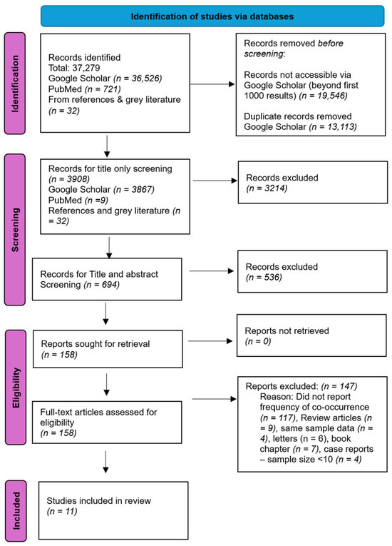
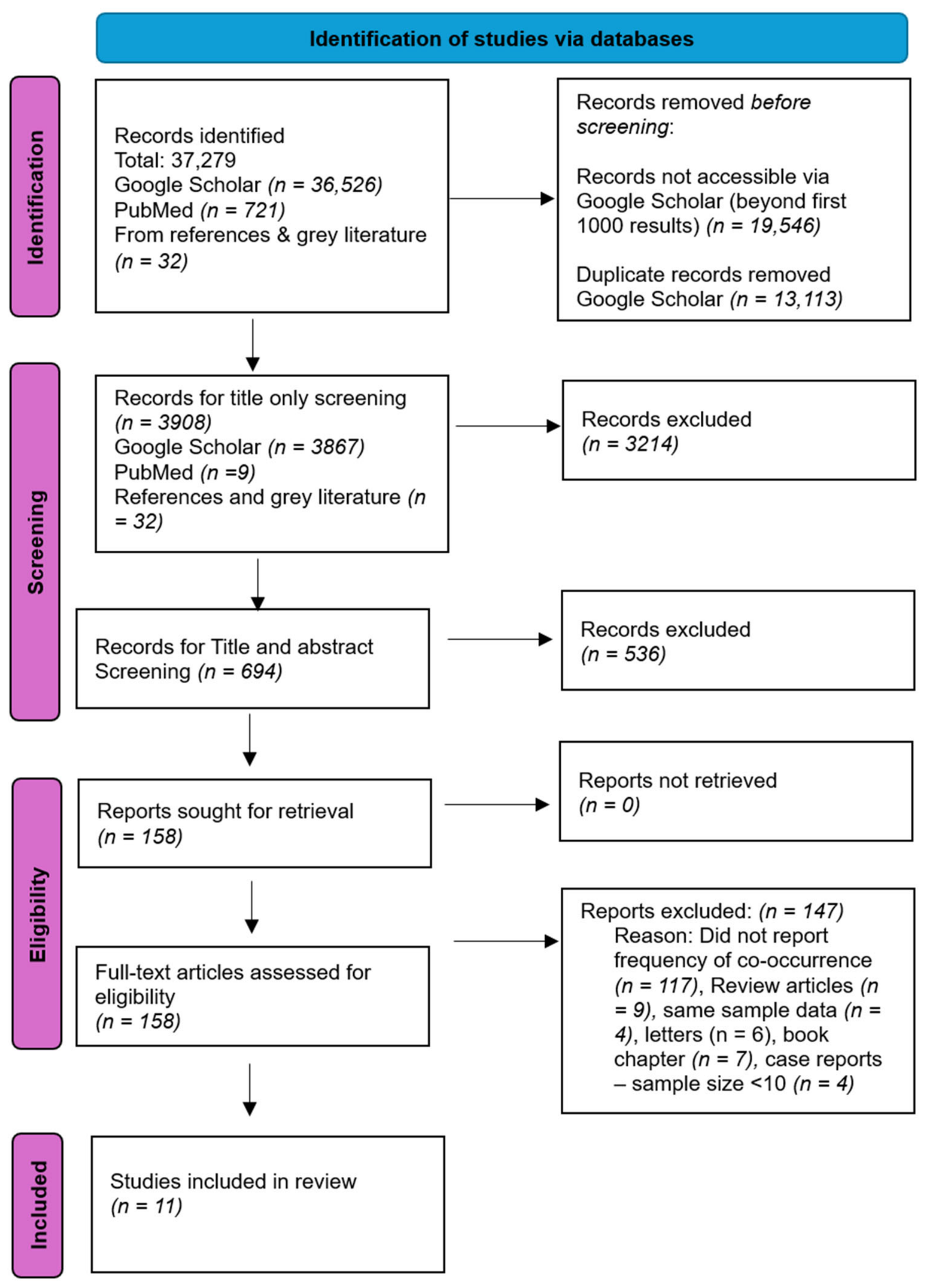
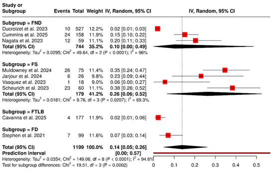
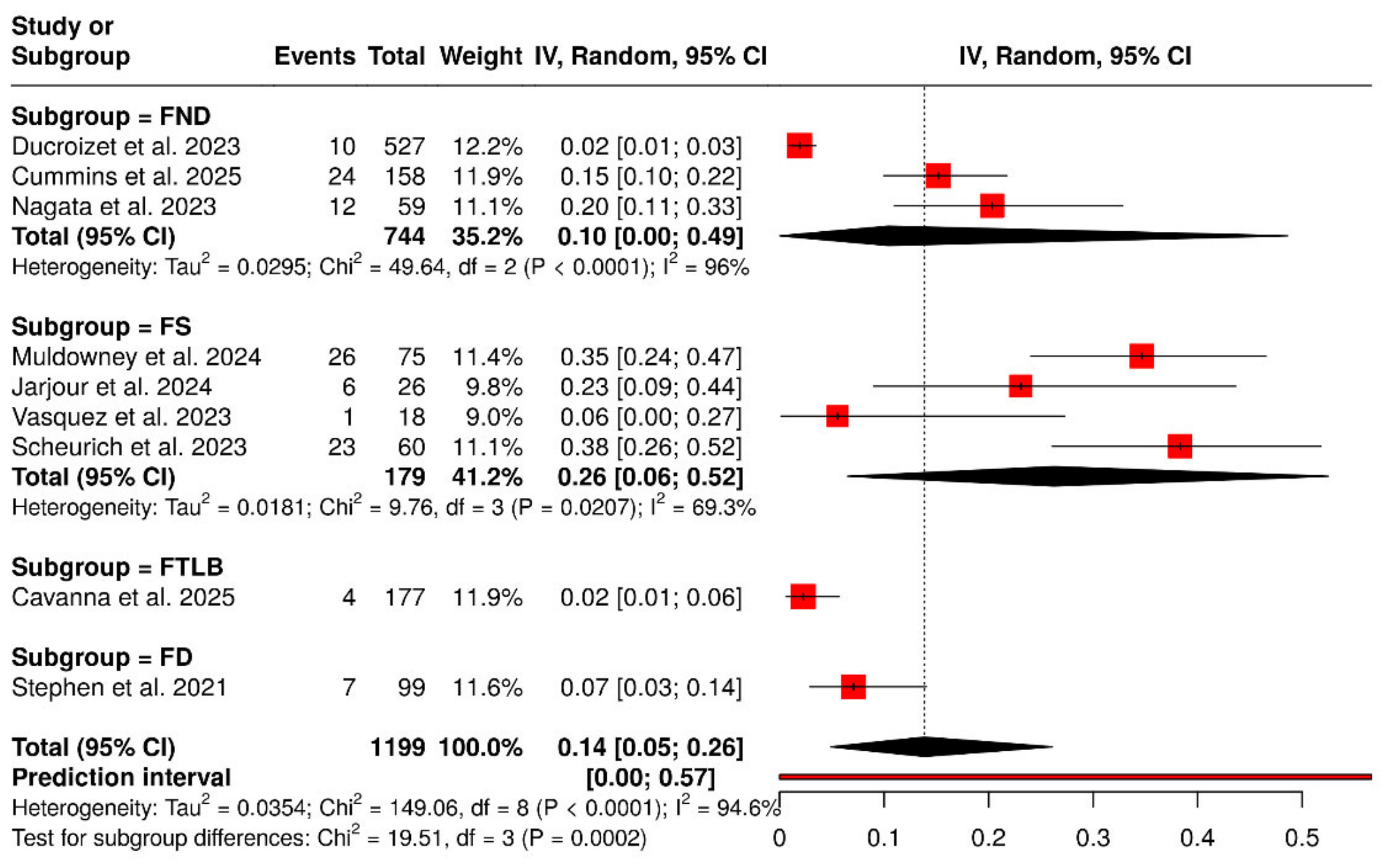
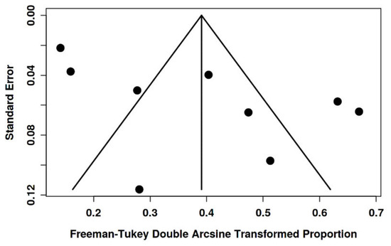
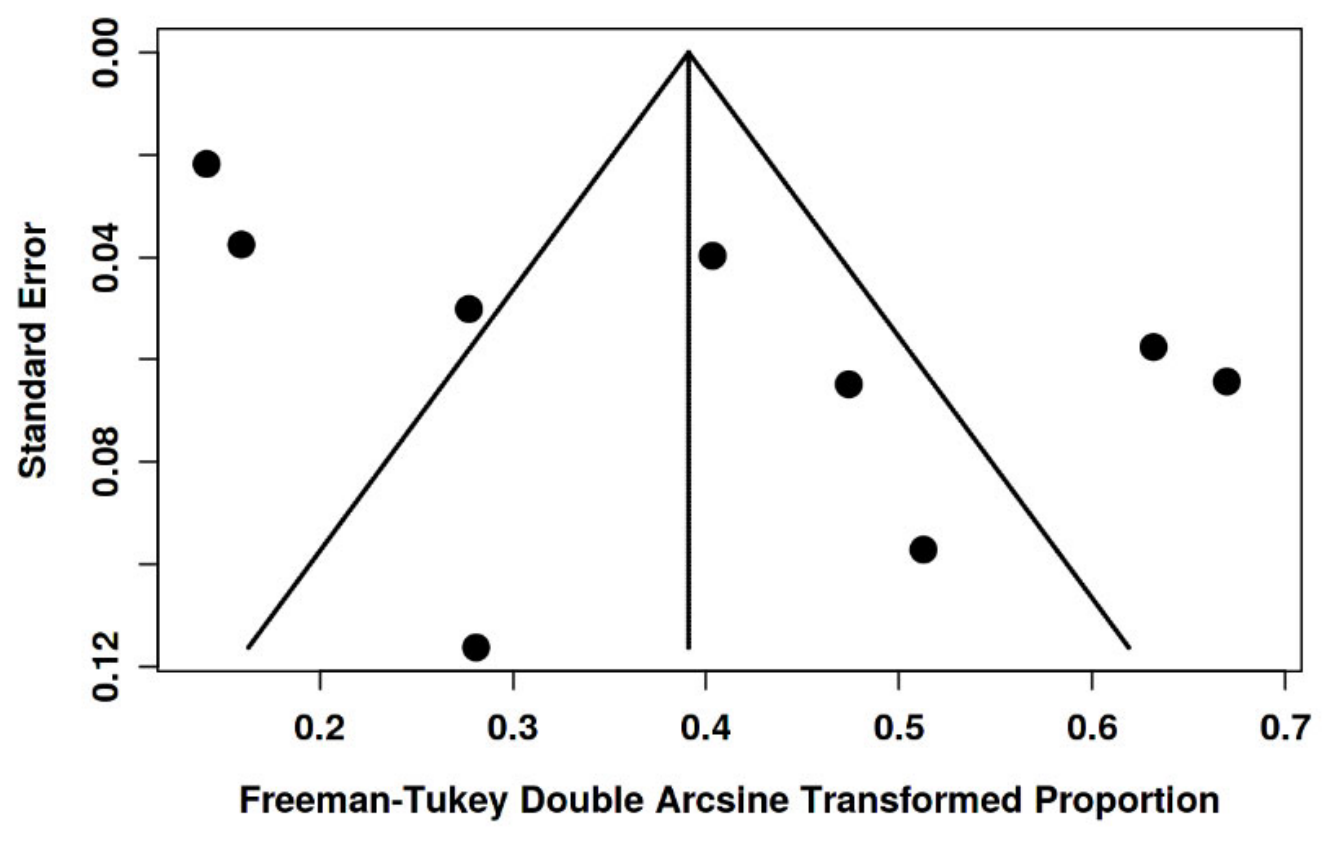

Disclaimer/Publisher’s Note: The statements, opinions and data contained in all publications are solely those of the individual author(s) and contributor(s) and not of MDPI and/or the editor(s). MDPI and/or the editor(s) disclaim responsibility for any injury to people or property resulting from any ideas, methods, instructions or products referred to in the content. |
© 2026 by the authors. Licensee MDPI, Basel, Switzerland. This article is an open access article distributed under the terms and conditions of the Creative Commons Attribution (CC BY) license.