


Highlights
What are the main findings?
- •
- In our study with 161 cases of pediatric hemimegalencephaly, isolated HME was the most common subtype, seizures typically began before age one (most often focal-onset with impaired awareness), and drug-resistant epilepsy affected 73% of the study sample.
- •
- In terms of neurosurgical procedures, functional procedures were more frequently reported than anatomical and the limited available literature suggests broadly comparable seizure outcomes.
- •
- Genetic testing (positive in only ~9.3% of cases) most often identified PIK3CA and NPRL3 variants, which could correlate with the fact that HME is primarily driven by somatic mosaic mutations in the PI3K–AKT–mTOR pathway, while associated neurocutaneous findings, especially Hypomelanosis of Ito and tuberous sclerosis, frequently served as visible clinical clues to the underlying diagnosis even without molecular confirmation.
What are the implications of the main findings?
- •
- Because HME often escapes genetic confirmation (due to mosaicism and limited tissue-specific testing), clinicians should rely on a combined approach both recognizing cutaneous and somatic markers alongside imaging and EEG findings to support earlier diagnosis and guide timely, individualized management.
- •
- Given the high rate of drug-resistant epilepsy and the comparable efficacy of functional versus anatomical hemispherectomy, early referral for multidisciplinary evaluation (neurology, genetics, radiology, neurosurgery) is essential to optimize seizure control and developmental outcomes, while larger, longer-term studies are still needed to refine surgical decision-making and explore emerging alternatives like mTOR-targeted therapy.
Abstract
Background/Objectives: To describe the clinical and genetic features of hemimegalencephaly in pediatric patients, and to highlight treatment outcomes in this population. Methods: This is a systematic literature review following PRISMA guidelines. We used PubMed, Google Scholar, and Scopus for this systematic literature review as databases. Clinical data, imaging findings, EEG patterns, genetic variants, associated syndromes and treatment modalities and outcomes were reviewed, analyzed and summarized in tables. A total of 161 pediatric patients with hemimegalencephaly are described. A risk of bias and sensitivity analysis was conducted. Results: Hemimegalencephaly is more frequently reported in males. The most common subtype was isolated hemimegalencephaly followed by the syndromic subtype, in particular the association with Ito’s hypomelanosis. The most common seizure type was focal onset with impaired awareness. Genetic analysis showed variants in TSC1, TSC2, NPRL3, and PIK3CA, supporting mTOR pathway involvement. Drug-resistant epilepsy was present in approximately 73.2% (118/161) of the sample size. Hemispherectomy, especially functional, in our sample was more frequently reported and the current limited available literature suggests potentially comparable seizure outcomes compared to anatomical hemispherectomy in terms of seizure reduction. Our biggest limitation is the lack of long-term neurological follow-up in the existing literature. Conclusions: Hemimegalencephaly presents with a wide clinical and genetic spectrum and frequently results in drug-resistant epilepsy and developmental impairment. Although surgery may offer seizure relief, outcomes vary and the optimal approach remains individualized. Early diagnosis is essential to enable timely intervention and coordinated care. A multidisciplinary team including neurology, genetics, radiology and neurosurgery is key to optimizing outcomes. Broader studies including milder cases and long follow-up are needed to fully understand disease progression and guide management.
1. Introduction
Hemimegalencephaly (HME) is a rare congenital brain malformation characterized by abnormal asymmetric neuronal proliferation leading to enlargement of one cerebral hemisphere [1,2,3]. Genetic factors and mutations in the mTOR (mammalian target of rapamycin) pathway are related to the pathogenesis of this disease [4]. Despite growing recognition, HME remains under-diagnosed, and epidemiological data are limited. It is estimated that HME accounts for approximately 1–3 per 1000 children diagnosed with epilepsy [5]. Clinically, HME often presents in early childhood with drug-resistant epilepsy (DRE), developmental delay, hemiparesis, and hemianopia, among other neurological abnormalities [6]. There are three main types of HME: isolated HME, syndromic HME occurring as part of a genetic or neurocutaneous disorder, and total HME which additionally involves the brainstem and cerebellum [4]. Given its early onset and the importance of the first years of life for neurological development, HME can significantly affect cognitive, motor, and social outcomes [5]. Early recognition improves management options, however delayed diagnosis often occurs, due to its rarity and variable presentation. Most patients with HME present with contralateral hemiparesis, cognitive impairment, and DRE often requiring neurosurgery [7]. Usually, medical therapy, diets, and neurostimulation serve only as bridges while awaiting surgery. Currently, the choice between anatomic and functional hemispherectomy approaches are actively debated given their outcomes and complications [7,8]. Consequently, managing HME requires balancing these surgical risks against the goal of halting cognitive decline, while emerging options like investigational transarterial embolization and targeted mTOR pathway inhibitors could be potential alternatives or adjuncts to surgery in the future with pharmacogenomics [9,10]. This systematic literature review summarizes current knowledge on HME in the pediatric population, focusing on clinical features, diagnostic evaluation, imaging characteristics, surgical approaches, and prognosis.
2. Materials and Methods
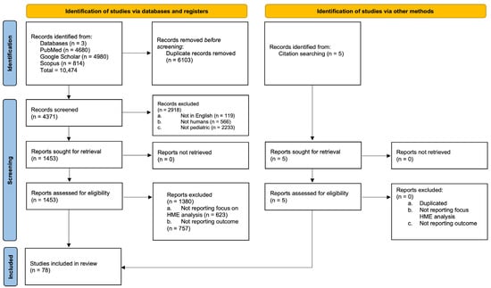
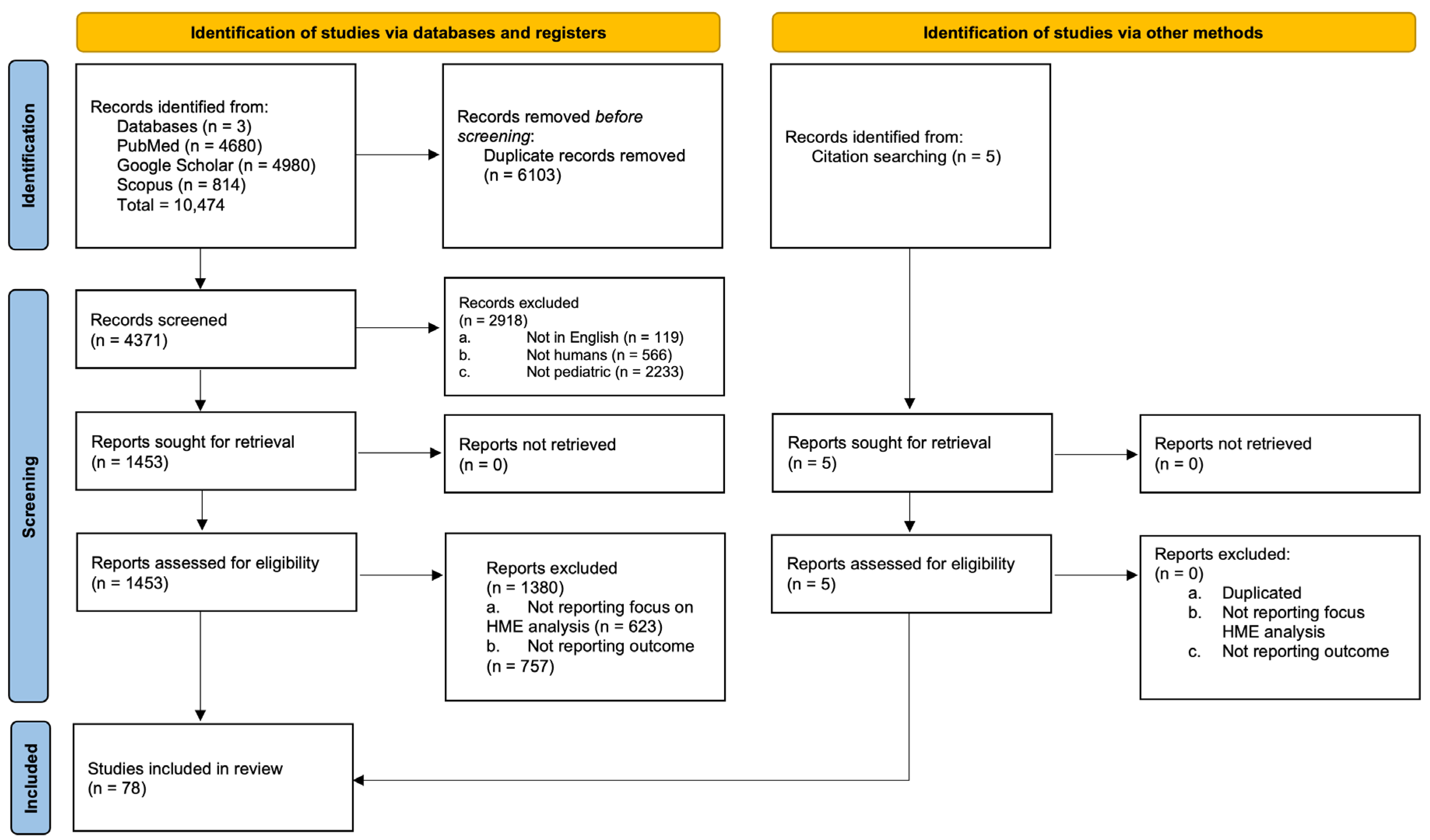
We performed a systematic review using the PRISMA 2020 guideline to gather information about demographics, types of HME, associated syndromes or clinical findings, associated epilepsy or drug-resistant epilepsy, and surgical management of HME. Figure 1 shows the results of the study using this guideline. PRISMA 2020 checklist see Supplementary Martial. The systematic review was not registered before its initiation, and a protocol was not prepared. Gray literature was used for the systematic review to fill knowledge gaps about imaging, EEG findings, and surgical complications not captured in the main systematic review to provide a more robust analysis given the lack of uniform data found in case studies and case reports which are the more prevalent studies reflecting these data.
Figure 1.
PRISMA.
Drug-resistant epilepsy was defined as failure of adequate trials of two tolerated, appropriately chosen and used antiepileptic drug schedules to achieve sustained seizure freedom as per the International League Against Epilepsy [11]. The proportion of DRE patients in our sample was calculated by identifying patients who met these criteria and were recognized by the authors as such.
To minimize patient overlap across studies and to generate a unique, non-redundant pooled cohort, publication year, patient age, and country of origin were used as the primary criteria for cross-referencing cases. The majority of studies included in this systematic review reported a single patient, typically presenting at a young age (ranging from neonatal to infancy), from geographically distinct countries. Furthermore, the studies contributing larger sample sizes originated from Italy, the United States, Turkey, and Japan, published in different years and involving patients of varying ages. Notably, these larger series report patients treated at the authors’ own institutions, indicating that the reported cases originate from a single center’s local patient population rather than being aggregated from other countries or referral centers abroad. Based on this distribution of publication years, countries, and patient ages, together with the single-center origin of the larger series, we infer that the degree of patient overlap, if present, is likely minimal.
2.1. Inclusion Criteria
- Language: Studies must be written in English.
- Population: Studies must include pediatric patients.
- Focus: Studies must report data on HME.
- Analysis: Studies must contain an analysis of HME and treatment.
- Outcomes reported:
- -
- Characteristics of the HME;
- -
- Treatment outcome;
- -
- Main conclusions of each study.
Exclusion criteria
- Linguistic exclusion: Studies not written in English were excluded.
- Animal studies: Studies involving animals were excluded.
- Nonrelevant focus: Articles not involving HME were excluded.
- Population exclusion: Studies not involving pediatric patients were excluded.
- Irrelevant HME analysis: Studies not containing an analysis of HME and treatment outcome were excluded.
2.2. Data Collection
We used PubMed, Google Scholar, and Scopus for this systematic literature review. The search was conducted between 15 December 2025, and 15 July 2026. We used an advanced search strategy with the following terms for Pubmed: (“hemimegalencephaly”[Title/Abstract] AND “pediatric”[Title/Abstract] AND “epilepsy”[Title/Abstract] AND “treatment”[Title/Abstract]) OR (“HME”[Title/Abstract] AND “pediatric”[Title/Abstract] AND “seizures”[Title/Abstract] AND (“surgery”[Title/Abstract] OR “hemispherectomy”[Title/Abstract] OR “hemispherotomy”[Title/Abstract])) OR (“hemimegalencephaly”[Title/Abstract] AND “pediatric”[Title/Abstract] AND “seizures”[Title/Abstract]). We used an advanced search strategy with the following terms for Google Scholar: (“hemimegalencephaly” OR “HME”) AND pediatric AND (epilepsy OR seizures) AND (treatment OR surgery OR hemispherectomy OR hemispherotomy OR outcome). We used an advanced search strategy with the following terms for Scopus:
- (“hemimegalencephaly” AND “pediatric” AND “epilepsy” AND “treatment”).
- (“HME” AND “pediatric” AND “seizures” AND (“surgery” OR “hemispherectomy” OR “hemispherotomy”)).
- (“hemimegalencephaly” AND “pediatric” AND “seizures”).
Variables included demographic information, medical and family history, genetics, seizure onset and semiology, localization, HME subtype, associated syndromes, neurological and imaging findings, Electroencephalogram (EEG) patterns, antiseizure medication use, and treatment response. Surgical variables included age at surgery, procedure type (e.g., functional hemispherectomy, anatomic hemispherectomy, hemispherotomy, callosotomy, lobectomy), and postoperative Engel classification.
2.3. Selection, Data Extraction, and Analysis
The primary reviewers (FM, MVM) screened all titles and abstracts and identified articles as being either “potentially relevant” or “irrelevant” to the research question based on the inclusion and exclusion criteria described above. Full-text copies of articles identified as potentially relevant were retrieved and individually assessed for inclusion in the review stage. Rayyan, a web platform designed to help in systematic literature reviews, was used by the reviewers and the supervisor to remove duplicates. Disagreement on inclusion or exclusion of studies was resolved by consensus achieved through the supervision process of this study on Rayyan. After reaching consensus, we collected the following information from each article: the author/year, methods, number of participants, and study design. We also extracted the main results, including each study’s outcome measures and main limitations. We analyzed the studies’ primary and secondary goals and gathered the main conclusions from each study by tabulating them into tables.
2.4. Quality/Risk of Bias Assessment
We used the proposed Murad method for methodological quality and synthesis of case series and case reports. This method analyzes 8 questions over 4 domains: selection, ascertainment, causality, and reporting [12]. Studies answering “yes” to ≥4/8 signaling questions were classified as low risk of bias. It is crucial to consider that 3 out of the 8 questions are only applicable for cases of adverse drug events which is the reason we considered 4 as our threshold for low risk of bias. The case series and case reports that meet criteria to be included in the study have a low risk of bias.
We used the Newcastle-Ottawa tool to perform the quality assessment in observational studies. A star system was used to perform quality assessments following the Newcastle-Ottawa Scale (NOS). A study was awarded 1 star for each numbered item within the selection and exposure categories. A maximum of 2 stars were awarded for comparability, 3 for exposure, and 4 for selection. The NOS ranges from 0 to 9 stars. We considered high-quality studies as those that achieved 7 or more stars, medium-quality studies as those with 4 to 6 stars, and poor-quality studies as those with fewer than 4 stars. The observational studies that meet criteria to be included in the study are high-quality studies.
Each study was assessed independently by the primary reviewers (FM, MVM). Discrepancies were resolved by consensus achieved through the supervision process of this study.
2.5. Sensitivity Analysis
To assess the robustness of the pooled findings, we conducted a post hoc sensitivity analysis by re-extracting study-level data (sample size, HME subtype, and DRE status) from the 78 included studies and recalculating key pooled estimates under four alternative exclusion scenarios: (1) removal of the single largest contributing series, (2) removal of all series with ≥8 patients, (3) restriction to single-patient case reports only, and (4) restriction to multi-patient case series only. Table 1 shows each scenario’s pooled proportions for HME subtype (isolated vs. syndromic) were recomputed from the underlying counts and compared against the base-case estimates derived from the full cohort to determine whether the direction and magnitude of the findings were sensitive to the influence of individual large series or to differential representation of case reports versus case series.
Table 1.
Sensitivity analysis.
In terms of HME subtype distribution, the direction of the finding (isolated more common than syndromic) is robust across every scenario. The isolated subtype stays the majority (60–74%) regardless of which studies are excluded. The magnitude shifts by up to ~14 points depending on whether case reports or case series dominate the subset, but the qualitative conclusion does not change.
In terms of surgery, FH remains more frequent than AH in every scenario. Removing Di Rocco et al. alone (one 15-patient series) widens the FH–AH gap from 37 points to 53 points, because that one study contributes 38% of all reported AH cases. More notably, restricting to case series only narrows the gap substantially (60.0% vs. 30.7%) compared with case reports (71.4% vs. 17.1%). This suggests the reported FH predominance is inflated by the case report literature plausibly causing a publication-pattern effect (e.g., individual FH cases may be more likely to be written up than AH cases) rather than a true difference in clinical practice.
3. Results
We reviewed 161 reported cases including 93 males and 68 females. Table 2 shows demographics from the study. The age range is 0–18. The mean age is 3.5 years. The clinical presentation of hemimegalencephaly is most commonly with seizures, with approximately 70% (109/160, one case did not report seizures) of our sample size developing seizures before the first year of life. Focal-onset impaired awareness seizures most commonly reported in 48.7% (78/160, 1 case did not report seizures) of the cases, followed by infantile spasms and generalized tonic–clonic seizures. The characterization and quantification of seizures is difficult given the minimal detail into seizure semiology. Only 15 cases meeting criteria for inclusion in the systematic review report positive genetic information with the most commonly reported variants being PIK3CA in four cases and NPRL3 in three cases, followed by TSC1 in three cases, and RHEB, TSC2, MTOR, AKT3, and TRIO each reported once. The origin of genetic testing between brain, blood, or buccal swab has been specified for those cases with available information. Despite the lack of specific genetic reports, 59 cases report associated neurocutaneous disorders such as 18 cases of Ito’s hypomelanosis, 13 cases of linear nevus syndrome, nine cases of tuberous sclerosis, three cases of epidermal nevus syndrome other than linear nevus syndrome, three cases of Proteus syndrome, and two cases of neurofibromatosis. Additional associated epilepsy syndromes include seven cases of Ohtahara syndrome and two cases of West syndrome. Finally, associated developmental disorders include one case of trisomy 21 and one case of autosomal dominant intellectual developmental disorder-44 with microcephaly (MRD44).
Table 2.
Study demographics.
Most of the reported cases were isolated HME up to 110, followed by 49 syndromic cases, and only two were total HME. Epilepsy is a common comorbidity. Drug-resistant epilepsy was present in approximately 73.2% (118/161) of the sample size. Phenobarbital is usually a first line given that seizures start in the neonatal period, but afterwards patients try multiple antiseizure medications without success which is the reason epilepsy surgery is common. Out of 161 patients, 110 (67%) had neurosurgeries. Seventy patients had a functional hemispherectomy, twenty-nine patients had an anatomical hemispherectomy, and eleven patients had other surgeries such as decortication or callosotomies. Table 3 shows the distribution of these findings. The HME type numbers correlate with the DRE yes/no variable to represent a total number of cases with DRE and HME.
Table 3.
HME characteristics.
In terms of diagnostic workup for the epilepsy, EEG in HME generally shows hemihypsarrhythmia, which later evolves into high-frequency background activity with almost continuous spikes, sharp waves, and spike-and-wave discharges over the malformed hemisphere that progressively involve the contralateral hemisphere [61,85]. Most of these cases end up having drug-resistant epilepsy with multiple anti-seizure medications requiring epilepsy surgery [61,85]. Some patients show asymmetric suppression-burst patterns, especially in early infancy [61].
Imaging also is part of the diagnostic workup for HME, and prenatal or postanal MRI typically shows hemispheric enlargement, often accompanied by ipsilateral ventriculomegaly, asymmetry, or dimorphism. Midline contralateral displacement and hypervascularization are also frequently observed [86,87]. Cortical abnormalities are also common, such as pachygyria or polymicrogyria, cortical thickening, gray matter heterotopia or gliosis [86,87]. The observed brain abnormalities underscore the importance of advanced prenatal imaging techniques, which has proven highly sensitive for detecting cortical abnormalities in utero if there are suspicions with prenatal ultrasounds. Imaging is also a crucial tool to decide approach to epilepsy surgery [1,35,56,63,66,67,75,82].
In terms of surgery, functional hemispherectomy (FH) (used commonly interchangeably with hemispherotomy given similar techniques) has become the most common procedure, over anatomical hemispherectomy (AH). FH was broadly comparable in terms of seizure control and long-term complications given no significant statistical difference comparted to AH [88]. However, it is important to consider the need for revision or reoperation in FH related to a less invasive procedure and sometimes incomplete disconnection [88,89]. The most common post-surgical complications included contralateral hemiparesis and hydrocephalus, while severe complications such as intracranial hemorrhage, excessive blood loss, and mortality have also been observed [88,90].
Post-surgical outcomes can be reported using the Engel classification as a standard score to understand seizure outcomes after epilepsy surgery [91]. In our review, only six studies reported Engel classifications as seen in Table 4. Nonetheless, these studies represent about 42.7% (47/110) of the sample size.
Table 4.
Engel classification for post-surgical outcomes.
Definition of different procedures:
- •
- Functional Hemispherectomy: Surgical disconnection of one cerebral hemisphere from the rest of the brain while leaving much of the hemisphere’s tissue in place.
- •
- Anatomic Hemispherectomy: Complete surgical removal of one cerebral hemisphere, including the cortex, subcortical structures, and corpus callosum.
- •
- Decortication: Surgical removal of the cerebral cortex from one hemisphere while sparing the underlying white matter and subcortical structure.
4. Discussion
This study provides a better understanding of hemimegalencephaly and its relationship with drug-resistant epilepsy. It is crucial to note that given the majority of studies are case reports and case studies, the reported information in most articles is biased, incomplete and not standardized, which is not a surprise, but it is necessary to mention as demonstrated in our bias assessment. From our data, we can extrapolate the information to generate awareness and promote earlier diagnosis and prompter individualized treatments.
Similar to other reported case reports and studies with large cohorts [90,92], our study identified that the isolated HME form was the most frequently reported which replicates broader and generalizable data. Most of those cases do not report genetic testing which could be a reasonable explanation to classify a case as isolated HME instead of syndrome if no neurocutaneous findings or other pathognomonic signs are noticeable on physical exams. This supports the idea of using genetic testing to inform and develop a better phenotype based on genetic variations to improve diagnostic precision and facilitate early treatment and management.
The genetic landscape of HME has shifted significantly with the discovery that it is largely a mosaicism-driven disorder. While previous understanding focused on broader developmental errors, modern genomic studies have solidified the role of the PI3K/AKT–mTOR pathway as the primary driver of this condition [7]. Additionally, the NPRL3 gene encodes for a subunit of the GATOR complex which helps regulate the mTOR signaling pathway [10]. This pathway is responsible for regulating fundamental cellular processes such as growth, proliferation, and differentiation. When a somatic mutation occurs early in neurogenesis, it leads to the characteristic uncontrolled overgrowth of a single cerebral hemisphere. These variants are typically mosaic, meaning they occur after fertilization and are only present in a subset of cells. This explains why standard blood-based genetic tests often return normal results, as the variant may be localized strictly to the affected brain tissue [7].
The inclusion of the TSC1 and TSC2 genes within this pathway provides a critical mechanistic link between HME and tuberous sclerosis complex [93,94]. These genes normally function as inhibitory “brakes” on the mTOR pathway; their loss of function leads to the same pathobiological endpoint as gain-of-function variants in genes like AKT3 or PIK3CA [93,94]. This endpoint is characterized by significant cellular hypertrophy and cortical dyslamination. Our study found positive genetic testing in only 9.3% (15/161) of the reviewed cases. Of those cases, 26.6% (4/15) were PIK3CA variants, 20% (3/15) were NPRL3 variants, 20% (3/15) were TSC1 variants and the missing 33.3% (5/15) were individual cases reporting RHEB, TSC2, MTOR, AKT3, and TRIO variants, about 6.66% (1/15) each. These results align with other studies with similar results for focal cortical malformations [95,96]. In clinical practice, identifying these variants is transformative for diagnosis, but in resource-limited settings where “deep” sequencing of brain tissue or saliva is unavailable, the clinician must pivot to a rigorous neuro-cutaneous examination to bridge the diagnostic gap [93,94].
In these environments, HME is often recognized as the neurological manifestation of a broader overgrowth syndrome through specific physical markers. For instance, the presence of hypopigmented streaks and swirls following Blaschko’s lines is a hallmark of Hypomelanosis of Ito, which often indicates chromosomal mosaicism highly associated with HME [22,34,63]. Similarly, identifying ash-leaf spots, facial angiofibromas, or Shagreen patches can immediately point toward a tuberous sclerosis diagnosis. These skin findings serve as accessible, non-invasive proxies for the underlying genetic disruptions that drive the brain’s malformation, allowing for a clinical classification even when molecular confirmation is out of reach [22,34,63].
Beyond skin-deep markers, other somatic findings provide essential prognostic value and hint at the timing of the initial genetic mutation. Macrocephaly is frequently observed at birth, reflecting the massive enlargement of the affected hemisphere. When HME is accompanied by homolateral facial hemihypertrophy or somatic hemihypertrophy, it suggests that the causal mutation occurred early enough in embryonic development to affect multiple germ layers, including both the mesoderm and ectoderm. This systemic involvement often signals a more complex clinical course, requiring the clinician to monitor for extracranial complications such as renal or cardiac issues depending on the suspected syndrome [22,34,63].
Clinical phenotyping in HME is not merely a tool for classification but a vital component of the management roadmap. Distinguishing between isolated HME and syndrome-associated HME dictates the urgency of various interventions. While isolated HME might focus primarily on the management of refractory epilepsy and potential surgical evaluation, syndrome-associated cases require a multidisciplinary approach to screen for systemic tumors or vascular malformations. By integrating these physical findings with the known molecular pathways, healthcare providers can better determine a patient’s prognosis and tailor their surveillance strategies to the specific risks associated with the underlying mosaic disorder.
Besides the clinical diversity and complexity of these cases, these patients present with seizures, most commonly focal-onset in nature given the unilateral lesion to the brain with correlates with our study’s data [61,85]. Additionally, infantile spasms are commonly associated as well given mutations in the mTOR pathway causing an abnormal activation, correlated in animal models with focal seizures, and an increased propensity for spasms, demonstrating the direct mechanistic connection probably in the setting of neuronal immaturity, cortical dysplasia, GABAergic pathways dysfunction, and chloride homeostasis dysregulation [97].
Epilepsy surgery is one of the most common approaches to DRE in cases with focal etiologies for epilepsy [88]. Our sample includes about 70% (110/161) of patients who had epilepsy surgery and 42.7% (47/110) of those cases report an Engel classification. Of this sample, more than 51.1% (24/47) of the patients were completely seizure free. Despite the small sample size, these results correlate with other studies with larger cohorts reporting excellent outcomes with the majority of patients achieving a Ia-Ic score with significant improvement in epilepsy control [51,86,90,93].
Despite the benefits from surgery, not every child is a candidate which has spurred interest in minimally invasive alternatives such as staged transarterial embolization for infants too unstable for open surgery, with early single-center data showing comparable seizure and developmental outcomes, though long-term safety and efficacy remain unestablished [9,98]. Also, the discovery that HME arises from somatic mosaic PI3K–AKT–mTOR pathway variants has opened the possibility of targeted medical therapy as an adjunct or alternative to surgery, but whether molecular diagnosis should redirect management remains actively debated rather than standard practice [93].
Other associated aspects before and after epilepsy surgery like developmental, language, and motor delay, intellectual disability, and psychosocial skills are generally not reported but should be managed in a multidisciplinary fashion with physical therapy, occupational therapy, early intervention programs, speech therapists, and neuropsychologists depending on the severity and individual needs of the case [99,100].
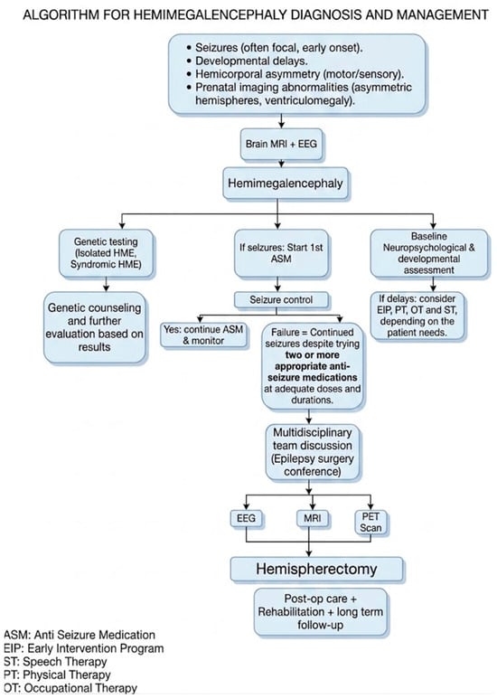
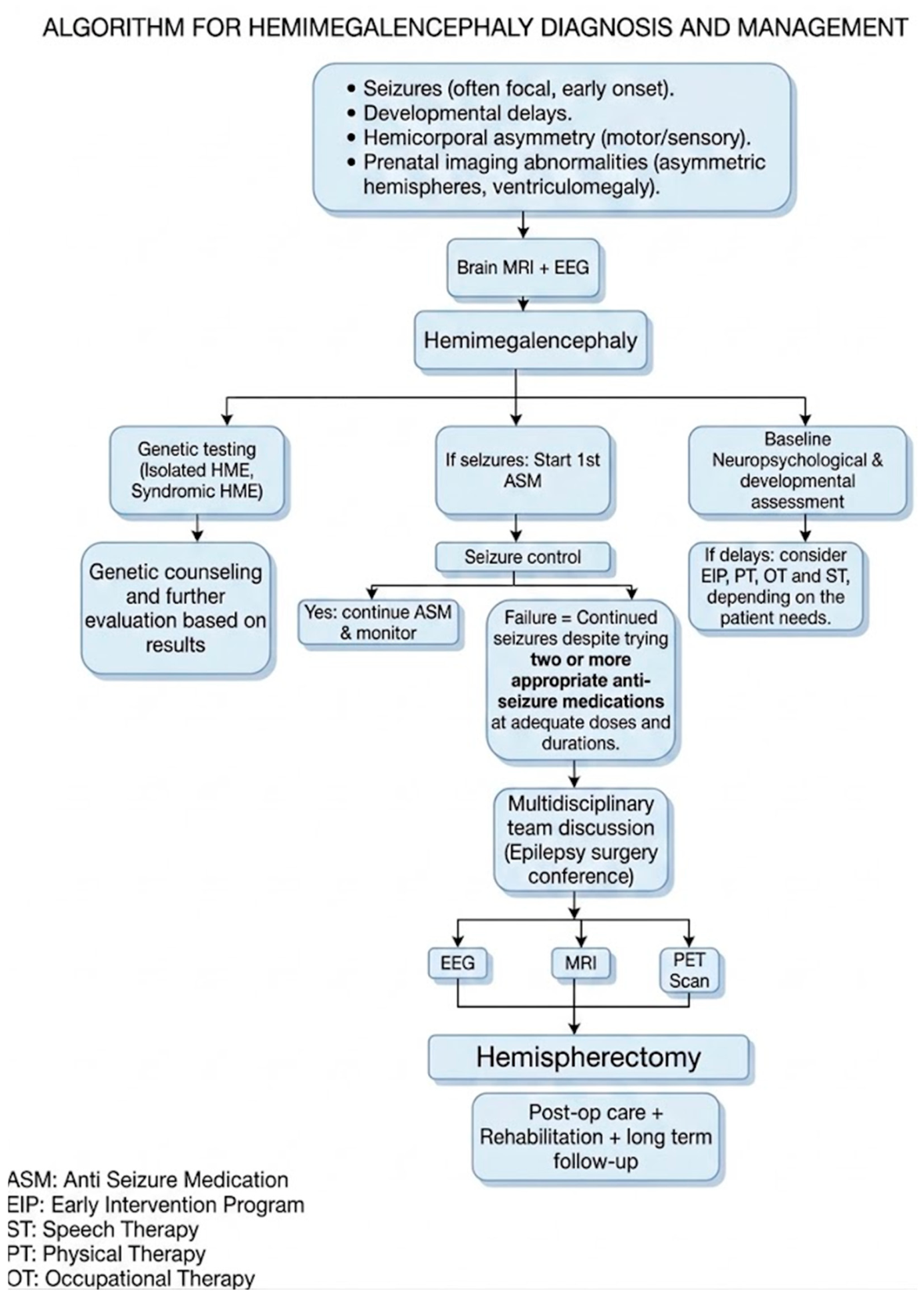
We propose a clinical framework (Figure 2) based on the presented information to guide the approach to HME cases. It should be emphasized that the transition from failure of antiseizure medications to hemispherectomy should also reflect the need for comprehensive multidisciplinary presurgical evaluation and individualized selection of surgical technique.
Figure 2.
Proposed clinical framework for hemimegalencephaly.
Limitations
One of the biggest limitations is the lack of long-term neurological follow-up in the existing literature. This gap hindered the ability to evaluate prolonged post-treatment outcomes. Additionally, the constantly changing and evolving field complicates the standardization of seizure semiology and DRE definition, especially when using primarily case reports and case studies to gather the data used in the manuscript as well as the comparability of surgical outcomes without a unique score such as Engel’s classification. Data about surgical outcomes and genetic testing is only reported in certain cases which is impactful for the final conclusion of the study. There is also a possibility of patient overlap given the rarity of this syndrome. However, this has been minimized by the strategies mentioned in the Section 2. Finally, other limitations are that we only included English-language studies and protocol nonregistration.
The data has been compared with other studies and reflect similar results despite the small sample with these specific data.
5. Conclusions
Hemimegalencephaly is a disease with a broad clinical and genetical spectrum. The findings in this study aid in the understanding of its diagnosis and outlines the spectrum of the known disease phenotype. Published HME cases frequently involve early-onset epilepsy. Hemispheric surgery can provide substantial seizure reduction in selected patients, despite the risk of postoperative complications. Early diagnosis and coordinated multidisciplinary care are essential for achieving the best clinical outcomes in patients with HME. Broader studies including possible milder phenotypes and long-term follow-up are needed to enhance understanding and refine management strategies of this disease. Future research must prioritize genetic testing, standardization of seizure semiology, and post-surgical outcomes to determine whether surgical or medical treatments offer lasting benefits for HME patients.
Supplementary Materials
The following supporting information can be downloaded at: https://www.mdpi.com/article/10.3390/children13101292/s1, Table S1: PRISMA 2020 checklist.
Author Contributions
F.M., M.V.M. and A.S.A. participated in drafting and revising the manuscript for content, including medical writing, in study concept and design, data acquisition, and study supervision or coordination. A.S.A. participated in revising the manuscript for content, including medical writing, in study concept and design, data acquisition, and study supervision or coordination. A.S.A. and D.M. participated in revising the manuscript for content, including medical writing, in study concept and design, data acquisition, and study supervision or coordination. A.S.A., R.J., J.C., Y.E. and A.R.T. participated in drafting and revising the manuscript for content, including medical writing, in study concept and design, data acquisition, analysis and interpretation of data, statistical analysis, and study supervision or coordination. All authors have read and agreed to the published version of the manuscript.
Funding
This research received no external funding.
Institutional Review Board Statement
Not applicable.
Informed Consent Statement
Not applicable.
Data Availability Statement
Data is available upon request.
Conflicts of Interest
The authors declare no conflicts of interest.
References
- Shim, S.; Shin, J.E.; Lee, S.M.; Baek, S.H.; Park, J.-S.; Han, J.H.; Park, M.S.; Kang, H.-C.; Park, E.K.; Eun, H.S. A Patient with Tuberous Sclerosis with Hemimegalencephaly Presenting with Intractable Epilepsy in the Early Neonatal Period: A Case Report. Perinatology 2022, 33, 201. [Google Scholar] [CrossRef] [Scilit]
- Di Rocco, C.; Battaglia, D.; Pietrini, D.; Piastra, M.; Massimi, L. Hemimegalencephaly: Clinical Implications and Surgical Treatment. Childs Nerv. Syst. 2006, 22, 852–866. [Google Scholar] [CrossRef] [Scilit] [PubMed]
- Pepi, C.; De Benedictis, A.; Rossi-Espagnet, M.C.; Cappelletti, S.; Da Rold, M.; Falcicchio, G.; Vigevano, F.; Marras, C.E.; Specchio, N.; De Palma, L. Hemispherotomy in Infants with Hemimegalencephaly: Long-Term Seizure and Developmental Outcome in Early Treated Patients. Brain Sci. 2022, 13, 73. [Google Scholar] [CrossRef] [Scilit] [PubMed]
- Jaiswal, V.; Hanif, M.; Sarfraz, Z.; Nepal, G.; Naz, S.; Mukherjee, D.; Ruxmohan, S. Hemimegalencephaly: A Rare Congenital Malformation of Cortical Development. Clin. Case Rep. 2021, 9, e05238. [Google Scholar] [CrossRef] [Scilit] [PubMed]
- Edmonds, B.; Ngo, J.P.; Groves, A.; Reyes, B.; Gott, R.A.; Chia, D.J.; Mirbaha, H.; Magaki, S.; Khanlou, N.; Pineles, S.L.; et al. Multi-Disciplinary Team Approach for Pediatric Hemimegalencephaly: Insights from a Single Institutional Case Series. Epilepsia Open 2024, 9, 2510–2517. [Google Scholar] [CrossRef] [Scilit] [PubMed]
- Liu, Q.; Ma, J.; Yu, G.; Zhang, Q.; Zhu, Y.; Wang, R.; Yu, H.; Liu, C.; Sun, Y.; Wang, W.; et al. Postoperative Seizure and Developmental Outcomes of Children with Hemimegalencephaly and Drug-Resistant Epilepsy. Seizure 2021, 92, 29–35. [Google Scholar] [CrossRef] [Scilit] [PubMed]
- Goel, K.; Phillips, H.W.; Chen, J.-S.; Ngo, J.; Edmonds, B.; Ha, P.X.; Wang, A.; Weil, A.; Russell, B.E.; Salamon, N.; et al. Hemispheric Epilepsy Surgery for Hemimegalencephaly: The UCLA Experience. Epilepsia 2024, 65, 57–72. [Google Scholar] [CrossRef] [Scilit] [PubMed]
- Chen, J.-S.; Harris, W.B.; Wu, K.J.; Phillips, H.W.; Tseng, C.-H.; Weil, A.G.; Fallah, A. Comparison of Hemispheric Surgery Techniques for Pediatric Drug-Resistant Epilepsy: An Individual Patient Data Meta-Analysis. Neurology 2023, 101, e410–e424. [Google Scholar] [CrossRef] [Scilit] [PubMed]
- Israel, S.; Wolff, N.; Ruffini, L.; Sanidas, G.; Lowe, C.R.; Chandereng, T.; Berl, M.M.; Tsuchida, T.N.; Anwar, T.; Pearl, M.S.; et al. Transarterial Embolization versus Hemispherectomy in Infants with Hemimegalencephaly and Drug-Resistant Epilepsy. Pediatr. Res. 2026, 99, 1523–1531. [Google Scholar] [CrossRef] [Scilit] [PubMed]
- Chandrasekar, I.; Tourney, A.; Loo, K.; Carmichael, J.; James, K.; Ellsworth, K.A.; Dimmock, D.; Joseph, M. Hemimegalencephaly and Intractable Seizures Associated with the NPRL3 Gene Variant in a Newborn: A Case Report. Am. J. Med. Genet. A 2021, 185, 2126–2130. [Google Scholar] [CrossRef] [Scilit] [PubMed]
- Kwan, P.; Arzimanoglou, A.; Berg, A.T.; Brodie, M.J.; Allen Hauser, W.; Mathern, G.; Moshé, S.L.; Perucca, E.; Wiebe, S.; French, J. Definition of Drug Resistant Epilepsy: Consensus Proposal by the Ad Hoc Task Force of the ILAE Commission on Therapeutic Strategies. Epilepsia 2010, 51, 1069–1077. [Google Scholar] [CrossRef] [Scilit] [PubMed]
- Murad, M.H.; Sultan, S.; Haffar, S.; Bazerbachi, F. Methodological Quality and Synthesis of Case Series and Case Reports. Evid.-Based Med. 2018, 23, 60–63. [Google Scholar] [CrossRef] [Scilit] [PubMed]
- Liu, A.M.K.; Chi, C.S.; Ming, K.A. A Case Report on Hemimegalencephaly. Tungs’ Med. J. 2015, 9, 136–141. [Google Scholar] [CrossRef] [Scilit]
- Alexopoulos, A.; Lachhwani, D.K.; Gupta, A.; Kotagal, P.; Harrison, A.M.; Bingaman, W.; Wyllie, E. Resective Surgery to Treat Refractory Status Epilepticus in Children with Focal Epileptogenesis. Neurology 2005, 64, 567–570. [Google Scholar] [CrossRef] [Scilit] [PubMed]
- Alfonso, I.; Vasconcellos, E.; Shuhaiber, H.H.; Yaylali, I.; Papazian, O. Bilateral Decreased Oxygenation During Focal Status Epilepticus in a Neonate with Hemimegalencephaly. J. Child Neurol. 2004, 19, 394–396. [Google Scholar] [CrossRef] [Scilit] [PubMed]
- Alfonso, I.; Papazian, O.; Litt, R.; Villalobos, R.; Acosta, J.I. Similar Brain SPECT Findings in Subclinical and Clinical Seizures in Two Neonates with Hemimegalencephaly. Pediatr. Neurol. 1998, 19, 132–134. [Google Scholar] [CrossRef] [Scilit] [PubMed]
- Alvarez, R.M.; García-Díaz, L.; Márquez, J.; Fajardo, M.; Rivas, E.; García-Lozano, J.C.; Antiñolo, G. Hemimegalencephaly: Prenatal Diagnosis and Outcome. Fetal Diagn. Ther. 2011, 30, 234–238. [Google Scholar] [CrossRef] [Scilit] [PubMed]
- Agrawal, A.; Dutta, G.; Singh, D.; Sachdeva, D.; Gupta, R. Hemimegalencephaly without Seizures: Report of a Case and Review of Literature. Rom. Neurosurg. 2017, 31, 306–309. [Google Scholar] [CrossRef] [Scilit]
- Bastos, H.; da Silva, P.F.S.; de Albuquerque, M.A.V.; Mattos, A.; Riesgo, R.S.; Ohlweiler, L.; Winckler, M.I.B.; Bragatti, J.A.; Duarte, R.D.; Zandoná, D.I. Proteus Syndrome Associated with Hemimegalencephaly and Ohtahara Syndrome: Report of Two Cases. Seizure 2008, 17, 378–382. [Google Scholar] [CrossRef] [Scilit] [PubMed][Green Version]
- Becherini, F.; Pisano, T.; Castagna, M.; Iannelli, A.; Guerrini, R. Progressive Hemispheric Shrinking in Hemimegalencephaly: A Possible Role for Seizure-Related Neuronal Loss. Dev. Med. Child Neurol. 2008, 50, 553–557. [Google Scholar] [CrossRef] [Scilit] [PubMed]
- Broumandi, D.D.; Hayward, U.M.; Benzian, J.M.; Gonzalez, I.; Nelson, M.D. Best Cases from the AFIP: Hemimegalencephaly. Radiographics 2004, 24, 843–848. [Google Scholar] [CrossRef] [Scilit] [PubMed]
- Calzolari, F.; Chirico, M.; Tamisari, L.; Di Rocco, C. Hemimegalencephaly Associated with Somatic Hemihypertrophy and a Malformation of the Feet: Case Report. Neuroradiology 1996, 38, 367–370. [Google Scholar] [CrossRef] [PubMed]
- Carozza, R.B.; Naftel, R.P.; Sarma, A.; Carter, E.G. Hemimegalencephaly and Intractable Focal Seizures Related to NPRL3 Mutation with Variable Familial Expressivity Treated with Anatomic Hemispherectomy. Ann. Child Neurol. Soc. 2023, 1, 239–243. [Google Scholar] [CrossRef] [Scilit] [PubMed]
- Chand, P.; Manglani, P.; Abbas, Q. Hemimegalencephaly: Seizure Outcome in an Infant after Hemispherectomy. J. Pediatr. Neurosci. 2018, 13, 106–108. [Google Scholar] [CrossRef] [Scilit] [PubMed]
- Chapman, K.; Cardenas, J.F. Hemimegalencephaly in a Patient with a Neurocutaneous Syndrome. Semin. Pediatr. Neurol. 2008, 15, 190–193. [Google Scholar] [CrossRef] [Scilit] [PubMed]
- Chrastina, J.; Novak, Z.; Brazdil, M.; Hermanova, M. Glioblastoma Multiforme in a Patient with Isolated Hemimegalencephaly. J. Neurol. Surg. Rep. 2015, 76, e160–e163. [Google Scholar] [CrossRef] [Scilit] [PubMed]
- Cornelius, L.P.; Raju, V.; Lalapet, R.A. A Case of Epilepsia Partialis Continua Due to Linear Nevus Syndrome with Hemimegalencephaly. J. Pediatr. Neurosci. 2017, 12, 203–205. [Google Scholar] [CrossRef] [Scilit] [PubMed]
- Cuddapah, V.A.; Thompson, M.; Blount, J.; Li, R.; Guleria, S.; Goyal, M. Hemispherectomy for Hemimegalencephaly Due to Tuberous Sclerosis and a Review of the Literature. Pediatr. Neurol. 2015, 53, 452–455. [Google Scholar] [CrossRef] [Scilit] [PubMed]
- Cusmai, R.; Curatolo, P.; Mangano, S.; Cheminal, R.; Echenne, B. Hemimegalencephaly and Neurofibromatosis. Neuropediatrics 1990, 21, 179–182. [Google Scholar] [CrossRef] [Scilit] [PubMed]
- Shields, W.D.; Shewmon, D.A.; Peacock, W.J.; LoPresti, C.M.; Nakagawa, J.; Yudovin, S. Surgery for the Treatment of Medically Intractable Infantile Spasms: A Cautionary Case. Epilepsia 1999, 40, 1305–1308. [Google Scholar] [CrossRef] [Scilit] [PubMed]
- Elting, J.W.; van der Naalt, J.; Fock, J.M. Mild Hypothermia for Refractory Focal Status Epilepticus in an Infant with Hemimegalencephaly. Eur. J. Paediatr. Neurol. 2010, 14, 452–455. [Google Scholar] [CrossRef] [Scilit] [PubMed]
- Galluzzi, P.; Cerase, A.; Strambi, M.; Buoni, S.; Fois, A.; Venturi, C. Hemimegalencephaly in Tuberous Sclerosis Complex. J. Child Neurol. 2002, 17, 677–680. [Google Scholar] [CrossRef] [Scilit] [PubMed]
- Ganguly, J.; Pal, S.; Basu, D.; Sengupta, P.; Pal, A.; Chatterjee, C.; Das, C. Hemimyoclonus: A Rare Presentation of Hemimegalencephaly. Neurol. India 2018, 66, S142–S145. [Google Scholar] [CrossRef] [Scilit] [PubMed]
- Gökçe, E.; Beyhan, M.; Ocak Karataş, Ş.F. Magnetic Resonance Imaging Findings of Two Cases with West Syndrome and Hypomelanosis of Ito with Hemimegalencephaly: A Report of Two Cases. Egypt. J. Radiol. Nucl. Med. 2022, 53, 136. [Google Scholar] [CrossRef] [Scilit]
- Golhar, S.; Chauhan, U.; Manwatkar, S.; Dhole, C. Isolated Nonsyndromic Hemimegalencephaly Presenting at Neonatal Age—A Case Report. Ann. Int. Med. Dent. Res. 2016, 2, 22–24. [Google Scholar]
- Gowda, V.K.; Raj, K.A.; Srinivas, S.M. Hemihypertrophy with Hemimegalencephaly Associated with Type 1b (Broad Pattern) Pigmentary Mosaicism. Karnataka Paediatr. J. 2023, 38, 55–56. [Google Scholar] [CrossRef] [Scilit]
- Griffiths, P.D.; Gardner, S.A.; Smith, M.; Rittey, C.; Powell, T. Hemimegalencephaly and Focal Megalencephaly in Tuberous Sclerosis Complex. AJNR Am. J. Neuroradiol. 1998, 19, 1935–1938. [Google Scholar] [PubMed]
- Guerra, M.P.; Cavalleri, F.; Migone, N.; Lugli, L.; Delalande, O.; Cavazzuti, G.B.; Ferrari, F. Intractable Epilepsy in Hemimegalencephaly and Tuberous Sclerosis Complex. J. Child Neurol. 2007, 22, 80–84. [Google Scholar] [CrossRef] [Scilit] [PubMed]
- Günbey, C.; Oğuz, K.K.; Bilginer, B.; Öztoprak, Ü.; Ilgaz, F.; Akalan, N.; Topçu, M.; Turanlı, G.; Yalnızoğlu, D. Clinical and Radiological Evaluation of Children with Hemimegalencephaly and Epilepsy: A Single-Center Study. Seizure 2025, 132, 186–191. [Google Scholar] [CrossRef] [Scilit] [PubMed]
- Guzzetta, F.; Battaglia, D.; Lettori, D.; Deodato, F.; Sani, E.; Randò, T.; Ricci, D.; Acquafondata, C.; Faienza, C. Epileptic Negative Myoclonus in a Newborn with Hemimegalencephaly. Epilepsia 2002, 43, 1106–1109. [Google Scholar] [CrossRef] [Scilit] [PubMed][Green Version]
- Kim, S.H.; Millichap, J.J.; Koh, S. Brain Inflammation in an Infant with Hemimegalencephaly, Escalating Seizures, and Epileptic Encephalopathy. Child Neurol. Open 2016, 3, 2329048X16633629. [Google Scholar] [CrossRef] [Scilit] [PubMed]
- Higurashi, N.; Hamano, S.; Oritsu, T.; Minamitani, M.; Sasaki, M.; Ida, H. Iomazenil Hyperfixation in Single Photon Emission Computed Tomography Study of Malformations of Cortical Development during Infancy. Eur. J. Paediatr. Neurol. 2011, 15, 372–375. [Google Scholar] [CrossRef] [Scilit] [PubMed]
- Honda, R.; Kaido, T.; Sugai, K.; Takahashi, A.; Kaneko, Y.; Nakagwa, E.; Sasaki, M.; Otsuki, T. Long-Term Developmental Outcome after Early Hemispherotomy for Hemimegalencephaly in Infants with Epileptic Encephalopathy. Epilepsy Behav. 2013, 29, 30–35. [Google Scholar] [CrossRef] [Scilit] [PubMed]
- Humbertclaude, V.T.; Coubes, P.A.; Robain, O.; Echenne, B.B. Early Hemispherectomy in a Case of Hemimegalencephaly. Pediatr. Neurosurg. 1997, 27, 268–271. [Google Scholar] [CrossRef] [Scilit] [PubMed]
- Jahan, R.; Mischel, P.S.; Curran, J.G.; Peacock, W.J.; Shields, D.W.; Vinters, H.V. Bilateral Neuropathologic Changes in a Child with Hemimegalencephaly. Pediatr. Neurol. 1997, 17, 344–349. [Google Scholar] [CrossRef] [Scilit] [PubMed]
- Kakish, D.; Tominna, M.; Krishnan, A. Hemimegalencephaly: Evolution from an Atypical Focal Early Appearance on Fetal MRI to More Conventional MR Findings. Cureus 2022, 14, e27976. [Google Scholar] [CrossRef] [Scilit] [PubMed]
- Kentab, A.Y. Hypomelanosis of Ito with Partial Motor Seizure and Hemimegaloencephaly: Case Report. Open Access J. Neurol. Neurosurg. 2017, 2, 36–39. [Google Scholar] [CrossRef] [Scilit]
- Kometani, H.; Sugai, K.; Saito, Y.; Nakagawa, E.; Sakuma, H.; Komaki, H.; Sasaki, M.; Adachi, Y.; Kaneko, Y.; Otsuki, T.; et al. Postnatal Evolution of Cortical Malformation in the “Non-Affected” Hemisphere of Hemimegalencephaly. Brain Dev. 2010, 32, 412–416. [Google Scholar] [CrossRef] [Scilit] [PubMed]
- Konkol, R.J.; Maister, B.H.; Wells, R.G.; Sty, J.R. Hemimegalencephaly: Clinical, EEG, Neuroimaging, and IMP-SPECT Correlation. Pediatr. Neurol. 1990, 6, 414–418. [Google Scholar] [CrossRef] [Scilit] [PubMed]
- Kumar, A.; Chetiwal, R.; Tanwar, S. Hemimegalencephaly with Adult-Onset Seizures and Normal Intellectual Function: A Rare Case Report. Arch. Med. Health Sci. 2023, 11, 274. [Google Scholar] [CrossRef] [Scilit] [PubMed]
- Lang, S.-S.; Goldberg, E.; Zarnow, D.; Johnson, M.P.; Storm, P.B.; Heuer, G.G. Prenatal Diagnosis of Hemimegalencephaly. World Neurosurg. 2014, 82, 241.e5–241.e8. [Google Scholar] [CrossRef] [Scilit] [PubMed]
- Lettori, D.; Battaglia, D.; Sacco, A.; Veredice, C.; Chieffo, D.; Massimi, L.; Tartaglione, T.; Chiricozzi, F.; Staccioli, S.; Mittica, A.; et al. Early Hemispherectomy in Catastrophic Epilepsy: A Neuro-Cognitive and Epileptic Long-Term Follow-Up. Seizure 2008, 17, 49–63. [Google Scholar] [CrossRef] [Scilit] [PubMed]
- Maher, C.O.; Cohen-Gadol, A.A.; Raffel, C. Cortical Resection for Epilepsy in Children with Linear Sebaceous Nevus Syndrome. Pediatr. Neurosurg. 2003, 39, 129–135. [Google Scholar] [CrossRef] [Scilit] [PubMed]
- Makridis, K.L.; Prager, C.; Tietze, A.; Atalay, D.A.; Triller, S.; Elger, C.E.; Thomale, U.-W.; Kaindl, A.M. Case Report: Hemispherotomy in the First Days of Life to Treat Drug-Resistant Lesional Epilepsy. Front. Neurol. 2021, 12, 818972. [Google Scholar] [CrossRef] [Scilit] [PubMed]
- Martínez Bermejo, A.; López Martín, V.; Arcas, J.; Roche, C.; Pérez Higueras, A.; Pascual Castroviejo, I. Hemimegalencephaly. Presentation of a case uncommon in neonatal age. An. Esp. Pediatr. 1992, 37, 317–320. [Google Scholar] [PubMed]
- Mathis, J.M.; Barr, J.D.; Albright, A.L.; Horton, J.A. Hemimegalencephaly and Intractable Epilepsy Treated with Embolic Hemispherectomy. AJNR Am. J. Neuroradiol. 1995, 16, 1076–1079. [Google Scholar] [PubMed]
- Mohamed, K.; Elmalik, E.E.; Bakry, M.; Bayoumi, M.A.A. Refractory Neonatal Seizures Caused by Hemimegalencephaly. BMJ Case Rep. 2022, 15, e251587. [Google Scholar] [CrossRef] [Scilit] [PubMed]
- Nagahama, Y.; Peters, D.; Kumonda, S.; Vesole, A.; Joshi, C.; Dlouhy, B.J.; Kawasaki, H. Delayed Diagnosis of Shunt Overdrainage Following Functional Hemispherotomy and Ventriculoperitoneal Shunt Placement in a Hemimegalencephaly Patient. Epilepsy Behav. Case Rep. 2017, 7, 34–36. [Google Scholar] [CrossRef] [Scilit] [PubMed]
- Nakashima, K.; Ogihara, M.; Kasai-Yoshida, E.; Tsuruta, S.; Nakagawa, M.; Kusakawa, I. Hemimegalencephaly Accompanied by Myoclonic Status Epilepticus. Pediatr. Neurol. 2012, 47, 55–58. [Google Scholar] [CrossRef] [Scilit] [PubMed]
- Ohta, Y.; Hiraiwa, M.; Murayama, K.; Nonaka-Mishima, M.; Kaneko, Y.; Yumoto, M.; Yotsumoto, H.; Iio, M. Hypometabolism and Dipole Localization in Hemimegalencephaly: A Case Report. Neuropediatrics 1994, 25, 255–258. [Google Scholar] [CrossRef] [Scilit] [PubMed]
- Ohtsuka, Y.; Ohno, S.; Oka, E. Electroclinical Characteristics of Hemimegalencephaly. Pediatr. Neurol. 1999, 20, 390–393. [Google Scholar] [CrossRef] [Scilit] [PubMed]
- Okanari, K.; Miyahara, H.; Itoh, M.; Takahashi, A.; Aizaki, K.; Nakagawa, E.; Otsuki, T.; Izumi, T. Hemimegalencephaly in a Patient with Coexisting Trisomy 21 and Hypomelanosis of Ito. J. Child Neurol. 2014, 29, 415–420. [Google Scholar] [CrossRef] [Scilit] [PubMed]
- Olmos-López, A.; von Son-de Fernex, F. Sindrome de West y epilepsia refractaria, asociada a hemimegalencefalia aislada, diagnóstico y tratamiento en segundo nivel: Reporte de un caso. Rev. Mex. Neuroci. 2016, 17, 120–128. [Google Scholar]
- Pavičić Klancir, K.; Habek, D.; Đuranović, V.; Tripalo Batoš, A.; Pejić Roško, S.; Stanojević, M. Hemispherotomy in an Infant with Hemimegalencephaly and Ohtahara Syndrome. Wien. Med. Wochenschr. 2024, 174, 107–110. [Google Scholar] [CrossRef] [Scilit] [PubMed]
- Pelayo, R.; Barasch, E.; Kang, H.; Marion, R.; Moshé, S.L. Progressively Intractable Seizures, Focal Alopecia, and Hemimegalencephaly. Neurology 1994, 44, 969–971. [Google Scholar] [CrossRef] [Scilit] [PubMed]
- Pepper, J.; Lo, W.B.; Agrawal, S.; Mohamed, R.; Horton, J.; Balloo, S.; Philip, S.; Basnet, A.; Wimalachandra, W.S.B.; Lawley, A.; et al. Functional Hemispherotomy for Epilepsy in the Very Young. J. Neurosurg. Pediatr. 2022, 30, 400–409. [Google Scholar] [CrossRef] [Scilit] [PubMed]
- Raus, I.; Vintan, A.M.; Coroiu, R.E. Hemimegalencephaly with Polymicrogyria—A Case Report. Clujul Med. 2016, 89, 293–296. [Google Scholar] [CrossRef] [Scilit] [PubMed]
- Rintahaka, P.J.; Chugani, H.T.; Messa, C.; Phelps, M.E. Hemimegalencephaly: Evaluation with Positron Emission Tomography. Pediatr. Neurol. 1993, 9, 21–28. [Google Scholar] [CrossRef] [Scilit] [PubMed]
- Rondagh, M.; de Vries, L.S.; van der Meeren, L.E.; Tromp, S.C.; Peeters-Scholte, C.M.P.C.D.; Toirkens, M.J.P.; Steggerda, S.J. Newborn with Refractory Seizures Due to Hemimegalencephaly and Tuberous Sclerosis Complex: Case Report and Literature Review. Neuropediatrics 2025, 56, 133–141. [Google Scholar] [CrossRef] [Scilit] [PubMed]
- Roy, S.G.; Tripathi, M.; Tripathi, M.; Ramanujam, B.; Singhal, A.; Bal, C. Ictal PET in Ohtahara Syndrome with Hemimegalencephaly. Clin. Nucl. Med. 2017, 42, e263. [Google Scholar] [CrossRef] [Scilit] [PubMed]
- Roza, E.; Andrei, R.; Teleanu, R.I. Hemimegalencephaly and Epileptic Encephalopathy Associated with a Variant of Uncertain Significance of the TRIO Gene. 2022. Available online: https://medicinamoderna.ro/wp-content/uploads/2022/06/Hemimegalencephaly-and-Epileptic-Encephalopathy-Associated-with-a-Variant-of-Uncertain-Significance-of-the-TRIO-Gene.pdf (accessed on 9 September 2026).
- Sakuma, H.; Iwata, O.; Sasaki, M. Longitudinal MR Findings in a Patient with Hemimegalencephaly Associated with Tuberous Sclerosis. Brain Dev. 2005, 27, 458–461. [Google Scholar] [CrossRef] [Scilit] [PubMed]
- Sakuta, R.; Aikawa, H.; Takashima, S.; Ryo, S. Epidermal Nevus Syndrome with Hemimegalencephaly: Neuropathological Study. Brain Dev. 1991, 13, 260–265. [Google Scholar] [CrossRef] [Scilit] [PubMed]
- Salinas, V.; Vega, P.; Piccirilli, M.V.; Chicco, C.; Ciraolo, C.; Christiansen, S.; Consalvo, D.; Perez-Maturo, J.; Medina, N.; González-Morón, D.; et al. Identification of a Somatic Mutation in the RHEB Gene through High Depth and Ultra-High Depth next Generation Sequencing in a Patient with Hemimegalencephaly and Drug Resistant Epilepsy. Eur. J. Med. Genet. 2019, 62, 103571. [Google Scholar] [CrossRef] [Scilit] [PubMed]
- Santana-Ramirez, A.; Farias-Serratos, F.; Sanchez-Corona, J.; Castañeda-Cisneros, G.; Farias-Serratos, N.M. Hemimegalencephaly with Facial Congenital Infiltrating Lipomatosis in a Child. Iran. J. Public Health 2014, 43, 1702–1709. [Google Scholar] [PubMed]
- Flores-Sarnat, L. Hemimegalencephaly Syndrome. Handb. Clin. Neurol. 2008, 87, 153–176. [Google Scholar] [CrossRef] [Scilit] [PubMed]
- Ajaj, S.; Bentaleb, F.; Elasfer, J.; Shlaibek, K. Hemimegalencephaly with Prominent Ipsilateral Facial Hypertrophy. Marshall J. Med. 2018, 4, 6. [Google Scholar] [CrossRef] [Scilit][Green Version]
- Serletis, D.; MacDonald, C.; Xu, Q.; Kazina, C.J.; Dakshinamurti, S.; Marin, S.; Del Bigio, M.R. Hemispherectomy for Hemimegalencephaly in a 6.5-Week-Old Infant with Tuberous Sclerosis Complex. Childs Nerv. Syst. 2022, 38, 1415–1419. [Google Scholar] [CrossRef] [Scilit] [PubMed]
- Kulkarni, S.D.; Deopujari, C.E.; Patil, V.A.; Sayed, R.J. Hemispherotomy in an Infant with Hemimegalencephaly. J. Pediatr. Neurosci. 2015, 10, 188–192. [Google Scholar] [CrossRef] [Scilit] [PubMed]
- Shiroishi, M.S.; Jackson, H.A.; Nelson, M.D.; Bluml, S.; Panigrahy, A. Contralateral Hemimicrencephaly in Neonatal Hemimegalencephaly. Pediatr. Radiol. 2010, 40, 1826–1830. [Google Scholar] [CrossRef] [Scilit] [PubMed]
- Taha, J.M.; Crone, K.R.; Berger, T.S. The Role of Hemispherectomy in the Treatment of Holohemispheric Hemimegaloencephaly. J. Neurosurg. 1994, 81, 37–42. [Google Scholar] [CrossRef] [Scilit] [PubMed]
- Wintermark, P.; Roulet-Perez, E.; Maeder-Ingvar, M.; Moessinger, A.C.; Gudinchet, F.; Meuli, R. Perfusion Abnormalities in Hemimegalencephaly. Neuropediatrics 2009, 40, 92–96. [Google Scholar] [CrossRef] [Scilit] [PubMed]
- Wolpert, S.M.; Cohen, A.; Libenson, M.H. Hemimegalencephaly: A Longitudinal MR Study. AJNR Am. J. Neuroradiol. 1994, 15, 1479–1482. [Google Scholar] [PubMed]
- Yoshioka, S.; Sugai, K.; Fujikawa, Y.; Komaki, H.; Nakagawa, E.; Sasaki, M. A case of hemimegalencephaly with slowly progressive expansion. No To Hattatsu 2007, 39, 432–435. [Google Scholar] [PubMed]
- Vigevano, F.; Bertini, E.; Boldrini, R.; Bosman, C.; Claps, D.; di Capua, M.; di Rocco, C.; Rossi, G.F. Hemimegalencephaly and Intractable Epilepsy: Benefits of Hemispherectomy. Epilepsia 1989, 30, 833–843. [Google Scholar] [CrossRef] [Scilit] [PubMed]
- Guerrini, R.; Dobyns, W.B. Malformations of Cortical Development: Clinical Features and Genetic Causes. Lancet Neurol. 2014, 13, 710–726. [Google Scholar] [CrossRef] [Scilit] [PubMed]
- Flores-Sarnat, L. Hemimegalencephaly: Part 1. Genetic, Clinical, and Imaging Aspects. J. Child Neurol. 2002, 17, 373–384; discussion 384. [Google Scholar] [CrossRef] [Scilit] [PubMed]
- Goel, K.; Ghadiyaram, A.; Krishnakumar, A.; Morden, F.T.C.; Higashihara, T.J.; Harris, W.B.; Shlobin, N.A.; Wang, A.; Karunungan, K.; Dubey, A.; et al. Hemimegalencephaly: A Systematic Comparison of Functional and Anatomic Hemispherectomy for Drug-Resistant Epilepsy. Neurosurgery 2024, 94, 666–678. [Google Scholar] [CrossRef] [Scilit] [PubMed]
- Pinto, A.L.R.; Lohani, S.; Bergin, A.M.R.; Bourgeois, B.F.D.; Black, P.M.; Prabhu, S.P.; Madsen, J.R.; Takeoka, M.; Poduri, A. Surgery for Intractable Epilepsy Due to Unilateral Brain Disease: A Retrospective Study Comparing Hemispherectomy Techniques. Pediatr. Neurol. 2014, 51, 336–343. [Google Scholar] [CrossRef] [Scilit] [PubMed]
- Salamon, N.; Andres, M.; Chute, D.J.; Nguyen, S.T.; Chang, J.W.; Huynh, M.N.; Chandra, P.S.; Andre, V.M.; Cepeda, C.; Levine, M.S.; et al. Contralateral Hemimicrencephaly and Clinical-Pathological Correlations in Children with Hemimegalencephaly. Brain 2006, 129, 352–365. [Google Scholar] [CrossRef] [Scilit] [PubMed]
- Durnford, A.J.; Rodgers, W.; Kirkham, F.J.; Mullee, M.A.; Whitney, A.; Prevett, M.; Kinton, L.; Harris, M.; Gray, W.P. Very Good Inter-Rater Reliability of Engel and ILAE Epilepsy Surgery Outcome Classifications in a Series of 76 Patients. Seizure—Eur. J. Epilepsy 2011, 20, 809–812. [Google Scholar] [CrossRef] [Scilit] [PubMed]
- Tinkle, B.T.; Schorry, E.K.; Franz, D.N.; Crone, K.R.; Saal, H.M. Epidemiology of Hemimegalencephaly: A Case Series and Review. Am. J. Med. Genet. A 2005, 139, 204–211. [Google Scholar] [CrossRef] [Scilit] [PubMed]
- Blumcke, I.; Budday, S.; Poduri, A.; Lal, D.; Kobow, K.; Baulac, S. Neocortical Development and Epilepsy: Insights from Focal Cortical Dysplasia and Brain Tumours. Lancet Neurol. 2021, 20, 943–955. [Google Scholar] [CrossRef] [Scilit] [PubMed]
- Gerasimenko, A.; Baldassari, S.; Baulac, S. mTOR Pathway: Insights into an Established Pathway for Brain Mosaicism in Epilepsy. Neurobiol. Dis. 2023, 182, 106144. [Google Scholar] [CrossRef] [Scilit] [PubMed]
- Lai, D.; Gade, M.; Yang, E.; Koh, H.Y.; Lu, J.; Walley, N.M.; Buckley, A.F.; Sands, T.T.; Akman, C.I.; Mikati, M.A.; et al. Somatic Variants in Diverse Genes Leads to a Spectrum of Focal Cortical Malformations. Brain 2022, 145, 2704–2720. [Google Scholar] [CrossRef] [Scilit] [PubMed]
- Lee, J.H.; Huynh, M.; Silhavy, J.L.; Kim, S.; Dixon-Salazar, T.; Heiberg, A.; Scott, E.; Bafna, V.; Hill, K.J.; Collazo, A.; et al. De Novo Somatic Mutations in Components of the PI3K-AKT3-mTOR Pathway Cause Hemimegalencephaly. Nat. Genet. 2012, 44, 941–945. [Google Scholar] [CrossRef] [Scilit] [PubMed]
- Bakouh, N.; Castaño-Martín, R.; Metais, A.; Dan, E.L.; Balducci, E.; Chhuon, C.; Lepicka, J.; Barcia, G.; Losito, E.; Lourdel, S.; et al. Chloride Deregulation and GABA Depolarization in MTOR-Related Malformations of Cortical Development. Brain 2025, 148, 549–563. [Google Scholar] [CrossRef] [Scilit] [PubMed]
- Pearl, M.S.; Tsuchida, T.N.; Oluigbo, C.; Kratimenos, P.; Anwar, T.; Kousa, Y.; Gaillard, W.D.; Chang, T. Definitive Treatment of Seizures Due to Hemimegalencephaly in Neonates and Young Infants by Transarterial Embolization: Technical Considerations for “Endovascular Embolic Hemispherectomy”. J. Neurointerv. Surg. 2023, 16, 81–87. [Google Scholar] [CrossRef] [Scilit] [PubMed]
- Puka, K.; Jones, M.; Mathern, G.W. Functional Cognitive and Language Outcomes after Cerebral Hemispherectomy for Hemimegalencephaly. Epilepsia 2021, 62, 2932–2940. [Google Scholar] [CrossRef] [Scilit] [PubMed]
- Jonas, R.; Nguyen, S.; Hu, B.; Asarnow, R.F.; LoPresti, C.; Curtiss, S.; de Bode, S.; Yudovin, S.; Shields, W.D.; Vinters, H.V.; et al. Cerebral Hemispherectomy: Hospital Course, Seizure, Developmental, Language, and Motor Outcomes. Neurology 2004, 62, 1712–1721. [Google Scholar] [CrossRef] [Scilit] [PubMed]

Disclaimer/Publisher’s Note: The statements, opinions and data contained in all publications are solely those of the individual author(s) and contributor(s) and not of MDPI and/or the editor(s). MDPI and/or the editor(s) disclaim responsibility for any injury to people or property resulting from any ideas, methods, instructions or products referred to in the content. |
© 2026 by the authors. Licensee MDPI, Basel, Switzerland. This article is an open access article distributed under the terms and conditions of the Creative Commons Attribution (CC BY) license.