


Abstract
Background/Objectives: Interleukin-27 (IL-27) is an obligate EBI3–p28 heterodimer, yet sepsis studies commonly measure and target the axis as a single entity. We tested whether both subunit transcripts are coordinately induced in septic monocytes and whether the axis has functional consequences. Methods: We integrated 272,993 whole-blood single-cell transcriptomes from 39 individuals, six additional single-cell cohorts and eight bulk cohorts, with two human macrophage datasets, a tolerant-like THP-1 model and caecal ligation and puncture in male C57BL/6 mice. Results: Across three cohorts (166 donor records), EBI3 ranked 137th of 13,872 genes whereas IL-27 ranked 9413th. Against post-cardiac-surgery controls, detection odds ratios (ORs) were 11.0 for EBI3 and 0.78 for IL-27 (ratio 13.3). Thirteen of 7164 septic monocytes carried both transcripts, matching the chance expectation (co-detection odds ratio 0.91, 95% CI 0.48–1.75) against 5.6–346.7 for concordantly transcribed benchmark complexes; the receiver-side module estimate was β = −0.0151. The imbalance was reproduced in human macrophages. In tolerant-like THP-1 cells, IL-27-associated immunoreactivity retained 79.3% of its acute value versus 14.7% for tumour necrosis factor (TNF). p28-directed intervention increased murine 120 h survival from 10% to 45%. Conclusions: Human sepsis shows marked IL-27 subunit discordance across producer and receiver compartments while retaining functional sensitivity of the p28-associated axis, defining a molecular-attribution gap between pathway activity and intact heterodimeric IL-27.
1. Introduction
Interleukin-27 (IL-27) is an obligate heterodimer of EBI3 and p28 [1] within a cytokine family that reuses subunits [2,3]. EBI3 also pairs with p35 in IL-35 [4,5]. Human and murine IL-27 biology differs: murine p28 can be secreted as IL-30, whereas isolated human p28 is retained and degraded [6]. Thus, detection of a single chain is not equivalent to detection of intact IL-27. Circulating “IL-27” is nevertheless commonly measured by immunoassay, and a meta-analysis of seven studies found that it discriminated septic from non-septic patients with moderate accuracy, with a summary area under the receiver-operating-characteristic curve (AUC) of 0.88 [7]. An anti-IL-27 antibody has also entered randomised phase 2 testing [8], making molecular attribution clinically relevant.
Sepsis is defined by organ dysfunction from a dysregulated host response to infection [9], remains a leading global cause of death [10], and includes reproducible immune endotypes and a transcriptionally defined immunosuppressed monocyte state (MS1) [11,12,13,14,15,16,17]. EBI3 elevation and p28-associated immunoreactivity have been reported separately in sepsis; their cellular coordination is the missing mechanistic link between a heterodimeric cytokine and a single-analyte clinical signal. Resolving that link is central to interpreting IL-27 measurement and to integrating the reported effects of IL-27 blockade in murine sepsis [18,19] with the inflammatory effects of endogenous IL-27 neutralisation in human monocytes [20].
We therefore tested, in cell-resolved human data, whether EBI3 and IL-27 are coordinately induced, whether they are co-detected in the same monocytes, and whether receptor-positive T and NK cells show a prespecified IL-27-associated response. Secondary analyses defined the EBI3-associated monocyte state and evaluated patient-level discrimination and aggregation effects.
2. Materials and Methods
2.1. Study Design and Data Sources
The study combines published single-cell and bulk transcriptomes with analyses of two human macrophage datasets, a macrophage-like cell model and murine caecal ligation and puncture (CLP). The principal cohort was GSE216009 (272,993 whole-blood single-cell transcriptomes from 39 individuals: 26 sepsis, seven cardiac surgery, six healthy volunteers, nine paired convalescent samples) with 30 surface markers. The cross-cohort meta-analysis used GSE216009, SCP548 and SCP3740 (166 donor records, two platforms). GSE167363 and GSE175453 provided ratio replication; GSE279451 provided an additional surface-protein replication context; GSE151263 contributed supporting cell-count context. Eight bulk cohorts were used for patient-level analyses and GSE87218 for chromatin accessibility. Two pairs of accessions are arms of single studies rather than independent cohorts and are treated as such: GSE85243 and GSE87218 are the transcriptome and chromatin arms of one experimental series, and GSE279448 and GSE279451 are the bulk and CITE-seq arms of one patient cohort. SCP548 and SCP3740 originate from the same institution and research programme, so donor overlap between them cannot be excluded from the deposited records; the leave-one-cohort-out analysis in Table S5 (Supplementary Materials) shows that the pooled estimate does not depend on treating them as independent.
2.2. Single-Cell Processing, Cell Identity and Surface-Protein Normalisation
Cells were filtered and annotated with Seurat v5.2.1 [21], with Harmony v2.0.5 [22] used for annotation and the Section 3.3 embedding. All detection, differential-expression, pseudobulk and meta-analytic models used non-integrated counts within cohort. No clinical label entered the integration.
Surface protein was measured by antibody-derived tags [23]. SoupX v1.6.2 [24] was applied to droplet-based cohorts; the nanowell principal cohort was processed without droplet-based ambient correction because it contains no empty droplets. Principal-cohort subset gates were repeated under centred log-ratio and dsb [25] normalisation. Eleven antibody-derived features in GSE175453 were removed before analysis. Because EBI3 is also a dendritic-cell gene, the main analysis was repeated after excluding ten dendritic-cell markers. Monocyte subsets were defined by surface CD14/CD16 gates [26,27].
2.3. Cross-Cohort Meta-Analysis and Subunit Detection Models
Cross-cohort effects were estimated from donor-level monocyte pseudobulk and pooled by random-effects meta-analysis [28]. The common universe comprised 13,872 genes. DerSimonian–Laird was used for the genome-wide screen; REML with Hartung–Knapp was the confirmatory estimator for EBI3. Paule–Mandel, fixed-effect, leave-one-cohort-out and prediction-interval analyses were prespecified sensitivity analyses.
A transcript was detected when at least one unique molecular identifier (UMI) was present; no imputation was used. Between-group detection used logit(P) ~ group + log (UMI) with patient-clustered SEs and t(G − 1) inference. Paired analyses used patient blocking. Ratios of odds ratios (ORs) were estimated from a stacked gene × group model so covariance between the two chains was retained. Pairs in which one chain had no positive cells in the comparator arm were separated from this model and were refitted by Firth penalised likelihood, with profile penalised-likelihood intervals.
2.4. Same-Cell Co-Detection and Benchmark Complexes
Same-cell co-detection was defined as joint detection of both transcripts and expressed as an OR against the product of marginal detection odds, with patient-clustered inference. Benchmark pairs were the three C1q combinations and S100A8/S100A9, selected for obligate or near-obligate complex formation and overlapping detection ranges. EBI3/IL23A was analysed separately because it contains EBI3. Pairs with fewer than ten positive cells in a group were not modelled.
Differential expression used patient-level pseudobulk comparing EBI3-positive and EBI3-negative septic monocytes. Thirty genes matched to EBI3 detection were run through the same pipeline as an empirical null. Additional cell-count and sequencing-depth matching tested the effect of arm size and depth. Pathway analysis used MSigDB Hallmark and Reactome collections; regulons used DoRothEA and decoupleR [29,30]. An L1 classifier [31] excluded EBI3, IL-27 and genes correlated with either at |r| > 0.30; folds were assigned by sample.
2.5. The EBI3-Associated State and Regulatory Analyses
Differential expression used patient-level pseudobulk, with patient as a blocking factor and sequencing depth as a covariate; patients with fewer than five cells in either arm were excluded. The subset-adjusted model added protein-defined monocyte subset. The unadjusted contrast was treated as a decomposition and the adjusted contrast as the primary subset-aware estimate.
Detection-matched placebo genes, cell-count matching and depth matching were prespecified calibrations. Hallmark comparisons used the fixed 50-set collection; the broader pathway analysis used Hallmark plus Reactome, with one false-discovery-rate (FDR) family.
DoRothEA confidence A–C regulons were scored with decoupleR cross-checking [29,30], excluding both subunits from regulons. The classifier used sample-level cross-validation and the same feature restrictions in every fold.
2.6. Receiver-Side, Patient-Level and Dilution Analyses
The receiver-side module comprised 41 genes induced by IL-27 across at least 10 of 12 T/NK subsets in a published single-cell cytokine atlas [32] and was applied unchanged. Module scores used binned control genes. Receptor-positive cells expressed both IL-27RA and IL6ST; receptor-negative cells expressed neither, with one-chain cells analysed separately. Within-patient contrasts were calibrated against a qualitative IL-15 positive control and 200 detection-matched receptor substitutions. The sensitivity of the module was characterised post hoc by adding an effect of known size to the module score of the dual-receptor-positive cells and refitting the identical patient-clustered model across a grid of effect sizes, taking exclusion of zero by the 95% interval as evidence of detection; the resulting minimum detectable effect is given in Table S17.
Patient-level analyses used one value per individual. Bulk models were composition-adjusted with marker scores [33] and used only for severity, outcome and cross-compartment discrimination. The EBI3-to-p28 ratio was calculated as (EBI3% + 0.1)/(p28% + 0.1). Four comparators were fixed in advance: healthy volunteers, cardiac surgery, convalescence and pooled non-infected controls. Comparator robustness used the floor and spread of four-arm AUCs, 500 detection-matched null objects and 2000 paired patient bootstraps. Dilution simulations resampled each individual’s cells while reducing monocyte content. The simulation makes no assumption about how either score scales with monocyte content. Rather than applying a dilution model, it resamples each individual’s own cells to a target monocyte fraction at a fixed total cell number and sums the observed counts, so the contribution of non-monocyte cells to each score is measured rather than imposed. The predictions were fixed before the simulation was run and included the differential prediction that MS1 would not be diluted, because its constituent genes are also expressed outside the monocyte compartment; a scaling assumption applied to both scores could not have produced the observed crossing.
2.7. Statistical Inference, Missing Data and Software
ORs and ratios of ORs used patient-clustered inference with CR1 correction and t(G − 1) reference distributions [34,35]. AUCs used DeLong intervals and paired tests [36,37]. Multiple testing used Benjamini–Hochberg within stated families. No single overarching correction was applied across the full set of claims. Multiplicity across claims was instead addressed by fixing the primary analyses and their criteria in advance and by calibrating each principal result against a null generated by passing matched objects through the identical pipeline, using detection-matched genes, structure-preserving permutation, matched gene pairs, matched receptor substitutions and placebo genes. This approach calibrates each claim against its own analysis pipeline but does not control a family-wise error rate across the paper. Secreted and cytokine annotations were assigned after ranking. The same pre-frozen complex definition was used for the secreted-proteome analysis. Weighted gene co-expression network analysis (WGCNA, v1.73) was applied to the same pseudobulk profiles.
Missing data were not imputed. Undetected transcripts were treated as zero counts; analyses excluded patients lacking required covariates, outcomes or cell counts. The patient-level EBI3-to-p28 ratio used a fixed 0.1 percentage-point offset.
Analyses used R 4.3.3 and Python 3.11; package versions, seeds and full QC thresholds are recorded in the analysis code. Terminology distinguishing molecular attribution from pathway-associated readouts is defined in Table S26 and used consistently throughout.
2.8. Experimental Methods
2.8.1. Public Human Macrophage Datasets
GSE85243 and GSE185433 were re-analysed for the response of EBI3 and IL-27 to six days of lipopolysaccharide (LPS) preconditioning. Three paired donors per dataset were included. The donor-level interaction ΔEBI3 − ΔIL-27 was tested by exact sign-flip permutation over all 26 assignments, with both one- and two-sided values reported. The experimental series that follows was designed to test transcript coordination and functional sensitivity of the p28-associated axis across human macrophage and murine systems.
2.8.2. THP-1 Macrophage-like Cell Model and Endotoxin Tolerance
THP-1 cells (ATCC, Manassas, VA, USA; TIB-202) were maintained in RPMI-1640 supplemented with 10% fetal bovine serum, 2 mM L-glutamine, 100 U/mL penicillin and 100 μg/mL streptomycin at 37 °C in a humidified atmosphere containing 5% CO2. Cells were differentiated into adherent macrophage-like cells with 25 ng/mL phorbol 12-myristate 13-acetate (PMA; Sigma-Aldrich, St. Louis, MO, USA, P8139) for 72 h, after which the PMA-containing medium was removed, the cells were washed and they were rested for a further 24 h in PMA-free complete medium.
The endotoxin-tolerance protocol followed the established THP-1 macrophage framework of Al-Shaghdali et al. [38]. Three states were generated. Untreated cells received complete medium alone. For acute stimulation, differentiated cells were exposed to 100 ng/mL Escherichia coli K12 lipopolysaccharide (LPS; InvivoGen (San Diego, CA, USA), tlrl-eklps) for 18 h. To generate the tolerant-like state, cells were preconditioned with 100 ng/mL K12-LPS for 24 h, the LPS-containing medium was removed, the cells were washed and they were rechallenged with 100 ng/mL K12-LPS for a further 18 h. Cells and conditioned supernatants were harvested at the end of the corresponding period.
2.8.3. Surface HLA-DR Flow Cytometry
Cells were harvested, washed in a PBS-based staining buffer and stained with an APC-conjugated anti-human HLA-DR antibody (clone L243; BioLegend (San Diego, CA, USA), 307610) for 30 min at 4 °C in the dark. Samples were acquired on an LSRFortessa X-20 flow cytometer (BD Biosciences, San Jose, CA, USA) using identical acquisition settings within each experimental batch and analysed in FlowJo 10.8.1 (Tree Star, Ashland, OR, USA).
After exclusion of debris and doublets, the HLA-DR-high population was defined as the discrete HLA-DR-positive population above a fluorescence-minus-one gating boundary. Once established, the gate was fixed and applied identically to every arm within the corresponding batch. The percentage of HLA-DR-high cells was the prespecified surface readout, and three wells were analysed per arm.
2.8.4. Soluble Mediators and Lactate Dehydrogenase Release
Cell-free conditioned supernatants were collected at harvest, clarified by centrifugation and stored frozen until analysis. TNF-α, IL-6 and IL-10 were quantified with commercial human sandwich enzyme-linked immunosorbent assay (ELISA) kits (Proteintech, Rosemont, IL, USA; TNF-α, KE00154; IL-6, KE00139; IL-10, KE00170) according to the manufacturer’s instructions. The IL-6 and IL-10 samples were assayed at a 1:5 dilution and the concentrations were corrected for that factor.
IL-27-associated immunoreactivity was quantified with a human IL-27 ELISA kit (Abcam (Cambridge, UK), ab83695) and interpolated from the plate-specific standard curve. Because this assay reports IL-27 immunoreactivity rather than chain-resolved quantification of EBI3 and p28, the readout is referred to throughout as IL-27-associated immunoreactivity.
Lactate dehydrogenase release was assessed with a Proteintech lactate dehydrogenase cytotoxicity assay kit, with absorbance read at 490 nm on a SpectraMax i3x multi-mode microplate reader (Molecular Devices, San Jose, CA, USA), as a membrane-integrity control. Because no maximum-release condition was included, the measurements were retained as optical-density values and compared between arms within the same plate rather than converted to percentage cytotoxicity. Eight wells were analysed per arm for the soluble readouts.
2.8.5. Bacterial-Association Assay
Association with labelled bacteria was assessed with a pHrodo Green Escherichia coli BioParticles conjugate (Invitrogen (Carlsbad, CA, USA), P35366). Lyophilised particles were resuspended at 1 mg/mL in pH-neutral uptake buffer, dispersed before use and added to the differentiated cells at a final concentration of 100 μg/mL. Untreated, acute-LPS and tolerant-like cells were incubated with the particles for 2 h at 37 °C, then washed, fixed with 4% paraformaldehyde and counterstained with 4′,6-diamidino-2-phenylindole.
Because pHrodo Green fluorescence increases on internalisation and acidification within the phagosome, pHrodo-positive nucleated cells were scored. Images were acquired with identical settings on an Axio Imager D2 fluorescence microscope (Carl Zeiss, Oberkochen, Germany) and analysed in Imaris 10.1.0 (Oxford Instruments, Abingdon, UK). Two to five randomly selected fields were acquired from each well and eight wells were analysed per arm; field-level percentages were averaged within each well before analysis, so the well, rather than the field or the individual cell, was the unit of analysis.
2.8.6. In Vitro Perturbation of the p28-Associated Axis
Recombinant human IL-27 (R&D Systems (Minneapolis, MN, USA), 2526-IL) was applied at 100 ng/mL for 16 h before the terminal lipopolysaccharide challenge, with vehicle-treated cells as the corresponding control.
For p28-directed perturbation, tolerant-like cells received 10 μg/mL of a functional-grade monoclonal antibody against IL-27 p28 (clone MM27-7B1; Invitrogen/eBioscience (San Diego, CA, USA), 16-7285-85) during the final challenge period, with an isotype-matched mouse IgG2a κ antibody (clone eBM2a; Invitrogen/eBioscience, 16-4724-85) applied at the same concentration. The recombinant-ligand and antibody experiments were conducted in separate batches and analysed only within their respective batches. Surface HLA-DR and secreted TNF-α were quantified as described above. The supplier lists this clone as reactive with mouse and states that it inhibits the activity of both mouse and human IL-27, which is the basis for its use in this human cell model; neutralisation and inhibition are listed among its reported applications, although the supplier records its species reactivity as mouse and we obtained no independent evidence of functional validation in a human system; the preparation is functional grade at greater than 90% purity by SDS-PAGE. No independent in-house specificity testing was performed beyond the isotype-matched control.
2.8.7. Caecal Ligation and Puncture
Male C57BL/6 mice aged 6–8 weeks and weighing 18–22 g were obtained from Shanghai Yishang Biotechnology Co., Ltd. (Shanghai, China) and were of specific-pathogen-free grade. All animal procedures were performed at the same facility. Animals were maintained under specific-pathogen-free conditions with free access to food and water, housed six per cage under a 12 h light/dark cycle with standard environmental enrichment consisting of nesting material and a shelter, and acclimatised for three days before surgery. Mice were anaesthetised with isoflurane. Under aseptic conditions, a 1 cm midline abdominal incision was made and the caecum was exteriorised. Approximately half of the caecal length was ligated with 4-0 silk without obstructing intestinal continuity, and the ligated caecum was punctured near the ligation site with a 21 G needle. The caecum was returned to the abdominal cavity and the incision was closed. Sham-operated mice underwent the same laparotomy and caecal manipulation without ligation or puncture. Immediately after surgery all mice received sterile saline at 1 mL per 20 g body weight for fluid resuscitation. Buprenorphine 0.1 mg/kg was given subcutaneously before surgery and every 8 h thereafter for 48 h. Predefined humane endpoints were loss of at least 20% of baseline body weight, or loss of at least 15% together with overt clinical abnormality; a body condition score of 2 or less out of 5; persistent inability to eat or drink; marked dehydration; sustained hunching, markedly reduced activity, or inability to ambulate normally or to reach food and water; markedly reduced responsiveness to stimuli or a moribund state; sustained respiratory distress or cyanosis; severe or uncontrolled haemorrhage; wound dehiscence or severe wound infection; and marked uncontrolled pain persisting despite analgesia. Animals reaching any endpoint were euthanised after veterinary consultation by carbon dioxide inhalation followed by cervical dislocation to confirm death. The parameters monitored were body weight, body condition score, food and water intake, activity and behaviour, posture, coat condition, hydration status, respiratory status, signs of pain and the surgical wound.
2.8.8. In Vivo p28-Directed Intervention, Survival and Peritoneal Macrophage Analysis
For p28-directed intervention, mice received a single intraperitoneal injection of 40 μg per mouse of an antigen-affinity-purified goat polyclonal antibody against mouse IL-27 p28/IL-30 (R&D Systems, AF1834) one hour before caecal ligation and puncture; the corresponding control goat immunoglobulin G (IgG) was given at the same time and by the same route. Sham animals provided surgery control. Animals were allocated to groups using a random number table. Because sham and CLP animals underwent different surgical procedures, sham-versus-CLP allocation could not be concealed from the surgeon. For the two CLP intervention groups, treatment identity (anti-p28 versus control IgG) was concealed from the surgeon and from the investigator who assessed the outcomes. No specific strategy was used to control for the order of surgery or for cage location. Survival to 120 h after surgery was the prespecified primary in vivo outcome; the 48 h large-to-small peritoneal macrophage ratio was a secondary outcome. Group sizes were based on previous experience with this model; no a priori power calculation was performed. No inclusion or exclusion criteria were set in advance. Survival was monitored for 120 h with 20 mice per group, with animals observed at 4-hourly intervals; deaths and euthanasia at a predefined humane endpoint were recorded as events at the observation time point at which they were first identified. No animals were excluded from the survival analysis. Expected sepsis-associated morbidity and mortality occurred; no unexpected adverse events were observed. This polyclonal reagent was raised against NS0-derived recombinant mouse IL-27 p28/IL-30 (Phe29–Ser234), antigen-affinity-purified, and is validated by the supplier for neutralisation of recombinant mouse IL-27 bioactivity. Polyclonal preparations carry a higher risk of off-target binding than monoclonal reagents; no independent specificity testing was performed, and the control goat IgG was matched for species and purification.
For peritoneal macrophage composition, a separate cohort was studied 48 h after surgery. At 48 h, mice were euthanised by carbon dioxide inhalation followed by cervical dislocation to confirm death, after which the peritoneal cavity was lavaged with 5 mL of pre-cooled PBS [39]. Recovered cells were washed and incubated with an Fc-blocking reagent (BD Pharmingen (San Jose, CA, USA), 553141) for 15 min at 4 °C, then surface-stained for 30 min at 4 °C in the dark with FITC-conjugated anti-mouse F4/80 (clone BM8; BioLegend, 123108) and PE-Cy7-conjugated anti-mouse I-A/I-E (clone M5/114.15.2; BioLegend, 107630). Samples were acquired on an LSRFortessa X-20 and analysed in FlowJo. After exclusion of debris and doublets, identical F4/80–MHC-II gates were applied across samples, and large and small peritoneal macrophages were resolved as F4/80-high MHC-II-low and F4/80-low MHC-II-high populations respectively [40]. The large-to-small ratio was calculated for each animal. Four mice were allocated to each group (sham, CLP + control IgG and CLP + anti-p28); one control-IgG sample was lost to technical failure during processing, so n = 3 contributed to that analysis. Thus, 12 mice were allocated to the 48 h cohort and 11 contributed data; across the survival and 48 h cohorts, 72 mice were allocated and 71 contributed to the reported analyses. The study protocol was not registered on a public platform; the design, group sizes and outcome measures were specified in advance in the animal ethics application (protocol code YISH-20230227-002).
2.8.9. Statistical Analysis of the Experimental Data
For the in vitro experiments the well was the unit of analysis; microscopy fields were averaged within wells and were not treated as independent observations. Two-group comparisons used Welch’s t test, and three-group comparisons one-way analysis of variance with Tukey’s multiple-comparison procedure. For the in vivo experiments the individual animal was the unit of analysis. Survival was compared by log-rank test, with hazard ratios and 95% confidence intervals from Cox proportional-hazards regression; the proportional-hazards assumption was assessed by scaled Schoenfeld residuals. Peritoneal macrophage ratios used one value per animal.
3. Results
3.1. EBI3 Ranks 137th and IL-27 9413th Among 13,872 Genes in Septic Monocytes
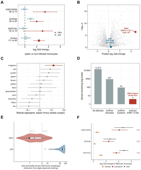
Monocyte pseudobulk profiles from three cohorts (166 donor records, two platforms) were compared between sepsis and non-infected controls (Figure 1A). EBI3 showed a pooled log2 fold change of +2.38 and ranked 137th/13,872; IL-27 ranked 9413th with a pooled effect of +0.21 (Figure 1B). Donor bootstrapping placed EBI3 in the top 5% in 78% of 300 resamples. Co-expression and secreted-proteome ranking independently converged on EBI3, while IL-27 remained far lower in the genome-wide ranking. The confirmatory REML/Hartung–Knapp estimate for EBI3 was +2.44 (95% CI +0.57 to +4.32; p = 0.030).
Figure 1.
Hypothesis-free nomination of EBI3. (A) Cross-cohort monocyte meta-analysis (166 donor records). (B) Genome-wide ranking of 13,872 shared genes. (C) Weighted gene co-expression network analysis (WGCNA) module context. (D) Secreted-protein/cytokine annotation. (E) Rank stability across 300 donor bootstraps. (F) Decomposition of the EBI3 fold change into a prevalence term (more positive cells) and an intensity term (more transcript per positive cell), in each of the three meta-analysis cohorts; the red value beside each cohort is the prevalence share of the total change. In (B), red marks EBI3, blue p28 and grey the remaining genes.
EBI3 remained highly ranked when genes were retained by detection rather than bulk-expression filters, avoiding exclusion of low-detection genes. The ranking was stable to alternative meta-analytic estimators and leave-one-cohort-out analyses.
WGCNA on the same donor-level pseudobulk placed EBI3 in a C1q-rich module (Figure 1C), providing convergent co-expression context for the EBI3-associated state.
Detection-matched null calibration placed EBI3 at the extreme positive tail in the principal cohort and SCP548, whereas IL-27 showed cohort-restricted elevation. This calibration reinforced EBI3 as the reproducible cross-cohort signal. Matching on total expression rather than on detection gave the same percentiles, and at IL-27’s own expression level reference genes reached detection odds ratios of about 3 at the 97.5th percentile of the matched distribution and up to 18.9 at the maximum. This argues against a global assay floor in that detection regime but does not quantify IL-27-specific dropout or guarantee that a given true effect would have been detected (Tables S9 and S10).
Annotation of the ranked 907 secreted proteins placed EBI3 near the top of the secreted set and among cytokines, while IL-27 remained low (Figure 1D). Other heteromeric secreted complexes showed coordinated partner-chain ranking, providing a biologically matched benchmark for the IL-27 subunits.
This population-level nomination motivated direct testing of whether EBI3 and p28 occur in the same cells.
Together, cross-cohort effect-size meta-analysis, donor resampling (Figure 1E), co-expression and secreted-proteome ranking consistently nominated EBI3 over IL-27, establishing a reproducible population-level subunit imbalance for cell-resolved testing.
3.2. Discordant IL-27 Subunit Induction Attenuates During Convalescence
Because IL-27 assembly requires both chains, the patient-level comparison was repeated with detection of EBI3 and p28 in the same monocytes of the principal cohort (Table 1). Against healthy volunteers, EBI3 rose strongly while p28 changed little. Against post-cardiac-surgery controls, the ORs were 11.0 (4.60–26.33) for EBI3 and 0.78 (0.50–1.20) for p28, with a ratio of ORs of 13.3 (5.1–34.7; Figure 2A,B).
Table 1.
Baseline characteristics of the principal cohort (GSE216009). The convalescent column comprises second time points from nine of the twenty-six septic patients; the cohort therefore contains 39 individuals in 48 samples. IQR, interquartile range.
Figure 2.
Discordant induction of the IL-27 subunits in septic monocytes. (A) EBI3 and IL-27 detection across clinical states, one point per sample with the median marked. (B) Ratio of disease-associated odds ratios. (C) Acute-to-convalescent change in nine patients. (D) EBI3-to-p28 detection ratio across monocyte subsets, one point per patient. Values above each subset are gene × subset interaction odds ratios against classical monocytes with 95% intervals; red bars are medians; open triangles are patients in whom p28 was detected but EBI3 was not, a ratio of zero that the logarithmic axis cannot show. (E) Patient-level difference-in-differences with and without subset standardisation. (F) Decomposition of the same increase in the principal cohort into within-subset induction, subset composition and their interaction, each as a share of the observed fold change on the log scale. In (E), grey denotes crude and red subset-standardised values. In (F), red bars are positive and blue negative contributions.
The same discordance appeared against non-infected intensive-care controls in the replication cohort with enough p28-positive cells (ratio of ORs 5.6, 1.6–20.3). It was driven mainly by prevalence: the prevalence term accounted for 87%, 92% and 75% of the EBI3 fold change across the three cohorts (Figure 1F). An additional CITE-seq cohort remained directionally concordant.
In nine patients sampled at acute sepsis and convalescence, EBI3 declined during recovery while p28 remained stable. The gene × time interaction was 0.067 (95% CI 0.010–0.461; p = 0.012; Figure 2C), demonstrating attenuation of the imbalance during convalescence.
The EBI3-to-p28 ratio was highest in the intermediate monocyte subset (Figure 2D), and subset adjustment with direct standardisation preserved the longitudinal interaction (Figure 2E).
Across protein-defined monocyte subsets, EBI3 detection was highest in intermediate and non-classical cells, whereas p28 remained low (gene × subset interaction versus classical monocytes: 8.4, 95% CI 2.8–25.8 for intermediate; 17.9, 2.9–110.0 for non-classical). The EBI3-to-p28 ratio therefore varies across the monocyte maturation axis while retaining the same direction of discordance.
Together, the longitudinal and subset analyses localise the discordance to the cellular level and show that it persists across the monocyte maturation axis.
3.3. EBI3-Positive Monocytes Define an HLA-DR-High State Distinct from MS1
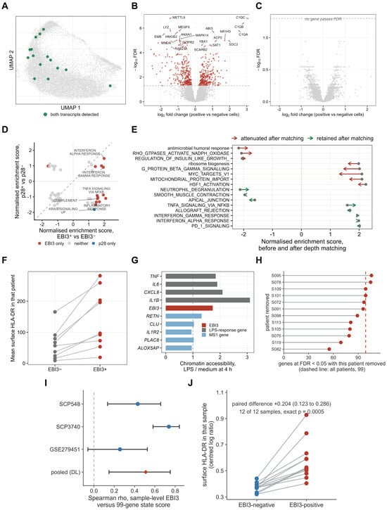
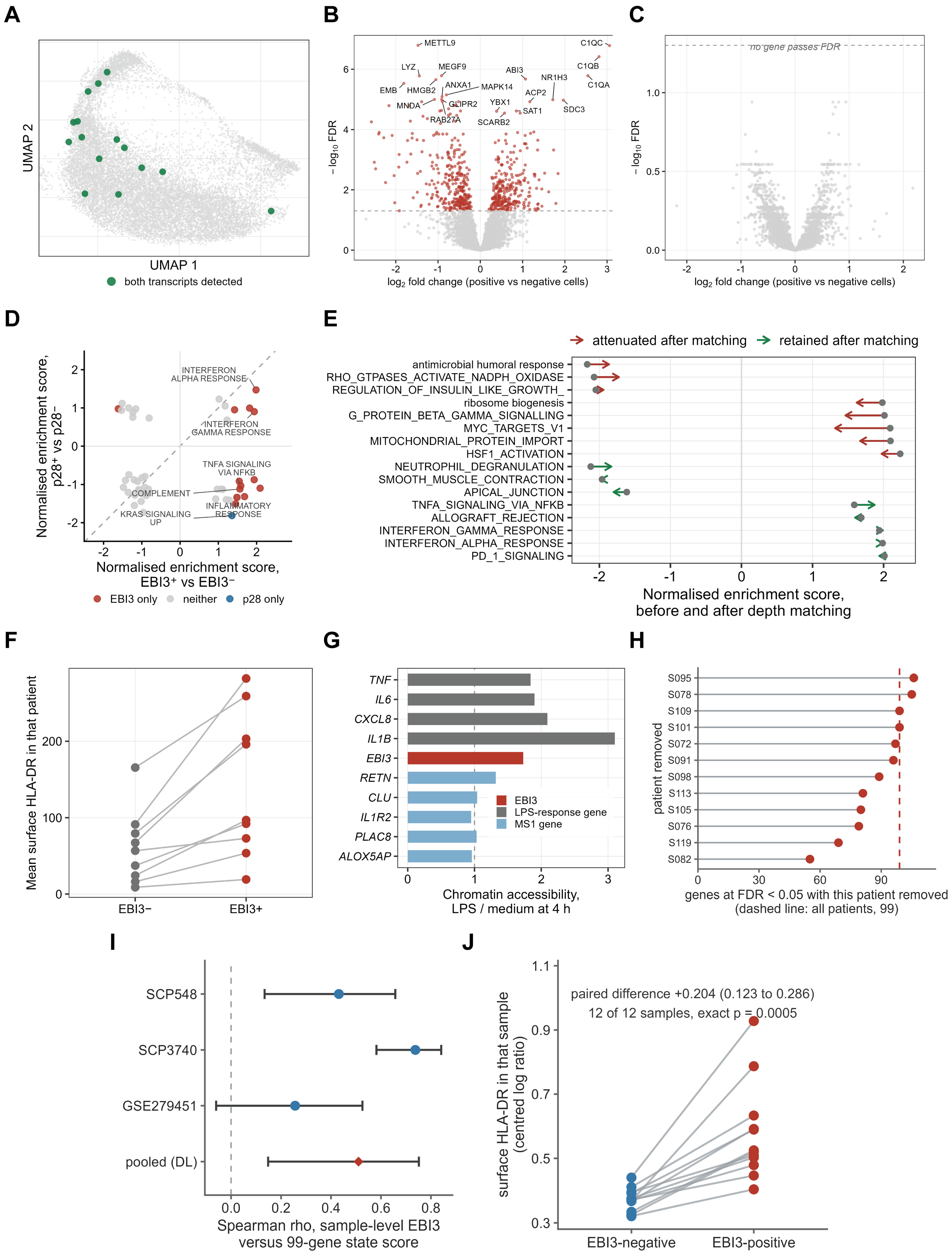
Cells carrying both transcripts were rare and scattered across the monocyte embedding (Figure 3A). EBI3-positive and p28-positive monocytes mapped to distinct transcriptional programmes: the EBI3-positive contrast yielded a substantial differential signature whereas the p28 contrast yielded none (Figure 3B,C). Hallmark enrichment scores were weakly correlated between the two contrasts (Figure 3D), and the EBI3-positive state was enriched for inflammatory and interferon-related programmes.
Figure 3.
The EBI3-associated state is distinct from p28 status and the MS1 monocyte state (MS1). (A) Monocytes with both transcripts detected, on the monocyte uniform manifold approximation and projection (UMAP) embedding: 15 of 15,958 monocytes pooled across all clinical states, against 11.6 expected from the marginal detection rates (the septic-only counts, 13 of 7164 against 11.9, are given in Section 3.4). (B) EBI3-positive versus EBI3-negative differential expression. (C) Corresponding p28 contrast. (D) Hallmark enrichment. (E) Depth-matched pathway comparison. (F) Surface HLA-DR. (G) EBI3-locus accessibility after lipopolysaccharide (LPS). (H) Leave-one-patient-out signature stability. (I) External projection of the 99-gene signature. (J) External protein-level replication. In (A), green marks monocytes with both transcripts detected and grey all others. In (I), blue circles are the individual cohorts and the red diamond the pooled estimate.
After depth matching, interferon-α/γ response, TNF/NF-κB signalling and inflammatory response remained recurrent features of the EBI3-associated state (Figure 3E). Together with subset-aware differential expression, these results define a reproducible transcriptional programme linked to EBI3 detection and distinct from p28 status.
EBI3-positive monocytes were HLA-DR-high, the reciprocal of the defining low-HLA-DR MS1 phenotype (Figure 3F). Within-patient decomposition attributed 94.7% of the EBI3 detection increase to induction within subsets, against −4.9% for subset composition (Figure 2F). Subset adjustment retained 99 differentially expressed genes and preserved C1QA/C1QC. Accessibility at the EBI3 locus rose 1.8-fold after 4 h of lipopolysaccharide in a single-donor chromatin series, alongside canonical response genes and unlike the MS1 markers (Figure 3G); with one donor, this observation is directionally consistent with the transcript data but cannot be generalised to the donor population, and it is not relied upon by any downstream inference. Separately, every leave-one-patient-out refit preserved the direction of all 99 genes (Figure 3H). The frozen 99-gene signature projected positively into all three external cohorts that lack surface protein (pooled Spearman ρ = +0.510, 95% CI +0.148 to +0.752; p = 0.008; Figure 3I). An L1 classifier predicted EBI3 status with cross-validated AUC 0.904 without selecting MS1 markers, and the same AUC was obtained when the MS1-defining genes were excluded from the candidate feature set outright (Table S16). The frozen five-gene MS1 score was lower in EBI3-positive monocytes in all twelve evaluable patients. The two states nonetheless co-existed within individuals: both were present in 24 of 26 septic patients and their patient-level frequencies were not inversely related (Spearman +0.145, p = 0.48), while within a patient an individual cell tended to carry one state or the other, with MS1-high defined by a median split of the frozen five-gene score. The pooled patient-clustered estimate of that cell-level association did not exclude the null (p = 0.114; Table S23). The surface phenotype replicated at the protein level in an independent CITE-seq cohort: in all twelve samples with sufficient cells, EBI3-positive monocytes carried more surface HLA-DR (paired difference +0.204, 95% CI 0.123–0.286, exact p = 0.0005; Figure 3J).
3.4. Rare Same-Cell Co-Detection of EBI3 and IL-27 Matches Chance Expectation
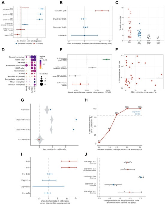
Within septic monocytes, 13 of 7164 cells carried both transcripts, against 11.9 expected from the marginal detection rates, and 97.7% of EBI3-positive cells lacked detectable p28. The pooled Fisher OR was 1.11 (p = 0.65) and the patient-clustered co-detection OR was 0.91 (95% CI 0.48–1.75). Benchmark complexes in the same cells showed materially higher co-detection (Figure 4A).
Figure 4.
IL-27 subunit discordance persists across same-cell and receiver-side analyses. (A) Same-cell co-detection odds for IL-27 and benchmark complexes. (B) Chain-to-chain odds-ratio ratios. (C) EBI3 and alternative α-chain detection. (D) Ligand and receptor-chain detection across blood cells. (E) IL-27-associated module in receptor-positive T and NK cells. (F) Patient-level association across monocyte EBI3 detection. (G) Structure-preserving co-detection null. (H) Recovery of known associations by simulation. (I) Pairwise chain-to-chain contrasts; two pre-frozen pairs are omitted from the panel because a chain has no positive cells in the control arm, which separates the model used here, and are given as Firth penalised-likelihood refits in Table 2. (J) Module response to exogenous cytokines. Throughout, red marks EBI3- or IL-27-containing series and blue the corresponding comparator (p28, benchmark complexes, or pairs and arms without EBI3); the green circle in (E) is the IL-15 positive control, and grey denotes background or unselected points.
A structure-preserving permutation reproduced the same separation. Shuffling IL-27 detection within patient × subset × depth strata left IL-27 at its structural null: an observed odds ratio of 0.92 against a null median of 1.02, at the 27th percentile of the permutation distribution. The C1q and calprotectin benchmarks gave observed odds ratios of 228.3, 346.7 and 5.63 against null medians of 6.40, 6.91 and 3.24, and exceeded every permutation (Figure 4G). Recovery tests showed that the pipeline detects positive associations when present (Figure 4H).
The benchmark lower confidence bounds were 2.65 or higher, compared with an upper bound of 1.75 for IL-27 (Figure 4B and Supplementary Figure S1). Alternative α-chain analyses reinforced localisation of the discordance to the EBI3–p28 pairing. Against healthy volunteers, where all three EBI3-containing pairs are estimable, the chain-to-chain ratios of ORs were 35.0 (7.4–165.1) for EBI3/p28 (IL-27), 61.6 (10.3–367.8) for EBI3/IL12A and 71.7 (18.9–272.8) for EBI3/IL23A; the corresponding estimate against post-cardiac-surgery controls for EBI3/p28 is the 13.3 reported above (Figure 4I and Table 2).
Table 2.
Chain-to-chain ratios of odds ratios for the eight pre-frozen complexes under both non-infected comparators. Values above 1 indicate stronger induction of the first-listed chain. Two pairs have no positive cells for one chain in the post-cardiac-surgery arm, which separates the patient-clustered model; they are refitted by Firth penalised likelihood and marked †. Both penalised intervals are wide and neither excludes unity, so these two entries are individually uninformative. On point estimates the separation is nonetheless complete under both comparators: every EBI3-containing pair exceeds every pair without EBI3 (1.85–13.5 versus 0.29–1.14 against post-cardiac-surgery controls). Full model output is in Table S12.
Alternative α-chain analysis further localised the discordance to the EBI3–p28 pairing (Figure 4C). The cell-resolved data therefore connect the reported diagnostic signal to a previously unrecognised molecular-attribution gap in coordinated EBI3–p28 production.
3.5. Receiver-Side Analysis Extends the Discordance Beyond Producer Cells
Receiver-side analysis provided an orthogonal test of IL-27-associated activity. In T and NK cells carrying both IL-27RA and IL6ST, the prespecified 41-gene module had β = −0.0151 (95% CI −0.0345 to +0.0043; Figure 4D,E). The parallel IL-15 analysis recovered a clear receptor-linked response (+0.0462), confirming the sensitivity of the design to receptor-associated signalling. Detection-matched receptor substitutions placed the observed IL-27 coefficient at the lower edge of the empirical null (95% range −0.0148 to +0.0258).
The dual-receptor definition provided the primary receiver-side estimate; IL-27RA alone gave +0.0133 and IL6ST alone −0.0273, further distinguishing the joint-receptor analysis from single-chain stratification. At the patient level, the module score did not follow the EBI3-positive monocyte fraction across its hundredfold range (Spearman ρ = +0.11, p = 0.60; Figure 4F).
Two independent human T-cell cytokine datasets [41,42] confirmed that IL-27 induces the 41-gene module (Figure 4J). Interferon-α and interleukin-6 also induced the programme, supporting its use as an IL-27-associated shared-response module.
3.6. The EBI3-to-p28 Ratio Provides Comparator-Stable, Cell-Dependent Discrimination
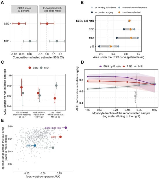
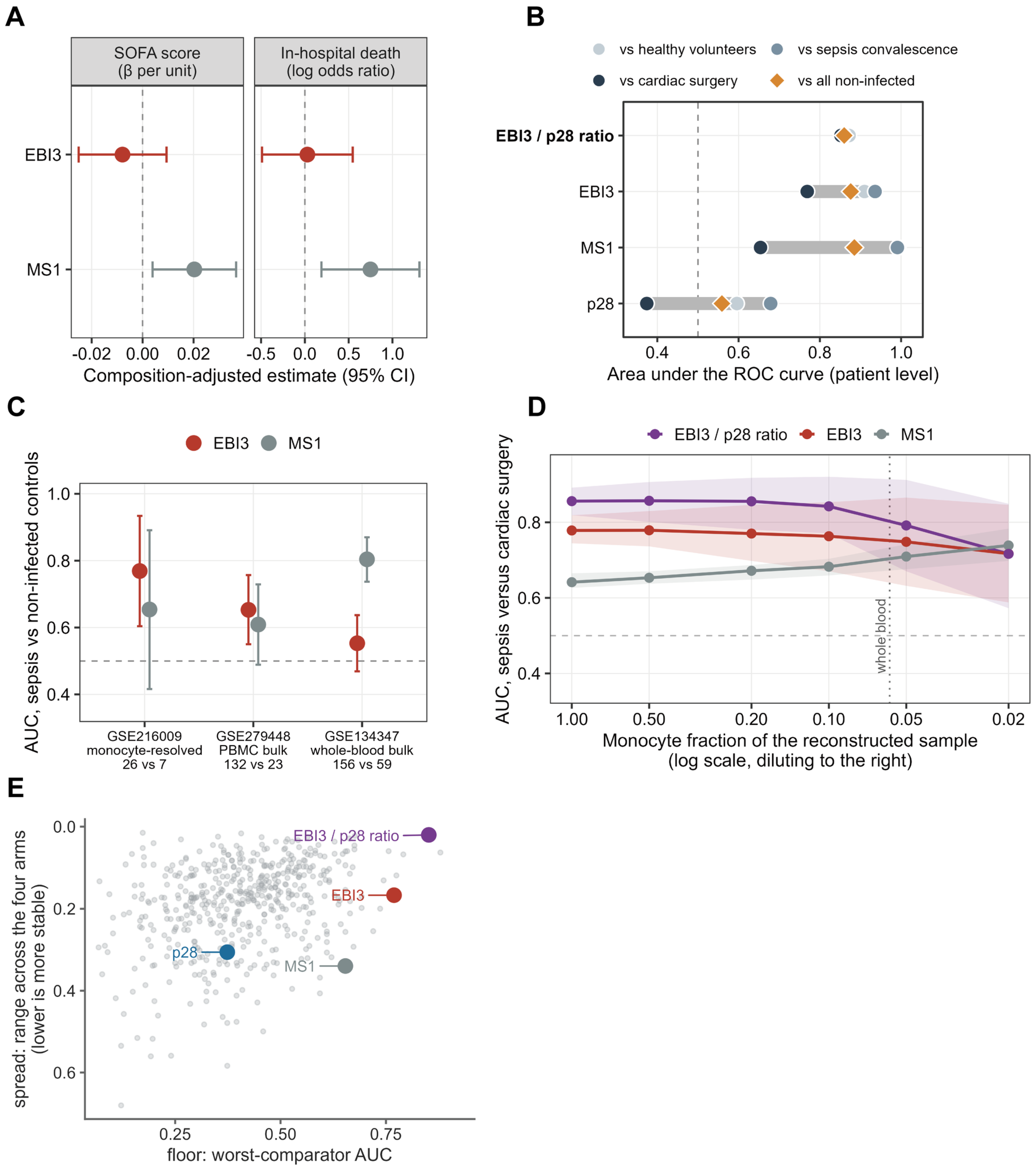
Among 348 septic patients, MS1 tracked the Sequential Organ Failure Assessment (SOFA) score (adjusted β = +0.020, p = 0.015; n = 345) and mortality (adjusted OR 2.11, 1.21–3.70), while EBI3 captured an orthogonal dimension of the response (SOFA β = −0.008, p = 0.37; mortality OR 1.03, 0.61–1.73; Figure 5A).
Figure 5.
The EBI3-to-p28 ratio provides a comparator-stable, cell-dependent signal. (A) Associations with Sequential Organ Failure Assessment (SOFA) score and mortality. (B) Discrimination across four non-infected comparator groups. (C) Discrimination after aggregation to PBMCs and whole blood. (D) Dilution with decreasing monocyte content. (E) Calibration against 500 detection-matched gene pairs.
Across four comparator arms, the EBI3-to-p28 ratio showed the smallest observed spread in patient-level discrimination: AUC 0.852–0.872 (spread 0.020), compared with 0.769–0.936 for EBI3 and 0.654–0.994 for MS1 (Figure 5B). This is a difference in spread rather than in magnitude: the ratio did not discriminate better than EBI3 alone in any individual arm (paired DeLong p ≥ 0.13), and adding p28 to a model already containing EBI3 did not improve fit in any arm (likelihood-ratio p ≥ 0.13; Table S22). Greater stability than EBI3 alone was not established by the paired patient bootstrap, whose interval for the difference in spread includes zero (Table S21).
Aggregation changed the scores in opposite directions. EBI3 discrimination fell from monocytes to peripheral blood mononuclear cells (PBMCs) to whole blood (0.769, 0.653, 0.553), whereas MS1 rose in whole blood (0.654, 0.609, 0.804; Figure 5C). Dilution simulations reproduced the same crossing pattern (Figure 5D). Monocyte content shaped the magnitude of the ratio, while comparator stability was retained. Its combined floor and spread lay beyond the corresponding detection-matched null distribution (Figure 5E).
3.7. Functional Validation Connects the p28-Associated Axis to Macrophage State and Survival
The experimental series extended the cell-resolved discovery into human macrophage and in vivo systems, testing whether the subunit imbalance is reproducible and functionally consequential.
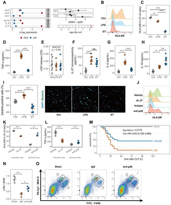
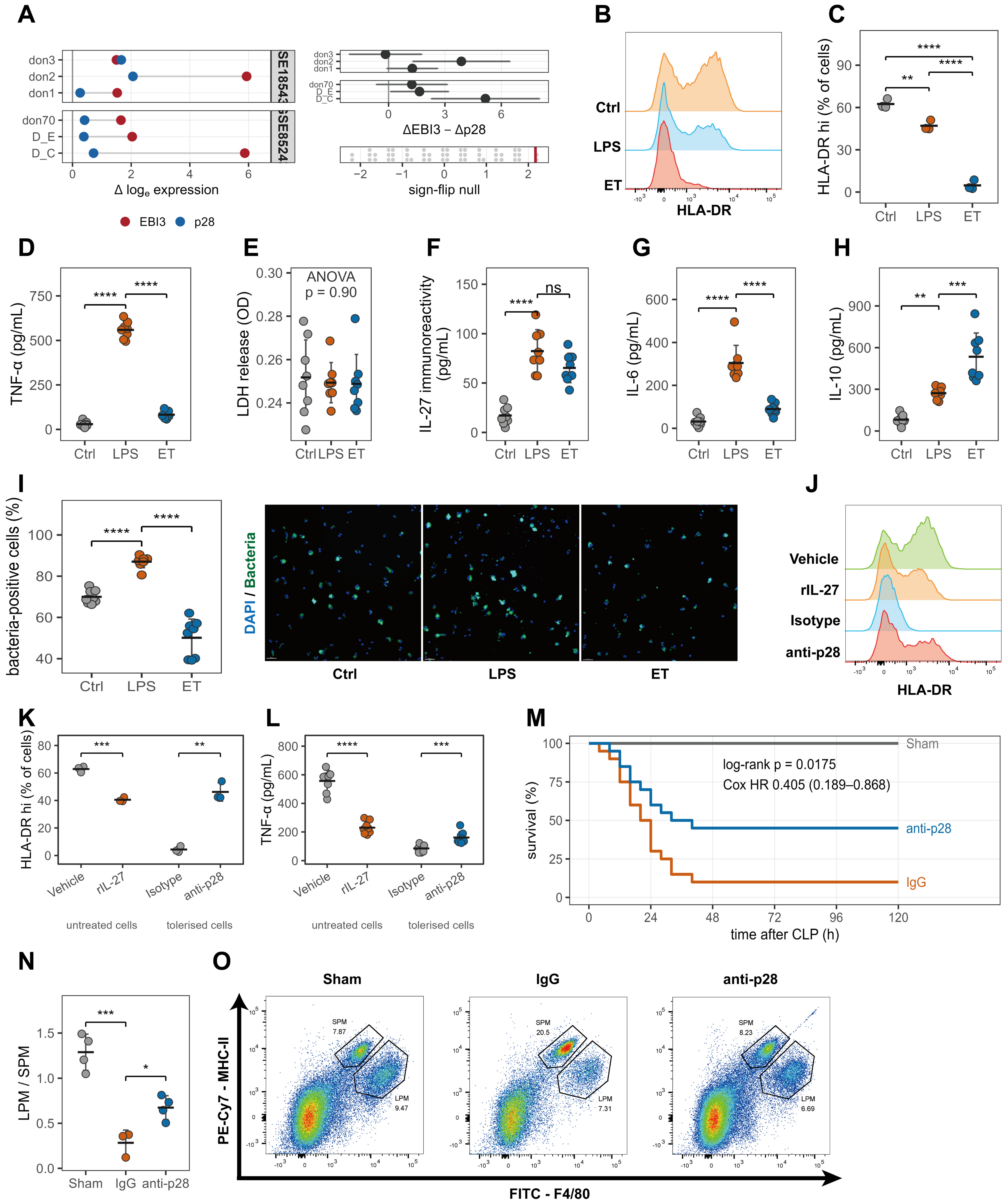
In GSE85243 and GSE185433, six days of LPS preconditioning increased EBI3 and IL-27 in all six paired donors, with a positive donor-level ΔEBI3 − ΔIL-27 interaction in five of six (exact sign-flip p = 0.0312 one-sided and 0.0625 two-sided). The interaction estimate was +2.17 on the natural-log expression scale (95% CI −0.21 to +4.55, study-balanced fixed-effect model), reproducing the direction of subunit imbalance in a human macrophage setting (Figure 6A).
Figure 6.
Experimental support for the IL-27-associated axis in human macrophages and mice. (A) Paired human macrophage donors after lipopolysaccharide preconditioning: the per-donor change in EBI3 and p28 in two public datasets (GSE185433 and GSE85243; six donors), the within-donor difference ΔEBI3 − Δp28, and the corresponding sign-flip null. (B) Surface HLA-DR distributions in the tolerant-like THP-1 model, from which the HLA-DR-high fractions in (C) were taken; an identical gate was applied to every arm. (C) Surface HLA-DR across untreated, acute lipopolysaccharide and tolerised arms (n = 3 per arm). (D) Secreted TNF-α for the same three arms (n = 8 per arm). (E) Lactate dehydrogenase release for the same three arms (n = 8 per arm), read as optical density and compared within the plate. (F) IL-27-associated immunoreactivity in conditioned medium (n = 8 per arm). (G) IL-6 in conditioned medium (n = 8 per arm). (H) IL-10 in conditioned medium (n = 8 per arm). (I) Cells associated with labelled bacteria (eight wells per arm; the well, not the field or the individual cell, is the unit of analysis), with representative fields (nuclei blue, labelled bacteria green; scale bar 50 μm). (J) Surface HLA-DR distributions after recombinant IL-27 in untreated cells and after the p28-directed antibody in tolerised cells, from which the fractions in (K) were taken; the same gate was applied throughout. (K) Surface HLA-DR for those four arms (n = 3 per arm). (L) TNF-α for the same four arms (n = 8 per arm). (M) Caecal ligation and puncture (CLP) survival after anti-p28 versus control immunoglobulin G (IgG) (n = 20 per group). (N) Peritoneal large-to-small macrophage ratio 48 h after caecal ligation and puncture (sham n = 4, CLP + IgG n = 3, CLP + anti-p28 n = 4). (O) Representative peritoneal flow plots for the three arms in (N), gated on F4/80 (FITC) against MHC-II (PE-Cy7); the small and large peritoneal macrophage gates are shown with their percentages, one animal per arm. Points are individual wells (in vitro) or animals (in vivo); the horizontal bar is the arm mean and whiskers span ±1 standard deviation (SD). Three-group comparisons are one-way analysis of variance with Tukey’s post-hoc test; the within-pair comparisons in (K,L) are Welch t-tests; (E) reports one-way analysis of variance only; (M) is a log-rank test with a Cox hazard ratio. * p < 0.05; ** p < 0.01; *** p < 0.001; **** p < 0.0001; ns = not significant.
In THP-1 macrophage-like cells, LPS preconditioning generated a tolerant-like state. Relative to the acute lipopolysaccharide arm, the tolerised arm had lower surface HLA-DR (47.1% to 4.7%; Figure 6B,C), TNF (559.3 to 82.4 pg/mL; Figure 6D) and IL-6 (304.2 to 89.4 pg/mL; Figure 6G), higher IL-10 (272.2 to 534.4 pg/mL; Figure 6H), and fewer cells associated with labelled bacteria (87.1% to 50.1%; Figure 6I); all five model-validation readouts moved in the expected direction. In the same supernatants, IL-27-associated immunoreactivity remained at 79.3% of its acute value, compared with 14.7% for TNF and 29.4% for IL-6 (Figure 6F).
The IL-27-associated readout therefore remained relatively preserved as the conventional inflammatory programme contracted, linking the transcript-level finding to a distinct secreted signal.
Recombinant IL-27 lowered HLA-DR from 62.9% to 40.5% (p = 2.1 × 10⁻⁴) and TNF from 556.9 to 230.1 pg/mL (p = 3.2 × 10⁻⁷), confirming that the readouts respond to the assembled ligand (Figure 6J–L).
A p28-directed antibody produced the reciprocal response in the tolerant-like arm, increasing HLA-DR from 4.3% to 46.2% (p = 0.0047) and TNF from 84.6 to 160.9 pg/mL (p = 9.1 × 10⁻⁴). The reciprocal shifts are shown in Figure 6J–L. Lactate dehydrogenase release remained stable across the experimental arms (Figure 6E and Table S31).
In CLP sepsis, anti-p28 treatment increased 120 h survival from 10% to 45% (log-rank p = 0.0175; Cox HR 0.405, 0.189–0.868; n = 20/group; Figure 6M). In a separate cohort, the large:small peritoneal macrophage ratio shifted from 0.28 with control IgG to 0.67 with anti-p28, toward the sham value of 1.29 (ANOVA p = 0.0001; treated vs. IgG p = 0.033; Figure 6N,O).
Together, the human macrophage and murine experiments connect the transcript-level discovery to functional p28-axis sensitivity, with reciprocal macrophage phenotypes in vitro and improved survival in vivo.
4. Discussion
4.1. Transcript-Level Discordance as the Central Biological Observation
The central biological advance is the identification of a sepsis-associated uncoupling of IL-27 subunit transcription. EBI3 and p28 diverge across protein-defined monocyte subsets, the magnitude of divergence exceeds that seen for concordant benchmark complexes, and the imbalance attenuates during convalescence.
The co-detection benchmark is especially informative: in the same cells and abundance range, the lowest benchmark confidence bound was 2.65, whereas the IL-27 upper bound was 1.75. The receiver-side analysis provides a second compartment-level readout, and both human macrophage datasets and the experimental model point in the same direction. Together, the data define a robust transcriptional coordination phenotype.
The phenotype is defined by coordinated divergence across the two chains: EBI3 induction is reproducible across cohorts, IL-27 remains largely unchanged across cohorts, and the imbalance diminishes with recovery. Our reanalysis of a published single-cell cytokine-response atlas [43] likewise separates the regulation of the two chains; the original report did not examine EBI3–p28 discordance. The EBI3-to-p28 ratio therefore captures a biologically coherent transcript-level coordination phenotype across clinical states and monocyte subsets.
Unpaired EBI3 is biologically plausible: activated human neutrophils release EBI3 without its heterodimeric partners [44], it has chaperone-like activity [45], and it binds interferon-γ and IL-10 [46]. These observations make unpaired EBI3 a plausible contributor to the sepsis-associated signal and motivate direct molecular resolution.
4.2. The EBI3-Associated Monocyte Phenotype Is Distinct from MS1
The EBI3-associated state is a stimulus-responsive programme embedded within conventional monocyte subsets. Within-subset induction accounted for 94.7% of the increase in EBI3 detection; subset adjustment retained 99 genes and C1QA/C1QC. EBI3-positive cells were HLA-DR-high, distinct from MS1, and the transcriptome predicted EBI3 status with AUC 0.904 without selecting MS1 markers.
The phenotype therefore represents an inducible functional state across conventional monocyte differentiation, separable from MS1 and independently reproduced at the surface-protein level.
4.3. Orthogonal Evidence from the Producer and Receiver Compartments
Three cell-resolved layers define the attribution problem in sequence. At the producer level, strong EBI3 induction was unmatched by IL-27; at the same-cell level, co-detection remained at chance and below the benchmark range of concordantly transcribed complexes; and at the receiver level, the dual-receptor module estimate remained near zero while IL-15 responsiveness was preserved. Producer, same-cell and receiver analyses therefore converge on the same molecular-attribution gap.
4.4. Functional Activity and Molecular Attribution Are Distinct Dimensions of the IL-27 Axis
The patient data show discordant subunit transcription and low same-cell co-detection, whereas p28-directed intervention improves survival in murine sepsis. Together, these findings separate two biologically relevant dimensions of the IL-27 axis: functional sensitivity to p28-directed perturbation and molecular attribution of the circulating signal.
The murine survival result establishes the biological consequence of p28-directed perturbation, while the human cell-resolved analyses define the molecular context in which that activity should be interpreted. This separation of functional sensitivity from molecular attribution is a central conceptual advance of the study. That inference is qualified by a species difference in subunit biology introduced above: murine p28 can be secreted autonomously as IL-30, whereas isolated human p28 is retained and degraded. A p28-directed antibody therefore engages a free-ligand pool in the mouse that the human compartment is not expected to contain to the same degree, so the murine experiment supports functional sensitivity of the p28-associated axis without implying that the same molecular species carries the human signal. The murine experiment is not offered as a new demonstration that p28-directed blockade improves survival in sepsis; that effect is established [18,19]. Its role here is to serve as an existence proof placed alongside the human transcript analyses within one study: it shows that functional sensitivity to p28-directed intervention can be present in the same disease in which coordinated heterodimer production is not evident, and that coexistence is what defines the attribution gap. The corresponding constraints on group size are given in Section 4.6.
The same framework extends to diagnostics: clinical performance and molecular attribution are complementary properties, and cross-subunit assay design provides the direct bridge between them.
4.5. Consequences for Biomarker Measurement and Translation
Clinical performance and molecular identity are related but distinct properties of a biomarker. Performance depends on comparator, compartment and assay epitope. The EBI3-to-p28 ratio was more stable across non-infected comparators than MS1, while aggregation diluted the monocyte-resolved EBI3 signal and shifted it in the opposite direction from MS1. Translation therefore depends on cell context as well as assay design. In-silico dilution showed that the ratio, like EBI3 itself, loses discrimination as monocyte content falls, so it is a within-monocyte quantity; measuring it clinically would require a monocyte-resolved rather than a whole-blood measurement.
A meta-analysis of circulating IL-27 protein reports an AUC of 0.88 for sepsis diagnosis [7], while the discovery programme was anchored to EBI3 [47] and the assay platform measures p28 [48]. Cross-subunit capture/detection, with antibodies recognising different chains, directly assigns signal to assembled IL-27 and provides the clearest route to link diagnostic performance to the heterodimer. That meta-analysis does not itself report the assay platform for any of its seven contributing studies. Three of the seven come from the programme cited here, which used a magnetic-bead multiplex assay and stated that the assay detects the p28 subunit [48]; for the remaining four, the assays are not recoverable from the meta-analysis, and we therefore do not claim that the chain-specificity issue generalises to them.
The intervention literature is also chain-dependent: murine blockade improves outcome [18,19], whereas neutralising endogenous IL-27 in human monocytes increases inflammatory output [20]. Taken together, these studies reinforce the need to interpret pathway activity and molecular attribution as distinct dimensions across species.
4.6. Limitations
This study has several limitations. The human analyses resolve subunit coordination at the level of single-cell transcripts and do not establish the molecular composition of the circulating protein signal. Cross-subunit measurement of assembled IL-27 alongside total EBI3 and p28 remains the direct test of the attribution framework, and the immunoassay used here has undefined chain specificity, which is why its output is reported throughout as IL-27-associated immunoreactivity rather than as IL-27. Same-cell, same-timepoint co-detection is an informative upstream indicator but is neither necessary nor sufficient for heterodimer assembly: translated protein can persist beyond its transcript, and secretion need not be synchronous with transcription, so its absence does not exclude asynchronous or trans-cellular pairing. The argument therefore does not rest on co-detection by itself but adds a receiver-side readout, which lies downstream of assembly, secretion and receptor engagement and is not subject to that objection. In the same vein, no chain-specific immunoassay, immunoblot or mass-spectrometric confirmation of the intact heterodimer was performed, so the retained immunoreactivity in tolerant-like cells cannot distinguish assembled IL-27 from free EBI3 or other cross-reactive material. This uncertainty is intrinsic to the molecular-attribution question addressed here and limits what the tolerance experiment can establish; chain-specific quantification is required for direct molecular resolution.
The experimental series is small. Each macrophage dataset contributes three paired donors; the donor-level interaction was positive in five of six and met the prespecified one-sided sign-flip threshold, but its effect-size interval includes zero and the two-sided value is 0.0625. The tolerant-like model uses an established cell line rather than primary cells. In the bacterial-association assay the tolerised arm yielded fewer scored cells per well than the other arms, so a contribution of cell density to that difference cannot be excluded, and chromatin accessibility was assessed in a single donor.
The antibody was given before induction, so the murine experiment tests prophylaxis rather than treatment of established sepsis, and the single 48 h harvest cannot separate prevention of the peritoneal macrophage shift from its reversal. The intervention establishes that the axis is functionally consequential but cannot assign the neutralised species: murine p28 is secreted autonomously as IL-30, whereas isolated human p28 is retained and degraded, and a p28-directed antibody binds p28 whether free or paired; therefore, the in vivo result constrains pathway activity rather than heterodimer identity. This is the same attribution limit that the human data describe, encountered here in the intervention itself. Only male mice were used, so the well-documented sex differences in the immune response to sepsis were not addressed and neither the survival nor the peritoneal result should be assumed to extend to females. The peritoneal endpoint rests on three to four animals per group, which is small for a three-group analysis of variance and leaves that comparison with wide uncertainty; group sizes throughout the in vivo work were set from previous experience with the model rather than from a power calculation.
The receiver-side result is a calibrated negative rather than an equivalence claim, since no smallest effect of interest was defined in advance for that module; a post-hoc characterisation places its minimum detectable effect at 0.0346 module-score units, three-quarters of the in vivo IL-15 response measured in the same cells, so a smaller response cannot be excluded. The EBI3-to-p28 ratio is accordingly presented as a comparator-stable readout rather than as a superior classifier: at this sample size, the p28 denominator contributes no detectable independent discrimination, which is the expected consequence of the near-null p28 induction that the study reports. The study reports a large number of estimators, normalisations, benchmark comparisons and permutation calibrations. Although the primary analyses and their criteria were fixed in advance and no overarching correction was applied across claims (Section 2.7), the volume of parallel analyses is itself a source of researcher degrees of freedom, and we note it as such; the direction of the principal findings was preserved across all of them. Finally, the contributing cohorts differ in platform, control definition and sampling time; the discordance phenotype was nonetheless consistent across them and across the prespecified sensitivity analyses.
5. Conclusions
In human sepsis, EBI3 and p28 are discordantly induced and rarely co-detected in the same monocyte. Against post-cardiac-surgery controls, EBI3 detection had OR 11.0 versus 0.78 for p28, ratio of ORs 13.3. The IL-27 co-detection upper bound was 1.75, below the benchmark lower bound of 2.65, and the receiver-side module estimate was β = −0.0151 (95% CI −0.0345 to +0.0043).
The imbalance was reproduced in human macrophages, IL-27-associated immunoreactivity remained relatively preserved in a tolerant-like state, and p28-directed intervention improved murine survival, establishing functional relevance of the p28-associated axis.
Together, these findings identify subunit discordance as a previously unrecognised feature of the IL-27 axis in human sepsis and expose a molecular-attribution gap between pathway-associated activity and attribution to intact heterodimeric IL-27. Cross-subunit measurement of assembled IL-27 alongside total EBI3 and p28 provides the direct molecular test of this framework.
Supplementary Materials
The following supporting information can be downloaded at: https://www.mdpi.com/article/10.3390/biomedicines14092035/s1, Section S1: Cohort replication and longitudinal support; Section S2: Single-cell object reconstruction and gating validation; Section S3: Cross-cohort nomination and robustness; Section S4: Same-cell co-detection and benchmark calibration; Section S5: The EBI3-associated transcriptional state; Section S6: Receiver-side calibration and positive-control validation; Section S7: Patient-level and cross-compartment validation; Section S8: Reproducibility, module definition and dataset roles; Section S9: Experimental validation: per-sample and per-mouse values.
Author Contributions
Conceptualization, Q.-S.S. and Y.Z.; methodology, Y.Z. and Q.-S.S.; software, Y.Z.; formal analysis, Y.Z.; investigation, Y.Z. and Q.-S.S.; data curation, Y.Z.; writing—original draft preparation, Y.Z.; writing—review and editing, Q.-S.S.; visualization, Y.Z.; supervision, Q.-S.S.; project administration, Q.-S.S. All authors have read and agreed to the published version of the manuscript.
Funding
This research received no external funding.
Institutional Review Board Statement
The study was conducted in accordance with the Declaration of Helsinki. Computational analyses used only publicly available, de-identified datasets generated under the ethical approvals reported in the original studies and involved no new human material. The animal experiments in Section 2.8 were approved by the Animal Ethics Committee of Shanghai Yishang Biotechnology Co., Ltd. (protocol code YISH-20230227-002, approved on 27 February 2023) and were performed in accordance with institutional and national guidelines for the care and use of laboratory animals. The cell-line experiments used the established THP-1 line and required no ethical approval.
Informed Consent Statement
Not applicable. No new human material was collected for this work; the human data analysed are de-identified and publicly available, generated under the informed consent obtained in the original studies.
Data Availability Statement
All transcriptomic data analysed are publicly available under the accession numbers given in Section 2.1 and Supplementary Sections S1 and S7; raw public data are not redistributed here and remain with the repositories cited there. The analysis code, the intermediate result tables underlying every figure and table, the final figure files, the raw laboratory workbooks and the dependency lockfiles are available on request from the corresponding author. No standalone software package was developed; the analysis scripts are the code of record and are retained with their versioned dependencies.
Conflicts of Interest
The authors declare no conflicts of interest.
References
- Pflanz, S.; Timans, J.C.; Cheung, J.; Rosales, R.; Kanzler, H.; Gilbert, J.; Hibbert, L.; Churakova, T.; Travis, M.; Vaisberg, E.; et al. IL-27, a heterodimeric cytokine composed of EBI3 and p28 protein, induces proliferation of naive CD4+ T cells. Immunity 2002, 16, 779–790. [Google Scholar] [CrossRef] [Scilit] [PubMed]
- Hunter, C.A.; Kastelein, R. Interleukin-27: Balancing protective and pathological immunity. Immunity 2012, 37, 960–969. [Google Scholar] [CrossRef] [Scilit] [PubMed]
- Yoshida, H.; Hunter, C.A. The immunobiology of interleukin-27. Annu. Rev. Immunol. 2015, 33, 417–443. [Google Scholar] [CrossRef] [Scilit] [PubMed]
- Collison, L.W.; Workman, C.J.; Kuo, T.T.; Boyd, K.; Wang, Y.; Vignali, K.M.; Cross, R.; Sehy, D.; Blumberg, R.S.; Vignali, D.A.A. The inhibitory cytokine IL-35 contributes to regulatory T-cell function. Nature 2007, 450, 566–569. [Google Scholar] [CrossRef] [Scilit] [PubMed]
- Devergne, O.; Birkenbach, M.; Kieff, E. Epstein-Barr virus-induced gene 3 and the p35 subunit of interleukin 12 form a novel heterodimeric hematopoietin. Proc. Natl. Acad. Sci. USA 1997, 94, 12041–12046. [Google Scholar] [CrossRef] [Scilit] [PubMed]
- Müller, S.I.; Friedl, A.; Aschenbrenner, I.; Bieren, J.E.-V.; Zacharias, M.; Devergne, O.; Feige, M.J. A folding switch regulates interleukin 27 biogenesis and secretion of its α-subunit as a cytokine. Proc. Natl. Acad. Sci. USA 2019, 116, 1585–1590. [Google Scholar] [CrossRef] [Scilit] [PubMed]
- Wang, Y.; Zhao, J.; Yao, Y.; Zhao, D.; Liu, S. Interleukin-27 as a diagnostic biomarker for patients with sepsis: A meta-analysis. Biomed Res. Int. 2021, 2021, 5516940. [Google Scholar] [CrossRef] [Scilit] [PubMed]
- Coherus Oncology, Inc. A Randomized Phase 2 Study of Casdozokitug in Combination with Toripalimab Plus Bevacizumab in Participants with Unresectable and/or Locally Advanced or Metastatic Hepatocellular Carcinoma. ClinicalTrials.gov Identifier NCT06679985. Available online: https://clinicaltrials.gov/study/NCT06679985 (accessed on 3 August 2026).
- Singer, M.; Deutschman, C.S.; Seymour, C.W.; Shankar-Hari, M.; Annane, D.; Bauer, M.; Bellomo, R.; Bernard, G.R.; Chiche, J.-D.; Coopersmith, C.M.; et al. The Third International Consensus Definitions for Sepsis and Septic Shock (Sepsis-3). JAMA 2016, 315, 801–810. [Google Scholar] [CrossRef] [Scilit] [PubMed]
- Rudd, K.E.; Johnson, S.C.; Agesa, K.M.; Shackelford, K.A.; Tsoi, D.; Kievlan, D.R.; Colombara, D.V.; Ikuta, K.S.; Kissoon, N.; Finfer, S.; et al. Global, regional, and national sepsis incidence and mortality, 1990–2017: Analysis for the Global Burden of Disease Study. Lancet 2020, 395, 200–211. [Google Scholar] [CrossRef] [Scilit] [PubMed]
- van der Poll, T.; Shankar-Hari, M.; Wiersinga, W.J. The immunology of sepsis. Immunity 2021, 54, 2450–2464. [Google Scholar] [CrossRef] [Scilit] [PubMed]
- Hotchkiss, R.S.; Monneret, G.; Payen, D. Sepsis-induced immunosuppression: From cellular dysfunctions to immunotherapy. Nat. Rev. Immunol. 2013, 13, 862–874. [Google Scholar] [CrossRef] [Scilit] [PubMed]
- Davenport, E.E.; Burnham, K.L.; Radhakrishnan, J.; Humburg, P.; Hutton, P.; Mills, T.C.; Rautanen, A.; Gordon, A.C.; Garrard, C.; Hill, A.V.S.; et al. Genomic landscape of the individual host response and outcomes in sepsis: A prospective cohort study. Lancet Respir. Med. 2016, 4, 259–271. [Google Scholar] [CrossRef] [Scilit] [PubMed]
- Burnham, K.L.; Davenport, E.E.; Radhakrishnan, J.; Humburg, P.; Gordon, A.C.; Hutton, P.; Svoren-Jabalera, E.; Garrard, C.; Hill, A.V.S.; Hinds, C.J.; et al. Shared and distinct aspects of the sepsis transcriptomic response to faecal peritonitis and pneumonia. Am. J. Respir. Crit. Care Med. 2017, 196, 328–339. [Google Scholar] [CrossRef] [Scilit] [PubMed]
- Scicluna, B.P.; van Vught, L.A.; Zwinderman, A.H.; Wiewel, M.A.; Davenport, E.E.; Burnham, K.L.; Nürnberg, P.; Schultz, M.J.; Horn, J.; Cremer, O.L.; et al. Classification of patients with sepsis according to blood genomic endotype: A prospective cohort study. Lancet Respir. Med. 2017, 5, 816–826. [Google Scholar] [CrossRef] [Scilit] [PubMed]
- Reyes, M.; Filbin, M.R.; Bhattacharyya, R.P.; Billman, K.; Eisenhaure, T.; Hung, D.T.; Levy, B.D.; Baron, R.M.; Blainey, P.C.; Goldberg, M.B.; et al. An immune-cell signature of bacterial sepsis. Nat. Med. 2020, 26, 333–340. [Google Scholar] [CrossRef] [Scilit] [PubMed]
- Leite, G.G.F.; de Brabander, J.; Michels, E.H.A.; Butler, J.M.; Cremer, O.L.; Scicluna, B.P.; Sweeney, T.E.; Reyes, M.; Salomao, R.; Peters-Sengers, H.; et al. Monocyte state 1 (MS1) cells in critically ill patients with sepsis or non-infectious conditions. Crit. Care 2024, 28, 88. [Google Scholar] [CrossRef] [Scilit] [PubMed]
- Wirtz, S.; Tubbe, I.; Galle, P.R.; Schild, H.J.; Birkenbach, M.; Blumberg, R.S.; Neurath, M.F. Protection from lethal septic peritonitis by neutralizing the biological function of interleukin 27. J. Exp. Med. 2006, 203, 1875–1881. [Google Scholar] [CrossRef] [Scilit] [PubMed]
- Bosmann, M.; Russkamp, N.F.; Strobl, B.; Roewe, J.; Balouzian, L.; Pache, F.; Radsak, M.P.; van Rooijen, N.; Zetoune, F.S.; Sarma, J.V.; et al. Interruption of macrophage-derived IL-27(p28) production by IL-10 during sepsis requires STAT3 but not SOCS3. J. Immunol. 2014, 193, 5668–5677. [Google Scholar] [CrossRef] [Scilit] [PubMed]
- Frangieh, M.; McHenry, A.; Phillips, R.; Ye, C.; Bernier, A.; Laffel, L.; Elyaman, W.; Bradshaw, E.M. IL-27: An endogenous constitutive repressor of human monocytes. Clin. Immunol. 2020, 217, 108498. [Google Scholar] [CrossRef] [Scilit] [PubMed]
- Hao, Y.; Hao, S.; Andersen-Nissen, E.; Mauck, W.M., III; Zheng, S.; Butler, A.; Lee, M.J.; Wilk, A.J.; Darby, C.; Zager, M.; et al. Integrated analysis of multimodal single-cell data. Cell 2021, 184, 3573–3587. [Google Scholar] [CrossRef] [Scilit] [PubMed]
- Korsunsky, I.; Millard, N.; Fan, J.; Slowikowski, K.; Zhang, F.; Wei, K.; Baglaenko, Y.; Brenner, M.; Loh, P.-R.; Raychaudhuri, S. Fast, sensitive and accurate integration of single-cell data with Harmony. Nat. Methods 2019, 16, 1289–1296. [Google Scholar] [CrossRef] [Scilit] [PubMed]
- Stoeckius, M.; Hafemeister, C.; Stephenson, W.; Houck-Loomis, B.; Chattopadhyay, P.K.; Swerdlow, H.; Satija, R.; Smibert, P. Simultaneous epitope and transcriptome measurement in single cells. Nat. Methods 2017, 14, 865–868. [Google Scholar] [CrossRef] [Scilit] [PubMed]
- Young, M.D.; Behjati, S. SoupX removes ambient RNA contamination from droplet-based single-cell RNA sequencing data. GigaScience 2020, 9, giaa151. [Google Scholar] [CrossRef] [Scilit] [PubMed]
- Mulè, M.P.; Martins, A.J.; Tsang, J.S. Normalizing and denoising protein expression data from droplet-based single cell profiling. Nat. Commun. 2022, 13, 2099. [Google Scholar] [CrossRef] [Scilit] [PubMed]
- Ziegler-Heitbrock, L.; Ancuta, P.; Crowe, S.; Dalod, M.; Grau, V.; Hart, D.N.; Leenen, P.J.M.; Liu, Y.-J.; MacPherson, G.; Randolph, G.J.; et al. Nomenclature of monocytes and dendritic cells in blood. Blood 2010, 116, e74–e80. [Google Scholar] [CrossRef] [Scilit] [PubMed]
- Wong, K.L.; Tai, J.J.-Y.; Wong, W.-C.; Han, H.; Sem, X.; Yeap, W.-H.; Kourilsky, P.; Wong, S.-C. Gene expression profiling reveals the defining features of the classical, intermediate, and nonclassical human monocyte subsets. Blood 2011, 118, e16–e31. [Google Scholar] [CrossRef] [Scilit] [PubMed]
- Viechtbauer, W. Conducting meta-analyses in R with the metafor package. J. Stat. Softw. 2010, 36, 1–48. [Google Scholar] [CrossRef] [Scilit]
- Garcia-Alonso, L.; Holland, C.H.; Ibrahim, M.M.; Turei, D.; Saez-Rodriguez, J. Benchmark and integration of resources for the estimation of human transcription factor activities. Genome Res. 2019, 29, 1363–1375. [Google Scholar] [CrossRef] [Scilit] [PubMed]
- Badia-i-Mompel, P.; Vélez Santiago, J.; Braunger, J.; Geiss, C.; Dimitrov, D.; Müller-Dott, S.; Taus, P.; Dugourd, A.; Holland, C.H.; Flores, R.O.R.; et al. decoupleR: Ensemble of computational methods to infer biological activities from omics data. Bioinform. Adv. 2022, 2, vbac016. [Google Scholar] [CrossRef] [Scilit] [PubMed]
- Friedman, J.; Hastie, T.; Tibshirani, R. Regularization paths for generalized linear models via coordinate descent. J. Stat. Softw. 2010, 33, 1–22. [Google Scholar] [CrossRef] [Scilit]
- Oesinghaus, L.; Becker, S.; Vornholz, L.; Papalexi, E.; Pangallo, J.; Moinfar, A.A.; Liu, J.; La Fleur, A.; Shulman, M.; Marrujo, S.; et al. A single-cell cytokine dictionary of human peripheral blood. bioRxiv 2025. [Google Scholar] [CrossRef] [Scilit] [PubMed]
- Newman, A.M.; Steen, C.B.; Liu, C.L.; Gentles, A.J.; Chaudhuri, A.A.; Scherer, F.; Khodadoust, M.S.; Esfahani, M.S.; Luca, B.A.; Steiner, D.; et al. Determining cell type abundance and expression from bulk tissues with digital cytometry. Nat. Biotechnol. 2019, 37, 773–782. [Google Scholar] [CrossRef] [Scilit] [PubMed]
- Zeileis, A. Econometric computing with HC and HAC covariance matrix estimators. J. Stat. Softw. 2004, 11, 1–17. [Google Scholar] [CrossRef] [Scilit]
- Cameron, A.C.; Gelbach, J.B.; Miller, D.L. Bootstrap-based improvements for inference with clustered errors. Rev. Econ. Stat. 2008, 90, 414–427. [Google Scholar] [CrossRef] [Scilit]
- Robin, X.; Turck, N.; Hainard, A.; Tiberti, N.; Lisacek, F.; Sanchez, J.-C.; Müller, M. pROC: An open-source package for R and S+ to analyze and compare ROC curves. BMC Bioinform. 2011, 12, 77. [Google Scholar] [CrossRef] [Scilit] [PubMed]
- DeLong, E.R.; DeLong, D.M.; Clarke-Pearson, D.L. Comparing the areas under two or more correlated receiver operating characteristic curves: A nonparametric approach. Biometrics 1988, 44, 837–845. [Google Scholar] [CrossRef] [Scilit] [PubMed]
- Al-Shaghdali, K.; Durante, B.; Hayward, C.; Beal, J.; Foey, A. Macrophage subsets exhibit distinct E. coli-LPS tolerisable cytokines associated with the negative regulators, IRAK-M and Tollip. PLoS ONE 2019, 14, e0214681. [Google Scholar] [CrossRef] [Scilit] [PubMed]
- Ray, A.; Dittel, B.N. Isolation of mouse peritoneal cavity cells. J. Vis. Exp. 2010, 35, 1488. [Google Scholar] [CrossRef] [Scilit] [PubMed]
- Ghosn, E.E.B.; Cassado, A.A.; Govoni, G.R.; Fukuhara, T.; Yang, Y.; Monack, D.M.; Bortoluci, K.R.; Almeida, S.R.; Herzenberg, L.A.; Herzenberg, L.A. Two physically, functionally, and developmentally distinct peritoneal macrophage subsets. Proc. Natl. Acad. Sci. USA 2010, 107, 2568–2573. [Google Scholar] [CrossRef] [Scilit] [PubMed]
- Cheng, J.; Myers, T.G.; Levinger, C.; Kumar, P.; Kumar, J.; Goshu, B.A.; Bosque, A.; Catalfamo, M. IL-27 induces IFN/STAT1-dependent genes and enhances function of TIGIT+ HIVGag-specific T cells. iScience 2022, 25, 103588. [Google Scholar] [CrossRef] [Scilit] [PubMed]
- Clenet, M.-L.; Laurent, C.; Lemaitre, F.; Farzam-Kia, N.; Tastet, O.; Devergne, O.; Lahav, B.; Girard, M.; Duquette, P.; Prat, A.; et al. The IL-27/IL-27R axis is altered in CD4+ and CD8+ T lymphocytes from multiple sclerosis patients. Clin. Transl. Immunol. 2021, 10, e1262. [Google Scholar] [CrossRef] [Scilit] [PubMed]
- Cui, A.; Huang, T.; Li, S.; Ma, A.; Pérez, J.L.; Sander, C.; Keskin, D.B.; Wu, C.J.; Fraenkel, E.; Hacohen, N. Dictionary of immune responses to cytokines at single-cell resolution. Nature 2024, 625, 377–384. [Google Scholar] [CrossRef] [Scilit] [PubMed]
- Cassatella, M.A.; Gardiman, E.; Arruda-Silva, F.; Bianchetto-Aguilera, F.; Gasperini, S.; Bugatti, M.; Vermi, W.; Larousserie, F.; Devergne, O.; Tamassia, N. Human neutrophils activated by TLR8 agonists, with or without IFNγ, synthesize and release EBI3, but not IL-12, IL-27, IL-35, or IL-39. J. Leukoc. Biol. 2020, 108, 1515–1526. [Google Scholar] [CrossRef] [Scilit] [PubMed]
- Watanabe, A.; Mizoguchi, I.; Hasegawa, H.; Katahira, Y.; Inoue, S.; Sakamoto, E.; Furusaka, Y.; Sekine, A.; Miyakawa, S.; Murakami, F.; et al. A chaperone-like role for EBI3 in collaboration with calnexin under inflammatory conditions. Front. Immunol. 2021, 12, 757669. [Google Scholar] [CrossRef] [Scilit] [PubMed]
- Scott, E.N.; Ye, C.; Yano, H.; Lipatova, Z.; Brunazzi, E.; Vignali, K.M.; Workman, C.J.; Vignali, D.A.A. Ebi3 binding to IFN-γ and IL-10 limits their function. J. Immunol. 2024, 213, 1115–1124. [Google Scholar] [CrossRef] [Scilit] [PubMed]
- Wong, H.R.; Cvijanovich, N.Z.; Hall, M.; Allen, G.L.; Thomas, N.J.; Freishtat, R.J.; Anas, N.; Meyer, K.; Checchia, P.A.; Lin, R.; et al. Interleukin-27 is a novel candidate diagnostic biomarker for bacterial infection in critically ill children. Crit. Care 2012, 16, R213. [Google Scholar] [CrossRef] [Scilit] [PubMed]
- Hanna, W.J.; Berrens, Z.; Langner, T.; Lahni, P.; Wong, H.R. Interleukin-27: A novel biomarker in predicting bacterial infection among the critically ill. Crit. Care 2015, 19, 378. [Google Scholar] [CrossRef] [Scilit] [PubMed]

Disclaimer/Publisher’s Note: The statements, opinions and data contained in all publications are solely those of the individual author(s) and contributor(s) and not of MDPI and/or the editor(s). MDPI and/or the editor(s) disclaim responsibility for any injury to people or property resulting from any ideas, methods, instructions or products referred to in the content. |
© 2026 by the authors. Licensee MDPI, Basel, Switzerland. This article is an open access article distributed under the terms and conditions of the Creative Commons Attribution (CC BY) license.