


Abstract
Background/Objectives: Risdiplam is a low-molecular-weight small-molecule modifier of SMN2 pre-mRNA splicing that was developed for spinal muscular atrophy (SMA) therapy and approved for the treatment of SMA as Evrysdi® (Roche, Basel, Switzerland). Vapromin® is a generic drug produced by JSC GENERIUM. In order to evaluate its biological activity and compare different batches of the reference drug and generic risdiplam, we performed comprehensive in vitro procedures. It is important to emphasize that this study did not assess the bioequivalence of the medicinal products for the purpose of comparing the biopharmaceutical quality of the generic and reference products. Instead, this study was designed specifically to compare their biological activity using cells derived from SMA patients and a reporter cell line. Methods: The biological activity of generic risdiplam was compared with that of the reference drug by assessing increases in SMN protein production and the relative transcription level of SMN2 mRNA in fibroblasts from SMA donors. Additionally, Exon 7 inclusion efficiency was evaluated using a constructed reporter cell line. Results: Using primary dermal fibroblasts from SMA probands, we demonstrated a concentration-dependent relationship between risdiplam concentration and SMN2 FL and SMN2 Δ7 transcript levels. At a risdiplam concentration of 0.18 μM, the relative SMN2 full-length transcript levels reached a maximum, with a mean 2.5-fold increase observed for both Evrysdi® (Roche, Basel, Switzerland) and Vapromin®. In primary fibroblasts derived from three SMA patients, the 2 SD quality range for Evrysdi® (Roche, Basel, Switzerland) was 98.0–106.7% (σ(log RP) = 0.0092), and the activities of all Vapromin® batches fell within this range. Using the reporter cell line, the quality range for Evrysdi® (Roche, Basel, Switzerland) was 88.32–117.3% (σ = 0.0309), and the Vapromin® values also fell within this range. Conclusions: This study experimentally confirmed that the in vitro biological activity of the generic drug Vapromin® is comparable to that of Evrysdi® (Roche, Basel, Switzerland). Further research should be conducted to confirm bioequivalence between the two products.
Keywords:
SMA; SMN; risdiplam; fibroblast; generic drug; Exon 7 inclusion; biologic activity; reporter cell line 1. Introduction
Spinal muscular atrophy (SMA) is a recessively inherited neuromuscular disorder caused by a deficiency in the survival motor neuron (SMN) protein due to a mutation in the SMN1 gene on chromosome 5q, leading to generalized symmetrical muscle weakness. SMA has an estimated prevalence of 1 to 2 per 100,000 individuals and an incidence of approximately 1 in 10,000 live births [1]. According to Jeong et al. [2], the global lifetime Bayesian prevalence of SMA across all ages was 0.010% (95% credible interval, 0.002–0.021). The clinical manifestation of the disease is highly variable. Patients with SMA are typically divided into four subgroups based on the age of disease onset and the developmental motor milestones achieved. In its most severe form, SMA is the leading genetic cause of infant mortality [3,4].
In addition to the SMN1 gene, the human genome also contains 1–8 copies of the centromeric survival motor neuron gene (SMN2), located on the same chromosome 5. Due to alternative pre-mRNA splicing, which predominantly generates mRNA transcripts lacking exon 7, expression of the SMN2 gene results in approximately 80–90% truncated, unstable/non-functional SMNΔ7 protein and only 10–20% full-length SMN protein, which is able to partially compensate for the absence of functional SMN1 expression in SMA. Therefore, a higher SMN2 copy number correlates with milder SMA phenotypes [5].
Currently, SMA remains incurable; however, progress has been made in the development of therapeutic approaches, including:
(1) Transplantation of intrathecal stem cells [6];
(2) Administration of exogenous SMN1 via recombinant viral vectors [7];
(3) Upregulation of SMN2 expression using low-molecular-weight histone deacetylase inhibitors [8,9];
(4) Correction of SMN2 splicing using antisense oligonucleotides and low-molecular-weight compounds to promote exon 7 inclusion in SMN2 mRNA transcripts, thereby increasing the production of full-length SMN protein [10,11,12]. Nevertheless, the long-term real-world effectiveness, safety, and differential impact of these approaches and registered drugs across SMA phenotypes remain areas of active investigation [1].
Risdiplam is a low-molecular-weight modifier of SMN2 pre-mRNA splicing developed for SMA therapy and approved for the treatment of SMA as Evrysdi® (Roche, Basel, Switzerland). Risdiplam received approval from the European Medicines Agency (EMA) in March 2021 based on pivotal clinical trials that established its efficacy and safety in both infantile-onset (Type 1) and later-onset (Types 2 and 3) SMA across a broad pediatric and young adult population. Further data on the efficacy, safety, pharmacokinetics, and pharmacodynamics of risdiplam can be found in the comprehensive review by Belančić et al. [1].
Switching SMA patients from another drug, nusinersen, to risdiplam has demonstrated effectiveness (non-inferiority), safety, and tolerability in a heterogeneous pediatric-and-adult “switch” cohort. This is expected to further improve the quality and standards of care, as well as the safety profile, for a notable proportion of SMA patients, particularly those who require a switch from nusinersen to other disease-modifying therapies (DMTs) for clinical or personal reasons [13].
Risdiplam has been reproduced by JSC GENERIUM under the brand name Vapromin® (GNR-115). Generic drugs are considered clinically equivalent to brand-name drugs when they demonstrate comparable safety and effectiveness. The use of generic pharmaceutical products is promoted to reduce costs and increase access to healthcare [14]. However, rigorous quality and safety assessments of pharmaceutical products are essential to achieve these goals. According to the EMA, a proposed generic medicinal product must demonstrate equivalence in biopharmaceutical quality to the corresponding reference medicinal product to enable bridging of the preclinical and clinical data generated for the reference product. The EMA therefore provides detailed requirements for the design, conduct, and evaluation of bioequivalence studies.
In the present study, we focused exclusively on evaluating the in vitro biological activity of the proposed generic risdiplam product and did not assess its bioequivalence or physicochemical characteristics. To evaluate biological activity, we performed a comparative analysis of the reference and generic drugs at two levels: SMN protein production and SMN2 mRNA transcript levels in treated fibroblasts derived from SMA donors. Additionally, exon 7 inclusion efficiency was assessed using a constructed reporter cell line.
2. Materials and Methods
2.1. Donors
Skin samples were obtained from three donors of European descent diagnosed with SMA (probands 1, 2, and 3, aged 38, 29, and 40 years, respectively). Skin biopsies were performed at a local medical facility under local anesthesia. The samples were delivered to the laboratory within 24 h of collection. The main inclusion criteria were a confirmed diagnosis and patient safety. According to multiplex ligation-dependent probe amplification (MLPA), all three donors harbored a homozygous deletion of exons 7 and 8 in the SMN1 gene. Probands 1 and 2 had three copies of the SMN2 gene, whereas proband 3 had four copies.
2.2. Processing Skin Fibroblasts
Skin samples, 3 mm in diameter, were delivered to the laboratory in cooled DMEM/F12 medium (PanEco, Moscow, Russia) containing a 1:100 solution of penicillin/streptomycin/amphotericin B (HiMedia, Mumbai, Maharashtra, India).
Mechanically minced biopsies were enzymatically dissociated in DMEM/F12 supplemented with 5% fetal bovine serum (Capricorn Scientific GmbH, Ebsdorfergrund, Germany), 0.2% dispase (Corning Inc., Corning, NY, USA), and 0.1 mg/mL collagenase type I (Thermo Fisher, Waltham, MA, USA) for 2 h. The cell suspension was filtered through a 70 μm cell strainer (Wuxi NEST Biotechnology Co., Ltd., Wuxi City, China).
Dissociated cells were pelleted at 200g and resuspended in DMEM/F12 medium containing 15% non-inactivated fetal bovine serum (HyClone, Logan, UT, USA), penicillin/streptomycin/amphotericin B, and 2 mM L-glutamine (Lonza Group, Basel, Switzerland). Cells were passaged upon reaching 90% confluence of the monolayer. Cell viability was determined by staining with a 0.4% trypan blue solution (PanEco, Moscow, Russia).
2.3. Calculation of the Fibroblast Population Doubling Time
Fibroblasts were seeded at a density of 1.5 × 10⁴ cells per 1 cm2 at each passage and cultured until they reached 90% confluence. Cells were detached from the plastic surface by trypsinization. Cell counting was performed using a Countess 3 automated cell counter (Thermo Fisher, Waltham, MA, USA). The population doubling time was calculated using the following formula:
where T2—doubling time;
Tk—the duration of one passage in hours;
Nk—the final number of cells harvested after Tk;
N0—the initial number of cells seeded;
ln—natural logarithm, ln (2) −0.693.
2.4. Cytotoxicity Assay
One hundred microliters of a cell suspension (6 × 10⁵ cells/mL) was seeded into each well of a 96-well plate and incubated for 24 h in a cell culture incubator (37 °C, 5% CO2). Subsequently, different concentrations of risdiplam (0.3 nM to 2.2 μM), prepared in culture medium, were added to the wells in triplicate, and the cells were incubated for an additional 20 h under the same conditions. Untreated wells containing culture medium alone served as controls. After a total exposure time of 44 h, XTT reagent (HiMedia, Mumbai, Maharashtra, India; Cat. No. CCK015) was added according to the manufacturer’s instructions. Optical density was measured at 492/690 nm using a spectrophotometer. Cytotoxicity was expressed as the percentage ratio of the absorbance of treated wells to that of the untreated control.
2.5. Drug Treatment of SMA Fibroblasts for In Vitro Evaluation of Exon 7 Inclusion Efficiency in SMN2 mRNA Splicing
Fibroblast suspensions from three SMA donors were added to the wells of a 96-well culture plate (Costar, Cambridge, MA, USA; Cat. No. 3599) at 100.0 μL/well and a concentration of 0.6 × 10⁶ cells/mL in culture medium. The cells were incubated at 37 ± 1 °C and a CO2 concentration of 5.0 ± 0.5%. Serial dilutions of risdiplam batches, at concentrations ranging from 0.0003 to 2.2 µM in cell culture medium, were added to the wells containing primary fibroblasts.
Cells were treated for 48 h under the same conditions. The Evrysdi® (Roche, Basel, Switzerland) batch B1056B11 was used as the reference standard (RS) for the calculation of relative activity. Independent serial dilutions of the RS were prepared for each plate. Wells containing cells and cell culture medium alone served as negative controls.
After incubation, the cells were washed three times with serum-free DMEM/F12 and twice with phosphate-buffered saline to remove residual medium. Subsequently, 100 μL of distilled water was added to each well to lyse the cells. Completion of lysis was monitored under a microscope using a 40× objective lens. The lysates were clarified by centrifugation at 1000 rpm for 5 min.
2.6. Total Protein and SMN Concentration Measurement
The concentration of total soluble protein in the samples was determined using a BCA Protein Assay Kit (#23227, Pierce BCA Protein Assay Kit; Thermo Fisher, Waltham, MA, USA). Samples were normalized to 1 g of total soluble protein based on the BCA analysis.
SMN protein levels in fibroblasts were quantified using an ELISA kit (#ADI-900-209, Enzo Life Sciences, Farmingdale, NY, USA) and expressed as nanograms per gram of total protein, according to previously described procedures [15,16].
2.7. RT-qPCR Analysis of Full-Length SMN2 mRNA and Exon 7 Deleted SMN2 mRNA
Total RNA from treated fibroblasts was isolated using the AllPrep DNA/RNA Kit (QIAGEN, Venlo, The Netherlands) and stored at −80 °C.
The mRNAs of SMN2 FL, SMN2 Δ7, and GAPDH were quantified using a single-tube RT-qPCR mix (Gen Terra, Moscow, Russia) and specific primers described in [17]:
SMN2 FL forw—GCTCACATTCCTTAAATTAAGGAGAAA;
SMN2 FL rev/SMN2 Δ7 rev—TCCAGATCTGTCTGATCGTTTCTT;
SMN2 Δ7 forw—TGGCTATCATACTGGCTATTATATGGAA;
GAPDH forw—CAACGGATTTGGTCGTATTGG;
GAPDH rev—TGATGGCAACAATATCCACTTACC.
RT-qPCR was carried out at the following temperatures for the indicated times: Step 1 (cDNA synthesis): 55 °C (15 min); Step 2: 95 °C (10 min); Step 3: 95 °C (15 s); and Step 4: 60 °C (1 min); Steps 3 and 4 were repeated for 40 cycles.
The Ct values for each mRNA were converted to mRNA abundance using the actual PCR efficiencies. SMN2 FL and Δ7 mRNAs were normalized to GAPDH and to the untreated controls and plotted as fold change relative to the control treatment.
2.8. Construction of a Reporter Cell Line Producing Luciferase upon Exon 7 Inclusion
The CHO SMN2-LUC MP 4G6 reporter cell line (JSC GENERIUM) was generated to evaluate the effectiveness of SMN2 mRNA splicing modifiers as follows: CHO cells were stably transformed with an expression cassette encoding a chimeric protein consisting of SMN2 gene exons 1–8 and introns 6 and 7, fused in a single reading frame with the firefly luciferase gene under the control of the native SMN2 promoter. The cell line produces luciferase upon successful inclusion of exon 7 in mature SMN2 mRNA. Thus, the luminescence intensity following the addition of the D-luciferin substrate is directly proportional to the ability of the drug to correct alternative splicing.
2.9. Determination of Exon 7 Inclusion Efficiency Using CHO SMN2-LUC MP 4G6 Reporter Cell Line
Different concentrations (0–60.2 µM) of the evaluated batches of Evrysdi® (Roche, Basel, Switzerland) and Vapromin® in cell culture medium (BalanCD, 94120, FUJIFILM Irvine Scientific, Santa Ana, CA, USA) were added to the wells in triplicate and incubated for 20 h in a cell culture incubator (37 °C, 5% CO2). Subsequently, 50 µL of luciferase substrate (G7940, Promega, Madison, WI, USA) was added to detect luminescence.
The specific activity of each test sample relative to the reference standard (RS) was determined using a four-parameter logistic function with GraphPad Prism 8.0 software (GraphPad Software, Inc.).
2.10. Western Blot
Fibroblasts from a healthy donor, an SMA patient, and an SMA patient treated with risdiplam were washed three times with serum-free medium and ice-cold phosphate-buffered saline (PBS). Cells were lysed on ice for 30 min in CHAPS-based lysis buffer (CHAPS BioChemica, Applichem GmbH, Darmstadt, Germany, A1099.0050) supplemented with phenylmethanesulfonyl fluoride (PMSF; Servicebio, Wuhan, China GC207002-5G). The total protein concentration in the cleared cell lysates was determined using the Bradford assay (Coomassie [Bradford] Protein Quantitative Assay Kit, Servicebio, Wuhan, China, G2001-250ML). Lysates were mixed with 4× SDS sample buffer containing β-mercaptoethanol and heated at 95 °C for 10 min. Equal amounts of protein (23 µg per lane) were separated by electrophoresis on a 10% SDS-polyacrylamide gel at 200 V for 50 min. Proteins were subsequently transferred onto methanol-activated polyvinylidene fluoride (PVDF) membranes using a semi-dry transfer system. Immediately after transfer, the membranes were blocked with 3% non-fat dry milk in Tris-buffered saline containing Tween 20 (TBST) for 1 h at room temperature. The PVDF membranes were then cut into strips and incubated overnight at 4 °C with mouse monoclonal antibodies against β-actin (1:1000; Servicebio, Wuhan, China, GB15689-100) or SMN (1:500; Invitrogen, Carlsbad, CA, USA, MA1-5878). Following washing with PBST, the membranes were incubated with horseradish peroxidase (HRP)-conjugated anti-mouse secondary antibody (1:1000; Dako, Agilent Technologies, Santa Clara, CA, USA, P0260) for 2 h at room temperature. The membranes were then washed extensively with PBST, and the immunoreactive bands were visualized using an enhanced chemiluminescence (ECL) substrate for HRP (Advansta, San Jose, CA, USA K-12045-D20). Molecular weight marker—Biolabmix RAV-10 (Novosibirsk, Russia, PS-1050) 6.5 to 270 kDa (6.5, 16, 30, 37, 52, 66, 95, 130, 175, 270 kDa).
2.11. Statistical Evaluation
A quality range (QR) approach was employed to evaluate the comparability of generic risdiplam (Vapromin®) with the reference product (Evrysdi® (Roche, Basel, Switzerland)) [18,19].
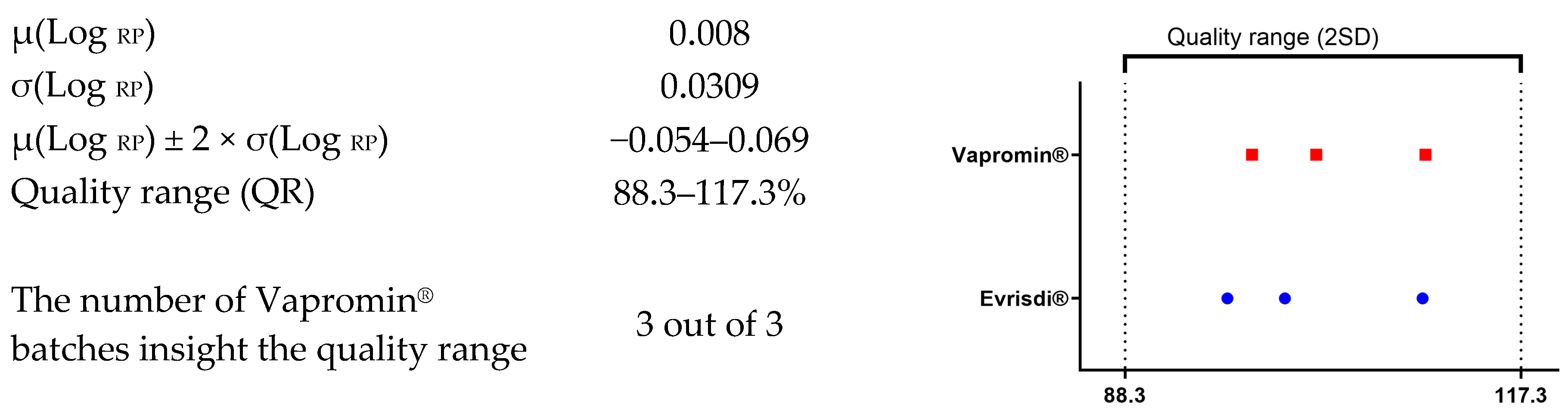
The QR limits were defined based on the variation observed in the reference product, calculated as the mean ± 2 standard deviations (SDs) according to the formula:
where:
µ(Log RP) ± 2 × σ(Log RP),
µ(Log RP)—arithmetic mean of the log-transformed values of % RSA obtained from the Evrysdi (Roche, Basel, Switzerland) LP® batches;
σ(Log RP)—standard deviation of the log-transformed values obtained from the Evrysdi (Roche, Basel, Switzerland) LP® batches.
To assess the comparability of the drugs in terms of specific activity, the quality range (QR) approach was used, as described in the FDA guidance “Development of Therapeutic Protein Biosimilars: Comparative Analytical Assessment and Other Quality-Related Considerations” (see [18,19] for details). Comparability was considered confirmed if the %RSA values obtained for individual batches of Vapromin® fell within the QR established for Evrysdi® (Roche, Basel, Switzerland). The QR was determined as follows: the %RSA values for individual batches of Evrysdi® (Roche, Basel, Switzerland) were log-transformed; the arithmetic mean (M) and standard deviation (SD) were calculated; and the M ± 2SD limits were back-transformed to the original %RSA scale.
SMN protein level versus drug log-concentration curves were fitted using a linear trend. The extra sum-of-squares F-test was used to assess the significance of the trend (i.e., whether the slope differed from zero) and to compare trend parameters between the drugs.
We were unable to identify a suitable function that accurately fitted the relationship between mRNA transcription levels and either drug concentration or log-concentration. Therefore, the correlation was assessed using Spearman’s r. The area under the transcription level curves (AUC), as well as the standard error of the AUC (SE of AUC), was calculated using the trapezoidal rule and DeLong’s method, respectively, and then compared between the drugs using the Z-test.
An unpaired Student’s t-test was used to compare the population doubling times of dermal fibroblasts from presumably healthy donors and SMA probands.
All calculations were performed using GraphPad Prism 8.0 and Microsoft Excel. A significance level of 0.05 was used throughout the analysis.
3. Results
Fibroblasts are the most relevant primary cell model for SMA studies, aside from neuronal cells, and are widely used in pathophysiological investigations. This study shows using fibroblasts from 3 SMA probands and healthy donors.
Dermal fibroblast cultures were established via enzymatic dissociation of skin biopsy samples. The cells adhered tightly to the plates and formed growth foci after 3–4 days of maintenance. Adherent cells acquired an elongated shape, with flattened cytoplasm. The doubling time of skin fibroblast cultures obtained from healthy donors did not differ significantly from that of SMA probands (p value = 0.5638). However, cells from SMA patients exhibited slower growth over several passages compared to cells from healthy donors. SMN2 expression in SMA proband skin fibroblasts was significantly lower than in healthy donors (p value 0.0015).
Three batches of Vapromin® and three batches of the original drug Evrysdi® (Roche, Basel, Switzerland) were analyzed in order to compare their biological properties.
3.1. Relative Transcription Level of SMN2 FL and SMN2 Δ7 in SMA Donors’ Fibroblasts
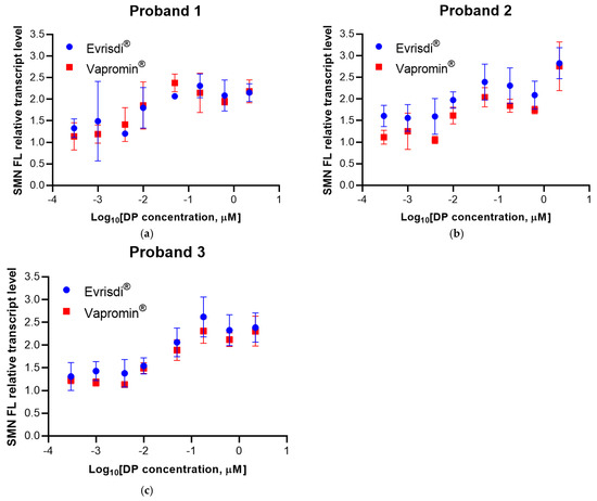
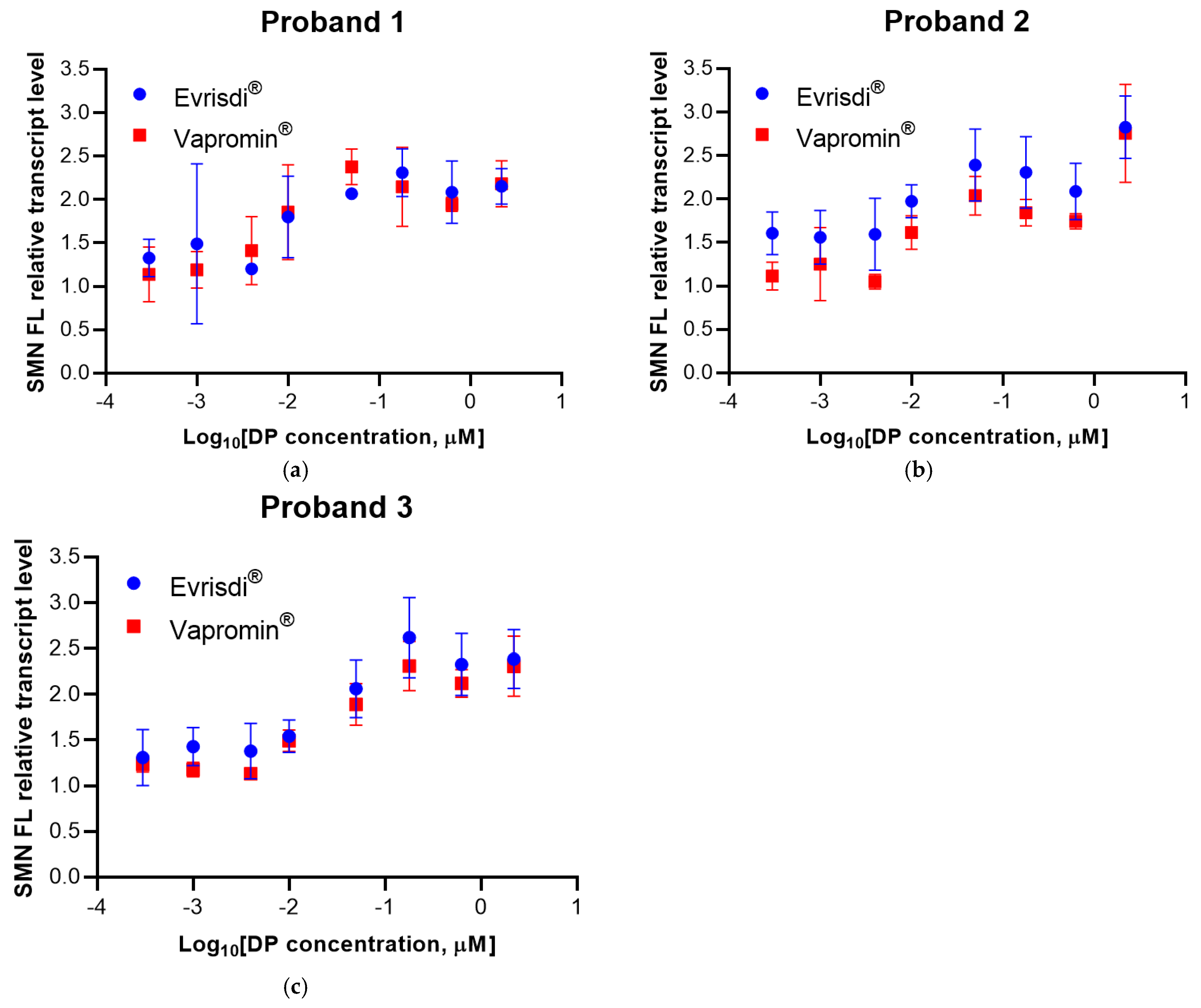
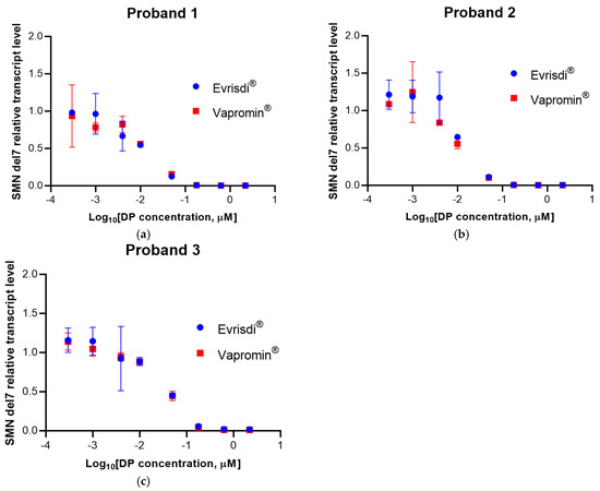
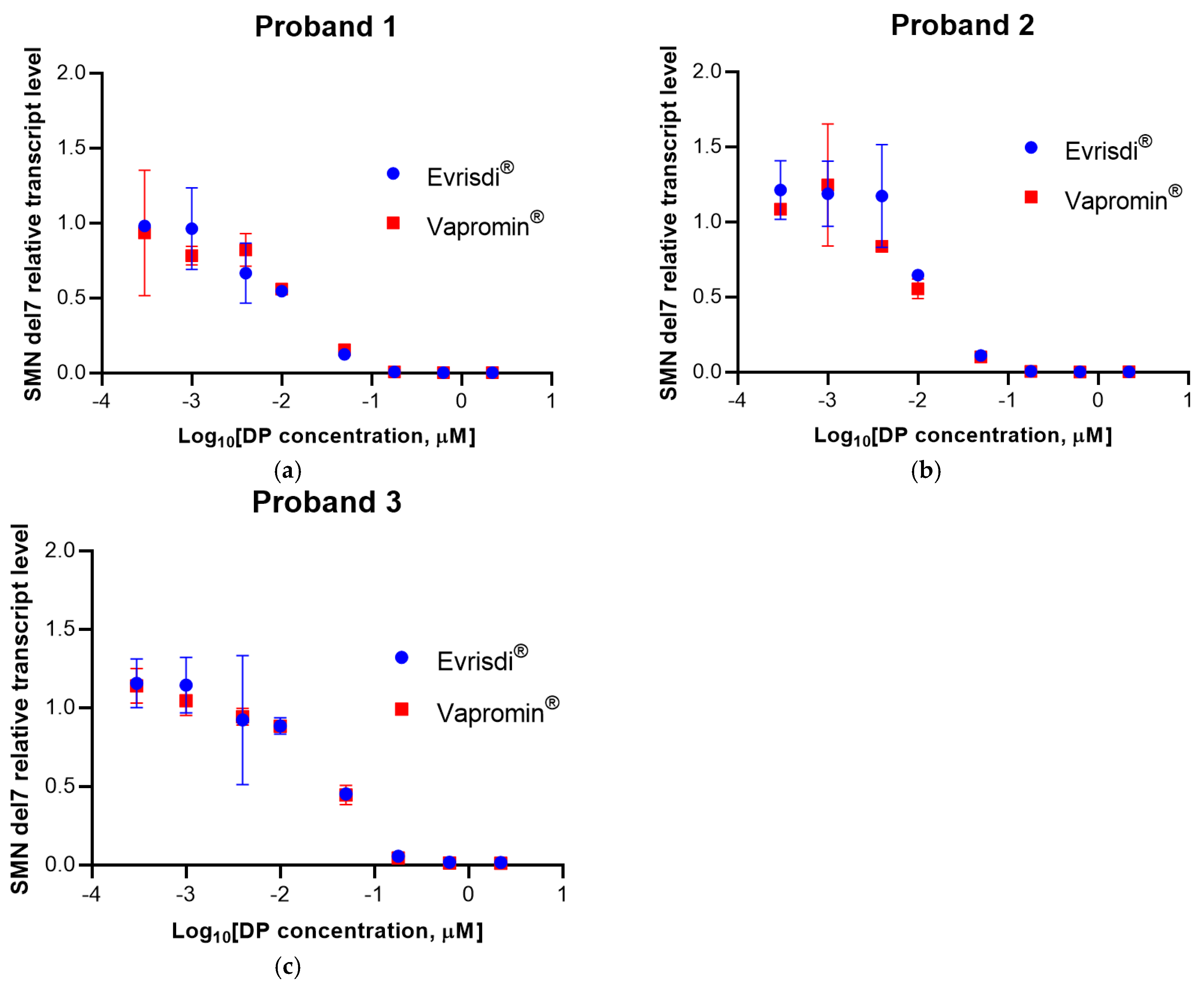
Fibroblasts from three SMA probands were exposed to increasing concentrations (0.0003 to 2.2 µM) of risdiplam preparations (either Evrysdi® (Roche, Basel, Switzerland) or Vapromin®), followed by measurement of the relative transcription levels of SMN2 FL and SMN2 Δ7, expressed as fold changes relative to the baseline level in fibroblasts from SMA probands without drugs (Figure 1 and Figure 2).
Figure 1.
Plots of SMN2 FL mRNA transcription levels versus each drug log-concentration in fibroblasts from three SMA probands (a–c). Transcription levels are expressed as fold changes relative to the baseline level in fibroblasts from SMA probands without drug. Mean transcription levels and standard deviations (based on triplicates) are shown for each drug concentration.
Figure 2.
Plots of SMN2 Δ7 mRNA transcription levels versus each drug log-concentration in fibroblasts from three SMA probands (a–c). Transcription levels are expressed as fold changes relative to the baseline level in fibroblasts from SMA probands without drug. Mean transcription levels and standard deviations (based on triplicates) are shown for each drug concentration.
Spearman’s r showed a significant correlation (positive for SMN2 FL; negative for SMN2 Δ7) between the transcription level and each drug log-concentration. Area under each curve (AUC) values were calculated, and the Z-test showed no significant differences in AUC between the drugs for each proband (Table 1 and Table 2).
Table 1.
Comparison of SMN2 FL mRNA transcription level curves in fibroblasts from three SMA probands.
Table 2.
Comparison of SMN2 Δ7 mRNA transcription level curves in fibroblasts from three SMA probands.
A relationship between risdiplam concentration and the transcription levels of SMN2 FL and SMN2 Δ7 was established. Addition of 0.18 μM risdiplam increased the relative transcription level of SMN2 FL by 2.5 times on average for both Evrysdi® (Roche, Basel, Switzerland) and Vapromin®. Simultaneously, the relative transcription level of SMN2 Δ7 decreased to 0 for all samples.
These data allow us to conclude that Evrysdi® (Roche, Basel, Switzerland) and Vapromin® have similar effects on the relative transcription levels of SMN2 FL and SMN2 Δ7 in the fibroblasts of SMA donors.
3.2. Evaluation of SMN Protein Production
SMN protein is considered to be unstable to repeated freeze–thaw cycles [20]. To avoid the influence of freeze–thawing, cell lysates were analyzed after a single cycle.
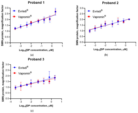
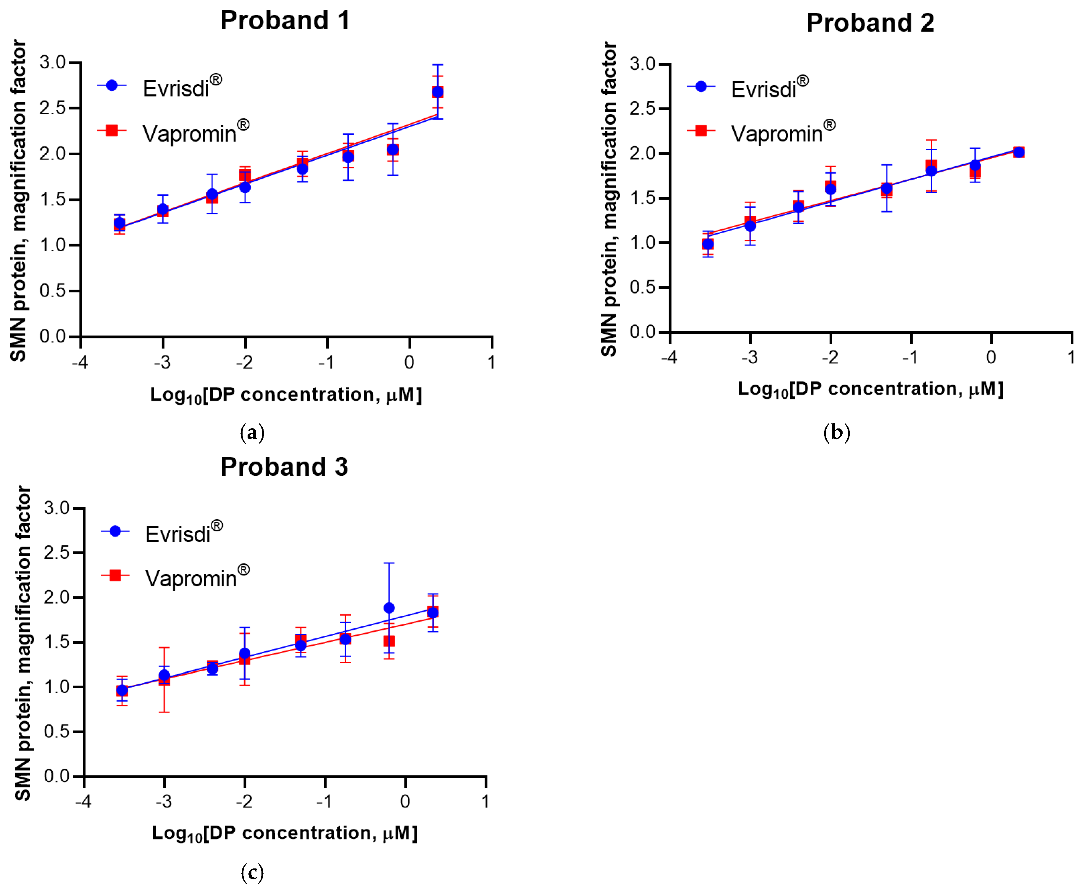
Fibroblasts from three SMA probands were exposed to increasing concentrations (0.0003 to 2.2 µM) of risdiplam preparations (either Evrysdi® (Roche, Basel, Switzerland) or Vapromin®), followed by relative SMA protein level measurement, expressed as fold changes relative to the baseline level in fibroblasts from SMA probands without drugs (Figure 3). Linear positive trends were established versus each drug log-concentration.
Figure 3.
Plots of SMN protein levels versus each drug log-concentration in fibroblasts from three SMA probands (a–c). SMN protein levels are expressed as fold changes relative to the baseline level in fibroblasts from SMA probands without drug. Mean levels and standard deviations (based on triplicates) are shown for each drug concentration.
Using the F-test for extra sum-of-squares, a significant difference of slopes from zero and the absence of significant differences in parameters between drugs for each proband were shown (Table 3).
Table 3.
Comparison of linear trends for SMN protein level in fibroblasts from three SMA probands.
The maximum increase in SMN protein levels was observed after treatment with 2.2 µM of the drug. The SMN protein level increased from 2 to 2.6 times (average value for three batches) compared to untreated fibroblasts.
These findings support the conclusion that Evrysdi® (Roche, Basel, Switzerland) and Vapromin® have similar effects on SMN protein production in fibroblasts from SMA donors.
3.3. Immunoblotting
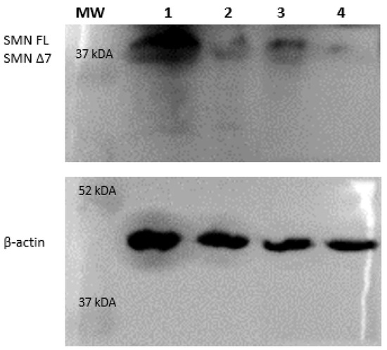
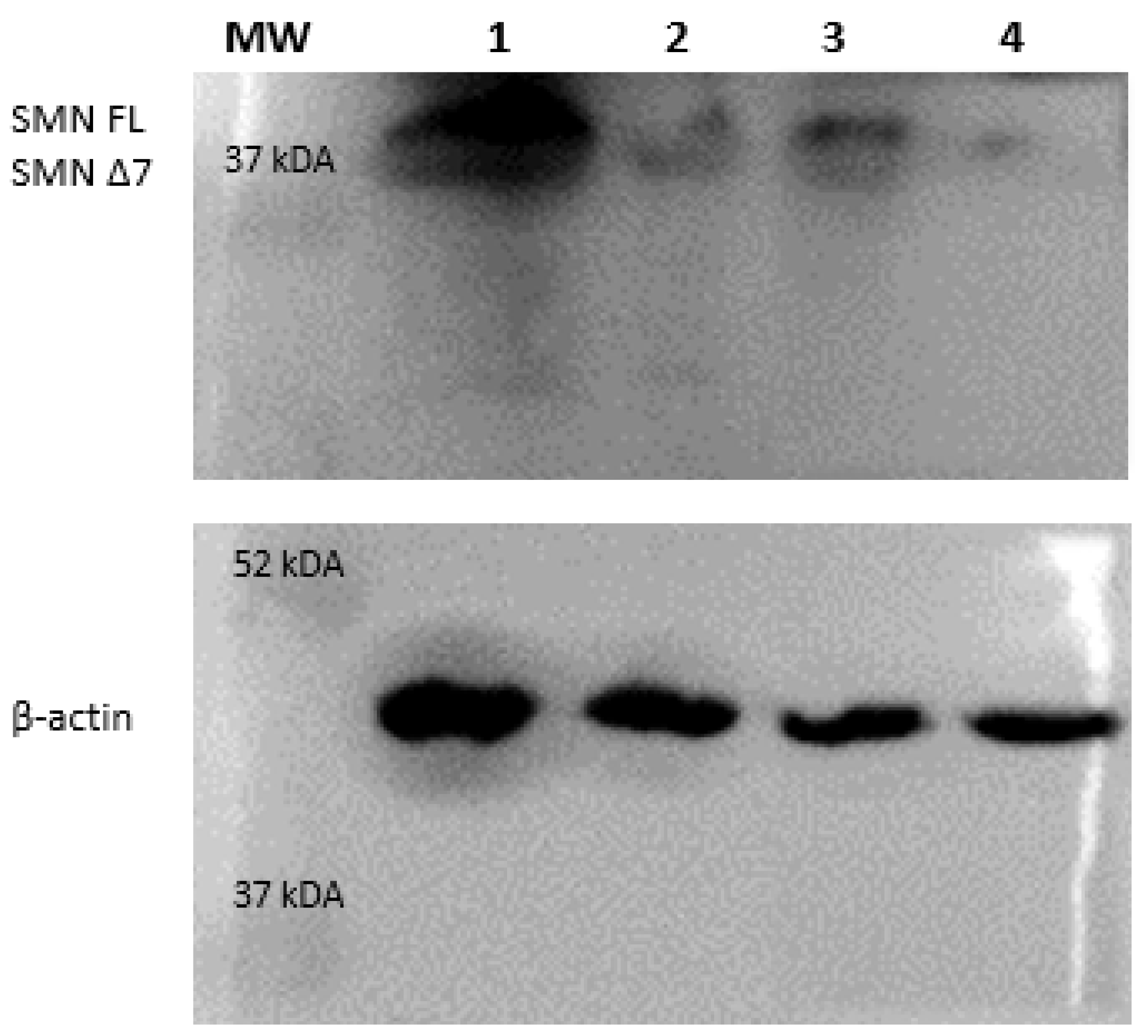
Western blot analysis confirmed the presence of full-length SMN2 protein in fibroblasts derived from patients with SMA following treatment with 2.2 µM risdiplam. Immunoblotting with anti-SMN monoclonal antibodies revealed two distinct bands, likely corresponding to the two major isoforms of SMN protein. These findings are consistent with previously published reports [21,22] (Figure 4).
Figure 4.
Western blot analysis of SMN protein expression in fibroblasts. The membrane was cut into two sections and probed with a monoclonal anti-SMN antibody and a monoclonal anti-β-actin antibody as a loading control. Lanes: 1, lysate from healthy donor fibroblasts; 2, lysate from SMA patient fibroblasts; 3, SMA patient fibroblasts treated with 2.2 µM Vapromin®; 4, SMA patient fibroblasts treated with 0.02 µM Vapromin®.
3.4. Cell Viability
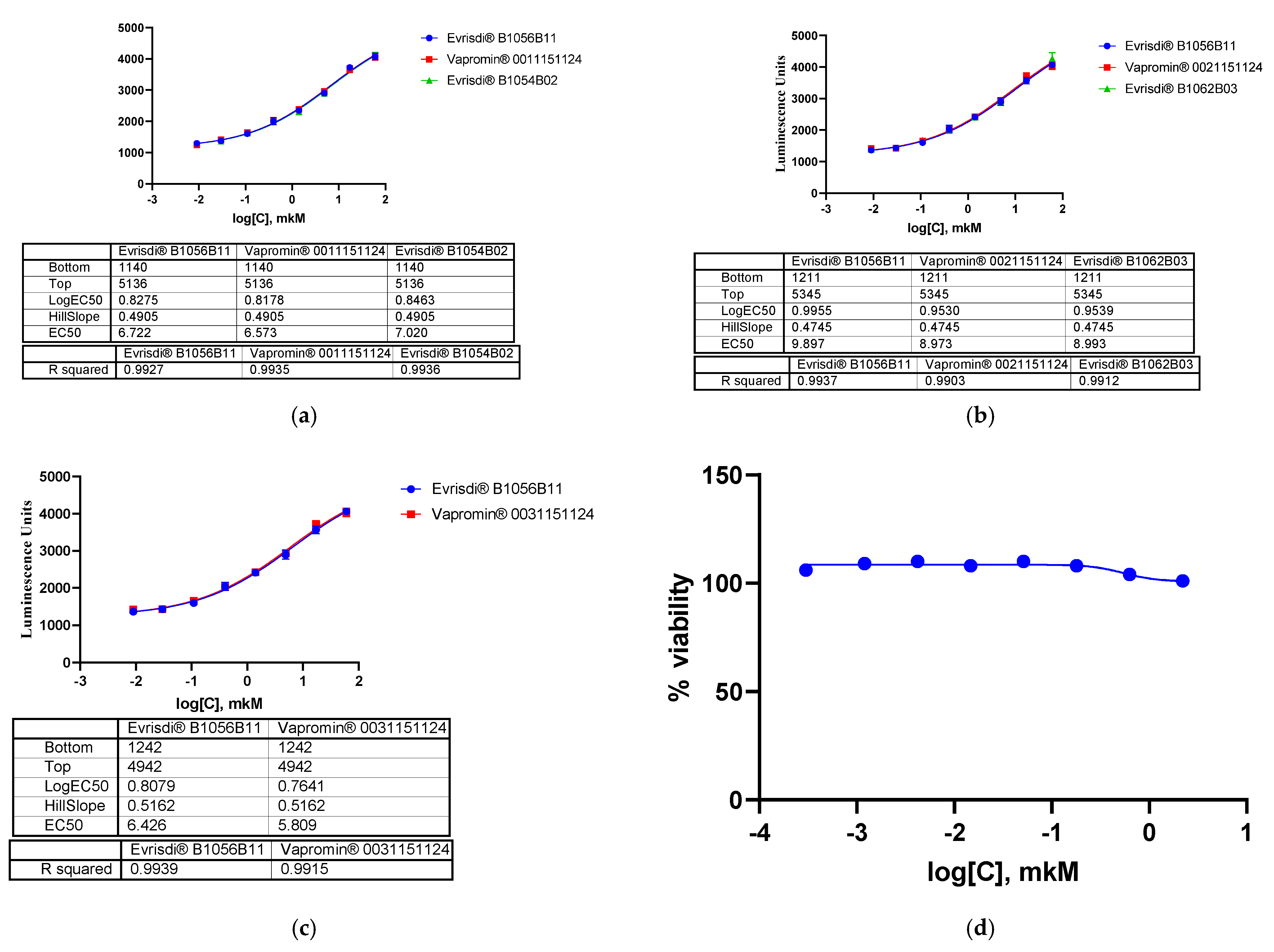
Cell viability was routinely assessed via visual inspection using light microscopy. Cell metabolic activity was assessed to determine whether the tested concentrations of risdiplam affected cell viability. The XTT assay demonstrated that treatment with risdiplam at concentrations ranging from 0.3 nM to 2.2 μM had no significant effect on the viability of fibroblast cells, indicating the absence of cytotoxicity within the tested concentration range (Figure 5d).
Figure 5.
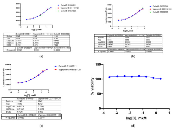
Plots showing the activity of different batches of risdiplam on the reporter cell line and cytotoxicity. (a–c): Each dot represents different drug log-concentrations versus responses of the reporter cell line in luminescence units. (d): Results of the XTT cytotoxicity assay following the treatment of fibroblasts with Vapromin® at concentrations ranging from 0.3 nM to 2.2 μM.
3.5. Results of Determination of Exon 7 Inclusion Efficiency Using the CHO SMN2-LUC MP 4G6 Reporter Cell Line
Addition of risdiplam to the reporter cell line CHO SMN2-LUC MP 4G6 led to a dose-dependent response (Figure 5).
The activity of each sample was calculated compared to the RS. The results are presented in Table 4.
Table 4.
Relative specific activity on the reporter cell culture.
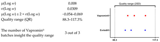
The individual values of relative specific activity for Vapromin® were compared with the QR (2SD) of Evrysdi® (Roche, Basel, Switzerland, Figure 6).
Figure 6.
Comparison of individual values obtained for Vapromin® with the quality range (2SD) of Evrysdi® (Roche, Basel, Switzerland).
The statistical analysis of exon 7 inclusion during mRNA splicing of the SMN2 gene on a reporter culture showed that the values of specific activity obtained for all batches of Vapromin® were within the QR of Evrysdi® (Roche, Basel, Switzerland).
4. Discussion
Despite the extensive clinical and preclinical use of DMTs, there is currently no standardized or broadly accepted in vitro framework for evaluating the efficacy of low-molecular-weight or antisense oligonucleotides targeting SMN2 splicing. Published studies frequently employ different cellular models, cell-seeding densities, and transfection strategies, often selected empirically and without systematic comparison [23].
In the present study, we compared the biological activity of the generic drug Vapromin® and its reference drug Evrysdi® (Roche, Basel, Switzerland)—the first approved orally administered low-molecular-weight drug for the therapy of SMA. Risdiplam distributes throughout both the central and peripheral nervous system and modulates splicing of SMN2 pre-mRNA toward the production of full-length mRNA to increase the levels of functional SMN protein [24]. Although it is generally not necessary to compare the in vitro biological activity of generic drugs, the clinical significance of risdiplam makes such comparison favorable.
Several assays are available to measure the biological function of SMN protein [25,26,27]. Primary fibroblasts are particularly suitable for this purpose, since they retain the genetic and epigenetic characteristics of the donor while being homogeneous and reproducible [28,29].
To assess the effectiveness of SMN2 pre-mRNA splicing and the amount of SMN protein produced, we used fibroblasts isolated from SMA probands with a homozygous deletion of exons 7–8 of the SMN1 gene and different numbers of the SMN2 gene copies.
Analyzing the obtained results for the relative transcription levels of SMN2 FL and SMN2 Δ7, we noted that, regardless of the drug (Evrysdi® (Roche, Basel, Switzerland) or Vapromin®), the level of SMN2 FL transcripts in fibroblasts of each proband changed in a concentration-dependent manner.
Signoria et al. studied the influence of the SMN2 copy number, age, and gender of the donor on the relative level of SMN2 FL transcription when treating patient fibroblasts with risdiplam and the antisense oligonucleotide nusinersen. The most considerable, however, the most variable change in the relative level of transcription was observed in patients with three copies of the SMN2 gene [29]. This may explain the results obtained in our study, since we used fibroblasts from patients with both three (probands 1 and 2) and four copies of the SMN gene (proband 3).
We also found that, at the maximum risdiplam concentration of 2.2 μM, the level of SMN2 FL transcripts was the same as the level of SMN FL in fibroblasts treated with 0.18 μM risdiplam. A similar effect was observed by other researchers when studying a saturating dose of antisense oligonucleotides [30]. The authors suggest that high concentrations of ASO targeting Intronic Splicing Silencer N1 promote the activation of the cryptic splicing site in intron 6 of the SMN2 gene.
Risdiplam enters cells primarily by passive diffusion due to its high lipophilicity, which allows it to cross cell membranes throughout the body, including the blood–brain barrier. Transcriptional changes should be confirmed by protein-level investigations, since changes in SMN expression levels may not be accompanied by effective changes in stable protein production, as the dynamics and half-lives of mRNA and proteins may not be comparable [3]. Using primary SMN donors’ fibroblasts, we characterized the in vitro response to SMN2 splicing modification risdiplam and demonstrated that Vapromin® caused a dose-dependent increase in SMN protein levels similar to Evrysdi (Roche, Basel, Switzerland).
Changes in SMN protein production in fibroblasts were also established, which is consistent with literature data. SMN protein levels have been studied in many cell types [27]. Notably, significant differences in SMN protein levels were found in platelets, erythrocytes, and PBMCs, highlighting the importance of investigating tissue-specific SMN expression [20,31]. It is considered that SMN protein levels in fibroblasts from healthy donors are several times higher than in SMA probands. Moreover, SMN protein production levels increase by 50% or more upon treatment of fibroblasts with mRNA splicing-correcting drugs [25,31]. Using imaging flow cytometry, it was shown that fibroblasts from SMA patients expressed SMN protein at 20–30% of normal levels [32]. A correlation was also established between SMN protein levels and the SMN2 copy number in fibroblasts, as well as with clinical characteristics, indicating that fibroblasts isolated from the skin of SMA carriers may be a reliable cell type for SMN biomarker studies. The lack of correlation between SMN protein levels in PBMCs and fibroblasts likely indicates differences between these tissues [27].
The CHO SMN2-LUC MP 4G6 reporter cell line was constructed to assess exon 7 inclusion during SMN2 mRNA splicing. It triggers the relevant signaling pathway in response to specific activation and allows visualization of the drug’s mechanism of action in vitro. This cell line could be used in a robust, sensitive, and reproducible in vitro assay for evaluating the efficacy of splicing modifiers targeting SMN2 splicing.
The cell line was used to determine the specific activity of each batch. The “quality range” method (formulas are given above) was also used for statistical analysis. The individual values obtained for the Vapromin® batches fell within the QR established for Evrysdi® (Roche, Basel, Switzerland).
5. Conclusions
Therefore, these data experimentally confirm that the in vitro biological activity of the generic drug Vapromin® is comparable to that of Evrysdi®. However, further investigation should be performed on a large number of batches to show equivalence testing (TOST).
Author Contributions
Conceptualization, O.S. and A.P.; methodology, O.S., R.A. and A.P.; validation, O.S., E.B., R.A., Y.G., A.G., I.K., N.K. and Y.B.; formal analysis, O.S., A.P. and A.K.; investigation, O.S., E.B., R.A., Y.G., A.G., I.K. and Y.B.; resources, A.P. and I.L.; data curation, O.S. and A.P.; writing—original draft preparation, O.S., A.K., A.P. and R.A.; writing—review and editing, O.S., A.P., A.K., Y.G., N.K. and R.K., visualization, O.S., A.P., A.G. and A.K.; supervision, A.P.; project administration, A.P., I.L. and R.S.; funding acquisition R.S. and R.K. All authors have read and agreed to the published version of the manuscript.
Funding
This research received no external funding.
Institutional Review Board Statement
The study was approved by the local independent ethical committee of JSC Generium (study protocol GNR-115 at 8 April 2024).
Informed Consent Statement
All donors gave written voluntary consent for sample collection and inclusion of analysis results in this study, including authorized cell storage and authorized future genetic analyses.
Data Availability Statement
The original contributions presented in this study are included in the article. Further inquiries can be directed to the corresponding author.
Conflicts of Interest
All authors are employees of JSC “Generium”. For the present study, there are no other relationships or activities that could be perceived to influence the submitted work. The authors alone are responsible for the content of this text.
Abbreviations
The following abbreviations are used in this manuscript:
| AUC | Area under curve |
| CHO | Chinese Hamster Ovary |
| DP | Drug product |
| DMTs | Disease-modifying therapies |
| EMA | European Medicines Agency |
| ECL | Enhanced chemiluminescence |
| FL | Full length |
| FDA | Food and Drug Administration |
| HRP | Horseradish peroxidase |
| JSC | Joint-Stock Company |
| mRNA | Messenger ribonucleic acid |
| MLPA | Multiplex ligation-dependent probe amplification |
| RS | Reference standard |
| PBS | Phosphate-buffered saline |
| PBST | Phosphate-buffered saline containing tween 20 |
| PD | Pharmacodynamics |
| PK | Pharmacokinetics |
| PVDF | Polyvinylidene fluoride |
| QR | Quality range |
| RSA | Relative specific activity |
| SD | Standard Deviation |
| SMA | Spinal muscular atrophy |
| SMN | Survival motor neuron |
| TBST | Tris-buffered saline containing Tween 20 |
| TOST | Two One-Sided Tests |
References
- Belančić, A.; Eustaquio, P.C.; Gkrinia, E.M.M.; Rački, V.; Pilipović, K.; Vitezić, D. Transforming Spinal Muscular Atrophy: From Pivotal Trials to Real-World Evidence and Future Therapeutic Frontiers in Types 1 and 2. Biomedicines 2025, 13, 1939. [Google Scholar] [CrossRef] [Scilit] [PubMed]
- Jeong, Y.D.; Son, Y.; Jeon, S.; Lee, K.; Kim, H.J.; Hwang, Y.; López-Gil, J.F.; Rahmati, M.; Woo, H.G.; Kang, J.; et al. Global Prevalence of Spinal Muscular Atrophy in 204 Countries and Territories, 1960-2024: A Systematic Review and Bayesian Modeling Study. Pediatr. Neurol. 2026, 180, 32–44. [Google Scholar] [CrossRef] [Scilit] [PubMed]
- Cherry, J.J.; Kobayashi, D.T.; Lynes, M.M.; Naryshkin, N.N.; Tiziano, F.D.; Zaworski, P.G.; Rubin, L.L.; Jarecki, J. Assays for the Identification and Prioritization of Drug Candidates for Spinal Muscular Atrophy. ASSAY Drug Dev. Technol. 2014, 12, 315–341. [Google Scholar] [CrossRef] [Scilit] [PubMed]
- Naryshkin, N.A.; Weetall, M.; Dakka, A.; Narasimhan, J.; Zhao, X.; Feng, Z.; Ling, K.K.Y.; Karp, G.M.; Qi, H.; Woll, M.G.; et al. SMN2 Splicing Modifiers Improve Motor Function and Longevity in Mice with Spinal Muscular Atrophy. Science 2014, 345, 688–693. [Google Scholar] [CrossRef] [Scilit] [PubMed]
- Butchbach, M.E.R. Copy Number Variations in the Survival Motor Neuron Genes: Implications for Spinal Muscular Atrophy and Other Neurodegenerative Diseases. Front. Mol. Biosci. 2016, 3, 7. [Google Scholar] [CrossRef] [Scilit] [PubMed]
- Corti, S.; Nizzardo, M.; Nardini, M.; Donadoni, C.; Salani, S.; Ronchi, D.; Simone, C.; Falcone, M.; Papadimitriou, D.; Locatelli, F.; et al. Embryonic Stem Cell-Derived Neural Stem Cells Improve Spinal Muscular Atrophy Phenotype in Mice. Brain 2010, 133, 465–481. [Google Scholar] [CrossRef] [Scilit] [PubMed]
- Dominguez, E.; Marais, T.; Chatauret, N.; Benkhelifa-Ziyyat, S.; Duque, S.; Ravassard, P.; Carcenac, R.; Astord, S.; De Moura, A.P.; Voit, T.; et al. Intravenous scAAV9 Delivery of a Codon-Optimized SMN1 Sequence Rescues SMA Mice. Hum. Mol. Genet. 2011, 20, 681–693. [Google Scholar] [CrossRef] [Scilit] [PubMed]
- Kernochan, L.E.; Russo, M.L.; Woodling, N.S.; Huynh, T.N.; Avila, A.M.; Fischbeck, K.H.; Sumner, C.J. The Role of Histone Acetylation in SMN Gene Expression. Hum. Mol. Genet. 2005, 14, 1171–1182. [Google Scholar] [CrossRef] [Scilit] [PubMed]
- Hauke, J.; Riessland, M.; Lunke, S.; Eyüpoglu, I.Y.; Blümcke, I.; El-Osta, A.; Wirth, B.; Hahnen, E. Survival Motor Neuron Gene 2 Silencing by DNA Methylation Correlates with Spinal Muscular Atrophy Disease Severity and Can Be Bypassed by Histone Deacetylase Inhibition. Hum. Mol. Genet. 2009, 18, 304–317. [Google Scholar] [CrossRef] [Scilit] [PubMed]
- Skordis, L.A.; Dunckley, M.G.; Yue, B.; Eperon, I.C.; Muntoni, F. Bifunctional Antisense Oligonucleotides Provide a Trans-Acting Splicing Enhancer That Stimulates SMN2 Gene Expression in Patient Fibroblasts. Proc. Natl. Acad. Sci. USA 2003, 100, 4114–4119. [Google Scholar] [CrossRef] [Scilit] [PubMed]
- Hua, Y.; Sahashi, K.; Hung, G.; Rigo, F.; Passini, M.A.; Bennett, C.F.; Krainer, A.R. Antisense Correction of SMN2 Splicing in the CNS Rescues Necrosis in a Type III SMA Mouse Model. Genes Dev. 2010, 24, 1634–1644. [Google Scholar] [CrossRef] [Scilit] [PubMed]
- Hua, Y.; Vickers, T.A.; Okunola, H.L.; Bennett, C.F.; Krainer, A.R. Antisense Masking of an hnRNP A1/A2 Intronic Splicing Silencer Corrects SMN2 Splicing in Transgenic Mice. Am. J. Hum. Genet. 2008, 82, 834–848. [Google Scholar] [CrossRef] [Scilit] [PubMed]
- Belančić, A.; Strbad, T.; Kučan Štiglić, M.; Vitezić, D. Switching from Nusinersen to Risdiplam: A Croatian Real-World Experience on Effectiveness and Safety. JPM 2024, 14, 244. [Google Scholar] [CrossRef] [Scilit] [PubMed]
- Alfonso-Cristancho, R.; Andia, T.; Barbosa, T.; Watanabe, J.H. Definition and Classification of Generic Drugs Across the World. Appl. Health Econ. Health Policy 2015, 13, 5–11. [Google Scholar] [CrossRef] [Scilit] [PubMed]
- Kobayashi, D.T.; Olson, R.J.; Sly, L.; Swanson, C.J.; Chung, B.; Naryshkin, N.; Narasimhan, J.; Bhattacharyya, A.; Mullenix, M.; Chen, K.S. Utility of Survival Motor Neuron ELISA for Spinal Muscular Atrophy Clinical and Preclinical Analyses. PLoS ONE 2011, 6, e24269. [Google Scholar] [CrossRef] [Scilit] [PubMed]
- Kobayashi, D.T.; Decker, D.; Zaworski, P.; Klott, K.; McGonigal, J.; Ghazal, N.; Sly, L.; Chung, B.; Vanderlugt, J.; Chen, K.S. Evaluation of Peripheral Blood Mononuclear Cell Processing and Analysis for Survival Motor Neuron Protein. PLoS ONE 2012, 7, e50763. [Google Scholar] [CrossRef] [Scilit] [PubMed][Green Version]
- Sivaramakrishnan, M.; McCarthy, K.D.; Campagne, S.; Huber, S.; Meier, S.; Augustin, A.; Heckel, T.; Meistermann, H.; Hug, M.N.; Birrer, P.; et al. Binding to SMN2 Pre-mRNA-Protein Complex Elicits Specificity for Small Molecule Splicing Modifiers. Nat. Commun. 2017, 8, 1476. [Google Scholar] [CrossRef] [Scilit] [PubMed]
- Tsong, Y.; Dong, X.; Shen, M. Development of Statistical Methods for Analytical Similarity Assessment. J. Biopharm. Stat. 2017, 27, 197–205. [Google Scholar] [CrossRef] [Scilit] [PubMed]
- Chow, S.-C. Challenging Issues in Assessing Analytical Similarity in Biosimilar Studies. Biosimilars 2015, 5, 33–39. [Google Scholar] [CrossRef] [Scilit]
- Zaworski, P.; Von Herrmann, K.M.; Taylor, S.; Sunshine, S.S.; McCarthy, K.; Risher, N.; Newcomb, T.; Weetall, M.; Prior, T.W.; Swoboda, K.J.; et al. SMN Protein Can Be Reliably Measured in Whole Blood with an Electrochemiluminescence (ECL) Immunoassay: Implications for Clinical Trials. PLoS ONE 2016, 11, e0150640. [Google Scholar] [CrossRef] [Scilit] [PubMed]
- Mattis, V.B.; Rai, R.; Wang, J.; Chang, C.-W.T.; Coady, T.; Lorson, C.L. Novel Aminoglycosides Increase SMN Levels in Spinal Muscular Atrophy Fibroblasts. Hum. Genet. 2006, 120, 589–601. [Google Scholar] [CrossRef] [Scilit] [PubMed]
- Le, T.T.; Pham, L.T.; Butchbach, M.E.R.; Zhang, H.L.; Monani, U.R.; Coovert, D.D.; Gavrilina, T.O.; Xing, L.; Bassell, G.J.; Burghes, A.H.M. SMNΔ7, the Major Product of the Centromeric Survival Motor Neuron (SMN2) Gene, Extends Survival in Mice with Spinal Muscular Atrophy and Associates with Full-Length SMN. Hum. Mol. Genet. 2005, 14, 845–857. [Google Scholar] [CrossRef] [Scilit] [PubMed]
- Tisnikar, M.S.; Zottel, A.; Kristan, K.; Lanišnik Rižner, T. Optimizing In Vitro Efficacy Assessment of the Antisense Oligonucleotide Nusinersen in Human Cellular Models. Pharmaceutics 2026, 18, 652. [Google Scholar] [CrossRef] [Scilit] [PubMed]
- Ratni, H.; Scalco, R.S.; Stephan, A.H. Risdiplam, the First Approved Small Molecule Splicing Modifier Drug as a Blueprint for Future Transformative Medicines. ACS Med. Chem. Lett. 2021, 12, 874–877. [Google Scholar] [CrossRef] [Scilit] [PubMed]
- Thi Man, N.; Humphrey, E.; Lam, L.T.; Fuller, H.R.; Lynch, T.A.; Sewry, C.A.; Goodwin, P.R.; MacKenzie, A.E.; Morris, G.E. A Two-Site ELISA Can Quantify Upregulation of SMN Protein by Drugs for Spinal Muscular Atrophy. Neurology 2008, 71, 1757–1763. [Google Scholar] [CrossRef] [Scilit] [PubMed]
- Piepers, S.; Cobben, J.-M.; Sodaar, P.; Jansen, M.D.; Wadman, R.I.; Meester-Delver, A.; Poll-The, B.T.; Lemmink, H.H.; Wokke, J.H.J.; Van Der Pol, W.-L.; et al. Quantification of SMN Protein in Leucocytes from Spinal Muscular Atrophy Patients: Effects of Treatment with Valproic Acid. J. Neurol. Neurosurg. Psychiatry 2011, 82, 850–852. [Google Scholar] [CrossRef] [Scilit] [PubMed][Green Version]
- Wadman, R.I.; Stam, M.; Jansen, M.D.; Van Der Weegen, Y.; Wijngaarde, C.A.; Harschnitz, O.; Sodaar, P.; Braun, K.P.J.; Dooijes, D.; Lemmink, H.H.; et al. A Comparative Study of SMN Protein and mRNA in Blood and Fibroblasts in Patients with Spinal Muscular Atrophy and Healthy Controls. PLoS ONE 2016, 11, e0167087. [Google Scholar] [CrossRef] [Scilit] [PubMed]
- Sturm, G.; Cardenas, A.; Bind, M.-A.; Horvath, S.; Wang, S.; Wang, Y.; Hägg, S.; Hirano, M.; Picard, M. Human Aging DNA Methylation Signatures Are Conserved but Accelerated in Cultured Fibroblasts. Epigenetics 2019, 14, 961–976. [Google Scholar] [CrossRef] [Scilit] [PubMed]
- Signoria, I.; Zwartkruis, M.M.; Geerlofs, L.; Perenthaler, E.; Faller, K.M.E.; James, R.; McHale-Owen, H.; Green, J.W.; Kortooms, J.; Snellen, S.H.; et al. Patient-Specific Responses to SMN2 Splice-Modifying Treatments in Spinal Muscular Atrophy Fibroblasts. Mol. Ther. Methods Clin. Dev. 2024, 32, 101379. [Google Scholar] [CrossRef] [Scilit] [PubMed]
- Wijaya, Y.O.S.; Niba, E.T.E.; Nishio, H.; Okamoto, K.; Awano, H.; Saito, T.; Takeshima, Y.; Shinohara, M. High Concentration or Combined Treatment of Antisense Oligonucleotides for Spinal Muscular Atrophy Perturbed SMN2 Splicing in Patient Fibroblasts. Genes 2022, 13, 685. [Google Scholar] [CrossRef] [Scilit] [PubMed]
- Czech, C.; Tang, W.; Bugawan, T.; Mano, C.; Horn, C.; Iglesias, V.A.; Fröhner, S.; Zaworski, P.G.; Paushkin, S.; Chen, K.; et al. Biomarker for Spinal Muscular Atrophy: Expression of SMN in Peripheral Blood of SMA Patients and Healthy Controls. PLoS ONE 2015, 10, e0139950. [Google Scholar] [CrossRef] [Scilit] [PubMed]
- Arakawa, M.; Arakawa, R.; Tatsumi, S.; Aoki, R.; Saito, K.; Nomoto, A. A Novel Evaluation Method of Survival Motor Neuron Protein as a Biomarker of Spinal Muscular Atrophy by Imaging Flow Cytometry. Biochem. Biophys. Res. Commun. 2014, 453, 368–374. [Google Scholar] [CrossRef] [Scilit] [PubMed]

Disclaimer/Publisher’s Note: The statements, opinions and data contained in all publications are solely those of the individual author(s) and contributor(s) and not of MDPI and/or the editor(s). MDPI and/or the editor(s) disclaim responsibility for any injury to people or property resulting from any ideas, methods, instructions or products referred to in the content. |
© 2026 by the authors. Licensee MDPI, Basel, Switzerland. This article is an open access article distributed under the terms and conditions of the Creative Commons Attribution (CC BY) license.