


Abstract
Background: Trace elements function as essential micronutrients involved in oxidative balance, mitochondrial activity, and cardiovascular metabolism. Cigarette smoking represents a significant source of toxic metals and may disrupt systemic trace element homeostasis. Alterations in micronutrient and metal balance may contribute to oxidative stress, endothelial dysfunction, and myocardial remodeling, which are central mechanisms in the pathogenesis of heart failure with preserved ejection fraction (HFpEF). This study aimed to investigate whether smokers with HFpEF exhibit distinct hair trace element profiles compared with smokers without HFpEF. Methods: In this prospective pilot study, scalp hair samples were collected from adults undergoing clinical evaluation for suspected cardiovascular disease. Trace element concentrations were determined using inductively coupled plasma mass spectrometry (ICP-MS). Participants were first stratified according to smoking status and subsequently, within the smoker subgroup, according to HFpEF diagnosis based on the Heart Failure Association Pre-test assessment, Echocardiography and natriuretic peptide score (HFA-PEFF) algorithm. Differences in trace element concentrations were analyzed using appropriate statistical tests, with multiple-comparison correction using the Benjamini–Hochberg false discovery rate (FDR). Active smoking was defined as ≥10 cigarettes per day for at least 1 year, and cumulative exposure was quantified in pack-years. Results: Fifty-eight participants were included, including 27 active smokers. In unadjusted analyses, several trace elements differed between smokers with HFpEF and those without HFpEF, including vanadium, lithium, aluminum, and copper. However, after FDR correction, only copper remained significantly elevated in smokers with HFpEF (q = 0.004). Hair copper concentrations were markedly higher in the HFpEF group compared with smokers without HFpEF. These differences were observed alongside echocardiographic features consistent with diastolic dysfunction and structural cardiac remodeling. Conclusions: In this hypothesis-generating pilot study, smokers with HFpEF demonstrated elevated hair copper concentrations, suggesting disturbances in trace element and micronutrient homeostasis. Altered copper metabolism may reflect oxidative stress-related cardiometabolic remodeling associated with HFpEF. These findings raise the hypothesis that cardiometabolic phenotype, rather than smoking exposure alone, may modulate trace element homeostasis in HFpEF; however, causal relationships cannot be established.
1. Introduction
Cigarette smoking remains one of the major modifiable risk factors for cardiovascular disease. Because tobacco smoke contains many different metals and metalloids, it can promote oxidative stress and disturb normal endothelial function. As a result, it may change the distribution of metals in the body and negatively affect mitochondrial activity. Beyond atherosclerosis, chronic exposure to tobacco smoke promotes oxidative stress, systemic inflammation, endothelial dysfunction, and neurohormonal activation—mechanisms implicated in the pathogenesis of heart failure with preserved ejection fraction (HFpEF) [1,2]. Long-term exposure to active smoking or passive inhalation of these elements may disrupt the body’s metal balance, which can influence inflammatory signaling and impair mitochondrial function—both key features in the pathophysiology of HFpEF [3].
HFpEF is characterized by impaired diastolic function, increased myocardial stiffness, and structural remodeling, frequently accompanied by systemic inflammatory activation and microvascular dysfunction [4,5]. Comorbidities such as obesity, hypertension, and metabolic disturbances further contribute to disease progression.
Trace elements represent essential micronutrients involved in mitochondrial respiration, antioxidant defense, and cardiovascular metabolism. Nutritional status and environmental exposure jointly determine systemic trace element homeostasis. Disturbances in micronutrient balance—including copper, zinc, and selenium—have been associated with oxidative stress, endothelial dysfunction, and cardiometabolic disease. Understanding how micronutrient-related trace element dysregulation contributes to heart failure phenotypes may provide new insights into metabolic and inflammatory mechanisms of HFpEF.
Trace elements participate in redox homeostasis and mitochondrial function. Dysregulation of essential and nonessential metals may influence myocardial remodeling and diastolic dysfunction. For example, copper is required for normal antioxidant defense, but when its metabolism becomes dysregulated, it can contribute to redox imbalance and the development of myocardial fibrosis, potentially worsening diastolic function [6]. Experimental studies also indicate that trace-element exposure may act together with oxidative stress to enhance the long-term cardiovascular toxicity associated with smoking.
Hair analysis provides a noninvasive matrix that reflects longer-term systemic exposure [7,8]. We hypothesized that active smokers with HFpEF would demonstrate a distinct trace element profile compared with smokers without HFpEF.
To our knowledge, no previous studies have investigated hair trace element profiles in smokers with HFpEF, despite the potential interaction between environmental exposure, micronutrient imbalance, and cardiometabolic remodeling. The primary endpoint of our analysis was the difference in hair concentrations of selected trace elements between smokers with and without HFpEF.
However, the biological relevance of circulating and tissue trace element alterations in cardiovascular disease remains complex, and whether these changes represent causal mechanisms or epiphenomena of systemic inflammation is still debated.
2. Patients and Methods
This prospective pilot study was conducted in 2025 at a tertiary academic center. Patients were consecutively enrolled after referral from cardiology clinics for evaluation of cardiovascular symptoms, including exertional dyspnea, fatigue, and, in some cases, stable angina. A structured patient selection process was applied, and all included participants met predefined inclusion and exclusion criteria.
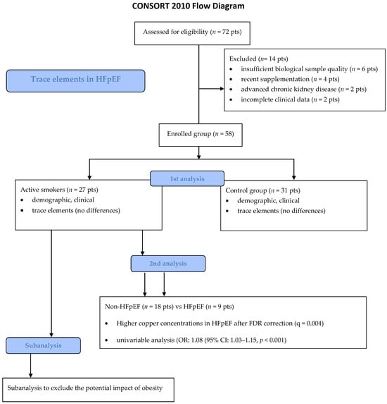
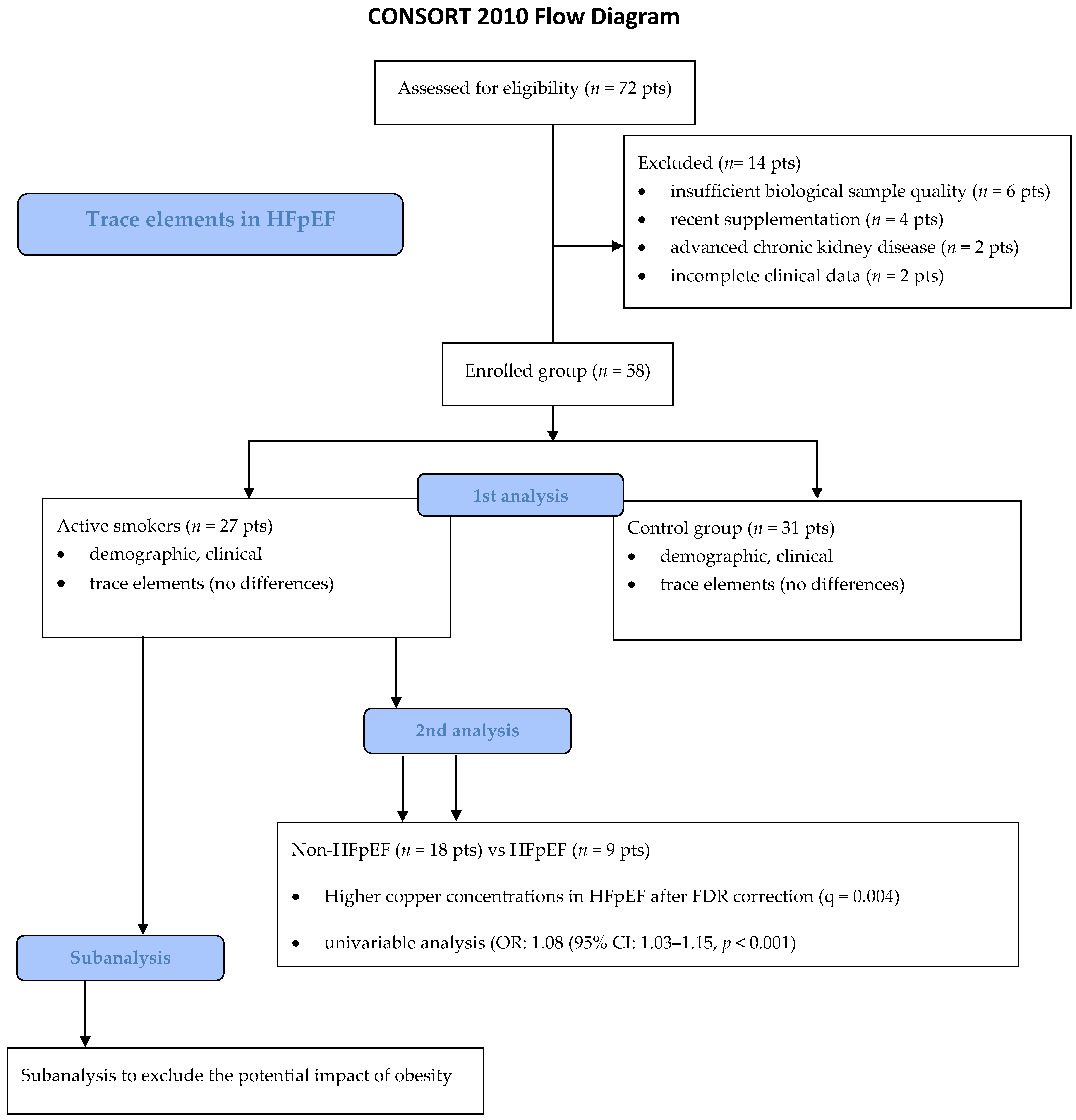
A total of 72 patients were initially screened for eligibility. Fourteen individuals were excluded due to predefined exclusion criteria (n = 6 for insufficient biological sample quality; n = 4 for recent supplementation; n = 2 for advanced chronic kidney disease, and n = 2 for incomplete clinical data). The final study population comprised 58 participants: 27 active smokers and 31 non-smokers. Among active smokers, 9 met criteria for HFpEF and 18 were classified as non-HFpEF, as presented in Figure 1.
Figure 1.
Flow chart.
Active smoking was defined as the daily consumption of at least 10 cigarettes for a minimum duration of one year. Smoking exposure was quantified in pack-years and included in subsequent analyses as a continuous variable.
2.1. Enrolment Criteria
2.1.1. Inclusion Criteria
Adults (≥18 years) electively admitted for evaluation of cardiovascular symptoms, including exertional dyspnea and fatigue, to internal medicine and hypertension clinics were screened.
2.1.2. Exclusion Criteria Included
- Previous cardiovascular interventions;
- Restrictive diet or food allergies;
- Advanced chronic kidney disease (stage 4–5);
- Active malignancy;
- Documented occupational or mental heavy metal exposure exceeding background levels;
- Any supplementation within last 12 months, including copper;
- Insufficient biological samples;
- Quality.
All patients underwent laboratory testing, transthoracic echocardiography, and—when clinically indicated—coronary angiography. Significant coronary artery disease was defined as ≥50% luminal stenosis on visual assessment.
2.2. HFpEF Diagnosis
Heart failure with preserved left ventricular ejection fraction (HFpEF) was diagnosed using the Heart Failure Association (HFA)-PEFF diagnostic algorithm [9] by two cardiologists independently, based on:
- Symptoms (exertional dyspnea, fatigue);
- Preserved left ventricular ejection fraction;
- Echocardiographic indices of diastolic dysfunction (E/A ratio, E/e′);
- Increased LAVI
- Reduced GLS;
- Elevated B-type natriuretic peptide (BNP);
- Body mass index (BMI) >30 kg/m2, when applicable.
2.3. Trace Element Examination
Scalp hair samples were collected according to a standardized protocol [10]. Trace elements examination: hair collection, preparation, and ICP-MS analysis. Hair samples (2–3 cm in length), untreated and free from dyes or chemical processing, were collected from the occipital region close to the scalp. The samples were cleaned by sequential washing in acetone, deionized water, 0.5% Triton X-100, and again deionized water, and then dried. Approximately 150–200 mg of dried hair was used for mineralization in the DigiTube system (SCP Science Ltd., Baie-D’Urfé, QC, Canada). For digestion, 4 mL of 65% nitric acid and 1 mL of 30% hydrogen peroxide were added. The samples were digested in a block system at 150 °C for 4 h. After cooling, the digested material was diluted 100-fold with ultrapure water [11]. The methodology was consistent across all samples to ensure comparability. Element concentrations were measured using an inductively coupled plasma mass spectrometer (ICP-MS, Agilent 7700×, Tokyo, Japan) equipped with a quadrupole mass analyzer and an Octopole Reaction System (ORS). To reduce polyatomic interferences and improve measurement accuracy, the ORS was operated in helium-collision mode. The instrument was tuned each day, and the operating parameters were kept constant: 1550 W RF power, plasma gas flow of 15 L min−1, nebulizer gas flow of 0.98 L min−1, and auxiliary gas flow of 0.9 L min−1. High-purity argon (99.999%) was used as both the carrier and plasma gas. A rhodium internal standard (20 μg L−1) was added to all samples and standards to correct for instrumental drift and matrix effects. Mathematical corrections were applied to eliminate any remaining isobaric overlaps. Multi-element calibration curves were prepared in the ranges of 1–500 μg L−1 for Ca, Fe, and Mg, and 0.05–100 μg L−1 for the other elements, with all correlation coefficients (R) greater than 0.9996. Method performance was verified using the certified reference material for human hair, NCS ZC 8100 2b (CISRI, Beijing, China), processed in the same manner as the moss samples. Limits of detection (LOD) ranged from 0.006 µg/g for Cd to 10 µg/g for Ca. The method showed high precision, with coefficients of variation (CV) of 1.6–3.8%, and good accuracy, with recoveries of 92–108% as previously described [12].
2.4. Endpoints
2.4.1. Primary Endpoints
The primary endpoint included trace element hair scalp analysis between the active smokers and patients who reported neither active nor passive nicotine in their medical history.
2.4.2. Secondary Endpoints
The secondary endpoint referred to active smokers who were divided according to HFpEF diagnosis. Hair trace element concentrations were compared between HFpEF patients and a group that did not meet HFpEF criteria.
2.4.3. Tertiary Endpoints
As the HFpEF group had significantly higher BMI values, we performed a further subanalysis to determine whether the secondary endpoint results were related to HFpEF diagnosis or to obesity.
2.5. Statistical Analysis
After the normality of the distribution of variables was tested with the Shapiro–Wilk test, the median (IQR) was used to describe the variables that did not follow the normal distribution. We used the t-test and the Mann–Whitney test to compare the variables between the groups. Statistical analysis was performed using JASP (Version 0.14.1, University of Amsterdam, Amsterdam, The Netherlands, 2020). Significance was set at p < 0.05.
Because multiple trace elements were analyzed simultaneously, the Benjamini–Hochberg false discovery rate (FDR) correction was applied to control for type I error. FDR-adjusted q-values < 0.05 were considered statistically significant.
Primary comparisons included smokers vs. non-smokers regarding demographic, clinical, and hair trace-element concentrations. Thereafter, the smoker group was subdivided by HFpEF vs. non-HFpEF diagnosis. Missing data were handled by pairwise deletion.
To account for potential confounding, an exploratory multivariable regression analysis was performed including age, body mass index, and smoking exposure (pack-years) as covariates, followed by copper concentration with HFpEF status as the dependent variable.
2.6. Bioethics Committee Approval
All participants provided written informed consent. The study was approved by the Bioethics Committee of Poznan University of Medical Sciences (protocol no. 411/24; 27 June 2024) and conducted in accordance with the Declaration of Helsinki.
3. Results
Fifty-eight patients (32 men [55%]) were included. The median age was 69 years (IQR, 64–76). There were no significant demographic, clinical, or laboratory differences in hair trace element concentrations between smokers and non-smokers, as presented in Table 1.
Table 1.
Demographic and clinical characteristics followed by laboratory and echocardiographic results in Active smokers and the control group.
In hair sample analysis for 21 trace elements, we did not find significant differences between the active smokers and the control group, as presented in Table 2.
Table 2.
The hair trace element concentrations in active smokers and the control group.
In the second step, we investigated the active-smoking population, including 27 participants. Among them, 9 met criteria for HFpEF and 18 were enrolled into the non-HFpEF group, as described in detail in Table 3.
Table 3.
Demographic and clinical characteristics. Laboratory and echocardiographic results.
In smokers with HFpEF, scalp hair concentrations of the following elements were significantly higher compared with smokers without HFpEF: Vanadium (p = 0.037), Lithium (p = 0.007), Aluminum (p = 0.018), and Copper (p < 0.001), as presented in Table 4.
Table 4.
Hair scalp trace elements analysis in active smokers with HFpEF and nonHFpEF subgroups.
We performed multiple testing correction using the Benjamini–Hochberg false discovery rate (FDR), revealing the significant difference only in Copper concentration (q = 0.004). The results are presented in Table 4.
The initially observed discrepancy in beryllium concentrations was identified as a unit inconsistency during data verification and has been corrected. After correction, no significant difference in beryllium levels was observed between groups.
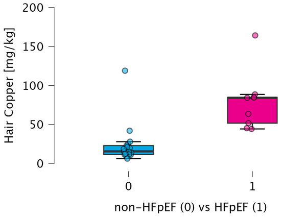
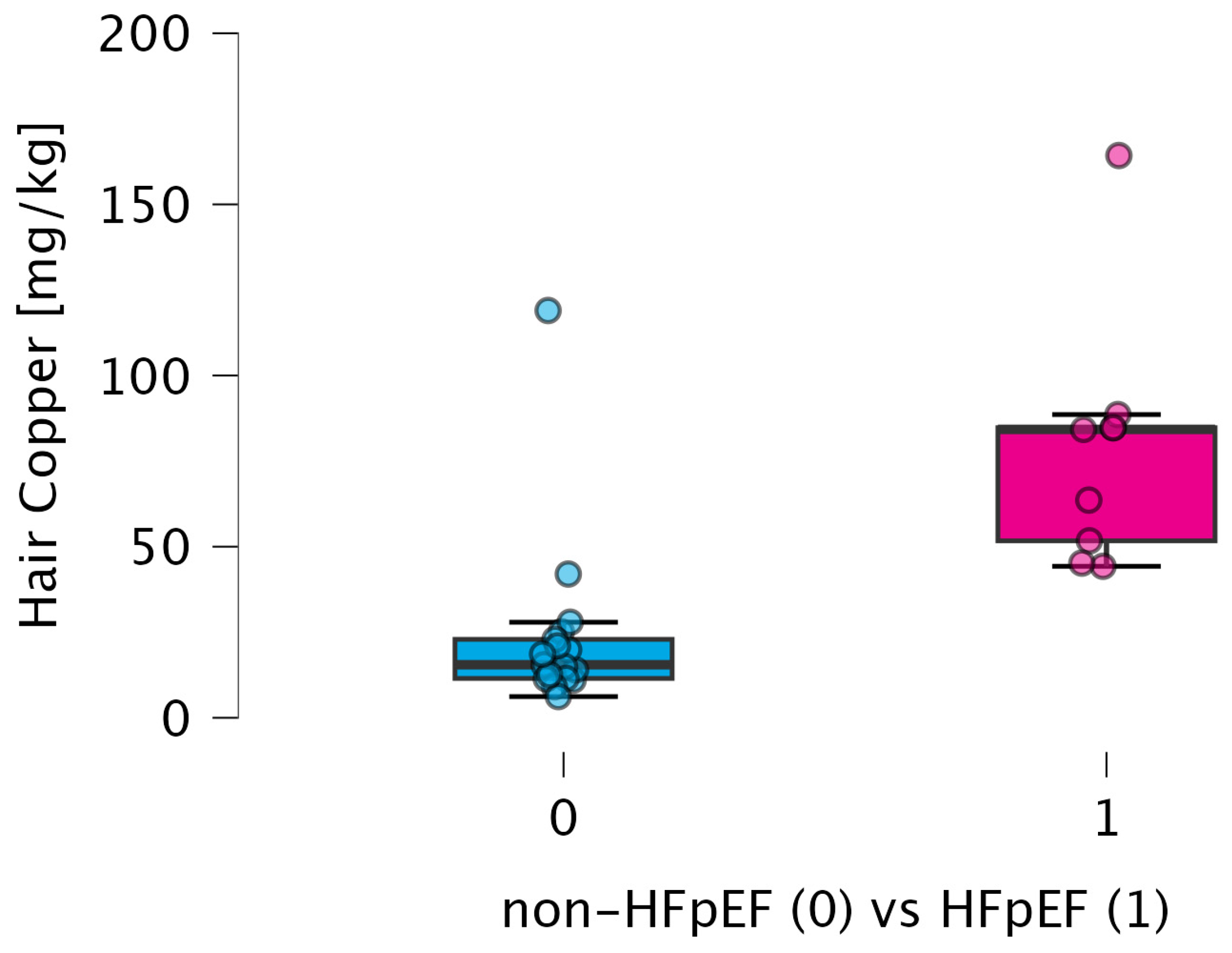
After adjustment for potential confounders, the association between elevated copper concentration and HFpEF remained directionally consistent, although the study was not powered for definitive multivariable inference. The differences in copper concentration between HFpEF and non-HFpEF subgroups are presented in Figure 2.
Figure 2.
The differences in copper concentrations between HFpEF and non-HFpEF subgroups. Abbreviations: HFpEF—heart failure with preserved ejection fraction, kg—kilogram, mg—milligram.
3.1. Multivariable Regression Analysis
In exploratory uni- and multivariable regression analysis adjusting for age, body mass index, and smoking exposure (pack-years), the association between copper concentration and HFpEF remained directionally consistent. However, the model did not reach statistical significance, likely due to limited sample size as presented in Table 5.
Table 5.
Univariate and multivariable logistic regression analysis for HFpEF. Multivariable model adjusted for age, body mass index, and smoking exposure (pack-years). Due to the limited sample size, the model was considered exploratory.
3.2. The Possible Role of Obesity
These biochemical differences were observed in parallel with echocardiographic evidence of structural and functional alterations, meeting the HFpEF criteria combined with clinical characteristics. To assess whether the observed differences were driven by obesity rather than HFpEF, we performed an additional subgroup analysis stratified by BMI. The active smoker group was subdivided into patients with a BMI over 30 kg/m2 and the non-obese population. Hair concentrations of 21 trace elements were investigated, and no significant differences in scalp trace element concentrations were observed (Table 6).
Table 6.
Hair scalp trace elements analysis in obese and non-obese subgroups.
4. Discussion
In this prospective pilot analysis of active smokers, we observed differences in scalp hair trace element concentrations between individuals with HFpEF and smokers without evidence of diastolic heart failure. In unadjusted analyses, several trace elements differed between smokers with and without HFpEF, including vanadium, lithium, aluminum, and copper. However, after adjustment for multiple comparisons using the Benjamini–Hochberg FDR procedure, only copper remained statistically significant. The selective elevation of copper, in contrast to other trace elements, may reflect its unique role in oxidative stress pathways and extracellular matrix remodeling [13,14]. Copper is actively incorporated into hair through binding to keratin-associated proteins and may accumulate under conditions of altered systemic distribution, particularly in states of chronic inflammation [15,16].
These findings suggest that alterations in trace element homeostasis may accompany the structural and functional myocardial changes characteristic of HFpEF in smokers. The analysis was based on hair samples, which reflect long-term exposure rather than short-term fluctuations in plasma. The primary endpoint was not significant; therefore, findings should be interpreted cautiously as secondary/exploratory.
Tobacco smoking remains a challenging problem with a significant impact on human morbidity and mortality [17,18]. Its impact on cardiovascular diseases, particularly premature atherosclerosis, is a major concern [19]. Cigarette smoke is a complex mixture containing numerous metals and metalloids capable of inducing oxidative stress and endothelial dysfunction [20]. Chronic exposure to active smoking or passive inhalation of these elements may alter systemic metal distribution, leading to altered inflammatory signaling and mitochondrial function [21]. Previous studies [22,23] highlighted dysregulation of several trace element concentrations, including copper, cadmium, and selenium, reflecting both environmental exposure and altered metabolic handling of metals, resulting in cardiovascular pathology. Such disturbances may contribute to the mechanisms recognized as central components of HFpEF pathophysiology. The lack of differences in trace element concentrations between smokers and non-smokers contrasts with previous reports [24,25]. This discrepancy may reflect differences in population characteristics, analytical methods, or the relatively small sample size of the present study. Importantly, the absence of differences between smokers and non-smokers suggests that smoking alone does not explain the observed copper elevation. Instead, HFpEF-related systemic changes, including inflammation and metabolic dysregulation, may play a dominant role [12,26,27].
In our study, which focused on the active-smoker population, copper showed the most consistent association with the HFpEF group. Several elements differed in our unadjusted comparisons; however, after FDR correction, only copper remained significantly elevated. The marked increase in hair copper levels observed in HFpEF patients (84.26 mg/kg vs. 15.00 mg/kg, p < 0.001; Table 4) becomes even more relevant when considered alongside the echocardiographic signs of structural and functional impairment shown in Table 3. In particular, the HFpEF group demonstrated lower global longitudinal strain (GLS) (14% vs. 19%, p < 0.001) and higher E/E’ ratios (11 vs. 8, p < 0.001), indicating reduced myocardial deformation and elevated left ventricular filling pressures. These findings should be interpreted within the framework of systemic inflammatory–resolution coupling rather than direct causality.
Copper is one of the essential trace elements that play pivotal roles in mitochondrial respiration and antioxidant defense [28]. Copper is a key micronutrient involved in mitochondrial respiration, oxidative phosphorylation, and antioxidant defense through enzymes such as superoxide dismutase and cytochrome c oxidase. Dysregulated copper homeostasis has been linked to cardiovascular remodeling, myocardial fibrosis, and metabolic dysfunction. Elevated copper levels may reflect altered systemic regulation of micronutrients under conditions of chronic inflammation and oxidative stress. Its dysregulated metabolism may contribute to myocardial remodeling and fibrosis by promoting oxidative stress and redox imbalance [29]. Copper plays an essential role as a cofactor for lysyl oxidase (LOX), the enzyme that enables collagen cross-linking within the extracellular matrix. When copper balance is disturbed, fibroblast activity may increase, leading to greater collagen deposition and myocardial stiffening. The incorporation of copper into hair shafts occurs during keratinization and reflects circulating bioavailable copper levels over time [30,31]. Increased deposition may therefore represent systemic redistribution rather than localized scalp-specific processes.
This mechanism aligns with our findings, where patients showed reduced septal and lateral E’ velocities, as presented in Table 3. Excessive intracellular copper can enhance reactive oxygen species generation and impair endothelial function [32], processes that may accelerate myocardial diastolic dysfunction. Elevated circulating copper concentrations have been associated with increased cardiovascular risk and adverse cardiac remodeling [33]. The markedly higher copper concentrations observed in our analysis among smokers with HFpEF may represent a marker of chronic oxidative stress or altered trace element homeostasis rather than direct cardiotoxicity. The absence of significant differences between smokers and non-smokers in the primary analysis suggests that HFpEF status, rather than smoking exposure alone, may be associated with alterations in trace element distribution. Moreover, the additional analysis stratified by obesity did not reveal differences in trace element concentrations, suggesting that the observed copper signal was unlikely to be explained solely by body mass index. As shown in Table 6, none of the 21 analyzed elements—including copper (p = 0.777)—differed significantly between obese and non-obese smokers. This supports the idea that the copper imbalance observed in our study is more closely related to heart failure and its accompanying systemic inflammatory response, rather than to metabolic alterations associated with excess body weight. Nonetheless, these findings should be interpreted cautiously, given the limited sample size. Moreover, dietary intake, except for dietary supplementation (including copper supplements), was not systematically assessed, which represents a limitation of our analysis.
Cigarette smoke contains numerous metals and metalloids capable of promoting oxidative stress [34]. Chronic exposure may alter systemic trace element distribution and redox balance. HFpEF is increasingly recognized as a syndrome characterized by systemic inflammation and coronary microvascular dysfunction, conditions that may influence metal handling, tissue redistribution, and excretion. This suggests that the homeostatic disturbances of these trace elements may be closely linked to the degree of structural remodeling and diastolic dysfunction observed in this population. Experimental data [35] further suggest a synergistic interaction between trace-element exposure and oxidative stress in mediating long-term cardiovascular toxicity associated with smoking. These findings should be interpreted as exploratory and hypothesis-generating, and larger studies integrating circulating and tissue trace element profiling are required to clarify the role of metal homeostasis in HFpEF.
From a nutritional perspective, trace element profiling may help identify micronutrient imbalance associated with cardiometabolic disease. If confirmed in larger studies, monitoring trace element status could provide insights into nutritional and environmental determinants of HFpEF pathophysiology.
Study Limitation
This study has several important limitations. First, the relatively small sample size and single-center design limit generalizability. Second, the absence of a healthy control group precludes conclusions regarding disease-specific alterations. Third, the observational design does not allow causal inference. Fourth, although exploratory multivariable adjustment was performed, residual confounding cannot be excluded. Fifth, dietary intake, renal function, and environmental exposure were not comprehensively assessed. Finally, hair analysis reflects long-term exposure but may not directly correspond to myocardial or intracellular concentrations.
Additionally, no formal sample size calculation was performed due to the pilot nature of the study, which further limits statistical power and the ability to detect smaller effect sizes.
A data inconsistency related to beryllium concentrations was identified and corrected during revision, highlighting the importance of careful interpretation of trace-level measurements.
5. Conclusions
In this exploratory pilot study, elevated hair copper concentrations were observed in smokers with HFpEF. These findings raise the hypothesis that trace element imbalance may reflect systemic cardiometabolic remodeling rather than smoking exposure alone. Further studies are required to determine whether copper represents a biomarker or a mechanistic contributor to HFpEF.
Author Contributions
Conceptualization, B.K. and T.U.; methodology, A.H.; software, I.S.; validation, B.K., A.H. and T.U.; formal analysis, T.U., K.B. and K.J.F.; investigation, I.S.; resources, B.K.; data curation, K.B.; writing—original draft preparation, B.K. and T.U.; writing—review and editing, I.S., A.H., Z.K., K.B., K.J.F. and A.T.; visualization, T.U.; supervision, A.H. All authors have read and agreed to the published version of the manuscript.
Funding
This research received no external funding.
Institutional Review Board Statement
The study was conducted in accordance with the Declaration of Helsinki and approved by the Poznan University of Medical Sciences Ethics Committee (protocol no. 411/24; 27 June 2024) for studies involving humans.
Informed Consent Statement
Informed consent was obtained from all subjects involved in the study.
Data Availability Statement
Data supporting the reported results can be obtained upon reasonable request by contacting the corresponding authors, due to privacy or ethical restrictions.
Conflicts of Interest
The authors declare no conflicts of interest.
References
- Suhrs, H.E.; Schroder, J.; Bové, K.B.; Mygind, N.D.; Frestad, D.; Michelsen, M.M.; Lange, T.; Gustafsson, I.; Kastrup, J.; Prescott, E. Inflammation, non-endothelial dependent coronary microvascular function and diastolic function—Are they linked? PLoS ONE 2020, 15, e0236035. [Google Scholar] [CrossRef] [Scilit]
- Doryńska, A.; Drohomirecka, A.; Topór-Mądry, R.; Łazarczyk, H.; Zieliński, T.; Rywik, T.M. Changes in the prevalence of heart failure phenotypes over time and their association with patient prognosis. Pol. Arch. Intern. Med. 2025, 135, 17119–17129. [Google Scholar] [CrossRef] [Scilit] [PubMed]
- Addissouky, T.A.; Sayed, E.L.; Majeed, M.I.; Ali, M.A.; Wang, Y.; El Baz, A.; Elarabany, N.; Khalil, A.A. Oxidative stress and inflammation: Elucidating mechanisms of smoking-attributable pathology for therapeutic targeting. Bull. Natl. Res. Cent. 2024, 48, 16. [Google Scholar] [CrossRef] [Scilit]
- Benowitz, N.L.; Burbank, A.D. Cardiovascular toxicity of nicotine: Implications for electronic cigarette use. Trends Cardiovasc. Med. 2016, 26, 515–523. [Google Scholar] [CrossRef] [Scilit]
- Cora, N.; Ghandour, J.; Pollard, C.M.; Desimine, V.L.; Ferraino, K.E.; Pereyra, J.M.; Valiente, R.; Lymperopoulos, A. Nicotine-induced adrenal beta-arrestin1 upregulation mediates tobacco-related hyperaldosteronism leading to cardiac dysfunction. World J. Cardiol. 2020, 12, 192–202. [Google Scholar] [CrossRef] [Scilit]
- Wang, Y.M.; Feng, L.S.; Xu, A.; Ma, X.H.; Zhang, M.T.; Zhang, J. Copper ions: The invisible killer of cardiovascular disease. Mol. Med. Rep. 2024, 30, 210–226. [Google Scholar] [CrossRef] [Scilit]
- Gramala, Z.; Kalus, O.; Maćkowiak, J.; Zalewska, K.; Karpiński, M.; Staniewski, A.; Szymańska, Z.; Zieliński, M.; Grobelna, M.; Zawadzki, P.; et al. Essential and Toxic Elements in Cardiovascular Disease: Pathophysiological Roles and the Emerging Contribution of Hair Mineral Analysis. Int. J. Mol. Sci. 2025, 26, 12145. [Google Scholar] [CrossRef] [Scilit] [PubMed]
- Bass, D.A.; Hickock, D.; Quig, D.; Urek, K. Trace element analysis in hair: Factors determining accuracy, precision, and reliability. Altern. Med. Rev. 2001, 6, 472–481. [Google Scholar]
- Pieske, B.; Tschöpe, C.; de Boer, R.A.; Fraser, A.G.; Anker, S.D.; Donal, E.; Edelmann, F.; Fu, M.; Guazzi, M.; Lam, C.S.P.; et al. How to diagnose heart failure with preserved ejection fraction: The HFA-PEFF diagnostic algorithm: A consensus recommendation from the Heart Failure Association (HFA) of the European Society of Cardiology (ESC). Eur. Heart J. 2019, 40, 3297–3317, Erratum in Eur. Heart J. 2021, 42, 1274. https://doi.org/10.1093/eurheartj/ehaa1016. [Google Scholar] [CrossRef] [Scilit] [PubMed]
- Urbanowicz, T.; Hanć, A.; Olasińska-Wiśniewska, A.; Komosa, A.; Filipiak, K.J.; Radziemski, A.; Matejuk, M.; Uruski, P.; Tykarski, A.; Jemielity, M. Relation between Systemic Inflammatory Index (SII) and Hair Trace Elements, Metals and Metalloids Concentration in Epicardial Coronary Artery Disease-Preliminary Report. Rev. Cardiovasc. Med. 2023, 24, 358–368. [Google Scholar] [CrossRef] [Scilit]
- Urbanowicz, T.; Spasenenko, I.; Królak, J.; Gabriel, K.; Krasińska, B.; Zawadzki, P.; Grobelna, M.; Zieliński, M.; Staniszewski, R.; Krasiński, Z.; et al. The relation between hair manganese accumulation, obesity, and coronary artery disease: Early observation. J. Elem. 2025, 30, 777–787. [Google Scholar] [CrossRef] [Scilit]
- Urbanowicz, T.; Hanć, A.; Kasperowicz, Z.; Adamczak, O.; Spasanenko, I.; Gabriel, K.; Tykarski, A.; Krasiński, Z.; Krasińska, B. Differences in Scalp Hair Trace Element Concentrations in Patients with Preserved Left Ventricular Ejection Fraction (HFpEF) Compared with Controls: A Hypothesis-Generating Study. J. Clin. Med. 2026, 15, 2029. [Google Scholar] [CrossRef] [Scilit]
- Boaru, D.L.; Leon-Oliva, D.; Castro-Martinez, P.; Garcia-Montero, C.; Fraile-Martinez, O.; García-González, B.; Pérez-González, I.; Michael Alhaddadin, M.N.; Barrena-Blázquez, S.; Lopez-Gonzalez, L.; et al. Cuproptosis: Current insights into its multifaceted role in disease, cancer, and translational/therapeutic opportunities. Biomed. Pharmacother. 2025, 190, 118422–118451. [Google Scholar] [CrossRef] [Scilit]
- Tapiero, H.; Townsend, D.M.; Tew, K.D. Trace elements in human physiology and pathology. Copper. Biomed. Pharmacother. 2003, 57, 386–398. [Google Scholar] [CrossRef] [Scilit]
- Jeong, S.Y.; Shim, H.Y.; Lee, Y.J.; Park, B. Association between Copper-Zinc Ratio in Hair and Neutrophil-Lymphocyte Ratio within the Context of a Normal White Blood Cell Count among Overweight or Obese Korean Individuals: A Pilot Study. Korean J. Fam. Med. 2021, 42, 240–244. [Google Scholar] [CrossRef] [Scilit]
- Borowczyk, K.; Suliburska, J.; Jakubowski, H. Demethylation of methionine and keratin damage in human hair. Amino Acids. 2018, 50, 537–546. [Google Scholar] [CrossRef] [Scilit]
- Lucki, M.; Chlebuś, E.; Wareńczak, A.; Daroszewski, P.; Lisiński, P. Assessment of CVD Risk Factors in Secondary Prevention after Ischemic Stroke Using the ICF. Int. J. Environ. Res. Public Health 2022, 19, 3368. [Google Scholar] [CrossRef] [Scilit] [PubMed]
- Moryson, W.; Stawinska-Witoszynska, B. Premature Mortality Due to Tobacco-Related Malignancies in Poland. Int. J. Gen. Med. 2021, 14, 2171–2182. [Google Scholar] [CrossRef] [Scilit] [PubMed]
- Le, A.; Peng, H.; Golinsky, D.; Di Scipio, M.; Lali, R.; Paré, G. What Causes Premature Coronary Artery Disease? Curr. Atheroscler. Rep. 2024, 26, 189–203. [Google Scholar] [CrossRef] [Scilit] [PubMed]
- Ishida, M.; Sakai, C.; Kobayashi, Y.; Ishida, T. Cigarette Smoking and Atherosclerotic Cardiovascular Disease. J. Atheroscler. Thromb. 2024, 31, 189–200. [Google Scholar] [CrossRef] [Scilit] [PubMed]
- Cabar, H.D.; Ersoy Karaçuha, M.; Yilmaz, M. The Interaction Between Concentration of Heavy Metal-Trace Elements and Non-Smoking Status of Adolescents in Sinop (Turkey). Biol. Trace Elem. Res. 2020, 194, 105–114. [Google Scholar] [CrossRef] [Scilit]
- Ji, R.; Wu, H.; Lin, H.; Li, Y.; Shi, Y. Cadmium and selenium blood levels in association with congestive heart failure in diabetic and prediabetic patients: A cross-sectional study from the national health and nutrition examination survey. Diabetol. Metab. Syndr. 2025, 17, 12. [Google Scholar] [CrossRef] [Scilit]
- Feng, J.; Yang, F.; Wu, H.; Xing, C.; Xue, H.; Zhang, L.; Zhang, C.; Hu, G.; Cao, H. Selenium protects against cadmium-induced cardiac injury by attenuating programmed cell death via PI3K/AKT/PTEN signaling. Environ. Toxicol. 2022, 37, 1185–1197. [Google Scholar] [CrossRef] [Scilit] [PubMed]
- Nakhaee, S.; Amirabadizadeh, A.; Ataei, M.; Ataei, H.; Zardast, M.; Shariatmadari, M.R.; Mousavi-Mirzaei, S.M.; Mehrpour, O. Comparison of serum concentrations of essential and toxic elements between cigarette smokers and non-smokers. Environ. Sci. Pollut. Res. Int. 2021, 28, 37672–37678. [Google Scholar] [CrossRef] [Scilit] [PubMed]
- Mansouri, B.; Azadi, N.A.; Sharafi, K.; Nakhaee, S. The effects of active and passive smoking on selected trace element levels in human milk. Sci. Rep. 2023, 13, 20756–20763. [Google Scholar] [CrossRef] [Scilit] [PubMed]
- Peikert, A.; Vacca, A.; Norata, G.D.; Schiattarella, G.G.; Osto, E. Cellular Interactions and Immunometabolic Mechanisms in Heart Failure with Preserved Ejection Fraction: From Molecular Mechanisms to Clinical Evidence. Circ. Heart Fail. 2026, 19, e012674. [Google Scholar] [CrossRef] [Scilit]
- Gatto, C.; Rusciano, M.R.; Visco, V.; Vecchione, C.; Ciccarelli, M. Pathophysiology of HFpEF: Insights from a Metabolic–Mitochondrial Perspective. Int. J. Mol. Sci. 2020, 27, 284. [Google Scholar] [CrossRef] [Scilit]
- Villeneuve, G.; Deschêne, K.; Roy, C.; Matte, J.J.; Lapointe, J.; Dalto, D.B. Effects of different combinations of dietary zinc and copper levels on the mitochondrial energy metabolism, oxidative balance, antioxidant system, and inflammation in weaned pigs. J. Anim. Sci. 2026, 5, skag069. [Google Scholar] [CrossRef] [Scilit]
- Liu, Z.Y.; Liu, Z.Y.; Lin, L.C.; Song, K.; Tu, B.; Zhang, Y.; Yang, J.J.; Zhao, J.Y.; Tao, H. Redox homeostasis in cardiac fibrosis: Focus on metal ion metabolism. Redox Biol. 2024, 71, 103109. [Google Scholar] [CrossRef] [Scilit]
- Dziedzic, E.A.; Tuzimek, A.; Gąsior, J.S.; Paleczny, J.; Junka, A.; Kwaśny, M.; Dąbrowski, M.; Jankowski, P. Investigation on the Association of Copper and Copper-to-Zinc-Ratio in Hair with Acute Coronary Syndrome Occurrence and Its Risk Factors. Nutrients 2022, 14, 4107. [Google Scholar] [CrossRef] [Scilit]
- Kempson, I.M.; Skinner, W.M.; Kirkbride, K.P. The occurrence and incorporation of copper and zinc in hair and their potential role as bioindicators: A review. J. Toxicol. Environ. Health B Crit. Rev. 2007, 10, 611–622. [Google Scholar] [CrossRef] [Scilit] [PubMed]
- Chen, X.; Cai, Q.; Liang, R.; Zhang, D.; Liu, X.; Zhang, M.; Xiong, Y.; Xu, M.; Liu, Q.; Li, P.; et al. Copper homeostasis and copper-induced cell death in the pathogenesis of cardiovascular disease and therapeutic strategies. Cell Death Dis. 2023, 14, 105. [Google Scholar] [CrossRef] [Scilit]
- Zhao, H.; Mei, K.; Hu, Q.; Wu, Y.; Xu, Y.; Qinling; Yu, P.; Deng, Y.; Zhu, W.; Yan, Z.; et al. Circulating copper levels and the risk of cardio-cerebrovascular diseases and cardiovascular and all-cause mortality: A systematic review and meta-analysis of longitudinal studies. Environ. Pollut. 2024, 340, 122711. [Google Scholar] [CrossRef] [Scilit] [PubMed]
- Ghoma, W.E.O.; Sevik, H.; Isinkaralar, K. Comparison of the rate of certain trace metals accumulation in indoor plants for smoking and non-smoking areas. Environ. Sci. Pollut. Res. Int. 2023, 30, 75768–75776. [Google Scholar] [CrossRef] [Scilit] [PubMed]
- Ghosh, B.; Chengala, P.P.; Shah, S.; Chen, D.; Karnam, V.; Wilmsen, K.; Yeung-Luk, B.; Sidhaye, V.K. Cigarette smoke-induced injury induces distinct sex-specific transcriptional signatures in mice tracheal epithelial cells. Am. J. Physiol. Lung Cell. Mol. Physiol. 2023, 325, L467–L476. [Google Scholar] [CrossRef] [Scilit] [PubMed]

Disclaimer/Publisher’s Note: The statements, opinions and data contained in all publications are solely those of the individual author(s) and contributor(s) and not of MDPI and/or the editor(s). MDPI and/or the editor(s) disclaim responsibility for any injury to people or property resulting from any ideas, methods, instructions or products referred to in the content. |
© 2026 by the authors. Licensee MDPI, Basel, Switzerland. This article is an open access article distributed under the terms and conditions of the Creative Commons Attribution (CC BY) license.