


Abstract
Background/Objectives: Bladder cancer treatment is frequently hindered by chemoresistance to agents such as cisplatin, a process in which autophagy is hypothesized to play a cytoprotective role. This study aimed to investigate the time-dependent transcriptional dynamics of autophagy-related genes in response to cisplatin in bladder cancer cell lines to better elucidate the molecular underpinnings of this resistance. Methods: Two human bladder cancer cell lines, T24 and 5637, were exposed to varying concentrations of cisplatin. Cell viability and half-maximal inhibitory concentration (IC50) values were determined at 24 and 48 h using the MTS assay. Subsequently, the relative mRNA expression levels of key autophagy-related genes (ULK1, BECN1, ATG5, ATG7, LC3B, SQSTM1/p62, LAMP1, and TFEB) were quantitatively analyzed via RT-qPCR at 0, 6, 24, and 48 h intervals. Results: Cisplatin exerted a dose- and time-dependent cytotoxic effect, with 5637 cells exhibiting significantly greater sensitivity compared to T24 cells. Transcriptional analysis revealed a dynamic, multiphasic modulation of the autophagic pathway: an early-phase upregulation of initiation genes (ULK1, BECN1), a mid-phase increase in autophagosome formation genes (ATG5, ATG7), and a late-phase alteration in lysosomal regulation genes (LAMP1, TFEB). Notably, the more chemoresistant T24 cells mounted a robust and sustained autophagic transcriptional response, whereas the sensitive 5637 cells demonstrated a more limited and transient reaction. Conclusions: Cisplatin modulates the autophagic pathway at the transcriptional level in a highly dynamic, time-dependent, and cell-line-specific manner. Interpreted alongside established functional evidence in the literature, the sustained autophagic gene expression observed in the resistant cells is consistent with a potential cytoprotective role, warranting further functional validation at the protein level. These findings map the temporal genetic landscape of cisplatin-induced autophagy, providing a theoretical framework for optimizing the timing of autophagy-targeted combination therapies in bladder cancer.
1. Introduction
Bladder cancer is one of the most prevalent urinary tract malignancies worldwide. Chemotherapy remains one of the main treatment options for this type of cancer [1]. Cisplatin is a cornerstone chemotherapy drug primarily used for bladder cancer treatment. However, the problem with cisplatin is that the patients’ response rate, especially in the metastatic stage, is very low, and most patients develop resistance, which is a major clinical issue [2]. One of the key mechanisms underlying this resistance is the autophagy process, which helps cells survive under stress conditions and can therefore decrease the efficacy of chemotherapy [3]. In this context, studying time-dependent changes in cisplatin-induced autophagy gene expression in bladder cancer cells is of vital importance in revealing the molecular mechanisms of chemoresistance [2]. Autophagy is an evolutionarily conserved lysosomal degradation process that has been demonstrated to play a main role in the adaptation of bladder cancer cells to chemotherapeutic agents and in the development of treatment resistance [4]. A study by Jobst found that autophagy supports cell survival during stress situations, like nutrient deprivation, through the controlled degradation and recycling of cellular components [4]. However, the fact that this process functions as a resistance mechanism to anti-cancer therapies has made autophagy modulation an attractive target for new therapeutic strategies [5]. This resistance mechanism plays a pivotal role, especially in the adaptive response of cells to platinum-based chemotherapeutic agents such as cisplatin [6]. Actually, overcoming the protective autophagy induced by chemotherapy may significantly increase the sensitivity of bladder cancer cells to cisplatin [3]. Notably, functional studies have confirmed that inhibiting this autophagic flux can successfully sensitize these resistant bladder cancer cells to cisplatin-induced death [7].
The activation of the autophagic cascade under chemotherapeutic stress is governed by a complex network of upstream signaling pathways. Cisplatin has been shown to induce autophagy in bladder cancer cells primarily through the activation of the AMPK (AMP-activated protein kinase) pathway and the subsequent inhibition of mTOR (mammalian target of rapamycin) signaling [8]. This regulatory shift promotes the nuclear translocation of master transcription factors such as TFEB (Transcription Factor EB), which orchestrates the expression of genes involved in both autophagosome formation and lysosomal biogenesis [9].
In this regard, understanding how cisplatin affects these upstream regulators and downstream autophagy pathways in bladder cancer cells is a basis for the development of new treatment approaches targeting resistance mechanisms [10]. In this paper, the time-dependent effects of cisplatin on autophagy gene expression in bladder cancer cell lines are thoroughly studied, thereby providing a comprehensive characterization of the transcriptional autophagic responses associated with chemotherapeutic resistance [11]. This study intends to serve as a reference for future personalized therapeutic approaches by identifying specific genetic mechanisms that play a key role in cisplatin resistance and regulate autophagic flux, which is shown to increase with tumor grade in bladder cancer cells [1,5]. More specifically, a comprehensive analysis of how autophagy gene expression profiles change over time is of great significance to understanding the molecular dynamics of chemoresistance in these cells [12]. Thus, elucidating the dual role of autophagy in supporting cancer cell survival and its complex interactions in tumor development is of vital importance for new cancer treatment strategies [13]. Considering the fundamental function of autophagy in maintaining cellular homeostasis, the dysregulation of this process in cancer cells is closely associated with tumor development, metastasis, and treatment resistance [14,15]. This dual role highlights that autophagy in cancer needs to be modulated very carefully [16]. For instance, autophagy inhibition is common in cisplatin-sensitive cells, whereas in cisplatin-resistant tumor cells, protective autophagy is observed to be highly activated [17]. This indicates that autophagy plays a central role in how cancer cells adapt to chemotherapeutic agents and develop resistance to treatment [17,18]. Therefore, to surpass cisplatin-induced chemoresistance in bladder cancer therapy, understanding the precise molecular chronology of autophagy and targeting these temporal processes are of utmost importance in the development of future therapeutic strategies [18]. While the functional status of autophagy is ultimately determined by protein-level flux, characterizing the temporal transcriptional landscape provides a fundamental blueprint of how cells program their long-term adaptive response to cisplatin stress.
2. Materials and Methods
2.1. Cell Line and Culture Conditions
Human bladder cancer cell lines T24 and 5637 (ATCC, Manassas, VA, USA) were utilized in this study. Cells were cultured in Dulbecco’s Modified Eagle (DMEM; high glucose; Gibco, Billings, MT, USA) supplemented with 10% fetal bovine serum (FBS; Capricorn Scientific, Ebsdorfergrund, Germany) and 1% penicillin/streptomycin (Gibco, Billings, MT, USA). The cell cultures were maintained in a humidified incubator containing 5% CO2 at 37 °C.
2.2. Cisplatin Treatment and Cell Viability Assay
Cisplatin (Sigma-Aldrich, St. Louis, MO, USA) was dissolved in 0.9% NaCl and administered to the cells at increasing concentrations. Cell viability was assessed at 24 and 48 h using the MTS assay (Sigma Chemical Co., St. Louis, MO, USA) following exposure to cisplatin concentrations ranging from 0 to 40 µM. Based on the obtained data, dose–response curves were generated, and half-maximal inhibitory concentration (IC50) values were calculated for each time point. For subsequent molecular analyses, sublethal doses (close to the IC50) that do not cause extreme acute cytotoxicity were selected to allow for the proper investigation of cellular responses.
2.3. RNA Isolation and cDNA Synthesis
Total RNA was isolated from the cells at defined time points (0, 6, 24, and 48 h) following cisplatin exposure. The isolation procedure was performed using the Qiagen RNeasy Mini Kit (Qiagen, Hilden, Germany) in accordance with the manufacturer’s instructions. The purity and concentration of the RNA were evaluated using spectrophotometric methods (NanoDrop 2000, Thermo Fisher Scientific, Waltham, MA, USA) and samples meeting the quality criteria (A260/280 ratio ~2.0) were subsequently utilized for complementary DNA (cDNA) synthesis (RevertAid First Strand cDNA Synthesis Kit (Thermo Fisher Scientific, Waltham, MA, USA)).
2.4. Real-Time Quantitative PCR (RT-qPCR) Analysis
The expression levels of specific genes representing different stages of the autophagy pathway were quantitatively analyzed using RT-qPCR (CFX96 Real-Time PCR System (Bio-Rad Laboratories, Hercules, CA, USA)). The analyzed gene panel included ULK1 and BECN1 (representing the initiation phase), ATG5, ATG7, and LC3B (representing the elongation phase), SQSTM1/p62 (representing selective autophagy), and LAMP1 and TFEB (representing lysosomal regulation) (Table 1). GAPDH was employed as the reference housekeeping gene for normalization. Relative gene expression levels were calculated using the 2−ΔΔCt method to evaluate the time-dependent dynamics of autophagy in response to cisplatin treatment.
Table 1.
Primer sequences for RT-qPCR analysis.
2.5. Statistical Analysis
All experiments were performed in at least three independent biological replicates. Data are presented as mean ± standard error of the mean (SEM). Statistical analyses and graph generation were conducted using (GraphPad Prism Version 9.0; GraphPad Software, Boston, MA, USA). Comparisons between the control group (0 h) and cisplatin-treated time points (6, 24, and 48 h) were performed using a one-way analysis of variance (ANOVA) followed by Dunnett’s post hoc test. A p-value < 0.05 was considered statistically significant.
3. Results
3.1. Time- and Dose-Dependent Effects of Cisplatin on Cell Viability
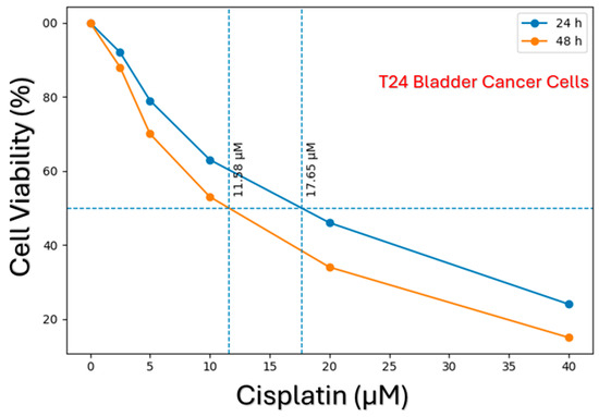
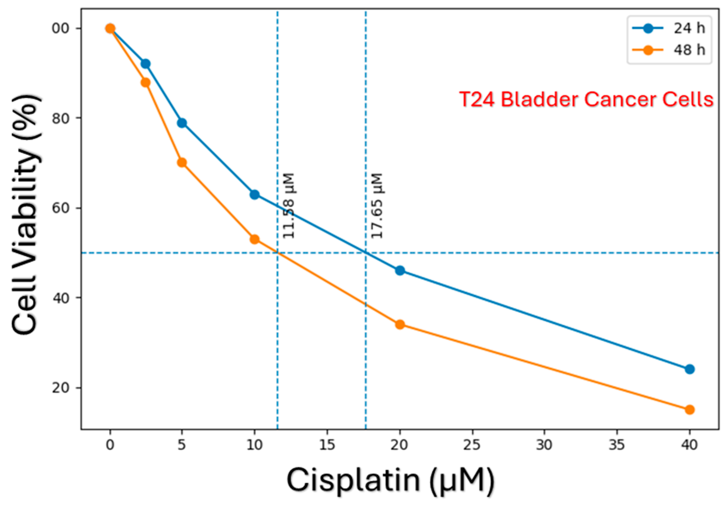
The cytotoxic effects of cisplatin on T24 and 5637 bladder cancer cell lines were evaluated in a time- and dose-dependent manner using the MTS assay. The resulting data demonstrated that cisplatin significantly reduced cell viability in both cell lines with increasing concentrations. The dose–response curves obtained for the T24 cell line are presented in Figure 1. In this cell line, a marked decrease in IC50 values was observed following 48 h of cisplatin exposure compared to 24 h of exposure. This indicates that the cytotoxic effect of cisplatin increases in a time-dependent manner.
Figure 1.
Time-dependent cytotoxic effect of cisplatin and determination of IC50 values in T24 bladder cancer cells. Cell viability was evaluated at 24 and 48 h time points following exposure to increasing concentrations of cisplatin (0–40 µM). The resulting dose–response curves demonstrate that cisplatin decreases cell viability in a dose- and time-dependent manner. The horizontal dashed line represents 50% cell viability, while the vertical dashed lines indicate the corresponding IC50 values at the respective time points. The lower IC50 values observed at 48 h of exposure indicate the increasing cytotoxic effect of cisplatin over time.
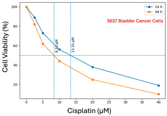
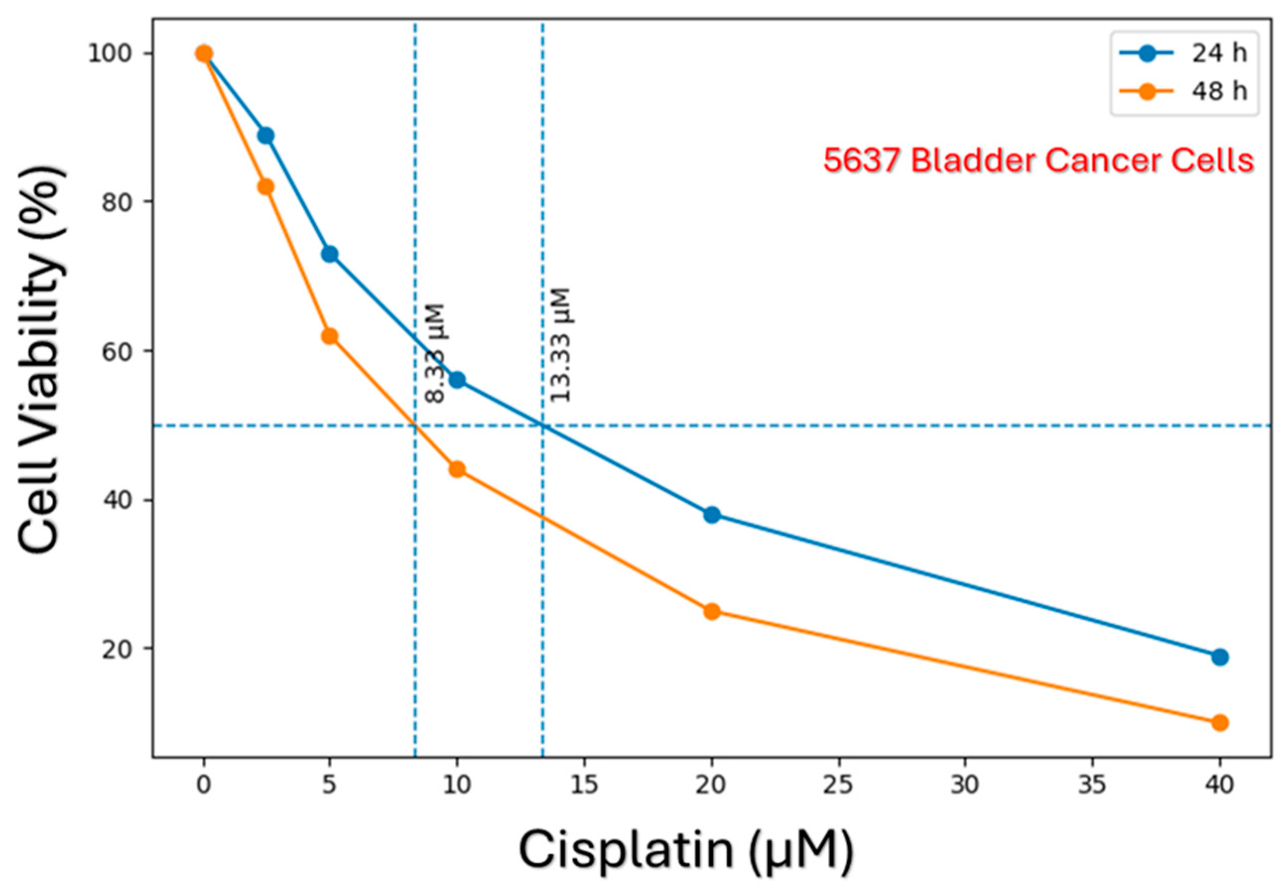
Similarly, the cell viability results obtained for the 5637 cell line are shown in Figure 2. A cisplatin-induced decrease in cell viability was also observed in 5637 cells, and the calculated IC50 values were notably lower compared to those of the T24 cells. This finding indicates that the 5637 cell line is inherently more sensitive to cisplatin-induced cytotoxicity. Overall, it was demonstrated that while cisplatin exhibits a time- and dose-dependent cytotoxic effect in both cell lines, the magnitude of this effect varies significantly depending on the specific cell line’s resistance profile.
Figure 2.
Time-dependent cytotoxic effect of cisplatin and determination of IC50 values in 5637 bladder cancer cells. Cells were exposed to varying concentrations of cisplatin for 24 and 48 h, and cell viability was assessed. The dose–response curves indicate a more pronounced sensitivity to cisplatin in 5637 cells. The horizontal dashed line represents 50% cell viability, while the vertical dashed lines indicate the calculated IC50 values. Lower IC50 values compared to T24 cells demonstrate that this cell line is more sensitive.
3.2. Time-Dependent Changes in Autophagy-Related Gene Expression
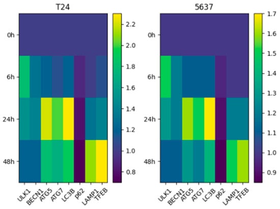
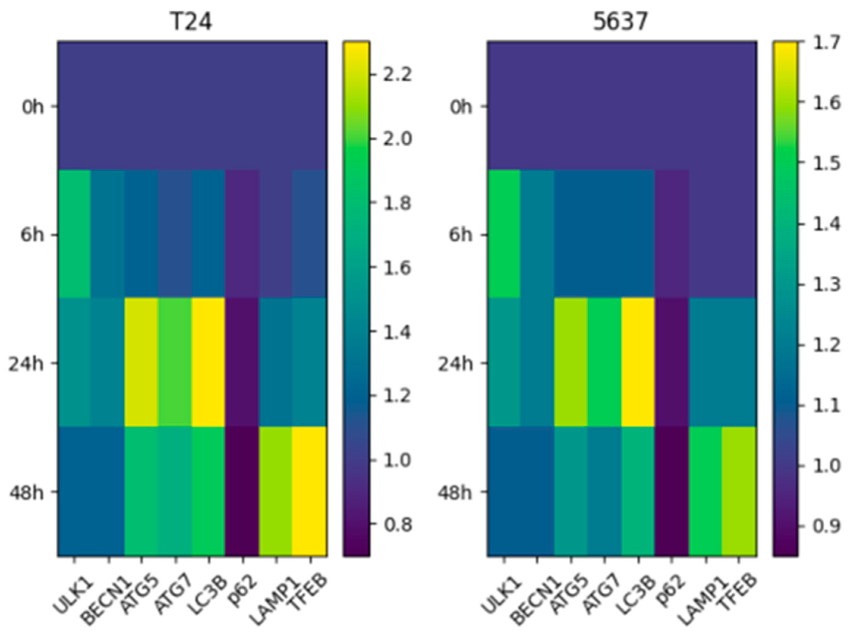
The modulatory effects of cisplatin treatment on autophagy-related gene expression were evaluated in a time-dependent manner. The transcriptional profiles obtained for both cell lines are presented as heatmaps in Figure 3.
Figure 3.
Time-dependent heatmap of autophagy-related gene expression in T24 and 5637 bladder cancer cells following cisplatin treatment. Relative expression levels (2−ΔΔCt) are shown for genes involved in autophagy initiation, elongation, selective autophagy, and lysosomal function across 0, 6, 24, and 48 h.
This quantitative analysis showed that genes representing distinct stages of the autophagic pathway are dynamically regulated over time. At the early time point (specifically 6 h), an upward trend was observed in the ULK1 and BECN1 genes, which govern the initiation stage of autophagy. This suggests that transcriptional program related to autophagy initiation is rapidly induced as a cellular stress response to initial cisplatin exposure. In the middle phase (24 h), the upregulation of ATG5 and ATG7 genes, which are responsible for autophagosome formation and elongation, was determined to continue. In contrast, the partial suppression or stabilization observed in certain genes indicates that autophagy is a non-linear and tightly regulated process.
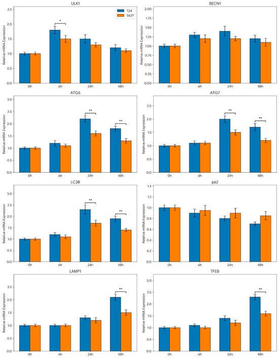
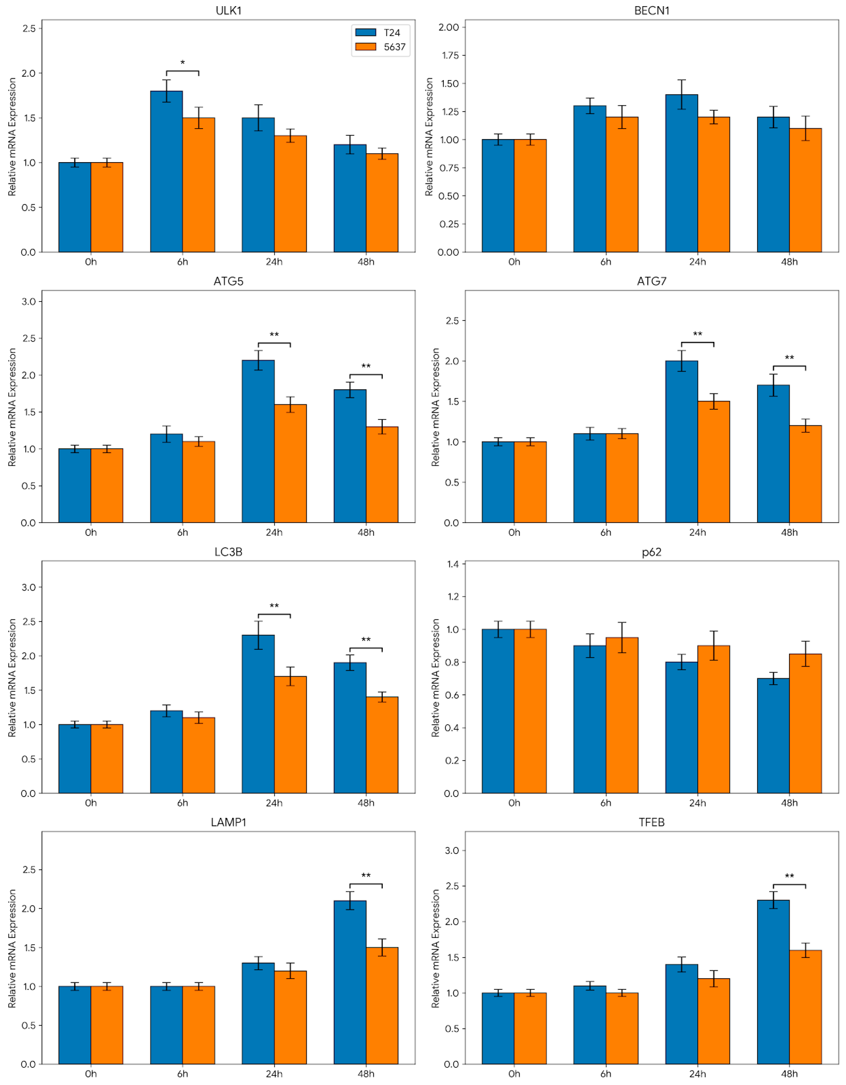
In the late phase (48 h), prominent transcriptional alterations were detected, particularly in the LAMP1 and TFEB genes associated with lysosomal regulation. These findings indicate that the autophagy–lysosome axis is heavily involved during prolonged cisplatin exposure, with downstream regulatory mechanisms coming into play in the advanced stages of the cellular response. Furthermore, the expression of the LC3B gene, a critical marker of autophagosome formation, was analyzed in a time-dependent manner, and the quantitative results are presented in Figure 4. It was determined that LC3B mRNA expression exhibited significant time-dependent changes, revealing cell-line-specific adaptive differences. While a more pronounced and sustainable expression pattern was observed in T24 cells, a more limited and transient transcriptional response was noted in 5637 cells. Overall, the findings reveal that cisplatin modulates the autophagic pathway at the mRNA level in a multi-stage manner, exhibiting distinct dynamics specific to the cell line (Figure 4).
Figure 4.
Time-dependent expression profiles of autophagy-related genes in T24 and 5637 bladder cancer cell lines following cisplatin treatment. Relative mRNA expression levels of genes involved in autophagy initiation (ULK1, BECN1), elongation (ATG5, ATG7, LC3B), selective autophagy (p62/SQSTM1), and lysosomal regulation (LAMP1, TFEB) were evaluated at 0, 6, 24, and 48 h post-treatment. Gene expression levels were calculated using the 2−ΔΔCt method and are presented as fold changes relative to the control group (0 h). Blue bars represent the T24 cell line, and orange bars represent the 5637 cell line. Data are expressed as mean ± SEM of at least three independent experiments. * p < 0.05, ** p < 0.01 indicating statistical significance between the T24 and 5637 cell lines at the corresponding time points.
3.3. Comparative Analysis Between Cell Lines
When the responses of T24 and 5637 bladder cancer cell lines to cisplatin were evaluated comparatively, prominent phenotypic and molecular differences were observed. According to the cell viability analyses, the 5637 cell line was shown to be highly sensitive to cisplatin, as evidenced by its significantly lower IC50 values (Figure 2). Conversely, the higher IC50 values of T24 cells demonstrate that this cell line exhibits a more chemoresistant phenotype (Figure 1).
The gene expression analyses strongly parallel this phenotypic divergence. A more distinct and sustainable increase in the expression of autophagy-related genes was observed in T24 cells (Figure 3 and Figure 4). Particularly, the sustained high-level expression of genes involved in the initiation and elongation stages suggests that the autophagic stress response operates as a highly organized and active process in these resistant cells. In contrast, the autophagic transcriptional response in the 5637 cell line was determined to be more limited and transient. Specifically, evaluating the time-dependent changes in LC3B and ATG genes revealed that the transcriptional drive for autophagy did not maintain continuity in this sensitive cell line, generating a fundamentally weaker adaptive response. This abbreviated response may directly contribute to the vulnerability of 5637 cells to cytotoxic stress.
Furthermore, the more pronounced upregulation of genes associated with lysosomal regulation (especially TFEB and LAMP1) in T24 cells indicates that the critical autophagy–lysosome axis functions more effectively in this cell line during prolonged stress. It is hypothesized that this molecular mechanism facilitates cellular adaptation to cisplatin, thereby promoting survival. Overall, it can be concluded that the robust and sustainable autophagic transcriptional response observed in the T24 cell line is consistent with a potential cytoprotective role at the transcriptional level, which likely contributes to the development of chemoresistance, whereas the weaker, transient autophagic activity in 5637 cells aligns with their increased sensitivity to the chemotherapeutic agent. The sustained transcriptional drive in T24 cells compared to the transient response in 5637 cells highlights a fundamental difference in how these cell lines program their long-term survival under cisplatin stress. While these transcriptional patterns provide a detailed temporal map of the genetic response to cisplatin, they represent a potential framework that warrants further validation through functional protein-level assays to confirm the complete autophagic flux.
4. Discussion
The effects of cisplatin on autophagy-related gene expression in bladder cancer cells were investigated in this research, demonstrating the dynamic, time-dependent, and phase-specific nature of this transcriptional process. The obtained results indicate that autophagy is not a static response but a temporally reprogrammed, multistage adaptive mechanism to cellular stress conditions. Particularly, the autophagic gene expression profiles that are activated in the early phase and differentiated in the late phase in response to cisplatin-induced damage highlight the critical importance of timing for optimizing therapeutic efficacy. In this regard, the increased expression of autophagy-regulatory genes such as ATG3 and ATG7 following cisplatin exposure [29], supports activation in the early stages of autophagic flux, while the expression profiles of other genes, such as ATG10, ATG14, and LC3B, may reflect autophagic adaptive responses in later stages [28]. A detailed analysis of these differentiated expression profiles can shed light on specific autophagy mechanisms associated with cisplatin resistance and provide new targets for bladder cancer therapy [30]. For example, it has been reported that increases in Beclin-1 and LC3 genes indicate a concentration- and time-dependent induction of autophagy followed by inhibition [31], underscoring the complex interactions of cisplatin with autophagic genes. In addition, the increased expression of major autophagy genes such as ATG5, ATG7, and LC3 in cisplatin combination therapies may indicate the potential of synergistic approaches to strengthen the autophagic response [32].
In this context, it is suggested that the transcriptional upregulation of autophagy-positive regulatory genes by agents such as Nectin-4-MMAE in bladder cancer cells could impact the tumor cells’ response to treatment and potentially offer new strategies for overcoming chemotherapeutic resistance [33]. These results provide evidence that the induction of autophagy likely leads to the enhancement of the cytotoxic effects of agents like Nectin-4-MMAE, thereby creating new therapeutic avenues in bladder cancer treatment [33]. Within this framework, alterations in the expression patterns of genes regulating autophagy, particularly the modulation of key effectors such as ATG5, ATG7, and LC3, may offer potential biomarkers and therapeutic targets for overcoming chemoresistance [2,16,33]. The increased expression of these genes in response to cisplatin treatment is consistent with a putative cytoprotective role of autophagy within these cancer cells [15]. Consequently, in scenarios where autophagy supports cancer cell survival, the combination therapy of autophagy inhibitors with cisplatin might result in enhanced chemotherapeutic efficacy [15].
Our current findings demonstrate that during the early phase (specifically around 6 h), there is a distinct upregulation in genes operating at the autophagy initiation steps (especially ULK1), implying the rapid activation of a cellular stress response against cisplatin. This early activation suggests that cells under cytotoxic stress prime the autophagic transcriptional program, potentially as a protective survival mechanism. In fact, early autophagic responses to chemotherapeutic agents have been shown to play a vital role in maintaining cellular homeostasis. For instance, in dichloroacetate combined cisplatin treatment, the expression of autophagy-related genes increases, supporting the premise that autophagy acts as a protective buffer against chemotherapeutic stress [32]. Thus, cisplatin-induced autophagy has the potential to increase chemoresistance in bladder cancer cells, making the targeted modulation of this pathway highly significant for future therapies [2]. Therefore, studying the role of autophagy in cancer development and investigating microRNAs related to dysregulated autophagy may aid in the development of promising therapeutic strategies [34].
During the middle phase (24 h), the upregulation of genes such as ATG5 and ATG7—which are crucial for the formation and elongation of the autophagosome—points to the progression of the autophagic process and its structurally active state. This suggests that the transcriptional drive for autophagy is not only initiated but maintained, supporting the active recycling of intracellular components. Conversely, the partial repression or stabilization of certain genes at this point implies that autophagy is not a simple linear increase but rather a tightly regulated cascade. This highlights the critical balancing role autophagy plays in cancer cell survival mechanisms, a balance that can be manipulated through strategic therapeutic interventions [11]. In the delayed phase, the reduction in specific autophagy genes raises the possibility that prolonged exposure to cisplatin might eventually disrupt autophagic flux or inhibit the process, thereby pushing the cell toward alternative death pathways, such as apoptosis. This reinforces the dynamic role of autophagy in cellular adaptation and suggests that modulating the autophagic response during long-term exposure could significantly enhance chemotherapy efficacy [29].
Late-phase (48 h) changes, particularly those related to lysosomal function and autophagy–lysosomal axis genes (LAMP1, TFEB), were highly significant. This finding indicates that during long-term cisplatin exposure, the transcriptional response is not limited to autophagosome formation; rather, lysosomal activity is heavily integrated into the adaptive process. Notably, the increased expression of TFEB suggests that lysosome biogenesis and autophagic flux are regulated in a highly coordinated manner. Consequently, the autophagy–lysosome axis emerges as a major component of cellular adaptation in the late phase, potentially driving the development of chemoresistance [12]. Hence, characterizing this dynamic progression could facilitate the development of promising treatment strategies [34]. As a master transcription factor regulating the expression of lysosomal biogenesis and autophagy genes, TFEB is highly likely to play a pivotal role in modulating this adaptive response [35,36,37]. In this regard, it has been reported that TFEB activation is associated with cisplatin resistance and may serve as a prognostic marker for chemoresistance in various malignancies, including ovarian cancer [38]. TFEB has been shown to stimulate doxorubicin resistance by promoting survival in colon and cervical cancer cells, while in glioblastoma, it alters cellular metabolism and reduces chemosensitivity [39]. Therefore, manipulating TFEB activity in bladder cancer cells by modulating the cisplatin-induced autophagic response holds considerable potential for enhancing therapeutic efficacy [38]. The dual role of TFEB in endosomal maturation and autophagy regulation is partially explained by its control over PtdIns3P levels, which affects lysosome positioning [40]. Thus, the complex interaction of TFEB between the endosomal system and autophagy presents a critical target for comprehensively understanding the bladder cancer cell response to cisplatin [40].
Comparative analysis of the cell lines revealed that 5637 cells are inherently more sensitive to cisplatin than T24 cells, as supported by their notably lower IC50 values. Concurrently, the autophagic transcriptional response in T24 cells was observed to be far more organized and sustainable, with lysosomal integration becoming particularly pronounced in the late phase. This molecular divergence suggests a correlation between a robust autophagic transcriptional response and the chemoresistant phenotype observed in specific cell lines. Conversely, in the more sensitive 5637 cells, the autophagic process lacked continuity, and lysosomal integration appeared insufficient, rendering these cells substantially more vulnerable to cytotoxic stress. This highlights the absolute necessity of developing personalized approaches in bladder cancer treatment and underscores the need for an in-depth investigation into how autophagy modulates chemotherapeutic responses. Given TFEB’s central role in regulating autophagy–lysosome biogenesis and metabolism [41,42,43], a detailed elucidation of cisplatin-induced TFEB activation mechanisms across different cell lines is vital for assessing its potential as a predictive biomarker [42,43,44]. Detailed analysis of TFEB nuclear translocation and its paracrine/autocrine regulation could yield novel strategies to overcome chemoresistance [45,46]. Furthermore, by controlling PtdIns3P levels, TFEB optimizes lysosomal degradation capacity, emerging as a key resistance mechanism [40]. Further molecular elucidation could provide a robust foundation for TFEB-targeted therapeutic agents.
These results support the well-documented notion that autophagy plays a paradoxical, dual role in cancer cells (acting as both a protective and cell-death-promoting mechanism) [47,48]. Often described as a ‘double-edged sword,’ autophagy can act as a tumor suppressor in early stages but transition into a pro-tumoral mechanism that manages metabolic stress in advanced tumors, thereby increasing chemoresistance [47,49,50]. Performing time-dependent analyses provides critical clues regarding the precise temporal phases at which autophagy should be therapeutically targeted. Early phase inhibition might reduce the cells’ ability to manage stress and lower their ‘metabolic fitness’ [48,51]; conversely, targeting lysosomal functions (e.g., with chloroquine-like agents) during the late phase can disrupt autophagic flux, leading to the lethal accumulation of cytotoxic products [52,53,54]. Understanding these kinetics and optimizing treatment protocols according to these temporal windows is emerging as a critical clinical approach for breaking chemotherapeutic resistance [55,56].
Study Limitations and Contextual Validation
While our study provides a comprehensive temporal map of autophagic gene expression, we acknowledge several critical limitations. Foremost, this study relies primarily on mRNA expression data obtained via RT-qPCR. Since transcriptional changes do not always perfectly correlate with functional protein activity or enzyme activation due to post-translational modifications [57]. the lack of Western blot validation (e.g., LC3-I to LC3-II conversion, p62 degradation) and functional autophagic flux assays (e.g., utilizing mRFP-GFP-LC3 reporters or lysosomal inhibitors like chloroquine or bafilomycin A1) limits the direct confirmation of actual autophagic executions [57,58,59,60,61]. It has been noted in the literature that chemotherapeutic agents may increase cytotoxic damage by blocking autophagic flow at the lysosomal stage rather than inducing true autophagic flux [53,55]. Furthermore, while we documented the upregulation of TFEB, we did not experimentally assess upstream regulatory events, such as mTOR/AMPK signaling alterations or TFEB nuclear translocation.
Nevertheless, our temporal transcriptional data robustly aligns with established functional models in the literature. Independent studies have previously demonstrated through Western blotting and Transmission Electron Microscopy (TEM) that cisplatin induces genuine autophagic flux in T24 and 5637 cell lines, characterized by significant LC3-II accumulation and p62 clearance [7]. Furthermore, the upstream regulation of this process via the AMPK/mTOR axis has been functionally mapped in bladder cancer models [8,9]. Our findings provide the vital temporal transcriptional blueprint that precedes and sustains these documented protein-level changes. Finally, we proposed a cytoprotective role for autophagy in the resistant T24 cell line. Although we did not perform functional rescue experiments or flow cytometry-based apoptosis assays combining cisplatin with autophagy inhibitors in this specific study, the functional causality of this mechanism is well-supported by external evidence. Data from established literature demonstrate that pharmacological inhibition (via chloroquine) or genetic silencing of essential autophagy genes (such as ATG7 or BECN1) significantly sensitize these specific bladder cancer cells to cisplatin-induced apoptosis [7]. Therefore, our temporal gene expression profiling accurately reflects the underlying genetic reprogramming that drives this established cytoprotective phenotype.
5. Conclusions
This study elucidates the time-dependent effects of cisplatin treatment on autophagy-related gene expression profiles in bladder cancer cell lines (T24 and 5637), exploring the molecular chronology of chemotherapeutic resistance. The data generated demonstrate that cisplatin-induced cellular stress triggers the transcriptional activation of key autophagy genes such as ULK1, BECN1, ATG5, and ATG7; however, the intensity and sustainability of this response depend heavily on the inherent chemoresistance profile of the cell line [62]. Specifically, in the phenotypically resistant T24 cells, the continually elevated expression of genes associated with the autophagic initiation and elongation complexes is perfectly in line with the “protective autophagy” concept [2,10]. It is highly probable that T24 cells preserve their metabolic homeostasis by employing this autophagic transcriptional program as a survival strategy against cisplatin, making this process a critical step in chemoresistance [2,4]. In contrast, the limited and transient autophagic response in 5637 cells implies that these cells remain much more vulnerable to cisplatin-induced DNA damage and oxidative stress [10,11].
Our transcriptional data, interpreted within the context of existing functional literature, supports the dual role of autophagy in cancer therapy [14,47]. From a clinical perspective, the silencing of genes such as Beclin-1, ATG5, and ATG12 has been demonstrated to sensitize bladder cancer cells to cisplatin [3,5]. In this sense, the robust genetic upregulation observed in T24 cells provides a strong theoretical basis supporting the therapeutic potential of autophagy inhibitors in combination with cisplatin, especially in chemoresistant cases [2,15]. Moreover, the marked late-phase changes in TFEB and LAMP1 further highlight lysosomal biogenesis as an integral part of chemotherapeutic adaptation [12,40]. Combining these detailed temporal genetic blueprints with functional autophagic flux analyses in future targeted studies will lay the groundwork for the development of more potent, precisely timed, and personalized treatment strategies to overcome chemoresistance in bladder cancer, provided that these transcriptional profiles are further validated in clinical settings [5,15].
Author Contributions
Conceptualization, S.Ü. and A.K.; methodology, S.Ü., V.K.A. and A.K.; software, V.K.A.; validation, S.Ü., Ö.K. and A.K.; formal analysis, S.Ü. and V.K.A.; investigation, S.Ü., V.K.A. and A.K.; resources, A.K.; data curation, S.Ü.; writing—original draft preparation, S.Ü.; writing—review and editing, G.L., M.B. and S.P.; visualization, S.Ü.; supervision, A.K.; project administration, A.K.; funding acquisition, A.K. All authors have read and agreed to the published version of the manuscript.
Funding
This work was funded by Project Contract No. BG16RFPR002-1.014-0007-C01—Personalized Innovative Medicine Competence Center-2 (PERIMED-2).
Institutional Review Board Statement
Not applicable.
Informed Consent Statement
Not applicable.
Data Availability Statement
The original contributions presented in this study are included in the article. Further inquiries can be directed to the corresponding author.
Acknowledgments
This study was supported by Pamukkale University Scientific Research Projects Coordination Unit (PAU-BAP) under project number 2025ALDEP014.
Conflicts of Interest
The authors declare no conflicts of interest.
Abbreviations
The following abbreviations are used in this manuscript:
| ATG | Autophagy-related gene |
| BECN1 | Beclin 1 |
| cDNA | Complementary DNA |
| DCA | Dichloroacetate |
| DMEM | Dulbecco’s Modified Eagle Medium |
| FBS | Fetal bovine serum |
| GAPDH | Glyceraldehyde 3-phosphate dehydrogenase |
| IC50 | Half-maximal inhibitory concentration |
| LAMP1 | Lysosomal-associated membrane protein 1 |
| LC3/LC3B | Microtubule-associated protein 1 light chain 3 |
| MMAE | Monomethyl auristatin E |
| mRFP-GFP | Monomeric red fluorescent protein-green fluorescent protein |
| MTS | 3-(4,5-dimethylthiazol-2-yl)-5-(3-carboxymethoxyphenyl)-2-(4-sulfophenyl)-2H-tetrazolium |
| PtdIns3P | Phosphatidylinositol-3-phosphate |
| RT-qPCR | Reverse transcription quantitative polymerase chain reaction |
| SQSTM1/p62 | Sequestosome 1 |
| TFEB | Transcription factor EB |
| ULK1 | Unc-51-like autophagy-activating kinase 1 |
References
- Shen, C.; Yan, Y.; Yang, S.; Wang, Z.; Wu, Z.; Li, Z.; Zhang, Z.; Lin, Y.; Li, P.; Hu, H. Construction and Validation of a Bladder Cancer Risk Model Based on Autophagy-Related Genes. Funct. Integr. Genom. 2023, 23, 46. [Google Scholar] [CrossRef] [Scilit]
- Chou, K.; Chang, A.; Hwang, T.I.-S.; Tsai, T.; Ho, C.; Liu, H.; Achudhan, D.; Tang, C.; Lin, Y. ABCC6 Transporter Contributed to Cisplatin Resistance on Bladder Cancer. Int. J. Med. Sci. 2025, 22, 3895. [Google Scholar] [CrossRef] [Scilit] [PubMed]
- Hwang, T.I.; Chen, P.-C.; Tsai, T.; Lin, J.; Chou, K.; Ho, C.; Chen, H.-E.; Chang, A. Hsa-MiR-30a-3p Overcomes the Acquired Protective Autophagy of Bladder Cancer in Chemotherapy and Suppresses Tumor Growth and Muscle Invasion. Cell Death Dis. 2022, 13, 390. [Google Scholar] [CrossRef] [Scilit]
- Jobst, M.; Kiss, E.; Gerner, C.; Marko, D.; Favero, G.D. Activation of Autophagy Triggers Mitochondrial Loss and Changes Acetylation Profile Relevant for Mechanotransduction in Bladder Cancer Cells. Arch. Toxicol. 2022, 97, 217. [Google Scholar] [CrossRef] [Scilit]
- Tantray, I.; Ojha, R.; Sharma, A. Non-Coding RNA and Autophagy: Finding Novel Ways to Improve the Diagnostic Management of Bladder Cancer. Front. Genet. 2023, 13, 1051762. [Google Scholar] [CrossRef] [Scilit] [PubMed]
- Huang, D.; Savage, S.R.; Calinawan, A.; Lin, C.; Zhang, B.; Wang, P.; Starr, T.K.; Birrer, M.J.; Paulovich, A.G. A Highly Annotated Database of Genes Associated with Platinum Resistance in Cancer. Oncogene 2021, 40, 6395. [Google Scholar] [CrossRef] [Scilit]
- Lin, J.-F.; Lin, Y.-C.; Tsai, T.-F.; Chen, H.-E.; Chou, K.-Y.; Hwang, T.I.-S. Cisplatin Induces Protective Autophagy through Activation of BECN1 in Human Bladder Cancer Cells. Drug Des. Devel. Ther. 2017, 11, 1517–1533. [Google Scholar] [CrossRef] [Scilit]
- Harhaji-Trajkovic, L.; Vilimanovich, U.; Kravic-Stevovic, T.; Bumbasirevic, V.; Trajkovic, V. AMPK-Mediated Autophagy Inhibits Apoptosis in Cisplatin-Treated Tumour Cells. J. Cell. Mol. Med. 2009, 13, 3644–3654. [Google Scholar] [CrossRef]
- Fan, Z.; Huangfu, X.; Liu, Z. Effect of Autophagy on Cisplatin-Induced Bladder Cancer Cell Apoptosis. Panminerva Med. 2017, 59, 1–8. [Google Scholar] [CrossRef] [Scilit] [PubMed]
- Sazonova, E.V.; Kopeina, G.S.; Imyanitov, E.N.; Zhivotovsky, B. Platinum Drugs and Taxanes: Can We Overcome Resistance? Cell Death Discov. 2021, 7, 155. [Google Scholar] [CrossRef] [Scilit]
- Ghasaban, F.T.; Maharati, A.; Akhlaghipour, I.; Moghbeli, M. MicroRNAs as the Critical Regulators of Autophagy-Mediated Cisplatin Response in Tumor Cells. Cancer Cell Int. 2023, 23, 80. [Google Scholar] [CrossRef] [Scilit]
- Mun, J.-Y.; Baek, S.-W.; Jeong, M.-S.; Jang, I.-H.; Lee, S.-R.; You, J.-Y.; Kim, J.; Yang, G.-E.; Choi, Y.H.; Kim, T.N.; et al. Stepwise Molecular Mechanisms Responsible for Chemoresistance in Bladder Cancer Cells. Cell Death Discov. 2022, 8, 450. [Google Scholar] [CrossRef] [Scilit] [PubMed]
- Zhang, J.; Xiang, Q.; Wu, M.; Lao, Y.; Xian, Y.; Xu, H.; Lin, Z. Autophagy Regulators in Cancer. Int. J. Mol. Sci. 2023, 24, 10944. [Google Scholar] [CrossRef] [Scilit] [PubMed]
- Kwantwi, L.B. The Dual Role of Autophagy in the Regulation of Cancer Treatment. Amino Acids 2024, 56, 7. [Google Scholar] [CrossRef] [Scilit] [PubMed]
- Wang, M.; Liu, M.; Yang, C.; Hu, Y.; Liao, X.; Liu, Q. Autophagy Modulation in Therapeutic Strategy of Breast Cancer Drug Resistance. J. Cancer 2024, 15, 5462. [Google Scholar] [CrossRef] [Scilit]
- Kumar, P.; Choudhary, A.; Kinger, S.; Jagtap, Y.A.; Prajapati, V.K.; Chitkara, D.; Chinnathambi, S.; Verma, R.K.; Mishra, A. Autophagy as a Potential Therapeutic Target in Regulating Improper Cellular Proliferation. Front. Pharmacol. 2025, 16, 1579183. [Google Scholar] [CrossRef] [Scilit]
- Mu, W.; Yao, Z.; Zhou, J.; Wang, C.; Chai, K.; Fan, Z.; Lv, G. Endoplasmic Reticulum Stress and Quality Control in Relation to Cisplatin Resistance in Tumor Cells. Front. Pharmacol. 2024, 15, 1419468. [Google Scholar] [CrossRef] [Scilit]
- Xiao, M.; Benoit, A.; Hasmim, M.; Duhem, C.; Vogin, G.; Berchem, G.; Noman, M.Z.; Janji, B. Targeting Cytoprotective Autophagy to Enhance Anticancer Therapies. Front. Oncol. 2021, 11, 626309. [Google Scholar] [CrossRef] [Scilit]
- Egan, D.F.; Shackelford, D.B.; Mihaylova, M.M.; Gelino, S.; Kohnz, R.A.; Mair, W.; Vasquez, D.S.; Joshi, A.; Gwinn, D.M.; Taylor, R.; et al. Phosphorylation of ULK1 (HATG1) by AMP-Activated Protein Kinase Connects Energy Sensing to Mitophagy. Science 2011, 331, 456–461. [Google Scholar]
- Settembre, C.; Di Malta, C.; Polito, V.A.; Garcia Arencibia, M.; Vetrini, F.; Erdin, S.; Erdin, S.U.; Huynh, T.; Medina, D.; Colella, P.; et al. TFEB Links Autophagy to Lysosomal Biogenesis. Science 2011, 332, 1429–1433. [Google Scholar] [CrossRef] [Scilit]
- Demishtein, A.; Fraiberg, M.; Berko, D.; Tirosh, B.; Elazar, Z.; Navon, A. SQSTM1/P62-Mediated Autophagy Compensates for Loss of Proteasome Polyubiquitin Recruiting Capacity. Autophagy 2017, 13, 1697–1708. [Google Scholar]
- Kirkin, V.; Lamark, T.; Sou, Y.S.; Bjørkøy, G.; Nunn, J.L.; Bruun, J.A.; Shvets, E.; McEwan, D.G.; Clausen, T.H.; Wild, P.; et al. A Role for NBR1 in Autophagosomal Degradation of Ubiquitinated Substrates. Mol. Cell 2009, 33, 505–516. [Google Scholar] [CrossRef] [Scilit] [PubMed]
- Mizushima, N.; Levine, B.; Cuervo, A.M.; Klionsky, D.J. Autophagy Fights Disease through Cellular Self-Digestion. Nature 2008, 451, 1069–1075. [Google Scholar] [CrossRef] [Scilit]
- Komatsu, M.; Waguri, S.; Ueno, T.; Iwata, J.; Murata, S.; Tanida, I.; Ezaki, J.; Mizushima, N.; Ohsumi, Y.; Uchiyama, Y. Impairment of Starvation-Induced and Constitutive Autophagy in Atg7-Deficient Mice. J. Cell Biol. 2005, 169, 425–434. [Google Scholar] [CrossRef] [Scilit]
- Kabeya, Y.; Mizushima, N.; Ueno, T.; Yamamoto, A.; Kirisako, T.; Noda, T.; Kominami, E.; Ohsumi, Y.; Yoshimori, T. LC3, a Mammalian Homologue of Yeast Apg8p, Is Localized in Autophagosome Membranes after Processing. EMBO J. 2000, 19, 5720–5728. [Google Scholar] [CrossRef] [Scilit]
- Huang, Q.; Zhan, L.; Cao, H.; Li, J.; Lyu, Y.; Guo, X.; Zhang, J.; Ji, L.; Ren, T.; An, J.; et al. Increased Mitochondrial Fission Promotes Autophagy and Hepatocellular Carcinoma Cell Survival through the ROS-Modulated Coordinated Regulation of the NFKB and TP53 Pathways. Autophagy 2016, 12, 999–1014. [Google Scholar] [CrossRef] [Scilit]
- Barber, R.D.; Harmer, D.W.; Coleman, R.A.; Clark, B.J. GAPDH as a Housekeeping Gene: Analysis of GAPDH MRNA Expression in a Panel of 72 Human Tissues. Genome Res. 2005, 21, 389–395. [Google Scholar] [CrossRef] [Scilit]
- Bordi, M.; Cegli, R.D.; Testa, B.; Nixon, R.A.; Ballabio, A.; Cecconi, F. A Gene Toolbox for Monitoring Autophagy Transcription. Cell Death Dis. 2021, 12, 1044. [Google Scholar] [CrossRef] [Scilit] [PubMed]
- Sumkhemthong, S.; Prompetchara, E.; Chanvorachote, P.; Chaotham, C. Cisplatin-Induced Hydroxyl Radicals Mediate pro-Survival Autophagy in Human Lung Cancer H460 Cells. Biol. Res. 2021, 54, 22. [Google Scholar] [CrossRef] [Scilit]
- Danish, F.; Qureshi, M.A.; Mirza, T.; Amin, W.; Sufiyan, S.; Naeem, S.; Arshad, F.; Mughal, N. Investigating the Association between the Autophagy Markers LC3B, SQSTM1/P62, and DRAM and Autophagy-Related Genes in Glioma. Int. J. Mol. Sci. 2024, 25, 572. [Google Scholar] [CrossRef] [Scilit] [PubMed]
- Alinaghi, M.; Mokarram, P.; Ahmadi, M.; Bozorg-ghalati, F. Biosynthesis of Palladium, Platinum, and Their Bimetallic Nanoparticles Using Rosemary and Ginseng Herbal Plants: Evaluation of Anticancer Activity. Sci. Rep. 2024, 14, 5798. [Google Scholar] [CrossRef] [Scilit]
- Qin, H.; Ge, Z.; Li, Q.; Shen, L. Metabolic Reprogramming Induced by DCA Enhances Cisplatin Sensitivity through Increasing Mitochondrial Oxidative Stress in Cholangiocarcinoma. Front. Pharmacol. 2023, 14, 1128312. [Google Scholar] [CrossRef] [Scilit]
- Wang, Y.; Nan, Y.; Ma, C.; Lü, X.; Wang, Q.; Huang, X.; Xue, W.; Fan, J.; Ju, D.; Ye, D.; et al. A Potential Strategy for Bladder Cancer Treatment: Inhibiting Autophagy to Enhance Antitumor Effects of Nectin-4-MMAE. Cell Death Dis. 2024, 15, 293. [Google Scholar] [CrossRef] [Scilit]
- Mao, C.; Xu, X.; Ding, Y.; Xu, N. Optimization of BCG Therapy Targeting Neutrophil Extracellular Traps, Autophagy, and MiRNAs in Bladder Cancer: Implications for Personalized Medicine. Front. Med. 2021, 8, 735590. [Google Scholar] [CrossRef] [Scilit]
- Boretto, C.; Actis, C.; Faris, P.; Cordero, F.; Beccuti, M.; Ferrero, G.; Muzio, G.; Moccia, F.; Autelli, R. Tamoxifen Activates Transcription Factor EB and Triggers Protective Autophagy in Breast Cancer Cells by Inducing Lysosomal Calcium Release: A Gateway to the Onset of Endocrine Resistance. Int. J. Mol. Sci. 2023, 25, 458. [Google Scholar] [CrossRef] [Scilit]
- Casey, A.; Liu, W.; Hein, L.K.; Sargeant, T.J.; Pederson, S.; Mäkinen, V. Transcriptional Targets of Senataxin and E2 Promoter Binding Factors Are Associated with Neuro-Degenerative Pathways during Increased Autophagic Flux. Sci. Rep. 2022, 12, 17665. [Google Scholar] [CrossRef] [Scilit]
- Frangež, Ž.; Gerard, D.; He, Z.; Gavriil, M.; Fernández-Marrero, Y.; Jafari, S.M.S.; Hunger, R.E.; Lucarelli, P.; Yousefi, S.; Sauter, T.; et al. ATG5 and ATG7 Expression Levels Are Reduced in Cutaneous Melanoma and Regulated by NRF1. Front. Oncol. 2021, 11, 721624. [Google Scholar] [CrossRef] [Scilit] [PubMed]
- Liu, W.; Wang, Y.; Xie, Y.; Dai, T.; Fan, M.; Li, C.; Zou, Y. Cisplatin Remodels the Tumor Immune Microenvironment via the Transcription Factor EB in Ovarian Cancer. Cell Death Discov. 2021, 7, 136. [Google Scholar] [CrossRef] [Scilit]
- Zhang, Y.; Ji, Y.; Tu, Y.; Li, Y. Autophagy in Doxorubicin Resistance: Basic Concepts, Therapeutic Perspectives and Clinical Translation. Front. Immunol. 2025, 16, 1642050. [Google Scholar] [CrossRef] [Scilit] [PubMed]
- Mathur, P.; Santos, C.D.B.; Lachuer, H.; Patat, J.; Latgé, B.; Radvanyi, F.; Goud, B.; Schauer, K. Transcription Factor EB Regulates Phosphatidylinositol-3-Phosphate Levels That Control Lysosome Positioning in the Bladder Cancer Model. Commun. Biol. 2023, 6, 114. [Google Scholar] [CrossRef] [Scilit] [PubMed]
- Gebrie, A. Transcription Factor EB as a Key Molecular Factor in Human Health and Its Implication in Diseases. SAGE Open Med. 2023, 11, 20503121231157209. [Google Scholar] [CrossRef] [Scilit]
- Tan, A.; Prasad, R.; Lee, C.; Jho, E. Past, Present, and Future Perspectives of Transcription Factor EB (TFEB): Mechanisms of Regulation and Association with Disease. Cell Death Differ. 2022, 29, 1433. [Google Scholar] [CrossRef] [Scilit] [PubMed]
- Wang, T.; Qin, Y.; Zeng, Y.; Jing, D.; Fan, G.; Liu, M.; Zhuo, Q.; Ji, S.; Chen, X.; Yu, X.; et al. A New Glance at Autophagolysosomal-Dependent or -Independent Function of Transcriptional Factor EB in Human Cancer. Acta Pharmacol. Sin. 2023, 44, 1536. [Google Scholar] [CrossRef] [Scilit]
- Zheng, B.; Qian, F.; Wang, X.; Wang, Y.; Zhou, B.; Fang, L. Neddylation Activated TRIM25 Desensitizes Triple-Negative Breast Cancer to Paclitaxel via TFEB-Mediated Autophagy. J. Exp. Clin. Cancer Res. 2024, 43, 177. [Google Scholar] [CrossRef] [Scilit]
- Gagliardi, S.; Mitruccio, M.; Corato, R.D.; Romanò, R.; Aloisi, A.; Rinaldi, R.; Alifano, P.; Guerra, F.; Bucci, C. Defects of Mitochondria-Lysosomes Communication Induce Secretion of Mitochondria-Derived Vesicles and Drive Chemoresistance in Ovarian Cancer Cells. Cell Commun. Signal. 2024, 22, 165. [Google Scholar] [CrossRef] [Scilit] [PubMed]
- Franco-Juárez, B.; Coronel-Cruz, C.; Hernández-Ochoa, B.; Gómez-Manzo, S.; Cárdenas-Rodríguez, N.; Arreguín-Espinosa, R.; Bandala, C.; Canseco-Ávila, L.M.; Ortega-Cuéllar, D. TFEB; Beyond Its Role as an Autophagy and Lysosomes Regulator. Cells 2022, 11, 3153. [Google Scholar] [CrossRef] [Scilit]
- Chavez-Dominguez, R.; Perez-Medina, M.; López-González, J.S.; Galicia-Velasco, M.; Aguilar-Cázares, D. The Double-Edge Sword of Autophagy in Cancer: From Tumor Suppression to Pro-Tumor Activity. Front. Oncol. 2020, 10, 578418. [Google Scholar] [CrossRef] [Scilit]
- White, E. The Role for Autophagy in Cancer. J. Clin. Investig. 2015, 125, 42. [Google Scholar] [CrossRef] [Scilit]
- Alvarez-Meythaler, J.G.; García-Mayea, Y.; Mir, C.; Kondoh, H.; Lleonart, M.E. Autophagy Takes Center Stage as a Possible Cancer Hallmark. Front. Oncol. 2020, 10, 586069. [Google Scholar] [CrossRef] [Scilit]
- Amaravadi, R.K.; Kimmelman, A.C.; White, E. Recent Insights into the Function of Autophagy in Cancer. Genes Dev. 2016, 30, 1913. [Google Scholar] [CrossRef] [Scilit] [PubMed]
- Jandrey, E.H.F.; Bezerra, M.; Inoue, L.T.; Furnari, F.B.; Camargo, A.A.; Costa, É.T. A Key Pathway to Cancer Resilience: The Role of Autophagy in Glioblastomas. Front. Oncol. 2021, 11, 652133. [Google Scholar] [CrossRef] [Scilit]
- Abbruzzese, C.; Catalogna, G.; Gallo, E.; Martino, S.D.; Mileo, A.M.; Carosi, M.; Dattilo, V.; Schenone, S.; Musumeci, F.; Lavia, P.; et al. The Small Molecule SI113 Synergizes with Mitotic Spindle Poisons in Arresting the Growth of Human Glioblastoma Multiforme. Oncotarget 2017, 8, 110743. [Google Scholar] [CrossRef] [Scilit] [PubMed]
- Golden, E.B.; Cho, H.; Jahanian, A.; Hofman, F.M.; Louie, S.G.; Schönthal, A.H.; Chen, T.C. Chloroquine Enhances Temozolomide Cytotoxicity in Malignant Gliomas by Blocking Autophagy. Neurosurg. Focus 2014, 37, E12. [Google Scholar] [CrossRef] [Scilit]
- Matteoni, S.; Abbruzzese, C.; Matarrese, P.; Luca, G.D.; Mileo, A.M.; Miccadei, S.; Schenone, S.; Musumeci, F.; Haas, T.L.; Sette, G.; et al. The Kinase Inhibitor SI113 Induces Autophagy and Synergizes with Quinacrine in Hindering the Growth of Human Glioblastoma Multiforme Cells. J. Exp. Clin. Cancer Res. 2019, 38, 202. [Google Scholar] [CrossRef] [Scilit]
- Hwang, Y.K.; Lee, D.-H.; Lee, E.C.; Oh, J.S. Importance of Autophagy Regulation in Glioblastoma with Temozolomide Resistance. Cells 2024, 13, 1332. [Google Scholar] [CrossRef] [Scilit]
- Jawhari, S.; Ratinaud, M.; Verdier, M. Glioblastoma, Hypoxia and Autophagy: A Survival-Prone ‘ménage-à-Trois’. Cell Death Dis. 2016, 7, e2434. [Google Scholar] [CrossRef] [Scilit]
- Yin, Z.; Pascual, C.; Klionsky, D.J. Autophagy: Machinery and Regulation. Microb. Cell 2016, 3, 588. [Google Scholar] [CrossRef] [Scilit]
- Ichimura, Y.; Komatsu, M. Pathophysiological Role of Autophagy: Lesson from Autophagy-Deficient Mouse Models. Exp. Anim. 2011, 60, 329. [Google Scholar] [CrossRef] [Scilit] [PubMed]
- Tamrakar, S.; Yashiro, M.; Kawashima, T.; Uda, T.; Terakawa, Y.; Kuwae, Y.; Ohsawa, M.; Ohata, K. Clinicopathological Significance of Autophagy-Related Proteins and Its Association With Genetic Alterations in Gliomas. Anticancer Res. 2019, 39, 1233. [Google Scholar] [CrossRef] [Scilit] [PubMed]
- Zhu, L.; Wang, Y.; He, J.; Tang, J.; Lv, W.; Hu, J. Cytoplasmic SQSTM1/P62 Accumulation Predicates a Poor Prognosis in Patients with Malignant Tumor. J. Cancer 2018, 9, 4072. [Google Scholar] [CrossRef] [Scilit]
- Mizushima, N. Autophagy in Protein and Organelle Turnover. Cold Spring Harb. Symp. Quant. Biol. 2011, 76, 397. [Google Scholar] [CrossRef] [Scilit] [PubMed]
- Lee, X.C.; Werner, E.; Falasca, M. Molecular Mechanism of Autophagy and Its Regulation by Cannabinoids in Cancer. Cancers 2021, 13, 1211. [Google Scholar] [CrossRef] [Scilit] [PubMed]

Disclaimer/Publisher’s Note: The statements, opinions and data contained in all publications are solely those of the individual author(s) and contributor(s) and not of MDPI and/or the editor(s). MDPI and/or the editor(s) disclaim responsibility for any injury to people or property resulting from any ideas, methods, instructions or products referred to in the content. |
© 2026 by the authors. Licensee MDPI, Basel, Switzerland. This article is an open access article distributed under the terms and conditions of the Creative Commons Attribution (CC BY) license.