


Abstract
Gastrointestinal (GI) cancers represent 26% of all cancer cases worldwide and are responsible for 35% of cancer deaths. These types of cancer have significant clinical challenges due to late detection and treatment resistance. The gut microbiome is a complex and adaptable ecosystem crucial for metabolism, intestinal barrier maintenance, and immune function, and establishes a strong two-way interaction within the host. Tumors affect both local and systemic microbial environments, and dysbiosis, an imbalance in microbiota, contributes to cancer development, immune evasion, and progression across different GI regions. In upper GI malignancies, microbial species such as Fusobacterium nucleatum and Porphyromonas gingivalis promote chronic inflammation, while gastric carcinogenesis develops from Helicobacter pylori-induced atrophy through secondary colonization by bacteria such as Streptococcus anginosus. In hepatopancreatobiliary and colorectal cancers, barrier disruption allows bacteria and lipopolysaccharides to translocate, disrupts bile acid metabolism, and causes genotoxic damage through agents like colibactin. These are just a few examples covered in this review. We covered a comprehensive and up to date overview of the major gut microbial communities associated with GI cancers and their role in oncogenesis. Additionally, the gut-derived microbial communities play an active role in cancer treatment outcomes. The microbiome influences patient responses to and side effects of chemotherapy, radiotherapy, and immune checkpoint blockades through drug metabolism, modulation of mucosal injury, and immune remodeling. Strategies like prebiotics, probiotics, and fecal microbiota transplantation (FMT) offer promising approaches to reduce treatment-related toxicities and overcome therapeutic resistance. These strategies are under active evaluation to address therapeutic resistance and reduce toxicity, highlighting the potential of microbiome-directed cancer medicine.
1. Introduction
Gastrointestinal (GI) cancers encompass a diverse group of malignancies arising throughout the digestive tract and associated organs. Unfortunately, GI cancers collectively account for 26% of global cancer incidence and 35% of cancer-related deaths [1], representing more than 5 million new cases and 3.4 million cancer deaths in 2020. Their clinical burden is compounded by nonspecific symptoms that can delay diagnosis and multiple universal risk factors like age, sex, obesity, infection, smoking, alcohol, and diet [2].
The gut microbiome is an ecosystem of microorganisms (bacteria, viruses, fungi, and archaea) that live within the GI tract. It is generally believed that the gut microbiome forms following colonization of the GI tract after birth. After 2.5 years of age, an infant’s gut microbiome displays the diversity and functional capacity similar to those of an adult [3]. A wide variety of factors influence the composition of the gut microbiome, including perinatal factors like mode of delivery or infant feeding and factors across the lifespan like genetics, infections, medication, and diet [4].
Notably, studies have indicated that the relationship between gut microbiome and GI cancers is bidirectional. While GI malignancies can alter the gut microbiome composition, the changes in gut microbiome may also contribute to tumorigenesis, treatment resistance, and immune dysregulation [5]. The numerous functions of the gut microbiome in digestion and metabolite synthesis, immune regulation, intestinal epithelial barrier maintenance, and pharmacokinetics are being investigated as potential mechanisms underlying the gut microbiome’s impact on GI cancers [6].
Collectively, the evidence highlights the crucial role that the gut microbiome plays in all aspects of GI cancer management. Although we have referred to GI cancers as a collective, primary tumors along the GI tract are uniquely influenced by the host gut microbiome and the local microbiome. This review differs from reviews centered on a single GI malignancy or a single domain of microbiome biology by providing a comprehensive overview of four interconnected domains relevant to GI malignancies: cancer-associated microbial composition, proposed oncogenic mechanisms, associated microbial metabolites, and therapeutic implications. By comparing these domains across individual GI cancer types, we aim to highlight cross-cutting patterns and potential microbiome-based opportunities for cancer prevention, diagnosis, and treatment.
2. Methodology
The narrative review was developed through a targeted literature search conducted using PubMed for published studies and ClinicalTrials.gov for relevant ongoing trials. The literature search was last updated in October 2026. Search terms were specific to each topic and combined cancer-specific terms with terms related to gut, oral, mucosal and tumor microbiomes; dysbiosis; microbial metabolites, carcinogenesis; treatment response; treatment toxicity, chemotherapy; radiotherapy; immune checkpoint blockade; and microbiome-directed therapies.
Search was focused on studies published from 1992 through 2026, and older sentinel studies were included to provide foundational mechanistic or clinical context. The literature was selected primarily based on its topical relevance to the microbiome, cancer-associated mechanisms, and clinical or therapeutic implications in GI malignancies. Although peer-reviewed studies were prioritized, selected conference reports were included when direct evidence was limited. Evidence from non-GI malignancies was considered in treatment-focused sections when GI-specific data were sparse.
Given the narrative nature of this review, literature selection was not conducted through a prespecified screening protocol, and formal counts of records were not maintained. As no quantitative analysis was performed, potential overlap between study populations was not systematically assessed. While literature selection was guided by topical relevance, evidence discussed in Section 6 was additionally evaluated in the context of study design, sample size, mechanistic evidence, and clinical applicability. No standardized risk-of-bias assessment or formal evidence-grading framework was applied. By integrating site-specific microbial mechanisms with clinical and therapeutic implications, this review aims to provide an integrated understanding of the microbiome role across the GI cancer continuum.
3. Role of Gut Microbiome in Upper GI Cancer
3.1. Esophageal Cancer
The two major types of esophageal cancer are squamous cell carcinoma (ESCC) and esophageal adenocarcinoma (EAC). These tumors have characteristic geographic distributions and microbial patterns. This warrants an independent understanding of the microbial signature of these subtypes.
3.1.1. Oral and Esophageal Microbiome Alterations Associated with Esophageal Cancer
The most common organisms associated with ESCC are F. nucleatum and P. gingivalis, along with other reported taxa, including Prevotella, Veillonella, Streptococcus, Actinomyces, Peptostreptococcus, and members of the family Enterobacteriaceae. Even though specific microbial signatures vary across cohorts and geographical regions, most analyses (paired tumor and adjacent tumor analysis) identified increased Fusobacterium and reduced Streptococcus populations within ESCC [7]. Tumor-based studies showed P. gingivalis is abundantly detected in ESCC tissue compared to healthy tissue. Gao et al. showed this number to be 61% in ESCC specimens compared to 12% in adjacent tissues [8]. A higher abundance was associated with poor prognosis. Similarly, Yamamura et al. demonstrated that the presence of F. nucleatum DNA is linked to poor survival and increased expression of the chemokine CCL20 [9], and an increased risk of ESCC metastasis [10].
Prospective studies have shown that microbiome alterations precede a cancer diagnosis. Peters et al. analyzed prediagnostic mouthwash samples to examine an association between baseline oral microbiome and risk of ESCC and EAC [11]. P. gingivalis was more abundant in the intratumoral region compared to adjacent tissues and was not detected in the healthy control tissue [8]. In Peters et al., the association between oral P. gingivalis and ESCC risk did not reach statistical significance, potentially owing to the limited number of incident ESCC cases. Based on microbial profiling of distal esophageal biopsies, gastroesophageal reflux disease (GERD) creates an ecological niche at the distal esophagus where the normal flora consisting of a Gram-positive, Streptococcus-rich community shifts to Gram-negative anaerobic organisms [12]. Organisms like Prevotella, Veillonella and Leptotrichia are enriched in esophageal tumor biopsies from patients with EAC [13]. These results are not consistent across cohorts. Peters et al. described that oral Tannerella forsythia abundance was associated with increased risk of EAC [11]. This study may reflect systemic exposure, periodontal health, etc., as the samples were collected orally rather than esophageally. Deshpande et al., who analyzed esophageal biopsies and saliva, reported Campylobacter enrichment in the esophageal microbiome of patients with Barrett’s esophagus. They integrated microbial profiling with host transcriptomic data to showcase the association between Campylobacter abundance, altered host gene response, and macrophage responses that changed according to bacterial isolates [14] but did not establish Campylobacter as an independent cause of EAC. Oral and esophageal microbial findings alone do not establish an intestinal origin or contribution.
3.1.2. Role of Gut Microbiota and Their Metabolites in Immune Modulation and Esophageal Carcinogenesis
Experimental studies suggest that F. nucleatum enters ESCC cells and promotes proliferation, migration, and invasion. Additionally, the relation between F. nucleatum and CCL20 expression suggests that the organism may influence the recruitment of immunoregulatory cells [9]. P. gingivalis has been shown to increase tumor cell proliferation and migration, and activate pathways related to NF-κB signaling. It also helps malignant cells to tolerate therapeutic stress by enhancing mitochondrial oxidative phosphorylation [15]. Clinically, positive P. gingivalis has been associated with poor prognosis. A cohort of patients with ESCC receiving neoadjuvant chemoimmunotherapy showed that tumors enriched in Streptococcus exhibited greater numbers of granzyme B-positive CD8+ T cells and fewer regulatory T cells (Tregs), which conferred a longer disease-free survival [16]. These human tumor-associated findings are observational and need further study before they can be used as a therapeutic intervention.
Putrescine, a polyamine involved in cellular proliferation, is increased following F. nucleatum infection and promotes ESCC cell proliferation [17]. Overall, ESCC is associated with colonization by oral pathogens that may contribute to the inflammatory signaling, immune remodeling, and treatment resistance. As of now, no organisms have been consistently implicated in ESCC carcinogenesis. EAC evidence is strongly centered on Barrett’s-associated microbial alterations rather than on established mechanistic causality.
3.2. Gastric Cancer
Gastric cancer and its relationship with microorganisms are understood as a continuous and cooperative process. Helicobacter pylori are established as a principal causative microorganism through initiation of chronic gastritis leading to progressive mucosal injury. The gastric atrophy and hypochlorhydria that follow provide a niche environment for certain oral and intestinal flora to flourish. These secondary microbial communities may facilitate inflammation and immune dysfunction. Thus, gastric carcinogenesis involves the direct oncogenic effect of H. pylori and indirect changes brought by the gastric microbiome [18].
3.2.1. Gut Microbiome Alterations Associated with Gastric Cancer
As chronic gastritis continues, the loss of acid-producing cells leads to an increase in gastric pH. This creates a niche that supports the growth of microorganisms that usually wouldn’t persist in the gastric mucosa. The gastric cancer-associated community generally has a reduced relative abundance of Helicobacter. Instead, there is an abundance of intestinal and oral taxa, which include Streptococcus, Lactobacillus, Prevotella, Veillonella, Fusobacterium, Parvimonas, Peptostreptococcus, Dialister, and Slackia. Coker et al. identified and validated a five-taxon signature comprising Peptostreptococcus stomatis, Streptococcus anginosus, Parvimonas micra, Slackia exigua, and Dialister pneumosintes, which distinguished gastric cancer from superficial gastritis with an area under the curve of approximately 0.82 [19]. Even though it is not established that each organism causally contributes to carcinogenesis, the functional importance of a broader dysbiotic microbial community independent of H. pylori is shown in experimental models. In H. pylori-infected INS-GAS mice, colonization with a restricted commensal microbiota increased the rate of gastritis compared to germ-free control mice [20]. These experiments confirm the role of non-H. pylori organisms as a co-promoter. Choi et al. showed that in patients who underwent endoscopic resection of early gastric cancer and eradication therapy, there was a reduction in the incidence of metachronous gastric cancer, and it also improved gastric glandular atrophy [21]. But this finding doesn’t completely eradicate the risk of cancer, especially in cases where there is extensive atrophy or intestinal metaplasia. This suggests that, beyond a point, premalignant epithelial and microbial changes persist despite removal of the organism.
3.2.2. Microbiome-Mediated Effects on Immunity and Oncogenic Pathways in Gastric Cancer
Persistent H. pylori infection leads to the histological progression from chronic active gastritis to glandular atrophy, metaplasia, dysplasia, and gastric adenocarcinoma [22]. The risk factors for this are bacterial virulence factors, host genetics, environmental exposures, gastritis and the severity of the gastritis. Among the bacterial virulence factors, Cytotoxin-associated gene A protein (CagA) has been shown to have the most mechanistic association with carcinogenesis. CagA-positive H. pylori strains deliver CagA into gastric epithelial cells using a type IV secretion system. This is phosphorylated by host kinases and interacts with SHP-2 tyrosine phosphatase and dysregulates downstream ERK/MAPK signaling, cellular proliferation, and epithelial modification. The oncogenic potential of CagA is also shown in a transgenic mouse model where CagA expression induced gastrointestinal and hematopoietic neoplasms [23]. Other H. pylori products include LPS, cytotoxin A, peptidoglycan, and outer membrane proteins, which take part in epithelial injury and bacterial persistence. Chronic infection activates NF-κB and results in the release of IL-8, which results in sustained recruitment of inflammatory cells like neutrophils, lymphocytes, and macrophages. Reactive oxygen species (ROS) and nitric oxide (NO) produced due to inflammation cause damage to DNA and cause genomic instability [24]. Tregs also contribute to limiting bacterial clearance. Among the non-H. pylori organisms associated with gastric cancer, S. anginosus has the most extensive mechanistic study. Fu et al. showed that S. anginosus was enriched in gastric lesions, and its colonization in conventional and germ-free mice led to chronic inflammation, atrophy, and tumorigenic changes. MAPK signaling was activated by the interaction of bacterial surface protein TMPC with host annexin A2 [25].
3.2.3. Microbial Metabolites Associated with Gastric Cancer
A separate analysis of 609 samples from 335 patients showed that Streptococcal dominant disease is associated with adverse clinical outcomes. The clinical isolate S. anginosus_ZJCC consumes arginine and produces ornithine through the arginine deiminase pathway. This creates an unfavorable condition for anti-tumor T cell activity [26]. In a separate study, Zhou et al. linked S. anginosus-derived methionine to gastric cancer progression using ATCC 33397, tumor-derived isolates RJ1-RJ3, and metE-deficient bacteria [27]. Separately, Liao et al. showed that S. anginosus-derived succinate activates SUCNR1–ABRAXAS1–PI3K/AKT signaling in experimental models [28]. Human tumor associations alone do not establish whether bacterial metabolites reach biologically effective concentrations. These mechanisms were studied separately and should not be assumed to coexist in every S. anginosus strain. The most coherent model of gastric carcinogenesis initiated by H. pylori involves chronic inflammation and oncogenic signaling, which lead to the remodeling of the local habitat through reduced gastric acidity and progressive atrophy. The altered oral and intestinal microbial community that colonizes adds inflammatory, metabolic, and immunosuppressive pressure. Among these secondary organisms, S. anginosus is an emerging mechanistic candidate. Further investigation incorporating diet and proton pump inhibitor usage, longitudinal evaluation and microbiome-based intervention are required to distinguish causal organisms.
4. Role of Gut Microbiome in Hepatobiliary Cancers
Hepatobiliary cancers remain a major clinical challenge due to their aggressive biological behavior, high propensity for metastasis, and limited response to conventional treatment modalities [29]. Hepatobiliary cancers broadly consist of hepatocellular carcinoma (HCC) and cholangiocarcinoma (CCA), with HCC accounting for approximately 80–90% of cases [30]. Although CCA is less prevalent (~2% of global cancer-related deaths annually), it is one of the most aggressive hepatobiliary cancers [31]. The pathophysiology of HCC and CCA involves complex processes characterized by genomic alterations, immune dysfunction, metabolic remodeling, oncogenic signaling, epigenetic regulation, and other factors. In addition to conventional risk factors, growing evidence now suggests that microbiota and its metabolites have an important role in initiation, progression, and therapeutic response of hepatobiliary cancers.
4.1. Hepatocellular Carcinoma
4.1.1. Gut Microbiome Alterations Associated with Hepatocellular Carcinoma
Distinct microbial signatures have been observed across different stages of HCC. Specifically, the enrichment of Lachnospiraceae incertae sedis, Bacteroides, and Clostridium cluster XlVa has been observed in patients with high tumor burden, while Actinobacteria has been seen in the early stages of HCC [32]. This evidence supports the notion that specific microbial communities may play an important role in the initiation and progression of HCC. Also, several studies suggest that HCC patients exhibit a distinct gut microbiota dysbiosis profile [33]. Notably, significant differences in gut microbial composition have been observed between HCC and non-HCC individuals, irrespective of cirrhosis status [34]. While identified bacterial taxa vary across studies, a broader dysbiotic phenotype remains consistent. Interestingly, some genera appear to change dynamically with the progression of disease [35]. For example, an increased abundance of Clostridium has been observed during HCC progression. In addition, other Firmicutes, including Lactobacillus and Streptococcus, have similarly been found to be increased in the gut of HCC patients [36]. Similarly, Ruminococcus is enriched in patients with HCC compared to other liver diseases [32,35,37]. Moreover, studies suggest that Ruminococcus abundance varies with tumor characteristics. Interestingly, its enrichment has been observed among HCC patients without liver microvascular invasion [38]. Furthermore, the members of the genus Enterococcus are also enriched in HCC [39,40]. Enterobacter ludwigii has been reported to be approximately 100-fold more abundant among HCC patients, displaying potential as a diagnostic biomarker [41]. Decreased abundance of Bifidobacterium and Actinobacteria in the gut has been associated with increasing HCC severity [42]. Other genera including Atopobium and Gemmiger have been reported to be enriched in the gut of HCC patients [36,43]. Among the phylum Bacteroidetes, Bacteroides, Desulfovibrio, and Parabacteroides are reported to be enriched in fecal samples of HCC patients [35,40]. Similarly, alterations in Proteobacteria have been observed in HCC. Within the phylum, an increased abundance of Escherichia coli and Shigella has also been observed [36,38]. In contrast, several bacterial species with beneficial functions, including Faecalibacterium, Prevotella, Akkermansia, and Subdoligranulum, are reduced in the gut of HCC patients [44]. In agreement with the above findings, a study by Yang et al. involving fecal samples from 364 hepatitis B virus (HBV)-associated HCC patients showed enrichment of Streptococcus and Escherichia, and Shigella in HCC patients compared to healthy controls, whereas Agathobacter, a genus from the phylum Firmicutes, was depleted markedly [38]. Li et al. further demonstrated HCC-related changes in the gut microbial composition by comparing 68 HCC patients with 33 liver cirrhosis (LC) and 34 normal controls. The study reported that 21 genera, including Ruminococcus, Roseburia, and Lachnospira, were abundant in HCC patients; conversely, 42 genera, including Phascolarctobacterium, Alistipes, Veillonella, and Faecalibacterium, were markedly reduced in HCC. In addition, approximately 35 species, including Lachnospira eligens, Phocaeicola vulgatus, Ruminococcus bicirculans, and Bacteroides uniformis, differed between HCC and LC. Compared to normal controls, HCC patients showed increased abundance of several Veillonella species including V. dispar, V. atypica, V. parvula, and V. T1-7 sp., whereas species such as Faecalibacterium, Phocaeicola dorei, and Bacteroides uniformis were decreased [37]. Therefore, HCC-associated distinct microbial alterations highlight the potential of the gut microbiome as a biomarker for HCC detection. Notably, Ruminococcus bicirculans and Odoribacter splanchnicus have been proposed as potential species-level biomarkers, while a combined assessment of Coprococcus, Coriobacterium and Atopobium showed considerable diagnostic potential at the genus level. Accumulating evidence suggests that gut microbial signatures vary according to disease progression and tumor stage [37]. Ren et al. identified approximately 13 genera, including Gemmiger and Parabacteroides, which were significantly enriched in early HCC patients, suggesting their potential use in early HCC diagnosis [43]. Another study by Zhang et al. reported higher abundance of Enterobacteriaceae and Enterococcus, and lower abundance of Bifidobacterium and Actinobacteria in patients with HCC. In addition, they also found that these alterations were significantly associated with more advanced HCC stages and poor prognosis, further suggesting that gut dysbiosis may be linked to HCC progression and clinical outcomes [39]. Moreover, the abundance of Streptococcus, Shigella, and Escherichia increased progressively with HCC progression and may therefore serve as a potential biomarker for HCC progression [38]. Studies by Ni et al. reported that pro-inflammatory bacteria were significantly enriched in HCC patients compared to healthy controls. They also proposed the degree of dysbiosis (Ddys) as a quantitative measure of microbial imbalance. According to their findings, the Ddys was significantly elevated in HCC patients and showed a gradual increase across HCC stages [45]. Subsequently, Huang et al. examined the association between gut microbiota and the expression of genes associated with tumor progression in 100 healthy controls and 113 HBV-related HCC patients. They found an increased abundance of Lachnospiraceae incertae sedis accompanied by decreased CD6 expression, whereas increased Bacteroides abundance was associated with lower MAPK10 expression [32]. Both patterns are associated with poor disease-free survival. These associations are identified through transcriptomic analysis but do not establish a direct causal relationship between gene expression, microbial abundance and survival outcomes. Further functional studies may be required to elucidate the underlying mechanisms and to validate the prognostic significance of these associations.
The origin of intratumoral microbiota remains poorly understood; however, current evidence suggests that it may arise from the biliary tract, hepatic circulation, or adjacent tissues [46]. During HCC development, increased gut dysbiosis and intestinal permeability may facilitate bacterial translocation through mucosal and mesenteric vasculature into the bloodstream [47]. The liver is continuously exposed to gut-derived microbial communities through the portal vein. Therefore, this anatomical connection raises the possibility that intratumoral bacteria may originate from the gut and subsequently contribute to tumor progression through interactions within the hepatic tumor microenvironment (TME) [48,49,50]. Accumulating evidence now suggests that HCC exhibits a distinct intratumoral microbial signature that differs from those of other cancers [46,51]. Studies by Sun et al. reported distinct shifts in the HCC-associated intratumoral microbiome compared to normal tissue which showed a significant reduction in Pseudomonas abundance, whereas Agrobacterium and Rhizobiaceae were significantly enriched in tumor tissues [52]. Another study by Huang et al. showed a higher abundance of Proteobacteria, Actinobacteriota, and Firmicutes, together with a lower abundance of Acidobacteriota and Patescibacteria at the phylum level. Notably, Bacilli, Actinobacteria, and Gammaproteobacteria were enriched in HCC tissues at the class level [46]. He et al. further characterized the intratumoral microbiome in HCC. Neisseria, Fusobacterium, and Enterobacteriaceae were significantly enriched in HCC tissues; however, the abundance of Pseudomonas was decreased. However, Proteobacteria showed a modest increase, while Actinobacteriota was significantly reduced in HCC tissues. Moreover, the abundance of Faecalibacterium, Dietzia, Hydrogenophaga, Megamonas, Chryseobacterium, Agathobacter, and Ruminococcaceae decreased significantly, whereas Fusobacterium, Neisseria, Lactobacillus, and Clostridia-UCG-014 were enriched in HCC tissues compared to normal tissues [51]. Interestingly, Sun et al. demonstrated enrichment of Actinobacteria within HCC tissues. A similar pattern has been observed among Firmicutes and Proteobacteria, both of which are reported to increase in the gut during HCC and were also elevated in HCC tissues. Furthermore, Enterobacteriaceae, considered a characteristic feature of HCC-associated gut dysbiosis, was also enriched in HCC tissues [52]. Collectively, these observations support a possible link between gut microbial dysbiosis and the establishment of the HCC intratumoral microbiome. Therefore, emerging evidence now reveals a compelling microbial fingerprint shared across gut and tumor tissue. The beneficial taxa, Faecalibacterium, members of the Ruminococcaceae family, and Agathobacter, are significantly depleted in both gut and HCC tissue, whereas Lactobacillus is consistently enriched [36,37,38,51]. This striking overlap suggests a profound gut–tumor microbial axis in HCC, positioning microbial dysbiosis as a potentially important feature. Therefore, the microbiome may represent a hidden link between the gut and the HCC TME. The mechanisms underlying gut–intratumoral microbial crosstalk remain unclear; however, increasing evidence now suggests that microbes associated with HCC risk may extend beyond the gut and might influence the tumor itself. However, these findings require cautious interpretations because low microbial biomass makes tissue analysis particularly susceptible to reagent and environmental contamination. Such factors may contribute to variability in the detection and interpretation of microbial findings across studies.
4.1.2. Microbiome-Mediated Effects on Regulating Oncogenic Pathways
The mechanisms by which the intratumoral microbiota influences HCC progression are not fully elucidated. However, based on evidence from HCC and other malignancies, three possible mechanisms have been proposed. One possible mechanism involves microbiota-mediated DNA damage and genomic instability. Intratumoral bacteria may promote chronic inflammation by disrupting epithelial barriers through mechanical interactions or secretion of bacterial toxins. Such genotoxic stress can activate oncogenic signaling pathways and facilitate malignant transformation. This possibility is particularly relevant in HCC because accumulating evidence suggests an increased abundance of Proteobacteria and Gammaproteobacteria at the class level. Members of Gammaproteobacteria can produce cytolethal distending toxin (CDT) [53]. CDT is a genotoxin composed of CdtA, CdtB, and CdtC subunits; among these subunits, CdtB has been shown to cause dose-dependent DNA damage [54]. Therefore, CDT-producing bacteria within HCC may promote tumorigenesis by directly inducing genomic damage.
The second possible mechanism involves the activation of tumor-associated signaling pathways. The abundances of Veillonella and Streptococcus increase progressively with disease advancement [33,38]. Notably, their association with lung cancer developmental signaling indicates their potential to influence tumor pathology beyond the gut [51]. Studies on CRC cells demonstrate that F. nucleatum can activate yes-associated protein 1 (YAP) signaling and suppress forkhead DNA-binding (FOXD3) signaling, which further reduces the transcription of methyltransferase-like protein 3 (METTL3). This subsequently halts N6-methyladenosine modification of kinesin family member 26B (KIF26B) mRNA, leading to enhanced stability and expression, ultimately promoting CRC metastasis [55]. Although the direct effects of these bacteria within HCC have not yet been established, such findings highlight the possibility that intratumoral bacteria actively hijack host signaling and the epitranscriptomic landscape to transform the tumor microenvironment into a permissive niche that promotes tumor growth, invasion, and HCC progression.
The third possible mechanism involves the direct remodeling of the TME. Bacterial infiltration into tumors may be facilitated by oxygen and chemotactic gradients within the TME [56]. Following colonization, they might influence tumor metabolism, invasion, immune checkpoint pathways, immune cell function, and metastatic behavior.
4.1.3. Microbial Metabolites Associated with Hepatocellular Carcinoma
Bile acids represent a major class of metabolites regulated by the gut microbiome [57,58,59]. Subsequently, the gut microbiome transforms the bile acids into active metabolites. Approximately 95% of the bile acids are subsequently reabsorbed through the gut–liver axis and transported back to the liver. The remaining 5% will reach the colon, where gut-derived microbial communities catalyze reactions such as dehydroxylation and convert primary bile acids into secondary bile acids such as deoxycholic acid (DCA) and lithocholic acid (LCA) [60,61,62]. Altogether, we can conclude that gut-derived microbial communities are critical determinants of the circulating bile acid profile and its downstream biological effects.
The gut microbiota/bile acid axis represents an important mediator linking microbial metabolism and tumor immunity in HCC. Accumulating evidence now suggests that bile acid derivatives from DCA and LCA (iso-allo-LCA, 3-oxo-LCA, and iso-DCA) can regulate macrophage polarization and T cell differentiation [63,64]. The metabolite 3-oxo-LCA has been shown to regulate Th17 differentiation through RORγt signaling pathways, whereas iso-DCA promotes Treg differentiation through a dendritic cell (DC)-mediated FOXP3 mechanism [57,65]. Similarly, iso-allo-LCA promotes Treg differentiation by activating FOXP3 expression through mitochondrial ROS generation and NR4A1 recruitment [65]. Recent preclinical studies suggest that DCA can promote M1 polarization, highlighting its role in shaping a pro-inflammatory environment. So, it is worth noting that dysregulated bile acid metabolism may directly contribute to the initiation and progression of HCC [66]. DCA has received particular attention as its association has been reported in studies related to DNA damage and altered cell survival [67,68]. Consistent with the above findings, the association between DCA and obesity-associated dysbiosis further promoted hepatic stellate cell (HSC) senescence and stimulated the secretion of cytokines that promote hepatocarcinogenesis. Similarly, alterations in bile acid metabolism have also been observed in non-alcoholic steatohepatitis (NASH)-associated HCC. In that particular study, an expansion of bile acid-metabolizing bacterial taxa, including Bacteroides, Clostridium, and Desulfovibrio, was observed, accompanied by increased taurochenodeoxycholic acid (TCDCA) levels. Conversely, the reduction in hydrophobic bile acids attenuated the NASH-mediated HCC progression [35]. Moreover, Ma et al. reported that gut microbiota-dependent bile acid remodeling promotes NKT cell accumulation through the CXCL16–CXCR6 axis. These accumulated NKT cells, in turn, inhibit HCC progression [50]. Huang et al. reported the association between bile acid-related taxa, including Lachnospiraceae incertae sedis, Clostridium X1Va, and Bacteroides, and transcriptomic alterations within the TME of patients with HBV-related HCC [32]. Further, Li et al. reported that TC-DCA and GC-DCA were closely associated with HCC, highlighting the potential value of bile acid metabolites as biomarkers of HCC [37].
The liver not only regulates the synthesis of bile acids, but it is also involved in absorbing nutrients from the portal vein and absorbing metabolites generated by gut microbiota, thus subsequently regulating glucose and lipid metabolism [69,70]. In addition, the gut microbiome also complements host metabolic functions by facilitating the digestion of complex polysaccharides and by synthesizing metabolites such as small fatty acids, vitamins, and amino acids [71,72,73,74,75,76]. Collectively, the gut microbiome can catalyze thousands of reactions not encoded by the human genome. Among these microbiota-derived metabolites, short-chain fatty acids (SCFAs) are particularly important and have emerged as an important mediator relevant to HCC development and progression [77]. Accumulating evidence now suggests that alterations in the gut microbial community can modify SCFA production which may influence T-cell activity and promote an immunosuppressive immune environment [33].
Distinct alterations in the gut microbiome have emerged as one of the promising features of nonalcoholic fatty liver disease (NAFLD)-associated HCC. A study by Behari et al. reported a profound enrichment of Enterobacteriaceae and Proteobacteria together with depletion of Erysipelotrichaceae and Oscillospiraceae and also identified a substantial enrichment of Veillonella parvula and Bacteroides caecimuris in NAFLD-HCC patients compared to controls. Interestingly, these microbial shifts are accompanied by increased expression of genes associated with butyrate/acetyl phosphate and acetate biosynthesis, together with elevated fecal levels of acetylphosphate, oxaloacetate, acetate, formate, and butyrate [33].
Experimental models of HCC have demonstrated an association between beneficial gut microbial population and SCFA abundance. Depletion of gut microbiota can lead to a reduction in SCFA level. For, e.g., the reduced Lactobacllus reuteri abundance was accompanied by decreased SCFA levels especially acetate. Studies demonstrated that L. reuteri-derived acetate suppressed IL-17A production by hepatic type 3 innate lymphoid cells (ILC3), thereby contributing an antitumor effect. Similarly, acetate produced by Bifidobacterium pseudolongum has been reported to inhibit NAFLD-associated hepatocellular carcinogenesis. Together these findings highlight acetate as a potential microbiota-derived metabolite that can modulate immune responses [78,79] in HCC. However, the effect of SCFAs is context-dependent. Notably, analysis of the gut/immune axis in patients suggested that SCFAs including butyrate and formate, may favor the expansion of IL-10+ effector Tregs and attenuate the expansion of cytotoxic CD8+ T cells [33]. SCFAs, especially propionate, have been shown to suppress the production of IL-22 and IL-17; therefore, they might dampen antitumor immunity [80]. In contrast, in another study butyrate enhances CD8+ T-cell function and also supports the survival of memory cells [81]. In addition, studies on butyrate suggest that it may be a critical antitumor signal and may be lost during HCC development. Therefore, the contrasting effects suggested that butyrate may act as a double-edged sword in cancer immunity. Depletion of butyrate-producing taxa is consistently demonstrated in HCC patients. This is notable given significant evidence demonstrating its anti-tumor effects. Butyrate supplementation has been shown to inhibit HCC proliferation and metastatic propensity, enhance the efficacy of sorafenib, and strengthen the antitumor efficacy of berberine by stabilizing PPARδ. Berberine promotes apoptosis by upregulating key apoptotic proteins such as cleaved Caspase-3 and BAX and inhibits the expression of the anti-apoptotic protein BCL2, while butyrate protects PPARδ by inhibiting the ubiquitin–proteasome system both in vitro and in vivo [82]. These findings position butyrate as an important metabolic mediator capable of suppressing HCC progression and enhancing treatment efficacy [43,82,83,84] (Figure 1). Altogether, these findings highlight the context-dependent effects of SCFAs on HCC.
Figure 1.
Gut-microbiota-derived metabolites modulate HCC progression through bile acids and short-chain fatty acid-associated pathways (Acetate, propionate, Butyrate, Formate). The gut microbiota-dependent bile acid pathway can promote natural killer T (NKT) cell accumulation through CXCL16-CXCR6 axis, which has been associated with inhibition of HCC progression. Formate and Butyrate may favour effector T regulatory cells (Tregs) and attenuate cytotoxic CD8+T cell expansion. Acetate from L. reuteri suppresses IL-17A production in ILC3s. Butyrate synergizes with berberine to stabilize PPAR-δ, thereby enhancing key pro-apoptotic regulators caspase-3/BAX and inhibits the expression of anti-apoptotic regulator protein BCL2.
4.2. Pancreatic Cancer
Pancreatic ductal adenocarcinoma (PDAC), which constitutes about 85% of pancreatic cancer cases, is one of the most lethal GI malignancies with late diagnosis, limited therapeutic interventions, and grim outcomes. Despite significant advancement in cancer research worldwide, the 5-year survival rate for pancreatic cancer remains dismal at 13% [85]. It is the third leading cause of cancer-related mortality in the US, after lung cancer and colorectal cancer [85]. Initially, the poor response of PDAC patients to therapy (especially immunotherapy) was attributed to its immunologically cold nature [86], TME [87] and extensive desmoplasia [88]. Recently, the gut microbiome [89] has emerged as a crucial mediator of PDAC carcinogenesis and treatment response. Understanding the interplay between gut microbiota and pancreatic cancer could lead to new strategies to improve patient outcomes.
4.2.1. Gut Microbiome Alterations Associated with Pancreatic Cancer
The human GI tract harbors a complex network of microbes vital for maintaining intestinal homeostasis, epithelial barrier integrity, and innate immunity [90]. Alteration in this microbiological network, characterized by a decline in beneficial bacterial species and an increase in opportunistic pathogenic species, has been linked to pancreatic tumorigenesis [91]. It is known that when the gut microbiome is altered, it may lead to tumorigenesis and can also affect the response to therapy in several cancers, including PDAC [92]. The hypothesis on how the gut microbiome reaches the pancreas proposes that it occurs via pancreatic or biliary ducts or via the blood circulation [92]; however, it is not yet validated.
Several studies have also shown a difference in the gut microbiota between PDAC patients and healthy individuals. In a study conducted by Ren et al., fecal samples from 85 PDAC patients and 57 matched healthy controls were collected and analyzed by MiSeq sequencing. The gut microbial diversity was significantly lower in PDAC patients. The gut microbiota was also unique in PDAC patients and contained significantly higher Bacteroidetes and lower Firmicutes and Proteobacteria when compared to healthy controls [93]. Rogers et al. characterized the bacterial composition of fecal samples from 50 patients undergoing pancreaticoduodenectomy and found that the microbial diversity of the fecal samples was significantly lower than that of healthy controls, and they were enriched with Klebsiella and Bacteroides [94]. A separate study by Half et al. examined the fecal microbiota of 30 PDAC patients, 13 healthy individuals, and 16 patients with NAFLD. This study did not find any differences in the microbial diversity between groups; however, it did show that PDAC patients had a distinct microbial profile when compared to controls and had a higher Bacteroidetes to Firmicutes ratio, which is consistent with the findings of Ren et al. [95]. PDAC patients also had an enrichment of Bifidobacterium, and Aykut et al. demonstrated that Ascomycota and Basidiomycota dominate the fungal communities in both the gut and tumor tissues of PDAC patients [96] as compared to healthy individuals [97]. Shifts in the balance of dominant phyla, such as Firmicutes and Bacteroidetes, can significantly impact host metabolism and immune regulation, contributing to the development of metabolic syndrome, chronic inflammation, and tumorigenesis [98]. H. pylori is notorious for its association with gastric cancer and yields various virulence factors that may disrupt host intracellular signaling pathways and lower the threshold for neoplastic transformation [99]. Of all the virulence factors, CagA and its pathogenicity island (cag PAI) and vacuolating cytotoxin A are the major pathogenic factors [100]. However, a study by Kumar et al. [101] showed a very low incidence of H. pylori among patients with PDAC.
4.2.2. Microbiome-Mediated Effects on Immunity and Oncogenic Pathways in Pancreatic Cancer
Microbial dysbiosis can reshape TME by activating persistent immune responses and chronic inflammation. A study by Sethi et al. demonstrated that depleting the gut microbiome in mouse models of pancreatic cancer, colon cancer, and melanoma significantly reduced tumor burden by modulating the immune response [89]. A recent study shows that low-protein diets (LPDs) can suppress PDAC progression in male mice by remodeling the gut microbiota and activating antitumor immunity [102]. Depleting the microbiome attenuated this effect, and fecal microbiota transplantation (FMT) from LPD-fed donors transferred the antitumor immunity to recipients. Mechanistically, LPD enriched a bacterial species called Blautia coccoides, which produced uridine diphosphate (UDP)-galactose to activate the macrophage P2Y14R–STAT1 axis. In advanced PDAC patients, reduced fecal B. coccoides and serum UDP-galactose correlated with poor prognosis. A. actinomycetemcomitans, also an oral bacterium that has been incriminated in PDAC, can lead to periodontal infections and increased nitrosamine production [103]. It can also induce DNA double-strand breaks in host cells, independently of apoptosis, and increase the risk of genome instability and subsequently enhance the risk of carcinogenesis [104]. Furthermore, the bacteria can produce CagE, which can have helicase activity, and its role in regulating DNA methylation expression is considered a possible mechanism of tumorigenesis [105]. Studies also suggested an increase in Wnt signaling activity and subsequent activation of the Wnt/β-catenin pathway has been shown to be essential in the initiation of PDAC [106].
As evidenced by the work of Riquelme et al., FMT from various human subjects (short term PDAC survivors (STS), long-term PDAC survivors (LTS) or control donors) to mice affects their tumor growth differently. FMT from LTS human donors to mice showed a significant reduction in their tumor growth; however, that effect was lost with the use of antibiotics altering the fecal microbiota [107]. These mice showed an increased number of Tregs and myeloid-derived suppressor cells (MDSCs), which are well-known immunosuppressive immune populations. Conversely, the mice that received FMT from LTS donors and did not receive antibiotic treatment were rich in activated CD8+ T-cells.
Hepatotropic viruses such as hepatitis B virus (HBV) and hepatitis C virus (HCV) have been incriminated in pancreatic oncogenesis. A meta-analysis by Zhao et al. showed that HBV (HR = 1.53, 95% CI) and HCV (HR = 1.82, 95% CI) infections were correlated with an increased risk of pancreatic cancer [108]. Although pathophysiological mechanisms which contribute to the development of PDAC in such patients are not fully elucidated, the anatomical proximity of these two organs, common origin of blood vessels and bile ducts and possibility of microbial migration may be some possible explanations. Furthermore, HBV has the ability to delay host immune system clearance of the virus by integrating the DNA, causing chromosomal instability and modulating the PI3K/AKT signaling pathway, which promotes proliferation, cell survival, and growth. The hepatitis B viral proteins (HBx proteins) can also promote oncogenesis by activation of HBV genes and by altering the normal cellular genes of the host [109]. It has been shown that fungal microbiota, including Candida, Saccharomyces, Aspergillus, or Malassezia spp., are involved in pancreatic oncogenesis [110]. One proposed mechanism is that ligation of mannose-binding lectin, which binds to glycans of the fungal wall, may lead to activation of the complement cascade and oncogenic progression [96].
4.2.3. Microbial Metabolites Associated with Pancreatic Cancer
Microbial metabolites, formed as a result of gut-microbial dysbiosis, can enter the pancreas and may alter metabolic pathways, stimulate pro-inflammatory signals, and influence the immune cell milieu, thereby promoting tumor progression and metastasis [111]. Although the precise mechanisms of the above-mentioned phenomenon are not completely elucidated, evidence supports the role of gut microbiota in the pathogenesis, progression, and therapy response of pancreatic cancer.
P. gingivalis can lead to an increased production of nitrosamines, which can be metabolized by Cytochrome P450, and its product can effectively interact with DNA and lead to the formation of DNA adducts that have carcinogenic potential if not repaired [103]. It also produces Porphyromonas Peptidyl Arginine Deaminase (PPAD) that has been linked with tumorigenesis via the P53 and KRAS axis [92]. F. nucleatum can produce a protein called Fap2, which binds and interacts with the human inhibitory immune checkpoint receptor, TIGIT (T cell immunoreceptor with Ig and ITIM domains), on natural killer (NK) cells and various T-cells. Therefore, reduced cytotoxic activity of NK and T cells compromises antitumor immune surveillance and also facilitates the maintenance of chronic inflammatory microenvironment [112]. By a mechanism similar to P. gingivalis, F. nucleatum can be involved with the TLRs and lead to carcinogenesis as discussed previously.
Several members of the gut microbial community, including Bacteroides fragilis, Bacteroides vulgatus, Listeria monocytogenes, Clostridium, Lactobacillus, Bifidobacterium, and Escherichia, are involved in the transformation of primary bile acids into secondary bile acids, either by deconjugation, oxidation, dehydroxylation, or epimerization [113]. Bile acids have multiple nuclear receptors, including Farnesoid X Receptor (FXR), liver-X receptor, CAR, vitamin D receptor (VDR), pregnane X receptor (PXR), and a non-nuclear receptor, Takeda G Protein-Coupled Receptor 5/G-protein-coupled bile acid receptor (TGR5), that may impact carcinogenesis [114,115]. Furthermore, bile acids can modulate the composition of the microbiome and facilitate bacterial translocation into tissues, which is a key step in the carcinogenesis of PDAC. Bile acids can also affect risk factors for PDAC such as pancreatitis and bile acid efflux disorders, type II diabetes, obesity, and hyperlipidemia; and they can reduce susceptibility to apoptosis, induce inflammatory mediators, and may perturb membranes and cellular movement [116]. Polyamines can be produced, accumulated, or used by gut bacteria such as E. coli, Enterococcus faecalis (E. faecalis), Staphylococcus aureus, Haemophilus influenzae, Neisseria flava, Pseudomonas aeruginosa, Campylobacter jejuni, Yersinia pestis, Vibrio cholerae, Bacteroides dorei, Bacteroides thetaiotaomicron, Bacteroides fragilis, Bacillus subtilis, and Proteus mirabilis. A mouse study revealed that bacterial polyamine biosynthetic capacity was upregulated and aggravated by tumor progression in PDAC, and there was a correlated elevated serum level of polyamines [117].
SCFA, metabolites from the gut microbiota, and cathelicidin-related antimicrobial peptides secreted by normal pancreatic β-cells protect against tissue inflammation and control pancreatic bacterial overgrowth. It has been shown that patients with PDAC have a higher abundance of lipopolysaccharide-producing bacteria and a reduction in beneficial microbes, such as butyrate-producing bacteria [118]. Butyrate is produced by certain gut bacteria and possesses anti-inflammatory and anti-neoplastic properties in regard to PDAC by means of “pro-differentiation, anti-proliferation, anti-invasion, pro-apoptosis” and chemo-sensitization effects. Another SCFA from the GI microbiota, acetate, induces insulin secretion via the microbiome-brain β-cell axis, controlling pancreatic bacterial overgrowth. Tryptophan metabolism can serve as an immunomodulatory factor by overexpression of indoleamine 2,3 dioxygenase1 which inhibits the maturation of dendritic cells, and T-cell proliferation [119]. Kynurenine suppresses the antitumor activity of CD8+ T-cells by activating the aryl hydrocarbon receptor (AHR). Kynureninase (KYNU), a kynurenine degrading enzyme, was reported to be expressed on antigen-presenting cells (APCs) which can deplete kynurenine, halt AHR activation, and restore anti-tumor activity in CD8+ T-cells [120]. Interestingly, Masoudi et. al. have demonstrated that PDAC patients have significantly higher expression of AHR receptor in their plasma samples as compared to that of healthy controls [121] and suggest AHR receptor as a potential therapeutic target.
Despite advances in medicine and the discovery of newer anticancer therapies, the prognosis of pancreatic cancer remains dismal. The gut microbiome has been recognized as involved in inflammatory state and immune responses in PDAC and its associated risk factors. We have seen that periodontal diseases increase the risk of developing PDAC. This is especially important as periodontal diseases should be avoided and promptly treated in patients with a family history of PDAC and those with known/suspected genetic mutations susceptible to the development of PDAC. Furthermore, the use of concomitant antibiotics can positively or negatively affect the treatment of PDAC. Modulating gut microbiomes can enhance the effect of therapy and patient outcomes.
4.3. Cholangiocarcinoma
CCA is an aggressive malignancy of the biliary tract that is historically considered rare but, in recent years, has become more common. A particularly notable aspect of this malignancy is its relationship with the gut microbiome: dysbiosis, metabolite translocation, and activation of immune pathways leading to chronic inflammation and immunosuppression. Chronic cholestasis seen in bile duct diseases such as primary sclerosing cholangitis increases metabolite levels, including conjugated bile acids, thereby creating an immunosuppressive microenvironment and further escalating tumor invasiveness [122]. Separately, gut microbial dysbiosis allows endotoxin influx, activating TLR/NF-κB signaling and resulting in chronic inflammation and tumorigenesis [123]. An increase in bile acid levels combined with gut microbial dysbiosis synergistically increases oncogenic signaling. This review talks about these three mechanisms: microbiome dysbiosis, bile acid and metabolite dysregulation, and immune microenvironment remodeling, and how they interact during CCA [122].
4.3.1. Gut Microbiome Alterations Associated with Cholangiocarcinoma
Gut microbiome dysbiosis weakens intestinal barrier integrity, disrupting the gut–liver axis and enabling bacterial translocation into the biliary tract. Microbiome dysbiosis is associated with CCA, but the specific bacterial changes depend on the tumor’s origin. In intrahepatic CCA, studies have shown a reduction in protective gut bacteria such as Faecalibacterium, Roseburia, and Bacteroides. Studies have also shown an increase in harmful bacterial groups, such as E. coli and Shigella [124]. Intrahepatic CCA (iCCA) is also associated with a decline in microbiota diversity compared with that of healthy individuals, likely due to the tumor microenvironment, which results in the selective survival of bacteria [125]. The iCCA patients also exhibit mycobiota dysbiosis, marked by an increase in Candida albicans and a depletion of the protective yeast Saccharomyces cerevisiae, which is more pronounced in advanced-stage disease (TNM III–IV) [126]. Extrahepatic CCA is associated with elevated levels of Alcaligenes faecalis, which may aid in early detection and monitoring of the disease [127]. Perihilar CCA, on the other hand, is linked with Enterococcus, which is associated with causing portal vein thrombosis and drug-resistant cancer [122]. This subtype-specific variation suggests that different biological mechanisms may be at play depending on tumor location, which means we need to study each subtype as a distinct disease process rather than assuming a single pathway across the disease. Enrichment of E. coli and Klebsiella in bile is a prognostic marker for survival in pancreaticobiliary cancer [128]. A rise in antibiotic-resistant bacteria: E. faecalis and Enterobacter is associated with a rise in postoperative infection and mortality cases [129]. A Mendelian randomization study showed a causal link between Eubacterium hallii group, Candidatus Soleaferrea, Flavonifractor, and biliary tract cancer. It also showed that Dorea and Lachnospiraceae ND3007 were protective against biliary tract cancer [130]. Tumors with vascular invasion are associated with increased Oscillospiraceae and decreased Eubacteriaceae, Allobaculum, and Pediococcus. These bacteria independently predict disease survival and recurrence [131]. These associations suggest that specific bacterial signatures could serve as prognostic biomarkers, helping identify patients at higher risk of recurrence or poor postoperative outcomes before clinical decline is apparent.
4.3.2. Microbiome-Mediated Effects on Immunity and Oncogenic Pathways in Cholangiocarcinoma
Gut microbial dysbiosis leads to a decline in tight junction protein levels, such as claudin-1 and occludin, resulting in weakening of the intestinal barrier. This allows the translocation of LPS, a microbial metabolite, into the bloodstream and into the liver via portal circulation. LPS triggers hepatic Kupffer cells to amplify pro-inflammatory cytokine production through coordinated activation of TLR4/MyD88 and IL-6/STAT3 signaling [122]. Chronic inflammation leads to oxidative damage to DNA and activation of dysregulated proliferative signaling pathways that bypass cell cycle checkpoints, resulting in progressive injury and oncogenesis [132]. Separately, LPS promotes tumor aggressiveness through METTL3-mediated PI3K/AKT activation, enhancing tumor cell migration and invasion [133]. LPS also suppresses antitumor immunity by triggering hepatic CXCL1 secretion that recruits MDSCs to the tumor microenvironment. Since LPS drives CCA progression through 3 distinct pathways, it is a valuable therapeutic target [134]. Ruminococcaceae is associated with increased IL-4 and decreased IL-6 levels in CCA patients with vascular invasion. This finding is surprising, as in an aggressive tumor we would not expect a rise in an anti-inflammatory cytokine profile [135]. However, the mechanism behind this remains unknown. Enrichment of Enterobacteriaceae in CCA is associated with elevated systemic inflammatory markers, including higher platelet-to-lymphocyte and neutrophil-to-lymphocyte ratios [122]. An increase in Bacteroides is associated with a better response to anti-PD1 Immunotherapy in biliary tract cancer. However, Proteobacteria showed a worse response with sintilimab–anlotinib [136,137].
4.3.3. Microbial Metabolites Associated with Cholangiocarcinoma
Conjugated bile acids increase in a stepwise pattern from healthy to benign to patients with CCA. Primary conjugated bile acids (GCA, TCDCA) increase with CCA, whereas secondary conjugated bile acids (GLCA, GUDCA) decrease, showing a malignancy-specific decline. Primary unconjugated bile acids (CA, CDCA), on the other hand, increase with cholestasis, which impairs normal clearance. Secondary unconjugated bile acids (DCA, LCA) decline, which directly reflects decreased bacterial function, specifically a reduction in 7α-dehydroxylation [138,139,140,141]. This bile acid ratio imbalance reinforces the idea that gut bacteria and bile acids exist in a two-way relationship rather than a simple one-way cause-and-effect chain, making it difficult to determine which is the primary driver of the disease progression.
A bile acid receptor FXR is typically suppressed in CCA. When activated by CDCA or GW4064, it triggers apoptosis in cancer cells via SHP activation, STAT3 inhibition, and a decline in Bcl-xL [142,143,144]. TGR5 is especially interesting due to the duality in its function and its stage-dependent switch. It drives proliferation via increased ROS, SRC activation, EGFR transactivation, and, finally, activation of the ERK1/2 pathway. Separately, TGR5 blocks apoptosis in cancer cells by phosphorylating CD95 [145,146,147]. However, it is protective in the early cholestatic disease via M2 macrophage polarization and its anti-inflammatory function. Paradoxically, in cases of established CCA tumors, it plays a pro-tumorigenic role by promoting proliferation and resistance to apoptosis [146,148]. The molecular trigger for the switch is not yet fully understood, which makes TGR5-targeted therapy in CCA difficult to deploy safely.
S1PR2, activated by conjugated bile acid, activates the ERK1/2/AKT pathway, which further increases the level of NF-κB and COX-2, increasing proinflammatory cytokines such as IL-6 and TNF-α. This process leads to chronic inflammation that promotes CCA progression [149,150,151]. S1PR2, when activated, upregulates COX-2/PGE2 signaling, which suppresses CD8+ T-cell activity while promoting Treg infiltration, thereby creating a local immunosuppressive environment [149,150]. Together, these two effects create a self-sustaining cycle in which inflammation and immune suppression continuously reinforce one another, accelerating CCA progression. Thus, gut microbiome dysbiosis, bile acid metabolism, and immune signaling emerge not as independent processes but as a connected entity. The fact that molecules such as LPS and TGR5 each act through multiple independent pathways suggests that multitargeted therapy, rather than single-pathway drugs, would be better suited to disrupting CCA progression. Therapeutic strategies targeting this axis, including antibiotics, probiotics, FMT, and bile acid receptor modulators, show real potential to disrupt the tumor-promoting pathways. The two-way relationship between bile acids and the gut microbiome makes it difficult to determine whether bacterial dysbiosis or bile acid disruption occurs first, calling the causal relationship between them into question. Further, the unknown trigger for TGR5 switching is something we could spend more time understanding, as it would open the door to matching treatment to disease status. As shown by the link between specific bacteria and immunotherapy responses discussed above, the gut microbiota not only influences disease risk but may also help predict and potentially improve patients’ treatment response.
5. Role of Gut Microbiome in Lower GI Cancers
5.1. Colorectal Cancer
Colorectal cancer (CRC) develops at the interface between the colonic epithelium and one of the most densely populated microbial ecosystems in the human body. This close contact creates continuous opportunities for two-way interactions between the gut microbiome and the tumor. This relationship has been previously conceptualized as a “gut–tumor–microbiome” axis [152,153], emphasizing that the tumor and the gut microbiome evolve together as interdependent compartments. Microbes and their metabolites can affect epithelial DNA damage, inflammation, immune surveillance, and treatment response. In turn, a growing tumor changes nutrient availability, oxygenation, barrier function, and mucosal architecture in ways that favor a distinct cancer-associated microbial community. Organisms enriched in stool or tumor tissue may therefore act as drivers, passengers, or both at different stages of tumorigenesis. Additionally, the sampling site is central to interpreting these relationships. Stool reflects the luminal ecosystem and is attractive for developing non-invasive biomarkers. Mucosal samples identify organisms in direct contact with the epithelium, and tumor tissue captures microbes located within the TME.
5.1.1. Gut Microbiome Alterations Associated with Colorectal Cancer
Several landmark studies support microbial involvement across CRC initiation, progression, and dissemination. Specific bacterial strains can affect epithelial, immune, and metabolic programs through several strain-dependent factors. Mucosal biofilms can bring communities of organisms into anatomic proximity to the epithelium, and this interaction can create metabolites that no single organism would generate alone. A tumor can then affect its associated microbiota by releasing nutrients and creating a hypoxic and inflamed niche. The distinction between “driver” and “passenger” is therefore not fixed. A microbe may help initiate damage, become dispensable after mutations accumulate and later re-emerge as a modifier of metastasis or therapy. This framework explains why a causal microbial process can be important in a subset of tumors without producing a universal taxonomic signature.
In 2019, two large meta-analyses showed that at least part of the stool microbiome signature associated with CRC is reproducible across different populations. One combined eight shotgun-metagenomic cohorts comprising 768 samples and identified 29 species consistently enriched in CRC. Models trained on several cohorts also performed more reliably in external datasets than models developed from a single cohort, showing the value of geographic and technical diversity during model development [154]. A second analysis of 969 metagenomes identified recurring taxonomic and functional signals, including greater representation of genes encoding choline trimethylamine lyase, and achieved a mean validation AUC of 0.84 [155]. Even though these studies did not define a universal CRC microbiome signature, they showed that a reproducible core signal exists across diverse populations and helped shift attention from individual taxa to a function-focused approach to microbiome research.
Over the past five years, CRC microbiome research has moved beyond identifying which microbes are enriched or depleted to ask which strains and functions matter, where they act, and whether they can be used clinically. Recent work on F. nucleatum illustrates why strain specificity matters. A comprehensive genomic analysis of 135 F. nucleatum isolates separated the F. nucleatum subspecies into two clades and showed that clade 2 (Fna C2) dominated the CRC niche. Fna C2 contained 195 associated genetic factors, including functions that may facilitate utilization of intestinal nutrients and persistence within the tumor environment. Further, administration of Fna C2 increased intestinal adenoma burden and altered the intestinal metabolome in mouse models [156]. This strain-level distinction may explain why studies measuring F. nucleatum only at the species level have produced inconsistent results. Simply detecting F. nucleatum therefore provides limited information about its biological activity.
Microbes may also influence dissemination beyond the primary tumor. In mouse models and human samples, CRC-associated impairment of the gut vascular barrier permitted bacterial translocation, and tumor-resident E. coli contributed to a premetastatic liver niche in experimental models [157]. This complements the earlier observation of viable Fusobacterium in matched metastases and suggests that microbes may influence dissemination by persisting within the tumor or by making distant tissues more permissive to tumor seeding. These findings do not show that bacteria are sufficient to cause metastasis, but they indicate that barrier failure and microbial translocation can modify the conditions under which metastases develop.
Recent pooled analyses have tested the validity of microbial biomarkers in larger and more diverse datasets. A recent study utilizing 3741 metagenomes from 18 cohorts, including 1471 CRCs, 702 adenomas, and 1568 controls achieved a mean AUC of 0.85 for CRC versus controls and identified 19 newly profiled species, distinct F. nucleatum clades, and stage-associated commensal subclades [158]. However, adenoma microbiomes were not clearly separable from controls. This limitation is clinically important as a case–control classifier for established cancer is not automatically an effective screening test for advanced precursors in an asymptomatic population. The same study also provides evidence for reverse causality. In stage IV CRC, many CRC-associated taxa were more abundant when the primary tumor remained in situ than after it had been resected, indicating that the tumor helps generate the stool signature [158]. Microbial features are therefore more likely to complement established screening tests than replace them. Prospective studies must determine whether they add clinically useful information beyond fecal immunochemical testing and other current approaches.
Although bacteria dominate CRC microbiome research, they only represent a part of the intestinal ecosystem. Metagenomic analyses indicate that fungal, archaeal, and viral factors may also play an important role. A four-kingdom analysis of 1368 samples from eight geographical cohorts found that a 16-feature bacterial–fungal–archaeal panel classified CRC with an AUC of 0.83 and outperformed corresponding single-kingdom models, with fungal features providing much of the incremental signal [159]. A separate analysis of 1329 metagenomes identified CRC-associated fungal patterns and found that Aspergillus rambellii promoted CRC-cell and xenograft growth in experimental models [160]. CRC mycobiome research and multi-kingdom correlations broaden the search for CRC mechanisms and biomarkers beyond bacteria and may provide us with multiple avenues of attack for novel therapeutic development.
Clinical evidence for microbiome-directed treatment remains limited. The RENMIN-215 open-label, single-arm phase II study treated 20 patients with refractory microsatellite-stable metastatic CRC using FMT, tislelizumab, and fruquintinib. The objective response rate was 20%, and median progression-free survival was 9.6 months, with no treatment-related deaths [161]. Because every patient received all three interventions and there was no control arm, the study cannot determine whether FMT added to treatment efficacy. Likewise, the antitumor effects of antibiotics, bacteriophages, metabolite blockades, or engineered bacteria in mice do not justify routine clinical use. Broad antibiotics could remove beneficial functions, select resistance, and alter drug metabolism. Future interventions will need to target a defined strain, virulence factor, or metabolite and use trial designs that can isolate the contribution of microbiome manipulation.
5.1.2. Microbiome-Mediated Effects on Immunity and Oncogenic Pathways in Colorectal Cancer
Several early studies established that CRC-associated bacteria could influence tumor biology rather than simply coexist with disease. A study showed that enterotoxigenic Bacteroides fragilis (ETBF), which produces B. fragilis toxin, accelerated colonic tumorigenesis in ApcMin/+ mice through a STAT3- and Th17-cell-dominant inflammatory response. Because the phenotype was induced experimentally in a susceptible host, the study supported a tumor-promoting role for ETBF rather than simple co-occurrence [162]. Two independent sequencing studies reported enrichment of Fusobacterium sequences, particularly Fusobacterium nucleatum, in human CRC tissue relative to matched non-neoplastic tissue [163,164]. Follow-up work demonstrated that F. nucleatum increased tumor multiplicity and recruited tumor-associated myeloid cells in mouse models [165], while its FadA adhesin bound epithelial E-cadherin and activated β-catenin signaling in preclinical CRC models [166]. Together, these studies provided early mechanistic links between specific bacteria, inflammatory signaling, and epithelial transformation.
Subsequently, evidence expanded from individual organisms to whole microbial communities. Fecal microbiota from patients with CRC increased colonic polyps, dysplasia, epithelial proliferation and Th1/Th17 responses when transferred to germ-free and carcinogen-treated mice. In contrast, control microbiota failed to reproduce the same phenotype, supporting a causal contribution of the gut microbiome under experimental conditions [167]. In a separate study investigating familial adenomatous polyposis, mucosal biofilms were shown to be enriched for colibactin-producing pks+ Escherichia coli and ETBF. Co-colonization of tumor-prone mice with both strains produced more IL-17, epithelial DNA damage, earlier tumors and greater mortality than either strain alone, showing that the genotoxic and inflammatory organisms acted synergistically [168]. Later, Bullman et al showed that Fusobacterium and its associated taxa were retained in paired primary CRCs and liver metastases. When patient-derived xenografts were treated with metronidazole, they showed reduced Fusobacterium load, cancer-cell proliferation and tumor growth [169]. Although metronidazole is not specific to Fusobacterium, these experiments raised the possibility that not only do these bacteria retained in metastatic tumors remain biologically active, but they are therapeutically relevant and can be targeted to modulate tumor biology.
Colibactin, a genotoxin encoded by the bacterial pks island, provides one of the clearest links between a defined bacterial product and its mutational footprint in human CRC. Repeated exposure of human intestinal organoids to pks+ E. coli generated characteristic single-base-substitution and indel patterns that were absent after exposure to an isogenic pks mutant. Closely matching signatures were subsequently identified, predominantly in CRC among 5876 human cancer genomes [170]. Improved analytical methods have since increased the sensitivity and specificity with which colibactin-associated mutations can be detected in organoids and CRC genomes [171]. Tumor sequencing has also linked colibactin to younger-onset CRC. Whole-genome analysis of 981 CRCs from 11 countries detected the colibactin-associated signatures SBS88 or ID18 in 21.1% of 802 microsatellite-stable cancers. Patients with signature-positive cancers were younger median age at diagnosis than those without the signatures (62 versus 67 years). These signatures were enriched among early clonal mutations, and ID18 was probabilistically assigned to approximately one quarter of APC driver indels in colibactin-positive cancers [172]. Notably, tumor microbial reads indicating current pks+ carriage did not correlate with the historical genomic signature. This apparent disconnect is biologically plausible since a transient exposure years or decades earlier could leave permanent mutations even after the responsible organism has disappeared. This study supports a hypothesis that exposure to colibactin earlier in life may accelerate the acquisition of initiating mutations in colorectal carcinogenesis. Establishing causation will require age-matched studies, historical or prospective exposure data, and, ultimately, evidence that preventing pks+ exposure reduces cancer incidence (Figure 2).
Figure 2.
Gut microbiota-mediated mechanisms in colorectal cancer. Gut microbiota contributes to colorectal cancer initiation and progression through genotoxic, immunologic, and metabolic mechanisms. pks+ E. Coli strains produce colibactin, which induces DNA damage. Enterotoxigenic B. Fragilis produces B. Fragilis toxin, which increases Th17-cell responses and promotes immune remodeling. F. nucleatum and bacterial metabolites further shape the tumor microenvironment in murine models; Fna C2 increases the number of colorectal adenomas. Collectively, these mechanisms contribute to tumor initiation and progression, invasion and metastasis, and modulation of responses to chemotherapy and immunotherapy. Abbreviations: B. fragilis, Bacteroides fragilis; CD4, cluster of differentiation 4; CD8, cluster of differentiation 8; DNA, deoxyribonucleic acid; E. coli, Escherichia coli; F. nucleatum, Fusobacterium nucleatum; Fna C2, F. nucleatum subsp. animalis clade 2; pks+, harboring the polyketide synthase genomic island; Th1, T helper 1; Th17, T helper 17; ↑, increases.
5.1.3. Microbial Metabolites Associated with Colorectal Cancer
An increasing body of evidence suggests that bacterial metabolites are key players in mediating the tumor-influencing effects of CRC-associated microbes. In CRC cells, patient-derived organoids, and mouse models, F. nucleatum altered microbial host metabolism and increased formate concentrations. Formate activated aryl hydrocarbon receptor signaling and promoted invasion, increased cancer stem-like features, and expanded Th17 cells. Further, formate supplementation increased tumor incidence and tumor size in mice [173]. Cong et al. identified that bile acid deoxycholic acid inhibits CD8+ T-cell effector function by suppressing Ca2+ NFAT2 signaling [174].
Microbial metabolism can also have protective effects. Carnobacterium maltaromaticum was selectively depleted in women with CRC and reduced intestinal tumor formation in two female mouse models. In those models, estrogen increased bacterial attachment through epithelial SLC3A2, and metabolic cross-feeding with other microbes increased intestinal vitamin D production and VDR signaling. Estrogen deprivation or disruption of the relevant host pathway removed the protective phenotype and increased tumor growth [175]. These results illustrate two important principles: the protective effects can depend on sex, endocrine state, and metabolic cooperation with local bacterial communities. This is an important qualification, as a bacterium cannot necessarily be classified as “beneficial” independently of the community and host in which it acts. Therefore, any therapeutic strategy would need to reproduce the relevant function and ecological setting, rather than simply adding the organism. Recent studies looking at microbiomes, metabolome, and treatment-response reinforce this point.
In a longitudinal rectal cancer cohort, 16S rRNA sequencing of 353 fecal specimens linked Bacteroides vulgatus-mediated nucleotide biosynthesis to resistance to neoadjuvant chemoradiotherapy. Nonresponding tumors upregulated DNA-repair and nucleoside-transport programs, while nucleoside supplementation or B. vulgatus gavage in experimental models protected cancer cells from 5-fluorouracil (5-FU) or irradiation [176]. The effect of bacterial metabolites is not limited to cytotoxic chemotherapy. In metastatic CRC, non-response to anti-PD-1 therapy was associated with greater F. nucleatum abundance and circulating succinate levels. In mice, F. nucleatum-derived succinate suppressed the cGAS-interferon-β pathway, reduced CD8+ T-cell trafficking, and induced therapy resistance. Experimentally, metronidazole administration lowered F. nucleatum and succinate levels, restoring sensitivity to anti-PD-1 therapy [177]. By contrast, a recent study reported that F. nucleatum-high microbiota and direct administration of the organism sensitized microsatellite-stable CRC models to anti-PD-1 therapy. In that context, intratumoral F. nucleatum-derived butyrate inhibited HDAC3/8 in CD8+ T cells, increased Tbx21 expression, and reduced T-cell exhaustion. Deleting a bacterial butyrate-production gene in an experimental model abolished the effect [178]. The two studies point in opposite directions, but it is important to note that they were conducted in different microbial and experimental contexts. Succinate-dominant and butyrate-dominant phenotypes may therefore arise under different conditions. The more useful question is not simply whether F. nucleatum is present, but which strain is present, where it resides, what it produces, and how those factors interact with tumor genotype and immune state.
Metabolomics can complement microbiome profiling by capturing the combined output of microbial, dietary, and host metabolism factors. Across four cohorts comprising 1251 participants, plasma and fecal profiling identified metabolites that changed along the adenoma–carcinoma sequence. Oleic acid increased across this sequence, whereas allocholic acid decreased. In functional experiments, oleic acid promoted tumor growth through its binding with α-enolase, while allocholic acid had the opposite effect on tumor growth through FXR1. A 17-metabolite plasma panel distinguished CRC from normal controls with AUCs ranging from 0.848–0.987 across the discovery and three validation cohorts [179]. The study therefore linked a clinically measurable metabolic signature to functional effects on tumor growth, rather than treating these metabolites as simple disease correlates.
The CRC microbiome field has progressed from reproducible associations to several credible causal mechanisms. The most promising examples connect ETBF-driven inflammation, F. nucleatum strain- and niche-specific effects, and colibactin-induced mutagenesis to defined host pathways. Recent work shows that microbial location and metabolic output can influence metastasis, antitumor immunity and treatment response, while large multi-cohort analyses support stool microbiome and metabolome signatures for established CRC. Translation now depends on resolving strains and functions, distinguishing historical exposure from current carriage, and testing biomarkers and interventions prospectively against clinical standards. The microbiome is best viewed not as a single cause of CRC, but as a modifiable component of a heterogeneous gut–tumor ecosystem.
5.2. Anal Malignancies
5.2.1. Gut Microbiome Alterations Associated with Anal Squamous Cell Carcinoma
CRC provides a useful framework for thinking about anal malignancies, but that framework cannot simply be transferred unchanged to a different disease context. Microbes could act locally within an anal tumor, indirectly through metabolites and immune signaling, or by modifying treatment response. However, the evidence is much less developed than in CRC and differs substantially between the two major histologic types. Anal Squamous Cell Carcinoma (ASCC) has a small but increasingly coherent literature, whereas direct microbiome evidence in Anorectal Mucosal Melanoma (AMM) remains preliminary.
Any microbiome model of ASCC must begin with human papillomavirus (HPV) biology. A systematic review and meta-analysis found HPV in more than 80% of anal cancers and estimated an overall HPV-attributable fraction approaching 90%. HPV16 became progressively more dominant from anal infection through high-grade squamous intraepithelial lesion (HSIL) to invasive cancer, supporting its central carcinogenic role [180]. The microbiome is therefore best considered as a potential modifier of HPV persistence, mucosal inflammation, or lesion progression and not as the primary carcinogenic driver.
That question matters clinically because HSIL is now a validated target for cancer prevention. In the phase III ANCHOR trial, treatment of biopsy-confirmed anal HSIL in people living with HIV reduced progression to invasive cancer by 57% when compared with active monitoring without treatment [181]. Although ANCHOR was not a microbiome study, it defines a clinically relevant setting in which microbial biomarkers could be useful: improving HSIL detection, identifying lesions most likely to persist or progress, or reducing unnecessary high-resolution anoscopies.
Early direct studies have identified changes in the local microbiome, but they have not yet defined a consistent ASCC-associated signature. Elnaggar et al. compared anorectal swabs from 44 women with lower-genital-tract dysplasia but no anal dysplasia, seven patients with anal dysplasia and 21 patients with localized ASCC. Overall microbial composition differed between ASCC and high-risk controls despite broadly similar alpha diversity. ASCC samples were enriched in anaerobic taxa including Peptoniphilus, Porphyromonas, Prevotella and Fusobacteria [182]. However, the small groups differed in age and other demographic characteristics, and the cross-sectional design could not determine whether these organisms preceded malignant transformation or were selected by the tumor environment.
A subsequent investigation has demonstrated that anal mucosal and stool microbiomes should not be treated as interchangeable. Among men who have sex with men and were living with well-controlled HIV, Brickman et al. found that 166 anal microbial profiles were compositionally distinct from 103 matched stool samples. Within the high-risk HPV-positive subgroup, HSIL was associated with differences in 28 bacterial species. Prevotella disiens, Sneathia amnii, Bacteroides fragilis and Fusobacterium equinum were among the enriched organisms, whereas the butyrate-producing Anaerostipes caccae was depleted [183]. Yet there was no clear separation of HSIL and lesion-free samples by overall beta diversity, and different Prevotella species changed in opposite directions. HPV16/18 was also more frequent among participants with HSIL, making it difficult to separate the microbial association from differences in viral oncogenic risk.
The clearest translational signal in ASCC microbiome research has come from asking what the microbiome is doing rather than simply which organisms are present. Serrano-Villar et al. studied 213 people living with HIV undergoing HSIL screening. Although they found no reproducible taxonomic signature of HSIL, metaproteomics identified increased expression of microbial proteins involved in glycolysis, cobalamin metabolism, branched-chain amino-acid metabolism and succinyl-CoA production. Direct measurement confirmed that intracellular cobalamin and succinyl-CoA concentrations were substantially higher in HSIL samples in both cohorts [184]. When combined as a metabolic test, cobalamin and succinyl-CoA achieved 96.6% sensitivity and 81.8% specificity for prevalent HSIL, compared with 91.2% sensitivity and only 34.1% specificity for anal cytology. The test reclassified approximately 82% of cytological false-positive results as true negatives. The improvement in specificity is potentially important because cytology can send many patients without HSIL for further invasive assessment. These findings represent the strongest current translational signal in the field because they suggest that microbial metabolism could improve an existing screening pathway. Importantly, this was a diagnostic study of prevalent HSIL, not a test of which lesions would later progress to cancer. The metabolite panel therefore requires prospective validation, as well as testing in women, people without HIV, and more geographically diverse populations.
This shift from composition to function is further reinforced by tissue-level metatranscriptomics. In 70 anal biopsies spanning low-grade lesions, HSIL and invasive ASCC, Lacunza et al. found enrichment of Fusobacterium nucleatum, F. gonidiaformans, B. fragilis and Campylobacter ureolyticus in cancer. ASCC was also enriched in microbial pathways involved in glycolysis and nucleotide, amino-acid and lipid biosynthesis. Transcripts annotated as the F. nucleatum adhesin FadA and the bacterial stress protein DnaK were more abundant in the cancer tissue [185]. These data show that invasive ASCC carries a distinct, transcriptionally active microbial profile. They do not, however, establish that FadA, DnaK, or the associated metabolic pathways drive malignant transformation.
Further, there is evidence to suggest that the microbiome may be altered by treatment. In a prospective cohort of 22 patients, chemoradiotherapy changed anorectal microbial composition, while several baseline microbial features were associated with greater treatment toxicity [186]. These observations remain exploratory, and no randomized microbiome-directed intervention has demonstrated benefit in ASCC. The most defensible current model is therefore an HPV-centered one in which the anorectal microbiome may modify local inflammation and metabolism and provide clinically useful biomarkers but is not yet an established cause or therapeutic target.
5.2.2. Gut Microbiome Alterations Associated with Anorectal Mucosal Melanoma
AMM arises from mucosal melanocytes and should not be assumed to share the microbial relationships observed in HPV-driven ASCC. Its immune environment also differs from that of cutaneous melanoma. In a molecular analysis of 101 mucosal melanomas, including 11 anorectal tumors, mucosal melanoma had a lower interferon-γ signature than cutaneous melanoma, while lower-body mucosal tumors were generally less immune-infiltrated than head-and-neck tumors [187]. This relatively immune-poor environment provides a rationale for studying whether local or intestinal microorganisms influence antitumor immunity. Because only 11 tumors were anorectal, these findings describe mucosal melanoma broadly rather than defining the immune biology of AMM specifically.
Direct evidence is currently severely limited to a recent conference report. In 16S rRNA sequencing analyses of 18 primary anorectal melanomas and six gastrointestinal metastases from cutaneous melanoma, the anorectal tumors were reported to contain a greater relative abundance of Firmicutes/Clostridia-associated taxa, including Lachnospiraceae and Ruminococcaceae. Gastrointestinal metastases from cutaneous melanoma showed relatively greater Fusobacteriaceae and Bacteroidaceae. In an orthotopic rectal melanoma model, vancomycin combined with anti-PD-1 therapy reduced early tumor growth [188]. The much larger cutaneous melanoma literature offers a research rationale but not a substitute for AMM-specific data. Multicohort metagenomic analyses have associated the gut microbiome with check point-inhibitor response, although the implicated organisms vary across geographic cohorts and no universal responder signature has emerged [189]. Small interventional studies have also shown that FMT can be combined with immunotherapy in cutaneous melanoma, but none of these trials provides an estimate of AMM-specific efficacy. Further, their non-randomized designs do not isolate the effect of microbiome manipulation from checkpoint therapy. For now, ASCC can be viewed as an emerging microbiome-biomarker field, particularly at the level of microbial metabolism and HSIL detection. In AMM, by contrast, the microbiome remains a biologically compelling but largely untested hypothesis requiring paired tumor, mucosal and stool profiling in dedicated prospective cohorts.
5.3. Small Intestinal Malignancies
5.3.1. Gut Microbiome Alterations Associated with Small Intestinal Neuroendocrine Tumors
Neuroendocrine tumors (NETs) constitute the most common cancer of the small intestine [190]. They arise from enterochromaffin cells, a specialized group of epithelial cells that detect chemical cues in the luminal microenvironment [191,192]. These signals can modulate serotonin production and alter communication with the enteric nervous system and beyond. Indigenous spore-forming bacteria and SCFAs, particularly butyrate, act directly on enterochromaffin cells to regulate tryptophan hydroxylase 1 expression and serotonin biosynthesis [193]. Butyrate also functions as a positive allosteric modulator of the serotonin transporter, accelerating re-uptake of serotonin [194].
Mulders et al. demonstrated that patients with midgut NETs exhibit an alteration in the fecal microbiome [195,196]. Patients with mid-gut NETs show reduced α-diversity compared with controls. β-diversity also differs markedly from both household-matched and unmatched controls. Differential abundance analyses show enrichment of Veillonella spp., particularly V. atypica and V. dispar, along with some streptococci. Importantly, Faecalibacterium (including F. prausnitzii, the major butyrate-producing species in the healthy human gut) is depleted. A microbial signature comprising 16 species can discriminate patients from controls with AUROC values of 0.86–0.89. This was first derived by 16S rRNA sequencing and subsequently validated by shotgun metagenomics, which also revealed shifts in microbial pathways involved in carbohydrate fermentation and nucleotide metabolism. A distinct pathway signature can even separate patients with carcinoid syndrome from those without. These associations, while consistent in stool, do not appear to reflect colonization of the tumors themselves. Tissue-level profiling of multifocal ileal NETs shows that microbial communities within tumors and adjacent normal mucosa are predominantly patient-specific rather than tumor-specific [197]. No single taxon is consistently enriched inside the neoplasms themselves. Any microbial contribution is therefore more likely to operate from the lumen or systemically than through bacteria residing within the tumor mass.
The loss of Faecalibacterium and the consequent reduction in local butyrate production forms a plausible mechanistic hypothesis. Lower butyrate diminishes positive allosteric modulation of serotonin transporter (SERT), increasing mucosal serotonin [194]. Midgut NET cells express functional 5-HT receptors and can respond to exogenous serotonin with increased proliferation mediated by ERK and JNK signaling [198]. Elevated local serotonin can therefore exert an autocrine or paracrine growth-promoting effect on the neoplastic cells or their precursors. In vivo, however, this remains an untested inference. No study has yet demonstrated that restoration of butyrate-producing bacteria or pharmacological modulation of SERT alters midgut NET growth or serotonin-related symptoms. The fecal microbial signatures also need independent, multi-center validation.
5.3.2. Gut Microbiome Alterations Associated with Small Bowel Adenocarcinoma
Small-bowel adenocarcinoma is molecularly distinct from colorectal adenocarcinoma and therefore requires its own microbiome characterization [199]. Direct studies of the microbial landscape of small-bowel adenocarcinoma (SBA) remain scarce. A dedicated tissue analysis performed by 16S rRNA sequencing of surgical specimens showed that tumors are dominated by Proteobacteria, Actinobacteria, Firmicutes, Bacteroidetes, Fusobacteria, and Cyanobacteria [200]. Phylum-level composition varies with tumor stage and anatomic site. Interestingly, abundance-based subgroups (Proteobacteria-dominant, Actinobacteria-dominant, and Firmicutes/Bacteroidetes-dominant) tracked with recurrence risk when stratified by TP53 and KRAS mutation status. Species-level or metabolite data are still lacking. The microbial contribution to SBA therefore remains largely undescribed.
5.3.3. Gut Microbiome Alterations Associated with Small Bowel B-Cell Lymphoma
Fecal metagenomic studies of primary intestinal B-cell lymphoma have demonstrated potentially causal links between the depletion of butyrate-producing bacteria and tumorigenesis. Lu et al. analyzed fecal samples from patients with primary intestinal B-cell lymphoma against healthy controls [201]. The cohort was defined by intestinal (rather than specifically small intestinal) involvement. Sequencing demonstrated reduced α-diversity and depletion of 33 species, five of which belonged to the genus Eubacterium. Eubacterium rectale, a butyrate-producing bacterium, was the most discriminant species by random forest analysis. Fecal butyrate and valerate levels were also concurrently lower. Transplanting these depleted communities into murine models caused intestinal inflammation and elevated TNF levels. The protective effect of this bacterium was demonstrated in Eμ-Myc mice, a transgenic model that spontaneously develops systemic Myc-driven B-cell lymphomas. E. rectale colonization halved lymphoma incidence (6 of 24 versus 12 of 24 controls) and delayed the onset of tumors. The same study also revealed an interesting mechanistic insight. MYD88 mutation was found in 7.9% gastrointestinal lymphomas versus 16.4% in extranodal diffuse large B-cell lymphoma. The authors proposed that intestinal tumorigenesis in lymphomas may be increased by extrinsic microbial stimulation whereas other extranodal sites depend on intrinsic mutation.
Evidence from other lymphoma lineages and from non-intestinal tumor models converges on the same metabolite (butyrate) but does not substitute for evidence in primary intestinal B-cell lymphoma. Shi et al. examined fecal samples from patients with extranodal NK/T-cell lymphoma, nasal type (NKTCL). This disease predominantly involves the nasopharynx and upper aerodigestive tract and is distinct in lineage from the B-cell entities above. Two Chinese cohorts were profiled, with validation of the Faecalibacterium prausnitzii finding in a publicly available Korean cohort [202]. Faecalibacterium prausnitzii was the most consistently depleted taxon. Lower abundance independently predicted shorter progression-free and overall survival in the Chinese cohorts. Both live F. prausnitzii and exogenous butyrate suppressed NKTCL growth in three human cell lines (KHYG-1, NKYS and YT) in-vitro. In in-vivo models, they suppressed the growth of RMA- and EL4-inoculated tumors (syngeneic murine lymphoma lines) in C57BL/6 mice. They also suppressed YT xenograft growth in nude mice. No intestinal tumor site was modeled. Mechanistically, butyrate increased histone acetylation at the SOCS1 promoter and upregulated SOCS1, which is an inhibitor of JAK-STAT signaling.
Wei et al. showed that high-fiber diets raised systemic and intratumoral butyrate and slowed the growth of subcutaneously implanted hematological tumor lines. Specifically, these included EL4 and A20 (murine T- and B-cell lymphoma lines) in C57BL/6 and BALB/c mice respectively, and K562 and Raji (human CML and human Burkitt’s lymphoma lines) in nude mice [203]. Antibiotic ablation of the microbiota abolished this benefit. Direct sodium butyrate administration reproduced the anti-tumor effect, induced apoptosis, and upregulated Fas, p21 and p27 through HDAC inhibition. Taken together, depletion of key butyrate-producing bacteria is observed in primary intestinal B-cell lymphoma, and restoration of these organisms or of butyrate itself limits tumorigenesis in experimental systems. The supporting interventional evidence derives from systemic and subcutaneous models rather than from tumors arising at the intestinal site.
5.3.4. Gut Microbiome Alterations Associated with Small Bowel T-Cell Lymphoma
Enteropathy-associated T-cell lymphoma is a rare and aggressive cancer of the small intestine. It is strongly associated with celiac disease, and individuals suffering from celiac disease have a lifetime risk of approximately 1–2% [204]. Refractory celiac disease type II is recognized as a pre-malignant state and carries substantial risk of progression to overt enteropathy-associated T-cell lymphoma (EATL). Celiac disease-associated dysbiosis is characterized by subtle shifts in the microbiome across pre-diagnostic, active and treated disease. No cohort in these analyses comprised refractory celiac disease [205]. Overall diversity is largely maintained, as reflected by preserved α-diversity metrics such as Shannon index. Butyrate-producing and fiber-associated taxa including Faecalibacterium, Agathobacter and Gemmiger are reduced, alongside enrichment of Helicobacter, Campylobacter and Haemophilus parainfluenzae. Concordance across disease stages is modest, and several taxa move in opposite directions between groups. Prevotella is depleted in the pre-diagnostic and active duodenal groups but enriched in active stool, and Akkermansia muciniphila is depleted before diagnosis yet enriched during active disease and on a gluten-free diet. Inter-study variation accounted for a substantially larger share of community variance than disease status. Interestingly, depletion of specific taxa is detectable even before the onset of clinical symptoms, although no adjusted community-level shift was resolved in the prospective group. They persist during active diseases and are incompletely reversed by a gluten-free diet.
Functional studies of the duodenal microbiota further reveal impaired microbial fiber metabolism in both active and treated celiac disease (16 newly diagnosed and 11 gluten-free diet patients against 26 controls, with refractory disease not represented) [206]. Fiber-degrading Prevotella species are reduced, and local SCFA production is diminished. Colonization with Prevotella restores small-intestinal SCFA production in germ-free mice. Supplementation with dietary inulin, a soluble fermentable fiber, can also support residual saccharolytic activity. This raises local SCFA levels and accelerates mucosal healing in gluten-sensitized HLA-DQ8 models (provided the necessary fiber-degrading microbes remain present). No dedicated microbiome profiling of established EATL has been reported. The microbial context currently linked to EATL risk is therefore that of celiac disease at large, in which loss of butyrate- and fiber-associated taxa persists through treatment [205,206]. Whether these changes extend to, or deepen in, refractory celiac disease type II has not been examined, and their relationship to malignant progression remains hypothetical. Whether restoration of these taxa or their metabolites can modify the trajectory from refractory celiac disease to lymphoma remains an open experimental question.
6. Role of Gut Microbiome in GI Therapy
The gut microbiome is increasingly recognized as a key mediator of cancer treatment, functioning not merely as a passive bystander but as an important determinant of therapeutic response across the major treatment modalities utilized in GI malignancies. Accumulating evidence suggests that gut microbes and their metabolites shape host response to chemotherapy, radiation therapy, and immunotherapy. Through a variety of converging mechanisms, including drug metabolism, regulation of the intestinal epithelial barrier, and modulation of local and systemic immunity, the gut microbiome plays a crucial role in both treatment effectiveness and tolerance [207]. Crucially, the gut microbiome provides a therapeutic target to both improve treatment effectiveness and reduce treatment-related toxicity, with investigations underway exploring the utility of dietary intervention, prebiotic and probiotic supplementation, antibiotic-mediated microbiome depletion, and fecal microbiota transplantation. The following sections, in turn, examine the relationship between the gut microbiome and cancer treatment and how these insights are being leveraged to advance patient care.
6.1. Gut Microbiome and Chemotherapy
The gut microbiome has emerged as an important regulator of chemotherapy response through several complementary mechanisms, including modulation of immune responses, alteration in drug metabolism, regulation of inflammatory pathways, and remodeling of the TME [208,209]. Critically, this relationship is bidirectional: chemotherapy, in turn, induces significant alterations in the gut microbiome, establishing a feedback loop that shapes antitumor efficacy and treatment-related toxicity.
6.1.1. Microbiome-Mediated Enhancement of Chemotherapy Efficacy
The gut microbiome can enhance chemotherapy efficacy by promoting antitumor immune responses and increasing tumor sensitivity to treatment. Cyclophosphamide represents one of the best-characterized examples of microbiome-dependent chemotherapy activity. In preclinical studies, cyclophosphamide has been shown to disrupt the intestinal barrier, allowing select Gram-positive bacteria, including Enterococcus hirae [210], to translocate into secondary lymphoid organs. This microbial translocation stimulates pathogenic Th17 immune responses, enhances dendritic cell activation, and promotes systemic antitumor immunity. Similarly, gut-resident bacterial species like B. intestinihominis accumulate in the colon and promote tumor infiltration of IFN-γ-producing γδ T cells [210]. Depletion of intestinal bacteria with antibiotics reduces these immune responses and decreases cyclophosphamide-mediated tumor control in mouse studies, demonstrating the crucial role of commensal microorganisms in optimal therapeutic activity [211]. Similarly, preclinical evidence suggests intestinal microbiota contributes to the efficacy of platinum-based chemotherapy, most consistently with oxaliplatin, via systemic immune modulation [212]. Commensal bacteria prime tumor-infiltrating myeloid cells through tonic, low-grade inflammatory signaling, enabling more effective ROS generation that supports oxaliplatin efficacy. Conversely, gut microbiome depletion impairs oxaliplatin efficacy [212].
Beyond directly modulating the microbial immune response, microbial metabolites such as SCFAs may also affect chemotherapy response. Butyrate, the primary fuel for colonocytes, potentiates oxaliplatin efficacy by enhancing cytotoxic CD8+ T-cell response [213]. Additionally, Butyrate and sodium propionate both downregulate thymidylate synthase, whose upregulation is a major mechanism in 5-FU resistance, to enhance 5-FU sensitivity in preclinical studies [214,215].
6.1.2. Microbiome-Mediated Chemotherapy Resistance
Although the microbiome can enhance chemotherapy efficacy, certain taxa contribute to treatment resistance. A well-characterized example is gemcitabine resistance in pancreatic cancer. Intratumoral Gammaproteobacteria expressing long-form cytidine deaminase metabolize and inactivate gemcitabine. Elimination of these bacteria with antibiotics restored gemcitabine sensitivity in preclinical models [216]. An analogous relationship exists in CRC, where Fusobacterium nucleatum promotes 5-FU and oxaliplatin resistance by activating autophagy [217].
While gut barrier disruption and bacterial translocation potentiated treatment efficacy in cyclophosphamide, the opposite has been observed in mouse studies with doxorubicin [218]. Following intestinal epithelial damage and gut bacterial translocation, the resulting systemic inflammation is associated with impaired doxorubicin response and poorer survival. Exogenous LPS administration recapitulated this response, whereas antibiotics were protective. Notably, baseline A. muciniphila abundance is positively associated with treatment response, consistent with a protective role of SCFAs. This apparent paradoxical role for antibiotics in chemotherapy modulation underscores the disease- and context-dependence of microbiome modulation.
6.1.3. Gut Microbiome and Chemotherapy-Associated Toxicity
While chemotherapy-associated GI toxicity is common and partly the result of rapidly dividing intestinal epithelial cells, chemotherapy-induced dysbiosis can further exacerbate GI toxicity, as occurs with irinotecan. Irinotecan is converted to the active metabolite SN-38, which is subsequently glucuronidated and excreted into the intestinal lumen as inactive SN-38G. Concurrently, irinotecan induces a compositional shift in the microbiome with enrichment in Clostridium cluster XI and Enterobacteriaceae, abundant in β-glucuronidase-producing taxa. Bacterial β-glucuronidases can deconjugate SN-38G, regenerating active SN-38 in the intestine and thereby worsening intestinal mucosal injury [219]. Intestinal injury and associated bacterial translocation drive TLR-mediated inflammatory signaling that sustains intestinal injury and perpetuates gut dysbiosis [220].
6.1.4. Microbiome-Based Strategies to Improve Chemotherapy Responses
The ability of the microbiome to regulate chemotherapy outcomes has stimulated interest in microbiome-directed therapeutic approaches [208,209]. Microbiome therapies foundationally consist of probiotics, live microbes, prebiotics, non-microbial substrates utilized by beneficial host microorganisms to confer health benefit, or synbiotics, which combine both prebiotics and probiotics [221].
Probiotics and synbiotics provide the most robust evidence, with numerous clinical trials demonstrating the benefit of Lactobacillus and Bifidobacterium probiotic and synbiotic supplementation in reducing chemotherapy-associated GI toxicity in CRC [222,223]. Although some observational data demonstrate survival benefit in this context [224], no prospective studies have been able to support this [225]. The ongoing LEONORA trial [226] aims to elucidate this question. The body of evidence for other GI malignancies is comparatively sparse but similarly demonstrates a reduction in GI toxicity without evidence of survival benefit [223].
FMT similarly aims to supplement beneficial gut commensals but provides added benefit of microbe-associated metabolites. The evidence is again sparse, with one phase 2 RCT assessing the effect of FMT in mitigating cancer-associated cachexia in gastro-esophageal cancer patients finding a statistically non-significant trend toward improved survival in the healthy donor FMT arm, though the primary endpoint of satiety was not met [227]. The overall body of evidence shows improvements in GI toxicity, though insufficient evidence exists to support a survival benefit [228].
6.2. Gut Microbiome and Radiotherapy
Radiation therapy (RT) is a key component of multimodal therapy in a variety of GI malignancies. However, treatment-related adverse effects, especially to the gut, may significantly impair patient quality of life or even limit efficacy. As with chemotherapy, the relationship between the gut microbiome and RT is bidirectional [229,230].
6.2.1. Radiation-Induced Alterations in Gut Microbiota
Radiation exposure induces substantial changes in both the composition and function of the gut microbiome. Direct radiation injury results in reduced mucus production, increased epithelial permeability, altered oxygen tension, disrupted metabolite transportation, and development of an inflammatory environment that causes dysbiosis. Radiation concurrently alters microbiome composition independent of intestinal injury [231].
Several microbiome patterns emerge in both preclinical and clinical studies, the most consistent being a reduction in α-diversity [232,233,234,235,236,237,238,239,240,241]. The evidence for specific taxonomic alterations is more nuanced. Clinical evidence demonstrates depletion of Firmicutes taxa (Bacillota), including Ruminococcaceae and Lachnospiraceae [233,242], following RT, alongside enrichment of Proteobacteria (Pseudomonadota) [233,237]. Some studies also show enrichment in Bacteroidetes (Bacteroidota) [176,243]. Notably, these dysbiotic changes may persist up to 12 weeks following treatment [232].
While definitive microbiome alterations attributable to RT require further elucidation, RT clearly induces significant dysbiosis. Given this bidirectional relationship, radiation-induced gut dysbiosis is clinically consequential, contributing to both treatment efficacy and radiation-associated toxicity.
6.2.2. Gut Microbiome Influences Radiation Therapy Efficacy
While RT induces significant microbial alterations, baseline gut microbiome composition reciprocally impacts RT efficacy and may possess prognostic potential. Bacteroidetes taxa are consistently linked with neoadjuvant chemoradiation resistance in observational studies of rectal cancer patients [176,242,243,244], with multiomic analysis of stool and serum from CRC patients suggesting a potential contribution from microbe-mediated nucleotide biosynthesis [176]. Fusobacterium enrichment has similarly conferred chemoradiotherapy (CRT) resistance in rectal cancer [244,245].
While detrimental bacterial taxa have been well-characterized, studies exploring beneficial taxa exhibit mixed results. The population garnering the most attention is SCFA-producing bacteria, particularly butyrate-producing species. Butyrate-producing microbe enrichment, including Roseburia, Coprococcus, and Faecalibacterium, has been observationally linked to positive CRT response in rectal cancer [242,246,247,248,249] and HCC [250]. Mechanistic exploration in mouse models of HCC suggests that loss of bacterial cyclic-di-AMP in gut dysbiosis, with downstream impairment in STING pathway activation, results in impaired antitumor immunity and RT resistance [250]. Similarly, enrichment of Ruminococcus species, which break down complex carbohydrates to cross-feed butyrate-producers, has shown similar patterns [251].
In addition to treatment response, the gut microbiome has been related to treatment-related toxicity. Greater α-diversity is associated with decreased odds of RT-related GI toxicity across various malignancies [240,241,250,252]. Regarding specific taxa, enrichment of Firmicutes was observed in patients without GI toxicity, whereas Bacteroidota and Fusobacteriota were associated with toxicity. These findings comport with preclinical experiments revealing causal linkages between Lachnospiraceae and Enterococcaceae enrichment, both Firmicutes taxa, with radioprotection mediated by microbe-derived propionate and tryptophan metabolites [253,254,255].
While the literature largely supports benefits of SCFA-producing microbial enrichment, an opposing line of evidence, known as the “butyrate paradox,” contradicts this finding. Clinical studies have linked both butyrate-producer enrichment and fecal butyrate levels to the development of radiation-associated GI toxicity [236,240,256]. These microbes have also been linked to impaired radiation efficacy via suppression of the STING pathway in preclinical studies [257], findings that have been validated in NSCLC (non-small cell lung cancer) patients [258]. Taken together, these observations suggest that butyrate is not inherently beneficial nor harmful; rather, its effect appears highly context-dependent, varying with anatomic location, host condition, and local concentration [259].
While baseline microbiome composition affects propensity for developing RT-induced toxicity, RT reciprocally induces gut dysbiosis, which further increases the risk for intestinal injury. The inflammatory microbial environment resulting from RT, even when transferred into healthy mice, increases sensitivity to intestinal injury [260]. Taken together, these data emphasize the bidirectional nature of microbiome–RT interactions. This suggests a potential role for gut microbiome modulation in both predicting treatment response and as a target to enhance treatment efficacy.
6.2.3. Microbiome-Based Strategies to Modulate Radiation Therapy
Prebiotics, Probiotics, and Synbiotics
Probiotic supplementation has emerged as the predominant therapy under investigation, with early evidence showing polymicrobial probiotics composed of Lactobacillus, Bifidobacterium, and Streptococcus species, which primarily produce lactate and acetate, improved RT-induced diarrhea in patients receiving adjuvant RT for sigmoid, rectal, or cervical cancer in one single-institution, randomized, placebo-controlled trial [261]. This has been recapitulated in patients with pelvic malignancies undergoing pelvic RT using probiotics consisting of L. acidophilus and B. longum [262]. Beyond composition, timing of probiotic administration impacts response. Prophylactic probiotic administration is consistently superior to therapeutic administration in reducing RT-induced GI toxicity [263,264]. The benefit of probiotic supplementation in this setting has been confirmed by several meta-analyses [225,265,266], though significant heterogeneity exists.
Prebiotics and synbiotics have similarly demonstrated benefits. In clinical trials, prebiotic supplementation with inulin showed preservation of beneficial commensals, Lactobacillus and Bifidobacterium, in patients with gynecologic malignancies receiving abdominal RT compared to placebo [267]. Synbiotics, which combine both probiotics and prebiotics, provide useful taxa and the substrates utilized to produce beneficial metabolites. Perhaps unsurprisingly then, synbiotic therapy of oat bran, blueberry husk, and L. plantarum probiotic shows superior microbial preservation and reduced intestinal fibrosis compared to fiber alone or control in rectal cancer patients undergoing neoadjuvant RT [268].
Fecal Microbiota Transplantation (FMT)
While probiotics transfer live microbes and prebiotics provide metabolic substrates, FMT utilizes a whole-scale approach, transferring entire microbial communities along with their associated metabolites. Although limited clinical data exist, existing data support potential benefit in chronic radiation enteritis symptoms [269], consistent with preclinical studies [270]. Preclinical in vitro and mouse in vivo mechanistic evaluation suggests indole-3-propionic acid [271], indole-3-acetaldehyde, and N-acetyl-5-hydroxytryptamine [272], which counteract mucosal inflammation and regenerate intestinal epithelium, are key mediators of FMT benefit.
The efficacy of FMT constituents has been explored as well. Washed microbiota transplantation (WMT), filtered and purified donor stool, transfers isolated gut microbiota, which aims to provide a safer and more precise alternative [273]. In a non-randomized prospective study of patients with pelvic or abdominal cancer with radiation enteritis, treatment with WMT produced greater symptom relief compared to routine therapy [274]. Mechanistic evaluation in mice implicated elevated SCFA levels activating PPAR- γ signaling as an essential pathway. WMT’s foil, fecal bacteria-free filtrate transplantation (FFT), a sterile, bacteria-depleted fraction of donor stool, has also conferred improved intestinal integrity, stimulated favorable microbiome alterations, and increased fecal SCFA in preclinical studies [275]. However, clinical evidence is confined to the treatment of Clostridioides difficile infection.
Antibiotics
The elimination of deleterious microbes with antibiotics has also been explored. As noted above, vancomycin has shown promise in potentiating RT-induced systemic antitumor immune responses in preclinical models [257]. In NSCLC patients, one open-label pilot study found that oral vancomycin treatment correlated with improved survival, though this exploratory investigation was limited to 17 patients; gut microbiome alterations noted included decreased α-diversity and depletion of butyrate-producers; decreased SCFA; and increased dendritic cell and T cell activation [258]. The data are mixed; however, several observational cohorts of head and neck cancer patients demonstrated worse outcomes with antibiotic exposure [276,277]. This discrepancy may be due to the lack of selective microbiome depletion in observational studies.
6.3. Gut Microbiome and Immunotherapy
Immune checkpoint blockade (ICB) has greatly improved cancer treatment, but a subset inexplicably fails to respond. The gut microbiome has been implicated as a contributor to this effect, first demonstrated in a pair of preclinical studies, and subsequent clinical trials corroborated this relationship in patients [278,279]. While heterogeneity exists in the specific bacterial taxa involved, the collective evidence firmly implicates the gut microbiome in mediating immunotherapy response.
6.3.1. Microbial Signatures of Immunotherapy Response
Effective immunotherapy response appears to require a functional gut microbiome, with α-diversity often utilized as an accessible proxy. Greater baseline gut microbial α-diversity has been associated with positive radiographic response in cohorts of HCC and mixed solid malignancies [280,281,282]. While this association has not been universally demonstrated across cohorts [189], taxa-specific patterns have been demonstrated, though with high inconsistency. The relative enrichment of Firmicutes, especially relative to Bacteroidetes, has correlated with positive immunotherapy response. This has been demonstrated in clinical studies of HCC [281,282,283,284,285], esophageal cancer [286], and in mixed cohorts [287]. Similarly, studies of hepatobiliary cancers [136] and gastric cancer [288] have linked relative enrichment in Firmicutes to positive radiographic response without a relative depletion of Proteobacteria, which is supported by several meta-analyses [289,290,291]. However, variability exists across clinical cohorts, geography, cancer types, and sequencing platforms. Notably, one large, cross-cohort analysis of patients with advanced melanoma found no single bacteria taxa to consistently predict response to immunotherapy [189].
The lack of taxonomic reproducibility has motivated a shift toward investigation of functional signatures. Enrichment of Akkermansia muciniphila, a SCFA producer, has been associated with favorable immunotherapy response, which was first established by Routy et al. [292]. This association has been subsequently recapitulated in observational clinical cohorts of melanoma [290,293], NSCLC [294], HCC [281], and mixed GI cancers [287]. However, this relationship is non-linear. Derosa et al. suggested an “overabundance paradox” [294]. In this prospective study of patients with advanced NSCLC, baseline A. muciniphila enrichment was associated with improved response and OS. In patients with recent antibiotic use, A. muciniphila and Clostridium were enriched, and both were associated with poor response to ICB. The use of broad-spectrum antibiotics collapses microbial richness, allowing A. muciniphila, an aerotolerant bacterium, to bloom unopposed [294]. This paradox underscores the context-dependence of microbiome signatures in predicting treatment response.
While enrichment in Firmicutes and A. muciniphila has been associated with treatment response, Proteobacteria enrichment has been connected to poor ICB response. Huang et al. noted that Proteobacteria enrichment conferred an unfavorable response to immunotherapy in solid malignancies in their systematic review [289]. Similar findings have been shown in a variety of GI malignancies [137,281,282,283,295]. Proteobacteria is a well-established marker of gut dysbiosis and intestinal epithelial dysfunction [296]. Epithelial injury increases epithelial oxygenation, allowing facultative anaerobes such as Proteobacteria to bloom while obligate anaerobes die, suggesting Proteobacteria enrichment may be a marker for gut dysbiosis rather than innately harmful.
Supporting a functional-based approach, gut microbiome profiles associated with improved treatment response are correlated with enrichment of SCFA-producing taxa [136,283,287,297,298]. SCFAs, specifically butyrate, serve as the primary energy source for colonocytes, which provides the biologic rationale for the importance of SCFA-producing microbes in immunotherapy. The healthy colon is a self-perpetuating anaerobic environment. Obligate anaerobes, including Firmicutes species, work in concert to generate butyrate, which provides energy for colonocytes via β-oxidation. This process maintains an anaerobic environment and supports antitumor immunity [299,300,301]. This anoxic environment is lost during intestinal injury and inflammation, allowing aerotolerant organisms, including Proteobacteria, to proliferate and displace beneficial commensals and activate an immunotolerant state [302].
Although several microbiome characteristics have been linked to positive immunotherapy response, the inconsistency across studies renders these findings exploratory at this time. Further investigation of the underlying mechanisms, elucidation of reproducibly predictive microbiome signatures, and external validation are required before clinical use can be considered.
6.3.2. Modulation of Immunotherapy Response
Immunotherapy has revolutionized cancer treatment, though a subset of patients inexplicably fails to respond. The gut microbiome appears to contribute to immunotherapy response and provides a therapeutic target for modulating response.
Prebiotics/Fiber
As discussed, SCFA-producing microbes have been observed to be enriched in favorable microbiome profiles. Fiber provides the substrate for SCFA and butyrate production. It is no surprise that increased dietary fiber intake has been associated with improved ICB response and greater relative abundance of beneficial taxa across a variety of malignancies in preclinical and observational studies [303,304,305]. Similarly, prebiotic supplementation has shown promise; the phase I/II ICON trial is exploring the impact of inulin gel supplementation, shown to enhance ICB response in preclinical models, on ICB response in advanced RCC [306]. Similarly, oat-β-glucan supplementation has been shown to enhance anti-PD-1 immunotherapy response in murine models of CRC, though this has yet to be demonstrated in prospective human studies [307].
Probiotics
Beyond enhancement of beneficial commensals, exogenous supplementation with probiotics has been explored. One well-studied probiotic is Clostridium butyricum MIYAIRI 588 (CBM588), a butyrate-producing anaerobe shown to beneficially modulate the gut microbiome in the setting of antibiotic-induced gut dysbiosis [308]. In retrospective analysis, CBM588 has been associated with improved survival in patients with NSCLC [309]. In subsequent clinical trials with metastatic RCC patients, probiotic administration was associated with improved PFS in patients receiving immunotherapy [310] and ICI with tyrosine kinase inhibitors [311]. The respective phase III trial is currently ongoing. Few studies exist specifically in GI cancers, however. The KEYNOTE-939 trial [312] tested pembrolizumab with EDP1503, a probiotic containing Bifidobacterium animalis, which demonstrated limited benefit with objective response in only 3 of 69 participants with advanced pMMR CRC, triple-negative breast cancer, or mixed anti-PD-1 refractory solid tumors [312]. Additional probiotic candidates, including Akkermansia muciniphila [313], Lactobacillus paracasei R3 [314], and additional multi-organism probiotics, are being investigated [315,316].
Fecal Microbiota Transplantation (FMT)
With its effectiveness demonstrated in the landmark preclinical studies, clinical use of FMT has developed significant interest. Studies using FMT from healthy or immunotherapy-responsive patients have shown potential benefit in improving objective response rates for immunotherapy-naïve patients [317] or renewing the effectiveness of ICB in previously resistant disease [318,319] in clinical trial settings. These results have been redemonstrated in trials of patients with GI malignancies [161,320,321]. A recent meta-analysis of FMT and ICI in solid malignancies reported a pooled objective response rate of 43% [322]. However, this study included trials with small patient samples, non-FMT treatments, and single-arm designs, making interpretation difficult. Metagenomic analysis suggests the potential benefit of FMT may be a consequence of successful integration of donor microbiota rather than increased diversity or adoption of specific bacterial species [323], consistent with the view that a functional microbial community, rather than any individual “beneficial” microbe, may underpin the potential benefit (Table 1).
Table 1.
Selected Microbiome Modulation Clinical Studies.
This review has certain limitations that should be acknowledged. First, the review is narrative in nature rather than systemic; therefore it may not cover all relevant studies in the field. Second, the selection of the literature may have introduced selection bias, potentially favouring studies reporting positive or significant studies. Despite these limitations, this review provides an overview of the current understanding of the subject, while highlighting important findings, emerging perspectives, and areas requiring further investigation.
7. Conclusions
Microbial dysbiosis and altered microbial metabolism have been reported in multiple GI cancers and may influence tumor development, immune evasion, and treatment response. However, the strength of the evidence varies considerably by cancer type and sampling site. In some settings, specific organisms/species or metabolites have clean mechanistic links to the underlying cancer biology; in others, the evidence remains associative and inconsistent. While greater microbial diversity and SCFA-producing taxa are often associated with better treatment responses, the Akkermansia overabundance and butyrate paradoxes show that these relationships are context-dependent. Across chemotherapy, radiotherapy and immune checkpoint blockade, gut microbes and their metabolites can influence treatment efficacy and toxicity through altered drug metabolism, changes in epithelial barrier function, and modulation of immune response. Prospective studies integrating tissue, mucosal, and stool multi-omics with longitudinal outcomes and interventions are needed to validate predictive biomarkers and establish causality. Microbiome-directed treatment strategies, including dietary modifications and FMT, remain promising but require large-scale studies to establish efficacy, feasibility and safety before routine clinical use.
Author Contributions
Conceptualization and original draft preparation—V.D., D.S.S., S.S., M.F.S.-N., U.V., A.T., S.B., A.B.K., N.R., A.R.A.H., S.C. and S.I., Writing—review and editing—V.D., D.S.S., S.S., M.F.S.-N., U.V., A.T., S.B., A.B.K., N.R., A.R.A.H., S.C. and S.I., Supervision—S.I. All authors have read and agreed to the published version of the manuscript.
Funding
This work was supported by grants from Department of Defense (DOD)—Grant Nos., HT9425-23-1-0306, HT9425-24-1-0014 and W81XWH-21-1-0665; Veteran Affairs (VA)—Grant Nos. I01BX004944-01 and I01CX002478-01A1, and funding by Department of General Surgery, University of Iowa Health care.
Institutional Review Board Statement
Not applicable.
Informed Consent Statement
Not applicable.
Data Availability Statement
No new data were created or analyzed in this study. Data sharing is not applicable to this article.
Conflicts of Interest
The authors declare no conflicts of interest. The authors declare that they have no known competing financial interests or personal relationships that could have appeared to influence the work reported in this paper.
Abbreviations
| 16S rRNA | 16S ribosomal RNA |
| YAP | Yes-associated protein |
| WMT | Washed microbiota transplantation |
| VDR | Vitamin D receptor |
| Tregs | T regulatory cells |
| TP53 | Tumor protein p53 |
| TNM | Tumor, Node, Metastasis |
| TNF-α | Tumor Necrosis Factor alpha |
| TME | Tumor microenvironment |
| TLR4 | Toll-Like Receptor 4 |
| Th17 | T helper 17 |
| TGR5 | Takeda G protein-coupled receptor 5 |
| TCDCA | Taurochenodeoxycholic acid |
| STAT3 | Signal Transducer and Activator of Transcription 3 |
| SOCS1 | Suppressor of cytokine signaling 1 |
| SHP | Small Heterodimer Partner |
| SERT | Serotonin transporter |
| SCFA | Short-chain fatty acid |
| SBS88 | SBS88: Single Base Substitution Signature 88 |
| SBA | Small-bowel adenocarcinoma |
| S1PR2 | Sphingosine-1-Phosphate Receptor 2 |
| RT | Radiation therapy |
| ROS | Reactive Oxygen Species |
| RORγt | Retinoic acid receptor-related orphan receptor gamma t |
| PXR | Pregnane X receptor |
| PPARδ | Peroxisome proliferator-activated receptor |
| PPAD | Porphyromonas Peptidyl Arginine Deaminase |
| pMMR | Mismatch repair proficient |
| PI3K | Phosphoinositide 3-Kinase |
| PGE2 | Prostaglandin E2 |
| PDAC | Pancreatic ductal adenocarcinoma |
| PD-1 | Programmed cell Death protein 1 |
| PAI | Pathogenicity island |
| NSCLC | Non-small cell lung cancer |
| NR4A1 | Nuclear Receptor Subfamily 4 |
| NO | Nitric oxide |
| NKTCL | Natural killer/T-cell lymphoma |
| NKT | Natural killer T cell |
| NK | Natural killer |
| NF-κB | Nuclear Factor kappa-light-chain-enhancer of activated B cells |
| NET | Neuroendocrine tumor |
| NASH | Nonalcoholic steatohepatitis |
| NAFLD | Nonalcoholic fatty liver disease |
| MyD88 | Myeloid Differentiation primary response 88 |
| METTL3 | Methyltransferase-Like 3 |
| MDSC | Myeloid-Derived Suppressor Cell |
| MAPK10 | Mitogen-activated protein kinase 10 |
| LPS | Lipopolysaccharide |
| LPD | Low-protein diets |
| LCA | Lithocholic Acid |
| LC | Liver cirrhosis |
| KRAS | Kirsten rat sarcoma viral oncogene homolog |
| KIF26B | Kinesin family member 26B |
| JAK-STAT | Janus kinase–signal transducer and activator of transcription |
| IL-6 | Interleukin 6 |
| IL-4 | Interleukin 4 |
| ID18 | Small Insertion and Deletion Signature 18 |
| iCCA | Intrahepatic Cholangiocarcinoma |
| ICB | Immune checkpoint blockade |
| HSIL | High-grade squamous intraepithelial lesion |
| HSC | Hepatic stellate cell |
| HPV | Human papillomavirus |
| HLA-DQ8 | Human leukocyte antigen DQ8 |
| HDAC | Histone deacetylase |
| HCV | Hepatitis C virus |
| HCC | Hepatocellular carcinoma |
| HBV | Hepatitis B virus |
| GUDCA | Glycoursodeoxycholic Acid |
| GLCA | Glycolithocholic Acid |
| GI | Gastrointestinal |
| GERD | Gastroesophageal reflux disease |
| GCA | Glycocholic Acid |
| FXR | Farnesoid X Receptor |
| FOXP3 | Forkhead box P3 |
| FOXD3 | Transcription factor forkhead box D3 |
| Fna C2 | Fusobacterium nucleatum subspecies animalis Clade 2 |
| FMT | Fecal Microbiota Transplantation |
| FFT | Fecal bacteria-free filtrate transplantation |
| ETBF | Enterotoxigenic Bacteroides fragilis |
| ESCC | Esophageal cancer are squamous cell carcinoma |
| ERK1/2 | Extracellular signal-Regulated Kinase 1/2 |
| EGFR | Epidermal Growth Factor Receptor |
| EATL | Enteropathy-associated T-cell lymphoma |
| EAC | Esophageal adenocarcinoma |
| Ddys | Degree of dysbiosis |
| DCA | Deoxycholic Acid |
| DC | Dendritic cell |
| CXCL16 | C-X-C motif chemokine ligand 16 |
| CXCL1 | C-X-C Motif Chemokine Ligand 1 |
| CRT | Chemoradiotherapy |
| CRC | Colorectal cancer |
| COX-2 | Cyclooxygenase-2 |
| cGAS | Cyclic GMP-AMP synthase |
| CDT | Cytolethal distending toxin |
| CDCA | Chenodeoxycholic Acid |
| CD95 | Cluster of Differentiation 95 |
| CD8 | Cluster of Differentiation 8 |
| CD6 | Cluster of Differentiation 6 |
| CCR6 | C-C motif chemokine receptor 6 |
| CCA | Cholangiocarcinoma |
| CBM588 | Clostridium butyricum MIYAIRI 588 |
| CASP3 | Caspase3 |
| CagA | Cytotoxin-associated gene A protein |
| Bcl-xL | B-cell lymphoma-extra large |
| BCL2 | B-cell lymphoma 2 |
| BAX | BCL2-associated X protein |
| AUROC | Area under the receiver operating characteristic curve |
| ASCC | Anal Squamous Cell Carcinoma |
| AMM | Anorectal Mucosal Melanoma |
| AKT | Protein Kinase B |
| 5-HT | 5-Hydroxytryptamine (serotonin) |
| 5-FU | 5-fluorouracil |
References
- Malkani, N.; Rashid, M.U. Systemic Diseases and Gastrointestinal Cancer Risk. J. Cancer Allied Spec. 2023, 9, 473. [Google Scholar] [CrossRef] [Scilit] [PubMed]
- Singh, A. Global burden of five major types of gastrointestinal cancer. Prz. Gastroenterol. 2024, 19, 236–254. [Google Scholar] [CrossRef] [Scilit] [PubMed]
- Thursby, E.; Juge, N. Introduction to the human gut microbiota. Biochem. J. 2017, 474, 1823–1836. [Google Scholar] [CrossRef] [Scilit] [PubMed]
- Wen, L.; Duffy, A. Factors Influencing the Gut Microbiota, Inflammation, and Type 2 Diabetes. J. Nutr. 2017, 147, 1468s–1475s. [Google Scholar] [CrossRef] [Scilit] [PubMed]
- Makkar, S.K.; Bishnupuri, K.S. The gut microbiome and gastrointestinal cancers: Mechanisms, biomarkers and therapeutic opportunities. Front. Physiol. 2025, 16, 1676796. [Google Scholar] [CrossRef] [Scilit] [PubMed]
- Jandhyala, S.M.; Talukdar, R.; Subramanyam, C.; Vuyyuru, H.; Sasikala, M.; Nageshwar Reddy, D. Role of the normal gut microbiota. World J. Gastroenterol. 2015, 21, 8787–8803. [Google Scholar] [CrossRef] [Scilit] [PubMed]
- Shao, D.; Vogtmann, E.; Liu, A.; Qin, J.; Chen, W.; Abnet, C.C.; Wei, W. Microbial characterization of esophageal squamous cell carcinoma and gastric cardia adenocarcinoma from a high-risk region of China. Cancer 2019, 125, 3993–4002. [Google Scholar] [CrossRef] [Scilit] [PubMed]
- Gao, S.; Li, S.; Ma, Z.; Liang, S.; Shan, T.; Zhang, M.; Zhu, X.; Zhang, P.; Liu, G.; Zhou, F.; et al. Presence of Porphyromonas gingivalis in esophagus and its association with the clinicopathological characteristics and survival in patients with esophageal cancer. Infect. Agent. Cancer 2016, 11, 3. [Google Scholar] [CrossRef] [Scilit] [PubMed]
- Yamamura, K.; Baba, Y.; Nakagawa, S.; Mima, K.; Miyake, K.; Nakamura, K.; Sawayama, H.; Kinoshita, K.; Ishimoto, T.; Iwatsuki, M.; et al. Human Microbiome Fusobacterium Nucleatum in Esophageal Cancer Tissue Is Associated with Prognosis. Clin. Cancer Res. 2016, 22, 5574–5581. [Google Scholar] [CrossRef] [Scilit] [PubMed]
- Li, Z.; Shi, C.; Zheng, J.; Guo, Y.; Fan, T.; Zhao, H.; Jian, D.; Cheng, X.; Tang, H.; Ma, J. Fusobacterium nucleatum predicts a high risk of metastasis for esophageal squamous cell carcinoma. BMC Microbiol. 2021, 21, 301. [Google Scholar] [CrossRef] [Scilit] [PubMed]
- Peters, B.A.; Wu, J.; Pei, Z.; Yang, L.; Purdue, M.P.; Freedman, N.D.; Jacobs, E.J.; Gapstur, S.M.; Hayes, R.B.; Ahn, J. Oral Microbiome Composition Reflects Prospective Risk for Esophageal Cancers. Cancer Res. 2017, 77, 6777–6787. [Google Scholar] [CrossRef] [Scilit] [PubMed]
- Yang, L.; Lu, X.; Nossa, C.W.; Francois, F.; Peek, R.M.; Pei, Z. Inflammation and intestinal metaplasia of the distal esophagus are associated with alterations in the microbiome. Gastroenterology 2009, 137, 588–597. [Google Scholar] [CrossRef] [Scilit] [PubMed]
- Lopetuso, L.R.; Severgnini, M.; Pecere, S.; Ponziani, F.R.; Boskoski, I.; Larghi, A.; Quaranta, G.; Masucci, L.; Ianiro, G.; Camboni, T.; et al. Esophageal microbiome signature in patients with Barrett’s esophagus and esophageal adenocarcinoma. PLoS ONE 2020, 15, e0231789. [Google Scholar] [CrossRef] [Scilit] [PubMed]
- Deshpande, N.P.; Riordan, S.M.; Gorman, C.J.; Nielsen, S.; Russell, T.L.; Correa-Ospina, C.; Fernando, B.S.M.; Waters, S.A.; Castaño-Rodríguez, N.; Man, S.M.; et al. Multi-omics of the esophageal microenvironment identifies signatures associated with progression of Barrett’s esophagus. Genome Med. 2021, 13, 133. [Google Scholar] [CrossRef] [Scilit] [PubMed]
- Liu, Y.; Zhou, F.; Yang, H.; Zhang, Z.; Zhang, J.; He, K.; Qian, M.; Li, R.; Sun, W.; Dai, N.; et al. Porphyromonas gingivalis promotes malignancy and chemo-resistance via GSK3β-mediated mitochondrial oxidative phosphorylation in human esophageal squamous cell carcinoma. Transl. Oncol. 2023, 32, 101656. [Google Scholar] [CrossRef] [Scilit] [PubMed]
- Wu, H.; Leng, X.; Liu, Q.; Mao, T.; Jiang, T.; Liu, Y.; Li, F.; Cao, C.; Fan, J.; Chen, L.; et al. Intratumoral Microbiota Composition Regulates Chemoimmunotherapy Response in Esophageal Squamous Cell Carcinoma. Cancer Res. 2023, 83, 3131–3144. [Google Scholar] [CrossRef] [Scilit] [PubMed]
- Ding, N.; Cheng, Y.; Liu, H.; Wu, Y.; Weng, Y.; Cui, H.; Cheng, C.; Zhang, W.; Cui, Y. Fusobacterium nucleatum Infection Induces Malignant Proliferation of Esophageal Squamous Cell Carcinoma Cell by Putrescine Production. Microbiol. Spectr. 2023, 11, e0275922. [Google Scholar] [CrossRef] [Scilit] [PubMed]
- Wizenty, J.; Sigal, M. Helicobacter pylori, microbiota and gastric cancer—Principles of microorganism-driven carcinogenesis. Nat. Rev. Gastroenterol. Hepatol. 2025, 22, 296–313. [Google Scholar] [CrossRef] [Scilit] [PubMed]
- Coker, O.O.; Dai, Z.; Nie, Y.; Zhao, G.; Cao, L.; Nakatsu, G.; Wu, W.K.; Wong, S.H.; Chen, Z.; Sung, J.J.Y.; et al. Mucosal microbiome dysbiosis in gastric carcinogenesis. Gut 2018, 67, 1024–1032. [Google Scholar] [CrossRef] [Scilit] [PubMed]
- Lertpiriyapong, K.; Whary, M.T.; Muthupalani, S.; Lofgren, J.L.; Gamazon, E.R.; Feng, Y.; Ge, Z.; Wang, T.C.; Fox, J.G. Gastric colonisation with a restricted commensal microbiota replicates the promotion of neoplastic lesions by diverse intestinal microbiota in the Helicobacter pylori INS-GAS mouse model of gastric carcinogenesis. Gut 2014, 63, 54–63. [Google Scholar] [CrossRef] [Scilit] [PubMed]
- Choi, I.J.; Kook, M.C.; Kim, Y.I.; Cho, S.J.; Lee, J.Y.; Kim, C.G.; Park, B.; Nam, B.H. Helicobacter pylori Therapy for the Prevention of Metachronous Gastric Cancer. N. Engl. J. Med. 2018, 378, 1085–1095. [Google Scholar] [CrossRef] [Scilit] [PubMed]
- Correa, P. Human gastric carcinogenesis: A multistep and multifactorial process—First American Cancer Society Award Lecture on Cancer Epidemiology and Prevention. Cancer Res. 1992, 52, 6735–6740. [Google Scholar] [PubMed]
- Ohnishi, N.; Yuasa, H.; Tanaka, S.; Sawa, H.; Miura, M.; Matsui, A.; Higashi, H.; Musashi, M.; Iwabuchi, K.; Suzuki, M.; et al. Transgenic expression of Helicobacter pylori CagA induces gastrointestinal and hematopoietic neoplasms in mouse. Proc. Natl. Acad. Sci. USA 2008, 105, 1003–1008. [Google Scholar] [CrossRef] [Scilit] [PubMed]
- Han, L.; Shu, X.; Wang, J. Helicobacter pylori-Mediated Oxidative Stress and Gastric Diseases: A Review. Front. Microbiol. 2022, 13, 811258. [Google Scholar] [CrossRef] [Scilit] [PubMed]
- Fu, K.; Cheung, A.H.K.; Wong, C.C.; Liu, W.; Zhou, Y.; Wang, F.; Huang, P.; Yuan, K.; Coker, O.O.; Pan, Y.; et al. Streptococcus anginosus promotes gastric inflammation, atrophy, and tumorigenesis in mice. Cell 2024, 187, 882–896.e817. [Google Scholar] [CrossRef] [Scilit] [PubMed]
- Yuan, L.; Pan, L.; Wang, Y.; Zhao, J.; Fang, L.; Zhou, Y.; Xia, R.; Ma, Y.; Jiang, Z.; Xu, Z.; et al. Characterization of the landscape of the intratumoral microbiota reveals that Streptococcus anginosus increases the risk of gastric cancer initiation and progression. Cell Discov. 2024, 10, 117. [Google Scholar] [CrossRef] [Scilit] [PubMed]
- Zhou, C.B.; Zhao, L.C.; Qin, Y.; Yu, J.; Li, W.; Feng, Q.; Tong, X.; Abuduaini, R.; Lu, S.Y.; Tang, H.; et al. Streptococcus anginosus-derived methionine promotes gastric cancer progression. Gut 2026, 75, 1279–1296. [Google Scholar] [CrossRef] [Scilit] [PubMed]
- Liao, F.; Rong, J.; He, C.; Chen, T.; Li, L.; Huang, Y.; Yang, Q.; Guo, J.; Li, N.; Liu, J.; et al. Streptococcus anginosus-generated succinate promotes the progression of gastric cancer via the succinate/SUCNR1/ABRAXAS1 axis. Cell Rep. 2026, 45, 117047. [Google Scholar] [CrossRef] [Scilit] [PubMed]
- Pant, K.; Gradilone, S.A. Hepatobiliary Cancers: Progress in Diagnosis, Pathogenesis, and Treatment. Technol. Cancer Res. Treat. 2022, 21, 15330338221097203. [Google Scholar] [CrossRef] [Scilit] [PubMed]
- Tischoff, I.; Tannapfel, A. Hepatocellular carcinoma and cholangiocarcinoma—Different prognosis, pathogenesis and therapy. Zentralbl Chir. 2007, 132, 300–305. [Google Scholar] [PubMed]
- Banales, J.M.; Marin, J.J.G.; Lamarca, A.; Rodrigues, P.M.; Khan, S.A.; Roberts, L.R.; Cardinale, V.; Carpino, G.; Andersen, J.B.; Braconi, C.; et al. Cholangiocarcinoma 2020: The next horizon in mechanisms and management. Nat. Rev. Gastroenterol. Hepatol. 2020, 17, 557–588. [Google Scholar] [CrossRef] [Scilit] [PubMed]
- Huang, H.; Ren, Z.; Gao, X.; Hu, X.; Zhou, Y.; Jiang, J.; Lu, H.; Yin, S.; Ji, J.; Zhou, L.; et al. Integrated analysis of microbiome and host transcriptome reveals correlations between gut microbiota and clinical outcomes in HBV-related hepatocellular carcinoma. Genome Med. 2020, 12, 102. [Google Scholar] [CrossRef] [Scilit] [PubMed]
- Behary, J.; Amorim, N.; Jiang, X.T.; Raposo, A.; Gong, L.; McGovern, E.; Ibrahim, R.; Chu, F.; Stephens, C.; Jebeili, H.; et al. Gut microbiota impact on the peripheral immune response in non-alcoholic fatty liver disease related hepatocellular carcinoma. Nat. Commun. 2021, 12, 187. [Google Scholar] [CrossRef] [Scilit] [PubMed]
- Zheng, R.; Wang, G.; Pang, Z.; Ran, N.; Gu, Y.; Guan, X.; Yuan, Y.; Zuo, X.; Pan, H.; Zheng, J.; et al. Liver cirrhosis contributes to the disorder of gut microbiota in patients with hepatocellular carcinoma. Cancer Med. 2020, 9, 4232–4250. [Google Scholar] [CrossRef] [Scilit] [PubMed]
- Xie, G.; Wang, X.; Huang, F.; Zhao, A.; Chen, W.; Yan, J.; Zhang, Y.; Lei, S.; Ge, K.; Zheng, X.; et al. Dysregulated hepatic bile acids collaboratively promote liver carcinogenesis. Int. J. Cancer 2016, 139, 1764–1775. [Google Scholar] [CrossRef] [Scilit] [PubMed]
- Zhang, H.L.; Yu, L.X.; Yang, W.; Tang, L.; Lin, Y.; Wu, H.; Zhai, B.; Tan, Y.X.; Shan, L.; Liu, Q.; et al. Profound impact of gut homeostasis on chemically-induced pro-tumorigenic inflammation and hepatocarcinogenesis in rats. J. Hepatol. 2012, 57, 803–812. [Google Scholar] [CrossRef] [Scilit] [PubMed]
- Li, X.; Yi, Y.; Wu, T.; Chen, N.; Gu, X.; Xiang, L.; Jiang, Z.; Li, J.; Jin, H. Integrated microbiome and metabolome analysis reveals the interaction between intestinal flora and serum metabolites as potential biomarkers in hepatocellular carcinoma patients. Front. Cell Infect. Microbiol. 2023, 13, 1170748. [Google Scholar] [CrossRef] [Scilit] [PubMed]
- Yang, J.; He, Q.; Lu, F.; Chen, K.; Ni, Z.; Wang, H.; Zhou, C.; Zhang, Y.; Chen, B.; Bo, Z.; et al. A distinct microbiota signature precedes the clinical diagnosis of hepatocellular carcinoma. Gut Microbes 2023, 15, 2201159. [Google Scholar] [CrossRef] [Scilit] [PubMed]
- Zhang, N.; Gou, Y.; Liang, S.; Chen, N.; Liu, Y.; He, Q.; Zhang, J. Dysbiosis of Gut Microbiota Promotes Hepatocellular Carcinoma Progression by Regulating the Immune Response. J. Immunol. Res. 2021, 2021, 4973589. [Google Scholar] [CrossRef] [Scilit] [PubMed]
- Ponziani, F.R.; Bhoori, S.; Castelli, C.; Putignani, L.; Rivoltini, L.; Del Chierico, F.; Sanguinetti, M.; Morelli, D.; Paroni Sterbini, F.; Petito, V.; et al. Hepatocellular Carcinoma Is Associated With Gut Microbiota Profile and Inflammation in Nonalcoholic Fatty Liver Disease. Hepatology 2019, 69, 107–120. [Google Scholar] [CrossRef] [Scilit] [PubMed]
- Zhang, L.; Wu, Y.N.; Chen, T.; Ren, C.H.; Li, X.; Liu, G.X. Relationship between intestinal microbial dysbiosis and primary liver cancer. Hepatobiliary Pancreat. Dis. Int. 2019, 18, 149–157. [Google Scholar] [CrossRef] [Scilit] [PubMed]
- Trivedi, Y.; Bolgarina, Z.; Desai, H.N.; Senaratne, M.; Swami, S.S.; Aye, S.L.; Mohammed, L. The Role of Gut Microbiome in Hepatocellular Carcinoma: A Systematic Review. Cureus 2023, 15, e43862. [Google Scholar] [CrossRef] [Scilit] [PubMed]
- Ren, Z.; Li, A.; Jiang, J.; Zhou, L.; Yu, Z.; Lu, H.; Xie, H.; Chen, X.; Shao, L.; Zhang, R.; et al. Gut microbiome analysis as a tool towards targeted non-invasive biomarkers for early hepatocellular carcinoma. Gut 2019, 68, 1014–1023. [Google Scholar] [CrossRef] [Scilit] [PubMed]
- Wang, Y.; Li, Y.; Lin, Y.; Cao, C.; Chen, D.; Huang, X.; Li, C.; Xu, H.; Lai, H.; Chen, H.; et al. Roles of the gut microbiota in hepatocellular carcinoma: From the gut dysbiosis to the intratumoral microbiota. Cell Death Discov. 2025, 11, 140. [Google Scholar] [CrossRef] [Scilit] [PubMed]
- Ni, J.; Huang, R.; Zhou, H.; Xu, X.; Li, Y.; Cao, P.; Zhong, K.; Ge, M.; Chen, X.; Hou, B.; et al. Analysis of the Relationship Between the Degree of Dysbiosis in Gut Microbiota and Prognosis at Different Stages of Primary Hepatocellular Carcinoma. Front. Microbiol. 2019, 10, 1458. [Google Scholar] [CrossRef] [Scilit] [PubMed]
- Huang, J.H.; Wang, J.; Chai, X.Q.; Li, Z.C.; Jiang, Y.H.; Li, J.; Liu, X.; Fan, J.; Cai, J.B.; Liu, F. The Intratumoral Bacterial Metataxonomic Signature of Hepatocellular Carcinoma. Microbiol. Spectr. 2022, 10, e0098322. [Google Scholar] [CrossRef] [Scilit] [PubMed]
- Tao, X.; Wang, N.; Qin, W. Gut Microbiota and Hepatocellular Carcinoma. Gastrointest. Tumors 2015, 2, 33–40. [Google Scholar] [CrossRef] [Scilit] [PubMed]
- Anand, S.; Mande, S.S. Host-microbiome interactions: Gut-Liver axis and its connection with other organs. npj Biofilms Microbiomes 2022, 8, 89. [Google Scholar] [CrossRef] [Scilit] [PubMed]
- Miele, L.; Valenza, V.; La Torre, G.; Montalto, M.; Cammarota, G.; Ricci, R.; Masciana, R.; Forgione, A.; Gabrieli, M.L.; Perotti, G.; et al. Increased intestinal permeability and tight junction alterations in nonalcoholic fatty liver disease. Hepatology 2009, 49, 1877–1887. [Google Scholar] [CrossRef] [Scilit] [PubMed]
- Ma, C.; Han, M.; Heinrich, B.; Fu, Q.; Zhang, Q.; Sandhu, M.; Agdashian, D.; Terabe, M.; Berzofsky, J.A.; Fako, V.; et al. Gut microbiome-mediated bile acid metabolism regulates liver cancer via NKT cells. Science 2018, 360, 6391. [Google Scholar] [CrossRef] [Scilit] [PubMed]
- He, Y.; Zhang, Q.; Yu, X.; Zhang, S.; Guo, W. Overview of microbial profiles in human hepatocellular carcinoma and adjacent nontumor tissues. J. Transl. Med. 2023, 21, 68. [Google Scholar] [CrossRef] [Scilit] [PubMed]
- Sun, L.; Ke, X.; Guan, A.; Jin, B.; Qu, J.; Wang, Y.; Xu, X.; Li, C.; Sun, H.; Xu, H.; et al. Intratumoural microbiome can predict the prognosis of hepatocellular carcinoma after surgery. Clin. Transl. Med. 2023, 13, e1331. [Google Scholar] [CrossRef] [Scilit] [PubMed]
- Jinadasa, R.N.; Bloom, S.E.; Weiss, R.S.; Duhamel, G.E. Cytolethal distending toxin: A conserved bacterial genotoxin that blocks cell cycle progression, leading to apoptosis of a broad range of mammalian cell lineages. Microbiology 2011, 157, 1851–1875. [Google Scholar] [CrossRef] [Scilit] [PubMed]
- Elwell, C.A.; Dreyfus, L.A. DNase I homologous residues in CdtB are critical for cytolethal distending toxin-mediated cell cycle arrest. Mol. Microbiol. 2000, 37, 952–963. [Google Scholar] [CrossRef] [Scilit] [PubMed]
- Chen, S.; Zhang, L.; Li, M.; Zhang, Y.; Sun, M.; Wang, L.; Lin, J.; Cui, Y.; Chen, Q.; Jin, C.; et al. Fusobacterium nucleatum reduces METTL3-mediated m(6)A modification and contributes to colorectal cancer metastasis. Nat. Commun. 2022, 13, 1248. [Google Scholar] [CrossRef] [Scilit] [PubMed]
- Xie, Y.; Xie, F.; Zhou, X.; Zhang, L.; Yang, B.; Huang, J.; Wang, F.; Yan, H.; Zeng, L.; Zhang, L.; et al. Microbiota in Tumors: From Understanding to Application. Adv. Sci. 2022, 9, e2200470. [Google Scholar] [CrossRef] [Scilit] [PubMed]
- Hang, S.; Paik, D.; Yao, L.; Kim, E.; Trinath, J.; Lu, J.; Ha, S.; Nelson, B.N.; Kelly, S.P.; Wu, L.; et al. Bile acid metabolites control T(H)17 and T(reg) cell differentiation. Nature 2019, 576, 143–148. [Google Scholar] [CrossRef] [Scilit] [PubMed]
- Bajaj, J.S.; Hylemon, P.B. Gut-liver axis alterations in alcoholic liver disease: Are bile acids the answer? Hepatology 2018, 67, 2074–2075. [Google Scholar] [CrossRef] [Scilit] [PubMed]
- Vallianou, N.; Christodoulatos, G.S.; Karampela, I.; Tsilingiris, D.; Magkos, F.; Stratigou, T.; Kounatidis, D.; Dalamaga, M. Understanding the Role of the Gut Microbiome and Microbial Metabolites in Non-Alcoholic Fatty Liver Disease: Current Evidence and Perspectives. Biomolecules 2021, 12, 56. [Google Scholar] [CrossRef] [Scilit] [PubMed]
- Hofmann, A.F. Chemistry and enterohepatic circulation of bile acids. Hepatology 1984, 4, 4S–14S. [Google Scholar] [PubMed]
- Hofmann, A.F. The enterohepatic circulation of bile acids in mammals: Form and functions. Front. Biosci. 2009, 14, 2584–2598. [Google Scholar] [CrossRef] [Scilit] [PubMed]
- Ticho, A.L.; Malhotra, P.; Dudeja, P.K.; Gill, R.K.; Alrefai, W.A. Intestinal Absorption of Bile Acids in Health and Disease. Compr. Physiol. 2019, 10, 21–56. [Google Scholar] [CrossRef] [Scilit]
- Sato, Y.; Atarashi, K.; Plichta, D.R.; Arai, Y.; Sasajima, S.; Kearney, S.M.; Suda, W.; Takeshita, K.; Sasaki, T.; Okamoto, S.; et al. Novel bile acid biosynthetic pathways are enriched in the microbiome of centenarians. Nature 2021, 599, 458–464. [Google Scholar] [CrossRef] [Scilit] [PubMed]
- Cai, J.; Sun, L.; Gonzalez, F.J. Gut microbiota-derived bile acids in intestinal immunity, inflammation, and tumorigenesis. Cell Host Microbe 2022, 30, 289–300. [Google Scholar] [CrossRef] [Scilit] [PubMed]
- Li, W.; Hang, S.; Fang, Y.; Bae, S.; Zhang, Y.; Zhang, M.; Wang, G.; McCurry, M.D.; Bae, M.; Paik, D.; et al. A bacterial bile acid metabolite modulates T(reg) activity through the nuclear hormone receptor NR4A1. Cell Host Microbe 2021, 29, 1366–1377.e1369. [Google Scholar] [CrossRef] [Scilit] [PubMed]
- Wang, L.; Gong, Z.; Zhang, X.; Zhu, F.; Liu, Y.; Jin, C.; Du, X.; Xu, C.; Chen, Y.; Cai, W.; et al. Gut microbial bile acid metabolite skews macrophage polarization and contributes to high-fat diet-induced colonic inflammation. Gut Microbes 2020, 12, 1819155. [Google Scholar] [CrossRef] [Scilit] [PubMed]
- Ahmad, F.; Saha, P.; Singh, V.; Wahid, M.; Mandal, R.K.; Nath Mishra, B.; Fagoonee, S.; Haque, S. Diet as a modifiable factor in tumorigenesis: Focus on microbiome-derived bile acid metabolites and short-chain fatty acids. Food Chem. 2023, 410, 135320. [Google Scholar] [CrossRef] [Scilit] [PubMed]
- Fu, T.; Coulter, S.; Yoshihara, E.; Oh, T.G.; Fang, S.; Cayabyab, F.; Zhu, Q.; Zhang, T.; Leblanc, M.; Liu, S.; et al. FXR Regulates Intestinal Cancer Stem Cell Proliferation. Cell 2019, 176, 1098–1112.e1018. [Google Scholar] [CrossRef] [Scilit] [PubMed]
- Bajaj, J.S. Alcohol, liver disease and the gut microbiota. Nat. Rev. Gastroenterol. Hepatol. 2019, 16, 235–246. [Google Scholar] [CrossRef] [Scilit] [PubMed]
- Fuchs, C.D.; Trauner, M. Role of bile acids and their receptors in gastrointestinal and hepatic pathophysiology. Nat. Rev. Gastroenterol. Hepatol. 2022, 19, 432–450. [Google Scholar] [CrossRef] [Scilit] [PubMed]
- Wei, X.; Li, N.; Wu, X.; Cao, G.; Qiao, H.; Wang, J.; Hao, R. The preventive effect of Glycyrrhiza polysaccharide on lipopolysaccharide-induced acute colitis in mice by modulating gut microbial communities. Int. J. Biol. Macromol. 2023, 239, 124199. [Google Scholar] [CrossRef] [Scilit] [PubMed]
- Hu, W.; Di, Q.; Liang, T.; Zhou, N.; Chen, H.; Zeng, Z.; Luo, Y.; Shaker, M. Effects of in vitro simulated digestion and fecal fermentation of polysaccharides from straw mushroom (Volvariella volvacea) on its physicochemical properties and human gut microbiota. Int. J. Biol. Macromol. 2023, 239, 124188. [Google Scholar] [CrossRef] [Scilit] [PubMed]
- Satheesh Babu, A.K.; Srinivasan, H.; Anandh Babu, P.V. Breaking bugs: Gut microbes metabolize dietary components and modulate vascular health. Crit. Rev. Food Sci. Nutr. 2024, 64, 12411–12419. [Google Scholar] [CrossRef] [Scilit] [PubMed]
- Cheng, H.; Zhang, D.; Wu, J.; Liu, J.; Zhou, Y.; Tan, Y.; Feng, W.; Peng, C. Interactions between gut microbiota and polyphenols: A mechanistic and metabolomic review. Phytomedicine 2023, 119, 154979. [Google Scholar] [CrossRef] [Scilit] [PubMed]
- Dodd, D.; Spitzer, M.H.; Van Treuren, W.; Merrill, B.D.; Hryckowian, A.J.; Higginbottom, S.K.; Le, A.; Cowan, T.M.; Nolan, G.P.; Fischbach, M.A.; et al. A gut bacterial pathway metabolizes aromatic amino acids into nine circulating metabolites. Nature 2017, 551, 648–652. [Google Scholar] [CrossRef] [Scilit] [PubMed]
- Degnan, P.H.; Taga, M.E.; Goodman, A.L. Vitamin B12 as a modulator of gut microbial ecology. Cell Metab. 2014, 20, 769–778. [Google Scholar] [CrossRef] [Scilit] [PubMed]
- Turnbaugh, P.J.; Ley, R.E.; Hamady, M.; Fraser-Liggett, C.M.; Knight, R.; Gordon, J.I. The human microbiome project. Nature 2007, 449, 804–810. [Google Scholar] [CrossRef] [Scilit] [PubMed]
- Hu, C.; Xu, B.; Wang, X.; Wan, W.H.; Lu, J.; Kong, D.; Jin, Y.; You, W.; Sun, H.; Mu, X.; et al. Gut microbiota-derived short-chain fatty acids regulate group 3 innate lymphoid cells in HCC. Hepatology 2023, 77, 48–64. [Google Scholar] [CrossRef] [Scilit] [PubMed]
- Song, Q.; Zhang, X.; Liu, W.; Wei, H.; Liang, W.; Zhou, Y.; Ding, Y.; Ji, F.; Ho-Kwan Cheung, A.; Wong, N.; et al. Bifidobacterium pseudolongum-generated acetate suppresses non-alcoholic fatty liver disease-associated hepatocellular carcinoma. J. Hepatol. 2023, 79, 1352–1365. [Google Scholar] [CrossRef] [Scilit] [PubMed]
- Dupraz, L.; Magniez, A.; Rolhion, N.; Richard, M.L.; Da Costa, G.; Touch, S.; Mayeur, C.; Planchais, J.; Agus, A.; Danne, C.; et al. Gut microbiota-derived short-chain fatty acids regulate IL-17 production by mouse and human intestinal gammadelta T cells. Cell Rep. 2021, 36, 109332. [Google Scholar] [CrossRef] [Scilit] [PubMed]
- Bachem, A.; Makhlouf, C.; Binger, K.J.; de Souza, D.P.; Tull, D.; Hochheiser, K.; Whitney, P.G.; Fernandez-Ruiz, D.; Dahling, S.; Kastenmuller, W.; et al. Microbiota-Derived Short-Chain Fatty Acids Promote the Memory Potential of Antigen-Activated CD8(+) T Cells. Immunity 2019, 51, 285–297.e285. [Google Scholar] [CrossRef] [Scilit] [PubMed]
- Shou, J.W.; Shaw, P.C. Berberine activates PPARdelta and promotes gut microbiota-derived butyric acid to suppress hepatocellular carcinoma. Phytomedicine 2023, 115, 154842. [Google Scholar] [CrossRef] [Scilit] [PubMed]
- Che, Y.; Chen, G.; Guo, Q.; Duan, Y.; Feng, H.; Xia, Q. Gut microbial metabolite butyrate improves anticancer therapy by regulating intracellular calcium homeostasis. Hepatology 2023, 78, 88–102. [Google Scholar] [CrossRef] [Scilit] [PubMed]
- Kumar, M.; Kaur, R.; Kanthaje, S.; Dhiman, R.K.; Chakraborti, A. Bacterial metabolite butyrate in modulating sorafenib-targeted microRNAs to curtail its resistance in hepatocellular carcinoma. J. Cancer Res. Clin. Oncol. 2023, 149, 5823–5839. [Google Scholar] [CrossRef] [Scilit] [PubMed]
- Siegel, R.L.; Kratzer, T.B.; Wagle, N.S.; Sung, H.; Jemal, A. Cancer statistics, 2026. CA Cancer J. Clin. 2026, 76, e70043. [Google Scholar] [CrossRef] [Scilit] [PubMed]
- Clark, C.E.; Beatty, G.L.; Vonderheide, R.H. Immunosurveillance of pancreatic adenocarcinoma: Insights from genetically engineered mouse models of cancer. Cancer Lett. 2009, 279, 1–7. [Google Scholar] [CrossRef] [Scilit] [PubMed]
- Feig, C.; Gopinathan, A.; Neesse, A.; Chan, D.S.; Cook, N.; Tuveson, D.A. The pancreas cancer microenvironment. Clin. Cancer Res. 2012, 18, 4266–4276. [Google Scholar] [CrossRef] [Scilit] [PubMed]
- Vaish, U.; Jain, T.; Are, A.C.; Dudeja, V. Cancer-Associated Fibroblasts in Pancreatic Ductal Adenocarcinoma: An Update on Heterogeneity and Therapeutic Targeting. Int. J. Mol. Sci. 2021, 22, 13408. [Google Scholar] [CrossRef] [Scilit] [PubMed]
- Sethi, V.; Kurtom, S.; Tarique, M.; Lavania, S.; Malchiodi, Z.; Hellmund, L.; Zhang, L.; Sharma, U.; Giri, B.; Garg, B.; et al. Gut Microbiota Promotes Tumor Growth in Mice by Modulating Immune Response. Gastroenterology 2018, 155, 33–37.e36. [Google Scholar] [CrossRef] [Scilit] [PubMed]
- Schepis, T.; De Lucia, S.S.; Nista, E.C.; Manilla, V.; Pignataro, G.; Ojetti, V.; Piccioni, A.; Gasbarrini, A.; Franceschi, F.; Candelli, M. Microbiota in Pancreatic Diseases: A Review of the Literature. J. Clin. Med. 2021, 10, 5920. [Google Scholar] [CrossRef] [Scilit] [PubMed]
- Signoretti, M.; Roggiolani, R.; Stornello, C.; Delle Fave, G.; Capurso, G. Gut microbiota and pancreatic diseases. Minerva Gastroenterol. Dietol. 2017, 63, 399–410. [Google Scholar] [CrossRef] [Scilit] [PubMed]
- Doocey, C.M.; Finn, K.; Murphy, C.; Guinane, C.M. The impact of the human microbiome in tumorigenesis, cancer progression, and biotherapeutic development. BMC Microbiol. 2022, 22, 53. [Google Scholar] [CrossRef] [Scilit] [PubMed]
- Ren, Z.; Jiang, J.; Xie, H.; Li, A.; Lu, H.; Xu, S.; Zhou, L.; Zhang, H.; Cui, G.; Chen, X.; et al. Gut microbial profile analysis by MiSeq sequencing of pancreatic carcinoma patients in China. Oncotarget 2017, 8, 95176–95191. [Google Scholar] [CrossRef] [Scilit] [PubMed]
- Rogers, M.B.; Aveson, V.; Firek, B.; Yeh, A.; Brooks, B.; Brower-Sinning, R.; Steve, J.; Banfield, J.F.; Zureikat, A.; Hogg, M.; et al. Disturbances of the Perioperative Microbiome Across Multiple Body Sites in Patients Undergoing Pancreaticoduodenectomy. Pancreas 2017, 46, 260–267. [Google Scholar] [CrossRef] [Scilit] [PubMed]
- Half, E.; Keren, N.; Reshef, L.; Dorfman, T.; Lachter, I.; Kluger, Y.; Reshef, N.; Knobler, H.; Maor, Y.; Stein, A.; et al. Fecal microbiome signatures of pancreatic cancer patients. Sci. Rep. 2019, 9, 16801. [Google Scholar] [CrossRef] [Scilit] [PubMed]
- Aykut, B.; Pushalkar, S.; Chen, R.; Li, Q.; Abengozar, R.; Kim, J.I.; Shadaloey, S.A.; Wu, D.; Preiss, P.; Verma, N.; et al. The fungal mycobiome promotes pancreatic oncogenesis via activation of MBL. Nature 2019, 574, 264–267. [Google Scholar] [PubMed]
- Kohi, S.; Macgregor-Das, A.; Dbouk, M.; Yoshida, T.; Chuidian, M.; Abe, T.; Borges, M.; Lennon, A.M.; Shin, E.J.; Canto, M.I.; et al. Alterations in the Duodenal Fluid Microbiome of Patients With Pancreatic Cancer. Clin. Gastroenterol. Hepatol. 2022, 20, e196–e227. [Google Scholar] [CrossRef] [Scilit] [PubMed]
- Fan, Y.; Pedersen, O. Gut microbiota in human metabolic health and disease. Nat. Rev. Microbiol. 2021, 19, 55–71. [Google Scholar] [CrossRef] [Scilit] [PubMed]
- Ahn, H.J.; Lee, D.S. Helicobacter pylori in gastric carcinogenesis. World J. Gastrointest. Oncol. 2015, 7, 455–465. [Google Scholar] [CrossRef] [Scilit] [PubMed]
- Valenzuela, M.A.; Canales, J.; Corvalan, A.H.; Quest, A.F. Helicobacter pylori-induced inflammation and epigenetic changes during gastric carcinogenesis. World J. Gastroenterol. 2015, 21, 12742–12756. [Google Scholar] [CrossRef] [Scilit] [PubMed]
- Kumar, S.; Metz, D.C.; Kaplan, D.E.; Goldberg, D.S. The association of Helicobacter pylori with pancreatic cancer. GastroHep 2020, 2, 157–164. [Google Scholar] [CrossRef] [Scilit] [PubMed]
- Chen, Y.; Nian, F.; Wu, S.; Yuan, T.; Ma, Y.; Cao, J.; Zhang, Y.; Liu, W.; Tang, W.; Zhang, D.; et al. Low-protein diet enhances antitumor immunity in pancreatic cancer through microbiota-derived UDP-galactose. Nat. Cancer 2026, 7, 1572–1591. [Google Scholar] [CrossRef] [Scilit] [PubMed]
- Li, Y.; Hecht, S.S. Metabolic Activation and DNA Interactions of Carcinogenic N-Nitrosamines to Which Humans Are Commonly Exposed. Int. J. Mol. Sci. 2022, 23, 4559. [Google Scholar] [CrossRef] [Scilit] [PubMed]
- Teshima, R.; Hanada, K.; Akada, J.; Kawano, K.; Yamaoka, Y. Aggregatibacter actinomycetemcomitans infection causes DNA double-strand breaks in host cells. Genes Cells 2018, 23, 264–273. [Google Scholar] [CrossRef] [Scilit] [PubMed]
- Sun, Z.; Xiong, C.; Teh, S.W.; Lim, J.C.W.; Kumar, S.; Thilakavathy, K. Mechanisms of Oral Bacterial Virulence Factors in Pancreatic Cancer. Front. Cell Infect. Microbiol. 2019, 9, 412. [Google Scholar] [CrossRef] [Scilit] [PubMed]
- Zhang, Y.; Morris, J.; Yan, W.; Schofield, H.K.; Gurney, A.; Simeone, D.M.; Millar, S.E.; Hoey, T.; Hebrok, M.; Pasca di Magliano, M. Canonical wnt signaling is required for pancreatic carcinogenesis. Cancer Res. 2013, 73, 4909–4922. [Google Scholar] [CrossRef] [Scilit] [PubMed]
- Riquelme, E.; Zhang, Y.; Zhang, L.; Montiel, M.; Zoltan, M.; Dong, W.; Quesada, P.; Sahin, I.; Chandra, V.; San Lucas, A.; et al. Tumor Microbiome Diversity and Composition Influence Pancreatic Cancer Outcomes. Cell 2019, 178, 795–806.e712. [Google Scholar] [CrossRef] [Scilit] [PubMed]
- Zhao, J.F.; Teng, Q.P.; Lv, Y.; Li, X.Y.; Ding, Y. Association between hepatitis B or hepatitis C virus infection and risk of pancreatic cancer: A systematic review and meta-analysis of cohort studies. Ther. Adv. Infect. Dis. 2023, 10, 20499361231212161. [Google Scholar] [CrossRef] [Scilit] [PubMed]
- Gheorghe, G.; Diaconu, C.C.; Ionescu, V.; Constantinescu, G.; Bacalbasa, N.; Bungau, S.; Gaman, M.A.; Stan-Ilie, M. Risk Factors for Pancreatic Cancer: Emerging Role of Viral Hepatitis. J. Pers. Med. 2022, 12, 83. [Google Scholar] [CrossRef] [Scilit] [PubMed]
- Wang, H.; Capula, M.; Krom, B.P.; Yee, D.; Giovannetti, E.; Deng, D. Of fungi and men: Role of fungi in pancreatic cancer carcinogenesis. Ann. Transl. Med. 2020, 8, 1257. [Google Scholar] [CrossRef] [Scilit] [PubMed]
- Krautkramer, K.A.; Fan, J.; Backhed, F. Gut microbial metabolites as multi-kingdom intermediates. Nat. Rev. Microbiol. 2021, 19, 77–94. [Google Scholar] [CrossRef] [Scilit] [PubMed]
- Gur, C.; Ibrahim, Y.; Isaacson, B.; Yamin, R.; Abed, J.; Gamliel, M.; Enk, J.; Bar-On, Y.; Stanietsky-Kaynan, N.; Coppenhagen-Glazer, S.; et al. Binding of the Fap2 protein of Fusobacterium nucleatum to human inhibitory receptor TIGIT protects tumors from immune cell attack. Immunity 2015, 42, 344–355. [Google Scholar] [CrossRef] [Scilit] [PubMed]
- Staley, C.; Weingarden, A.R.; Khoruts, A.; Sadowsky, M.J. Interaction of gut microbiota with bile acid metabolism and its influence on disease states. Appl. Microbiol. Biotechnol. 2017, 101, 47–64. [Google Scholar] [CrossRef] [Scilit] [PubMed]
- Li, T.; Chiang, J.Y. Nuclear receptors in bile acid metabolism. Drug Metab. Rev. 2013, 45, 145–155. [Google Scholar] [CrossRef] [Scilit] [PubMed]
- Ding, L.; Yang, L.; Wang, Z.; Huang, W. Bile acid nuclear receptor FXR and digestive system diseases. Acta Pharm. Sin. B 2015, 5, 135–144. [Google Scholar] [CrossRef] [Scilit] [PubMed]
- Malhotra, P.; Palanisamy, R.; Caparros-Martin, J.A.; Falasca, M. Bile Acids and Microbiota Interplay in Pancreatic Cancer. Cancers 2023, 15, 3573. [Google Scholar] [CrossRef] [Scilit] [PubMed]
- Mendez, R.; Kesh, K.; Arora, N.; Di Martino, L.; McAllister, F.; Merchant, N.; Banerjee, S.; Banerjee, S. Microbial dysbiosis and polyamine metabolism as predictive markers for early detection of pancreatic cancer. Carcinogenesis 2020, 41, 561–570. [Google Scholar] [CrossRef] [Scilit] [PubMed]
- Cheng, C.; Wang, J.; Xie, J.; Yin, H.; Xu, Z.; Xie, Y.; He, T.; Jiang, Z.; Wang, Y.; Wu, W.; et al. Gut microbiota and pancreatic cancer: Tumorigenesis, progression, and clinical applications. Cancer Biol. Med. 2026, 23, 678–702. [Google Scholar] [CrossRef] [Scilit] [PubMed]
- Bracho-Sanchez, E.; Hassanzadeh, A.; Brusko, M.A.; Wallet, M.A.; Keselowsky, B.G. Dendritic Cells Treated with Exogenous Indoleamine 2,3-Dioxygenase Maintain an Immature Phenotype and Suppress Antigen-specific T cell Proliferation. J. Immunol. Regen. Med. 2019, 5, 100015. [Google Scholar] [CrossRef] [Scilit] [PubMed]
- Giacomantonio, M.A.; Vijayan, V.V.; Nair, P.G.; Pathak, G.P.; Kennedy, B.E.; Paulo, J.A.; McMillen, T.; Banerjee, A.; Kumar, V.; Sultonova, M.; et al. Subversion of kynurenine-induced AHR activation in CD8 T cells by kynureninase-expressing antigen-presenting cells. Cell Rep. 2026, 45, 117149. [Google Scholar] [CrossRef] [Scilit] [PubMed]
- Masoudi, S.; Hassanzadeh Nemati, A.; Fazli, H.R.; Beygi, S.; Moradzadeh, M.; Pourshams, A.; Mohamadkhani, A. An Increased Level of Aryl Hydrocarbon Receptor in Patients with Pancreatic Cancer. Middle East J. Dig. Dis. 2019, 11, 38–44. [Google Scholar] [CrossRef] [Scilit] [PubMed]
- Wang, L.; Qiao, W.; Zhen, X.; Zhang, Y.; Dong, Z. Targeting the gut-liver axis in cholangiocarcinoma: Mechanisms, therapeutic advances, and future directions. Front. Oncol. 2025, 15, 1646897. [Google Scholar] [CrossRef] [Scilit] [PubMed]
- Wang, X.; Fang, Y.; Liang, W.; Cai, Y.; Wong, C.C.; Wang, J.; Wang, N.; Lau, H.C.; Jiao, Y.; Zhou, X.; et al. Gut-liver translocation of pathogen Klebsiella pneumoniae promotes hepatocellular carcinoma in mice. Nat. Microbiol. 2025, 10, 169–184. [Google Scholar] [CrossRef] [Scilit] [PubMed]
- Zhang, Q.; Zhou, J.; Zhai, D.; Jiang, Q.; Yang, M.; Zhou, M. Gut microbiota regulates the ALK5/NOX1 axis by altering glutamine metabolism to inhibit ferroptosis of intrahepatic cholangiocarcinoma cells. Biochim. Biophys. Acta (BBA) -Mol. Basis Dis. 2024, 1870, 167152. [Google Scholar] [CrossRef] [Scilit] [PubMed]
- Lederer, A.K.; Rasel, H.; Kohnert, E.; Kreutz, C.; Huber, R.; Badr, M.T.; Dellweg, P.K.E.; Bartsch, F.; Lang, H. Gut Microbiota in Diagnosis, Therapy and Prognosis of Cholangiocarcinoma and Gallbladder Carcinoma-A Scoping Review. Microorganisms 2023, 11, 2363. [Google Scholar] [CrossRef] [Scilit] [PubMed]
- Zhang, L.; Chen, C.; Chai, D.; Kuang, T.; Deng, W.; Wang, W. Alterations of gut mycobiota profiles in intrahepatic cholangiocarcinoma. Front. Microbiol. 2022, 13, 1090392. [Google Scholar] [CrossRef] [Scilit] [PubMed]
- Di Carlo, P.; Serra, N.; Fasciana, T.M.A.; Giammanco, A.; D’Arpa, F.; Rea, T.; Napolitano, M.S.; Lucchesi, A.; Cascio, A.; Sergi, C.M. Microbial profile in bile from pancreatic and extra-pancreatic biliary tract cancer. PLoS ONE 2024, 19, e0294049. [Google Scholar] [CrossRef] [Scilit] [PubMed]
- Di Carlo, P.; Serra, N.; D’Arpa, F.; Agrusa, A.; Gulotta, G.; Fasciana, T.; Rodolico, V.; Giammanco, A.; Sergi, C. The microbiota of the bilio-pancreatic system: A cohort, STROBE-compliant study. Infect. Drug Resist. 2019, 12, 1513–1527. [Google Scholar] [CrossRef] [Scilit] [PubMed]
- Cammann, S.; Karabulut, S.; DeTemple, D.E.; Oldhafer, F.; Kulik, U.; Schroeter, A.; Vondran, F.W.R.; Klempnauer, J.; Kleine, M.; Timrott, K.; et al. Antibiotic-Resistant Bacteria Colonizing the Bile Duct Are Associated with Increased Morbidity and Mortality after Resection of Extrahepatic Cholangiocarcinoma. Surg. Infect. 2022, 23, 270–279. [Google Scholar] [CrossRef] [Scilit] [PubMed]
- Chen, Z.; Shi, W.; Chen, K.; Lu, C.; Li, X.; Li, Q. Elucidating the causal association between gut microbiota and intrahepatic cholangiocarcinoma through Mendelian randomization analysis. Front. Microbiol. 2023, 14, 1288525. [Google Scholar] [CrossRef] [Scilit] [PubMed]
- Jia, X.; Lu, S.; Zeng, Z.; Liu, Q.; Dong, Z.; Chen, Y.; Zhu, Z.; Hong, Z.; Zhang, T.; Du, G.; et al. Characterization of Gut Microbiota, Bile Acid Metabolism, and Cytokines in Intrahepatic Cholangiocarcinoma. Hepatology 2020, 71, 893–906. [Google Scholar] [CrossRef] [Scilit] [PubMed]
- Tilg, H.; Adolph, T.E.; Trauner, M. Gut-liver axis: Pathophysiological concepts and clinical implications. Cell Metab. 2022, 34, 1700–1718. [Google Scholar] [CrossRef] [Scilit] [PubMed]
- Ke, J.; Zhang, C.J.; Wang, L.Z.; Xie, F.S.; Wu, H.Y.; Li, T.; Bian, C.W.; Wu, R.L. Lipopolysaccharide promotes cancer cell migration and invasion through METTL3/PI3K/AKT signaling in human cholangiocarcinoma. Heliyon 2024, 10, e29683. [Google Scholar] [CrossRef] [Scilit] [PubMed]
- Zhang, Q.; Ma, C.; Duan, Y.; Heinrich, B.; Rosato, U.; Diggs, L.P.; Ma, L.; Roy, S.; Fu, Q.; Brown, Z.J.; et al. Gut Microbiome Directs Hepatocytes to Recruit MDSCs and Promote Cholangiocarcinoma. Cancer Discov. 2021, 11, 1248–1267. [Google Scholar] [CrossRef] [Scilit] [PubMed]
- Deng, T.; Li, J.; He, B.; Chen, B.; Liu, F.; Chen, Z.; Zheng, J.; Shi, Z.; Zhang, T.; Deng, L.; et al. Gut microbiome alteration as a diagnostic tool and associated with inflammatory response marker in primary liver cancer. Hepatol. Int. 2022, 16, 99–111. [Google Scholar] [CrossRef] [Scilit] [PubMed]
- Mao, J.; Wang, D.; Long, J.; Yang, X.; Lin, J.; Song, Y.; Xie, F.; Xun, Z.; Wang, Y.; Wang, Y.; et al. Gut microbiome is associated with the clinical response to anti-PD-1 based immunotherapy in hepatobiliary cancers. J. Immunother. Cancer 2021, 9, e003334. [Google Scholar] [CrossRef] [Scilit] [PubMed]
- Jin, S.; Zhao, R.; Zhou, C.; Zhong, Q.; Shi, J.; Su, C.; Li, Q.; Su, X.; Chi, H.; Lu, X.; et al. Feasibility and tolerability of sintilimab plus anlotinib as the second-line therapy for patients with advanced biliary tract cancers: An open-label, single-arm, phase II clinical trial. Int. J. Cancer 2023, 152, 1648–1658. [Google Scholar] [CrossRef] [Scilit] [PubMed]
- Proungvitaya, S.; Sombattheera, S.; Boonsiri, P.; Limpaiboon, T.; Wongkham, S.; Wongkham, C.; Titapun, A.; Proungvitaya, T. Diagnostic value of serum bile acid composition patterns and serum glycocholic acid levels in cholangiocarcinoma. Oncol. Lett. 2017, 14, 4943–4948. [Google Scholar] [CrossRef] [Scilit] [PubMed]
- Zhang, X.; Yang, Z.; Shi, Z.; Zhu, Z.; Li, C.; Du, Z.; Zhang, Y.; Wang, Z.; Jiao, Z.; Tian, X.; et al. Analysis of bile acid profile in plasma to differentiate cholangiocarcinoma from benign biliary diseases and healthy controls. J. Steroid Biochem. Mol. Biol. 2021, 205, 105775. [Google Scholar] [CrossRef] [Scilit] [PubMed]
- Rejchrt, S.; Hroch, M.; Repak, R.; Fejfar, T.; Douda, T.; Kohoutova, D.; Peterova, E.; Bures, J. Investigation of 23 Bile Acids in Liver Bile in Benign and Malignant Biliary Stenosis: A Pilot Study. Gastroenterol. Res. Pract. 2019, 2019, 5371381. [Google Scholar] [CrossRef] [Scilit] [PubMed]
- Wang, W.; Tian, S.L.; Jin, D.; Liu, B.; Wang, W.; Chang, H.; Chen, C.; Yu, Z.; Wang, Y.Z.; Li, Y.L. The role of bile acid subtypes in the diagnosis of cholangiocarcinoma. Asia Pac. J. Clin. Oncol. 2022, 18, e163–e172. [Google Scholar] [CrossRef] [Scilit] [PubMed]
- Wang, W.; Zhan, M.; Li, Q.; Chen, W.; Chu, H.; Huang, Q.; Hou, Z.; Man, M.; Wang, J. FXR agonists enhance the sensitivity of biliary tract cancer cells to cisplatin via SHP dependent inhibition of Bcl-xL expression. Oncotarget 2016, 7, 34617–34629. [Google Scholar] [CrossRef] [Scilit] [PubMed]
- Girisa, S.; Henamayee, S.; Parama, D.; Rana, V.; Dutta, U.; Kunnumakkara, A.B. Targeting Farnesoid X receptor (FXR) for developing novel therapeutics against cancer. Mol. BioMed. 2021, 2, 21. [Google Scholar] [CrossRef] [Scilit] [PubMed]
- Chen, H.; Zhu, B.; Zhao, L.; Liu, Y.; Zhao, F.; Feng, J.; Jin, Y.; Sun, J.; Geng, R.; Wei, Y. Allicin Inhibits Proliferation and Invasion in Vitro and in Vivo via SHP-1-Mediated STAT3 Signaling in Cholangiocarcinoma. Cell Physiol. Biochem. 2018, 47, 641–653. [Google Scholar] [CrossRef] [Scilit] [PubMed]
- Reich, M.; Klindt, C.; Deutschmann, K.; Spomer, L.; Häussinger, D.; Keitel, V. Role of the G Protein-Coupled Bile Acid Receptor TGR5 in Liver Damage. Dig. Dis. 2017, 35, 235–240. [Google Scholar] [CrossRef] [Scilit] [PubMed]
- Reich, M.; Deutschmann, K.; Sommerfeld, A.; Klindt, C.; Kluge, S.; Kubitz, R.; Ullmer, C.; Knoefel, W.T.; Herebian, D.; Mayatepek, E.; et al. TGR5 is essential for bile acid-dependent cholangiocyte proliferation in vivo and in vitro. Gut 2016, 65, 487–501. [Google Scholar] [CrossRef] [Scilit] [PubMed]
- Sato, H.; Macchiarulo, A.; Thomas, C.; Gioiello, A.; Une, M.; Hofmann, A.F.; Saladin, R.; Schoonjans, K.; Pellicciari, R.; Auwerx, J. Novel potent and selective bile acid derivatives as TGR5 agonists: Biological screening, structure-activity relationships, and molecular modeling studies. J. Med. Chem. 2008, 51, 1831–1841. [Google Scholar] [CrossRef] [Scilit] [PubMed]
- Di Giorgio, C.; Urbani, G.; Marchianò, S.; Biagioli, M.; Bordoni, M.; Bellini, R.; Massa, C.; Lachi, G.; Cari, L.; Morretta, E.; et al. Liver GPBAR1 Associates With Immune Dysfunction in Primary Sclerosing Cholangitis and Its Activation Attenuates Cholestasis in Abcb4−/− Mice. Liver Int. 2025, 45, e16235. [Google Scholar] [CrossRef] [Scilit] [PubMed]
- Liu, R.; Li, X.; Qiang, X.; Luo, L.; Hylemon, P.B.; Jiang, Z.; Zhang, L.; Zhou, H. Taurocholate Induces Cyclooxygenase-2 Expression via the Sphingosine 1-phosphate Receptor 2 in a Human Cholangiocarcinoma Cell Line. J. Biol. Chem. 2015, 290, 30988–31002. [Google Scholar] [CrossRef] [Scilit] [PubMed]
- Liu, R.; Zhao, R.; Zhou, X.; Liang, X.; Campbell, D.J.; Zhang, X.; Zhang, L.; Shi, R.; Wang, G.; Pandak, W.M.; et al. Conjugated bile acids promote cholangiocarcinoma cell invasive growth through activation of sphingosine 1-phosphate receptor 2. Hepatology 2014, 60, 908–918. [Google Scholar] [CrossRef] [Scilit] [PubMed]
- Wang, Y.; Aoki, H.; Yang, J.; Peng, K.; Liu, R.; Li, X.; Qiang, X.; Sun, L.; Gurley, E.C.; Lai, G.; et al. The role of sphingosine 1-phosphate receptor 2 in bile-acid-induced cholangiocyte proliferation and cholestasis-induced liver injury in mice. Hepatology 2017, 65, 2005–2018. [Google Scholar] [CrossRef] [Scilit] [PubMed]
- Wang, Q.; Ding, H. Synergistic Interactions Between Gut and Intratumoral Microbiota: A New Perspective on the Oncogenesis and Treatment of Colorectal Cancer. Dig. Dis. Sci. 2026, 71, 1253–1264. [Google Scholar] [CrossRef] [Scilit] [PubMed]
- Suryansh, S.; Kannan, A.B.; Iyer, S.; Jain, T. Bugging In: Rethinking Colorectal Cancer Pathogenesis Through the Gut-Tumor-Microbiota Axis. Dig. Dis. Sci. 2026, 71, 3273–3276. [Google Scholar] [CrossRef] [Scilit] [PubMed]
- Wirbel, J.; Pyl, P.T.; Kartal, E.; Zych, K.; Kashani, A.; Milanese, A.; Fleck, J.S.; Voigt, A.Y.; Palleja, A.; Ponnudurai, R.; et al. Meta-analysis of fecal metagenomes reveals global microbial signatures that are specific for colorectal cancer. Nat. Med. 2019, 25, 679–689. [Google Scholar] [CrossRef] [Scilit] [PubMed]
- Thomas, A.M.; Manghi, P.; Asnicar, F.; Pasolli, E.; Armanini, F.; Zolfo, M.; Beghini, F.; Manara, S.; Karcher, N.; Pozzi, C.; et al. Metagenomic analysis of colorectal cancer datasets identifies cross-cohort microbial diagnostic signatures and a link with choline degradation. Nat. Med. 2019, 25, 667–678. [Google Scholar] [CrossRef] [Scilit] [PubMed]
- Zepeda-Rivera, M.; Minot, S.S.; Bouzek, H.; Wu, H.; Blanco-Miguez, A.; Manghi, P.; Jones, D.S.; LaCourse, K.D.; Wu, Y.; McMahon, E.F.; et al. A distinct Fusobacterium nucleatum clade dominates the colorectal cancer niche. Nature 2024, 628, 424–432. [Google Scholar] [CrossRef] [Scilit] [PubMed]
- Bertocchi, A.; Carloni, S.; Ravenda, P.S.; Bertalot, G.; Spadoni, I.; Lo Cascio, A.; Gandini, S.; Lizier, M.; Braga, D.; Asnicar, F.; et al. Gut vascular barrier impairment leads to intestinal bacteria dissemination and colorectal cancer metastasis to liver. Cancer Cell 2021, 39, 708–724.e711. [Google Scholar] [CrossRef] [Scilit] [PubMed]
- Piccinno, G.; Thompson, K.N.; Manghi, P.; Ghazi, A.R.; Thomas, A.M.; Blanco-Miguez, A.; Asnicar, F.; Mladenovic, K.; Pinto, F.; Armanini, F.; et al. Pooled analysis of 3,741 stool metagenomes from 18 cohorts for cross-stage and strain-level reproducible microbial biomarkers of colorectal cancer. Nat. Med. 2025, 31, 2416–2429. [Google Scholar] [CrossRef] [Scilit] [PubMed]
- Liu, N.N.; Jiao, N.; Tan, J.C.; Wang, Z.; Wu, D.; Wang, A.J.; Chen, J.; Tao, L.; Zhou, C.; Fang, W.; et al. Multi-kingdom microbiota analyses identify bacterial-fungal interactions and biomarkers of colorectal cancer across cohorts. Nat. Microbiol. 2022, 7, 238–250. [Google Scholar] [CrossRef] [Scilit] [PubMed]
- Lin, Y.; Lau, H.C.; Liu, Y.; Kang, X.; Wang, Y.; Ting, N.L.; Kwong, T.N.; Han, J.; Liu, W.; Liu, C.; et al. Altered Mycobiota Signatures and Enriched Pathogenic Aspergillus rambellii Are Associated With Colorectal Cancer Based on Multicohort Fecal Metagenomic Analyses. Gastroenterology 2022, 163, 908–921. [Google Scholar] [CrossRef] [Scilit] [PubMed]
- Zhao, W.; Lei, J.; Ke, S.; Chen, Y.; Xiao, J.; Tang, Z.; Wang, L.; Ren, Y.; Alnaggar, M.; Qiu, H.; et al. Fecal microbiota transplantation plus tislelizumab and fruquintinib in refractory microsatellite stable metastatic colorectal cancer: An open-label, single-arm, phase II trial (RENMIN-215). eClinicalMedicine 2023, 66, 102315. [Google Scholar] [CrossRef] [Scilit] [PubMed]
- Wu, S.; Rhee, K.J.; Albesiano, E.; Rabizadeh, S.; Wu, X.; Yen, H.R.; Huso, D.L.; Brancati, F.L.; Wick, E.; McAllister, F.; et al. A human colonic commensal promotes colon tumorigenesis via activation of T helper type 17 T cell responses. Nat. Med. 2009, 15, 1016–1022. [Google Scholar] [CrossRef] [Scilit] [PubMed]
- Kostic, A.D.; Gevers, D.; Pedamallu, C.S.; Michaud, M.; Duke, F.; Earl, A.M.; Ojesina, A.I.; Jung, J.; Bass, A.J.; Tabernero, J.; et al. Genomic analysis identifies association of Fusobacterium with colorectal carcinoma. Genome Res. 2012, 22, 292–298. [Google Scholar] [CrossRef] [Scilit] [PubMed]
- Castellarin, M.; Warren, R.L.; Freeman, J.D.; Dreolini, L.; Krzywinski, M.; Strauss, J.; Barnes, R.; Watson, P.; Allen-Vercoe, E.; Moore, R.A.; et al. Fusobacterium nucleatum infection is prevalent in human colorectal carcinoma. Genome Res. 2012, 22, 299–306. [Google Scholar] [CrossRef] [Scilit] [PubMed]
- Kostic, A.D.; Chun, E.; Robertson, L.; Glickman, J.N.; Gallini, C.A.; Michaud, M.; Clancy, T.E.; Chung, D.C.; Lochhead, P.; Hold, G.L.; et al. Fusobacterium nucleatum potentiates intestinal tumorigenesis and modulates the tumor-immune microenvironment. Cell Host Microbe 2013, 14, 207–215. [Google Scholar] [CrossRef] [Scilit] [PubMed]
- Rubinstein, M.R.; Wang, X.; Liu, W.; Hao, Y.; Cai, G.; Han, Y.W. Fusobacterium nucleatum promotes colorectal carcinogenesis by modulating E-cadherin/β-catenin signaling via its FadA adhesin. Cell Host Microbe 2013, 14, 195–206. [Google Scholar] [CrossRef] [Scilit] [PubMed]
- Wong, S.H.; Zhao, L.; Zhang, X.; Nakatsu, G.; Han, J.; Xu, W.; Xiao, X.; Kwong, T.N.Y.; Tsoi, H.; Wu, W.K.K.; et al. Gavage of Fecal Samples From Patients With Colorectal Cancer Promotes Intestinal Carcinogenesis in Germ-Free and Conventional Mice. Gastroenterology 2017, 153, 1621–1633.e1626. [Google Scholar] [CrossRef] [Scilit] [PubMed]
- Dejea, C.M.; Fathi, P.; Craig, J.M.; Boleij, A.; Taddese, R.; Geis, A.L.; Wu, X.; DeStefano Shields, C.E.; Hechenbleikner, E.M.; Huso, D.L.; et al. Patients with familial adenomatous polyposis harbor colonic biofilms containing tumorigenic bacteria. Science 2018, 359, 592–597. [Google Scholar] [CrossRef] [Scilit] [PubMed]
- Bullman, S.; Pedamallu, C.S.; Sicinska, E.; Clancy, T.E.; Zhang, X.; Cai, D.; Neuberg, D.; Huang, K.; Guevara, F.; Nelson, T.; et al. Analysis of Fusobacterium persistence and antibiotic response in colorectal cancer. Science 2017, 358, 1443–1448. [Google Scholar] [CrossRef] [Scilit] [PubMed]
- Pleguezuelos-Manzano, C.; Puschhof, J.; Rosendahl Huber, A.; van Hoeck, A.; Wood, H.M.; Nomburg, J.; Gurjao, C.; Manders, F.; Dalmasso, G.; Stege, P.B.; et al. Mutational signature in colorectal cancer caused by genotoxic pks(+) E. coli. Nature 2020, 580, 269–273. [Google Scholar] [CrossRef] [Scilit] [PubMed]
- Rosendahl Huber, A.; Pleguezuelos-Manzano, C.; Puschhof, J.; Ubels, J.; Boot, C.; Saftien, A.; Verheul, M.; Trabut, L.T.; Groenen, N.; van Roosmalen, M.; et al. Improved detection of colibactin-induced mutations by genotoxic E. coli in organoids and colorectal cancer. Cancer Cell 2024, 42, 487–496.e486. [Google Scholar] [CrossRef] [Scilit] [PubMed]
- Diaz-Gay, M.; Dos Santos, W.; Moody, S.; Kazachkova, M.; Abbasi, A.; Steele, C.D.; Vangara, R.; Senkin, S.; Wang, J.; Fitzgerald, S.; et al. Geographic and age variations in mutational processes in colorectal cancer. Nature 2025, 643, 230–240. [Google Scholar] [CrossRef] [Scilit] [PubMed]
- Ternes, D.; Tsenkova, M.; Pozdeev, V.I.; Meyers, M.; Koncina, E.; Atatri, S.; Schmitz, M.; Karta, J.; Schmoetten, M.; Heinken, A.; et al. The gut microbial metabolite formate exacerbates colorectal cancer progression. Nat. Metab. 2022, 4, 458–475. [Google Scholar] [CrossRef] [Scilit] [PubMed]
- Cong, J.; Liu, P.; Han, Z.; Ying, W.; Li, C.; Yang, Y.; Wang, S.; Yang, J.; Cao, F.; Shen, J.; et al. Bile acids modified by the intestinal microbiota promote colorectal cancer growth by suppressing CD8(+) T cell effector functions. Immunity 2024, 57, 876–889.e811. [Google Scholar] [CrossRef] [Scilit] [PubMed]
- Li, Q.; Chan, H.; Liu, W.X.; Liu, C.A.; Zhou, Y.; Huang, D.; Wang, X.; Li, X.; Xie, C.; Liu, W.Y.; et al. Carnobacterium maltaromaticum boosts intestinal vitamin D production to suppress colorectal cancer in female mice. Cancer Cell 2023, 41, 1450–1465.e1458. [Google Scholar] [CrossRef] [Scilit] [PubMed]
- Teng, H.; Wang, Y.; Sui, X.; Fan, J.; Li, S.; Lei, X.; Shi, C.; Sun, W.; Song, M.; Wang, H.; et al. Gut microbiota-mediated nucleotide synthesis attenuates the response to neoadjuvant chemoradiotherapy in rectal cancer. Cancer Cell 2023, 41, 124–138.e126. [Google Scholar] [CrossRef] [Scilit] [PubMed]
- Jiang, S.S.; Xie, Y.L.; Xiao, X.Y.; Kang, Z.R.; Lin, X.L.; Zhang, L.; Li, C.S.; Qian, Y.; Xu, P.P.; Leng, X.X.; et al. Fusobacterium nucleatum-derived succinic acid induces tumor resistance to immunotherapy in colorectal cancer. Cell Host Microbe 2023, 31, 781–797.e789. [Google Scholar] [CrossRef] [Scilit] [PubMed]
- Wang, X.; Fang, Y.; Liang, W.; Wong, C.C.; Qin, H.; Gao, Y.; Liang, M.; Song, L.; Zhang, Y.; Fan, M.; et al. Fusobacterium nucleatum facilitates anti-PD-1 therapy in microsatellite stable colorectal cancer. Cancer Cell 2024, 42, 1729–1746.e1728. [Google Scholar] [CrossRef] [Scilit] [PubMed]
- Sun, Y.; Zhang, X.; Hang, D.; Lau, H.C.; Du, J.; Liu, C.; Xie, M.; Pan, Y.; Wang, L.; Liang, C.; et al. Integrative plasma and fecal metabolomics identify functional metabolites in adenoma-colorectal cancer progression and as early diagnostic biomarkers. Cancer Cell 2024, 42, 1386–1400.e1388. [Google Scholar] [CrossRef] [Scilit] [PubMed]
- Lin, C.; Franceschi, S.; Clifford, G.M. Human papillomavirus types from infection to cancer in the anus, according to sex and HIV status: A systematic review and meta-analysis. Lancet Infect. Dis. 2018, 18, 198–206. [Google Scholar] [CrossRef] [Scilit] [PubMed]
- Palefsky, J.M.; Lee, J.Y.; Jay, N.; Goldstone, S.E.; Darragh, T.M.; Dunlevy, H.A.; Rosa-Cunha, I.; Arons, A.; Pugliese, J.C.; Vena, D.; et al. Treatment of Anal High-Grade Squamous Intraepithelial Lesions to Prevent Anal Cancer. N. Engl. J. Med. 2022, 386, 2273–2282. [Google Scholar] [CrossRef] [Scilit] [PubMed]
- Elnaggar, J.H.; Huynh, V.O.; Lin, D.; Hillman, R.T.; Abana, C.O.; El Alam, M.B.; Tomasic, K.C.; Karpinets, T.V.; Kouzy, R.; Phan, J.L.; et al. HPV-related anal cancer is associated with changes in the anorectal microbiome during cancer development. Front. Immunol. 2023, 14, 1051431. [Google Scholar] [CrossRef] [Scilit] [PubMed]
- Brickman, C.E.; Agnello, M.; Imam, N.; Camejo, P.; Pino, R.; Carroll, L.N.; Chein, A.; Palefsky, J.M. Distinct anal microbiome is correlated with anal cancer precursors in MSM with HIV. Aids 2024, 38, 1476–1484. [Google Scholar] [CrossRef] [Scilit] [PubMed]
- Serrano-Villar, S.; Tincati, C.; Raju, S.C.; Saenz, J.S.; Moreno, E.; Bargiela, R.; Cabello-Ubeda, A.; Sendagorta, E.; Kurz, A.; Perez Molina, J.A.; et al. Microbiome-derived cobalamin and succinyl-CoA as biomarkers for improved screening of anal cancer. Nat. Med. 2023, 29, 1738–1749. [Google Scholar] [CrossRef] [Scilit] [PubMed]
- Lacunza, E.; Fink, V.; Salas, M.E.; Gun, A.M.; Basiletti, J.A.; Picconi, M.A.; Golubicki, M.; Robbio, J.; Kujaruk, M.; Iseas, S.; et al. Transcriptome and microbiome-immune changes across preinvasive and invasive anal cancer lesions. JCI Insight 2024, 9, e180907. [Google Scholar] [CrossRef] [Scilit] [PubMed]
- Lin, D.; El Alam, M.B.; Jaoude, J.A.; Kouzy, R.; Phan, J.L.; Elnaggar, J.H.; Resendiz, B.; Medrano, A.Y.D.; Lynn, E.J.; Nguyen, N.D.; et al. Microbiome Dynamics During Chemoradiation Therapy for Anal Cancer. Int. J. Radiat. Oncol. Biol. Phys. 2022, 113, 974–984. [Google Scholar] [CrossRef] [Scilit] [PubMed]
- Vos, J.L.; Traets, J.J.; Qiao, X.; Seignette, I.M.; Peters, D.; Wouters, M.W.; Hooijberg, E.; Broeks, A.; van der Wal, J.E.; Karakullukcu, M.B.; et al. Diversity of the immune microenvironment and response to checkpoint inhibitor immunotherapy in mucosal melanoma. JCI Insight 2024, 9, e179982. [Google Scholar] [CrossRef] [Scilit] [PubMed]
- Parmar, P.; MacBeth, M.; Karr, M.; Nusbacher, N.; Bagby, S.M.; Whitty, P.; Michel, K.; Tobin, R.P.; McCarter, M.; Medina, T.; et al. Abstract 7931: Distinct microbiota composition in anorectal mucosal melanoma is associated with resistance to immune checkpoint inhibition. Cancer Res. 2026, 86. [Google Scholar] [CrossRef] [Scilit]
- Lee, K.A.; Thomas, A.M.; Bolte, L.A.; Bjork, J.R.; de Ruijter, L.K.; Armanini, F.; Asnicar, F.; Blanco-Miguez, A.; Board, R.; Calbet-Llopart, N.; et al. Cross-cohort gut microbiome associations with immune checkpoint inhibitor response in advanced melanoma. Nat. Med. 2022, 28, 535–544. [Google Scholar] [CrossRef] [Scilit] [PubMed]
- Mousavi, S.E.; Ilaghi, M.; Mahdavizadeh, V.; Ebrahimi, R.; Aslani, A.; Yekta, Z.; Nejadghaderi, S.A. A population-based study on incidence trends of small intestine cancer in the United States from 2000 to 2020. PLoS ONE 2024, 19, e0307019. [Google Scholar] [CrossRef] [Scilit] [PubMed]
- Yano, J.M.; Yu, K.; Donaldson, G.P.; Shastri, G.G.; Ann, P.; Ma, L.; Nagler, C.R.; Ismagilov, R.F.; Mazmanian, S.K.; Hsiao, E.Y. Indigenous bacteria from the gut microbiota regulate host serotonin biosynthesis. Cell 2015, 161, 264–276. [Google Scholar] [CrossRef] [Scilit] [PubMed]
- Bellono, N.W.; Bayrer, J.R.; Leitch, D.B.; Castro, J.; Zhang, C.; O’Donnell, T.A.; Brierley, S.M.; Ingraham, H.A.; Julius, D. Enterochromaffin Cells Are Gut Chemosensors that Couple to Sensory Neural Pathways. Cell 2017, 170, 185–198.e116. [Google Scholar] [CrossRef] [Scilit] [PubMed]
- Reigstad, C.S.; Salmonson, C.E.; Rainey, J.F., 3rd; Szurszewski, J.H.; Linden, D.R.; Sonnenburg, J.L.; Farrugia, G.; Kashyap, P.C. Gut microbes promote colonic serotonin production through an effect of short-chain fatty acids on enterochromaffin cells. FASEB J. 2015, 29, 1395–1403. [Google Scholar] [CrossRef] [Scilit] [PubMed]
- Cai, J.; Cheung, J.; Cheung, S.W.M.; Chin, K.T.C.; Leung, R.W.K.; Lam, R.S.T.; Sharma, R.; Yiu, J.H.C.; Woo, C.W. Butyrate acts as a positive allosteric modulator of the 5-HT transporter to decrease availability of 5-HT in the ileum. Br. J. Pharmacol. 2024, 181, 1654–1670. [Google Scholar] [CrossRef] [Scilit] [PubMed]
- Mulders, M.C.F.; Audhoe, A.S.; Van Koetsveld, P.M.; Feelders, R.A.; Hofland, L.J.; de Herder, W.W.; Kraaij, R.; Hofland, J. Midgut neuroendocrine tumor patients have a depleted gut microbiome with a discriminative signature. Eur. J. Cancer 2024, 197, 113472. [Google Scholar] [CrossRef] [Scilit] [PubMed]
- Mulders, M.C.F.; Van Koetsveld, P.M.; Feelders, R.A.; Hofland, L.J.; de Herder, W.W.; Kraaij, R.; Hofland, J. Gut microbial and functional alterations lead to metagenomic signatures for midgut neuroendocrine tumor patients and for carcinoid syndrome. Endocr. Relat. Cancer 2025, 32, e240145. [Google Scholar] [CrossRef] [Scilit] [PubMed]
- Lee, I.T.; Dohlman, A.; Gao, T.; Zhang, Z.; Kasai, Y.; Kim, G.E.; Thirlwell, C.; Nakakura, E.; Meyerson, M.; Mäkinen, N. Characterization of tumor-associated microbiome in multifocal small intestinal neuroendocrine tumors (SI-NETs). ESMO Gastrointest. Oncol. 2025, 9, 100229. [Google Scholar] [CrossRef] [Scilit] [PubMed]
- Drozdov, I.; Kidd, M.; Gustafsson, B.I.; Svejda, B.; Joseph, R.; Pfragner, R.; Modlin, I.M. Autoregulatory effects of serotonin on proliferation and signaling pathways in lung and small intestine neuroendocrine tumor cell lines. Cancer 2009, 115, 4934–4945. [Google Scholar] [CrossRef] [Scilit] [PubMed]
- Schrock, A.B.; Devoe, C.E.; McWilliams, R.; Sun, J.; Aparicio, T.; Stephens, P.J.; Ross, J.S.; Wilson, R.; Miller, V.A.; Ali, S.M.; et al. Genomic Profiling of Small-Bowel Adenocarcinoma. JAMA Oncol. 2017, 3, 1546–1553. [Google Scholar] [CrossRef] [Scilit] [PubMed]
- Pan, H.; Cheng, H.; Wang, H.; Ge, W.; Yuan, M.; Jiang, S.; Wan, X.; Dong, Y.; Liu, Z.; Zhao, R.; et al. Molecular profiling and identification of prognostic factors in Chinese patients with small bowel adenocarcinoma. Cancer Sci. 2021, 112, 4758–4771. [Google Scholar] [CrossRef] [Scilit] [PubMed]
- Lu, H.; Xu, X.; Fu, D.; Gu, Y.; Fan, R.; Yi, H.; He, X.; Wang, C.; Ouyang, B.; Zhao, P.; et al. Butyrate-producing Eubacterium rectale suppresses lymphomagenesis by alleviating the TNF-induced TLR4/MyD88/NF-κB axis. Cell Host Microbe 2022, 30, 1139–1150.e1137. [Google Scholar] [CrossRef] [Scilit] [PubMed]
- Shi, Z.; Li, M.; Zhang, C.; Li, H.; Zhang, Y.; Zhang, L.; Li, X.; Li, L.; Wang, X.; Fu, X.; et al. Butyrate-producing Faecalibacterium prausnitzii suppresses natural killer/T-cell lymphoma by dampening the JAK-STAT pathway. Gut 2025, 74, 557–570. [Google Scholar] [CrossRef] [Scilit] [PubMed]
- Wei, W.; Sun, W.; Yu, S.; Yang, Y.; Ai, L. Butyrate production from high-fiber diet protects against lymphoma tumor. Leuk. Lymphoma 2016, 57, 2401–2408. [Google Scholar] [CrossRef] [Scilit] [PubMed]
- Marchi, E.; Craig, J.W.; Kalac, M. Current and upcoming treatment approaches to uncommon subtypes of PTCL (EATL, MEITL, SPTCL, and HSTCL). Blood 2024, 144, 1898–1909. [Google Scholar] [CrossRef] [Scilit] [PubMed]
- Prendergast, P.J.; Bishop, H.V.; Herbold, C.W.; Verdu, E.F.; Dobson, R.C.J.; Day, A.S.; Ogilvie, O.J. Comprehensive cross-cohort analysis reveals global gut microbiome signatures of celiac disease. Commun. Med. 2026, 6, 392. [Google Scholar] [CrossRef] [Scilit] [PubMed]
- Wulczynski, M.; Constante, M.; Galipeau, H.J.; Blom, J.; Rueda, G.H.; El-Chaar, N.; Superdock, D.K.; Jiang, S.; David, L.A.; Murray, J.A.; et al. Small intestinal microbial fiber metabolism dysfunction in celiac disease. Nat. Commun. 2026, 17, 2698. [Google Scholar] [CrossRef] [Scilit] [PubMed]
- Fernandes, M.R.; Aggarwal, P.; Costa, R.G.F.; Cole, A.M.; Trinchieri, G. Targeting the gut microbiota for cancer therapy. Nat. Rev. Cancer 2022, 22, 703–722. [Google Scholar] [CrossRef] [Scilit] [PubMed]
- Zitvogel, L.; Ma, Y.; Raoult, D.; Kroemer, G.; Gajewski, T.F. The microbiome in cancer immunotherapy: Diagnostic tools and therapeutic strategies. Science 2018, 359, 1366–1370. [Google Scholar] [CrossRef] [Scilit] [PubMed]
- Helmink, B.A.; Khan, M.A.W.; Hermann, A.; Gopalakrishnan, V.; Wargo, J.A. The microbiome, cancer, and cancer therapy. Nat. Med. 2019, 25, 377–388. [Google Scholar] [CrossRef] [Scilit] [PubMed]
- Daillere, R.; Vetizou, M.; Waldschmitt, N.; Yamazaki, T.; Isnard, C.; Poirier-Colame, V.; Duong, C.P.M.; Flament, C.; Lepage, P.; Roberti, M.P.; et al. Enterococcus hirae and Barnesiella intestinihominis Facilitate Cyclophosphamide-Induced Therapeutic Immunomodulatory Effects. Immunity 2016, 45, 931–943. [Google Scholar] [CrossRef] [Scilit] [PubMed]
- Viaud, S.; Saccheri, F.; Mignot, G.; Yamazaki, T.; Daillère, R.; Hannani, D.; Enot, D.P.; Pfirschke, C.; Engblom, C.; Pittet, M.J.; et al. The Intestinal Microbiota Modulates the Anticancer Immune Effects of Cyclophosphamide. Science 2013, 342, 971–976. [Google Scholar] [CrossRef] [Scilit] [PubMed]
- Iida, N.; Dzutsev, A.; Stewart, C.A.; Smith, L.; Bouladoux, N.; Weingarten, R.A.; Molina, D.A.; Salcedo, R.; Back, T.; Cramer, S.; et al. Commensal bacteria control cancer response to therapy by modulating the tumor microenvironment. Science 2013, 342, 967–970. [Google Scholar] [CrossRef] [Scilit] [PubMed]
- He, Y.; Fu, L.; Li, Y.; Wang, W.; Gong, M.; Zhang, J.; Dong, X.; Huang, J.; Wang, Q.; Mackay, C.R.; et al. Gut microbial metabolites facilitate anticancer therapy efficacy by modulating cytotoxic CD8(+) T cell immunity. Cell Metab. 2021, 33, 988–1000.e1007. [Google Scholar] [CrossRef] [Scilit] [PubMed]
- Kim, N.; Lee, Y.; Kim, T.; Kim, J.; Yang, C. Enhancement of 5-fluorouracil efficacy in colorectal cancer cells through thymidylate synthase inhibition by sodium propionate. Vitr. Cell Dev. Biol. Anim. 2025, 61, 838–847. [Google Scholar] [CrossRef] [Scilit] [PubMed]
- Kim, N.; Yang, C. Sodium Butyrate Enhances 5-Fluorouracil Sensitivity Through Thymidylate Synthase Regulation and Cell-Cycle Modulation in Gastric Cancer. Mol. Nutr. Food Res. 2025, 69, e70152. [Google Scholar] [CrossRef] [Scilit] [PubMed]
- Geller, L.T.; Barzily-Rokni, M.; Danino, T.; Jonas, O.H.; Shental, N.; Nejman, D.; Gavert, N.; Zwang, Y.; Cooper, Z.A.; Shee, K.; et al. Potential role of intratumor bacteria in mediating tumor resistance to the chemotherapeutic drug gemcitabine. Science 2017, 357, 1156–1160. [Google Scholar] [CrossRef] [Scilit] [PubMed]
- Yu, T.; Guo, F.; Yu, Y.; Sun, T.; Ma, D.; Han, J.; Qian, Y.; Kryczek, I.; Sun, D.; Nagarsheth, N.; et al. Fusobacterium nucleatum Promotes Chemoresistance to Colorectal Cancer by Modulating Autophagy. Cell 2017, 170, 548–563.e516. [Google Scholar] [CrossRef] [Scilit] [PubMed]
- Bawaneh, A.; Wilson, A.S.; Levi, N.; Howard-McNatt, M.M.; Chiba, A.; Soto-Pantoja, D.R.; Cook, K.L. Intestinal Microbiota Influence Doxorubicin Responsiveness in Triple-Negative Breast Cancer. Cancers 2022, 14, 2698. [Google Scholar] [CrossRef] [Scilit] [PubMed]
- Wallace, B.D.; Wang, H.; Lane, K.T.; Scott, J.E.; Orans, J.; Koo, J.S.; Venkatesh, M.; Jobin, C.; Yeh, L.A.; Mani, S.; et al. Alleviating cancer drug toxicity by inhibiting a bacterial enzyme. Science 2010, 330, 831–835. [Google Scholar] [CrossRef] [Scilit] [PubMed]
- Wong, D.V.; Lima-Junior, R.C.; Carvalho, C.B.; Borges, V.F.; Wanderley, C.W.; Bem, A.X.; Leite, C.A.; Teixeira, M.A.; Batista, G.L.; Silva, R.L.; et al. The Adaptor Protein Myd88 Is a Key Signaling Molecule in the Pathogenesis of Irinotecan-Induced Intestinal Mucositis. PLoS ONE 2015, 10, e0139985. [Google Scholar] [CrossRef] [Scilit] [PubMed]
- Swanson, K.S.; Gibson, G.R.; Hutkins, R.; Reimer, R.A.; Reid, G.; Verbeke, K.; Scott, K.P.; Holscher, H.D.; Azad, M.B.; Delzenne, N.M.; et al. The International Scientific Association for Probiotics and Prebiotics (ISAPP) consensus statement on the definition and scope of synbiotics. Nat. Rev. Gastroenterol. Hepatol. 2020, 17, 687–701. [Google Scholar] [CrossRef] [Scilit] [PubMed]
- Yang, M.; Wang, L.; Luo, C.; Shang, J.; Zheng, X.; Qian, J.; Yang, R. Efficacy and safety of probiotics in preventing chemotherapy-related diarrhea in patients with colorectal cancer: A systematic review and meta-analysis based on 18 randomized trials. Medicine 2025, 104, e43126. [Google Scholar] [CrossRef] [Scilit] [PubMed]
- Yao, B.; Wei, W.; Zhang, H. Efficacy of probiotics or synbiotics supplementation on chemotherapy-induced complications and gut microbiota dysbiosis in gastrointestinal cancer: A systematic review and meta-analysis. Eur. J. Clin. Nutr. 2025, 79, 616–626. [Google Scholar] [CrossRef] [Scilit] [PubMed]
- Wang, C.; Nie, L.; Li, X.; Sun, K.; Du, H.; Song, S.; Wang, F.; Shen, X. Effects of combined dietary fiber and probiotic supplementation on gut microbiota, immune function, and clinical outcomes in patients with advanced colorectal cancer: A retrospective cohort study. Support. Care Cancer 2026, 34, 318. [Google Scholar] [CrossRef] [Scilit] [PubMed]
- Qureshi, Z.; Jamil, A.; Altaf, F.; Siddique, R. Efficacy and Safety of Probiotics as Adjunctive Therapy in Cancer Treatment: A Comprehensive Systematic Review and Meta-Analysis. Am. J. Clin. Oncol. 2025, 48, 148–161. [Google Scholar] [PubMed]
- Schottker, B.; Kopp-Schneider, A.; Biehl, L. Protocol of the LEONORA randomized clinical trial: Lower gastrointestinal symptom burden by prophylaxis with synbiotics after colorectal cancer surgery. BMC Cancer 2026, 26, 455. [Google Scholar] [CrossRef] [Scilit] [PubMed]
- de Clercq, N.C.; van den Ende, T.; Prodan, A.; Hemke, R.; Davids, M.; Pedersen, H.K.; Nielsen, H.B.; Groen, A.K.; de Vos, W.M.; van Laarhoven, H.W.M.; et al. Fecal Microbiota Transplantation from Overweight or Obese Donors in Cachectic Patients with Advanced Gastroesophageal Cancer: A Randomized, Double-blind, Placebo-Controlled, Phase II Study. Clin. Cancer Res. 2021, 27, 3784–3792. [Google Scholar] [CrossRef] [Scilit] [PubMed]
- Wekking, D.; Ende, T.V.D.; Bijlsma, M.F.; Vidal-Itriago, A.; Nieuwdorp, M.; van Laarhoven, H.W.M. Fecal microbiota transplantation to enhance cancer treatment outcomes across different cancer types: A systematic literature review. Cancer Treat. Rev. 2025, 140, 103025. [Google Scholar] [CrossRef] [Scilit] [PubMed]
- Yi, Y.; Lu, W.; Shen, L.; Wu, Y.; Zhang, Z. The gut microbiota as a booster for radiotherapy: Novel insights into radio-protection and radiation injury. Exp. Hematol. Oncol. 2023, 12, 48. [Google Scholar] [CrossRef] [Scilit] [PubMed]
- Yang, Q.; Qin, B.; Hou, W.; Qin, H.; Yin, F. Pathogenesis and therapy of radiation enteritis with gut microbiota. Front. Pharmacol. 2023, 14, 1116558. [Google Scholar] [CrossRef] [Scilit] [PubMed]
- Wang, Q.; Xu, G.; Yan, O.; Wang, S.; Wang, X. Radiation-induced injury and the gut microbiota: Insights from a microbial perspective. Ther. Adv. Gastroenterol. 2025, 18, 17562848251347347. [Google Scholar] [CrossRef] [Scilit] [PubMed]
- Zhang, Y.; Xue, J.; Lin, H.; Wang, K.; Tan, X.; Jiao, X.; Jiang, H. Persistent dysbiosis of intestinal and oral microbiota after neoadjuvant radiotherapy for rectal cancer: Implications for surgery and microbiome intervention. Front. Oncol. 2026, 16, 1786044. [Google Scholar] [CrossRef] [Scilit] [PubMed]
- Lu, L.; Huang, Q.; Sun, C.; Kang, S.; Jin, P.; Wang, X.; Luo, X.; Li, J. Effects of total abdominal irradiation on gut microbiota and metabolome during acute tissue injury. Front. Microbiol. 2025, 16, 1702694. [Google Scholar] [CrossRef] [Scilit] [PubMed]
- Park, M.; Choi, Y.Y.; Lee, Y.; Cho, M. Single radiation exposure induces gut microbiota dysbiosis and decreases short-chain fatty acid metabolism and intestinal barrier integrity in mice. Front. Cell Infect. Microbiol. 2025, 15, 1654976. [Google Scholar] [CrossRef] [Scilit] [PubMed]
- Zhao, T.S.; Xie, L.W.; Cai, S.; Xu, J.Y.; Zhou, H.; Tang, L.F.; Yang, C.; Fang, S.; Li, M.; Tian, Y. Dysbiosis of Gut Microbiota Is Associated With the Progression of Radiation-Induced Intestinal Injury and Is Alleviated by Oral Compound Probiotics in Mouse Model. Front. Cell Infect. Microbiol. 2021, 11, 717636. [Google Scholar] [CrossRef] [Scilit] [PubMed]
- Henson, C.C.; Green, K.; Slater, R.; McLaughlin, J.; Hann, M.; Barraclough, L.; Burden, S.; Gillespie, L.; Ward, T.; Probert, C. Feasibility Study Exploring the Effect of Pelvic Radiotherapy on the Intestinal Microbiome and Metabolome to Improve the Detection and Management of Gastrointestinal Toxicity. Clin. Oncol. (R. Coll. Radiol.) 2026, 50, 103994. [Google Scholar] [CrossRef] [Scilit] [PubMed]
- Wang, Z.; Wang, Q.; Wang, X.; Zhu, L.; Chen, J.; Zhang, B.; Chen, Y.; Yuan, Z. Gut microbial dysbiosis is associated with development and progression of radiation enteritis during pelvic radiotherapy. J. Cell Mol. Med. 2019, 23, 3747–3756. [Google Scholar] [CrossRef] [Scilit] [PubMed]
- Zheng, J.; She, H.; Han, R.; Tang, J.; Dou, Y.; Lu, C.; Huang, D.; Lin, C.; Wu, D.; He, C.; et al. Dapk2 dysfunction leads to Mic60 lactylation and mitochondrial metabolic reprogramming, promoting lung cancer EGFR-TKI resistance and metastasis. Dev. Cell 2025, 60, 3267–3284.e3267. [Google Scholar] [CrossRef] [Scilit] [PubMed]
- Yu, C.; Lin, Y.; Cui, X.; Huang, X.; Zhang, Y.; Kang, M.; Lu, J. Characteristics of gut microbiota changes in patients with nasopharyngeal carcinoma during radiotherapy. J. Transl. Med. 2026, 24, 876. [Google Scholar] [CrossRef] [Scilit] [PubMed]
- Reis Ferreira, M.; Andreyev, H.J.N.; Mohammed, K.; Truelove, L.; Gowan, S.M.; Li, J.; Gulliford, S.L.; Marchesi, J.R.; Dearnaley, D.P. Microbiota- and Radiotherapy-Induced Gastrointestinal Side-Effects (MARS) Study: A Large Pilot Study of the Microbiome in Acute and Late-Radiation Enteropathy. Clin. Cancer Res. 2019, 25, 6487–6500. [Google Scholar] [CrossRef] [Scilit] [PubMed]
- Zheng, Z.; Sun, S.; Zhu, J.; Jiang, Q.; Shen, J.; Zhen, H.; Guan, H.; Wang, W.; Hu, X.; Zhang, F.; et al. Relationship Between Intestinal Flora and Acute Radiation Enteritis in Patients With Advanced Cervical Cancer Undergoing Concurrent Chemoradiotherapy. Adv. Radiat. Oncol. 2026, 11, 101968. [Google Scholar] [CrossRef] [Scilit] [PubMed]
- Yi, Y.; Shen, L.; Shi, W.; Xia, F.; Zhang, H.; Wang, Y.; Zhang, J.; Wang, Y.; Sun, X.; Zhang, Z.; et al. Gut Microbiome Components Predict Response to Neoadjuvant Chemoradiotherapy in Patients with Locally Advanced Rectal Cancer: A Prospective, Longitudinal Study. Clin. Cancer Res. 2021, 27, 1329–1340. [Google Scholar] [CrossRef] [Scilit] [PubMed]
- Huang, J.; Qin, Q.; Li, X.; Jiang, K.; Xu, J.; Mao, Y.; Kang, W.; Gao, R.; Cheng, Y.; Zhao, W.; et al. Bacteroides-associated NAD(+) depletion correlates with exacerbated radiation-induced colorectal injury and impaired mucosal proliferative capacity. Gut Microbes 2026, 18, 2641260. [Google Scholar] [CrossRef] [Scilit] [PubMed]
- Stepanyan, A.; Kotsafti, A.; Rosato, A.; Castagliuolo, I.; Scarpa, M.; Scarpa, M.; Group, I.S. Gut microbiota-associated predictors as biomarkers of neoadjuvant treatment response in rectal cancer-a systematic review. Br. J. Cancer 2026, 135, 139–151. [Google Scholar] [CrossRef] [Scilit] [PubMed]
- De Dosso, S.; Deantonio, L.; Iezzi, G.; Christoforidis, D.; Vannelli, A.; Frattini, M.; Garo, M.L. Fusobacterium nucleatum and response to treatment in colon and rectal cancer: A systematic review. Cancer Treat. Rev. 2026, 149, 103182. [Google Scholar] [CrossRef] [Scilit] [PubMed]
- Emoto, S.; Inoue, R.; Murai, S.; Inagaki, Y.; Nozawa, H.; Sasaki, K.; Murono, K.; Kaneko, K.; Yokoyama, Y.; Abe, S.; et al. A pilot study of gut mucosal and faecal microbiota in rectal cancer: Associations with histological response and adverse events following preoperative chemoradiotherapy. Colorectal Dis. 2025, 27, e70106. [Google Scholar] [CrossRef] [Scilit] [PubMed]
- Chen, H.; Zeng, M.; Batool, S.S.; Zhao, Y.; Yu, Z.; Zhou, J.; Liu, K.; Huang, J. Metagenomic analysis reveals effects of gut microbiome in response to neoadjuvant chemoradiotherapy in advanced rectal cancer. Genomics 2024, 116, 110951. [Google Scholar] [CrossRef] [Scilit] [PubMed]
- Iacovacci, J.; Serafini, M.S.; Avuzzi, B.; Badenchini, F.; Cicchetti, A.; Devecchi, A.; Dispinzieri, M.; Doldi, V.; Giandini, T.; Gioscio, E.; et al. Intestinal microbiota composition is predictive of radiotherapy-induced acute gastrointestinal toxicity in prostate cancer patients. eBioMedicine 2024, 106, 105246. [Google Scholar] [CrossRef] [Scilit] [PubMed]
- Zhu, H.; Guo, H.; Sun, N.; Xiao, R.; Ji, B.; Jiang, R.; Dong, F.; Yao, C.; Wang, X.; Li, R.; et al. Faecalibaculum rodentium Alleviates Ionizing Radiation-Induced Damage in Mice by Improving Intestinal Integrity and Hematopoiesis via Its Metabolite Butyrate. Adv. Sci. 2026, 13, e09383. [Google Scholar] [CrossRef] [Scilit] [PubMed]
- Li, Z.; Zhang, Y.; Hong, W.; Wang, B.; Chen, Y.; Yang, P.; Zhou, J.; Fan, J.; Zeng, Z.; Du, S. Gut microbiota modulate radiotherapy-associated antitumor immune responses against hepatocellular carcinoma Via STING signaling. Gut Microbes 2022, 14, 2119055. [Google Scholar] [CrossRef] [Scilit] [PubMed]
- Roesel, R.; Strati, F.; Basso, C.; Epistolio, S.; Spina, P.; Djordjevic, J.; Sorrenti, E.; Villa, M.; Cianfarani, A.; Mongelli, F.; et al. Combined tumor-associated microbiome and immune gene expression profiling predict response to neoadjuvant chemo-radiotherapy in locally advanced rectal cancer. Oncoimmunology 2025, 14, 2465015. [Google Scholar] [CrossRef] [Scilit] [PubMed]
- Jang, B.S.; Chung, M.G.; Lee, D.S. Association between gut microbial change and acute gastrointestinal toxicity in patients with prostate cancer receiving definitive radiation therapy. Cancer Med. 2023, 12, 20727–20735. [Google Scholar] [CrossRef] [Scilit] [PubMed]
- Guo, H.; Chou, W.C.; Lai, Y.; Liang, K.; Tam, J.W.; Brickey, W.J.; Chen, L.; Montgomery, N.D.; Li, X.; Bohannon, L.M.; et al. Multi-omics analyses of radiation survivors identify radioprotective microbes and metabolites. Science 2020, 370, 6516. [Google Scholar] [CrossRef] [Scilit] [PubMed]
- Xie, L.W.; Cai, S.; Lu, H.Y.; Tang, F.L.; Zhu, R.Q.; Tian, Y.; Li, M. Microbiota-derived I3A protects the intestine against radiation injury by activating AhR/IL-10/Wnt signaling and enhancing the abundance of probiotics. Gut Microbes 2024, 16, 2347722. [Google Scholar] [CrossRef] [Scilit] [PubMed]
- He, K.Y.; Lei, X.Y.; Wu, D.H.; Zhang, L.; Li, J.Q.; Li, Q.T.; Yin, W.T.; Zhao, Z.L.; Liu, H.; Xiang, X.Y.; et al. Akkermansia muciniphila protects the intestine from irradiation-induced injury by secretion of propionic acid. Gut Microbes 2023, 15, 2293312. [Google Scholar] [CrossRef] [Scilit] [PubMed]
- Ferreira, M.R.; Sands, C.J.; Li, J.V.; Andreyev, J.N.; Chekmeneva, E.; Gulliford, S.; Marchesi, J.; Lewis, M.R.; Dearnaley, D.P. Impact of Pelvic Radiation Therapy for Prostate Cancer on Global Metabolic Profiles and Microbiota-Driven Gastrointestinal Late Side Effects: A Longitudinal Observational Study. Int. J. Radiat. Oncol. Biol. Phys. 2021, 111, 1204–1213. [Google Scholar] [CrossRef] [Scilit] [PubMed]
- Uribe-Herranz, M.; Rafail, S.; Beghi, S.; Gil-de-Gomez, L.; Verginadis, I.; Bittinger, K.; Pustylnikov, S.; Pierini, S.; Perales-Linares, R.; Blair, I.A.; et al. Gut microbiota modulate dendritic cell antigen presentation and radiotherapy-induced antitumor immune response. J. Clin. Investig. 2020, 130, 466–479. [Google Scholar] [CrossRef] [Scilit] [PubMed]
- Feigenberg, S.J.; Costabile, F.; Tanes, C.; Bittinger, K.; O’Connor, R.; Agarwal, D.; Skoufos, G.; Salaris, S.; Hatzigeorgiou, A.; Kostopoulos, N.; et al. Enhancing outcomes in medically inoperable early-stage NSCLC with gut-targeted antibiotics and stereotactic body radiotherapy: Results from a randomized pilot study. J. Immunother. Cancer 2025, 13, e011356. [Google Scholar] [CrossRef] [Scilit] [PubMed]
- Guan, X.; Li, W.; Meng, H. A double-edged sword: Role of butyrate in the oral cavity and the gut. Mol. Oral Microbiol. 2021, 36, 121–131. [Google Scholar] [CrossRef] [Scilit] [PubMed]
- Gerassy-Vainberg, S.; Blatt, A.; Danin-Poleg, Y.; Gershovich, K.; Sabo, E.; Nevelsky, A.; Daniel, S.; Dahan, A.; Ziv, O.; Dheer, R.; et al. Radiation induces proinflammatory dysbiosis: Transmission of inflammatory susceptibility by host cytokine induction. Gut 2018, 67, 97–107. [Google Scholar] [CrossRef] [Scilit] [PubMed]
- Delia, P.; Sansotta, G.; Donato, V.; Frosina, P.; Messina, G.; De Renzis, C.; Famularo, G. Use of probiotics for prevention of radiation-induced diarrhea. World J. Gastroenterol. 2007, 13, 912–915. [Google Scholar] [CrossRef] [Scilit] [PubMed]
- Demers, M.; Dagnault, A.; Desjardins, J. A randomized double-blind controlled trial: Impact of probiotics on diarrhea in patients treated with pelvic radiation. Clin. Nutr. 2014, 33, 761–767. [Google Scholar] [CrossRef] [Scilit] [PubMed]
- Wang, Q.; Wang, S.; Tang, Y.; Wen, F.; Shu, P.; Zhou, J.; Ren, M.; Shen, Y.; Wang, X. Efficacy of probiotics combined with glutamine compound for acute radiation enteritis during neoadjuvant chemoradiotherapy in locally advanced rectal cancer: A randomized controlled trial (NCT05406882). J. Clin. Oncol. 2026, 44, 3624. [Google Scholar] [CrossRef] [Scilit]
- Venkidesh, B.S.; Acharya, M.; Narasimhamurthy, R.K.; Murali, T.S.; Rao, B.S.S.; Mumbrekar, K.D. Multi-strain bacterial combination mitigates pelvic irradiation-induced gut damage by preserving gut integrity, inhibiting inflammation and apoptosis. Sci. Rep. 2026, 16, 17608. [Google Scholar] [CrossRef] [Scilit] [PubMed]
- Yang, R.; Liu, W.; Cai, S.; Feng, X.; Chen, Y.; Cheng, X.; Ma, J.; Ma, W.; Tian, Z.; Yang, W. Evaluation of the efficacy of probiotics in the chemoradiotherapy of colorectal cancer: A meta-analysis of Randomized Controlled Trials. BMC Gastroenterol. 2025, 25, 312. [Google Scholar] [CrossRef] [Scilit] [PubMed]
- Liu, M.M.; Li, S.T.; Shu, Y.; Zhan, H.Q. Probiotics for prevention of radiation-induced diarrhea: A meta-analysis of randomized controlled trials. PLoS ONE 2017, 12, e0178870. [Google Scholar] [CrossRef] [Scilit] [PubMed]
- Garcia-Peris, P.; Velasco, C.; Lozano, M.A.; Moreno, Y.; Paron, L.; de la Cuerda, C.; Breton, I.; Camblor, M.; Garcia-Hernandez, J.; Guarner, F.; et al. Effect of a mixture of inulin and fructo-oligosaccharide on Lactobacillus and Bifidobacterium intestinal microbiota of patients receiving radiotherapy: A randomised, double-blind, placebo-controlled trial. Nutr. Hosp. 2012, 27, 1908–1915. [Google Scholar] [PubMed]
- Stene, C.; Xu, J.; Fallone de Andrade, S.; Palmquist, I.; Molin, G.; Ahrne, S.; Thorlacius, H.; Johnson, L.B.; Jeppsson, B. Synbiotics protected radiation-induced tissue damage in rectal cancer patients: A controlled trial. Clin. Nutr. 2025, 49, 33–41. [Google Scholar] [CrossRef] [Scilit] [PubMed]
- Ding, X.; Li, Q.; Li, P.; Chen, X.; Xiang, L.; Bi, L.; Zhu, J.; Huang, X.; Cui, B.; Zhang, F. Fecal microbiota transplantation: A promising treatment for radiation enteritis? Radiother. Oncol. 2020, 143, 12–18. [Google Scholar] [CrossRef] [Scilit] [PubMed]
- Cui, M.; Xiao, H.; Li, Y.; Zhou, L.; Zhao, S.; Luo, D.; Zheng, Q.; Dong, J.; Zhao, Y.; Zhang, X.; et al. Faecal microbiota transplantation protects against radiation-induced toxicity. EMBO Mol. Med. 2017, 9, 448–461. [Google Scholar] [CrossRef] [Scilit] [PubMed]
- Xiao, H.W.; Cui, M.; Li, Y.; Dong, J.L.; Zhang, S.Q.; Zhu, C.C.; Jiang, M.; Zhu, T.; Wang, B.; Wang, H.C.; et al. Gut microbiota-derived indole 3-propionic acid protects against radiation toxicity via retaining acyl-CoA-binding protein. Microbiome 2020, 8, 69. [Google Scholar] [CrossRef] [Scilit] [PubMed]
- Tu, Y.; Luo, L.; Zhou, Q.; Ni, J.; Tang, Q. Fecal Microbiota Transplantation Repairs Radiation Enteritis Through Modulating the Gut Microbiota-Mediated Tryptophan Metabolism. Radiat. Res. 2024, 201, 572–585. [Google Scholar] [CrossRef] [Scilit] [PubMed]
- Zhang, T.; Lu, G.; Zhao, Z.; Liu, Y.; Shen, Q.; Li, P.; Chen, Y.; Yin, H.; Wang, H.; Marcella, C.; et al. Washed microbiota transplantation vs. manual fecal microbiota transplantation: Clinical findings, animal studies and in vitro screening. Protein Cell 2020, 11, 251–266. [Google Scholar] [CrossRef] [Scilit] [PubMed]
- Wang, W.; Yu, Y.; Wang, R.; Wang, Y.; Ding, X.; Lu, G.; Lu, C.; Liang, C.; Zhang, S.; Yi, B.; et al. Holdemanella biformis augments washed microbiota transplantation for the treatment of radiation enteritis. Gut 2026, 75, 289–301. [Google Scholar] [CrossRef] [Scilit] [PubMed]
- Zhang, H.; Dong, M.; Zheng, J.; Yang, Y.; He, J.; Liu, T.; Wei, H. Fecal bacteria-free filtrate transplantation is proved as an effective way for the recovery of radiation-induced individuals in mice. Front. Cell Infect. Microbiol. 2023, 13, 1343752. [Google Scholar] [CrossRef] [Scilit] [PubMed]
- Chen, B.; Ngan, W.T.Y.; Chu, T.S.M.; Ng, C.W.K.; Wong, E.W.Y.; Lau, E.H.L.; Cheng, S.C.C.; Chan, C.P.L.; Chan, A.H.K.; Johnson, D.; et al. Impact of Antibiotic Use in the Primary Treatment of Nasopharyngeal Carcinoma. Cancers 2026, 18, 2082. [Google Scholar] [CrossRef] [Scilit] [PubMed]
- Nenclares, P.; Bhide, S.A.; Sandoval-Insausti, H.; Pialat, P.; Gunn, L.; Melcher, A.; Newbold, K.; Nutting, C.M.; Harrington, K.J. Impact of antibiotic use during curative treatment of locally advanced head and neck cancers with chemotherapy and radiotherapy. Eur. J. Cancer 2020, 131, 9–15. [Google Scholar] [CrossRef] [Scilit] [PubMed]
- Sivan, A.; Corrales, L.; Hubert, N.; Williams, J.B.; Aquino-Michaels, K.; Earley, Z.M.; Benyamin, F.W.; Lei, Y.M.; Jabri, B.; Alegre, M.L.; et al. Commensal Bifidobacterium promotes antitumor immunity and facilitates anti-PD-L1 efficacy. Science 2015, 350, 1084–1089. [Google Scholar] [CrossRef] [Scilit] [PubMed]
- Vétizou, M.; Pitt, J.M.; Daillère, R.; Lepage, P.; Waldschmitt, N.; Flament, C.; Rusakiewicz, S.; Routy, B.; Roberti, M.P.; Duong, C.P.; et al. Anticancer immunotherapy by CTLA-4 blockade relies on the gut microbiota. Science 2015, 350, 1079–1084. [Google Scholar] [CrossRef] [Scilit] [PubMed]
- Cheng, X.; Wang, J.; Gong, L.; Dong, Y.; Shou, J.; Pan, H.; Yu, Z.; Fang, Y. Composition of the Gut Microbiota Associated with the Response to Immunotherapy in Advanced Cancer Patients: A Chinese Real-World Pilot Study. J. Clin. Med. 2022, 11, 5479. [Google Scholar] [CrossRef] [Scilit] [PubMed]
- Zheng, Y.; Wang, T.; Tu, X.; Huang, Y.; Zhang, H.; Tan, D.; Jiang, W.; Cai, S.; Zhao, P.; Song, R.; et al. Gut microbiome affects the response to anti-PD-1 immunotherapy in patients with hepatocellular carcinoma. J. Immunother. Cancer 2019, 7, 193. [Google Scholar] [CrossRef] [Scilit] [PubMed]
- Wu, H.; Zheng, X.; Pan, T.; Yang, X.; Chen, X.; Zhang, B.; Peng, L.; Xie, C. Dynamic microbiome and metabolome analyses reveal the interaction between gut microbiota and anti-PD-1 based immunotherapy in hepatocellular carcinoma. Int. J. Cancer 2022, 151, 1321–1334. [Google Scholar] [CrossRef] [Scilit] [PubMed]
- Lu, W.; Wang, Y.; Zhang, J.; Li, Y.; Huang, L.; Yang, W.; Zhou, S.; Zhou, M.; Chen, Y.; Wu, R.; et al. Fecal microbiome and metabolome dynamics during immunotherapy-based total neoadjuvant therapy in rectal cancer: Associations with treatment response and toxicity. Front. Immunol. 2026, 17, 1871586. [Google Scholar] [CrossRef] [Scilit] [PubMed]
- Lee, P.C.; Wu, C.J.; Hung, Y.W.; Lee, C.J.; Chi, C.T.; Lee, I.C.; Yu-Lun, K.; Chou, S.H.; Luo, J.C.; Hou, M.C.; et al. Gut microbiota and metabolites associate with outcomes of immune checkpoint inhibitor-treated unresectable hepatocellular carcinoma. J. Immunother. Cancer 2022, 10, e004779. [Google Scholar] [CrossRef] [Scilit] [PubMed]
- Xin, Y.; Peng, G.; Song, W.; Zhou, X.; Huang, X.; Cao, X. Gut microbiota as a prognostic biomarker for unresectable hepatocellular carcinoma treated with anti-PD-1 therapy. Front. Genet. 2024, 15, 1366131. [Google Scholar] [CrossRef] [Scilit] [PubMed]
- Lee, S.; White, J.R.; Kelly, R.J.; Zaidi, A.H.; Feliciano, J.L.; Hales, R.K.; Voong, K.R.; Battafarano, R.J.J.; Jobe, B.A.; Yang, S.C.; et al. Distinct tumor microbiome and metabolomic profiles to inform responses of patients with esophageal cancer to neoadjuvant chemoradiotherapy and immunotherapy. J. Clin. Oncol. 2024, 42, 4084. [Google Scholar] [CrossRef] [Scilit]
- Peng, Z.; Cheng, S.; Kou, Y.; Wang, Z.; Jin, R.; Hu, H.; Zhang, X.; Gong, J.F.; Li, J.; Lu, M.; et al. The Gut Microbiome Is Associated with Clinical Response to Anti-PD-1/PD-L1 Immunotherapy in Gastrointestinal Cancer. Cancer Immunol. Res. 2020, 8, 1251–1261. [Google Scholar] [CrossRef] [Scilit] [PubMed]
- Han, Z.; Cheng, S.; Dai, D.; Kou, Y.; Zhang, X.; Li, F.; Yin, X.; Ji, J.; Zhang, Z.; Wang, X.; et al. The gut microbiome affects response of treatments in HER2-negative advanced gastric cancer. Clin. Transl. Med. 2023, 13, e1312. [Google Scholar] [CrossRef] [Scilit] [PubMed]
- Huang, C.; Li, M.; Liu, B.; Zhu, H.; Dai, Q.; Fan, X.; Mehta, K.; Huang, C.; Neupane, P.; Wang, F.; et al. Relating Gut Microbiome and Its Modulating Factors to Immunotherapy in Solid Tumors: A Systematic Review. Front. Oncol. 2021, 11, 642110. [Google Scholar] [CrossRef] [Scilit] [PubMed]
- Gopalakrishnan, V.; Spencer, C.N.; Nezi, L.; Reuben, A.; Andrews, M.C.; Karpinets, T.V.; Prieto, P.A.; Vicente, D.; Hoffman, K.; Wei, S.C.; et al. Gut microbiome modulates response to anti-PD-1 immunotherapy in melanoma patients. Science 2018, 359, 97–103. [Google Scholar] [CrossRef] [Scilit] [PubMed]
- Simpson, R.C.; Shanahan, E.R.; Batten, M.; Reijers, I.L.M.; Read, M.; Silva, I.P.; Versluis, J.M.; Ribeiro, R.; Angelatos, A.S.; Tan, J.; et al. Diet-driven microbial ecology underpins associations between cancer immunotherapy outcomes and the gut microbiome. Nat. Med. 2022, 28, 2344–2352. [Google Scholar] [CrossRef] [Scilit] [PubMed]
- Routy, B.; Le Chatelier, E.; Derosa, L.; Duong, C.P.M.; Alou, M.T.; Daillere, R.; Fluckiger, A.; Messaoudene, M.; Rauber, C.; Roberti, M.P.; et al. Gut microbiome influences efficacy of PD-1-based immunotherapy against epithelial tumors. Science 2018, 359, 91–97. [Google Scholar] [CrossRef] [Scilit] [PubMed]
- Matson, V.; Fessler, J.; Bao, R.; Chongsuwat, T.; Zha, Y.; Alegre, M.L.; Luke, J.J.; Gajewski, T.F. The commensal microbiome is associated with anti-PD-1 efficacy in metastatic melanoma patients. Science 2018, 359, 104–108. [Google Scholar] [CrossRef] [Scilit] [PubMed]
- Derosa, L.; Routy, B.; Thomas, A.M.; Iebba, V.; Zalcman, G.; Friard, S.; Mazieres, J.; Audigier-Valette, C.; Moro-Sibilot, D.; Goldwasser, F.; et al. Intestinal Akkermansia muciniphila predicts clinical response to PD-1 blockade in patients with advanced non-small-cell lung cancer. Nat. Med. 2022, 28, 315–324. [Google Scholar] [CrossRef] [Scilit] [PubMed]
- Dean, A.P.; Comito, O.R.; Yusoff, I.; Rao, S.; Johansson, M.; Navadgi, S.; Webber, L.; Watanabe, Y. Potential effect of the gut microbiome on tumour response to anti-cancer treatment in patients diagnosed with pancreatic ductal adenocarcinoma. J. Clin. Oncol. 2025, 43, 757. [Google Scholar] [CrossRef] [Scilit]
- Litvak, Y.; Byndloss, M.X.; Tsolis, R.M.; Baumler, A.J. Dysbiotic Proteobacteria expansion: A microbial signature of epithelial dysfunction. Curr. Opin. Microbiol. 2017, 39, 1–6. [Google Scholar] [CrossRef] [Scilit] [PubMed]
- Lee, P.C.; Wu, C.J.; Hung, Y.W.; Lee, C.J.; Mon, H.C.; Chi, C.T.; Lee, I.C.; Kuo, Y.L.; Chou, S.H.; Luo, J.C.; et al. Distinct gut microbiota but common metabolomic signatures between viral and MASLD HCC contribute to outcomes of combination immunotherapy. Hepatology 2026, 84, 411–423. [Google Scholar] [CrossRef] [Scilit] [PubMed]
- Nomura, M.; Nagatomo, R.; Doi, K.; Shimizu, J.; Baba, K.; Saito, T.; Matsumoto, S.; Inoue, K.; Muto, M. Association of Short-Chain Fatty Acids in the Gut Microbiome With Clinical Response to Treatment With Nivolumab or Pembrolizumab in Patients With Solid Cancer Tumors. JAMA Netw. Open 2020, 3, e202895. [Google Scholar] [CrossRef] [Scilit] [PubMed]
- Fernandez, E.; Wargo, J.A.; Helmink, B.A. The Microbiome and Cancer: A Translational Science Review. JAMA 2025, 333, 2188–2196. [Google Scholar] [PubMed]
- Luu, M.; Riester, Z.; Baldrich, A.; Reichardt, N.; Yuille, S.; Busetti, A.; Klein, M.; Wempe, A.; Leister, H.; Raifer, H.; et al. Microbial short-chain fatty acids modulate CD8(+) T cell responses and improve adoptive immunotherapy for cancer. Nat. Commun. 2021, 12, 4077. [Google Scholar] [CrossRef] [Scilit] [PubMed]
- Bachem, A.; Clarke, M.; Kong, G.; Tarasova, I.; Dryburgh, L.; Kosack, L.; Lee, A.R.; Newland, L.D.; Wagner, T.; Bawden, E.; et al. Microbiota-derived butyrate promotes a FOXO1-induced stemness program and preserves CD8(+) T cell immunity against melanoma. Immunity 2025, 58, 2799–2813.e2798. [Google Scholar] [CrossRef] [Scilit] [PubMed]
- Byndloss, M.X.; Olsan, E.E.; Rivera-Chavez, F.; Tiffany, C.R.; Cevallos, S.A.; Lokken, K.L.; Torres, T.P.; Byndloss, A.J.; Faber, F.; Gao, Y.; et al. Microbiota-activated PPAR-gamma signaling inhibits dysbiotic Enterobacteriaceae expansion. Science 2017, 357, 570–575. [Google Scholar] [CrossRef] [Scilit] [PubMed]
- Spencer, C.N.; McQuade, J.L.; Gopalakrishnan, V.; McCulloch, J.A.; Vetizou, M.; Cogdill, A.P.; Khan, M.A.W.; Zhang, X.; White, M.G.; Peterson, C.B.; et al. Dietary fiber and probiotics influence the gut microbiome and melanoma immunotherapy response. Science 2021, 374, 1632–1640. [Google Scholar] [CrossRef] [Scilit] [PubMed]
- Fields, B.C.; Traweek, R.S.; Jiang, K.; Witt, R.G.; Damania, A.; Chiang, Y.J.; Ajami, N.; Nassif-Haddad, E.; Keung, E.Z.; Chelvanambi, M.; et al. High dietary fiber is associated with improved outcomes in patients with melanoma and sarcoma treated with immunotherapy regardless of gut microbiome dysbiosis and social vulnerability. Cancer 2026, 132, e70335. [Google Scholar] [CrossRef] [Scilit] [PubMed]
- Somodi, C.; Dora, D.; Horvath, M.; Szegvari, G.; Lohinai, Z. Gut microbiome changes and cancer immunotherapy outcomes associated with dietary interventions: A systematic review of preclinical and clinical evidence. J. Transl. Med. 2025, 23, 756. [Google Scholar] [CrossRef] [Scilit] [PubMed]
- Benlaifaoui, M.; Richard, C.; Hunter, S.; Mendez-Salazar, E.O.; Kourtian, S.; Malo, J.; Prifti, D.K.; Ponce, M.; Khalfi, S.; Belkaid, W.; et al. Dietary intervention through bacterial-derived butyrate elicits anti-tumor activity and increases anti-PD-1 response. Gut Microbes 2026, 18, 2699457. [Google Scholar] [CrossRef] [Scilit] [PubMed]
- Yang, S.; Lu, H.; Jin, M.; Pei, W.; Wang, Z.; Ni, J.; Shi, Y.; Zhang, S.; Li, Y.; Li, C.; et al. Oat-beta-glucan potentiates anti-PD-1 efficacy through Faecalibacterium prausnitzii-derived butyrate and indole-3-propionic acid. Cell Host Microbe 2026, 34, 1333–1349.e1310. [Google Scholar] [CrossRef] [Scilit] [PubMed]
- Hagihara, M.; Yamashita, R.; Matsumoto, A.; Mori, T.; Kuroki, Y.; Kudo, H.; Oka, K.; Takahashi, M.; Nonogaki, T.; Yamagishi, Y.; et al. The impact of Clostridium butyricum MIYAIRI 588 on the murine gut microbiome and colonic tissue. Anaerobe 2018, 54, 8–18. [Google Scholar] [CrossRef] [Scilit] [PubMed]
- Tomita, Y.; Ikeda, T.; Sakata, S.; Saruwatari, K.; Sato, R.; Iyama, S.; Jodai, T.; Akaike, K.; Ishizuka, S.; Saeki, S.; et al. Association of Probiotic Clostridium butyricum Therapy with Survival and Response to Immune Checkpoint Blockade in Patients with Lung Cancer. Cancer Immunol. Res. 2020, 8, 1236–1242. [Google Scholar] [CrossRef] [Scilit] [PubMed]
- Dizman, N.; Meza, L.; Bergerot, P.; Alcantara, M.; Dorff, T.; Lyou, Y.; Frankel, P.; Cui, Y.; Mira, V.; Llamas, M.; et al. Nivolumab plus ipilimumab with or without live bacterial supplementation in metastatic renal cell carcinoma: A randomized phase 1 trial. Nat. Med. 2022, 28, 704–712. [Google Scholar] [CrossRef] [Scilit] [PubMed]
- Ebrahimi, H.; Meza, L.A.; Lee, K.; Malhotra, J.; Alcantara, M.; Zengin, Z.B.; Dizman, N.; Govindarajan, A.; Hsu, J.; Llamas-Quitiquit, M.; et al. Effect of CBM588 in combination with cabozantinib plus nivolumab for patients (pts) with metastatic renal cell carcinoma (mRCC): A randomized clinical trial. J. Clin. Oncol. 2023, 41, LBA104. [Google Scholar] [CrossRef] [Scilit]
- Wang, J.S.; Arrowsmith, E.R.; Beck, J.T.; Friedmann, J.; Jamal, R.; McHale, D.; Gardner, H.; Chisamore, M.J.; Ulahannan, S.V. Phase 1/2, open-label study of oral bacterial supplementation (EDP1503) plus pembrolizumab in participants with advanced or metastatic microsatellite-stable colorectal cancer, triple-negative breast cancer, and checkpoint inhibitor-relapsed tumors. Investig. New Drugs 2025, 43, 894–903. [Google Scholar] [CrossRef] [Scilit] [PubMed]
- Yang, Z.; Liang, L.; Li, X.; Peng, X.; Qian, X.; Huang, Z.; Wang, X.; Hu, Q.; Fu, X. Akkermansia muciniphila enhances the antitumour efficacy of alphaPD-1 therapy in gastric cancer by remodelling the tumour immune microenvironment. Toxicol. Appl. Pharmacol. 2026, 507, 117711. [Google Scholar] [CrossRef] [Scilit] [PubMed]
- Song, W.; Liu, J.; Yang, J.; Chen, T.; Zhu, J.; Liu, X. Colonizable probiotic Lactobacillus paracasei R3 enhances ICI therapy via modulating PBMCs differentiation. Front. Microbiol. 2025, 16, 1547964. [Google Scholar] [CrossRef] [Scilit] [PubMed]
- Su, X.; Jin, J.; Huang, Y.; Hou, H.; Li, Z.; Cao, C.; Wang, X.; Li, F.; Deng, Z.; Zhang, M. Tumor-suppressing multi-enterobacteria enhance the anti-PD-1/PD-L1 efficacy in microsatellite stable colorectal cancer. Sci. Rep. 2026, 16, 14069. [Google Scholar] [CrossRef] [Scilit] [PubMed]
- Perets, R.; Meshner, S.; Tirosh, O.; Eshar, S.; Metz-Breiner, A.; Ben-Shabat, S.K.; Ben-Yehuda, H.; Haber, E.; Cohen-Asis, M.; Maurice-Dror, C.; et al. Preliminary results from a FIH, open-label phase 1 study with BMC128, a rationally designed live bacterial consortium, in combination with nivolumab. J. Clin. Oncol. 2024, 42, 8631. [Google Scholar] [CrossRef] [Scilit]
- Duttagupta, S.; Messaoudene, M.; Hunter, S.; Desilets, A.; Jamal, R.; Mihalcioiu, C.; Belkaid, W.; Marcoux, N.; Fidelle, M.; Suissa, D.; et al. Fecal microbiota transplantation plus immunotherapy in non-small cell lung cancer and melanoma: The phase 2 FMT-LUMINate trial. Nat. Med. 2026, 32, 1337–1350. [Google Scholar] [CrossRef] [Scilit] [PubMed]
- Baruch, E.N.; Youngster, I.; Ben-Betzalel, G.; Ortenberg, R.; Lahat, A.; Katz, L.; Adler, K.; Dick-Necula, D.; Raskin, S.; Bloch, N.; et al. Fecal microbiota transplant promotes response in immunotherapy-refractory melanoma patients. Science 2021, 371, 602–609. [Google Scholar] [CrossRef] [Scilit] [PubMed]
- Davar, D.; Dzutsev, A.K.; McCulloch, J.A.; Rodrigues, R.R.; Chauvin, J.M.; Morrison, R.M.; Deblasio, R.N.; Menna, C.; Ding, Q.; Pagliano, O.; et al. Fecal microbiota transplant overcomes resistance to anti-PD-1 therapy in melanoma patients. Science 2021, 371, 595–602. [Google Scholar] [CrossRef] [Scilit] [PubMed]
- Kim, Y.; Kim, G.; Kim, S.; Cho, B.; Kim, S.Y.; Do, E.J.; Bae, D.J.; Kim, S.; Kweon, M.N.; Song, J.S.; et al. Fecal microbiota transplantation improves anti-PD-1 inhibitor efficacy in unresectable or metastatic solid cancers refractory to anti-PD-1 inhibitor. Cell Host Microbe 2024, 32, 1380–1393.e1389. [Google Scholar] [CrossRef] [Scilit] [PubMed]
- Zhang, Y.; Xu, X.; Wang, S.; Yin, X.; Zhang, B.; Zhu, Z.; Ji, R.; Zhu, J.; He, H.; Cheng, S.; et al. Fecal microbiota transplantation combined with anti-PD-1 therapy in refractory microsatellite-stable gastric cancer: A phase I feasibility and safety study. J. Immunother. Cancer 2026, 14, e013823. [Google Scholar] [CrossRef] [Scilit] [PubMed]
- Lin, A.; Huang, L.; Jiang, A.; Zhu, L.; Mou, W.; Li, Y.; Zhang, C.; Liu, Z.; Zhang, J.; Cheng, Q.; et al. Microbiota boost immunotherapy? A meta-analysis dives into fecal microbiota transplantation and immune checkpoint inhibitors. BMC Med. 2025, 23, 341. [Google Scholar] [CrossRef] [Scilit] [PubMed]
- Fessler, J.L.; Olm, M.R.; Engleman, E.G.; Sonnenburg, J.L. Integration of donor microbiota following FMT correlates with anti-PD-1 response in melanoma. Nat. Commun. 2026, 17, 7022. [Google Scholar] [CrossRef] [Scilit] [PubMed]
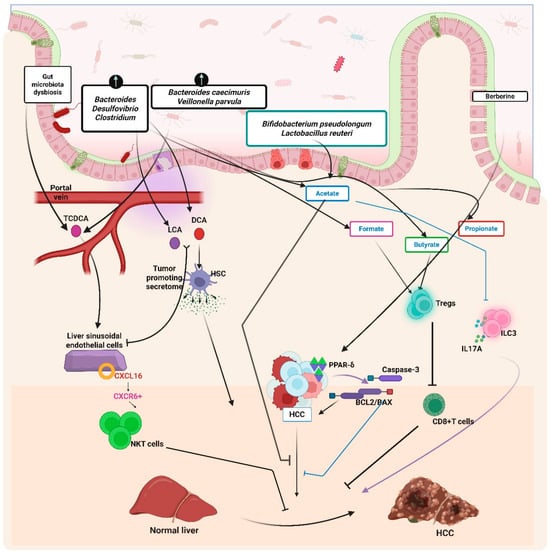
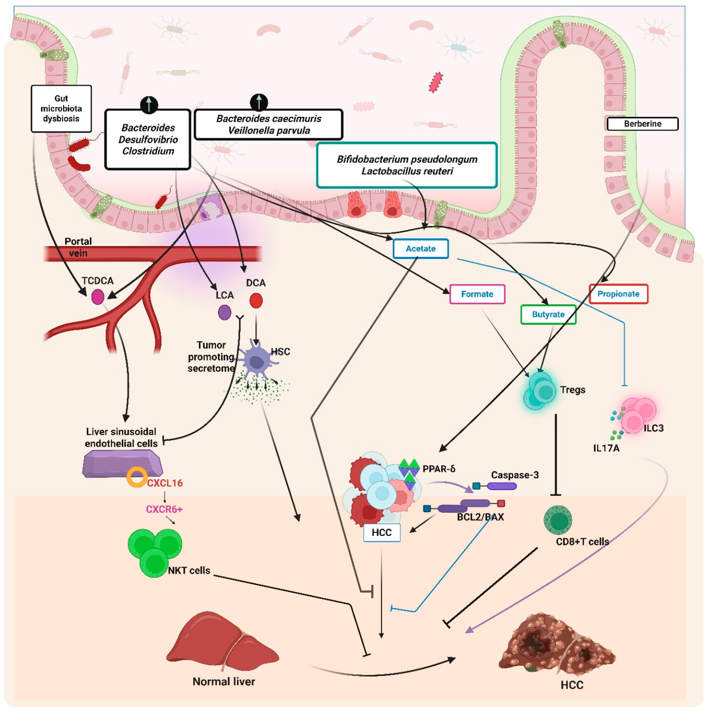
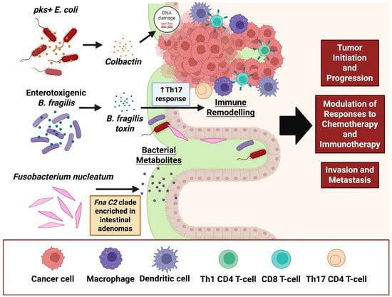
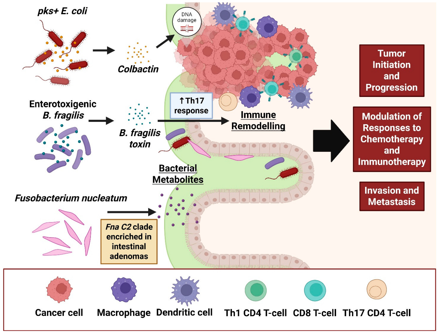

Disclaimer/Publisher’s Note: The statements, opinions and data contained in all publications are solely those of the individual author(s) and contributor(s) and not of MDPI and/or the editor(s). MDPI and/or the editor(s) disclaim responsibility for any injury to people or property resulting from any ideas, methods, instructions or products referred to in the content. |
© 2026 by the authors. Licensee MDPI, Basel, Switzerland. This article is an open access article distributed under the terms and conditions of the Creative Commons Attribution (CC BY) license.