


Abstract
Background: Excessive left stellate ganglion (LSG) activity contributes to ventricular arrhythmias (VAs) after myocardial ischemia reperfusion (I/R). C-C motif chemokine ligand 4 (CCL4) is an inflammatory chemokine involved in neuroimmune signaling. However, whether CCL4 contributes to LSG activation and VAs after myocardial I/R remains unclear. Methods: We enrolled 98 patients with acute myocardial infarction undergoing emergency percutaneous coronary intervention (PCI) and assessed circulating CCL4, norepinephrine (NE), heart rate variability, and 24-h VAs. In beagles, CCL4 or CCL4 plus a neutralizing antibody was locally microinjected into the LSG before myocardial I/R. LSG neural activity and function, myocardial sympathetic and inflammatory indices, ventricular electrophysiology, and VAs were evaluated. C-C chemokine receptor type 5 (CCR5) expression and localization, together with local immune inflammatory changes, were assessed within the LSG. Results: Patients who developed VAs during post-PCI monitoring had higher circulating CCL4 levels both before PCI and at 1 h after PCI reperfusion, and the increase in CCL4 from pre-PCI to 1 h post-PCI was significantly greater. CCL4 at 1 h post-PCI correlated with sympathetic-related indices and 24-h ventricular premature beat burden, and remained independently associated with VAs after multivariable adjustment. In beagles, myocardial I/R increased endogenous CCL4 mRNA expression in the LSG. Local CCL4 administration enhanced LSG neural activity and function, increased myocardial TH-positive area and pro-inflammatory cytokine mRNA expression, shortened effective refractory period and action potential duration at 90% repolarization, and promoted VAs, whereas CCL4 neutralization attenuated these effects. CCR5 partially colocalized with tyrosine hydroxylase-positive sympathetic neurons. CCR5 expression and c-Fos activation were increased after I/R and further enhanced by CCL4. CCL4 also increased the numbers of Iba1-, CD3-, and CD11c-positive cells and inflammatory responses within the LSG. Conclusions: Clinically, circulating CCL4 was associated with VAs after PCI. In the canine I/R model, local CCL4 administration enhanced LSG sympathetic activity and ventricular arrhythmia susceptibility, potentially involving CCR5-related and local inflammatory processes.
1. Introduction
Acute myocardial infarction (AMI) remains a major cause of cardiovascular morbidity and mortality worldwide [1]. Timely reperfusion is essential for limiting myocardial injury but can also induce myocardial ischemia reperfusion (I/R) injury [2,3,4]. Malignant ventricular arrhythmias (VAs) are among the most serious complications following myocardial I/R and may lead to sudden cardiac death [5].
Excessive cardiac sympathetic activation is an important contributor to myocardial I/R-associated VAs [6,7]. The left stellate ganglion (LSG) is a major component of cardiac sympathetic regulation, and increased LSG neural activity after myocardial I/R enhances cardiac sympathetic output and contributes to ventricular electrical instability and VAs [8,9]. Emerging evidence further indicates that the LSG represents not only a sympathetic neural relay but also a dynamic neuroimmune microenvironment, in which local inflammatory activation can enhance sympathetic neuronal excitability and increase susceptibility to VAs [10,11]. These observations highlight a potential role for neuroimmune interactions within the LSG in myocardial I/R-associated VAs.
C-C motif chemokine ligand 4 (CCL4), also known as macrophage inflammatory protein-1β, is a member of the CC chemokine family with well-established chemotactic and immunoregulatory functions [12,13]. Growing evidence implicates CCL4 in the pathophysiology of cardiovascular diseases. Circulating CCL4 levels have been associated with infarct size in patients with AMI undergoing successful percutaneous coronary intervention (PCI) [14]. A recent study further suggested that CCL4 may exert cardioprotective effects by promoting regulatory T-cell recruitment into ischemic myocardium and attenuating myocardial I/R injury [15]. Beyond myocardial immune regulation, CCL4 has also been implicated in neuroimmune signaling under pathological conditions and may influence neural function within an inflammatory microenvironment [16,17]. CCL4 signals, at least in part, through C-C chemokine receptor type 5 (CCR5), which is expressed in both neural and immune cell populations [18,19]. However, whether CCL4 contributes to LSG activation and VAs after myocardial I/R remains unclear.
In this study, we first examined dynamic changes in circulating CCL4 before and after PCI, as well as their associations with sympathetic-related indices and VAs in patients with AMI undergoing emergency PCI. We then used a canine myocardial I/R model to investigate whether local CCL4 modulates LSG neural activity and function, myocardial sympathetic and inflammatory responses, ventricular electrophysiology and VAs. We further assessed CCR5 expression and localization, together with local immune inflammatory remodeling within the LSG, to explore potential mechanisms linking CCL4 to LSG activation and VAs.
2. Materials and Methods
2.1. Clinical Study Population
This prospective observational study was conducted at Renmin Hospital of Wuhan University between 1 January 2024 and 1 January 2025. Patients with AMI presenting within 12 h of symptom onset and undergoing successful emergency PCI were enrolled. AMI was diagnosed according to contemporary guideline criteria [2,20]. Successful PCI was defined as achieving Thrombolysis in Myocardial Infarction (TIMI) grade 3 flow in the culprit artery following PCI. Recruitment was non-consecutive and depended on the availability of research personnel, the feasibility of timely blood sampling, and patients’ willingness to participate. Major exclusion criteria included persistent/permanent atrial fibrillation or pre-existing sustained arrhythmias requiring immediate treatment, implanted pacemaker/defibrillator, cardiogenic shock or hemodynamic instability, active infection, inflammatory disease, or malignant disease, and severe hepatic or renal dysfunction. Patients with unsuccessful PCI or requiring repeat intervention due to failed initial reperfusion, as well as those with incomplete essential study data, were excluded from the final analysis. Ultimately, 98 patients were included in the final analysis. Baseline clinical characteristics, including demographics, cardiovascular risk factors, medications on admission, STEMI status, and infarct-related artery were collected. Laboratory assessments were also performed at admission, mainly including creatine kinase-myocardial band (CK-MB), myoglobin (Myo), high-sensitivity cardiac troponin T (hs-TnT), and low-density lipoprotein cholesterol (LDL-C). All patients received guideline-directed medical therapy following PCI, with individualized treatment adjustments according to their clinical conditions and comorbidities. The study was approved by the institutional ethics committee (WDRY2022-K164), and all participants provided written informed consent.
2.2. 24-h Holter Monitoring and Heart Rate Variability (HRV) Analysis
All patients were admitted to the coronary care unit (CCU) immediately and underwent 24-h Holter monitoring after PCI using a Holter monitoring system (Beneware, Hangzhou, China). Electrocardiogram (ECG) recordings were automatically analyzed using the corresponding commercially available software (HolterSystem 1.4.0.0, Beneware, Hangzhou, China), which identified and quantified VA events over the 24-h monitoring period. VAs included: (1) ventricular premature beats (VPBs, defined as isolated ectopic QRS complexes), (2) non-sustained ventricular tachycardia (NsVT, ≥3 consecutive VPBs lasting < 30 s), (3) sustained ventricular tachycardia (sVT, ≥30 s), and (4) ventricular fibrillation (VF, chaotic electrical activity without identifiable QRS complexes). No independent manual adjudication of VA events was performed. The total number of VPBs during the 24-h monitoring period was recorded as the VPB burden. Patients were classified into the VA and non-VA groups based on the presence or absence of VAs during 24-h Holter monitoring after PCI. Patients with at least one VPB or any other recorded VA were assigned to the VA group, without applying a minimum VPB burden threshold. Patients without any VPBs or other recorded VAs were assigned to the non-VA group. In an exploratory subgroup analysis, patients were further categorized according to the occurrence of sVT/VF.
Because patients remained clinically stable during CCU monitoring, HRV was assessed over the entire 24-h recording period. Frequency-domain HRV parameters included low-frequency power (LF, 0.04–0.15 Hz), high-frequency power (HF, 0.15–0.40 Hz), and LF to HF ratio (LF/HF), all automatically calculated by the software.
2.3. Measurement of Human Serum CCL4 and Norepinephrine (NE)
Peripheral venous blood samples (5 mL) were collected from the antecubital vein under sterile conditions before PCI and at 1 h after successful PCI reperfusion. The 1-h post-PCI time point was defined from the restoration of coronary blood flow during PCI. At this time point, all patients had been transferred to the CCU, where blood samples were collected, followed immediately by the initiation of 24-h Holter monitoring. Blood samples were allowed to clot at room temperature for 60 min and then centrifuged at 3000 rpm for 15 min at 4 °C. Serum was aliquoted and stored at −80 °C until analysis. Serum CCL4 was measured at both time points, whereas serum NE was measured at 1 h post-PCI. Serum CCL4 and NE concentrations were quantified using commercially available enzyme-linked immunosorbent assay (ELISA) kits (ELK Biotech, Wuhan, China, catalog numbers: ELK1117 for CCL4 and ELK9243 for NE) according to the manufacturer’s instructions. The detection ranges were 15.63–1000 pg/mL for CCL4 and 78.13–5000 pg/mL for NE, with sensitivities of 6.2 and 24.1 pg/mL. The manufacturer-reported intra- and inter-assay coefficients of variation (CVs) were <8% and <10%, respectively, for both assays. All samples were measured in duplicate, and the mean values were used for subsequent analyses. ΔCCL4 was defined as the difference between the 1 h post-PCI and pre-PCI CCL4 concentrations. For exploratory stratified analyses, patients were categorized into low- and high-CCL4 groups according to the median serum CCL4 concentration at 1 h post-PCI.
2.4. Animal Experimental Groups
Adult male beagles (body weight, 8–12 kg) were obtained from Marshall (Beijing) and housed under standard laboratory conditions at the Center for Experimental Animals, Renmin Hospital of Wuhan University. All experimental procedures were approved by the Institutional Animal Care and Use Committee of Renmin Hospital of Wuhan University (No. WDRM20230202C) and were performed in accordance with institutional guidelines for the care and use of laboratory animals. Twenty-four beagles were randomly assigned to four groups (n = 6 per group) using a computer-generated randomization sequence: Control, I/R, I/R + CCL4, and I/R + CCL4 + anti-CCL4. All outcome assessments and subsequent data analyses were conducted by investigators blinded to group allocation. All 24 animals completed the experimental protocol, with no deaths, losses, or exclusions. All experimental endpoints were assessed using six biological replicates per group (n = 6), with each animal considered an independent biological replicate.
2.5. Animal Preparation and LSG Microinjection
Beagles were anesthetized with 3% sodium pentobarbital (30 mg/kg for induction and 2 mg/kg/h for maintenance), intubated, and mechanically ventilated with room air (16 breaths/min; tidal volume 26–28 mL/kg). Arterial blood pressure (BP) was continuously monitored through femoral artery catheterization with a pressure transducer attached. BP and surface ECG were recorded with the Lead 7000 Lab System (Jinjiang Inc., Chengdu, China). A heating pad was used to maintain the core body temperature of the beagles at 36.5 ± 1.5 °C.
Under sterile surgical conditions, a left thoracotomy was performed at the third intercostal space to expose the LSG. The Control and I/R groups received 100 μL vehicle consisting of phosphate-buffered saline (PBS) containing 0.1% bovine serum albumin. The I/R + CCL4 group received 0.5 μg recombinant canine CCL4 (R&D Systems, Minneapolis, MN, USA, catalog number: 5839-MB) in the same vehicle, whereas the I/R + CCL4 + anti-CCL4 group received 0.5 μg CCL4 preincubated with 30 μg canine CCL4 neutralizing antibody (R&D Systems, Minneapolis, MN, USA) for 30 min and prepared in the same vehicle, with a final volume of 100 μL in all groups.
Local microinjection was performed using a microsyringe equipped with a fine needle. The injection solution was slowly delivered at four sites within the LSG, with approximately 25 μL administered at each site. The needle was retained in place for approximately 10 s after each injection before withdrawal to minimize reflux. Subsequent experimental procedures were initiated 30 min after LSG microinjection. The dose and injection volume of CCL4 were selected based on our previous experience with local microinjection of inflammatory mediators into the LSG and previously reported studies of local CCL4 administration [21,22,23]. Investigators performing the LSG interventions were blinded to treatment allocation.
2.6. Myocardial I/R Protocol and Experimental Sequence
The heart was exposed through a left thoracotomy at the fourth intercostal space. The left anterior descending (LAD) coronary artery was identified and dissected approximately 1–2 mm distal to the first diagonal branch. A 3-0 silk suture was passed beneath the LAD, and myocardial ischemia was induced by tightening the ligature. After 45 min of ischemia, the ligature was released to restore coronary blood flow. Successful ischemia was confirmed by persistent ST-segment elevation and regional myocardial pallor or cyanosis, whereas reperfusion was confirmed by recovery of myocardial color and partial normalization of ST-segment elevation. Control animals underwent identical surgical exposure without LAD occlusion. Surface ECG and LSG neural activity were recorded simultaneously throughout the subsequent 60-min reperfusion period. Following completion of this recording period, blood samples were collected, followed sequentially by measurements of effective refractory period (ERP), action potential duration at 90% repolarization (APD90), LSG function and VF threshold. Finally, the animals were euthanized, and tissue samples were collected.
2.7. LSG Neural Activity and Function Assessment
LSG neural activity was recorded using two tungsten-coated microelectrodes inserted into the LSG, with a ground electrode placed in the chest wall. The recording electrodes were connected to a differential amplifier, and electrical signals were amplified 30–50-fold, band-pass filtered at 300–1000 Hz, recorded and analyzed using a PowerLab data acquisition system (AD Instruments, New South Wales, Australia). Neural discharges with a signal-to-noise ratio > 3:1 were considered valid discharges. Firing frequency and discharge amplitude were quantified for subsequent analysis. LSG function was assessed by measuring the maximal percentage increase in systolic blood pressure (SBP) relative to pre-stimulation baseline during standardized high-frequency electrical stimulation (20 Hz, 0.1 ms pulse duration). Five incremental stimulation intensities were applied (5, 7.5, 10, 12.5, and 15 V) sequentially, with each stimulation maintained for 15 s. Sufficient recovery time was allowed between stimulations, and the next stimulation was initiated after systolic blood pressure had returned to the pre-stimulation baseline.
2.8. Measurement of Canine Serum and Myocardial NE
Peripheral venous blood (5 mL) was collected from the jugular vein and allowed to clot at room temperature for 60 min, followed by centrifugation at 3000 rpm for 15 min at 4 °C. Serum was separated and stored at −80 °C until analysis. For myocardial samples, peri-infarct tissue from the left ventricle was rapidly harvested and snap-frozen in liquid nitrogen. Approximately 100 mg of myocardial tissue was homogenized and centrifuged, and the resulting supernatant was collected for NE measurement. Serum and myocardial NE levels were quantified using commercially available ELISA kits (IBL International GmbH, Hamburg, Germany, catalog number: RE59261) according to the manufacturer’s instructions. The detection range for NE was 200–20,000 pg/mL, with a sensitivity of 20 pg/mL. The manufacturer-reported intra- and inter-assay CVs were 7.4% and 12.5%, respectively. All samples were measured in duplicate, and the mean values were used for subsequent analyses. Myocardial NE content was normalized to tissue wet weight and expressed as ng/g tissue, whereas serum NE concentrations were expressed as pg/mL.
2.9. Ventricular Electrophysiological Measurements
ERP was measured at the left ventricular base (LVB), midventricular region (LVM), and apex (LVA) using a programmed S1–S2 stimulation protocol consisting of eight consecutive S1 stimuli at a basic cycle length of 330 ms followed by a premature S2 stimulus. The initial S1–S2 coupling interval was 250 ms and was progressively shortened in 10-ms steps until loss of ventricular capture. The coupling interval was then returned to the preceding captured interval and decreased in 2-ms steps for precise determination of ERP. ERP was defined as the longest S1–S2 coupling interval that failed to elicit a ventricular response. ERP measurements were performed at 30 min after LSG microinjection and repeated after myocardial I/R. Simultaneous monophasic action potential recordings were obtained from all three sites using custom-fabricated Ag-AgCl catheters during steady-state pacing (S1–S1 = 330 ms, maintained for 30 s to ensure electrophysiological stability). APD90 was defined as the interval from the rapid upstroke of the monophasic action potential to the time point at which 90% repolarization was completed. APD90 was measured at 30 min after LSG microinjection and repeated after myocardial I/R. VF threshold was assessed at the right ventricular apex using 20-Hz burst stimulation (0.1-ms pulse duration, 10 s), starting at 2 V and increasing in 2-V increments until VF occurred. The lowest voltage inducing VF was defined as the VF threshold, and VF was terminated immediately by electrical defibrillation.
2.10. Canine VAs and HRV Analysis
Immediately after 45 min of myocardial ischemia, coronary blood flow was restored, surface ECG was recorded continuously throughout the subsequent 60-min reperfusion period using a PowerLab data acquisition system (AD Instruments, New South Wales, Australia) for the assessment of VAs. VAs were classified according to the Lambeth Conventions II, and VPBs, NsVT, sVT, and VF were recorded and quantified over the entire 60-min recording period. In cases of sVT or VF, electrical defibrillation and standard resuscitation procedures were performed as necessary, consistent with the protocol described in a previous study [24]. Following successful resuscitation, ECG monitoring was resumed for the remainder of the predefined 60-min observation period. All animals completed the scheduled monitoring period. For HRV analysis, the first eligible continuous 5-min sinus-rhythm segment was selected chronologically from the 60-min ECG recording period by an investigator blinded to experimental group allocation. Segments containing ectopic beats, arrhythmic events, significant recording artifacts, or obvious nonstationary were excluded. A minimum of 5 consecutive minutes of stable sinus rhythm was required, without RR interval interpolation or beat replacement. Power spectral density was estimated using the Lomb–Scargle periodogram implemented in the LabChart HRV module (version 8.1.13, ADInstruments, New South Wales, Australia). Frequency-domain parameters, including LF power (0.04–0.15 Hz), HF power (0.15–0.40 Hz), and the LF/HF ratio, were calculated using identical analysis settings across all experimental groups.
2.11. Immunofluorescence and Immunohistochemical Analysis
LSG tissues were fixed in 4% paraformaldehyde, paraffin-embedded, and sectioned at 5 μm. For immunofluorescence analysis, sections were subjected to double staining for tyrosine hydroxylase (TH) and c-Fos to assess sympathetic neuronal activation, and the percentage of c-Fos-positive cells among TH-positive neurons was quantified. Separate sections were stained for CCR5 and TH to evaluate the localization of CCR5 within sympathetic neuronal regions. CCR5 immunoreactivity within TH-positive regions was quantified as the percentage of CCR5-positive area within the TH-positive area. Additional sections were stained for Iba1, CD3, and CD11c to assess immune-related cell populations within the LSG. For Iba1, CD3, and CD11c staining, marker-positive cells were counted in three randomly selected non-overlapping fields per section. All immunofluorescence staining was performed using commercially available antibodies (Servicebio, Wuhan, China, catalog number: GB12181 for TH, dilution, 1:500; GB12069 for c-Fos, dilution, 1:200; GB11505 for CCR5, dilution, 1:500; GB12105 for Iba1, dilution, 1:500; GB15014 for CD3, dilution, 1:500; GB12059 for CD11c, dilution, 1:500) according to the manufacturer’s protocols, with nuclei counterstained using 4′,6-diamidino-2-phenylindole (DAPI, Servicebio, Wuhan, China, catalog number: G1012).
For myocardial immunohistochemistry, peri-infarct myocardial tissues were fixed in 4% paraformaldehyde, paraffin-embedded, and sectioned at 5 μm. Sections were incubated with a primary anti-TH antibody (Servicebio, Wuhan, China, catalog number: GB12181, dilution, 1:500) and visualized using 3,3′-diaminobenzidine (DAB). TH-positive area was quantified in three randomly selected non-overlapping fields per section and expressed as the percentage of DAB-positive area relative to the total valid myocardial tissue area.
All images were acquired under identical microscopic settings. For each animal, three non-overlapping sections were analyzed, with three randomly selected non-overlapping fields examined per section. Measurements were averaged across sections and fields to obtain a single value for each animal, which was used as the statistical unit. Quantitative analyses were performed using ImageJ software 1.52a (National Institutes of Health, Bethesda, MD, USA). Image acquisition and quantitative analyses were performed by investigators blinded to group allocation.
2.12. Quantitative PCR (qPCR)
LSG and peri-infarct myocardial tissues were rapidly collected, snap-frozen in liquid nitrogen, and stored at −80 °C until RNA extraction. Total RNA was isolated separately from LSG and peri-infarct myocardial tissues using TRIzol Reagent (Servicebio, Wuhan, China, catalog number: G3013). RNA concentration and purity were determined spectrophotometrically, with all samples having A260/280 ratios between 1.8 and 2.0. First-strand cDNA was synthesized from 1 μg of total RNA using RevertAid First Strand cDNA Synthesis Kit (Servicebio, Wuhan, China, catalog number: G3331) in a 20 μL reaction volume. qPCR was performed using FastStart Universal SYBR Green Master Mix (Servicebio, Wuhan, China, catalog number: G3320). In LSG tissues, relative mRNA expression levels of CCL4, CCR5, interleukin-6 (IL-6), interleukin-1β (IL-1β), and tumor necrosis factor-α (TNF-α) were quantified. In peri-infarct myocardial tissues, mRNA expression levels of IL-6, IL-1β, and TNF-α were quantified. Relative gene expression was determined using the 2−ΔΔCt method, with glyceraldehyde-3-phosphate dehydrogenase (GAPDH) as the reference gene. All reactions were performed in triplicate, and melt-curve analysis confirmed amplification specificity. Amplification efficiencies were determined using standard curves generated from serial dilutions of canine cDNA and calculated using the formula E = (10−1/slope − 1) × 100%. The amplification efficiencies of all primer pairs ranged from 90% to 110%, supporting their suitability for relative gene expression analysis using the 2−ΔΔCt method. Primer sequences used for qPCR were as follows: canine CCL4, forward 5′-TCCTACTGCCTGCTGCTT-3′ and reverse 5′-GCTGGTCTCAAAGTAATCTGC-3′; canine IL-6, forward 5′-GAGGGCTGTTCGGATAATGTAG-3′ and reverse 5′-TGACTGGAGAAAGGAATGCC-3′; canine IL-1β, forward 5′-CTGCCAAGACCTGAACCAC-3′ and reverse 5′-AGCTACAATGACTGACACGAA-3′; canine TNF-α, forward 5′-CAACTGGAGAAGGGTGATCG-3′ and reverse 5′-GATTCCAAAGTACACCTGCC-3′; canine CCR5, forward 5′-CACCCATCCGGCATCTCTTT-3′ and reverse 5′-GTGGTGGGGACATGAGAAGG-3′; canine GAPDH, forward 5′-TGCACCACCAACTGCTTAGC-3′ and reverse 5′-GGCATGGACTGTGGTCATGAG-3′.
2.13. Statistical Analysis
Continuous variables were assessed for normality using the Shapiro–Wilk test. Normally distributed variables are presented as mean ± standard deviation (SD), whereas non-normally distributed variables are presented as median (25th percentile, 75th percentile). Categorical variables are presented as number and percentage. Comparisons between two independent groups were performed using the independent-samples t-test or Mann–Whitney U test, as appropriate. Categorical variables were compared using the χ2 test or Fisher’s exact test, as appropriate. Associations between continuous variables were evaluated using Spearman rank correlation analysis. Multivariable logistic regression analysis was performed to evaluate the independent association between circulating CCL4 at 1 h post-PCI and VAs. Odds ratios (ORs) and 95% confidence intervals (CIs) were calculated. Multicollinearity among covariates was assessed before model construction. Receiver operating characteristic (ROC) analysis was performed to evaluate the discriminatory ability of circulating CCL4 for VAs, and the area under the curve (AUC) was calculated. For animal experiments, comparisons among multiple independent groups were performed using one-way analysis of variance (ANOVA) followed by Tukey’s multiple-comparison test when the data met parametric assumptions. For VPB and NsVT counts, normality was assessed using the Shapiro–Wilk test for within-group observations and ANOVA residuals, and homogeneity of variance was evaluated using Levene’s test. Given some variance heterogeneity, Kruskal–Wallis tests and Games–Howell post hoc comparisons were additionally performed as sensitivity analyses to assess the robustness of the original ANOVA findings. The incidence of sVT/VF in canine experiments was compared among the four groups using the Fisher–Freeman–Halton exact test. Exact 95% confidence intervals for incidence were calculated using the Clopper–Pearson method. Outcomes measured repeatedly within the same animals across time points or stimulation intensities were analyzed using two-way repeated-measures ANOVA followed by Tukey’s multiple-comparison test. A two-sided p < 0.05 was considered statistically significant. Prism 8.0 (GraphPad Software, Inc., La Jolla, CA, USA) and SPSS 26.0 (IBM Corp., Armonk, NY, USA) were used for all analyses.
3. Results
3.1. Elevated Circulating CCL4 Is Associated with Sympathetic-Related Indices and VAs in Patients with AMI After PCI Reperfusion
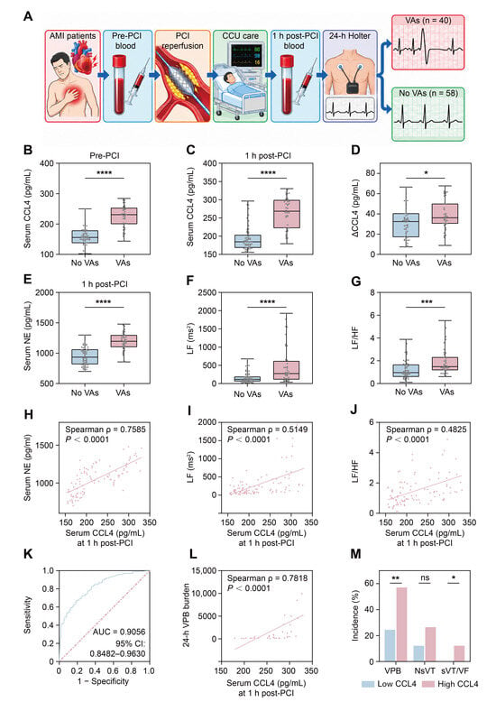
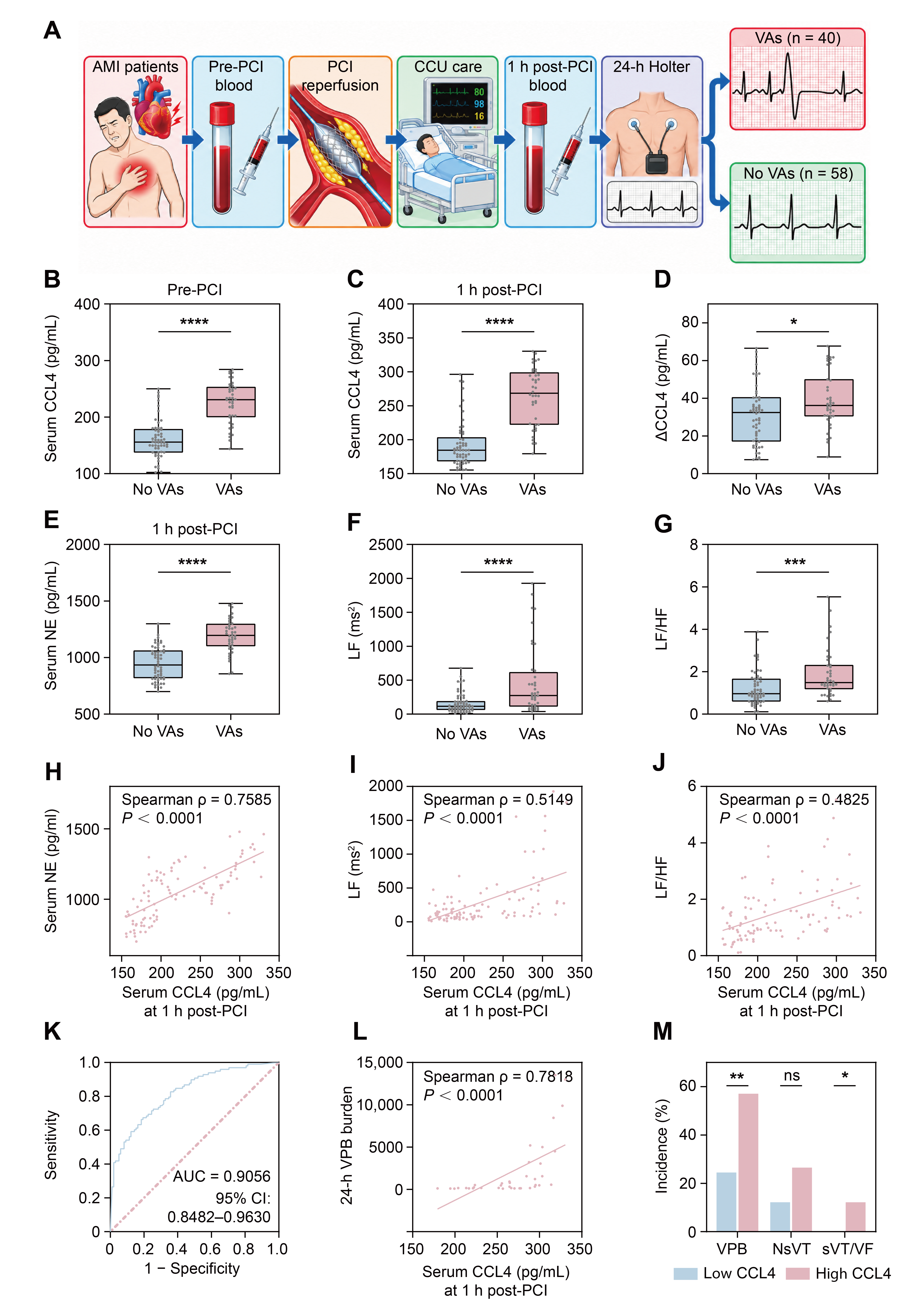
Age, sex, cardiovascular risk factors, medications on admission, and admission laboratory parameters (CK-MB, myoglobin, hs-TnT, and LDL-C) showed no statistically significant differences between patients with and without VAs (Table 1). Patients were classified according to the occurrence of VAs during the 24-h post-PCI monitoring period. Compared with patients without VAs, those with VAs had significantly higher circulating CCL4 levels both before PCI and at 1 h after reperfusion. Furthermore, the increase in CCL4 from pre-PCI to 1 h post-PCI was significantly greater in the VA group (Table 1 and Figure 1A–D). Patients with VAs exhibited higher serum NE levels at 1 h post-PCI and higher LF and LF/HF during 24-h Holter monitoring than those without VAs (Figure 1E–G). Circulating CCL4 at 1 h post-PCI was positively correlated with serum NE, LF, and LF/HF (Figure 1H–J). Multivariable logistic regression analysis further showed that circulating CCL4 at 1 h post-PCI remained independently associated with VAs after adjustment for age, sex, and left ventricular ejection fraction (LVEF), with the association remaining significant after additional adjustment for serum NE at 1 h post-PCI and LF/HF (Table 2). ROC analysis revealed that serum CCL4 levels at 1 h post-PCI discriminated between patients with and without VAs, with an AUC of 0.9056 (95% CI: 0.8482–0.9630; Figure 1K). Among patients with VAs, circulating CCL4 was also strongly correlated with 24-h VPB burden (Figure 1L). Consistently, when the cohort was stratified according to the median circulating CCL4 level, patients in the high-CCL4 group had higher LF, LF/HF, and serum NE levels than those in the low-CCL4 group. The incidence of VPBs was also higher in the high-CCL4 group, as was the incidence of sVT/VF, whereas the difference in NsVT incidence did not reach statistical significance (Table 3 and Figure 1M). Among the 98 patients, six experienced sVT/VF after PCI reperfusion. Compared with patients without sVT/VF, those with sVT/VF also exhibited higher serum CCL4 levels both before and at 1 h after PCI, accompanied by higher serum NE levels and LF/HF (Table S1).
Table 1.
Clinical characteristics of AMI patients with and without VAs after PCI reperfusion.
Figure 1.
Elevated circulating CCL4 is associated with sympathetic-related indices and VAs in patients with AMI after PCI reperfusion. (A) Schematic overview of the clinical study. Patients with AMI underwent emergency PCI, with blood samples collected before PCI and at 1 h after PCI, followed by 24-h Holter monitoring. Patients were classified according to the occurrence of VAs. (B,C) Serum CCL4 levels before PCI and at 1 h post-PCI, showing higher levels in patients with VAs. (D) ΔCCL4 (1 h post-PCI minus pre-PCI), showing a greater increase in patients with VAs. (E) Serum NE levels at 1 h post-PCI, showing higher levels in patients with VAs. (F,G) LF and LF/HF derived from 24-h Holter monitoring, showing higher values in patients with VAs. (H–J) Correlations of serum CCL4 at 1 h post-PCI with serum NE at 1 h post-PCI, LF, and LF/HF, showing positive associations. (K) ROC analysis of serum CCL4 at 1 h post-PCI for discrimination of VA status. (L) Correlation between serum CCL4 at 1 h post-PCI and 24-h VPB burden among patients with VAs, showing a positive association. (M) Incidence of VPBs, NsVT, and sVT/VF in patients stratified into low- and high-CCL4 groups according to the median serum CCL4 level at 1 h post-PCI, showing higher VPB and sVT/VF incidences in the high-CCL4 group. AMI, acute myocardial infarction; PCI, percutaneous coronary intervention; CCU, coronary care unit; CCL4, C-C motif chemokine ligand 4; VAs, ventricular arrhythmias; NE, norepinephrine; HRV, heart rate variability; LF, low-frequency power; HF, high-frequency power; LF/HF, LF to HF ratio; ROC, receiver operating characteristic; AUC, area under the curve; VPB, ventricular premature beat; NsVT, non-sustained ventricular tachycardia; sVT, sustained ventricular tachycardia; VF, ventricular fibrillation. For box-and-whisker plots (B–G), the center line indicates the median, the box represents the 25th to 75th percentiles, and the whiskers extend from the minimum to the maximum values; individual dots represent individual patients. Comparisons between groups were performed using the independent-samples t-test or Mann–Whitney U test, as appropriate. Correlations were assessed using Spearman rank correlation analysis. Categorical variables in (M) were compared using the χ2 test or Fisher’s exact test, as appropriate. ns, not significant; * p < 0.05; ** p < 0.01; *** p < 0.001; **** p < 0.0001.
Table 2.
Multivariable logistic regression analysis of factors associated with VAs.
Table 3.
Clinical characteristics and VAs according to circulating CCL4 levels.
3.2. Myocardial I/R Increases CCL4 mRNA Expression in the LSG, While Local CCL4 Administration Enhances LSG Neural Activity and Function
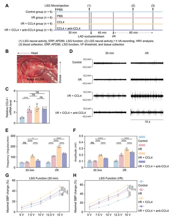
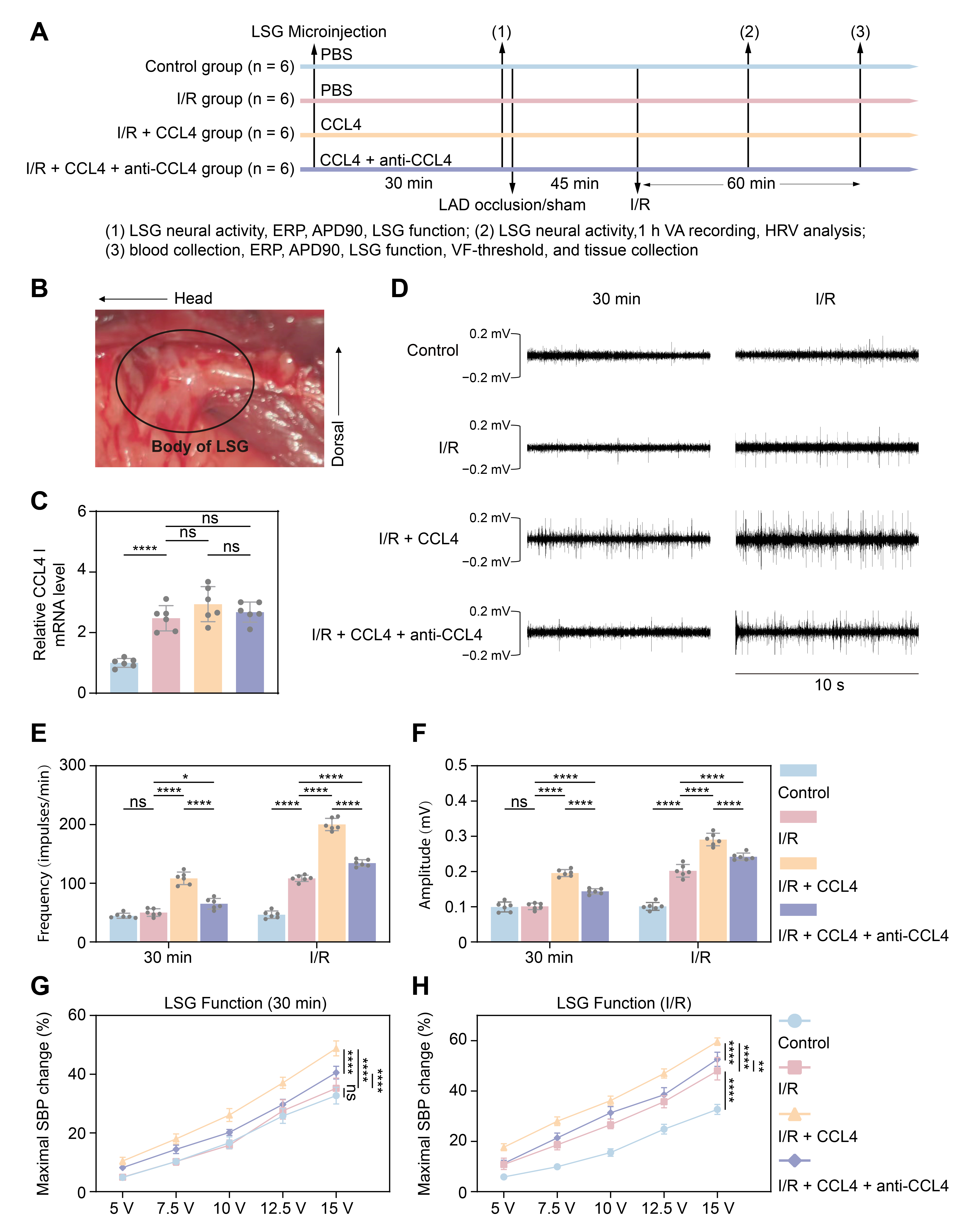
Beagles were assigned to the Control, I/R, I/R + CCL4, and I/R + CCL4 + anti-CCL4 groups (n = 6 each), with the experimental protocol and LSG anatomy shown in Figure 2A,B. No statistically significant differences in baseline heart rate (HR), SBP, diastolic BP (DBP), or mean BP (MBP) were observed among the four experimental groups (Supplementary Table S2). CCL4 mRNA expression in the LSG was significantly increased after myocardial I/R compared with the Control group, whereas no further significant differences were observed among the I/R, I/R + CCL4, and I/R + CCL4 + anti-CCL4 groups (Figure 2C). Representative recordings of LSG neural activity at 30 min after microinjection and after myocardial I/R are shown in Figure 2D. Quantitative analysis confirmed increased firing frequency and amplitude in the I/R + CCL4 group, whereas CCL4 neutralization attenuated these changes (Figure 2E,F). Consistent with the neural recordings, local CCL4 administration enhanced LSG function during graded electrical stimulation. At 30 min after microinjection, the I/R + CCL4 group exhibited greater SBP responses than the Control and I/R groups, whereas CCL4 neutralization attenuated this effect (Figure 2G). After myocardial I/R, LSG function was increased in the I/R group and further enhanced by CCL4 administration, while CCL4 neutralization partially reversed these changes (Figure 2H).
Figure 2.
Myocardial I/R increases CCL4 mRNA expression in the LSG, while local CCL4 administration enhances LSG neural activity and function. (A) Experimental protocol. Beagles were assigned to the Control, I/R, I/R + CCL4, and I/R + CCL4 + anti-CCL4 groups. PBS, CCL4, or CCL4 plus neutralizing antibody was locally microinjected into the LSG. Thirty minutes after microinjection, LSG neural activity was recorded, followed sequentially by ERP, APD90, and LSG function assessments before myocardial ischemia. Following 45 min of LAD occlusion, surface ECG and LSG neural activity were recorded simultaneously throughout the subsequent 60-min reperfusion period, during which spontaneous VAs were monitored and HRV was analyzed. Blood samples were then collected, followed sequentially by ERP, APD90, LSG function, and VF-threshold measurements. Induced VF was immediately terminated by electrical defibrillation. Finally, the animals were euthanized, and tissue samples were collected. (B) Representative image showing the anatomical location of the LSG. (C) Relative CCL4 mRNA expression in the LSG, showing increased expression after myocardial I/R. (D) Representative recordings of LSG neural activity 30 min after microinjection and after myocardial I/R. (E,F) Quantification of LSG firing frequency and amplitude, showing enhanced neural activity following local CCL4 administration and attenuation by CCL4 neutralization. (G,H) LSG function during graded electrical stimulation at 30 min after microinjection and after myocardial I/R, showing enhanced LSG function following CCL4 administration and attenuation by CCL4 neutralization. LSG, left stellate ganglion; I/R, ischemia reperfusion; CCL4, C-C motif chemokine ligand 4; PBS, phosphate-buffered saline; LAD, left anterior descending coronary artery; ERP, effective refractory period; APD90, action potential duration at 90% repolarization; SBP, systolic blood pressure. For quantitative analyses, n = 6 independent animals per group. Data are presented as mean ± SD. Comparisons in (C) were performed using one-way ANOVA followed by Tukey’s post hoc test. Comparisons in (E–H) were performed using two-way repeated-measures ANOVA followed by Tukey’s post hoc test. ns, not significant; * p < 0.05; ** p < 0.01; **** p < 0.0001.
3.3. CCL4 Increases Myocardial TH Immunoreactivity and Inflammation and Alters HRV Indices After Myocardial I/R
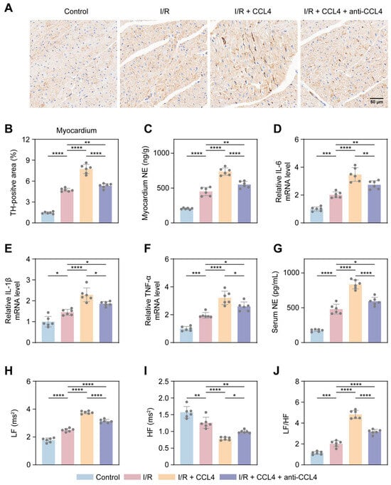
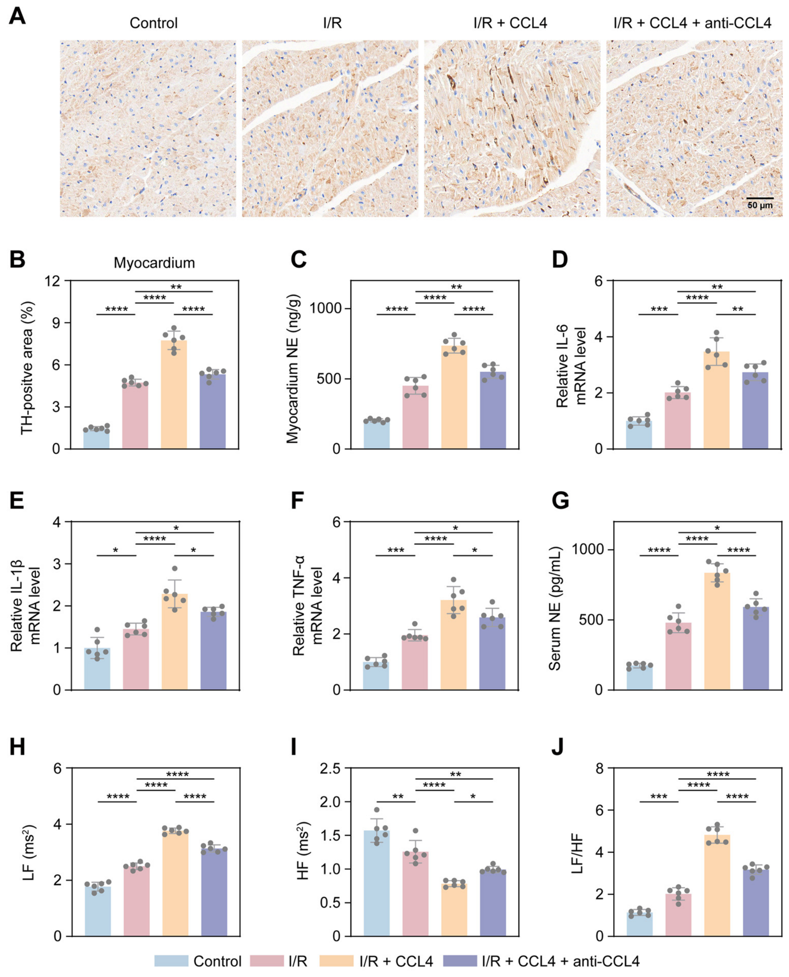
Immunohistochemical analysis of the peri-infarct myocardium showed an increased TH-positive area after myocardial I/R, which was further increased by local CCL4 administration and attenuated by CCL4 neutralization (Figure 3A,B). Myocardial NE levels showed a similar pattern, with further elevation following CCL4 administration and attenuation by CCL4 neutralization (Figure 3C). Myocardial mRNA levels of IL-6, IL-1β, and TNF-α were increased after I/R and further elevated following CCL4 administration, whereas CCL4 neutralization attenuated these increases (Figure 3D–F). Consistent with enhanced myocardial sympathetic activation and inflammatory responses, serum NE levels were increased following myocardial I/R and further elevated by CCL4 administration (Figure 3G). HRV analysis further showed increased LF and LF/HF and decreased HF following CCL4 administration, whereas these alterations were partially attenuated by CCL4 neutralization (Figure 3H–J).
Figure 3.
CCL4 increases myocardial TH immunoreactivity and inflammation and alters HRV indices after myocardial I/R. (A) Representative immunohistochemical images showing TH-positive staining (brown) and nuclei (blue) in the peri-infarct myocardium from the Control, I/R, I/R + CCL4, and I/R + CCL4 + anti-CCL4 groups. Scale bar = 50 μm. (B) Quantification of myocardial TH-positive area, showing increased TH-positive area after myocardial I/R, further enhancement by CCL4, and attenuation by CCL4 neutralization. (C) Myocardial NE content, showing increased levels after myocardial I/R and further elevation following CCL4 administration. (D–F) Relative myocardial mRNA expression levels of IL-6 (D), IL-1β (E), and TNF-α (F), showing increased expression after myocardial I/R, further elevation following CCL4 administration, and attenuation by CCL4 neutralization. (G) Serum NE levels, showing a similar pattern of increase and attenuation by CCL4 neutralization. (H–J) HRV indices, including LF, HF, and LF/HF, showing increased LF (H) and LF/HF (J) and decreased HF (I) after CCL4 administration, with partial reversal by CCL4 neutralization. I/R, ischemia reperfusion; CCL4, C-C motif chemokine ligand 4; TH, tyrosine hydroxylase; NE, norepinephrine; IL-6, interleukin-6; IL-1β, interleukin-1β; TNF-α, tumor necrosis factor-α; LF, low-frequency power; HF, high-frequency power; LF/HF, LF to HF ratio. For quantitative analyses, n = 6 independent animals per group. Data are presented as mean ± SD. Comparisons among multiple groups were performed using one-way ANOVA followed by Tukey’s post hoc test. * p < 0.05; ** p < 0.01; *** p < 0.001; **** p < 0.0001.
3.4. CCL4 Aggravates Ventricular Electrical Instability After Myocardial I/R
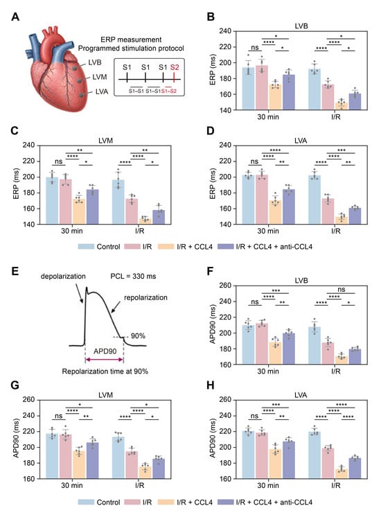
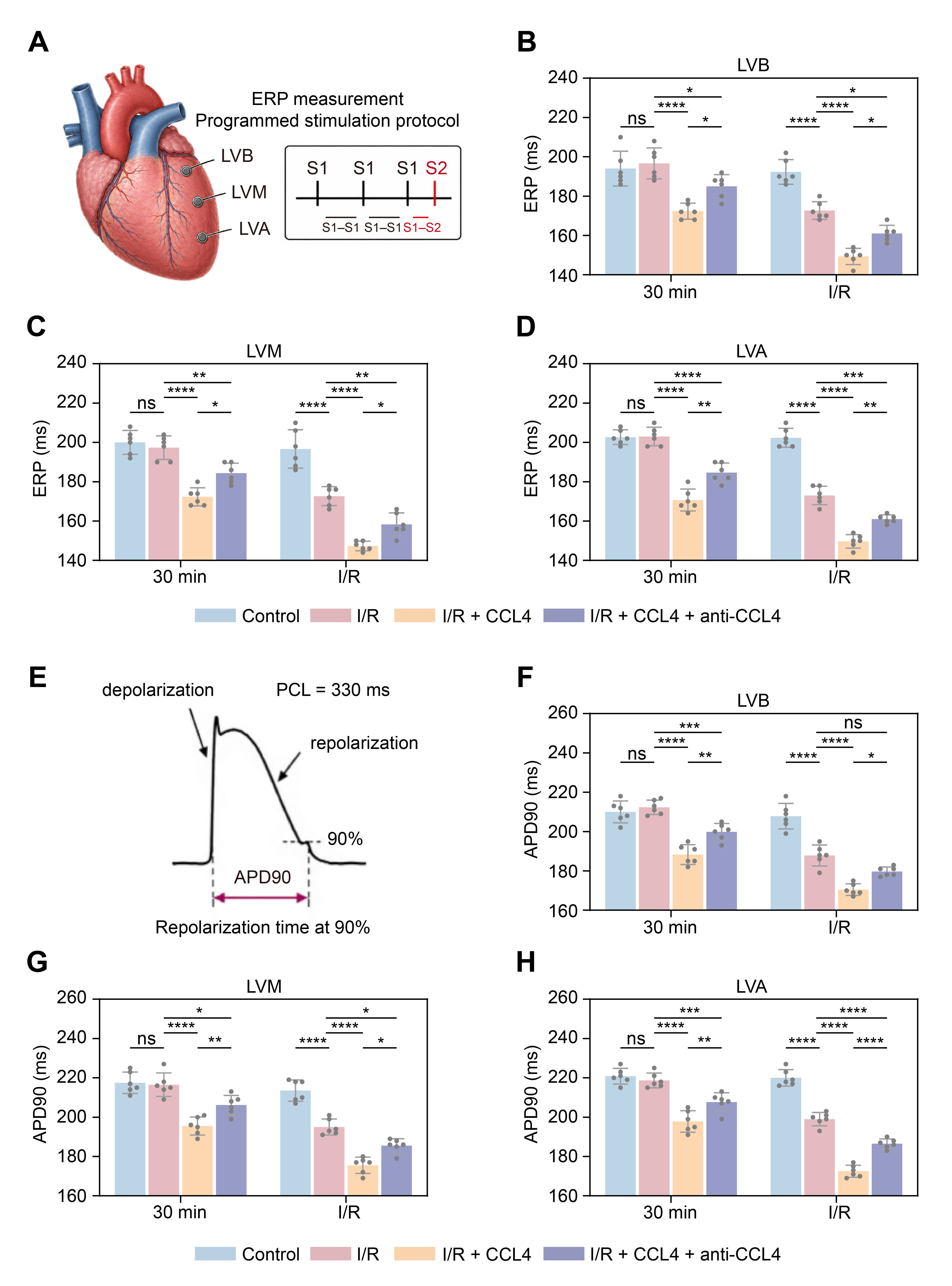
Ventricular effective refractory period (ERP) was assessed at the left ventricular base (LVB), midventricular region (LVM), and apex (LVA) (Figure 4A). At 30 min after LSG microinjection, ERP was comparable between the Control and I/R groups but was significantly shortened following local CCL4 administration, with partial attenuation by CCL4 neutralization (Figure 4B–D). After myocardial I/R, ERP was further shortened in the I/R group and was lowest in the I/R + CCL4 group, whereas CCL4 neutralization partially reversed these changes. A similar pattern was observed for action potential duration at 90% repolarization (APD90). At 30 min after microinjection, APD90 was significantly shortened by CCL4 at all three ventricular sites, whereas CCL4 neutralization attenuated this effect (Figure 4E–H). After myocardial I/R, APD90 was further reduced in the I/R group and shortened most prominently in the I/R + CCL4 group, with partial recovery following CCL4 neutralization.
Figure 4.
CCL4 aggravates ventricular electrical instability after myocardial I/R. (A) Schematic illustration of ERP measurement at the LVB, LVM, and LVA using a programmed S1–S2 stimulation protocol. (B–D) ERP measured at the LVB (B), LVM (C), and LVA (D) at 30 min after LSG microinjection and after myocardial I/R, showing ERP shortening following local CCL4 administration and attenuation by CCL4 neutralization. (E) Schematic illustration of APD90 measurement using monophasic action potential recordings at a pacing cycle length of 330 ms. (F–H) APD90 measured at the LVB (F), LVM (G), and LVA (H) at 30 min after LSG microinjection and after myocardial I/R, showing APD90 shortening following CCL4 administration and attenuation by CCL4 neutralization. ERP, effective refractory period; LVB, left ventricular base; LVM, left ventricular midventricular region; LVA, left ventricular apex; APD90, action potential duration at 90% repolarization; PCL, pacing cycle length; LSG, left stellate ganglion; I/R, ischemia reperfusion; CCL4, C-C motif chemokine ligand 4. For quantitative analyses, n = 6 independent animals per group. Data are presented as mean ± SD. Comparisons were performed using two-way repeated-measures ANOVA followed by Tukey’s post hoc test. ns, not significant; * p < 0.05; ** p < 0.01; *** p < 0.001; **** p < 0.0001.
3.5. CCL4 Promotes VAs After Myocardial I/R
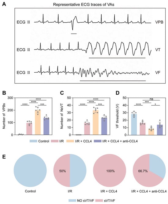
Representative ECG recordings showed VPBs, VT, and VF during the post-I/R period (Figure 5A). Compared with I/R alone, local CCL4 administration increased the numbers of VPBs and NsVT, whereas CCL4 neutralization attenuated these VA events (Figure 5B,C). The VF threshold was reduced after myocardial I/R and further decreased following CCL4 administration. CCL4 neutralization increased the VF threshold compared with the I/R + CCL4 group, to a level comparable with that in the I/R group (Figure 5D). The incidence of sVT/VF was 0% in the Control group, 50% in the I/R group, 100% in the I/R + CCL4 group, and 66.7% in the I/R + CCL4 + anti-CCL4 group (Figure 5E).
Figure 5.
CCL4 promotes VAs after myocardial I/R. (A) Representative ECG waveforms illustrating VPB, VT, and VF, redrawn from original recordings for visual clarity. The traces are presented schematically and are not intended for quantitative interpretation of time intervals or voltage amplitudes. (B,C) Numbers of VPBs (B) and NsVT (C) episodes during the post-I/R recording period, showing increased arrhythmic burden after CCL4 administration and attenuation by CCL4 neutralization. (D) VF threshold, showing a reduction after myocardial I/R and a further decrease following CCL4 administration, with partial recovery after CCL4 neutralization. (E) Incidence of sVT/VF in the four experimental groups, showing the highest incidence in the I/R + CCL4 group. ECG, electrocardiogram; VPB, ventricular premature beat; VT, ventricular tachycardia; NsVT, non-sustained ventricular tachycardia; VF, ventricular fibrillation; I/R, ischemia reperfusion; CCL4, C-C motif chemokine ligand 4; sVT, sustained ventricular tachycardia. For quantitative analyses, n = 6 independent animals per group. Data in (B–D) are presented as mean ± SD. Data in (E) are presented as percentages, the corresponding event counts and exact 95% confidence intervals (CIs), provided here for clarity, were as follows: Control, 0/6 (0%; 95% CI: 0–45.9%); I/R, 3/6 (50.0%; 95% CI: 11.8–88.2%); I/R + CCL4, 6/6 (100%; 95% CI: 54.1–100%); and I/R + CCL4 + anti-CCL4, 4/6 (66.7%; 95% CI: 22.3–95.7%). Comparisons in (B–D) were performed using one-way ANOVA followed by Tukey’s post hoc test. The overall sVT/VF incidence was compared using the Fisher–Freeman–Halton exact test (p = 0.0048), followed by pairwise Fisher’s exact tests with Holm adjustment. ns, not significant; * p < 0.05; *** p < 0.001; **** p < 0.0001.
3.6. CCL4 Enhances CCR5 Immunoreactivity in TH-Positive Regions and Sympathetic Neuronal Activation in the LSG After Myocardial I/R
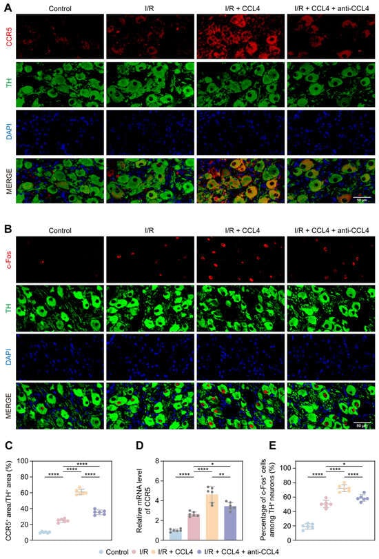
Immunofluorescence staining demonstrated partial colocalization of CCR5 with TH-positive sympathetic neurons within the LSG. Quantitative analysis of CCR5-positive area within TH-positive regions showed a significant increase after myocardial I/R compared with the Control group. This increase was further enhanced by local CCL4 administration, whereas CCL4 neutralization markedly attenuated the CCL4-associated increase (Figure 6A,C). Consistently, CCR5 mRNA expression was elevated after I/R and further increased in the I/R + CCL4 group, whereas CCL4 neutralization partially reversed this change (Figure 6D). In parallel, c-Fos activation in TH-positive sympathetic neurons was increased after myocardial I/R and further enhanced by local CCL4 administration, while CCL4 neutralization attenuated this effect (Figure 6B,E).
Figure 6.
CCL4 enhances CCR5 immunoreactivity in TH-positive regions and sympathetic neuronal activation in the LSG after myocardial I/R. (A) Representative immunofluorescence images showing CCR5 (red), TH (green), and DAPI (blue) in the LSG, with partial colocalization of CCR5 signals with TH-positive sympathetic neurons. Scale bar = 50 μm. (B) Representative immunofluorescence images showing c-Fos (red), TH (green), and DAPI (blue) in the LSG. c-Fos activation in TH-positive neurons was increased after myocardial I/R and further enhanced by CCL4 administration, with partial attenuation following CCL4 neutralization. Scale bar = 50 μm. (C) Quantification of CCR5-positive area within TH-positive regions, showing an increased percentage of CCR5-positive area within TH-positive regions after myocardial I/R, further enhancement by CCL4 administration, and attenuation by CCL4 neutralization. (D) Relative CCR5 mRNA expression in the LSG, showing increased expression after myocardial I/R, further elevation following CCL4 administration, and partial attenuation by CCL4 neutralization. (E) Percentage of c-Fos-positive cells among TH-positive neurons, showing increased sympathetic neuronal activation after myocardial I/R, further enhancement by CCL4 administration, and attenuation by CCL4 neutralization. CCR5, C-C chemokine receptor type 5; TH, tyrosine hydroxylase; DAPI, 4′,6-diamidino-2-phenylindole; LSG, left stellate ganglion; I/R, ischemia reperfusion; CCL4, C-C motif chemokine ligand 4. For quantitative analyses, n = 6 independent animals per group. Data are presented as mean ± SD. Comparisons among multiple groups were performed using one-way ANOVA followed by Tukey’s post hoc test. * p < 0.05; ** p < 0.01; **** p < 0.0001.
3.7. CCL4 Increases Immune-Related Marker-Positive Cells and Inflammatory Responses Within the LSG After Myocardial I/R
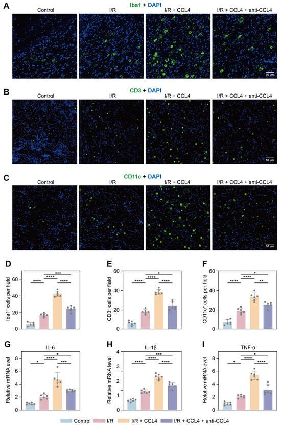
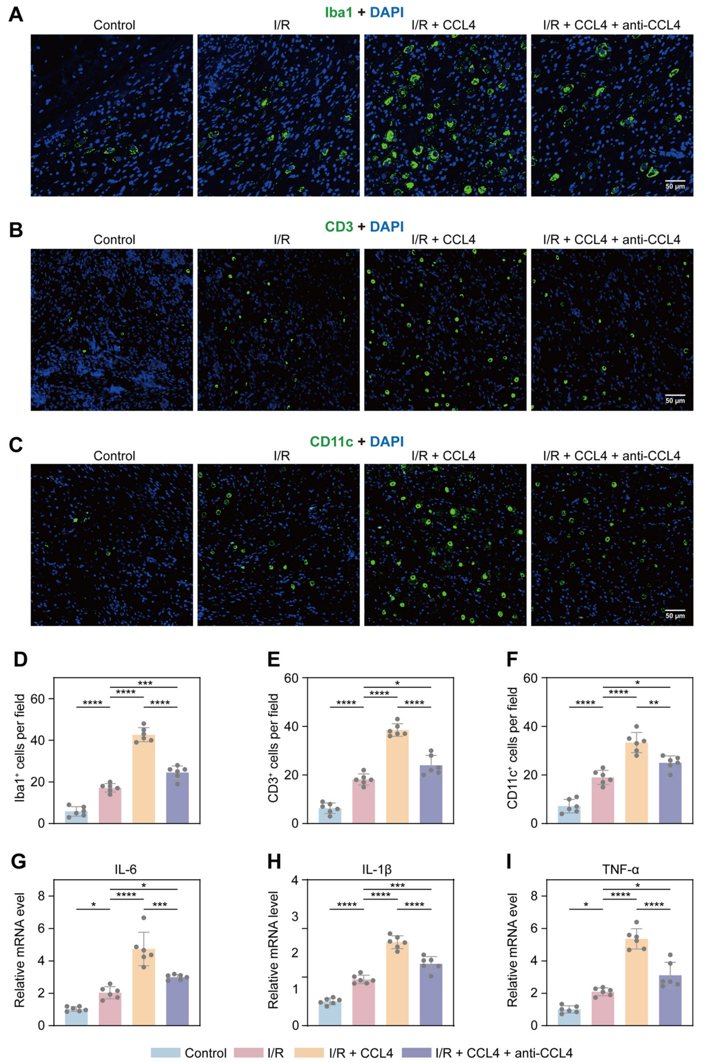
Immunofluorescence analysis showed that myocardial I/R increased the numbers of Iba1-positive, CD3-positive, and CD11c-positive cells within the LSG. These cellular changes were further enhanced by local CCL4 administration, whereas CCL4 neutralization significantly attenuated the increases (Figure 7A–F). Consistent with these findings, mRNA expression levels of IL-6, IL-1β, and TNF-α were increased after myocardial I/R and further elevated in the I/R + CCL4 group, while CCL4 neutralization partially reversed these changes (Figure 7G–I).
Figure 7.
CCL4 increases immune-related marker-positive cells and inflammatory responses within the LSG following myocardial I/R. (A–C) Representative immunofluorescence images showing Iba1-positive cells (A), CD3-positive cells (B), and CD11c-positive cells (C) in the LSG. Iba1, CD3, and CD11c signals are shown in green, and DAPI-stained nuclei are shown in blue. Scale bar = 50 μm. (D–F) Quantification of Iba1-positive cells (D), CD3-positive cells (E), and CD11c-positive cells (F) per field, showing increased numbers of marker-positive cells after myocardial I/R, further enhancement following CCL4 administration, and attenuation by CCL4 neutralization. (G–I) Relative mRNA expression of IL-6 (G), IL-1β (H), and TNF-α (I) in the LSG, showing increased inflammatory activation after myocardial I/R, further enhancement by CCL4 administration, and attenuation by CCL4 neutralization. I/R, ischemia reperfusion; CCL4, C-C motif chemokine ligand 4; IL-6, interleukin-6; IL-1β, interleukin-1β; TNF-α, tumor necrosis factor-α; DAPI, 4′,6-diamidino-2-phenylindole. For quantitative analyses, n = 6 independent animals per group. Data are presented as mean ± SD. Comparisons among multiple groups were performed using one-way ANOVA followed by Tukey’s post hoc test. * p < 0.05; ** p < 0.01; *** p < 0.001; **** p < 0.0001.
4. Discussion
Chemokines are increasingly recognized as clinically relevant inflammatory mediators in ischemic heart disease [25,26]. Circulating CCL4 has been associated with infarct size in patients with first-time STEMI undergoing successful PCI [14]. Chemokine-related inflammatory signaling also has been implicated in arrhythmogenic remodeling [27,28]. Our findings extend these observations by suggesting an association between circulating CCL4 and VAs after PCI. Elevated pre-PCI CCL4 may reflect a pre-existing inflammatory burden, whereas the post-PCI increase may indicate an enhanced reperfusion-associated inflammatory response. A similar pattern was observed in the exploratory subgroup analysis stratified by sVT/VF occurrence, although several baseline characteristics differed between the groups and the small number of sVT/VF events warrants cautious interpretation. Enhanced sympathetic activity is a well-established contributor to VAs after I/R [29,30]. Consistent with this, the concurrent elevation of circulating NE and sympathetic-related HRV indices supports the presence of enhanced sympathetic activity in patients with VAs. Although HRV-derived indices have recognized limitations in assessing autonomic activity [31,32], the increase in circulating NE provides complementary evidence. Interestingly, circulating CCL4 was positively correlated with NE, LF, and LF/HF. These findings suggest a potential relationship between circulating CCL4 and sympathetic activation, although a causal link remains to be established.
The LSG occupies a critical extracardiac level between central sympathetic control and the heart [8,33,34]. In the present study, myocardial I/R increased CCL4 mRNA expression within the LSG, indicating endogenous CCL4 transcriptional upregulation. However, the origin and relationship of circulating and LSG-associated CCL4 remain unclear. Circulating CCL4 may primarily reflect systemic inflammation, whereas LSG-associated CCL4 may arise from local production. Nevertheless, circulating CCL4 may also enter the ganglion, and locally released CCL4 may potentially contribute to circulating levels. Thus, the two may represent interconnected components of the inflammatory response after myocardial I/R. Regardless of its precise origin, the key functional question is whether CCL4 can modulate LSG activity. Accumulating evidence indicates that inflammatory mediators such as IL-1β, IL-6 and IL-17A within the LSG can actively influence sympathetic neural function rather than merely reflect local inflammation [11,21,35]. As a pro-inflammatory chemokine, CCL4 may represent an additional mediator acting within the LSG. Consistent with this possibility, local CCL4 administration enhanced LSG neural activity and function before the onset of myocardial I/R, indicating that its effect on the LSG does not require preceding ischemic injury. Myocardial I/R itself also increased LSG activity and function, consistent with previous evidence of sympathetic ganglionic activation after myocardial injury [36,37]. Notably, the effects of CCL4 remained evident after I/R, whereas CCL4 neutralization attenuated these changes.
Continuous ambulatory recordings in canine models have shown that bursts of LSG nerve activity frequently precede VAs [9]. Selective optogenetic inhibition of the LSG stabilizes ventricular electrophysiology and suppresses ischemia-induced VAs [38]. Consistent with these observations, CCL4-induced LSG activation was associated with greater ventricular electrical instability, with electrophysiological changes evident even before I/R, suggesting a pre-existing arrhythmogenic substrate that may be further aggravated by I/R. The apparent discrepancy with the previously reported cardioprotective effects of CCL4 in ischemic myocardium may reflect its tissue-specific actions [15]. Local CCL4 administration increased myocardial TH-positive area and NE levels in the peri-infarct myocardium. Excessive adrenergic stimulation can directly alter ventricular ion-channel function and intracellular Ca2+ handling, abbreviate refractoriness, increase spatial heterogeneity of repolarization, and promote afterdepolarizations and triggered activity [39,40]. CCL4 administration was also accompanied by increased myocardial expression of IL-6, IL-1β, and TNF-α. Enhanced catecholaminergic drive can promote oxidative stress and inflammatory activation within the injured myocardium, while inflammatory mediators can in turn modify ion-channel function, gap-junction coupling, and Ca2+-handling proteins, thereby increasing conduction and repolarization heterogeneity and facilitating both triggered activity and re-entry [41,42,43]. The observed myocardial sympathetic and inflammatory changes may contribute to the electrophysiological alterations and increased arrhythmic susceptibility associated with CCL4-induced LSG activation. Beyond these mechanisms, previous studies have implicated mitochondrial energetic dysfunction, impaired intercellular electrical coupling, and progressive structural remodeling [7,44,45]. However, none of the downstream mechanisms discussed above were directly investigated in the present study, and their potential contributions require further mechanistic investigation.
CCR5, one of the major receptors through which CCL4 exerts its biological effects, is expressed in both neural and immune-cell populations [16,46]. In the present study, CCR5 was partially colocalized with TH-positive sympathetic neurons. Moreover, myocardial I/R increased CCR5 immunoreactivity within TH-positive regions and enhanced c-Fos activation in sympathetic neurons, and both responses were further augmented by local CCL4 administration and attenuated by CCL4 neutralization. Chemokine receptors expressed by neurons can modulate membrane excitability, intracellular signaling, and synaptic transmission under inflammatory conditions [18,47,48]. One possible interpretation is that CCL4 may directly influence sympathetic neuronal excitability through neuronal CCR5. However, because CCR5 was not selectively inhibited or genetically manipulated in the present study, a direct CCL4–CCR5 signaling mechanism remains unconfirmed. An additional and potentially complementary mechanism involves remodeling of the local immune inflammatory microenvironment. CCL4 also has well-established chemotactic activity toward several immune-cell populations [49,50]. Consistent with these properties, myocardial I/R increased the numbers of Iba1-, CD3-, and CD11c-positive cells within the LSG, and these responses were further enhanced by CCL4 administration. Although these markers lack absolute cell-type specificity, the findings suggest increased local immune-cell presence, potentially involving monocyte/macrophage-lineage cells, T lymphocytes, and dendritic or other myeloid cells. Differences in the immune-cell populations involved may also partly explain the contrasting effects of CCL4 [15]. However, these findings cannot distinguish immune-cell recruitment from changes in resident cell populations or increased marker expression. Together with increased inflammatory cytokine expression, these findings suggest a potential contribution of local immune inflammatory changes to sympathetic activation, although the underlying cellular mechanisms remain unclear.
Limitations
The relatively small clinical sample size may limit the statistical robustness and generalizability of these findings, warranting further validation in larger, independent cohorts. Additionally, the lack of systematic assessment of inflammatory markers, electrolyte levels, and blood gas parameters during Holter monitoring, together with individual variations in medication use, may have introduced residual confounding. The animal experiments were also limited by the relatively small sample size (n = 6 per group). Larger, adequately powered animal studies are needed to confirm these observations. Although CCL4 mRNA expression increased in the LSG following myocardial I/R, corresponding endogenous protein levels and their temporal dynamics were not assessed. Therefore, whether the administered dose and local exposure reflect pathophysiological conditions remains uncertain, and the dose–response relationship of CCL4 within the canine LSG requires further investigation. Moreover, the distribution of injected CCL4 and its potential effects on circulating CCL4 concentrations were not evaluated, so systemic absorption and secondary myocardial effects cannot be excluded. The relationship between circulating and LSG-associated CCL4 also remains unresolved, requiring further tissue-specific measurements and source identification. Myocardial area at risk, infarct size, and ventricular function were not directly assessed, and blood gas and electrolyte measurements were unavailable for intergroup comparison. Therefore, despite standardized experimental conditions, potential influences of unmeasured physiological differences and variations in myocardial injury severity on the post-I/R electrophysiological findings cannot be excluded. Finally, the invasive nature of local LSG delivery presents additional challenges for potential clinical translation, particularly regarding delivery feasibility and safety.
5. Conclusions
Clinically, circulating CCL4 was associated with VAs after PCI. In the canine I/R model, local CCL4 administration enhanced LSG sympathetic activity and ventricular arrhythmia susceptibility, potentially involving CCR5-related and local inflammatory processes.
Supplementary Materials
The following supporting information can be downloaded at: https://www.mdpi.com/article/10.3390/biomedicines14112533/s1, Table S1: Clinical characteristics of AMI patients with and without sVT/VF after PCI reperfusion; Table S2: Baseline physiological characteristics of the experimental animals.
Author Contributions
Conceptualization: C.W., W.S., Z.Z. and H.J.; Methodology: C.W., W.S., Z.Z. and H.X.; Investigation: C.W., W.S., Z.Z., H.X. and W.Z.; Formal analysis: C.W., W.S. and Z.Z.; Data curation: C.W., W.S. and Z.Z.; Visualization: C.W., W.S. and Z.Z.; Writing—original draft: C.W., W.S. and Z.Z.; Writing—review and editing: C.W., W.S., Z.Z., H.X., W.Z. and H.J.; Funding acquisition: W.S. and H.J.; Resources: H.J.; Project administration: H.J.; Supervision: H.J. All authors have read and agreed to the published version of the manuscript.
Funding
This research was funded by grants from the National Natural Science Foundation of China (Grant No. 82370286) and the Open Fund of Hubei Key Laboratory (Grant No. SZ202404).
Institutional Review Board Statement
The clinical study was conducted in accordance with the Declaration of Helsinki and approved by the Ethics Committee of Renmin Hospital of Wuhan University (WDRY2022-K164), approval date 28 July 2022. The animal study protocol was approved by the Institutional Animal Care and Use Committee of Renmin Hospital of Wuhan University (WDRM20230202C), approval date 2 June 2023.
Informed Consent Statement
Informed consent was obtained from all subjects involved in the study.
Data Availability Statement
All data used during the current study are available from the corresponding author on request.
Conflicts of Interest
The authors declare no conflicts of interest.
References
- Stark, B.A.; DeCleene, N.K.; Desai, E.C.; Hsu, J.M.; Johnson, C.O.; Lara-Castor, L.; LeGrand, K.E.; Bhoomadevi, A.; Aalipour, M.A.; Aalruz, H.; et al. Global, regional, and national burden of cardiovascular diseases and risk factors in 204 countries and territories, 1990–2023. J. Am. Coll. Cardiol. 2025, 86, 2167–2243. [Google Scholar] [CrossRef] [Scilit] [PubMed]
- Byrne, R.A.; Rossello, X.; Coughlan, J.J.; Barbato, E.; Berry, C.; Chieffo, A.; Claeys, M.J.; Dan, G.A.; Dweck, M.R.; Galbraith, M.; et al. 2023 esc guidelines for the management of acute coronary syndromes. Eur. Heart J. 2023, 44, 3720–3826. [Google Scholar] [CrossRef] [Scilit] [PubMed]
- Welt, F.G.P.; Batchelor, W.; Spears, J.R.; Penna, C.; Pagliaro, P.; Ibanez, B.; Drakos, S.G.; Dangas, G.; Kapur, N.K. Reperfusion injury in patients with acute myocardial infarction: Jacc scientific statement. J. Am. Coll. Cardiol. 2024, 83, 2196–2213. [Google Scholar] [CrossRef] [Scilit] [PubMed]
- Heusch, G. Myocardial ischaemia-reperfusion injury and cardioprotection in perspective. Nat. Rev. Cardiol. 2020, 17, 773–789. [Google Scholar] [CrossRef] [Scilit] [PubMed]
- van der Weg, K.; Prinzen, F.W.; Gorgels, A.P. Editor’s choice- reperfusion cardiac arrhythmias and their relation to reperfusion-induced cell death. Eur. Heart J. Acute Cardiovasc. Care 2019, 8, 142–152. [Google Scholar] [CrossRef] [Scilit] [PubMed]
- Shen, M.J.; Zipes, D.P. Role of the autonomic nervous system in modulating cardiac arrhythmias. Circ. Res. 2014, 114, 1004–1021. [Google Scholar] [CrossRef] [Scilit] [PubMed]
- Herring, N.; Kalla, M.; Paterson, D.J. The autonomic nervous system and cardiac arrhythmias: Current concepts and emerging therapies. Nat. Rev. Cardiol. 2019, 16, 707–726. [Google Scholar] [CrossRef] [Scilit] [PubMed]
- Ajijola, O.A.; Shivkumar, K. Neural remodeling and myocardial infarction: The stellate ganglion as a double agent. J. Am. Coll. Cardiol. 2012, 59, 962–964. [Google Scholar] [CrossRef] [Scilit] [PubMed]
- Zhou, S.; Jung, B.C.; Tan, A.Y.; Trang, V.Q.; Gholmieh, G.; Han, S.W.; Lin, S.F.; Fishbein, M.C.; Chen, P.S.; Chen, L.S. Spontaneous stellate ganglion nerve activity and ventricular arrhythmia in a canine model of sudden death. Heart Rhythm. 2008, 5, 131–139. [Google Scholar] [CrossRef] [Scilit] [PubMed]
- Lei, Q.; Jiang, Z.; Shao, Y.; Liu, X.; Li, X. Stellate ganglion, inflammation, and arrhythmias: A new perspective on neuroimmune regulation. Front. Cardiovasc. Med. 2024, 11, 1453127. [Google Scholar] [CrossRef] [Scilit] [PubMed]
- Peng, C.; Lu, Y.; Li, R.; Zhang, L.; Liu, Z.; Xu, X.; Wang, C.; Hu, R.; Tan, W.; Zhou, L.; et al. Neuroimmune modulation mediated by IL-6: A potential target for the treatment of ischemia-induced ventricular arrhythmias. Heart Rhythm. 2024, 21, 610–619. [Google Scholar] [CrossRef] [Scilit] [PubMed]
- Chen, K.; Bao, Z.; Tang, P.; Gong, W.; Yoshimura, T.; Wang, J.M. Chemokines in homeostasis and diseases. Cell Mol. Immunol. 2018, 15, 324–334. [Google Scholar] [CrossRef] [Scilit] [PubMed]
- Nagarsheth, N.; Wicha, M.S.; Zou, W. Chemokines in the cancer microenvironment and their relevance in cancer immunotherapy. Nat. Rev. Immunol. 2017, 17, 559–572. [Google Scholar] [CrossRef] [Scilit] [PubMed]
- Orn, S.; Breland, U.M.; Mollnes, T.E.; Manhenke, C.; Dickstein, K.; Aukrust, P.; Ueland, T. The chemokine network in relation to infarct size and left ventricular remodeling following acute myocardial infarction. Am. J. Cardiol. 2009, 104, 1179–1183. [Google Scholar] [CrossRef] [Scilit] [PubMed]
- Zhang, W.; Jiang, X.; Liu, J.; Yu, P.; Zeng, X.; Guo, C. Srage attenuates myocardial ischemia-reperfusion injury by recruiting tregs through chemokine ccl4. Eur. Heart J. 2024, 45, ehae666-3707. [Google Scholar] [CrossRef] [Scilit]
- Rostène, W.; Kitabgi, P.; Parsadaniantz, S.M. Chemokines: A new class of neuromodulator? Nat. Rev. Neurosci. 2007, 8, 895–903. [Google Scholar] [CrossRef] [Scilit] [PubMed]
- Sowa, J.E.; Tokarski, K. Cellular, synaptic, and network effects of chemokines in the central nervous system and their implications to behavior. Pharmacol. Rep. 2021, 73, 1595–1625. [Google Scholar] [CrossRef] [Scilit] [PubMed]
- Cao, Y.; Wang, J.; Li, X.; Chen, X.; Xiong, J.; Wang, T.; Sun, H.; Wang, Z. Microglial ccl4-ccr5 signaling links systemic inflammation to synaptic loss and memory deficits in chronic kidney disease. J. Neuroinflamm. 2026, 23, 252. [Google Scholar] [CrossRef] [Scilit] [PubMed]
- Bambal, V.; Kodihalli Basavaraj, P.; Vanaja, K. Chemokine receptor type-5: A key regulator of immunity, disease pathogenesis, and emerging therapeutic target. Inflammopharmacology 2025, 33, 4477–4498. [Google Scholar] [CrossRef] [Scilit] [PubMed]
- Thygesen, K.; Alpert, J.S.; Jaffe, A.S.; Chaitman, B.R.; Bax, J.J.; Morrow, D.A.; White, H.D. Fourth universal definition of myocardial infarction (2018). Eur. Heart J. 2019, 40, 237–269. [Google Scholar] [CrossRef] [Scilit] [PubMed]
- Deng, J.; Zhou, X.; Wang, M.; Wang, M.; Zhou, L.; Meng, G.; Wang, Y.; Wang, Z.; Zhang, Y.; Zhou, Z.; et al. The effects of interleukin 17A on left stellate ganglion remodeling are mediated by neuroimmune communication in normal structural hearts. Int. J. Cardiol. 2019, 279, 64–71. [Google Scholar] [CrossRef] [Scilit] [PubMed]
- Myers, R.D.; Lopez-Valpuesta, F.J.; Miñano, F.J.; Wooten, M.H.; Barwick, V.S.; Wolpe, S.D. Fever and feeding in the rat: Actions of intrahypothalamic interleukin-6 compared to macrophage inflammatory protein-1 beta (mip-1 beta). J. Neurosci. Res. 1994, 39, 31–37. [Google Scholar] [CrossRef] [Scilit] [PubMed]
- Saika, F.; Kiguchi, N.; Kobayashi, Y.; Fukazawa, Y.; Kishioka, S. Cc-chemokine ligand 4/macrophage inflammatory protein-1β participates in the induction of neuropathic pain after peripheral nerve injury. Eur. J. Pain. 2012, 16, 1271–1280. [Google Scholar] [CrossRef] [Scilit] [PubMed]
- Peng, J.Q.; Zhou, M.M.; Xu, L.; Wang, X.; Tang, Y.H.; Liu, Y. Targeted m-channel activation in the left stellate ganglion protects against ischemia-induced ventricular arrhythmias in canines. J. Am. Heart Assoc. 2025, 14, e039059. [Google Scholar] [CrossRef] [Scilit] [PubMed]
- Safa, A.; Rashidinejad, H.R.; Khalili, M.; Dabiri, S.; Nemati, M.; Mohammadi, M.M.; Jafarzadeh, A. Higher circulating levels of chemokines cxcl10, ccl20 and ccl22 in patients with ischemic heart disease. Cytokine 2016, 83, 147–157. [Google Scholar] [CrossRef] [Scilit] [PubMed]
- Andersen, T.; Ueland, T.; Ghukasyan Lakic, T.; Åkerblom, A.; Bertilsson, M.; Aukrust, P.; Michelsen, A.E.; James, S.K.; Becker, R.C.; Storey, R.F.; et al. C-x-c ligand 16 is an independent predictor of cardiovascular death and morbidity in acute coronary syndromes. Arterioscler. Thromb. Vasc. Biol. 2019, 39, 2402–2410. [Google Scholar] [CrossRef] [Scilit] [PubMed]
- Li, S.; Jiang, Z.; Chao, X.; Jiang, C.; Zhong, G. Identification of key immune-related genes and immune infiltration in atrial fibrillation with valvular heart disease based on bioinformatics analysis. J. Thorac. Dis. 2021, 13, 1785–1798. [Google Scholar] [CrossRef] [Scilit] [PubMed]
- Ninni, S.; Ortmans, S.; Seunes, C.; Coisne, A.; Marechal, X.; Modine, T.; Gherraert, C.; Lefebvre, P.; Woitrain, E.; Staels, B.; et al. Postoperative atrial fibrillation following cardiac surgery is associated with pre-existing inflammatory dysregulation in epicardial adipose tissue. Insights from transcriptomic analysis. Arch. Cardiovasc. Dis. Suppl. 2019, 11, 105–106. [Google Scholar] [CrossRef] [Scilit]
- Kabir, R.A.; Doytchinova, A.; Liu, X.; Adams, D.; Straka, S.; Chen, L.S.; Shen, C.; Lin, S.F.; Everett, T.H.t.; Chen, P.S. Crescendo skin sympathetic nerve activity and ventricular arrhythmia. J. Am. Coll. Cardiol. 2017, 70, 3201–3202. [Google Scholar] [CrossRef] [Scilit] [PubMed]
- Zhang, P.; Liang, J.J.; Cai, C.; Tian, Y.; Dai, M.Y.; Wong, J.; Everett, T.H.t.; Wittwer, E.D.; Barsness, G.W.; Chen, P.S.; et al. Characterization of skin sympathetic nerve activity in patients with cardiomyopathy and ventricular arrhythmia. Heart Rhythm. 2019, 16, 1669–1675. [Google Scholar] [CrossRef] [Scilit] [PubMed]
- Reyes del Paso, G.A.; Langewitz, W.; Mulder, L.J.; van Roon, A.; Duschek, S. The utility of low frequency heart rate variability as an index of sympathetic cardiac tone: A review with emphasis on a reanalysis of previous studies. Psychophysiology 2013, 50, 477–487. [Google Scholar] [CrossRef] [Scilit] [PubMed]
- Eckberg, D.L. Point:Counterpoint: Respiratory sinus arrhythmia is due to a central mechanism vs. Respiratory sinus arrhythmia is due to the baroreflex mechanism. J. Appl. Physiol. (1985) 2009, 106, 1740–1742; discussion 1744. [Google Scholar] [CrossRef] [Scilit] [PubMed]
- van Weperen, V.Y.H.; Vaseghi, M. The brain-heart axis: Effects of cardiovascular disease on the CNS and opportunities for central neuromodulation. Nat. Rev. Neurosci. 2026, 27, 159–177. [Google Scholar] [CrossRef] [Scilit] [PubMed]
- Yu, L.; Huang, B.; Wang, Z.; Wang, S.; Wang, M.; Li, X.; Zhou, L.; Meng, G.; Yuan, S.; Zhou, X.; et al. Impacts of renal sympathetic activation on atrial fibrillation: The potential role of the autonomic cross talk between kidney and heart. J. Am. Heart Assoc. 2017, 6, e004716. [Google Scholar] [CrossRef] [Scilit] [PubMed]
- Wang, M.; Li, S.; Zhou, X.; Huang, B.; Zhou, L.; Li, X.; Meng, G.; Yuan, S.; Wang, Y.; Wang, Z.; et al. Increased inflammation promotes ventricular arrhythmia through aggravating left stellate ganglion remodeling in a canine ischemia model. Int. J. Cardiol. 2017, 248, 286–293. [Google Scholar] [CrossRef] [Scilit] [PubMed]
- Chatterjee, N.A.; Singh, J.P. Autonomic modulation and cardiac arrhythmias: Old insights and novel strategies. Europace 2021, 23, 1708–1721. [Google Scholar] [CrossRef] [Scilit] [PubMed]
- Pius-Sadowska, E.; Machaliński, B. Pleiotropic activity of nerve growth factor in regulating cardiac functions and counteracting pathogenesis. ESC. Heart Fail. 2021, 8, 974–987. [Google Scholar] [CrossRef] [Scilit] [PubMed]
- Yu, L.; Zhou, L.; Cao, G.; Po, S.S.; Huang, B.; Zhou, X.; Wang, M.; Yuan, S.; Wang, Z.; Wang, S.; et al. Optogenetic modulation of cardiac sympathetic nerve activity to prevent ventricular arrhythmias. J. Am. Coll. Cardiol. 2017, 70, 2778–2790. [Google Scholar] [CrossRef] [Scilit] [PubMed]
- Waldron, N.H.; Fudim, M.; Mathew, J.P.; Piccini, J.P. Neuromodulation for the treatment of heart rhythm disorders. JACC Basic. Transl. Sci. 2019, 4, 546–562. [Google Scholar] [CrossRef] [Scilit] [PubMed]
- Mehra, R.; Tjurmina, O.A.; Ajijola, O.A.; Arora, R.; Bolser, D.C.; Chapleau, M.W.; Chen, P.S.; Clancy, C.E.; Delisle, B.P.; Gold, M.R.; et al. Research opportunities in autonomic neural mechanisms of cardiopulmonary regulation: A report from the national heart, lung, and blood institute and the national institutes of health office of the director workshop. JACC Basic. Transl. Sci. 2022, 7, 265–293. [Google Scholar] [CrossRef] [Scilit] [PubMed]
- Grune, J.; Yamazoe, M.; Nahrendorf, M. Electroimmunology and cardiac arrhythmia. Nat. Rev. Cardiol. 2021, 18, 547–564. [Google Scholar] [CrossRef] [Scilit] [PubMed]
- Lazzerini, P.E.; Abbate, A.; Boutjdir, M.; Capecchi, P.L. Fir(e)ing the rhythm: Inflammatory cytokines and cardiac arrhythmias. JACC Basic. Transl. Sci. 2023, 8, 728–750. [Google Scholar] [CrossRef] [Scilit] [PubMed]
- Tonko, J.B.; Lambiase, P.D. The proarrhythmogenic role of autonomics and emerging neuromodulation approaches to prevent sudden death in cardiac ion channelopathies. Cardiovasc. Res. 2024, 120, 114–131. [Google Scholar] [CrossRef] [Scilit] [PubMed]
- Benitah, J.P.; Pereira, L.; Perrier, R.; Mercadier, J.J.; Sabourin, J.; Gómez, A.M. Local calcium dynamics and signalling in cardiomyocytes. Nat. Rev. Cardiol. 2026, 23, 816–831. [Google Scholar] [CrossRef] [Scilit] [PubMed]
- Maack, C.; Eschenhagen, T.; Hamdani, N.; Heinzel, F.R.; Lyon, A.R.; Manstein, D.J.; Metzger, J.; Papp, Z.; Tocchetti, C.G.; Yilmaz, M.B.; et al. Treatments targeting inotropy: A position paper of the committees on translational research and acute heart failure of the heart failure association of the european society of cardiology. Eur. Heart J. 2019, 40, 3626–3644. [Google Scholar] [CrossRef] [Scilit] [PubMed]
- Sorce, S.; Myburgh, R.; Krause, K.H. The chemokine receptor ccr5 in the central nervous system. Prog. Neurobiol. 2011, 93, 297–311. [Google Scholar] [CrossRef] [Scilit] [PubMed]
- Festa, B.P.; Siddiqi, F.H.; Jimenez-Sanchez, M.; Rubinsztein, D.C. Microglial cytokines poison neuronal autophagy via ccr5, a druggable target. Autophagy 2024, 20, 949–951. [Google Scholar] [CrossRef] [Scilit] [PubMed]
- Feng, Y.Q.; Xu, Z.Z.; Wang, Y.T.; Xiong, Y.; Xie, W.; He, Y.Y.; Chen, L.; Liu, G.Y.; Li, X.; Liu, J.; et al. Targeting c-c chemokine receptor 5: Key to opening the neurorehabilitation window after ischemic stroke. Front. Cell. Neurosci. 2022, 16, 876342. [Google Scholar] [CrossRef] [Scilit]
- Liu, J.; Zhang, X.; Cheng, Y.; Cao, X. Dendritic cell migration in inflammation and immunity. Cell. Mol. Immunol. 2021, 18, 2461–2471. [Google Scholar] [CrossRef] [Scilit] [PubMed]
- Ebert, L.M.; McColl, S.R. Up-regulation of ccr5 and ccr6 on distinct subpopulations of antigen-activated cd4+ t lymphocytes. J. Immunol. 2002, 168, 65–72. [Google Scholar] [CrossRef] [Scilit] [PubMed]

Disclaimer/Publisher’s Note: The statements, opinions and data contained in all publications are solely those of the individual author(s) and contributor(s) and not of MDPI and/or the editor(s). MDPI and/or the editor(s) disclaim responsibility for any injury to people or property resulting from any ideas, methods, instructions or products referred to in the content. |
© 2026 by the authors. Licensee MDPI, Basel, Switzerland. This article is an open access article distributed under the terms and conditions of the Creative Commons Attribution (CC BY) license.