


Abstract
Background/Objectives: Postoperative recurrence of chronic subdural hematoma (CSDH) has been associated with several clinical, laboratory, and radiological factors. However, the biological differences between radiological types of CSDH remain poorly understood. Matrix metalloproteinases (MMPs), particularly MMP-2 and MMP-9, are involved in inflammation, angiogenesis, and extracellular matrix remodeling. This study examined factors associated with CSDH recurrence and explored the relationship between MMP concentrations and hematoma architecture and density. Methods: This retrospective study included 100 consecutive patients who underwent surgery for CSDH. Recurrence was assessed over 3 months. Serum and hematoma MMP-2 and MMP-9 concentrations were measured in a subgroup of 22 patients, with serum concentrations additionally compared with those of 16 controls. Factors associated with recurrence were analyzed using Firth penalized logistic regression. Associations between MMP concentrations and radiological characteristics were examined in an exploratory analysis, with correction for multiple comparisons. Results: Postoperative recurrence occurred in 20 patients (20.0%). Separated hematoma architecture (OR 14.10, 95% CI 4.51–48.82; p < 0.001) and higher neutrophil-to-lymphocyte ratio (OR 1.078, 95% CI 1.011–1.182; p = 0.023) were independently associated with recurrence. Serum MMP-2 concentrations were higher in separated hematomas, while serum MMP-9 concentrations were higher in trabecular hematomas. Hematoma MMP-9 concentrations also differed according to hematoma density. All three associations remained significant after false discovery rate correction (adjusted p = 0.042). Neither serum nor hematoma MMP-2 and MMP-9 concentrations were significantly associated with postoperative recurrence. Conclusions: Separated hematoma architecture and higher NLR were independently associated with postoperative recurrence of CSDH. Differences in MMP-2 and MMP-9 concentrations across radiological hematoma subtypes suggest potential biological heterogeneity, although no significant association with recurrence was demonstrated. These exploratory biomarker findings require confirmation in larger studies, particularly given the small biomarker subgroup and limited precision of some estimates.
1. Introduction
Chronic subdural hematoma (CSDH) is one of the most common neurosurgical conditions, with an increasing incidence associated with population aging and the widespread use of antithrombotic therapy [1,2]. CSDH predominantly affects older adults, particularly those aged 70 years or older. Burr-hole drainage is the standard surgical treatment and provides favorable outcomes in most patients. Nevertheless, postoperative recurrence remains a significant clinical challenge, with reported recurrence rates ranging from 5% to 30% [3].
The pathogenesis of CSDH and its postoperative recurrence involves persistent inflammation, angiogenesis, repeated microhemorrhages, fibrinolysis, and extracellular matrix (ECM) remodeling within the hematoma membranes [1,2]. These processes contribute to the formation of fragile neovessels, increased vascular permeability, and continued hematoma expansion. Several inflammatory and angiogenic mediators are involved, including interleukin-6 (IL-6), interleukin-8 (IL-8), and vascular endothelial growth factor (VEGF). These mediators help maintain local inflammation and promote neovascularization within CSDH membranes [1]. Several clinical, laboratory, radiological, and biological factors have been associated with recurrence following surgical treatment of CSDH. Among radiological characteristics, hematoma architecture, particularly the separated subtype, has been associated with an increased risk of postoperative recurrence [3,4]. Different CT-defined hematoma architectures may reflect variations in membrane organization, inflammatory activity, and vascular remodeling, although the biological processes underlying these radiological patterns remain incompletely characterized.
Matrix metalloproteinases (MMPs) are zinc-dependent enzymes involved in the degradation and remodeling of the extracellular matrix. Among them, MMP-2 and MMP-9 contribute to the degradation of basement membrane components, particularly type IV collagen, and participate in inflammatory processes, angiogenesis, and vascular remodeling [5]. In CSDH, these enzymes may contribute to the formation and remodeling of fragile blood vessels within the hematoma membranes, potentially increasing vascular permeability and facilitating repeated microhemorrhages [6,7]. Differences in MMP-2 and MMP-9 concentrations may also reflect variations in local inflammatory activity and hematoma organization. Although previous studies have investigated MMP expression in CSDH, little is known about the relationship between MMP-2 and MMP-9 concentrations, CT-defined hematoma characteristics, and postoperative recurrence.
The role of MMP-2 and MMP-9 in the pathophysiology of CSDH has been examined in several studies. Hua et al. investigated the concentrations of both metalloproteinases in patients with CSDH, while our earlier study compared their perioperative profiles in acute and chronic subdural hematomas [6,8]. Schack et al. reported differences in MMP-9 concentrations between radiological subtypes of CSDH [9]. However, little is known about how both MMP-2 and MMP-9 concentrations in serum and hematoma fluid relate to CT-defined hematoma architecture and density. The primary aim of the present study was to identify clinical, laboratory, and radiological factors associated with postoperative recurrence in 100 patients with CSDH. In addition, we examined the associations between MMP-2 and MMP-9 concentrations in preoperative serum and hematoma fluid and radiological characteristics of CSDH in a subgroup of 22 patients. By examining both metalloproteinases across different radiological phenotypes, this exploratory analysis extends earlier observations and may provide new insights into the biological heterogeneity of CSDH.
2. Patients and Methods
2.1. Patients and Inclusion/Exclusion Criteria
After applying the inclusion and exclusion criteria, 100 consecutive patients with chronic subdural hematoma were included in the study. All patients were treated at the Department of Neurosurgery, Regional Specialist Hospital in Lublin, Poland, between 4 April 2024 and 31 March 2026. Clinical, laboratory, and radiological data were collected for all patients. Recurrence was assessed in the entire cohort during a 3-month follow-up period. MMP-2 and MMP-9 concentrations were measured in a subgroup of 22 patients with CSDH, selected based on the availability of biological samples and complete MMP-2 and MMP-9 measurements. This biomarker analysis was exploratory and was conducted separately from the recurrence analysis performed in the entire cohort. The biomarker subgroup partially overlapped with the CSDH cohort from our earlier study [8], and MMP-2 and MMP-9 measurements from overlapping patients were reused. However, the present study addressed different research questions, focusing on postoperative CSDH recurrence and the associations of MMP concentrations with CT-defined hematoma architecture and density. A control group comprising 16 patients undergoing surgery for degenerative spine disease was included for comparison of serum MMP-2 and MMP-9 concentrations. The inclusion and exclusion criteria are presented in Table 1.
Table 1.
Inclusion and exclusion criteria.
2.2. Surgical Procedure and Follow-Up
All patients with symptomatic chronic subdural hematoma underwent surgical treatment by single burr-hole craniotomy under general anesthesia. Intravenous antibiotics were administered before surgery. After hematoma evacuation, the subdural cavity was irrigated with warmed Ringer’s solution, and a closed-system subdural drain was placed in all patients. In all patients, the subdural drain was removed 48 h after surgery. Bilateral chronic subdural hematomas were treated with bilateral single burr-hole craniostomy and were considered as a single case for analysis.
A postoperative CT scan was performed on the first day after surgery to assess hematoma evacuation. All patients underwent a clinical follow-up examination at the Department of Neurosurgery 3 months after surgical treatment. Additional CT examinations during follow-up were performed in patients presenting with clinical symptoms suggestive of hematoma recurrence rather than routinely in all patients. Postoperative recurrence was defined as symptomatic reaccumulation of CSDH within 3 months after the initial surgery, confirmed by CT and requiring repeat surgical evacuation. Patients without clinical symptoms suggestive of recurrence during the follow-up period were classified as having no clinically significant recurrence.
2.3. MMP-2 and MMP-9 Measurements
Peripheral blood samples were collected from patients in the biomarker subgroup approximately 30 min before surgery. Hematoma fluid samples were collected intraoperatively during surgical evacuation. Both blood and hematoma fluid samples were collected into identical tubes containing a clot activator and centrifuged at 2000 rpm for 15 min. The resulting serum and hematoma supernatant were transferred into sterile polypropylene tubes and stored at −80 °C until analysis.
MMP-2 and MMP-9 concentrations were measured using commercially available enzyme-linked immunosorbent assay (ELISA) kits (Human MMP-2 Quantikine ELISA Kit, catalog no. MMP200, and Human MMP-9 Quantikine ELISA Kit, catalog no. DMP900; R&D Systems, Minneapolis, MN, USA), according to the manufacturer’s instructions. All analyses were performed at the Department of Medical Chemistry, Medical University of Lublin, Poland.
2.4. Clinical, Laboratory, and Radiological Variables
Demographic, clinical, laboratory, and radiological data were obtained from the medical records. Demographic data included age and gender. Clinical data included the Markwalder Grading Scale (MGS) score, patient-reported history of head trauma, antithrombotic therapy, diabetes mellitus (DM), arterial hypertension (AH), and hypercholesterolemia. History of head trauma was assessed based on information recorded in the medical documentation at admission. The absence of reported head trauma indicated that no preceding traumatic event had been reported and did not necessarily imply that no trauma had occurred.
Laboratory data included white blood cell (WBC), neutrophil, lymphocyte, hemoglobin (HGB), and platelet (PLT) counts, blood glucose, C-reactive protein (CRP), estimated glomerular filtration rate (eGFR), prothrombin time (PT), activated partial thromboplastin time (APTT), international normalized ratio (INR), neutrophil-to-lymphocyte ratio (NLR), platelet-to-lymphocyte ratio (PLR), and systemic immune-inflammation index (SII). NLR was calculated by dividing the absolute neutrophil count by the absolute lymphocyte count. PLR was calculated by dividing the platelet count by the absolute lymphocyte count. SII was calculated by multiplying the platelet count by the neutrophil count and dividing the result by the lymphocyte count.
Radiological data included hematoma laterality, maximum hematoma thickness, midline shift (MLS), hematoma density, hematoma architecture, and postoperative pneumocephalus. Hematoma density was classified as hypodense, isodense, mixed-density, or hyperdense. Hematoma architecture was classified as homogeneous, laminar, separated, or trabecular, according to the classification proposed by Nakaguchi et al. [4]. All CT examinations were independently evaluated by two physicians. Any discrepancies in radiological classification were resolved through joint review and consensus.
In the exploratory biomarker subgroup of 22 patients, preoperative serum and hematoma fluid MMP-2 and MMP-9 concentrations were analyzed in relation to CT-defined hematoma architecture, density, and postoperative recurrence. Clinical, laboratory, and radiological factors associated with recurrence were assessed in the entire cohort of 100 patients.
2.5. Statistical Analysis
All statistical analyses were performed using Python version 3.13.5, with NumPy version 2.3.5, SciPy version 1.17.0, and statsmodels version 0.14.6. Data distribution was assessed using the Shapiro–Wilk test. Continuous variables were compared using Student’s t-test or the Mann–Whitney U test, as appropriate, while categorical variables were compared using the χ2 test or Fisher’s exact test. No a priori sample size calculation was performed, as this retrospective study included all eligible patients treated during the study period. The precision of the observed recurrence rate was assessed using an exact binomial 95% confidence interval.
Given the limited number of recurrence events (n = 20) and the risk of sparse-data bias, the multivariable analysis was performed using Firth’s penalized logistic regression. The primary model was restricted to two variables: separated hematoma architecture and neutrophil-to-lymphocyte ratio (NLR). These variables were selected based on their clinical and biological relevance, while limiting model complexity and reducing the risk of overfitting. The model was intended to assess the association of these two variables with recurrence rather than to develop a comprehensive clinical prediction model. An exploratory assessment of sample size adequacy was performed for the two-variable regression model using a target global shrinkage factor of 0.90. This assessment was based on the number of model parameters and the apparent Cox–Snell R2 derived from the corresponding conventional logistic regression model.
The absence of reported head trauma was not included in the primary multivariable model because trauma history was based on patient-reported information and was therefore susceptible to recall bias. Furthermore, only six patients had no reported history of head trauma, resulting in sparse data and potentially unstable effect estimates. Trauma history was retained in the descriptive and univariable analyses.
Differences in preoperative serum and hematoma fluid MMP-2 and MMP-9 concentrations according to CT-defined hematoma architecture and density were assessed using the Kruskal–Wallis test. Where post hoc pairwise comparisons were performed, p-values were adjusted using the Holm method. The Benjamini–Hochberg false discovery rate (FDR) correction was applied across eight comparisons examining the associations of serum and hematoma fluid MMP-2 and MMP-9 concentrations with CT-defined hematoma architecture and density. Associations between MMP concentrations and postoperative recurrence were assessed using the Mann–Whitney U test. Given the small biomarker subgroup (n = 22), all MMP-related analyses were considered exploratory and hypothesis-generating.
Odds ratios (ORs) with profile-likelihood-based 95% confidence intervals (95% CIs) were calculated for the Firth regression model. All statistical tests were two-sided, and a p-value < 0.05 was considered statistically significant. For analyses involving multiple comparisons, statistical significance was assessed using the corresponding adjusted p-values. Effect estimates were interpreted with particular attention to confidence interval width and the limited number of recurrence events.
3. Results
3.1. Baseline Characteristics
A total of 100 consecutive patients with chronic subdural hematoma were included in the study. The median age was 74 years (IQR, 71–82 years), and 70.0% of the patients were male. Unilateral hematomas were present in 67.0% of patients, while 33.0% had bilateral hematomas. The most common hematoma architecture was homogeneous (32.0%). During the 3-month follow-up, postoperative recurrence requiring repeat surgery occurred in 20.0% of patients. The baseline characteristics of the study population are summarized in Table 2.
Table 2.
Baseline characteristics of patients with chronic subdural hematoma (n = 100).
3.2. Factors Associated with Postoperative Recurrence
Postoperative recurrence occurred in 20 of 100 patients (20.0%; exact 95% CI: 12.7–29.2%). Patients with postoperative recurrence had higher NLR, SII, CRP, and APTT values and lower hemoglobin concentrations than patients without recurrence. In the univariable analysis, postoperative recurrence also differed according to reported trauma history, hematoma density, and hematoma architecture. Separated hematomas were present in 70.0% of patients with recurrence compared with 11.3% of patients without recurrence. No significant differences were found in age, gender, antithrombotic therapy, comorbidities, renal function, hematoma laterality, hematoma thickness, or postoperative pneumocephalus. The clinical, laboratory, and radiological characteristics of patients with and without postoperative recurrence are compared in Table 3.
Table 3.
Comparison of patients with and without postoperative recurrence.
3.3. Multivariable Analysis of Factors Associated with Postoperative Recurrence
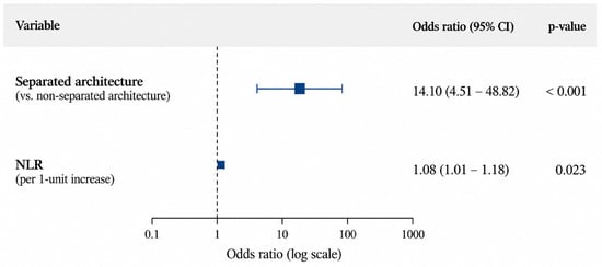
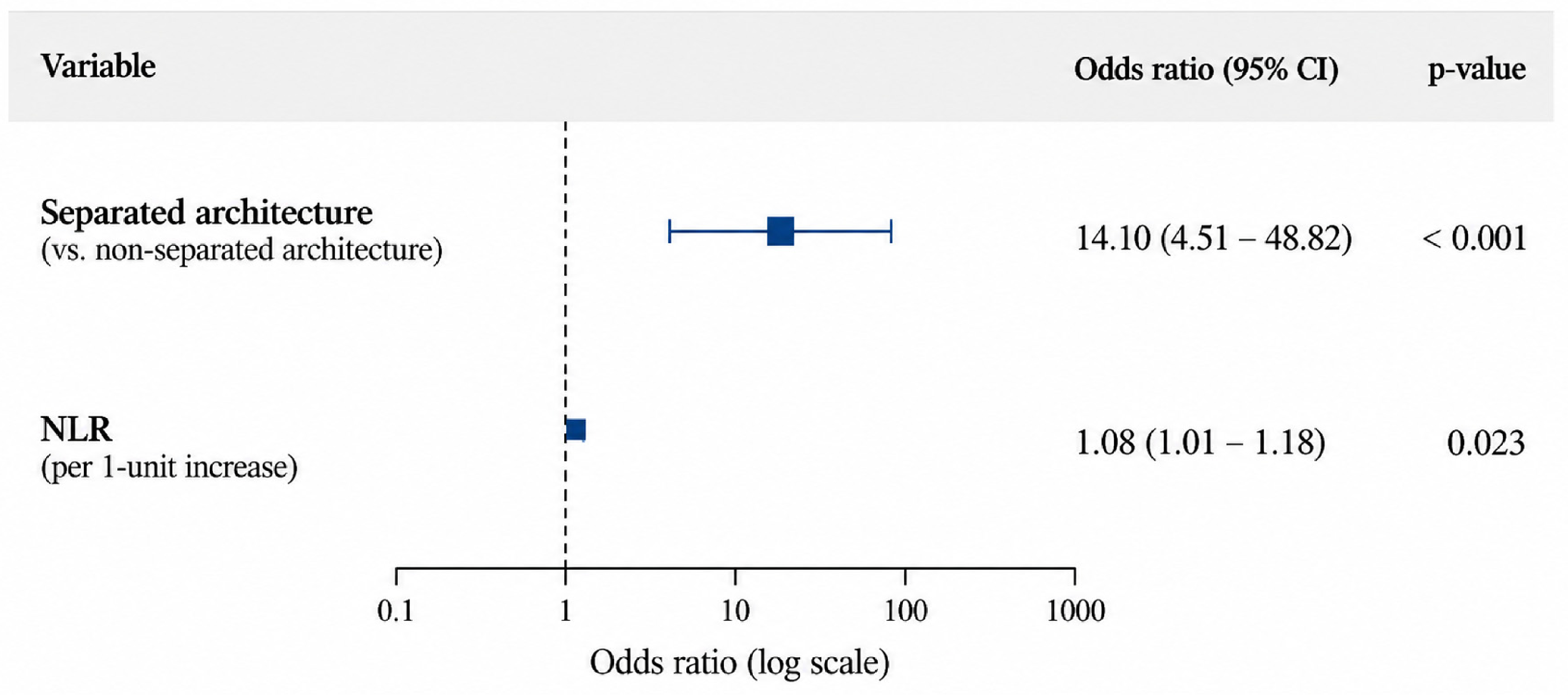
Given the limited number of recurrence events, the multivariable Firth penalized logistic regression model was restricted to two clinically relevant variables: separated hematoma architecture and neutrophil-to-lymphocyte ratio (NLR). Both variables remained independently associated with postoperative recurrence. Separated hematoma architecture showed the strongest association with recurrence (OR 14.10, 95% CI 4.51–48.82; p < 0.001), while higher NLR was also associated with increased odds of recurrence (OR 1.078 per 1-unit increase, 95% CI 1.011–1.182; p = 0.023) (Table 4, Figure 1).
Table 4.
Multivariable logistic regression model for postoperative recurrence.
Figure 1.
Forest plot of factors independently associated with postoperative recurrence of chronic subdural hematoma, identified using multivariable Firth’s penalized logistic regression. The model included separate hematoma architecture and neutrophil-to-lymphocyte ratio (NLR). Squares represent adjusted odds ratios (ORs), and horizontal lines indicate profile penalized likelihood 95% confidence intervals (CIs). The dashed vertical line indicates an OR of 1 (no association). NLR was analyzed per 1-unit increase, and non-separated hematoma architecture served as the reference category.
An exploratory sample size assessment yielded an apparent Cox–Snell R2 of 0.273 and an estimated global shrinkage factor of 0.937 for the two-variable model. The sample size corresponding to a target shrinkage factor of 0.90 was approximately 56 patients under these assumptions. However, these estimates were based on apparent model performance and may be optimistic.
3.4. Perioperative MMP-2 and MMP-9 Profiles
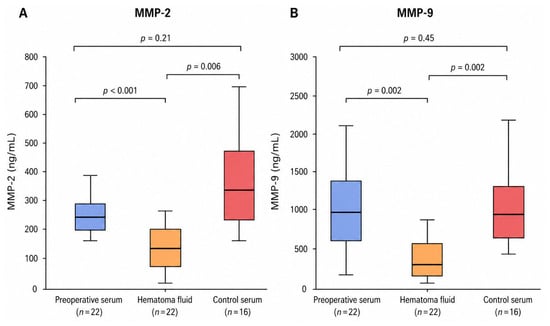
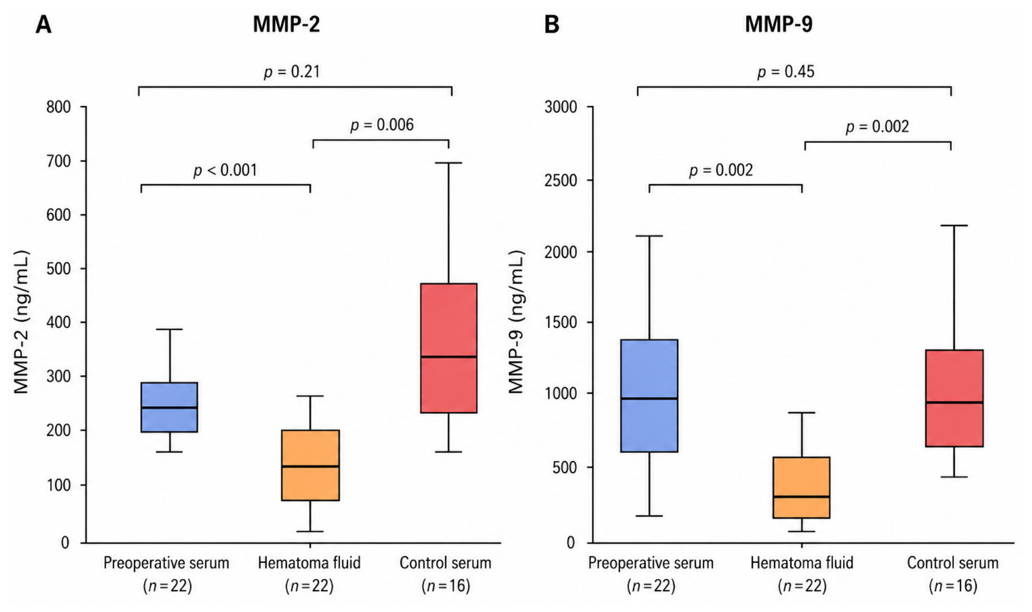
No statistically significant differences in preoperative serum MMP-2 or MMP-9 concentrations were observed between patients with CSDH and control subjects. In the exploratory biomarker subgroup, both MMP-2 and MMP-9 concentrations were significantly lower in hematoma fluid than in paired preoperative serum samples (Table 5, Figure 2).
Table 5.
Comparison of MMP-2 and MMP-9 concentrations between patients with CSDH and control subjects and between serum and hematoma fluid in patients with CSDH.
Figure 2.
Comparison of MMP-2 (A) and MMP-9 (B) concentrations in preoperative serum, hematoma fluid, and control serum. Box plots show the median, interquartile range, and minimum–maximum values. Holm-adjusted post hoc p-values are shown.
3.5. Associations Between MMP-2 and MMP-9 Concentrations and Radiological Characteristics of CSDH
In the exploratory biomarker subgroup, serum MMP-2 and MMP-9 concentrations differed significantly according to hematoma architecture. Higher serum MMP-2 concentrations were observed in separated hematomas (p = 0.016), whereas higher serum MMP-9 concentrations were found in trabecular hematomas (p = 0.014). Hematoma fluid MMP-9 concentrations also differed significantly according to hematoma density (p = 0.011).
These three associations remained statistically significant after Benjamini–Hochberg false discovery rate (FDR) correction (all FDR-adjusted p = 0.042). No other significant associations were observed between MMP concentrations and the radiological characteristics examined (Table 6).
Table 6.
Associations between MMP-2 and MMP-9 concentrations and CT-defined hematoma characteristics in patients with chronic subdural hematoma (n = 22).
3.6. MMP Profiles and Postoperative Recurrence
No significant associations were observed between serum or hematoma MMP-2 and MMP-9 concentrations and postoperative recurrence (Table 7).
Table 7.
Comparison of MMP-2 and MMP-9 concentrations according to postoperative recurrence.
4. Discussion
4.1. Demographic and Clinical Characteristics
Chronic subdural hematoma predominantly affects older adults and occurs more frequently in men, findings that were also observed in our cohort, in which the median patient age was 74 years and 70% of patients were male. Although advanced age, male gender, and neurological status on admission have been extensively investigated as potential factors associated with recurrence, the available evidence remains inconsistent. Several previous studies, including recent meta-analyses, have not consistently identified age or gender as independent predictors of postoperative recurrence [3,10,11,12]. Similarly, neurological status assessed using the Glasgow Coma Scale or the Markwalder Grading Scale has not consistently been associated with recurrence [13,14]. In our cohort, no statistically significant associations were observed between age, gender, or Markwalder grade and postoperative recurrence.
4.2. Comorbidities and Medication
Among the analyzed comorbidities, diabetes mellitus has been extensively investigated as a potential factor associated with chronic subdural hematoma recurrence; however, the available evidence remains inconclusive. Likewise, hypercholesterolemia, arterial hypertension, cardiovascular disease, pulmonary disease, and chronic kidney disease have not been consistently identified as independent predictors of recurrence [13,14,15]. In our cohort, no statistically significant associations were observed between the analyzed comorbidities and postoperative recurrence. Antithrombotic therapy was also not significantly associated with recurrence. Studies and recent meta-analyses have suggested that antithrombotic therapy, particularly anticoagulant use, may increase the risk of postoperative recurrence [16]. The lack of a significant association in our cohort may reflect differences in patient characteristics, treatment strategies or sample size.
4.3. Time from Trauma
Only a few studies have investigated the influence of the interval between head trauma and surgery on chronic subdural hematoma recurrence. Kim et al. reported that patients undergoing surgery during the late subacute stage (15–28 days after trauma) had a lower recurrence rate than those treated during the chronic stage (>28 days) [17]. The relationship between a history of head trauma and postoperative recurrence remains uncertain. A systematic review and meta-analysis did not identify a history of trauma as a significant risk factor for CSDH recurrence [18]. Furthermore, establishing an accurate history of head trauma may be particularly challenging in older patients, as minor traumatic events may be forgotten or considered clinically insignificant, and the exact timing of injury is often uncertain [19].
In our cohort, the absence of a reported history of head trauma was associated with recurrence in the univariable analysis. This variable was not retained in the final multivariable model, and the observed association should be interpreted cautiously. The absence of reported trauma does not necessarily indicate that no traumatic event occurred. Recall bias and uncertainty regarding the timing of minor head injuries may limit the reliability of this variable and complicate assessment of the relationship between trauma-to-surgery interval and postoperative recurrence. Head trauma may influence MMP-2 and MMP-9 profiles through local inflammation and tissue remodeling. The relationship between trauma history and MMP concentrations was not directly examined in our study, and this possible association remains uncertain.
4.4. Laboratory Findings
Among the analyzed laboratory parameters, several markers of systemic inflammation were associated with postoperative recurrence. Patients who developed recurrence had significantly higher NLR, SII, and CRP values than those without recurrence. Importantly, NLR remained independently associated with recurrence in the multivariable Firth-penalized logistic regression model after adjustment for separated hematoma architecture. Each 1-unit increase in NLR was associated with an 8% increase in the odds of recurrence (OR 1.08, 95% CI 1.01–1.18; p = 0.023). These findings are consistent with previous reports showing higher NLR values in patients who developed recurrence [20,21] and support the potential role of systemic inflammation in the pathophysiology of chronic subdural hematoma recurrence. The limited number of recurrence events should be considered when interpreting this association.
Higher SII and CRP values were also found in patients with recurrence. This may indicate a stronger systemic inflammatory response in these patients. Studies have suggested that SII may be a useful and easily available marker of recurrence risk [22]. CRP has also been associated with inflammation in chronic subdural hematoma [23]. However, SII and CRP were not included in the final multivariable model, and their independent associations with recurrence could not be established.
Hemoglobin concentration was lower in patients with recurrence, whereas platelet count and white blood cell count were not significantly different between the groups. Studies have generally not identified hemoglobin concentration as an independent predictor of chronic subdural hematoma recurrence [24,25], and the lack of an association with white blood cell count in our cohort is consistent with previous reports [26]. APTT was also higher in patients with recurrence; however, as this association was observed only in the univariable analysis, its clinical significance remains uncertain and requires confirmation in larger cohorts.
4.5. Radiological Findings
Radiological characteristics are among the strongest and best-established factors associated with chronic subdural hematoma recurrence. Nakaguchi et al. demonstrated that hematoma architecture on computed tomography is associated with postoperative recurrence, with the separated subtype carrying the highest risk [4]. Similar findings have subsequently been reported by other investigators [27,28]. Consistent with these observations, separated hematoma architecture was independently associated with recurrence in our cohort (OR 14.10, 95% CI 4.51–48.82; p < 0.001). The wide confidence interval, however, indicates limited precision of the effect estimate and warrants cautious interpretation.
Hematoma density was also associated with recurrence in the univariable analysis, whereas hematoma laterality, hematoma thickness, and postoperative pneumocephalus were not significantly associated with recurrence. Previous studies have also investigated these radiological characteristics as potential risk factors for recurrence, although their associations have been less consistent than those reported for hematoma architecture [28,29]. Radiological prognostic factors have also been examined in surgically treated acute hematoma [30].
The association between hematoma architecture and recurrence may also reflect underlying biological differences between radiological subtypes. In the exploratory biomarker subgroup (n = 22), serum MMP-2 and MMP-9 concentrations differed according to hematoma architecture, suggesting that different radiological phenotypes of chronic subdural hematoma may be associated with distinct biological profiles.
4.6. MMP-2 and MMP-9 Profiles and Their Associations with Hematoma Characteristics
The biological mechanisms underlying the different radiological appearances of chronic subdural hematoma remain poorly understood [1]. Hematoma architecture and density may reflect different stages of CSDH development and changes in the local tissue environment [6,31]. Inflammation, angiogenesis, and extracellular matrix remodeling play important roles in these processes [32].
Serum MMP-2 concentrations were highest in separated hematomas, a subtype associated with repeated microhemorrhages and active hematoma development. By comparison, the highest serum MMP-9 concentrations were found in trabecular hematomas, which represent a later stage of organization [4]. MMP-2 (gelatinase A) degrades type IV collagen and participates in tissue remodeling and angiogenesis [5]. MMP-9 (gelatinase B) is involved in inflammatory responses, basement membrane degradation, and increased vascular permeability [1,5]. Both enzymes may contribute to the formation of fragile vessels and repeated bleeding within CSDH membranes [1,6,33]. The expression of both enzymes has also been demonstrated in CSDH membranes [34].
Higher serum MMP-2 concentrations in separated hematomas may be related to tissue remodeling and angiogenesis, whereas elevated serum MMP-9 concentrations in trabecular hematomas could reflect inflammatory processes during hematoma organization. Differences in inflammatory markers between radiological subtypes of CSDH have been reported previously [9,35]. One study found higher levels of IL-8 and other inflammatory mediators in trabecular hematomas [35]. Although these observations may help explain our results, the underlying mechanisms remain uncertain because MMP activity and membrane histology were not examined.
Genetic factors may also contribute to the biological heterogeneity of CSDH, although their role remains poorly understood. Apolipoprotein E (APOE) polymorphisms have been associated with differences in neuroinflammatory responses, vascular integrity, and extracellular matrix remodeling [36]. In particular, the APOE ε4 allele has been linked to increased MMP-9 activity and blood–brain barrier dysfunction through activation of the cyclophilin A–MMP-9 pathway [36]. These mechanisms have mainly been investigated in Alzheimer’s disease and other cerebrovascular disorders, and their relevance to CSDH remains uncertain.
Presenilin-1 and presenilin-2, which are involved in amyloid precursor protein processing and other cellular signaling pathways, may also be relevant when considering the relationship between neurodegeneration and vascular pathology [37]. However, there is currently insufficient evidence to establish a direct role for these proteins in CSDH development or recurrence. Inflammation, angiogenesis, and extracellular matrix remodeling are well-recognized processes in CSDH pathophysiology [38], their potential relationship with APOE- and presenilin-associated pathways remains speculative. As genetic markers and neurodegenerative conditions were not specifically evaluated in our cohort, no conclusions regarding their association with hematoma architecture, MMP concentrations, or postoperative recurrence can be drawn.
Schack et al. identified differences in immunological profiles, including MMP-9 levels, across radiological subtypes of CSDH [9]. Our analysis showed associations between serum MMP-2 and MMP-9 concentrations and hematoma architecture, as well as between hematoma fluid MMP-9 concentrations and hematoma density. All three associations remained statistically significant after FDR correction. Hematoma density, however, depends on several factors, including blood degradation products and hematoma composition, and cannot be considered a direct measure of MMP activity. Studies have investigated MMP-2 and MMP-9 in CSDH; evidence regarding the association between MMP-2 concentrations and CT-defined hematoma architecture remains limited. The observed association between serum MMP-2 concentrations and separated hematoma architecture extends previous findings and suggests a potential relationship between MMP-2 and the biological heterogeneity of CSDH.
Unlike Hua et al. [6], we observed lower MMP-2 and MMP-9 concentrations in hematoma fluid than in paired serum samples. Su et al. also reported higher serum MMP-9 concentrations in patients with CSDH [7]. Differences in patient populations, laboratory methods, and sample processing may account for these discrepancies. Preoperative serum MMP-2 and MMP-9 concentrations did not differ significantly between patients with CSDH and controls. Their potential value in distinguishing radiological subtypes therefore requires further investigation, and no conclusions can be drawn regarding their diagnostic usefulness. These observations remain exploratory because the biomarker subgroup comprised only 22 patients.
4.7. MMP Profiles and Postoperative Recurrence
Neither serum nor hematoma MMP-2 and MMP-9 concentrations were significantly associated with postoperative recurrence. In contrast, separated hematoma architecture and higher NLR remained independently associated with recurrence in the multivariable analysis. MMP concentrations also differed according to hematoma architecture and density. These exploratory findings suggest a possible relationship between MMP concentrations and radiological hematoma characteristics, although an association with postoperative recurrence could not be established.
CSDH recurrence involves several biological processes, including persistent inflammation, angiogenesis, repeated microhemorrhages, increased vascular permeability, and extracellular matrix remodeling [1,39]. MMPs are involved in some of these processes and may contribute to changes occurring within the hematoma membranes. The association between MMP concentrations and radiological characteristics may reflect these underlying processes, although the present study does not establish a direct link between MMPs and recurrence. The small biomarker subgroup (n = 22) also limits the interpretation of the negative recurrence findings.
5. Study Limitations
The retrospective, single-center design may limit the generalizability of the results. Although only 20 recurrence events were observed, the multivariable analysis was restricted to two clinically relevant variables and performed using Firth’s penalized logistic regression to reduce the risk of sparse-data bias. An exploratory sample size assessment suggested that the cohort met the selected shrinkage criterion for this model. This assessment was based on apparent model performance and may therefore be optimistic. The wide confidence interval for separated hematoma architecture also indicates limited precision of the effect estimate. The association between the absence of reported head trauma and recurrence should be interpreted cautiously, as only six patients had no reported history of trauma and the information was susceptible to recall bias.
The associations between MMP concentrations and radiological characteristics remained significant after FDR correction; these findings should be interpreted cautiously given the small biomarker subgroup (n = 22). MMP concentrations were measured at a single perioperative time point, precluding the assessment of temporal changes. Given the observational design, these findings do not establish causal relationships or allow reliable conclusions regarding the association between MMP concentrations and postoperative recurrence.
The follow-up period was limited to 3 months, and recurrence was defined as symptomatic hematoma reaccumulation requiring repeat surgery. Asymptomatic recurrences, those occurring after the follow-up period, and patients treated at other institutions may have been missed. The actual recurrence rate may therefore have been underestimated. External validation in larger, prospective, multicenter cohorts is needed to confirm the observed associations.
6. Conclusions
Separated hematoma architecture and higher NLR were independently associated with postoperative recurrence of chronic subdural hematoma. These findings support the relevance of radiological hematoma characteristics and systemic inflammatory markers in the assessment of recurrence risk.
MMP-2 and MMP-9 concentrations differed according to hematoma architecture and density, with three associations remaining statistically significant after FDR correction. These findings extend previous observations on MMP profiles in CSDH and suggest potential biological differences between radiological hematoma subtypes. However, no significant association between MMP concentrations and postoperative recurrence was observed. Given the exploratory nature of the biomarker analysis, these findings warrant further investigation in larger, prospective cohorts.
Author Contributions
Conceptualization, B.K.; methodology, B.K., D.L.-K. and J.K.; formal analysis, B.K.; investigation, B.K., D.L.-K. and J.K.; data curation, B.K.; visualization, B.K.; writing—original draft preparation, B.K.; writing—review and editing, B.K., M.K., D.L.-K., J.K., C.G. and R.M.; supervision, B.K., C.G. and R.M.; project administration, B.K. All authors have read and agreed to the published version of the manuscript.
Funding
This research received no external funding.
Institutional Review Board Statement
The study was conducted in accordance with the Declaration of Helsinki and approved by the Ethics Committee of the John Paul II Catholic University of Lublin (protocol code 13/2024, 3 April 2024).
Informed Consent Statement
Written informed consent was obtained from participants in accordance with the study protocol approved by the Bioethics Committee.
Data Availability Statement
The data generated and analyzed during this study are not publicly available due to privacy considerations related to patient data.
Acknowledgments
Artificial intelligence tools were used exclusively for language editing and translation. The authors take full responsibility for the content of the manuscript.
Conflicts of Interest
The authors declare no conflicts of interest.
References
- Edlmann, E.; Giorgi-Coll, S.; Whitfield, P.C.; Carpenter, K.L.H.; Hutchinson, P.J. Pathophysiology of chronic subdural haematoma: Inflammation, angiogenesis and implications for pharmacotherapy. J. Neuroinflamm. 2017, 14, 108. [Google Scholar] [CrossRef] [Scilit] [PubMed] [PubMed Central]
- Holl, D.C.; Volovici, V.; Dirven, C.M.F.; Peul, W.C.; van Kooten, F.; Jellema, K.; van der Gaag, N.A.; Miah, I.P.; Kho, K.H.; den Hertog, H.M.; et al. Pathophysiology and Nonsurgical Treatment of Chronic Subdural Hematoma: From Past to Present to Future. World Neurosurg. 2018, 116, 402–411.e2. [Google Scholar] [CrossRef] [Scilit] [PubMed]
- Maroufi, S.F.; Farahbakhsh, F.; Macdonald, R.L.; Khoshnevisan, A. Risk factors for recurrence of chronic subdural hematoma after surgical evacuation: A systematic review and meta-analysis. Neurosurg. Rev. 2023, 46, 270. [Google Scholar] [CrossRef] [Scilit] [PubMed]
- Nakaguchi, H.; Tanishima, T.; Yoshimasu, N. Factors in the natural history of chronic subdural hematomas that influence their postoperative recurrence. J. Neurosurg. 2001, 95, 256–262. [Google Scholar] [CrossRef] [Scilit]
- Nagase, H.; Visse, R.; Murphy, G. Structure and function of matrix metalloproteinases and TIMPs. Cardiovasc. Res. 2006, 69, 562–573. [Google Scholar] [CrossRef] [Scilit]
- Hua, C.; Zhao, G.; Feng, Y.; Yuan, H.; Song, H.; Bie, L. Role of Matrix Metalloproteinase-2, Matrix Metalloproteinase-9, and Vascular Endothelial Growth Factor in the Development of Chronic Subdural Hematoma. J. Neurotrauma 2016, 33, 65–70. [Google Scholar] [CrossRef] [Scilit] [PubMed] [PubMed Central]
- Su, G.J.; Gao, J.; Wu, C.W.; Zou, J.F.; Zhang, D.; Zhu, D.L.; Liu, J.; Zhang, J.H.; Huang, X.J. Serum Levels of MMP-8 and MMP-9 as Markers in Chronic Subdural Hematoma. J. Clin. Med. 2022, 11, 902. [Google Scholar] [CrossRef] [Scilit] [PubMed] [PubMed Central]
- Kulesza, B.; Krakowiak, M.; Luchowska-Kocot, D.; Kurzepa, J.; Grochowski, C.; Maciejewski, R. Perioperative Matrix Metalloproteinase-2 and Matrix Metalloproteinase-9 Profiles in Acute and Chronic Subdural Hematomas. J. Clin. Med. 2026, 15, 6499. [Google Scholar] [CrossRef] [Scilit]
- Schack, A.; Rønn Jensen, T.S.; Binderup, T.; Fugleholm, K. Immunoprofile of Radiologic Chronic Subdural Hematoma Subtypes. World Neurosurg. 2025, 193, 815–824. [Google Scholar] [CrossRef] [Scilit] [PubMed]
- Yamamoto, H.; Hirashima, Y.; Hamada, H.; Hayashi, N.; Origasa, H.; Endo, S. Independent predictors of recurrence of chronic subdural hematoma: Results of multivariate analysis performed using a logistic regression model. J. Neurosurg. 2003, 98, 1217–1221. [Google Scholar] [CrossRef] [Scilit]
- Lampros, M.; Katsiou, I.-T.; Kafritsas, G.; Metaxas, D.; Zagorianakou, P.; Zigouris, A.; Skiada, D.; Michos, E.; Pachatouridis, D.; Alexiou, G.A.; et al. Risk factors for recurrence in patients surgically treated for chronic subdural hematomas: A single institutional experience. Surgeries 2025, 6, 19. [Google Scholar] [CrossRef] [Scilit]
- Abdullah, H.M.; Dakurah, T.; Akoto, H.; Osei, K.A.; Yeboah, K.; Agyeman, K.D.; Wepeba, G.; Bankah, P.; Boatey, J.; Ametefe, M.; et al. Predictors of recurrence of chronic subdural haematoma in a cohort study of patients presenting in a sub-Saharan African teaching hospital. BMC Neurol. 2022, 22, 346. [Google Scholar] [CrossRef] [Scilit]
- Liu, L.X.; Cao, X.D.; Ren, Y.M.; Zhou, L.X.; Yang, C.H. Risk factors for recurrence of chronic subdural hematoma: A single-center experience. World Neurosurg. 2019, 132, e506–e513. [Google Scholar] [CrossRef] [Scilit]
- Lee, S.W.; Sin, E.G. Risk factors for the recurrence of chronic subdural hematoma. Korean J. Neurotrauma 2024, 20, 80–89. [Google Scholar] [CrossRef] [Scilit]
- Torihashi, K.; Sadamasa, N.; Yoshida, K.; Narumi, O.; Chin, M.; Yamagata, S. Independent predictors for recurrence of chronic subdural hematoma: A review of 343 consecutive surgical cases. Neurosurgery 2008, 63, 1125–1129. [Google Scholar] [CrossRef] [Scilit]
- Wang, H.; Zhang, M.; Zheng, H.; Xia, X.; Luo, K.; Guo, F.; Qian, C. The effects of antithrombotic drugs on the recurrence and mortality in patients with chronic subdural hematoma: A meta-analysis. Medicine 2019, 98, e13972. [Google Scholar] [CrossRef] [Scilit]
- Kim, D.I.; Kim, J.H.; Kang, H.I.; Moon, B.G.; Kim, J.S.; Kim, D.R. Impact of time interval between trauma onset and burr-hole surgery on recurrence of late subacute or chronic subdural hematoma. J. Korean Neurosurg. Soc. 2016, 59, 498–504. [Google Scholar] [CrossRef] [Scilit]
- Mishra, R.; Deora, H.; Florez-Perdomo, W.A.; Moscote-Salazar, L.R.; Garcia-Ballestas, E.; Rahman, M.M.; Shrivastava, A.; Raj, S.; Chavda, V.; Montemurro, N. Clinical and Radiological Characteristics for Recurrence of Chronic Subdural Hematoma: A Systematic Review and Meta-Analysis. Neurol. Int. 2022, 14, 683–695. [Google Scholar] [CrossRef] [Scilit] [PubMed] [PubMed Central]
- Sundblom, J.; Sandberg, E.; Ronne-Engström, E. Trauma Mechanisms and Surgical Outcomes in the Elderly Patient with Chronic Subdural Hematoma. Can. Geriatr. J. 2022, 25, 40–48. [Google Scholar] [CrossRef] [Scilit] [PubMed] [PubMed Central]
- Ertuğrul, B.; Kömürcü, H. Inflammatory and Metabolic Biomarkers Associated with Chronic Subdural Hematoma Recurrence: A Single-Center Retrospective Cohort Study. Clin. Pract. 2026, 16, 193. [Google Scholar] [CrossRef] [Scilit]
- Dobran, M.; Carrassi, E.; Di Rienzo, A.; Lattanzi, S.; Capece, M.; Aiudi, D.; Iacoangeli, M. Preoperative neutrophil-to-lymphocyte ratio as a predictor of chronic subdural hematoma recurrence. Interdiscip. Neurosurg. 2022, 28, 101436. [Google Scholar] [CrossRef] [Scilit]
- Vaibhav, K.; Sahu, A.; Prasad, R.S.; Deb, D.; Kumar, D.; Karimi, A. Prediction of postoperative recurrence of chronic subdural hematoma using preoperative systemic immune inflammation index and eosinophils. Asian J. Neurosurg. 2024, 19, 707–714. [Google Scholar] [CrossRef] [Scilit]
- Hansen, J.T.; Tursø, T.S.V.; Olsen, M.H.; Binderup, T.; Fugleholm, K.; Jensen, T.S.R. Subdural and systemic C-reactive protein in patients with chronic subdural hematoma recurrence. Can. J. Neurol. Sci. 2026, 53, 247–253. [Google Scholar] [CrossRef] [Scilit]
- Barić, H.; Komljenović, S.; Bilić, K.; Migo, W.; Vitowanu, J.M.; Desnica, A.; Mrak, G. Hematologic indices and chronic subdural hematoma: A single-center cohort study. Asian J. Neurosurg. 2024, 19, 735–740. [Google Scholar] [CrossRef] [Scilit]
- Zawy Alsofy, S.; Lewitz, M.; Meyer, K.; Fortmann, T.; Wilbers, E.; Nakamura, M.; Ewelt, C. Retrospective analysis of risk factors for recurrence of chronic subdural haematoma after surgery. J. Clin. Med. 2024, 13, 805. [Google Scholar] [CrossRef] [Scilit]
- Güresir, Á.; Coch, C.; Heine, A.; Mass, E.; Lampmann, T.; Vatter, H.; Velten, M.; Schmitz, M.-T.; Güresir, E.; Wach, J. Red blood cell distribution width to platelet count ratio facilitates preoperative prediction of recurrence in surgically treated chronic subdural hematoma. Front. Neurol. 2022, 13, 884231. [Google Scholar] [CrossRef] [Scilit]
- Kwon, S.M.; Lee, M.H.; Seo, Y.; Kim, Y.I.; Oh, H.-J.; Kim, K.H.; Choi, K.-S.; Chong, K. A radiological assessment of chronic subdural hematomas. Korean J. Neurotrauma 2022, 18, 12–21. [Google Scholar] [CrossRef] [Scilit]
- Miah, I.P.; Tank, Y.; Rosendaal, F.R.; Peul, W.C.; Dammers, R.; Lingsma, H.F.; Hertog, H.M.D.; Jellema, K.; van der Gaag, N.A.; Dutch Chronic Subdural Hematoma Research Group. Radiological prognostic factors of chronic subdural hematoma recurrence: A systematic review and meta-analysis. Neuroradiology 2021, 63, 27–40. [Google Scholar] [CrossRef] [Scilit]
- You, C.G.; Zheng, X.S. Postoperative pneumocephalus increases the recurrence rate of chronic subdural hematoma. Clin. Neurol. Neurosurg. 2018, 166, 56–60. [Google Scholar] [CrossRef] [Scilit]
- Kulesza, B.; Krakowiak, M.; Brzozowska, A.; Grochowski, C.; Maciejewski, R. Clinical, Laboratory, and Radiological Predictors of Three-Month Outcome After Surgical Treatment of Acute Extra-Axial Hematomas. J. Clin. Med. 2026, 15, 5797. [Google Scholar] [CrossRef] [Scilit]
- Chen, H.; Colasurdo, M.; Malhotra, A.; Gandhi, D.; Bodanapally, U.K. Advances in chronic subdural hematoma and membrane imaging. Front. Neurol. 2024, 15, 1366238. [Google Scholar] [CrossRef] [Scilit] [PubMed] [PubMed Central]
- Trieu, M.; Thomas, A.J. Pathophysiology of chronic subdural hematoma—New insights. Ther. Adv. Neurol. Disord. 2026, 19, 17562864261435399. [Google Scholar] [CrossRef] [Scilit]
- Arunachalam Sakthiyendran, N.; Gonzalez-Salido, J.; Perkins, T.; Enriquez-Marulanda, A.; Dasenbrock, H.; Holsapple, J. The subdural membrane: Unraveling the pathophysiology and treatment of chronic subdural hematoma. Front. Surg. 2025, 12, 1694048. [Google Scholar] [CrossRef] [Scilit]
- Nakagawa, T.; Kodera, T.; Kubota, T. Expression of matrix metalloproteinases in the chronic subdural haematoma membrane. Acta Neurochir. 2000, 142, 61–66. [Google Scholar] [CrossRef] [Scilit] [PubMed]
- Svedung Wettervik, T.; Sundblom, J.; Ronne-Engström, E. Inflammatory biomarkers differentiate the stage of maturation in chronic subdural hematomas. J. Neuroimmunol. 2023, 381, 578127. [Google Scholar] [CrossRef] [Scilit] [PubMed]
- Alkhalifa, A.E.; Al-Ghraiybah, N.F.; Odum, J.; Shunnarah, J.G.; Austin, N.; Kaddoumi, A. Blood-Brain Barrier Breakdown in Alzheimer’s Disease: Mechanisms and Targeted Strategies. Int. J. Mol. Sci. 2023, 24, 16288. [Google Scholar] [CrossRef] [Scilit] [PubMed] [PubMed Central]
- Sun, Y.; Islam, S.; Michikawa, M.; Zou, K. Presenilin: A Multi-Functional Molecule in the Pathogenesis of Alzheimer’s Disease and Other Neurodegenerative Diseases. Int. J. Mol. Sci. 2024, 25, 1757. [Google Scholar] [CrossRef] [Scilit] [PubMed] [PubMed Central]
- Xu, Z.; Yang, S.; Zheng, G.; Chen, H.; Wan, X.; Xu, H.; Xu, Y.; Wang, J.; Tong, M.; Tu, Q. A novel paradigm for chronic subdural hematoma: Regenerative medicine strategies targeting the pathological microenvironment. Front. Cell Dev. Biol. 2026, 14, 1757623. [Google Scholar] [CrossRef] [Scilit]
- Georgountzos, G.; Gkalonakis, I.; Anastasopoulos, L.; Stranjalis, G.; Kalamatianos, T. Biofluid Biomarkers in the Prognosis of Chronic Subdural Hematoma: A Systematic Scoping Review. Diagnostics 2023, 13, 2449. [Google Scholar] [CrossRef] [Scilit] [PubMed] [PubMed Central]

Disclaimer/Publisher’s Note: The statements, opinions and data contained in all publications are solely those of the individual author(s) and contributor(s) and not of MDPI and/or the editor(s). MDPI and/or the editor(s) disclaim responsibility for any injury to people or property resulting from any ideas, methods, instructions or products referred to in the content. |
© 2026 by the authors. Licensee MDPI, Basel, Switzerland. This article is an open access article distributed under the terms and conditions of the Creative Commons Attribution (CC BY) license.