


Abstract
Diabesotension, the coexistence of diabetes mellitus, obesity, and hypertension, represents an increasingly prevalent and clinically significant cardiometabolic syndrome. Epidemiological evidence highlights strong bidirectional relationships among these conditions, which together markedly elevate the risk of cardiovascular disease, stroke, nephropathy, and premature mortality. The pathophysiology of diabesotension is multifactorial, involving insulin resistance, chronic hyperglycemia, obesity-associated inflammation, endothelial dysfunction, sympathetic overactivity, and activation of the renin–angiotensin–aldosterone system (RAAS), culminating in adverse metabolic and hemodynamic interactions. Clinically, the synergistic presence of diabetes and hypertension accelerates both microvascular and macrovascular complications, leading to poorer outcomes than either condition alone. Recognizing diabesotension as a distinct clinical entity may facilitate more comprehensive care models and improved long-term outcomes, highlighting the need for continued research to refine risk stratification and optimize individualized treatment strategies. In this review, we focus on the genes implicated in key hallmarks of diabesotension, including insulin resistance, inflammation, RAAS activation, and oxidative stress, relevant clinical management strategies, and AI-based models for risk stratification of this triad of diseases.
1. Introduction
Diabesotension, defined as the coexistence of diabetes mellitus, obesity, and hypertension, represents a severe cardiometabolic phenotype characterized by the convergence of three major chronic diseases that frequently co-occur and share overlapping pathophysiological mechanisms. Although diabesotension shares features with broader cardiometabolic syndrome, it is defined by the co-occurrence of diabetes, obesity and hypertension, providing a clinically distinct framework for interrogating the shared and interacting mechanisms that underpin their combined disease burden. Individuals with diabesotension exhibit elevated risk of cardiovascular disease, stroke, nephropathy, and premature mortality [1,2]. Epidemiological studies reveal that more than half of patients with type 2 diabetes develop hypertension, while individuals with hypertension are at greater risk of impaired glucose tolerance and diabetes [3,4]. This bidirectional association highlights the need to view diabesotension as a distinct entity rather than as three isolated conditions.
The pathogenesis of diabesotension is multifactorial and involves complex metabolic and hemodynamic interactions. Insulin resistance, a common feature in type 2 diabetes, plays a central role by promoting hyperinsulinemia, sympathetic overactivity, sodium retention, and endothelial dysfunction, all of which contribute to elevated blood pressure [5,6]. Chronic hyperglycemia further exacerbates vascular injury through oxidative stress and advanced glycation end-products, while obesity and low-grade systemic inflammation accelerate the progression of both disorders [7,8]. In addition, activation of the RAAS and increased arterial stiffness create a physiological environment conducive to the development and maintenance of hypertension in diabetic individuals [9].
The clinical consequences of diabesotension are profound, with patients experiencing higher rates of both microvascular and macrovascular complications compared to those with either diabetes or hypertension alone [2,10]. Hypertension accelerates the progression of diabetic nephropathy, retinopathy, and neuropathy, while diabetes worsens the vascular damage associated with high blood pressure. Together, they significantly increase the risk of coronary artery disease, stroke, heart failure, and end-stage renal disease [1]. This synergistic effect underscores the importance of early detection, aggressive risk factor modification, and comprehensive treatment strategies tailored to address both conditions simultaneously.
Effective management of diabesotension requires a holistic and multidisciplinary approach. Lifestyle modification, including dietary adjustments, physical activity, and weight reduction, serves as the foundation of therapy [11,12]. Pharmacological interventions are equally critical, with angiotensin-converting enzyme inhibitors (ACEIs) and angiotensin receptor blockers (ARBs) preferred for their renoprotective and cardioprotective properties [13,14]. Recent advances in glucose-lowering therapies, such as sodium-glucose cotransporter 2 (SGLT2) inhibitors and glucagon-like peptide-1 (GLP-1) receptor agonists, provide additional cardiovascular and renal benefits [15]. By recognizing diabesotension as a unique clinical syndrome, healthcare systems can promote integrated care models that reduce long-term complications and improve patient outcomes.
2. Genetic Basis Underlying Diabesotension
2.1. Diabesotension-Associated Genes
Diabesotension arises from a multifactorial interplay of numerous genes that collectively influence disease susceptibility, onset, and progression. Over the past decade, thousands of genes have been identified as being associated with obesity, diabetes mellitus, and hypertension. Disease-associated genes were retrieved from the National Center for Biotechnology Information (NCBI) Gene database (https://www.ncbi.nlm.nih.gov/gene/) (accessed on 31 August 2025) [16]. Separate searches were conducted using the disease terms “obesity”, “hypertension”, and “diabetes” (date of retrieval: 30 August 2025). Genes returned as being associated with each disease term were collected and gene identifiers were standardized before comparison. After removal of duplicate entries, 2191 genes associated with obesity, 1349 genes associated with hypertension, and 2494 genes associated with diabetes were identified. The three disease-associated gene sets were intersected using Venny 2.1 (https://bioinfogp.cnb.csic.es/tools/venny/) (accessed on 31 August 2025), resulting in 443 genes common to all three conditions. These overlapping genes were defined as candidate diabesotension-related genes and were used for subsequent analyses (Figure 1A). All 443 genes were then subjected to PANTHER v16.0 (https://www.pantherdb.org) (accessed on 31 August 2025) to categorize them into protein classes [17]. Of the 443 overlapping genes, 437 were successfully annotated (Figure 1B) and subsequently subjected to enrichment analysis using the Human Phenotype Ontology (HPO) and biological pathway databases, including KEGG, Reactome, and WikiPathways in g:Profiler (https://biit.cs.ut.ee/gprofiler/gost) (accessed on 31 August 2025) [18]. Benjamini–Hochberg FDR and p-adj value ≤ 0.05 were used to determine the statistical significance of human phenotype and biological pathways.
Figure 1.
List of diabesotension-related genes. (A) A total of 443 diabesotension-related genes were retrieved from the National Center for Biotechnology Information (NCBI; https://www.ncbi.nlm.nih.gov) (accessed on 31 August 2025). (B) A total of 437 diabesotension-related genes were categorized into 21 primary protein classes. # represents the number.
Most of the identified genes were prominently classified as metabolite interconversion enzymes (21.8%), followed by intercellular signal molecules (14.6%) and protein-modifying enzymes (12.2%) (Figure 1B). Metabolite interconversion enzymes (MIEs), including hydrolases, lyases, oxidoreductases, and transferases, are crucial in obesity as they influence lipid, carbohydrate, and amino acid metabolism, leading to metabolic dysfunction and insulin resistance [19]. Their dysregulation is also observed in diabetes, particularly in glucokinase, which controls insulin secretion and glucose storage, and whose impaired activity can result in hyperglycemia, inflammation, and complications such as non-alcoholic fatty liver disease (NAFLD). Meanwhile, in hypertension, key MIEs include cytochrome P450 (CYP) enzymes, particularly CYP4A and CYP4F, which convert arachidonic acid to 20-hydroxyeicosatetraenoic acid (20-HETE), a molecule that elevates blood pressure [20,21,22]. Other important MIEs include 11β-hydroxysteroid dehydrogenases (11β-HSDs), which activate glucocorticoids, and angiotensin-converting enzyme (ACE), a known drug target that converts angiotensin I to angiotensin II [23,24].
In obesity, intercellular signals involve hormones such as leptin and insulin, inflammatory cytokines from TLR-activated adipose tissue, mitogen-activated protein kinases (ERK, JNK, p38) that regulate appetite and inflammation, and fatty acids that influence metabolism [25,26]. Disruptions in downstream pathways, particularly PI3K/AKT and JAK/STAT, contribute to metabolic dysfunction, insulin resistance, and related complications [27,28]. In diabetes, similar mechanisms are at play, where hormones like insulin and glucagon, islet-derived paracrine factors (somatostatin and growth factors), and adipokines (TNF-α, IL-6, and resistin) regulate insulin sensitivity and islet function. Dysregulated adipokine release and paracrine signaling exacerbate insulin resistance and accelerate diabetes progression [29,30,31,32]. In hypertension, neurohormones such as angiotensin II and norepinephrine can also trigger downstream PI3K/AKT signaling via activation of G protein-coupled receptors [33]. Protein-modifying enzymes, on the other hand, contribute to obesity, diabetes, and hypertension through post-translational modifications (PTMs) that regulate metabolism, insulin signaling, lipid homeostasis, and vascular function [34]. For example, a negative regulator, Protein Tyrosine Phosphatase 1B (PTP1B), is overexpressed in obesity and disrupts insulin signaling by dephosphorylating the insulin receptor and insulin receptor substrate 1 (IRS-1) [35,36]. Likewise, histone deacetylases (HDACs) affect lipid metabolism, and dysregulation of HDAC3 and HDAC6 can promote weight gain and metabolic disorders [37,38].
2.2. Genes Involved in Hallmarks of Diabesotension
2.2.1. Insulin Resistance
The regulation of insulin sensitivity and glucose metabolism is orchestrated by a complex interplay of genetic and molecular pathways encompassing hormonal signaling, lipid metabolism, and inflammatory responses (Table 1). Among these, AGT modulates insulin sensitivity through renin–angiotensin system-dependent mechanisms, with specific haplotypes (such as TCG) linked to increased T2DM susceptibility [39]. Downstream of insulin receptor signaling, AKT1 serves as a pivotal kinase in the PI3K-Akt cascade, directly promoting glucose uptake; its impaired activation contributes to insulin resistance, whereas enhanced Akt signaling improves systemic metabolic homeostasis [27,28]. The lipid transporter CD36 regulates fatty acid uptake and oxidation, maintaining metabolic balance under physiological conditions but promoting lipid accumulation and insulin resistance in states of nutrient excess [40]. Similarly, FOXO1, a transcriptional regulator integrating insulin and nutrient signals, promotes hepatic gluconeogenesis and lipid oxidation while suppressing glucose uptake and insulin secretion [41].
Inflammatory mediators such as IL6 and TNF disrupt insulin receptor function through SOCS3- and NF-κB-dependent mechanisms, reinforcing the inflammatory–metabolic feedback loop characteristic of obesity-associated insulin resistance [42,43]. Core components of insulin signaling, including INS, INSR, IRS1, and IRS2, are essential for maintaining glucose homeostasis; genetic or functional disruption of these elements impairs insulin secretion, receptor binding, and downstream signaling, resulting in hyperinsulinemia and glucose intolerance [44,45]. Stress-activated and nutrient-sensing kinases, MAPK8 (JNK1), MTOR, and PTEN, further modulate insulin pathway integrity, where chronic mTORC1 activation, JNK1-mediated IRS inhibition, or excessive PTEN activity converge to dampen insulin sensitivity [46].
Genes governing mitochondrial function and lipid metabolism, such as PPARA, PPARGC1A, and SREBF1, shape systemic insulin responsiveness through their control of oxidative and biosynthetic pathways. PPARα activation enhances fatty acid oxidation and suppresses inflammation, thereby improving insulin sensitivity, while the coactivator PGC-1α sustains mitochondrial biogenesis and thermogenesis; loss of PGC-1α function impairs lipid metabolism and promotes insulin resistance under nutrient excess [47]. Conversely, elevated SREBP1 activity perturbs insulin receptor and Akt phosphorylation, leading to mitochondrial dysfunction and dysregulated lipid storage [48,49]. Other key modulators include PIK3R1, where pathogenic variants compromise PI3K-dependent signaling [50]; SOCS3 and STAT3 [51,52], which inhibit IRS1 and Akt activation in skeletal muscle; and PTPN1 (PTP1B), which attenuates insulin receptor phosphorylation, contributing to cardiac insulin resistance [53]. The insulin-responsive glucose transporter SLC2A4 (GLUT4) remains a crucial effector of glucose uptake, with its upregulation ameliorating insulin resistance in metabolic disease contexts [54].
Finally, additional loci, including NOS3, PRKAG2, and TRIB3, integrate vascular, energetic, and stress responses into insulin signaling networks. NOS3 variants influence nitric oxide-mediated endothelial and metabolic regulation [55]; PRKAG2 mutations impair AMPK activation and myocardial glucose uptake [56]; and TRIB3 modulates glucose-induced negative feedback on insulin action [57]. Collectively, these genes delineate a convergent regulatory framework in which inflammation, lipid handling, mitochondrial dynamics, and kinase signaling intersect to determine tissue-specific insulin sensitivity and overall glucose homeostasis.
Table 1.
List of 26 diabesotension-associated genes involved in insulin resistance.
2.2.2. Inflammation
Inflammation constitutes a major molecular axis linking metabolic and vascular dysfunction in diabesotension. Several genes central to immune signaling, lipid metabolism, and endothelial activation contribute to this pathophysiology (Table 2). AKT1, a key effector of PI3K signaling, regulates acute inflammatory responses and vascular permeability by controlling leukocyte extravasation and endothelial barrier integrity [77]. CXCL8 (IL-8) orchestrates neutrophil recruitment and activation through CXCR1/2 and ACKR1 signaling, a process modulated by chemokine dimerization and post-translational modification [78]. The pro-inflammatory cytokine IL12B enhances insulin resistance and endothelial dysfunction, with elevated IL-12 levels linking systemic inflammation to β-cell stress and atherosclerotic risk [79]. Similarly, IL6 serves as both a predictive and pathogenic mediator across diabetes subtypes, including type 1, type 2, and gestational diabetes, and contributes to diabetic complications such as nephropathy, neuropathy, and cognitive impairment [80].
Hyperglycemia-activated signaling cascades further amplify inflammatory injury through mitogen-activated protein kinases. MAPK1, MAPK3, and MAPK14 (p38α) mediate stress-induced and pro-inflammatory responses in vascular and renal tissues. MAPK1 promotes oxidative stress and cardiac remodeling, while MAPK3 activation drives renal inflammation and fibrosis in diabetic nephropathy [81,82]. MAPK14 functions as a central stress transducer, regulating cytokine gene expression and inflammatory signaling, and is emerging as a target for inflammation-driven metabolic disease [83]. Transcriptional regulators NFKB1 and TNF integrate inflammatory signaling with metabolic dysfunction. NF-κB polymorphisms modulate cardiovascular inflammation in type 2 diabetes, particularly through interactions with other immune mediators, while TNF, secreted by adipocytes and immune cells, promotes chronic low-grade inflammation that underlies obesity-induced insulin resistance and endothelial dysfunction [84,85]. Finally, RETN links adipose-derived inflammation to vascular pathology by promoting arterial inflammation and atherogenesis, thereby connecting obesity, hypertension, and insulin resistance through macrophage-driven signaling [86]. Collectively, these genes delineate an inflammatory network that integrates metabolic stress with vascular remodeling, serving as a molecular bridge between diabetes and hypertension.
Table 2.
List of 10 diabesotension-associated genes involved in inflammation.
2.2.3. Neurohormonal Regulation (RAAS)
Neurohormonal mechanisms, encompassing central insulin signaling, neurotrophic regulation, and RAAS interactions, are integral to diabesotension pathophysiology [9]. Sixteen diabesotension-associated genes have been reported to be involved in RAAS, as listed in Table 3. AKT1 supports neuronal insulin signaling, partially restoring AS160 phosphorylation and glucose uptake when Akt2 function is impaired, thereby influencing neuroendocrine regulation and metabolic homeostasis [87]. Transcriptional and apoptotic regulators such as ATF4, BCL2, and FOXO3 mediate neuronal plasticity, excitability, and survival. ATF4 controls GABAB receptor trafficking and synaptic transmission via cytoskeletal signaling [88], while the anti-apoptotic BCL-2 family maintains mitochondrial integrity in neurons under metabolic stress [89]. FOXO3 integrates stress responses and neuronal maintenance, preventing axonal degeneration and influencing susceptibility to neurodegenerative disorders associated with metabolic imbalance [90].
Neurotrophic and insulin signaling pathways intersect through BDNF, NTRK2 (TrkB), and IRS1. Reduced BDNF levels in diabetes link metabolic dysfunction to impaired neuroprotection [91], whereas NTRK2 activation by BDNF triggers MAPK, PLCγ, and PI3K pathways essential for neuronal differentiation and synaptic maintenance [92]. IRS1, a central mediator of neuronal insulin signaling, regulates PI3K–Akt activity; its reduced function in diabetes disrupts neuronal and neurovascular integrity [93]. MAPK family members, including MAPK1, MAPK3, MAPK8, and MAPK14, also play multifaceted roles in neurohormonal regulation. MAPK1 and JNK3 contribute to neurotoxicity and degeneration [94]; MAPK14 impairs autophagy and BACE1 clearance, linking metabolic stress to neurodegeneration [95]; and MAPK3 facilitates leptin-dependent transcriptional regulation of neuronal responses controlling appetite and energy balance [96]. Similarly, MAPK8 (JNK1/3) orchestrates hypothalamic POMC and AgRP neuron activity, influencing feeding behavior, thermogenesis, and systemic energy balance [97].
Inflammatory transcription factors such as NFKB1 connect nutrient-induced stress to hypothalamic inflammation and neuroendocrine dysregulation, representing a shared pathway between metabolic and neurodegenerative disease [98]. Additional signaling regulators, including PIK3R1, RAC1, SORT1, and TP53, contribute variably to neuronal survival, synaptic remodeling, and insulin resistance-related neuroendocrine dysfunction. PIK3R1 mutations impair AKT phosphorylation and neuronal insulin signaling [99], RAC1 inhibition prevents hypertension and renal damage [100], SORT1 mediates neuronal survival and death across CNS and metabolic tissues [101], and TP53, through its mitochondrial apoptotic activity, promotes β-cell dysfunction and neuronal vulnerability under metabolic stress [102]. Collectively, these neurohormonal regulators illustrate how metabolic and neural circuits converge through insulin, trophic, and inflammatory signaling to maintain systemic energy and vascular homeostasis.
Table 3.
List of 16 diabesotension-associated genes involved in neurohormonal regulation.
2.2.4. Oxidative Stress
Oxidative stress represents a key pathogenic mechanism linking metabolic dysfunction and vascular injury in diabesotension (Table 4). This process arises from an imbalance between reactive oxygen species (ROS) generation and antioxidant defense, governed by a complex interplay of enzymatic, signaling, and transcriptional regulators [103,104]. Central antioxidant enzymes, including CAT, GPX3, SOD1, SOD2, SOD3, and NQO1, constitute the first line of defense against oxidative damage by detoxifying ROS and maintaining cellular redox equilibrium [105,106,107,108,109,110]. Reduced activity or expression of these enzymes in diabetes or obesity enhances mitochondrial dysfunction and tissue injury, promoting insulin resistance and endothelial impairment. HMOX1, through heme degradation and carbon monoxide production, exerts cytoprotective and anti-inflammatory effects [111], while MAOA, CYBB, NOX4, and NOX5 act as major enzymatic sources of ROS, driving oxidative injury in renal, cardiac, and vascular tissues [112,113,114,115].
Transcriptional control of oxidative homeostasis is largely coordinated by NFE2L2 (NRF2), a master regulator of antioxidant responses, and NFKB1, which integrates oxidative, inflammatory, and apoptotic signaling. Crosstalk between NRF2 and NF-κB pathways determines whether oxidative stress leads to adaptation or cellular injury [116]. Similarly, redox-sensitive kinases such as MAPK14, MAPK8, and MTOR transduce oxidative cues into metabolic and inflammatory outcomes, modulating autophagy, apoptosis, and cellular resilience [117,118,119]. The interplay between these kinases and mitochondrial regulators like AKT1 and PINK1 influences tissue-specific responses to oxidative burden, from adaptive hypertrophy to apoptosis [120,121].
Cellular stress and survival are further modulated by BCL2, TP53, and CDKN1A, which govern mitochondrial integrity, cell-cycle arrest, and senescence under oxidative pressure. Their dysregulation contributes to β-cell failure, adipose tissue aging, and systemic insulin resistance [89,122,123]. Pro-inflammatory cytokines and chemokines, such as IL1A, IL6, TNF, and CXCL8, amplify oxidative injury through sustained activation of NADPH oxidases and inflammatory transcription factors, perpetuating metabolic inflammation and vascular dysfunction [124,125,126,127]. Meanwhile, metabolic regulators such as FOXO1, PTEN, and SIRT1 act as redox-sensitive switches that coordinate antioxidant gene expression and insulin signaling via PI3K–Akt and AMPK pathways [128,129,130]. Structural and enzymatic mediators including ICAM1, PTGS2, and XDH further link oxidative stress to endothelial activation, vascular remodeling, and lipid oxidation [131,132,133].
Together, these genes delineate a tightly interconnected redox regulatory network in which impaired antioxidant capacity, excessive ROS production, and maladaptive stress signaling converge to drive the metabolic, inflammatory, and vascular complications characteristic of diabesotension.
Table 4.
List of 32 diabesotension-associated genes involved in oxidative stress.
3. The Pathogenesis of Diabesotension
Diabesotension develops through the interplay of insulin resistance, inflammation, neurohormonal dysregulation, and oxidative stress. These interconnected hallmarks form an integrated pathogenic network linking diabetes, obesity, and hypertension, as reflected by the shared and functionally interconnected genes identified (Figure 2). Several genes, such as AKT1, PIK3R1 and PTEN, are repeatedly implicated across the hallmarks, highlighting the shared molecular basis to the pathogenesis of diabesotension. For example, dysregulation of Akt signaling impairs glucose uptake [27] and alters endothelial and inflammatory responses [136], thereby linking insulin resistance to elevated inflammation and vascular dysfunction. Similarly, pathogenic alterations in PIK3R1 compromise PI3K-dependent insulin signaling, resulting in profound insulin resistance and metabolic dysfunction [137,138]. Overexpression of PIK3R1 was reported to mimic glucocorticoid, a stress hormone that is known to induce insulin resistance, hyperglycemia, and dyslipidemia as long-term effects [138], linking insulin resistance to neurohormonal regulation. PTEN, the negative regulator of phosphatidylinositol 3-kinase (P13K), was suggested to be associated with metabolic disorder by affecting insulin sensitivity, inflammation, and neurohormonal regulation via the regulation of vagal function through vagus nerve activity. Mice with impaired efferent vagal output were found to exhibit peripheral insulin resistance, glucose intolerance, and a pro-inflammatory state in peripheral metabolic tissues and serum [139]. Collectively, these shared genes and the underlying mechanisms reveal the interconnected relationships among hallmarks, creating a permissive environment for the pathogenesis of diabesotension.
Figure 2.
Convergence of genes across biological hallmarks associated with diabesotension. Venn diagram illustrating the distribution and recurrence of genes across the major biological hallmarks examined, including insulin resistance, inflammation, oxidative stress, and neurohormonal regulation. Numbers indicate the number of genes unique to or shared among the respective hallmarks, while gene symbols within overlapping regions identify recurrent genes contributing to multiple biological processes.
The hallmarks of diabesotension are suggested to be dynamically related throughout its pathogenesis (Figure 3). Insulin resistance, which represents the fundamental anomaly in metabolic disorders, particularly diabetes, acts as the initial trigger for the subsequent pathogenic events. Disruption of insulin sensitivity decreases glucose uptake in insulin-sensitive tissues, increases glycogenolysis, alters insulin secretion, and increases lipid accumulation via lipolysis in adipose tissue [140], which can lead to obesity. Moreover, insulin resistance can compromise mitochondrial function, further contributing to its severity. The decrement in mitochondrial oxidative capacity and impairment of lipid metabolism in obese and diabetic patients were reported to increase the availability of intramyocellular fatty acids, potentially being channeled towards lipotoxic lipid species biosynthesis such as ceramide and diacylglycerol, in which both are associated with insulin resistance [141]. Then, elevated adipose tissue in diabetes and obese individuals notably increased inflammatory cytokines such as IL-6, IL-1β, MCP-1, and TNF-α. These cytokines produced by adipose tissues are found to increase dyslipidemia and further contribute to insulin resistance [142]. Prolonged insulin resistance further alters vascular and renal sodium handling by altering neurohormonal regulation via RAAS activity. Overactive RAAS can increase angiotensin II and aldosterone release, which will eventually raise sodium reabsorption and blood pressure [143,144]. Furthermore, the PI3K pathway is impaired in an insulin-resistant state, which can cause vasoconstriction and elevate blood pressure, since insulin plays a profound role in vasodilation by activating the P13K pathway, which can promote endothelial nitric oxide production [145].
Figure 3.
A schematic diagram illustrating the hallmarks of diabesotension, highlighting the interplay among insulin resistance, inflammation, RAAS/neurohormonal dysregulation, and oxidative stress. The figure was created using Microsoft PowerPoint, with icons obtained from Servier Medical Art (https://smart.servier.com) (accessed on 1 September 2026) and Bioicons (https://bioicons.com) (accessed on 1 September 2026).
Low-grade chronic inflammation observed in obese individuals serves as a significant catalyst that sustains and exacerbates insulin resistance while facilitating vascular dysfunction [142,146]. Studies have found that both innate and adaptive immune cells contribute to obesity-linked insulin resistance by promoting inflammation, particularly in adipose tissue. They found that increased adipose tissue macrophages, T cells (particularly CD8+ effector/effector memory T cells, and CD4+ Th1 cells), and natural killer T cells are associated with the development of insulin resistance [147,148,149,150]. Mechanistically, these immune cells cause insulin resistance through the release of type 1 cytokines, such as IL-1β, TNFα, and IFNγ [151], and activation of IKK/NF-kB, JNK, MAPK, and JAK/JAK2/STAT1 pathways [146]. Inflammation is also associated with hypertension via the dysfunction of perivascular adipose tissue (PVAT), particularly under conditions of obesity and insulin resistance. PVAT dysfunction eventually increased the release of pro-inflammatory paracrine factors, contributing to vascular dysregulation and elevated blood pressure [152,153]. Examples of paracrine factors secreted by PVAT include adipokines, cytokines, and chemokines, with TNF-α and interleukins (ILs) being known to induce endothelial dysfunction, arterial stiffness, and microvascular injury hypertension [153,154], all of which are key pathological features of hypertension. Notably, the secretion of adiponectin, an important anti-inflammatory adipokine, is reduced in the PVAT of obese and diabetic individuals [155], thereby creating a permissive environment that favors vascular inflammation and dysfunction. Inflammation also creates a self-sustaining inflammatory metabolic feedback loop by intensifying oxidative stress in diabesotension. Adipose tissue accumulation induces the synthesis of pro-inflammatory cytokines [142], which consequently increases the generation of reactive oxygen species (ROS) [156]. ROS production further induces the release of pro-inflammatory cytokines [157], causing damage to the cellular components driving the progression of diabesotension. Moreover, prolonged inflammation sustains sympathetic nervous system (SNS) activation, promoting renal sodium reabsorption, impairing pressure natriuresis, and elevating blood pressure [143,144,158]. Specifically, stimulation of TNF-α and IL-1β in the paraventricular nucleus of the hypothalamus was shown to increase the release of adrenocorticotropic hormone, responsible for enhancing cardiac sympathetic afferent reflexes and outflow, both of which are significant factors for the development of hypertension [159]. Collectively, inflammation plays a profound role in the progression of diabesotension as it dynamically interacts with insulin resistance, oxidative stress, and neurohormonal regulation.
Neurohormonal pathways serve as a critical bridge through which insulin resistance, inflammation, and oxidative stress translate into sustained diabesotension. For example, adipose tissue can induce endothelial dysfunction by secreting paracrine signals and hormones, known as adipokines [152]. Excessive secretion of vasoactive adipokines such as angiotensinogen, angiotensin II, and aldosterone, as well as increased plasma renin activity, is observed in obesity, which can alter endothelial integrity [160,161]. Notably, endothelial dysfunction is also contributed to by parasympathetic and sympathetic regulation. Parasympathetic tone of the autonomic nervous system was shown to be reduced in obese individuals, leading to increased heart rate and reduced baroreflex sensitivity [161]. Meanwhile, sympathetic outflow increases, causing hepatic glucose disorder, alteration in insulin resistance, and increased blood pressure or hypertension [159]. In addition, neuroinflammation of the hypothalamus can also cause diabesotension. This neurohormonal dysregulation happens as immune cells that reside in the CNS, particularly microglia, produce sustained pro-inflammatory cytokines in response to TNF stimulation by adipose tissue, increased saturated fatty acids, and glucocorticoids [162]. Increased hypothalamic neuroinflammation further exacerbates diabesotension by increasing oxidative stress, endoplasmic reticulum stress, neurodegeneration, insulin resistance, and leptin resistance [159,163]. Besides the hypothalamus, in vivo studies have reported that a high-fat diet can increase pro-inflammatory cytokines such as IL-1β, IL-6, and TNF-α in other brain regions, including the cerebellum [163], hippocampus [164], amygdala, and brainstem [165], suggesting widespread involvement of brain regions in the facilitation of neuroinflammation-induced metabolic diseases. Briefly, obesity-driven adipokine, autonomic nervous system dysregulation, and central neuroinflammation–oxidative stress represent the downstream effects of pathogenic neurohormonal dysregulation. These factors also interact with insulin resistance, inflammation, and oxidative stress during the development of diabesotension.
Oxidative stress occurs when the generation of free radicals exceeds the antioxidant defenses, leading to disruption of redox signaling and molecular damage [166]. Oxidative stress represents both a downstream consequence and an upstream driver of diabesotension hallmarks. For instance, inflammation is a downstream consequence of oxidative stress, as it is able to induce monocyte and macrophage activation and increases procytokine expression [167,168]. This was proven in the case of hyperglycemia in diabetes patients, where oxidative stress-induced inflammation occurred physiologically [169,170]. This condition is exacerbated in obese individuals, as excess adipose tissue secretes proinflammatory molecules, particularly adipokines [152]. Likewise, insulin resistance is also linked to oxidative stress. Studies have observed a direct role of ROS damage in the etiology of many chronic diseases, including insulin resistance in diabetes patients [171,172]. Hyperinsulinemia, one of the features of insulin resistance, causes the PI3-kinase signaling pathway to phosphorylate Rac rather than PIP2, leading to enhanced NOX4 activity [173]. Eventually, this leads to elevated intracellular ROS production. The increased ROS subsequently activates casein kinase-2 (CK2), which in turn stimulates the retromer complex [174]. Activation of the retromer promotes downstream signaling to the trans-Golgi network, causing GLUT4 to be targeted to lysosomal degradation rather than being translocated to the plasma membrane [175]. Consequently, glucose levels remain elevated under oxidative conditions. ROS have also been involved with neurohormonal dysregulation, especially in hypertension and heart failure [176,177]. It is reported that ROS signaling at mitochondria-associated neuronal microdomains and membranes contributes to the activation of the sympathetic nervous system, which has been previously explained as a cause of hypertension [177]. In addition, excessive ROS levels also impair synaptic plasticity, especially within autonomic regions such as the brainstem and hypothalamus. Such alteration effects neuronal signal transmission, leading to imbalance between the parasympathetic and sympathetic nervous systems [178], whose balance is crucial for normal blood pressure regulation of the human body.
4. The Interplay Between Gene–Environment and Gene–Gene Interactions in Diabesotension-Related Genes
Diabesotension is a complex multifactorial phenotype in which genetic susceptibility interacts with environmental and behavioral exposures to shape the shared pathophysiology of obesity, T2D and hypertension. One of the earliest studies on diabesotension-related genes, as summarized in Table 1, investigated the association between SREBF1 variants and the insulin sensitivity response [48]. Gene–diet interactions have also been reported for SREBF1, a transcriptional regulator involved in lipid and glucose metabolism. In a 6-week fish oil supplementation study of 210 adults, three SREBF1 variants (rs12953299, rs4925118, and rs4925115) were associated with differences in the insulin response to supplementation, while rs12953299 and rs4925115 were additionally associated with changes in insulin sensitivity. Notably, these variants were not associated with fasting insulin concentrations or insulin sensitivity at baseline, suggesting that their metabolic effects may become apparent in response to specific dietary exposures. These findings provide evidence that SREBF1 genetic variation may modify the metabolic response to omega-3 fatty acid intake. However, given the relatively small sample size and short intervention period, replication in larger and more diverse populations is needed to establish the robustness and potential clinical relevance of these gene–diet interactions [48].
Potential gene–environment interplay in T2DM has also been suggested for genetic variation within the NF-κB inflammatory pathway. In a cohort of 487 elderly individuals, two NFKB1 variants, rs7667496 and rs28362491, were associated with an increased risk of T2DM (OR = 1.68 and 1.67, respectively) [70]. Importantly, these associations remained significant after adjustment for age, sex, hypertension, BMI, and cholesterol, indicating that the genetic effects were independent of several established demographic and metabolic risk factors. Given the central role of NF-κB signaling in inflammatory responses to metabolic and environmental stimuli, these findings raise the possibility that NFKB1 variation may influence individual susceptibility to T2DM in the presence of obesity, hypertension, dyslipidemia, or other pro-inflammatory exposures [70]. However, no formal gene–environment interaction was tested. Therefore, further studies are required to determine whether these factors modify the effects of NFKB1 variants on T2DM risk.
Genetic variation in NTRK2 has also been implicated in inter-individual differences in renal function, a phenotype closely influenced by metabolic and cardiovascular factors (Table 3). In 848 Mexican American participants, the NTRK2 variants rs1036915 and rs1187274, which were in perfect linkage disequilibrium, were significantly associated with eGFR (p = 0.017). Carriers of the minor G allele of rs1036915 exhibited higher eGFR than carriers of the major A allele (104 versus 98 mL/min/1.73 m2). Notably, this association persisted after accounting for age, sex, diabetes, diabetes duration, systolic blood pressure, and antihypertensive medication, suggesting an independent contribution of NTRK variation to renal function in the presence of important metabolic and cardiovascular exposures [92].
For gene–gene interactions, a case–control study investigated the potential gene–gene interactions among genetic variants in three renin–angiotensin system (RAS) genes, ACE, AGT, and AGTR1, in relation to T2D risk [39]. The study included 290 patients with T2DM and 267 control participants. Significant differences in allele frequencies between cases and controls were observed for AGT variants, whereas no significant associations were detected for ACE or AGTR1. The TCG haplotype of AGT was associated with an increased risk of T2DM (OR = 1.92, 95% CI: 1.15-3.20). However, interaction analyses found no evidence of significant gene–gene interactions among ACE, AGT, and AGTR1. Instead, the associations between AGT variants and T2DM appeared to be independent, suggesting that genetic variation within the RAS, particularly in AGT, may modestly contribute to T2DM susceptibility [39].
A study of 452 patients with T2DM investigated the individual and combined effects of genetic variants within the NF-κB1 inflammatory pathway on susceptibility to hypertension and dyslipidemia [84]. Polymorphisms in TLR2, TLR4, TLR9, NF-κB1, IL6, IL6R, IL10, and ICAM1 were evaluated among patients with T2DM alone, T2DM with hypertension, and T2DM with both hypertension and dyslipidemia. The TLR9 rs352140 CT genotype was associated with an approximately 2.8–2.94 fold increased risk of hypertension and hypertension with dyslipidemia, whereas the NF-κB1 rs4648068 GG genotype was associated with a 64.8% reduction in dyslipidemia risk. Analysis of combined genetic effects showed that the TLR9/IL6R/NF-κB G-C-T risk haplotype was associated with a 3.26-fold increased risk of hypertension, while the IL10/TLR4/NF-κB1 G-T-A protective haplotype was associated with an 82% reduction in dyslipidemia risk. These findings suggest that interactions among genetic variants within inflammatory signaling pathways may synergistically influence susceptibility to hypertension and dyslipidemia in patients with T2DM [84].
5. AI-Driven Prediction of Diabesotension Risk
Recent advances in ML and AI have enabled increasingly sophisticated risk prediction and stratification models for metabolic disorders via integration of multi-omics and clinical data. However, none of the studies identified in this review developed a unified predictive model specifically for the diabesotension triad of obesity, T2D, and hypertension. Instead, existing models have predominantly focused on individual diseases, particularly T2D and hypertension, or their associated complications, while obesity remains comparatively underrepresented in integrated predictive modeling. Table 5 summarizes 26 studies employing AI-based approaches in T2D and hypertension research. Previously, ensemble and gradient-boosting approaches, particularly XGBoost, have demonstrated strong performance in detecting high-risk subgroups of T2D using polygenic risk scores (PRS), metabolic risk score (MRS), imaging-derived features, and demographics as variables [179,180,181]. Meanwhile, a study by [182] applied a multi-omics sparse regression model to predict diabetes risk, achieving discrimination even among individuals without overt prediabetes by integrating genome-wide polygenic scores with proteomic and metabolomic profiles. Complementing genetic risk, epigenetic and methylation-based tree-ensemble models have shown added predictive value when integrated with standard clinical variables, thereby improving long-term T2D prediction across independent cohorts [183].
Table 5.
List of ML-driven prediction models for T2DM, hypertension, or obesity from 2016 to 2026.
Deep learning approaches have also been applied to longitudinal electronic health records (EHRs) and continuous glucose monitoring (CGM) data in order to predict temporally resolved risk, patient subtyping, and digital biomarker development [184]. Ref. [194], on the other hand, applied a deep learning model called T-BEHRT on a large-scale HER dataset to facilitate scalable, automated screening and refined cardiovascular risk assessment in individuals with diabetes. A study by [185] also applied DL on the CGM datasets, where the model achieved high sensitivity and specificity for dysglycemia detection, demonstrating accurate discrimination between diabetic and non-diabetic participants. The author of [199] developed GlucoNet-MM, an interpretable DL model that integrates multi-task learning (MTL) and decision transformer (DT) for patient-specific blood glucose monitoring. A multimodal AI model integrating EHRs, CGM, lifestyle, microbiome, imaging, and genetic data further revealed the multifactorial determinants of post-prandial glucose variability, supporting a holistic and personalized view of glycemic regulation in normal, pre-diabetic and T2D patients [187].
Beyond T2D, machine learning models have been widely applied to hypertension and diabetes-related complications, including nephropathy, retinopathy, and cardiovascular disease. Tree-based and ensemble methods, particularly random forest and XGBoost, consistently outperformed linear classifiers in predicting hypertension risk using a combination of demographic, anthropometric, lifestyle, and clinical features, while offering interpretable feature importance through SHAP-based explainability [193,202,204]. Support vector machines, neural networks, and hybrid clustering-classification methods have similarly demonstrated robust performance across diverse populations, leveraging both clinical variables and higher-dimensional molecular data [195,196,197]. Notably, AI-driven imaging systems called EyeArt have achieved specialist-level performance in detecting diabetic retinopathy, highlighting the clinical readiness of AI models for large-scale deployment. However, the clinical translation of AI models more broadly remains dependent on robust external validation across diverse populations and healthcare settings, appropriate regulatory evaluation, and successful integration into existing clinical workflows. Differences in patient demographics, imaging protocols, healthcare infrastructure, data availability, and model performance across populations may limit generalizability, while implementation challenges related to cost, interoperability, clinician acceptance, data privacy, and ongoing performance monitoring must also be addressed before widespread deployment.
Despite these advances, the current evidence highlights a substantial gap between disease-specific AI prediction and an integrated diabesotension model. A true diabesotension prediction framework would need to model obesity, T2D, and hypertension simultaneously rather than treating each condition as an isolated outcome. Such a framework would ideally integrate longitudinal anthropometric, glycemic, blood-pressure, biochemical, lifestyle, medication, and demographic data with genomic and other multi-omics information to capture the shared and interacting biological mechanisms underlying the three conditions. Multi-task learning or multimodal architectures may be particularly suitable, as they could accommodate correlated outcomes while identifying both shared and disease-specific predictors. Importantly, model development would require adequate representation of individuals across different disease trajectories, combinations of obesity, T2D, and hypertension, and diverse ancestral populations, followed by external validation in independent multi-ancestry cohorts. For models incorporating genetic information, ancestry-specific allele frequencies, effect sizes, and the cross-population transferability of PRS should also be explicitly evaluated. Evaluation should extend beyond discrimination to include calibration, interpretability, clinical utility, and the ability to predict progression from a single metabolic disorder to coexisting diabesotension. Developing such an integrated framework represents an important direction for future research and may enable earlier identification of individuals at risk of developing the interconnected metabolic triad.
6. Clinical Management Strategies
The clinical management of diabetes, hypertension, and obesity requires an integrated approach because these conditions frequently coexist and share metabolic and cardiovascular risk factors. Pharmacological treatment remains a central component of disease management, although the therapeutic strategies differ substantially across the three conditions. FDA-approved DrugBank data demonstrate considerable diversity in the pharmacological classes used for diabetes and hypertension, whereas a comparatively smaller range of drugs is indicated for obesity (Figure 4) [205]. Notably, there was no overlap between antihypertensive drugs and those used for either diabetes or obesity, highlighting the largely distinct pharmacological mechanisms currently targeted in hypertension management.
Figure 4.
Overview and overlap of drugs associated with diabetes, hypertension, and obesity from the DrugBank database (accessed on 30 August 2026). Drugs identified from the three drug datasets were classified according to their major pharmacological classes and therapeutic indications. The Venn diagram illustrates shared drugs among diabetes, hypertension, and obesity. Four drugs, semaglutide, liraglutide, exenatide, and tirzepatide, were shared between the diabetes and obesity datasets. No shared drugs were identified between diabetes and hypertension or between hypertension and obesity, and no drug was common to all three conditions. Drugs explicitly classified as withdrawn or discontinued in the respective datasets were excluded.
In contrast, an important therapeutic overlap was observed between diabetes and obesity. Four drugs, semaglutide, liraglutide, exenatide, and tirzepatide, were shared between the diabetes and obesity datasets. Semaglutide, liraglutide, and exenatide act through glucagon-like peptide-1 receptor (GLP-1R) signaling, while tirzepatide targets both glucose-dependent insulinotropic polypeptide (GIP) and GLP-1 receptors. According to a study by Mayer et al. (2024) [206], semaglutide effectively lowered BMI, body weight and HbA1c, and liraglutide has been reported to improve glycemic control and reduce body weight in adults with T2DM [207]. The convergence of these therapies across diabetes and obesity reflects the close biological relationship between glucose homeostasis, appetite regulation, energy balance, and adiposity. However, meta-analysis studies reported that liraglutide significantly reduced systolic blood pressure (SBP) compared with placebo in individuals with or without pathoglycemia, with greater reductions observed at higher doses, although the effect was attenuated beyond one year of treatment [208]. Similarly, semaglutide was associated with clinically meaningful blood pressure reductions in normotensive individuals without diabetes [209].
Both exenatide and tirzepatide have also demonstrated beneficial effects on body weight and metabolic control. One year of exenatide treatment resulted in significant weight reduction and improved glycemic control, accompanied by decreased postprandial glucagon, active GLP-1, and total GIP levels [210]. Similarly, tirzepatide produced statistically significant and clinically meaningful weight reduction compared with placebo, with particularly pronounced effects among individuals with overweight or obesity without diabetes, while maintaining an acceptable safety profile [211]. However, Gill et al. (2010) reported that 12-week exenatide treatment had minimal effects on heart rate but showed trends toward improved blood pressure, weight loss, and systolic blood pressure [212]. Meanwhile, the SURMOUNT-1 ambulatory blood pressure monitoring substudy demonstrated that tirzepatide reduced 24-hour ambulatory blood pressure in adults with overweight and above [213]. These findings suggest that GLP-1 receptor agonists may contribute to blood pressure reduction, potentially through their effects on excess body weight. However, the efficacy of all four drugs specifically in individuals with both obesity and hypertension remains to be established.
7. Conclusions
In summary, diabesotension constitutes a genetically and biologically unified cardiometabolic continuum, underpinned by shared polygenic architectures, pleiotropic regulatory networks and gene–environment interactions that converge on insulin resistance, adipose dysfunction and vascular dysregulation. Moving forward, translational studies should prioritize functional validation of shared susceptibility genes implicated across T2D, obesity, and hypertension, particularly those involved in insulin signaling, adipogenesis, inflammation, lipid metabolism, and vascular homeostasis, using CRISPR-based perturbation, organoid models, and relevant in vivo systems. Concurrently, predictive models should progress beyond single-omic approaches toward integrative architectures, such as multimodal deep neural networks, graph neural networks, and transformer-based models, capable of jointly incorporating multi-omics, clinical, and lifestyle data to predict the diabesotension triad and its progression. These models should undergo external validation across ancestrally and geographically diverse populations, followed by prospective evaluation of their clinical utility. Ultimately, biomarker- or genotype-stratified prospective trials comparing pathway-guided interventions with conventional risk-factor-based management will be necessary to determine whether multi-omic risk stratification improves prevention, treatment selection, and long-term cardiometabolic outcomes. Such studies would provide a practical framework for translating genetic and multi-omic discoveries into the precise prevention and treatment of diabesotension.
Author Contributions
Conceptualization, M.-R.A.-Z.; methodology, M.-R.A.-Z.; formal analysis, M.-R.A.-Z.; investigation, M.-R.A.-Z.; data curation, N.A.; writing—original draft preparation, S.A., M.A.Z., M.Z.H.Z., S.M.M.I. and L.V.; writing—review and editing, M.-R.A.-Z., N.A. and A.H.; visualization, M.-R.A.-Z.; supervision, M.-R.A.-Z. and A.H.; project administration, M.-R.A.-Z.; funding acquisition, S.A. All authors have read and agreed to the published version of the manuscript.
Funding
This research was registered with National Medical Research Register (NMRR ID-26-02400-MP4). The study received no external research funding. The article processing charge (APC) was funded by the Institute for Medical Research (IMR), National Institutes of Health (NIH), Ministry of Health Malaysia.
Institutional Review Board Statement
Not applicable.
Informed Consent Statement
Not applicable.
Data Availability Statement
No new data were created or analyzed in this study. Data sharing is not applicable to this article.
Acknowledgments
During the preparation of this manuscript, the authors used ChatGPT 5.3 (OpenAI) https://chat.openai.com/, (accessed between 31 August 2025 until 28 January 2026) in order to assist with language enhancement, sentence restructuring, and the improvement of textual clarity and conciseness. It was not used to generate, draft, summarize, or interpret the scientific content presented in Table 1, Table 2, Table 3 and Table 4. After using this tool/service, the authors reviewed and edited the content as needed and take(s) full responsibility for the content of the published article. AI tools were not used to generate original scientific ideas, analyze data, or draw conclusions. The authors have reviewed and edited the output and take full responsibility for the content of this publication.
Conflicts of Interest
The authors declare no conflicts of interest.
References
- Petrie, J.R.; Guzik, T.J.; Touyz, R.M. Diabetes, Hypertension, and Cardiovascular Disease: Clinical Insights and Vascular Mechanisms. Can. J. Cardiol. 2018, 34, 575–584. [Google Scholar] [CrossRef] [Scilit]
- Abdullah-Zawawi, M.-R.; Abdul Jalal, M.I.; Afiqah-Aleng, N.; Kamal-Chinakarppen, S.-J.; Md Shahri, N.A.A.; Sulaiman, S.A.; Chin, S.F.; Mohamed-Hussein, Z.-A.; Jamal, R.; Abdul Murad, N.A. Bioinformatics-Led Identification of Pathophysiological Hallmark Genes in Diabesotension via Graph Clustering Method. J. Diabetes Metab. Disord. 2025, 24, 141. [Google Scholar] [CrossRef] [Scilit]
- El Alami, H.; Bouqdayr, M.; Errafii, K.; Lkhider, M.; Al Idrissi, N.; Ghazal, H.; Wakrim, L.; Abidi, O.; Khlil, N.; Naamane, A.; et al. Risk Factors for Hypertension in Patients with Type 2 Diabetes Mellitus in the Casablanca-Settat Region, Morocco: A Cross-Sectional Study. Pan Afr. Med. J. 2025, 51, 38. [Google Scholar] [CrossRef] [Scilit]
- Yuan, Y.; Isasi, C.R.; Al-Rousan, T.; Ghosh, A.K.; Mullachery, P.H.; Palta, P.; Makarem, N. Associations of Concurrent Hypertension and Type 2 Diabetes With Mortality Outcomes: A Prospective Study of U.S. Adults. Diabetes Care 2025, 48, 1241–1250. [Google Scholar] [CrossRef] [Scilit]
- Sinha, S.; Haque, M. Insulin Resistance Is Cheerfully Hitched with Hypertension. Life 2022, 12, 564. [Google Scholar] [CrossRef] [Scilit]
- Guo, H.; Wu, H.; Li, Z. The Pathogenesis of Diabetes. Int. J. Mol. Sci. 2023, 24, 6978. [Google Scholar] [CrossRef] [Scilit]
- Hussain, A. Chronic Hyperglycemia and Cardiovascular Dysfunction: An in-Depth Exploration of Metabolic and Cellular Pathways in Type 2 Diabetes Mellitus. Cardiovasc. Diabetol. Endocrinol. Rep. 2025, 11, 39. [Google Scholar] [CrossRef] [Scilit]
- Li, Y.; Liu, Y.; Liu, S.; Gao, M.; Wang, W.; Chen, K.; Huang, L.; Liu, Y. Diabetic Vascular Diseases: Molecular Mechanisms and Therapeutic Strategies. Signal Transduct. Target. Ther. 2023, 8, 152. [Google Scholar] [CrossRef] [Scilit]
- Hsueh, W.A.; Wyne, K. Renin-Angiotensin-Aldosterone System in Diabetes and Hypertension. J. Clin. Hypertens. 2011, 13, 224–237. [Google Scholar] [CrossRef] [Scilit]
- Roy, B. Pathophysiological Mechanisms of Diabetes-Induced Macrovascular and Microvascular Complications: The Role of Oxidative Stress. Med. Sci. 2025, 13, 87. [Google Scholar] [CrossRef] [Scilit]
- Elhag, W.; El Ansari, W. Medical Weight Management: A Multidisciplinary Approach. In Weight Management—Challenges and Opportunities; IntechOpen: London, UK, 2022. [Google Scholar]
- Thalia, O.P. Lifestyle Interventions for Managing Diabetes and Hypertension: A Holistic Approach. IDOSR J. Biochem. Biotechnol. ALLIED FIELDS 2024, 9, 13–17. [Google Scholar] [CrossRef] [Scilit]
- Sobhy, M.; Eletriby, A.; Ragy, H.; Kandil, H.; Saleh, M.A.; Farag, N.; Guindy, R.; Bendary, A.; Nayel, A.M.E.; Shawky, A.; et al. ACE Inhibitors and Angiotensin Receptor Blockers for the Primary and Secondary Prevention of Cardiovascular Outcomes: Recommendations from the 2024 Egyptian Cardiology Expert Consensus in Collaboration with the CVREP Foundation. Cardiol. Ther. 2024, 13, 707–736. [Google Scholar] [CrossRef] [Scilit]
- Avula, A.; Johal, L.K.; Ali, F.; Amir, S.; Yadav, S.; Murtuza, M.; Zia, U.; Janjua, F.F.; Raza, A.; Lal, K.; et al. ACE Inhibitors and ARBs in Chronic Kidney Disease: A Systematic Review of Randomized Controlled Trials on Albuminuria Reduction, EGFR Decline, and Safety. Cureus 2025, 17, e93707. [Google Scholar] [CrossRef] [Scilit]
- Liakos, A.; Karagiannis, T.; Avgerinos, I.; Bekiari, E. SGLT-2 Inhibitors and GLP-1 Receptor Agonists as Combination Therapy in Type 2 Diabetes. Curr. Diab. Rep. 2026, 26, 1. [Google Scholar] [CrossRef] [Scilit]
- Sayers, E.W.; Beck, J.; Bolton, E.E.; Bourexis, D.; Brister, J.R.; Canese, K.; Comeau, D.C.; Funk, K.; Kim, S.; Klimke, W.; et al. Database Resources of the National Center for Biotechnology Information. Nucleic Acids Res. 2021, 49, D10–D17. [Google Scholar] [CrossRef] [Scilit]
- Mi, H.; Ebert, D.; Muruganujan, A.; Mills, C.; Albou, L.-P.; Mushayamaha, T.; Thomas, P.D. PANTHER Version 16: A Revised Family Classification, Tree-Based Classification Tool, Enhancer Regions and Extensive API. Nucleic Acids Res. 2021, 49, D394–D403. [Google Scholar] [CrossRef] [Scilit]
- Raudvere, U.; Kolberg, L.; Kuzmin, I.; Arak, T.; Adler, P.; Peterson, H.; Vilo, J. G:Profiler: A Web Server for Functional Enrichment Analysis and Conversions of Gene Lists (2019 Update). Nucleic Acids Res. 2019, 47, W191–W198. [Google Scholar] [CrossRef] [Scilit]
- Čolak, E.; Pap, D. The Role of Oxidative Stress in the Development of Obesity and Obesity-Related Metabolic Disorders. J. Med. Biochem. 2021, 40, 1–9. [Google Scholar] [CrossRef] [Scilit]
- Roman, R.J. 20-HETE and Hypertension. Hypertension 2024, 81, 2012–2015. [Google Scholar] [CrossRef] [Scilit]
- Wu, C.-C.; Gupta, T.; Garcia, V.; Ding, Y.; Schwartzman, M.L. 20-HETE and Blood Pressure Regulation. Cardiol. Rev. 2014, 22, 1–12. [Google Scholar] [CrossRef] [Scilit]
- Froogh, G.; Garcia, V.; Schwartzman, M.L. The CYP/20-HETE/GPR75 Axis in Hypertension. Adv. Pharmacol. 2022, 94, 1–25. [Google Scholar] [CrossRef] [Scilit]
- Sandeep, T.C.; Walker, B.R. Pathophysiology of Modulation of Local Glucocorticoid Levels by 11β-Hydroxysteroid Dehydrogenases. Trends Endocrinol. Metab. 2001, 12, 446–453. [Google Scholar] [CrossRef] [Scilit]
- Gomez-Sanchez, E.P.; Gomez-Sanchez, C.E. 11β-Hydroxysteroid Dehydrogenases: A Growing Multi-Tasking Family. Mol. Cell. Endocrinol. 2021, 526, 111210. [Google Scholar] [CrossRef] [Scilit]
- Rogero, M.; Calder, P. Obesity, Inflammation, Toll-Like Receptor 4 and Fatty Acids. Nutrients 2018, 10, 432. [Google Scholar] [CrossRef] [Scilit]
- Jin, X.; Qiu, T.; Li, L.; Yu, R.; Chen, X.; Li, C.; Proud, C.G.; Jiang, T. Pathophysiology of Obesity and Its Associated Diseases. Acta Pharm. Sin. B 2023, 13, 2403–2424. [Google Scholar] [CrossRef] [Scilit]
- Savova, M.S.; Mihaylova, L.V.; Tews, D.; Wabitsch, M.; Georgiev, M.I. Targeting PI3K/AKT Signaling Pathway in Obesity. Biomed. Pharmacother. 2023, 159, 114244. [Google Scholar] [CrossRef] [Scilit]
- Huang, X.; Liu, G.; Guo, J.; Su, Z. The PI3K/AKT Pathway in Obesity and Type 2 Diabetes. Int. J. Biol. Sci. 2018, 14, 1483–1496. [Google Scholar] [CrossRef] [Scilit]
- Kwon, H.; Pessin, J.E. Adipokines Mediate Inflammation and Insulin Resistance. Front. Endocrinol. 2013, 4, 71. [Google Scholar] [CrossRef] [Scilit]
- Onyango, A.N. Mechanisms of the Regulation and Dysregulation of Glucagon Secretion. Oxid. Med. Cell. Longev. 2020, 2020, 3089139. [Google Scholar] [CrossRef] [Scilit]
- Ruan, H.; Lodish, H.F. Insulin Resistance in Adipose Tissue: Direct and Indirect Effects of Tumor Necrosis Factor-α. Cytokine Growth Factor Rev. 2003, 14, 447–455. [Google Scholar] [CrossRef] [Scilit]
- Dunmore, S.J.; Brown, J.E.P. The Role of Adipokines in β-Cell Failure of Type 2 Diabetes. J. Endocrinol. 2013, 216, T37–T45. [Google Scholar] [CrossRef] [Scilit]
- Zhang, F.; Armando, I.; Jose, P.A.; Zeng, C.; Yang, J. G Protein-Coupled Receptor Kinases in Hypertension: Physiology, Pathogenesis, and Therapeutic Targets. Hypertens. Res. 2024, 47, 2317–2336. [Google Scholar] [CrossRef] [Scilit]
- Yang, Y.; Wu, J.; Zhou, W.; Ji, G.; Dang, Y. Protein Posttranslational Modifications in Metabolic Diseases: Basic Concepts and Targeted Therapies. MedComm 2024, 5, e752. [Google Scholar] [CrossRef] [Scilit]
- Kołodziej-Sobczak, D.; Sobczak, Ł.; Łączkowski, K.Z. Protein Tyrosine Phosphatase 1B (PTP1B): A Comprehensive Review of Its Role in Pathogenesis of Human Diseases. Int. J. Mol. Sci. 2024, 25, 7033. [Google Scholar] [CrossRef] [Scilit]
- Venable, C.L.; Frevert, E.U.; Kim, Y.B.; Fischer, B.M.; Kamatkar, S.; Neel, B.G.; Kahn, B.B. Overexpression of Protein-Tyrosine Phosphatase-1B in Adipocytes Inhibits Insulin-Stimulated Phosphoinositide 3-Kinase Activity without Altering Glucose Transport or Akt/Protein Kinase B Activation. J. Biol. Chem. 2000, 275, 18318–18326. [Google Scholar] [CrossRef] [Scilit]
- Sun, Z.; Miller, R.A.; Patel, R.T.; Chen, J.; Dhir, R.; Wang, H.; Zhang, D.; Graham, M.J.; Unterman, T.G.; Shulman, G.I.; et al. Hepatic Hdac3 Promotes Gluconeogenesis by Repressing Lipid Synthesis and Sequestration. Nat. Med. 2012, 18, 934–942. [Google Scholar] [CrossRef] [Scilit]
- Li, Z.; Song, Y.; Li, Z.; Liu, S.; Yi, S.; Zhang, Z.; Yu, T.; Li, Y. Role of Protein Lysine Acetylation in the Pathogenesis and Treatment of Obesity and Metabolic Syndrome. Curr. Obes. Rep. 2025, 14, 24. [Google Scholar] [CrossRef] [Scilit]
- Joyce-Tan, S.M.; Zain, S.M.; Abdul Sattar, M.Z.; Abdullah, N.A. Renin-Angiotensin System Gene Variants and Type 2 Diabetes Mellitus: Influence of Angiotensinogen. J. Diabetes Res. 2016, 2016, 1–7. [Google Scholar] [CrossRef] [Scilit]
- Garcia, N.; Mellergaard, M.; Salomon, C.; Sepulveda, P.; Kristensen, P.; Handberg, A. CD36 and Its Role in Obesity. Obes. Rev. 2026, 27, e70039. [Google Scholar] [CrossRef] [Scilit]
- Lee, S.; Dong, H.H. FoxO Integration of Insulin Signaling with Glucose and Lipid Metabolism. J. Endocrinol. 2017, 233, R67–R79. [Google Scholar] [CrossRef] [Scilit]
- Tanti, J.-F.; Ceppo, F.; Jager, J.; Berthou, F. Implication of Inflammatory Signaling Pathways in Obesity-Induced Insulin Resistance. Front. Endocrinol. 2012, 3, 181. [Google Scholar] [CrossRef] [Scilit]
- McArdle, M.A.; Finucane, O.M.; Connaughton, R.M.; McMorrow, A.M.; Roche, H.M. Mechanisms of Obesity-Induced Inflammation and Insulin Resistance: Insights into the Emerging Role of Nutritional Strategies. Front. Endocrinol. 2013, 4, 52. [Google Scholar] [CrossRef] [Scilit]
- Guo, S. Insulin Signaling, Resistance, and Metabolic Syndrome: Insights from Mouse Models into Disease Mechanisms. J. Endocrinol. 2014, 220, T1–T23. [Google Scholar] [CrossRef] [Scilit]
- Mir, M.M.; Jeelani, M.; Alharthi, M.H.; Rizvi, S.F.; Sohail, S.K.; Wani, J.I.; Sabah, Z.U.; BinAfif, W.F.; Nandi, P.; Alshahrani, A.M.; et al. Unraveling the Mystery of Insulin Resistance: From Principle Mechanistic Insights and Consequences to Therapeutic Interventions. Int. J. Mol. Sci. 2025, 26, 2770. [Google Scholar] [CrossRef] [Scilit]
- Stanciu, S.M.; Jinga, M.; Miricescu, D.; Stefani, C.; Nica, R.I.; Stanescu-Spinu, I.-I.; Vacaroiu, I.A.; Greabu, M.; Nica, S. MTOR Dysregulation, Insulin Resistance, and Hypertension. Biomedicines 2024, 12, 1802. [Google Scholar] [CrossRef] [Scilit]
- Supruniuk, E.; Mikłosz, A.; Chabowski, A. The Implication of PGC-1α on Fatty Acid Transport across Plasma and Mitochondrial Membranes in the Insulin Sensitive Tissues. Front. Physiol. 2017, 8, 923. [Google Scholar] [CrossRef] [Scilit]
- Bouchard-Mercier, A.; Rudkowska, I.; Lemieux, S.; Couture, P.; Pérusse, L.; Vohl, M.-C. SREBF1 Gene Variations Modulate Insulin Sensitivity in Response to a Fish Oil Supplementation. Lipids Health Dis. 2014, 13, 152. [Google Scholar] [CrossRef] [Scilit]
- Peng, Y.; Yang, X.; Luo, X.; Liu, C.; Cao, X.; Wang, H.; Guo, L. Novel Mechanisms Underlying Anti-Polycystic Ovary like Syndrome Effects of Electroacupuncture in Rats: Suppressing SREBP1 to Mitigate Insulin Resistance, Mitochondrial Dysfunction and Oxidative Stress. Biol. Res. 2020, 53, 50. [Google Scholar] [CrossRef] [Scilit]
- Thauvin-Robinet, C.; Auclair, M.; Duplomb, L.; Caron-Debarle, M.; Avila, M.; St-Onge, J.; Le Merrer, M.; Le Luyer, B.; Héron, D.; Mathieu-Dramard, M.; et al. PIK3R1 Mutations Cause Syndromic Insulin Resistance with Lipoatrophy. Am. J. Hum. Genet. 2013, 93, 141–149. [Google Scholar] [CrossRef] [Scilit]
- Zhang, L.; Chen, Z.; Wang, Y.; Tweardy, D.J.; Mitch, W.E. Stat3 Activation Induces Insulin Resistance via a Muscle-Specific E3 Ubiquitin Ligase Fbxo40. Am. J. Physiol. Endocrinol. Metab. 2020, 318, E625–E635, Erratum in Am. J. Physiol. Endocrinol. Metab. 2022, 322, E330. https://doi.org/10.1152/ajpendo.00480.2019_COR. [Google Scholar] [CrossRef] [Scilit]
- Jorgensen, S.B.; O’Neill, H.M.; Sylow, L.; Honeyman, J.; Hewitt, K.A.; Palanivel, R.; Fullerton, M.D.; Öberg, L.; Balendran, A.; Galic, S.; et al. Deletion of Skeletal Muscle SOCS3 Prevents Insulin Resistance in Obesity. Diabetes 2013, 62, 56–64. [Google Scholar] [CrossRef] [Scilit]
- Nguyen, T.D.; Schwarzer, M.; Schrepper, A.; Amorim, P.A.; Blum, D.; Hain, C.; Faerber, G.; Haendeler, J.; Altschmied, J.; Doenst, T. Increased Protein Tyrosine Phosphatase 1B (PTP1B) Activity and Cardiac Insulin Resistance Precede Mitochondrial and Contractile Dysfunction in Pressure-Overloaded Hearts. J. Am. Heart Assoc. 2018, 7, e008865. [Google Scholar] [CrossRef] [Scilit]
- Jin, Y.; Cheng, X.; Wu, J.; Zhu, Y.; Xu, H.; Cao, C. Enhanced SLC2A4 Expression Mitigates Insulin Resistance in Gestational Diabetes Mellitus via the FoxO Signaling Pathway in Vitro. Exp. Ther. Med. 2025, 30, 1–9. [Google Scholar] [CrossRef] [Scilit]
- Garme, Y.; Saravani, R.; Galavi, H.R. Association of Nitric Oxide Synthase 3 Gene Polymorphism with the Risk of Type 2 Diabetes. Biomed. Rep. 2017, 7, 85–89. [Google Scholar] [CrossRef] [Scilit]
- Thorn, S.L.; Gollob, M.H.; Harper, M.-E.; Beanlands, R.S.; deKemp, R.A.; DaSilva, J.N. Chronic AMPK Activity Dysregulation Produces Myocardial Insulin Resistance in the Human Arg302Gln-PRKAG2 Glycogen Storage Disease Mouse Model. EJNMMI Res. 2013, 3, 48. [Google Scholar] [CrossRef] [Scilit]
- Zhang, W.; Liu, J.; Tian, L.; Liu, Q.; Fu, Y.; Garvey, W.T. TRIB3 Mediates Glucose-Induced Insulin Resistance via a Mechanism That Requires the Hexosamine Biosynthetic Pathway. Diabetes 2013, 62, 4192–4200. [Google Scholar] [CrossRef] [Scilit]
- Zhang, Z.; Liu, H.; Liu, J. Akt Activation: A Potential Strategy to Ameliorate Insulin Resistance. Diabetes Res. Clin. Pract. 2019, 156, 107092. [Google Scholar] [CrossRef] [Scilit]
- Glatz, J.F.C.; Angin, Y.; Steinbusch, L.K.M.; Schwenk, R.W.; Luiken, J.J.F.P. CD36 as a Target to Prevent Cardiac Lipotoxicity and Insulin Resistance. Prostaglandins Leukot. Essent. Fat. Acids 2013, 88, 71–77. [Google Scholar] [CrossRef] [Scilit]
- Goudriaan, J.R.; Dahlmans, V.E.H.; Teusink, B.; Ouwens, D.M.; Febbraio, M.; Maassen, J.A.; Romijn, J.A.; Havekes, L.M.; Voshol, P.J. CD36 Deficiency Increases Insulin Sensitivity in Muscle, but Induces Insulin Resistance in the Liver in Mice. J. Lipid Res. 2003, 44, 2270–2277. [Google Scholar] [CrossRef] [Scilit]
- Park, S.; Cha, H.-N.; Shin, M.-G.; Park, S.; Kim, Y.; Kim, M.-S.; Shin, K.-H.; Thoudam, T.; Lee, E.J.; Wolfe, R.R.; et al. Inhibitory Regulation of FOXO1 in PPARδ Expression Drives Mitochondrial Dysfunction and Insulin Resistance. Diabetes 2024, 73, 1084–1098. [Google Scholar] [CrossRef] [Scilit]
- Teaney, N.A.; Cyr, N.E. FoxO1 as a Tissue-Specific Therapeutic Target for Type 2 Diabetes. Front. Endocrinol. 2023, 14, 1286838. [Google Scholar] [CrossRef] [Scilit]
- Rehman, K.; Akash, M.S.H.; Liaqat, A.; Kamal, S.; Qadir, M.I.; Rasul, A. Role of Interleukin-6 in Development of Insulin Resistance and Type 2 Diabetes Mellitus. Crit. Rev. Eukaryot. Gene Expr. 2017, 27, 229–236. [Google Scholar] [CrossRef] [Scilit]
- Kushi, R.; Hirota, Y.; Ogawa, W. Insulin Resistance and Exaggerated Insulin Sensitivity Triggered by Single-Gene Mutations in the Insulin Signaling Pathway. Diabetol. Int. 2021, 12, 62–67. [Google Scholar] [CrossRef] [Scilit]
- Rojek, A.; Wikiera, B.; Noczynska, A.; Niedziela, M. Syndrome of Congenital Insulin Resistance Caused by a Novel INSR Gene Mutation. J. Clin. Res. Pediatr. Endocrinol. 2023, 15, 312–317. [Google Scholar] [CrossRef] [Scilit]
- Toyoshima, Y.; Nakamura, K.; Taguchi, Y.; Tokita, R.; Takeuchi, S.; Osawa, H.; Teramoto, N.; Sugihara, H.; Yoshizawa, F.; Yamanouchi, K.; et al. Deletion of IRS-1 Leads to Growth Failure and Insulin Resistance with Downregulation of Liver and Muscle Insulin Signaling in Rats. Sci. Rep. 2025, 15, 649. [Google Scholar] [CrossRef] [Scilit]
- Hirose, M.; Inoue, K.; Matoba, S.; Tatebe, T.; Tokita, S.; Dodo, Y.; Tomishima, T.; Hasegawa, A.; Honda, A.; Ozaki, M.; et al. Disruption of Insulin Receptor Substrate 2 (IRS2) Causes Non-Obese Type 2 Diabetes with β-Cell Dysfunction in the Golden (Syrian) Hamster. Sci. Rep. 2024, 14, 17450. [Google Scholar] [CrossRef] [Scilit]
- Turkyilmaz, A.; Akin, M.N.; Kasap, B.; Ozdemİr, C.; Demirtas Bilgic, A.; Edgunlu, T.G. AKT1 and MAPK8: New Targets for Gestational Diabetes Mellitus? Fetal Pediatr. Pathol. 2024, 43, 427–435. [Google Scholar] [CrossRef] [Scilit]
- Yoon, M.-S. The Role of Mammalian Target of Rapamycin (MTOR) in Insulin Signaling. Nutrients 2017, 9, 1176. [Google Scholar] [CrossRef] [Scilit]
- Coto, E.; Díaz-Corte, C.; Tranche, S.; Gómez, J.; Alonso, B.; Iglesias, S.; Reguero, J.R.; López-Larrea, C.; Coto-Segura, P. Gene Variants in the NF-KB Pathway (NFKB1, NFKBIA, NFKBIZ) and Their Association with Type 2 Diabetes and Impaired Renal Function. Hum. Immunol. 2018, 79, 494–498. [Google Scholar] [CrossRef] [Scilit]
- Lin, Y.; Wang, Y.; Li, P. PPARα: An Emerging Target of Metabolic Syndrome, Neurodegenerative and Cardiovascular Diseases. Front. Endocrinol. 2022, 13, 1074911. [Google Scholar] [CrossRef] [Scilit]
- Kleiner, S.; Mepani, R.J.; Laznik, D.; Ye, L.; Jurczak, M.J.; Jornayvaz, F.R.; Estall, J.L.; Chatterjee Bhowmick, D.; Shulman, G.I.; Spiegelman, B.M. Development of Insulin Resistance in Mice Lacking PGC-1α in Adipose Tissues. Proc. Natl. Acad. Sci. USA 2012, 109, 9635–9640. [Google Scholar] [CrossRef] [Scilit]
- Santos, J.L.; Krause, B.J.; Cataldo, L.R.; Vega, J.; Salas-Pérez, F.; Mennickent, P.; Gallegos, R.; Milagro, F.I.; Prieto-Hontoria, P.; Riezu-Boj, J.I.; et al. PPARGC1A Gene Promoter Methylation as a Biomarker of Insulin Secretion and Sensitivity in Response to Glucose Challenges. Nutrients 2020, 12, 2790. [Google Scholar] [CrossRef] [Scilit]
- Li, Y.Z.; Di Cristofano, A.; Woo, M. Metabolic Role of PTEN in Insulin Signaling and Resistance. Cold Spring Harb. Perspect. Med. 2020, 10, a036137. [Google Scholar] [CrossRef] [Scilit]
- Mohallem, R.; Aryal, U.K. Regulators of TNFα Mediated Insulin Resistance Elucidated by Quantitative Proteomics. Sci. Rep. 2020, 10, 20878. [Google Scholar] [CrossRef] [Scilit]
- Wang, Q.; Li, X.; Wushoulaji, K.; Wang, J.; Wan, L.; Yang, Y.; Gong, X. Exploring the Biological Functions and Immune Regulatory Roles of IRAK3, TNFRSF1A, CX3CR1, and JUNB in T2DM Combined with MAFLD: Integrated Bioinformatics and Single-Cell Analysis. Front. Immunol. 2025, 16, 1587225. [Google Scholar] [CrossRef] [Scilit]
- Di Lorenzo, A.; Fernández-Hernando, C.; Cirino, G.; Sessa, W.C. Akt1 Is Critical for Acute Inflammation and Histamine-Mediated Vascular Leakage. Proc. Natl. Acad. Sci. USA 2009, 106, 14552–14557. [Google Scholar] [CrossRef] [Scilit]
- Cambier, S.; Gouwy, M.; Proost, P. The Chemokines CXCL8 and CXCL12: Molecular and Functional Properties, Role in Disease and Efforts towards Pharmacological Intervention. Cell. Mol. Immunol. 2023, 20, 217–251. [Google Scholar] [CrossRef] [Scilit]
- Wegner, M.; Winiarska, H.; Bobkiewicz-Kozłowska, T.; Dworacka, M. IL-12 Serum Levels in Patients with Type 2 Diabetes Treated with Sulphonylureas. Cytokine 2008, 42, 312–316. [Google Scholar] [CrossRef] [Scilit]
- Jin, Z.; Zhang, Q.; Liu, K.; Wang, S.; Yan, Y.; Zhang, B.; Zhao, L. The Association between Interleukin Family and Diabetes Mellitus and Its Complications: An Overview of Systematic Reviews and Meta-Analyses. Diabetes Res. Clin. Pract. 2024, 210, 111615. [Google Scholar] [CrossRef] [Scilit]
- Jubaidi, F.F.; Zainalabidin, S.; Taib, I.S.; Abdul Hamid, Z.; Mohamad Anuar, N.N.; Jalil, J.; Mohd Nor, N.A.; Budin, S.B. The Role of PKC-MAPK Signalling Pathways in the Development of Hyperglycemia-Induced Cardiovascular Complications. Int. J. Mol. Sci. 2022, 23, 8582. [Google Scholar] [CrossRef] [Scilit]
- Lim, A.K.H.; Nikolic-Paterson, D.J.; Ma, F.Y.; Ozols, E.; Thomas, M.C.; Flavell, R.A.; Davis, R.J.; Tesch, G.H. Role of MKK3–P38 MAPK Signalling in the Development of Type 2 Diabetes and Renal Injury in Obese Db/Db Mice. Diabetologia 2009, 52, 347–358. [Google Scholar] [CrossRef] [Scilit]
- Madkour, M.M.; Anbar, H.S.; El-Gamal, M.I. Current Status and Future Prospects of P38α/MAPK14 Kinase and Its Inhibitors. Eur. J. Med. Chem. 2021, 213, 113216. [Google Scholar] [CrossRef] [Scilit]
- Zuo, C.; Cheng, J.; Yang, D.; Liu, Y.; Chen, H.; Lu, X.; Guo, M.; Yang, Z.; Wang, Z.; Wang, Y.; et al. Synergistic Effects of NF-ΚB1 and Inflammatory Pathway Polymorphisms on Hypertension and Dyslipidemia Susceptibility in Type 2 Diabetes. Diabetol. Metab. Syndr. 2025, 17, 269. [Google Scholar] [CrossRef] [Scilit]
- Alzamil, H. Elevated Serum TNF-α Is Related to Obesity in Type 2 Diabetes Mellitus and Is Associated with Glycemic Control and Insulin Resistance. J. Obes. 2020, 2020, 1–5. [Google Scholar] [CrossRef] [Scilit]
- Park, H.K.; Kwak, M.K.; Kim, H.J.; Ahima, R.S. Linking Resistin, Inflammation, and Cardiometabolic Diseases. Korean J. Intern. Med. 2017, 32, 239–247. [Google Scholar] [CrossRef] [Scilit]
- Sharma, M.; Dey, C.S. Role of Akt Isoforms in Neuronal Insulin Signaling and Resistance. Cell. Mol. Life Sci. 2021, 78, 7873–7898. [Google Scholar] [CrossRef] [Scilit]
- Corona, C.; Pasini, S.; Liu, J.; Amar, F.; Greene, L.A.; Shelanski, M.L. Activating Transcription Factor 4 (ATF4) Regulates Neuronal Activity by Controlling GABABR Trafficking. J. Neurosci. 2018, 38, 6102–6113. [Google Scholar] [CrossRef] [Scilit]
- Perez-Serna, A.A.; Guzman-Llorens, D.; Dos Santos, R.S.; Marroqui, L. Bcl-2 and Bcl-XL in Diabetes: Contributions to Endocrine Pancreas Viability and Function. Biomedicines 2025, 13, 223. [Google Scholar] [CrossRef] [Scilit]
- Santo, E.E.; Paik, J. FOXO in Neural Cells and Diseases of the Nervous System. Curr. Top. Dev. Biol. 2018, 127, 105–118. [Google Scholar] [CrossRef] [Scilit]
- Rozanska, O.; Uruska, A.; Zozulinska-Ziolkiewicz, D. Brain-Derived Neurotrophic Factor and Diabetes. Int. J. Mol. Sci. 2020, 21, 841. [Google Scholar] [CrossRef] [Scilit]
- Thameem, F.; Voruganti, V.S.; Blangero, J.; Comuzzie, A.G.; Abboud, H.E. Evaluation of Neurotrophic Tyrosine Receptor Kinase 2 (NTRK2) as a Positional Candidate Gene for Variation in Estimated Glomerular Filtration Rate (EGFR) in Mexican American Participants of San Antonio Family Heart Study. J. Biomed. Sci. 2015, 22, 23. [Google Scholar] [CrossRef] [Scilit]
- Lavin, D.P.; White, M.F.; Brazil, D.P. IRS Proteins and Diabetic Complications. Diabetologia 2016, 59, 2280–2291. [Google Scholar] [CrossRef] [Scilit]
- Sidarala, V.; Kowluru, A. The Regulatory Roles of Mitogen-Activated Protein Kinase (MAPK) Pathways in Health and Diabetes: Lessons Learned from the Pancreatic β-Cell. Recent Pat. Endocr. Metab. Immune Drug Discov. 2017, 10, 76–84. [Google Scholar] [CrossRef] [Scilit]
- Alam, J.; Scheper, W. Targeting Neuronal MAPK14/P38α Activity to Modulate Autophagy in the Alzheimer Disease Brain. Autophagy 2016, 12, 2516–2520. [Google Scholar] [CrossRef] [Scilit]
- Kalil, G.Z.; Haynes, W.G. Sympathetic Nervous System in Obesity-Related Hypertension: Mechanisms and Clinical Implications. Hypertens. Res. 2012, 35, 4–16. [Google Scholar] [CrossRef] [Scilit]
- Wen, X.; Zhang, B.; Wu, B.; Xiao, H.; Li, Z.; Li, R.; Xu, X.; Li, T. Signaling Pathways in Obesity: Mechanisms and Therapeutic Interventions. Signal Transduct. Target. Ther. 2022, 7, 298, Correction in Signal Transduct. Target. Ther. 2022, 7, 369. https://doi.org/10.1038/s41392-022-01188-4. [Google Scholar] [CrossRef] [Scilit]
- Cai, D.; Liu, T. Inflammatory Cause of Metabolic Syndrome via Brain Stress and NF-ΚB. Aging 2012, 4, 98–115. [Google Scholar] [CrossRef] [Scilit]
- Huang-Doran, I.; Tomlinson, P.; Payne, F.; Gast, A.; Sleigh, A.; Bottomley, W.; Harris, J.; Daly, A.; Rocha, N.; Rudge, S.; et al. Insulin Resistance Uncoupled from Dyslipidemia Due to C-Terminal PIK3R1 Mutations. JCI Insight 2016, 1, e88766. [Google Scholar] [CrossRef] [Scilit]
- Parvanova, A.; Reseghetti, E.; Abbate, M.; Ruggenenti, P. Mechanisms and Treatment of Obesity-Related Hypertension—Part 1: Mechanisms. Clin. Kidney J. 2024, 17, sfad282. [Google Scholar] [CrossRef] [Scilit]
- Su, X.; Chen, X.; Wang, B.; Chen, L.; Dai, C. Emerging Roles of Sortilin in Affecting the Metabolism of Glucose and Lipid Profiles. Bosn. J. Basic Med. Sci. 2021, 22, 340. [Google Scholar] [CrossRef] [Scilit]
- Guo, D.; Fang, L.; Yu, X.; Wang, C.; Wang, Y.; Guo, W. Different Roles of TP53 Codon 72 Polymorphism in Type 2 Diabetes and Its Complications: Evidence from a Case-Control Study on a Chinese Han Population. Int. J. Gen. Med. 2021, 14, 4259–4268. [Google Scholar] [CrossRef] [Scilit]
- Caturano, A.; Rocco, M.; Tagliaferri, G.; Piacevole, A.; Nilo, D.; Di Lorenzo, G.; Iadicicco, I.; Donnarumma, M.; Galiero, R.; Acierno, C.; et al. Oxidative Stress and Cardiovascular Complications in Type 2 Diabetes: From Pathophysiology to Lifestyle Modifications. Antioxidants 2025, 14, 72. [Google Scholar] [CrossRef] [Scilit]
- An, Y.; Xu, B.-T.; Wan, S.-R.; Ma, X.-M.; Long, Y.; Xu, Y.; Jiang, Z.-Z. The Role of Oxidative Stress in Diabetes Mellitus-Induced Vascular Endothelial Dysfunction. Cardiovasc. Diabetol. 2023, 22, 237. [Google Scholar] [CrossRef] [Scilit]
- Abu Khadra, K.M.; Bataineh, M.I.; Khalil, A.; Saleh, J. Oxidative Stress and Type 2 Diabetes: The Development and the Pathogenesis, Jordanian Cross-Sectional Study. Eur. J. Med. Res. 2024, 29, 370. [Google Scholar] [CrossRef] [Scilit]
- Langhardt, J.; Flehmig, G.; Klöting, N.; Lehmann, S.; Ebert, T.; Kern, M.; Schön, M.R.; Gärtner, D.; Lohmann, T.; Dressler, M.; et al. Effects of Weight Loss on Glutathione Peroxidase 3 Serum Concentrations and Adipose Tissue Expression in Human Obesity. Obes. Facts 2018, 11, 475–490. [Google Scholar] [CrossRef] [Scilit]
- Dzięgielewska-Gęsiak, S.; Wysocka, E.; Fatyga, E.; Muc-Wierzgoń, M. Relationship of SOD-1 Activity in Metabolic Syndrome and/or Frailty in Elderly Individuals. Metabolites 2024, 14, 514. [Google Scholar] [CrossRef] [Scilit]
- Lee, W.-S.; Ham, W.; Kim, J. Roles of NAD(P)H:Quinone Oxidoreductase 1 in Diverse Diseases. Life 2021, 11, 1301. [Google Scholar] [CrossRef] [Scilit]
- Liu, Y.; Qi, W.; Richardson, A.; Van Remmen, H.; Ikeno, Y.; Salmon, A.B. Oxidative Damage Associated with Obesity Is Prevented by Overexpression of CuZn- or Mn-Superoxide Dismutase. Biochem. Biophys. Res. Commun. 2013, 438, 78–83. [Google Scholar] [CrossRef] [Scilit]
- Cui, R.; Gao, M.; Qu, S.; Liu, D. Overexpression of Superoxide Dismutase 3 Gene Blocks High-Fat Diet-Induced Obesity, Fatty Liver and Insulin Resistance. Gene Ther. 2014, 21, 840–848. [Google Scholar] [CrossRef] [Scilit]
- Abraham, N.G.; Junge, J.M.; Drummond, G.S. Translational Significance of Heme Oxygenase in Obesity and Metabolic Syndrome. Trends Pharmacol. Sci. 2016, 37, 17–36. [Google Scholar] [CrossRef] [Scilit]
- Merce, A.P.; Ionică, L.N.; Bînă, A.M.; Popescu, S.; Lighezan, R.; Petrescu, L.; Borza, C.; Sturza, A.; Muntean, D.M.; Creţu, O.M. Monoamine Oxidase Is a Source of Cardiac Oxidative Stress in Obese Rats: The Beneficial Role of Metformin. Mol. Cell. Biochem. 2023, 478, 59–67. [Google Scholar] [CrossRef] [Scilit]
- Turgeon, J.; Haddad, P.; Dussault, S.; Groleau, J.; Maingrette, F.; Perez, G.; Rivard, A. Protection against Vascular Aging in Nox2-Deficient Mice: Impact on Endothelial Progenitor Cells and Reparative Neovascularization. Atherosclerosis 2012, 223, 122–129. [Google Scholar] [CrossRef] [Scilit]
- Pavlov, T.S.; Palygin, O.; Isaeva, E.; Levchenko, V.; Khedr, S.; Blass, G.; Ilatovskaya, D.V.; Cowley, A.W.; Staruschenko, A. NOX4-dependent Regulation of ENaC in Hypertension and Diabetic Kidney Disease. FASEB J. 2020, 34, 13396–13408. [Google Scholar] [CrossRef] [Scilit]
- Mallik, S.; Paria, B.; Firdous, S.M.; Ghazzawy, H.S.; Alqahtani, N.K.; He, Y.; Li, X.; Gouda, M.M. The Positive Implication of Natural Antioxidants on Oxidative Stress-Mediated Diabetes Mellitus Complications. J. Genet. Eng. Biotechnol. 2024, 22, 100424. [Google Scholar] [CrossRef] [Scilit]
- Gutiérrez-Cuevas, J.; Galicia-Moreno, M.; Monroy-Ramírez, H.C.; Sandoval-Rodriguez, A.; García-Bañuelos, J.; Santos, A.; Armendariz-Borunda, J. The Role of NRF2 in Obesity-Associated Cardiovascular Risk Factors. Antioxidants 2022, 11, 235. [Google Scholar] [CrossRef] [Scilit]
- Qiu, M.; Huang, Y.; Zhou, X.; Yu, J.; Li, J.; Wang, W.; Zippi, M.; Fiorino, S.; Hong, W. Hyperlipidemia Exacerbates Acute Pancreatitis via Interactions between P38MAPK and Oxidative Stress. Cell. Signal. 2025, 125, 111504. [Google Scholar] [CrossRef] [Scilit]
- Feng, J.; Lu, S.; Ou, B.; Liu, Q.; Dai, J.; Ji, C.; Zhou, H.; Huang, H.; Ma, Y. The Role of JNk Signaling Pathway in Obesity-Driven Insulin Resistance. Diabetes Metab. Syndr. Obes. 2020, 13, 1399–1406. [Google Scholar] [CrossRef] [Scilit]
- Zhao, D.; Yang, J.; Yang, L. Insights for Oxidative Stress and MTOR Signaling in Myocardial Ischemia/Reperfusion Injury under Diabetes. Oxid. Med. Cell. Longev. 2017, 2017, 6437467. [Google Scholar] [CrossRef] [Scilit]
- Almeida, E.A.; Mehndiratta, M.; Madhu, S.V.; Kar, R.; Puri, D. PINK1 and Oxidative Stress in Lean and Obese Patients with Type 2 Diabetes Mellitus. J. Diabetes Complicat. 2023, 37, 108542. [Google Scholar] [CrossRef] [Scilit]
- Fernandez-Twinn, D.S.; Blackmore, H.L.; Siggens, L.; Giussani, D.A.; Cross, C.M.; Foo, R.; Ozanne, S.E. The Programming of Cardiac Hypertrophy in the Offspring by Maternal Obesity Is Associated with Hyperinsulinemia, AKT, ERK, and MTOR Activation. Endocrinology 2012, 153, 5961–5971. [Google Scholar] [CrossRef] [Scilit]
- Al-Baqeri, A.; AL-Habori, M.; Al-Eryani, E.; Saif-Ali, R. The Interplay of Tumor Suppressor P53 and Oxidative Stress in Pancreatic β-Cell Apoptosis and Insulin Resistance in Newly Diagnosed Type 2 Diabetes Mellitus. Diabetol. Metab. Syndr. 2025, 17, 402. [Google Scholar] [CrossRef] [Scilit]
- Wondmkun, Y.T. Obesity, Insulin Resistance, and Type 2 Diabetes: Associations and Therapeutic Implications. Diabetes Metab. Syndr. Obes. 2020, 13, 3611–3616. [Google Scholar] [CrossRef] [Scilit]
- Masenga, S.K.; Kabwe, L.S.; Chakulya, M.; Kirabo, A. Mechanisms of Oxidative Stress in Metabolic Syndrome. Int. J. Mol. Sci. 2023, 24, 7898. [Google Scholar] [CrossRef] [Scilit]
- Oguntibeju, O.O. Type 2 Diabetes Mellitus, Oxidative Stress and Inflammation: Examining the Links. Int. J. Physiol. Pathophysiol. Pharmacol. 2019, 11, 45–63. [Google Scholar]
- Manna, P.; Jain, S.K. Obesity, Oxidative Stress, Adipose Tissue Dysfunction, and the Associated Health Risks: Causes and Therapeutic Strategies. Metab. Syndr. Relat. Disord. 2015, 13, 423–444. [Google Scholar] [CrossRef] [Scilit]
- Saucedo, R.; Ortega-Camarillo, C.; Ferreira-Hermosillo, A.; Díaz-Velázquez, M.F.; Meixueiro-Calderón, C.; Valencia-Ortega, J. Role of Oxidative Stress and Inflammation in Gestational Diabetes Mellitus. Antioxidants 2023, 12, 1812. [Google Scholar] [CrossRef] [Scilit]
- Ponugoti, B.; Dong, G.; Graves, D.T. Role of Forkhead Transcription Factors in Diabetes-Induced Oxidative Stress. Exp. Diabetes Res. 2012, 2012, 1–7. [Google Scholar] [CrossRef] [Scilit]
- Olivares-Vicente, M.; Herranz-López, M. The Interplay Between Oxidative Stress and Lipid Composition in Obesity-Induced Inflammation: Antioxidants as Therapeutic Agents in Metabolic Diseases. Int. J. Mol. Sci. 2025, 26, 8544. [Google Scholar] [CrossRef] [Scilit]
- Meng, T.; Qin, W.; Liu, B. SIRT1 Antagonizes Oxidative Stress in Diabetic Vascular Complication. Front. Endocrinol. 2020, 11, 568861. [Google Scholar] [CrossRef] [Scilit]
- Świątkiewicz, I.; Wróblewski, M.; Nuszkiewicz, J.; Sutkowy, P.; Wróblewska, J.; Woźniak, A. The Role of Oxidative Stress Enhanced by Adiposity in Cardiometabolic Diseases. Int. J. Mol. Sci. 2023, 24, 6382. [Google Scholar] [CrossRef] [Scilit]
- Hu, K.; Guo, Y.; Li, Y.; Zhou, S.; Lu, C.; Cai, C.; Yang, H.; Li, Y.; Wang, W. Identification and Validation of PTGS2 Gene as an Oxidative Stress-Related Biomarker for Arteriovenous Fistula Failure. Antioxidants 2023, 13, 5. [Google Scholar] [CrossRef] [Scilit]
- Widjaja, N.A.; Caesar, L.A.; Nova, S.; Ardianah, E. Beyond the Scale: Investigating Adiponectin, ICAM-1, and VCAM-1 as Metabolic Markers in Obese Adolescents with Metabolic Syndrome. J. Obes. 2023, 2023, 1–11. [Google Scholar] [CrossRef] [Scilit]
- Bae, C.-S.; Lee, Y.; Ahn, T. Therapeutic Treatments for Diabetes Mellitus-Induced Liver Injury by Regulating Oxidative Stress and Inflammation. Appl. Microsc. 2023, 53, 4. [Google Scholar] [CrossRef] [Scilit]
- Guo, Y.; Liu, Z.; Wang, M. NFKB1-Mediated Downregulation of MicroRNA-106a Promotes Oxidative Stress Injury and Insulin Resistance in Mice with Gestational Hypertension. Cytotechnology 2021, 73, 115–126. [Google Scholar] [CrossRef] [Scilit]
- García-Prieto, C.F.; Hernández-Nuño, F.; Del Rio, D.; Ruiz-Hurtado, G.; Aránguez, I.; Ruiz-Gayo, M.; Somoza, B.; Fernández-Alfonso, M.S. High-Fat Diet Induces Endothelial Dysfunction through a down-Regulation of the Endothelial AMPK-PI3K-Akt-ENOS Pathway. Mol. Nutr. Food Res. 2015, 59, 520–532. [Google Scholar] [CrossRef] [Scilit]
- Winnay, J.N.; Solheim, M.H.; Dirice, E.; Sakaguchi, M.; Noh, H.-L.; Kang, H.J.; Takahashi, H.; Chudasama, K.K.; Kim, J.K.; Molven, A.; et al. PI3-Kinase Mutation Linked to Insulin and Growth Factor Resistance in Vivo. J. Clin. Invest. 2016, 126, 1401–1412. [Google Scholar] [CrossRef] [Scilit]
- Tsay, A.; Wang, J.-C. The Role of PIK3R1 in Metabolic Function and Insulin Sensitivity. Int. J. Mol. Sci. 2023, 24, 12665. [Google Scholar] [CrossRef] [Scilit]
- Li, Y.Z.; Son, J.E.; Vo, N.; Bellisimo, C.; Lee, J.H.; Rapps, J.; Pollock-Tahiri, E.; Ghebreselassie, M.; Chen, C.T.; Bazinet, R.P.; et al. PTEN Regulates Vagal-Insulin Signaling to Optimize Autonomic Output Determining Peripheral Inflammatory and Metabolic Homeostasis. Nat. Commun. 2025, 17, 774. [Google Scholar] [CrossRef] [Scilit]
- Gonzalez-Franquesa, A.; Patti, M.-E. Insulin Resistance and Mitochondrial Dysfunction. Mitochondrial Dyn. Cardiovasc. Med. 2017, 982, 465–520. [Google Scholar] [CrossRef] [Scilit]
- Sergi, D.; Naumovski, N.; Heilbronn, L.K.; Abeywardena, M.; O’Callaghan, N.; Lionetti, L.; Luscombe-Marsh, N. Mitochondrial (Dys)Function and Insulin Resistance: From Pathophysiological Molecular Mechanisms to the Impact of Diet. Front. Physiol. 2019, 10, 532. [Google Scholar] [CrossRef] [Scilit]
- Uti, D.E.; Atangwho, I.J.; Omang, W.A.; Alum, E.U.; Obeten, U.N.; Udeozor, P.A.; Agada, S.A.; Bawa, I.; Ogbu, C.O. Cytokines as Key Players in Obesity Low Grade Inflammation and Related Complications. Obes. Med. 2025, 54, 100585. [Google Scholar] [CrossRef] [Scilit]
- Artunc, F.; Schleicher, E.; Weigert, C.; Fritsche, A.; Stefan, N.; Häring, H.-U. The Impact of Insulin Resistance on the Kidney and Vasculature. Nat. Rev. Nephrol. 2016, 12, 721–737. [Google Scholar] [CrossRef] [Scilit]
- Brosolo, G.; Da Porto, A.; Bulfone, L.; Vacca, A.; Bertin, N.; Scandolin, L.; Catena, C.; Sechi, L.A. Insulin Resistance and High Blood Pressure: Mechanistic Insight on the Role of the Kidney. Biomedicines 2022, 10, 2374. [Google Scholar] [CrossRef] [Scilit]
- Das, U.N. Renin-Angiotensin-Aldosterone System in Insulin Resistance and Metabolic Syndrome. J. Transl. Int. Med. 2016, 4, 66–72. [Google Scholar] [CrossRef] [Scilit]
- Wu, H.; Ballantyne, C.M. Metabolic Inflammation and Insulin Resistance in Obesity. Circ. Res. 2020, 126, 1549–1564. [Google Scholar] [CrossRef] [Scilit]
- Shin, K.C.; Hwang, I.; Choe, S.S.; Park, J.; Ji, Y.; Kim, J.I.; Lee, G.Y.; Choi, S.H.; Ching, J.; Kovalik, J.-P.; et al. Macrophage VLDLR Mediates Obesity-Induced Insulin Resistance with Adipose Tissue Inflammation. Nat. Commun. 2017, 8, 1087. [Google Scholar] [CrossRef] [Scilit]
- McLaughlin, T.; Liu, L.-F.; Lamendola, C.; Shen, L.; Morton, J.; Rivas, H.; Winer, D.; Tolentino, L.; Choi, O.; Zhang, H.; et al. T-Cell Profile in Adipose Tissue Is Associated with Insulin Resistance and Systemic Inflammation in Humans. Arterioscler. Thromb. Vasc. Biol. 2014, 34, 2637–2643. [Google Scholar] [CrossRef] [Scilit]
- Khan, I.M.; Perrard, X.Y.; Brunner, G.; Lui, H.; Sparks, L.M.; Smith, S.R.; Wang, X.; Shi, Z.-Z.; Lewis, D.E.; Wu, H.; et al. Intermuscular and Perimuscular Fat Expansion in Obesity Correlates with Skeletal Muscle T Cell and Macrophage Infiltration and Insulin Resistance. Int. J. Obes. 2015, 39, 1607–1618. [Google Scholar] [CrossRef] [Scilit]
- Wensveen, F.M.; Jelenčić, V.; Valentić, S.; Šestan, M.; Wensveen, T.T.; Theurich, S.; Glasner, A.; Mendrila, D.; Štimac, D.; Wunderlich, F.T.; et al. NK Cells Link Obesity-Induced Adipose Stress to Inflammation and Insulin Resistance. Nat. Immunol. 2015, 16, 376–385. [Google Scholar] [CrossRef] [Scilit]
- Wu, H.; Ballantyne, C.M. Skeletal Muscle Inflammation and Insulin Resistance in Obesity. J. Clin. Investig. 2017, 127, 43–54. [Google Scholar] [CrossRef] [Scilit]
- Padilla, J.; Vieira-Potter, V.J.; Jia, G.; Sowers, J.R. Role of Perivascular Adipose Tissue on Vascular Reactive Oxygen Species in Type 2 Diabetes: A Give-and-Take Relationship. Diabetes 2015, 64, 1904–1906. [Google Scholar] [CrossRef] [Scilit][Green Version]
- Koenen, M.; Hill, M.A.; Cohen, P.; Sowers, J.R. Obesity, Adipose Tissue and Vascular Dysfunction. Circ. Res. 2021, 128, 951–968. [Google Scholar] [CrossRef] [Scilit]
- Nosalski, R.; Guzik, T.J. Perivascular Adipose Tissue Inflammation in Vascular Disease. Br. J. Pharmacol. 2017, 174, 3496–3513. [Google Scholar] [CrossRef] [Scilit]
- Aghamohammadzadeh, R.; Unwin, R.D.; Greenstein, A.S.; Heagerty, A.M. Effects of Obesity on Perivascular Adipose Tissue Vasorelaxant Function: Nitric Oxide, Inflammation and Elevated Systemic Blood Pressure. J. Vasc. Res. 2015, 52, 299–305. [Google Scholar] [CrossRef] [Scilit]
- Daiber, A.; Steven, S.; Vujacic-Mirski, K.; Kalinovic, S.; Oelze, M.; Di Lisa, F.; Münzel, T. Regulation of Vascular Function and Inflammation via Cross Talk of Reactive Oxygen and Nitrogen Species from Mitochondria or NADPH Oxidase-Implications for Diabetes Progression. Int. J. Mol. Sci. 2020, 21, 3405. [Google Scholar] [CrossRef] [Scilit]
- Marseglia, L.; Manti, S.; D’Angelo, G.; Nicotera, A.; Parisi, E.; Di Rosa, G.; Gitto, E.; Arrigo, T. Oxidative Stress in Obesity: A Critical Component in Human Diseases. Int. J. Mol. Sci. 2014, 16, 378–400. [Google Scholar] [CrossRef] [Scilit]
- Hall, J.E.; Mouton, A.J.; da Silva, A.A.; Omoto, A.C.M.; Wang, Z.; Li, X.; do Carmo, J.M. Obesity, Kidney Dysfunction, and Inflammation: Interactions in Hypertension. Cardiovasc. Res. 2021, 117, 1859–1876. [Google Scholar] [CrossRef] [Scilit]
- Han, C.; Rice, M.W.; Cai, D. Neuroinflammatory and Autonomic Mechanisms in Diabetes and Hypertension. Am. J. Physiol. Endocrinol. Metab. 2016, 311, E32-41. [Google Scholar] [CrossRef] [Scilit]
- Ferreira, N.S.; Tostes, R.C.; Paradis, P.; Schiffrin, E.L. Aldosterone, Inflammation, Immune System, and Hypertension. Am. J. Hypertens. 2021, 34, 15–27. [Google Scholar] [CrossRef] [Scilit]
- Fatima, N.; Patel, S.N.; Hussain, T. Angiotensin II Type 2 Receptor: A Target for Protection Against Hypertension, Metabolic Dysfunction, and Organ Remodeling. Hypertension 2021, 77, 1845–1856. [Google Scholar] [CrossRef] [Scilit]
- Lohmeier, T.E.; Iliescu, R. The Sympathetic Nervous System in Obesity Hypertension. Curr. Hypertens. Rep. 2013, 15, 409–416. [Google Scholar] [CrossRef] [Scilit]
- Alexaki, V.I. The Impact of Obesity on Microglial Function: Immune, Metabolic and Endocrine Perspectives. Cells 2021, 10, 1584. [Google Scholar] [CrossRef] [Scilit]
- Kang, E.-B.; Koo, J.-H.; Jang, Y.-C.; Yang, C.-H.; Lee, Y.; Cosio-Lima, L.M.; Cho, J.-Y. Neuroprotective Effects of Endurance Exercise Against High-Fat Diet-Induced Hippocampal Neuroinflammation. J. Neuroendocrinol. 2016, 28. [Google Scholar] [CrossRef] [Scilit]
- Almeida-Suhett, C.P.; Graham, A.; Chen, Y.; Deuster, P. Behavioral Changes in Male Mice Fed a High-Fat Diet Are Associated with IL-1β Expression in Specific Brain Regions. Physiol. Behav. 2017, 169, 130–140. [Google Scholar] [CrossRef] [Scilit]
- Griendling, K.K.; Camargo, L.L.; Rios, F.J.; Alves-Lopes, R.; Montezano, A.C.; Touyz, R.M. Oxidative Stress and Hypertension. Circ. Res. 2021, 128, 993–1020. [Google Scholar] [CrossRef] [Scilit]
- Bae, H.; Jeong, C.H.; Cheng, W.N.; Hong, K.; Seo, H.G.; Han, S.G. Oxidative Stress-Induced Inflammatory Responses and Effects of N-Acetylcysteine in Bovine Mammary Alveolar Cells. J. Dairy Res. 2017, 84, 418–425. [Google Scholar] [CrossRef] [Scilit]
- Elmarakby, A.A.; Sullivan, J.C. Relationship between Oxidative Stress and Inflammatory Cytokines in Diabetic Nephropathy. Cardiovasc. Ther. 2012, 30, 49–59. [Google Scholar] [CrossRef] [Scilit]
- Zhang, Y.; Yuan, D.; Yao, W.; Zhu, Q.; Liu, Y.; Huang, F.; Feng, J.; Chen, X.; Huang, Y.; Chi, X.; et al. Hyperglycemia Aggravates Hepatic Ischemia Reperfusion Injury by Inducing Chronic Oxidative Stress and Inflammation. Oxid. Med. Cell. Longev. 2016, 2016, 3919627. [Google Scholar] [CrossRef] [Scilit]
- Kumar, P.; Swain, M.M.; Pal, A. Hyperglycemia-Induced Inflammation Caused down-Regulation of 8-OxoG-DNA Glycosylase Levels in Murine Macrophages Is Mediated by Oxidative-Nitrosative Stress-Dependent Pathways. Int. J. Biochem. Cell Biol. 2016, 73, 82–98. [Google Scholar] [CrossRef] [Scilit]
- Pieri, B.L.d.S.; Rodrigues, M.S.; Farias, H.R.; Silveira, G.d.B.; Ribeiro, V.d.S.G.d.C.; Silveira, P.C.L.; De Souza, C.T. Role of Oxidative Stress on Insulin Resistance in Diet-Induced Obesity Mice. Int. J. Mol. Sci. 2023, 24, 12088. [Google Scholar] [CrossRef] [Scilit]
- Ore, A.; Adewale, A.A.; Kehinde, S.A.; Faniyi, T.O.; Oladeji, A.D.; Rufus, P.C.; Akande, A.A.; Chukwuemeka, I.E. Potential Roles of Oxidative Stress and Insulin Resistance in Diisononyl Phthalate Induced Dyslipidemia and Hepatosteatosis in BALB/c Mice. Adv. Redox Res. 2022, 5, 100038. [Google Scholar] [CrossRef] [Scilit]
- Campa, C.C.; Ciraolo, E.; Ghigo, A.; Germena, G.; Hirsch, E. Crossroads of PI3K and Rac Pathways. Small GTPases 2015, 6, 71–80. [Google Scholar] [CrossRef] [Scilit]
- Ma, J.; Nakagawa, Y.; Kojima, I.; Shibata, H. Prolonged Insulin Stimulation Down-Regulates GLUT4 through Oxidative Stress-Mediated Retromer Inhibition by a Protein Kinase CK2-Dependent Mechanism in 3T3-L1 Adipocytes. J. Biol. Chem. 2014, 289, 133–142. [Google Scholar] [CrossRef] [Scilit]
- Hurrle, S.; Hsu, W.H. The Etiology of Oxidative Stress in Insulin Resistance. Biomed. J. 2017, 40, 257–262. [Google Scholar] [CrossRef] [Scilit]
- Gallo, G.; Rubattu, S.; Volpe, M. Mitochondrial Dysfunction in Heart Failure: From Pathophysiological Mechanisms to Therapeutic Opportunities. Int. J. Mol. Sci. 2024, 25, 2667. [Google Scholar] [CrossRef] [Scilit]
- Wang, R.; Wang, M.; Du, D.; Shan, Z.; Bi, L.; Chen, Q.-H. Brain-Targeted Reactive Oxygen Species in Hypertension: Unveiling Subcellular Dynamics, Immune Cross-Talk, and Novel Therapeutic Pathways. Antioxidants 2025, 14, 408. [Google Scholar] [CrossRef] [Scilit]
- Beckhauser, T.F.; Francis-Oliveira, J.; De Pasquale, R. Reactive Oxygen Species: Physiological and Physiopathological Effects on Synaptic Plasticity. J. Exp. Neurosci. 2016, 10, 23–48. [Google Scholar] [CrossRef] [Scilit]
- Huang, Y.-J.; Chen, C.; Yang, H.-C. AI-Enhanced Integration of Genetic and Medical Imaging Data for Risk Assessment of Type 2 Diabetes. Nat. Commun. 2024, 15, 4230. [Google Scholar] [CrossRef] [Scilit]
- Hahn, S.-J.; Kim, S.; Choi, Y.S.; Lee, J.; Kang, J. Prediction of Type 2 Diabetes Using Genome-Wide Polygenic Risk Score and Metabolic Profiles: A Machine Learning Analysis of Population-Based 10-Year Prospective Cohort Study. EBioMedicine 2022, 86, 104383. [Google Scholar] [CrossRef] [Scilit]
- Rafie, Z.; Talab, M.S.; Koor, B.E.Z.; Garavand, A.; Salehnasab, C.; Ghaderzadeh, M. Leveraging XGBoost and Explainable AI for Accurate Prediction of Type 2 Diabetes. BMC Public Health 2025, 25, 3688. [Google Scholar] [CrossRef] [Scilit]
- Carrasco-Zanini, J.; Pietzner, M.; Wheeler, E.; Kerrison, N.D.; Langenberg, C.; Wareham, N.J. Multi-Omic Prediction of Incident Type 2 Diabetes. Diabetologia 2024, 67, 102–112. [Google Scholar] [CrossRef] [Scilit]
- Cheng, Y.; Gadd, D.A.; Gieger, C.; Monterrubio-Gómez, K.; Zhang, Y.; Berta, I.; Stam, M.J.; Szlachetka, N.; Lobzaev, E.; Wrobel, N.; et al. Development and Validation of DNA Methylation Scores in Two European Cohorts Augment 10-Year Risk Prediction of Type 2 Diabetes. Nat. Aging 2023, 3, 450–458. [Google Scholar] [CrossRef] [Scilit]
- Jin, Q.; Zhang, H.; Szczerbinski, L.; Zhu, J.; Gerych, W.; Xu, X.; Wang, K.; Hsu, S.; Mandla, R.; Deutsch, A.J.; et al. Opportunistic Screening of Type 2 Diabetes with Deep Metric Learning Using Electronic Health Records. Sci. Rep. 2025, 15, 41892. [Google Scholar] [CrossRef] [Scilit]
- Shao, J.; Pan, Y.; Xue, J.; Pan, H.; Wang, J.; Li, S.; Nie, Z.; Li, Y.; Tian, Z.; Zhao, Y.; et al. A Deep Learning-Derived Digital Biomarker of Dysglycemia and Its Association with Genetic Risk of Type 2 Diabetes. Npj Metab. Health Dis. 2025, 3, 46. [Google Scholar] [CrossRef] [Scilit]
- Chen, S.-F.; Lee, S.E.; Sadaei, H.J.; Park, J.-B.; Khattab, A.; Chen, J.-F.; Henegar, C.; Wineinger, N.E.; Muse, E.D.; Torkamani, A. Meta-Prediction of Coronary Artery Disease Risk. Nat. Med. 2025, 31, 2277–2288, Correction in Nat. Med. 2025, 31, 3204. https://doi.org/10.1038/s41591-025-03925-y. [Google Scholar] [CrossRef] [Scilit]
- Carletti, M.; Pandit, J.; Gadaleta, M.; Chiang, D.; Delgado, F.; Quartuccio, K.; Fernandez, B.; Raygoza Garay, J.A.; Torkamani, A.; Miotto, R.; et al. Multimodal AI Correlates of Glucose Spikes in People with Normal Glucose Regulation, Pre-Diabetes and Type 2 Diabetes. Nat. Med. 2025, 31, 3121–3127. [Google Scholar] [CrossRef] [Scilit]
- Khattab, A.; Chen, S.-F.; Sadaei, H.J.; Wineinger, N.; Torkamani, A. A Two-Stage Machine Learning Approach for Personalized Type 2 Diabetes Screening Across Diverse Populations. Preprint 2024. [Google Scholar] [CrossRef] [Scilit]
- Barnett, E.J.; Zhang-James, Y.; Hess, J.L.; Glatt, S.J.; Faraone, S.V. Using Genomic Context Informed Genotype Data and Within-Model Ancestry Adjustment to Classify Type 2 Diabetes. Commun. Med. 2025, 5, 479. [Google Scholar] [CrossRef] [Scilit]
- Alqahtani, S.A.M. Comparative Machine Learning Models for Hypertension Prediction in a Cohort of Patients with Diabetes Using Routine Clinical Variables. Arch. Endocrinol. Metab. 2025, 69, 1–8. [Google Scholar] [CrossRef] [Scilit]
- Hosseini Sarkhosh, S.M.; Esteghamati, A.; Hemmatabadi, M.; Daraei, M. Predicting Diabetic Nephropathy in Type 2 Diabetic Patients Using Machine Learning Algorithms. J. Diabetes Metab. Disord. 2022, 21, 1433–1441. [Google Scholar] [CrossRef] [Scilit]
- Okwudili, U.H.; Ukachukwu, O.O.; Chijindu, V.C.; Ezea, M.O.; Ishaq, B. An Improved Performance Model for Artificial Intelligence-Based Diabetes Prediction. J. Electr. Syst. Inf. Technol. 2025, 12, 25. [Google Scholar] [CrossRef] [Scilit]
- Ji, W.; Zhang, Y.; Cheng, Y.; Wang, Y.; Zhou, Y. Development and Validation of Prediction Models for Hypertension Risks: A Cross-Sectional Study Based on 4,287,407 Participants. Front. Cardiovasc. Med. 2022, 9, 928948. [Google Scholar] [CrossRef] [Scilit]
- Rao, S.; Li, Y.; Nazarzadeh, M.; Canoy, D.; Mamouei, M.; Hassaine, A.; Salimi-Khorshidi, G.; Rahimi, K. Systolic Blood Pressure and Cardiovascular Risk in Patients With Diabetes: A Prospective Cohort Study. Hypertension 2023, 80, 598–607. [Google Scholar] [CrossRef] [Scilit]
- Wu, H.; Yang, S.; Huang, Z.; He, J.; Wang, X. Type 2 Diabetes Mellitus Prediction Model Based on Data Mining. Inform. Med. Unlocked 2018, 10, 100–107. [Google Scholar] [CrossRef] [Scilit]
- Kaur, H.; Kumari, V. Predictive Modelling and Analytics for Diabetes Using a Machine Learning Approach. Appl. Comput. Inform. 2022, 18, 90–100. [Google Scholar] [CrossRef] [Scilit]
- Held, E.; Cape, J.; Tintle, N. Comparing Machine Learning and Logistic Regression Methods for Predicting Hypertension Using a Combination of Gene Expression and Next-Generation Sequencing Data. BMC Proc. 2016, 10, 34. [Google Scholar] [CrossRef] [Scilit]
- Lim, J.I.; Regillo, C.D.; Sadda, S.R.; Ipp, E.; Bhaskaranand, M.; Ramachandra, C.; Solanki, K.; Dubiner, H.; Levy-Clarke, G.; Pesavento, R.; et al. Artificial Intelligence Detection of Diabetic Retinopathy. Ophthalmol. Sci. 2023, 3, 100228. [Google Scholar] [CrossRef] [Scilit]
- Maqsood, S.; Sarwar, M.A.; Belousovienė, E.; Maskeliūnas, R. GlucoNet-MM: A Multimodal Attention-based Multi-task Learning Framework with Decision Transformer for Personalised and Explainable Blood Glucose Forecasting. Diabetes Obes. Metab. 2025, 27, 7416–7430. [Google Scholar] [CrossRef] [Scilit]
- Chang, W.; Liu, Y.; Xiao, Y.; Yuan, X.; Xu, X.; Zhang, S.; Zhou, S. A Machine-Learning-Based Prediction Method for Hypertension Outcomes Based on Medical Data. Diagnostics 2019, 9, 178. [Google Scholar] [CrossRef] [Scilit]
- Nour, M.; Polat, K. Automatic Classification of Hypertension Types Based on Personal Features by Machine Learning Algorithms. Math. Probl. Eng. 2020, 2020, 2742781, Corrigendum in Math. Probl. Eng. 2020, 2020, 8986139. https://doi.org/10.1155/2020/8986139. [Google Scholar] [CrossRef] [Scilit]
- Islam, M.M.; Alam, M.J.; Maniruzzaman, M.; Ahmed, N.A.M.F.; Ali, M.S.; Rahman, M.J.; Roy, D.C. Predicting the Risk of Hypertension Using Machine Learning Algorithms: A Cross Sectional Study in Ethiopia. PLoS ONE 2023, 18, e0289613. [Google Scholar] [CrossRef] [Scilit]
- Martinez-Ríos, E.; Montesinos, L.; Alfaro-Ponce, M. A Machine Learning Approach for Hypertension Detection Based on Photoplethysmography and Clinical Data. Comput. Biol. Med. 2022, 145, 105479. [Google Scholar] [CrossRef] [Scilit]
- Zhao, H.; Zhang, X.; Xu, Y.; Gao, L.; Ma, Z.; Sun, Y.; Wang, W. Predicting the Risk of Hypertension Based on Several Easy-to-Collect Risk Factors: A Machine Learning Method. Front. Public Health 2021, 9, 619429. [Google Scholar] [CrossRef] [Scilit]
- Wishart, D.S.; Knox, C.; Guo, A.C.; Cheng, D.; Shrivastava, S.; Tzur, D.; Gautam, B.; Hassanali, M. DrugBank: A Knowledgebase for Drugs, Drug Actions and Drug Targets. Nucleic Acids Res. 2008, 36, D901–D906. [Google Scholar] [CrossRef] [Scilit]
- Mayer, C.S.; Fontelo, P. Semaglutide Use in People with Obesity and Type 2 Diabetes from Real-World Utilization Data: An Analysis of the All of US Program. Diabetes Obes. Metab. 2024, 26, 4989–4995. [Google Scholar] [CrossRef] [Scilit]
- Yousef, C.C.; Thomas, A.; Matar, M.A.; Ghandoura, L.; Aldossary, I.; Almuhanna, S.M.; Alhussain, F.; AL Bisher, F.B.; Aljohani, R.M.; Balubaid, A.N.; et al. Liraglutide Effects on Glycemic Control and Weight in Patients with Type 2 Diabetes Mellitus: A Real-World, Observational Study and Brief Narrative Review. Diabetes Res. Clin. Pract. 2021, 177, 108871. [Google Scholar] [CrossRef] [Scilit]
- Zhao, X.; Huang, K.; Zheng, M.; Duan, J. Effect of Liraglutide on Blood Pressure: A Meta-Analysis of Liraglutide Randomized Controlled Trials. BMC Endocr. Disord. 2019, 19, 4. [Google Scholar] [CrossRef] [Scilit]
- Kennedy, C.; Hayes, P.; Salama, S.; Hennessy, M.; Fogacci, F. The Effect of Semaglutide on Blood Pressure in Patients without Diabetes: A Systematic Review and Meta-Analysis. J. Clin. Med. 2023, 12, 772. [Google Scholar] [CrossRef] [Scilit]
- Kishimoto, M.; Noda, M. Effects of Exenatide in a Morbidly Obese Patient with Type 2 Diabetes. Diabetes Ther. 2014, 5, 323–332. [Google Scholar] [CrossRef] [Scilit][Green Version]
- Cerchi, E.; Santo, P.A.d.E.; de Oliveira, M.C.; Janovsky, C.C.P.S.; Halpern, B. Effects of Tirzepatide on Weight Management in Patients with and without Diabetes: A Systematic Review and Meta-Analysis. Int. J. Obes. 2025, 49, 2415–2425. [Google Scholar] [CrossRef] [Scilit]
- Gill, A.; Hoogwerf, B.J.; Burger, J.; Bruce, S.; Macconell, L.; Yan, P.; Braun, D.; Giaconia, J.; Malone, J. Effect of Exenatide on Heart Rate and Blood Pressure in Subjects with Type 2 Diabetes Mellitus: A Double-Blind, Placebo-Controlled, Randomized Pilot Study. Cardiovasc. Diabetol. 2010, 9, 6. [Google Scholar] [CrossRef] [Scilit]
- de Lemos, J.A.; Linetzky, B.; le Roux, C.W.; Laffin, L.J.; Vongpatanasin, W.; Fan, L.; Hemmingway, A.; Ahmad, N.N.; Bunck, M.C.; Stefanski, A. Tirzepatide Reduces 24-Hour Ambulatory Blood Pressure in Adults With Body Mass Index ≥27 Kg/m 2: SURMOUNT-1 Ambulatory Blood Pressure Monitoring Substudy. Hypertension 2024, 81, e41–e43. [Google Scholar] [CrossRef] [Scilit]
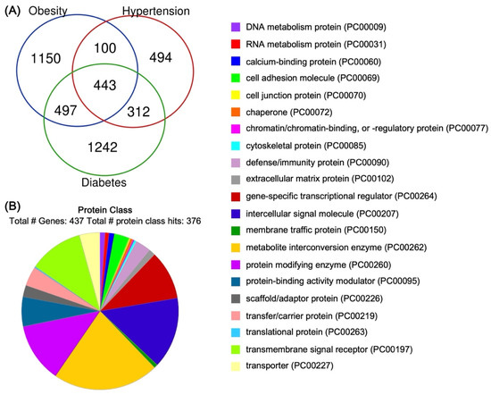
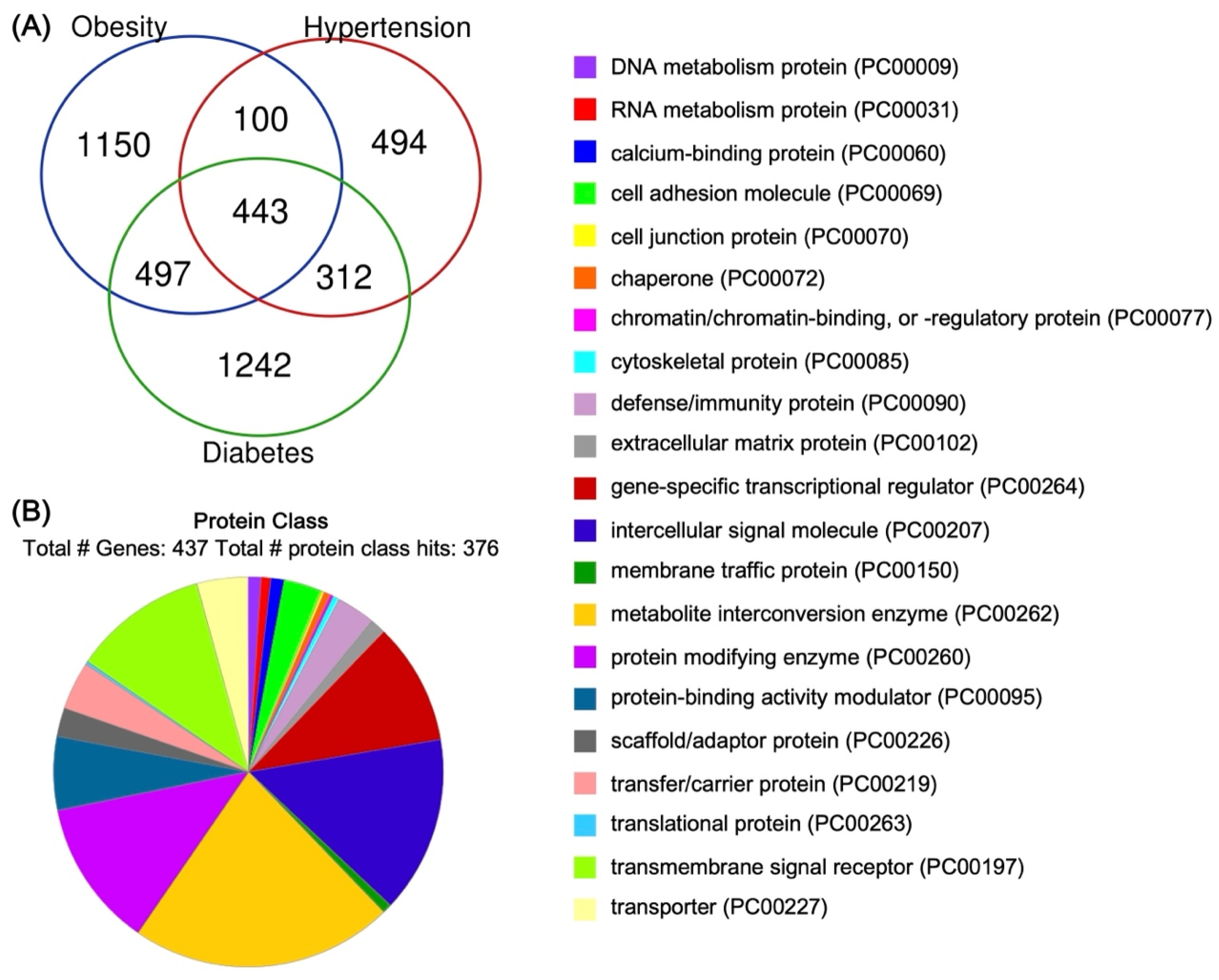
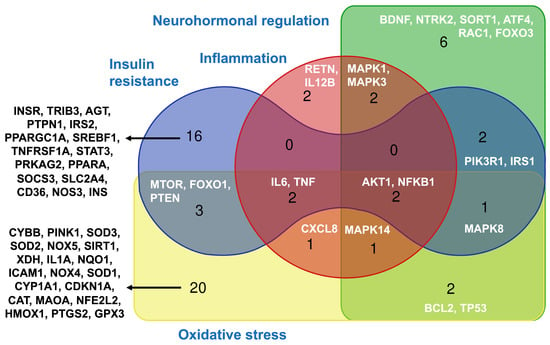
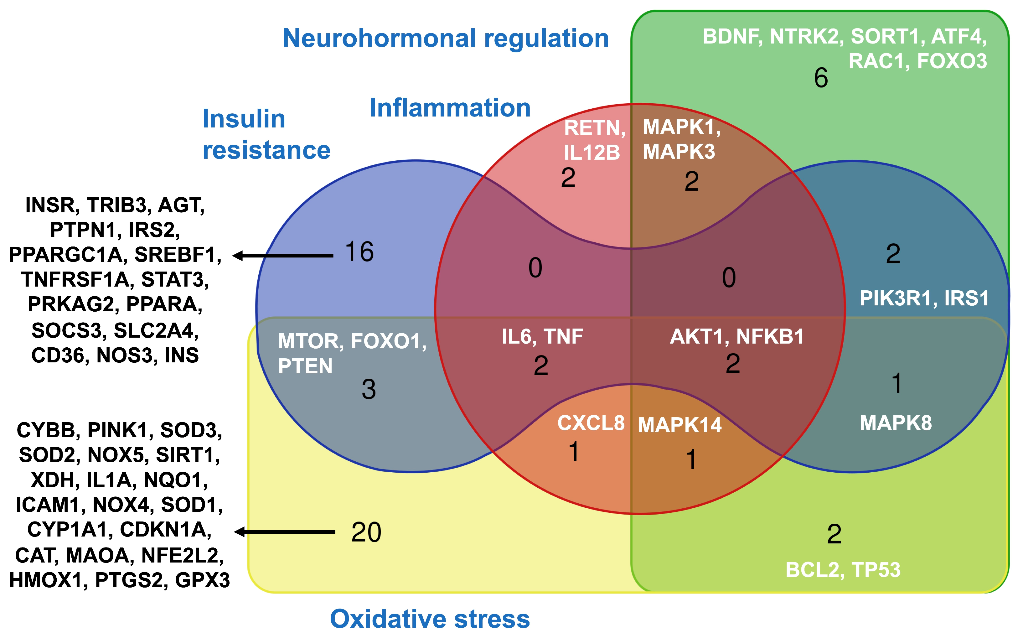
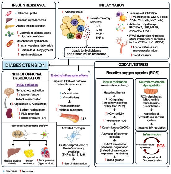
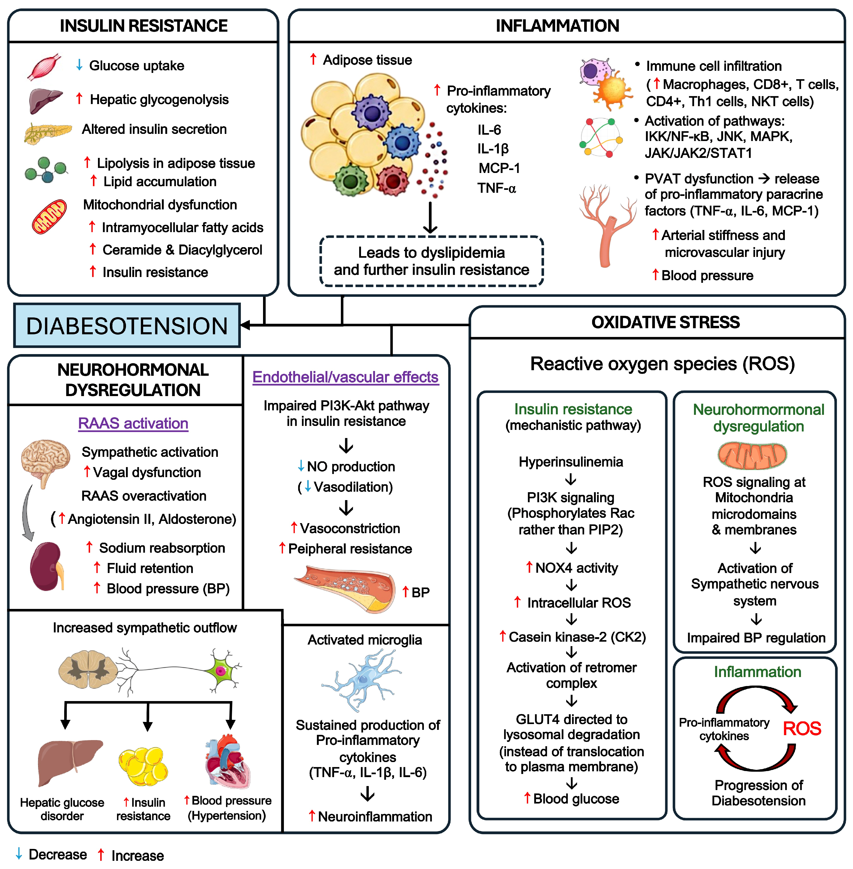
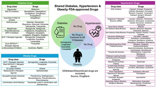
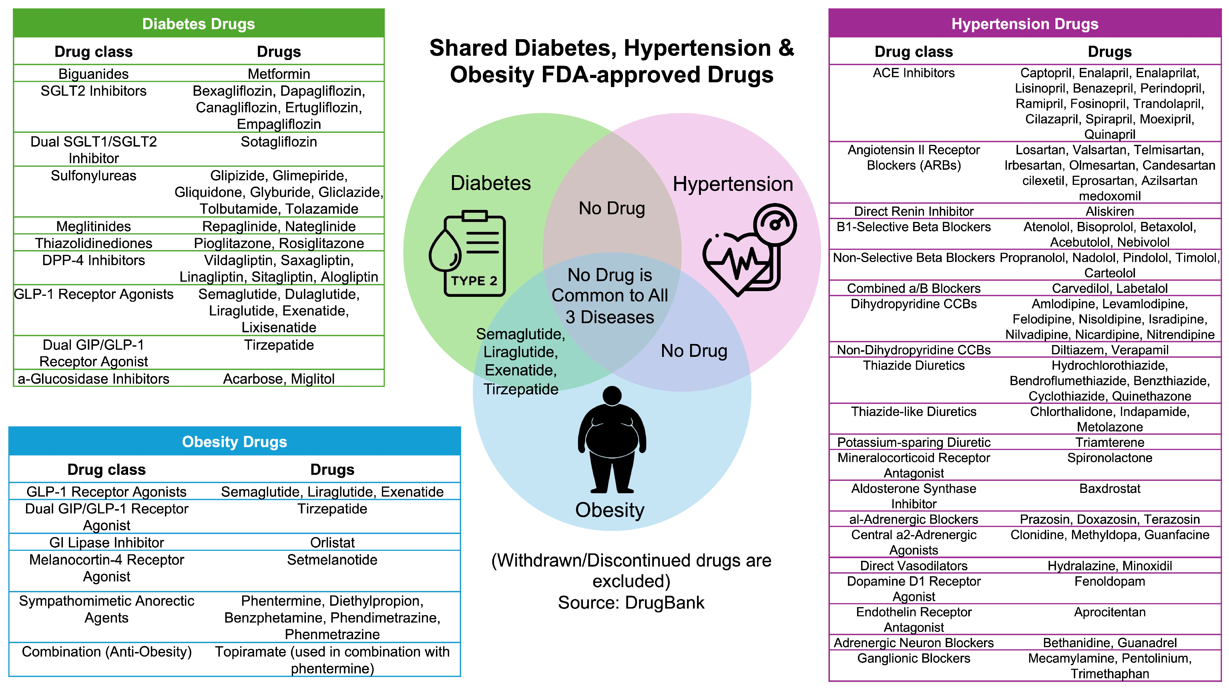

Disclaimer/Publisher’s Note: The statements, opinions and data contained in all publications are solely those of the individual author(s) and contributor(s) and not of MDPI and/or the editor(s). MDPI and/or the editor(s) disclaim responsibility for any injury to people or property resulting from any ideas, methods, instructions or products referred to in the content. |
© 2026 by the authors. Licensee MDPI, Basel, Switzerland. This article is an open access article distributed under the terms and conditions of the Creative Commons Attribution (CC BY) license.