


Abstract
Extracorporeal membrane oxygenation (ECMO) is a continuously evolving and increasingly utilized life-support therapy. ECMO requires systemic anticoagulation, which exposes patients to an increased risk of heparin-induced thrombocytopenia (HIT). Clinical experience with alternative anticoagulants in this setting remains limited. The 2022 Extracorporeal Life Support Organization (ELSO)—Anticoagulation Guidelines provide no specific recommendations regarding anticoagulant selection for ECMO patients with HIT. This article aims to review current practices, available evidence, and most recent advances concerning the use of alternative anticoagulants in ECMO patients with HIT. In patients with a high suspicion or confirmed diagnosis of HIT, management includes discontinuing all forms of heparin exposure and initiating an alternative anticoagulant, such as a direct thrombin inhibitor and/or factor Xa inhibitor. Direct thrombin inhibitors act independently of antithrombin and have a short half-life, providing a more consistent and predictable anticoagulation effect. Most available data, primarily from retrospective studies, describe the use of argatroban in ECMO patients with HIT. Bivalirudin has also been used as an alternative anticoagulant in this population, with no significant increase in bleeding or thrombotic complications. However, the current evidence remains limited to small, retrospective, single-center or case–control studies. Fondaparinux has shown effectiveness in the HIT setting and appears to have a low risk of complications. Factor XIIa inhibitors represent a novel class of anticoagulants currently under investigation, evaluated only in animal models. Growing clinical experience with alternative anticoagulants, particularly direct thrombin inhibitors, suggests that their use will likely become a primary focus in ECMO anticoagulation management in the coming years.
1. Introduction
Extracorporeal membrane oxygenation (ECMO) is a form of mechanical circulatory support used in patients with severe respiratory or cardiac failure. ECMO should be considered in cases of acute, potentially reversible respiratory failure and in severe cardiogenic shock refractory to conventional management. In recent years, the indications for ECMO have expanded to include various clinical scenarios, including its use during cardiopulmonary resuscitation (eCPR), as a bridge to lung or heart transplantation, for rewarming in accidental deep hypothermia, and for resuscitation following severe trauma [1].
ECMO can be established as a venovenous (VV-ECMO), primarily used for respiratory failure, and venoarterial (VA-ECMO), which supports patients with cardiac failure. The main principle of ECMO involves draining blood from large vessels into an oxygenator, where venous blood is enriched with oxygen and carbon dioxide is removed. The oxygenated blood is then returned to either the venous (VV-ECMO) or arterial (VA-ECMO) circulation using a centrifugal pump.
Contact between blood and the artificial surfaces of the ECMO circuit introduces a significant risk of circuit thrombosis, requiring systemic anticoagulation. Exposure of blood to non-endothelial materials, such as the tubing and oxygenator, activates complex inflammatory and coagulation pathways. As a result ECMO disturbs hemostatic balance and homeostasis [2]. Inflammation triggers coagulation activation, impairs natural anticoagulant mechanisms, and disrupts the fibrinolytic system. The ensuing hyperinflammation elevates acute-phase reactant proteins and stimulates various immune pathways. Persistent activation of these systems leads to platelet and coagulation factor consumption, increasing the risk of severe bleeding. This phenomenon is described as “slow” disseminated intravascular coagulation (DIC).
Bleeding complications in patients receiving ECMO can be classified as minor, such as epistaxis, surgical wound bleeding, cannulation site bleeding, or gastrointestinal bleeding, and major, life-threatening, such as intracranial hemorrhage, pericardial tamponade, pulmonary, or retroperitoneal bleeding. The overall incidence of bleeding has been reported to be as high as 49% [3], whereas the incidence of intracranial hemorrhage or infarction is about 8% [4].
Multiple studies have attempted to identify risk factors associated with bleeding during ECMO and found that higher SAPS III (Simplified Acute Physiology Score) score, longer ECMO duration, prolonged aPTT, post-surgical ECMO, low inflammatory response, hemoglobin < 9 g/dL, fibrinogen < 2 g/L, pH < 7.12, and body mass index below 25 kg/m2 are associated with an increased risk of bleeding [5,6,7]. Patients who experience bleeding have longer ICU and hospital stays, and higher mortality rates [5]. In contrast, thromboembolic complications may range from deep vein thrombosis to acute thrombosis of the oxygenator or circuit tubing, which may lead to system failure, one of the most critical emergencies during ECMO [8].
To prevent and manage thromboembolic events, systemic anticoagulation remains the standard of care. The most commonly used anticoagulant in ECMO patients is unfractionated heparin (UFH). It is a mixture of glycosaminoglycans of different molecular weights that acts primarily by binding to antithrombin (AT) to form a complex that inhibits thrombin and factor Xa [9]. Although the therapeutic goal is to achieve a stable plasma heparin concentration, the relationship between heparin dosage and effect is non-linear [10].
UFH offers several advantages, including a well-established mechanism of action, low cost, ease of titration, rapid onset, and reversibility with protamine [11]. Moreover, anticoagulation can be monitored with point-of-care devices, such as activated clotting time (ACT), or through laboratory tests such as activated partial thromboplastin clotting time (aPTT), anti-factor Xa activity, or viscoelastic testing. For these reasons, current guidelines continue to recommend UFH as the first-line anticoagulant during ECMO support [12].
During states of hyperinflammation, the anticoagulant efficacy of UFH may be diminished due to increased binding of heparin to acute-phase proteins, such as factor VIII and fibrinogen, as well as decreased plasma concentrations of AT [5]. A key limitation of heparin therapy lies in its nonspecific binding to multiple targets, including AT, endothelial cells, circulating plasma proteins, and macrophages. These interactions alter its pharmacokinetic properties and dose–response relationship, contributing to complications such as heparin resistance and heparin-induced thrombocytopenia (HIT) [12].
2. Heparin-Induced Thrombocytopenia—HIT
2.1. Types and Incidence
To date, the understanding of HIT in patients receiving ECMO remains limited. Most studies reporting HIT in this context are constrained by small sample sizes, single-center designs, and retrospective nature. As a result, the literature is inconsistent, particularly regarding the prevalence and mortality rates of HIT among ECMO patients [13,14]. Given the increasing use of ECMO, this topic is of growing clinical importance, and updated systematic data on HIT in this population are needed.
Heparin can induce two distinct types of thrombocytopenia. Type I HIT, a non-immune form, typically develops within the first few days of heparin therapy and is more common than type II. In contrast, type II HIT is an immune-mediated complication of anticoagulant therapy that can occur with both UFH and low-molecular-weight heparin [15]. Type II HIT is characterized by a >50% reduction in platelet count from the post-heparin peak value [15], typically developing 5 to 10 days after heparin initiation. Key pathological features include the formation of heparin-dependent, platelet-activating IgG antibodies, which generate a state of hypercoagulability [16]. These IgG antibodies bind to platelet factor 4 (PF4)-heparin complexes, leading to intravascular activation through FcyRIIA receptor cross-linking (Figure 1). This process induces a prothrombotic and hypercoagulable state, ultimately resulting in thrombocytopenia [17].
Figure 1.
Schematic illustration of heparin-induced thrombocytopenia (HIT): IgG antibodies bind to platelet factor 4 (PF4)–heparin complexes, resulting in cross-linking of FcγRIIA receptors on platelets and monocytes, which triggers their activation and promotes a prothrombotic state.
The reported incidence of Type II HIT in adults ranges from 0.2% to 5% [16,18], although available data are inconsistent, suggesting rates from <0.36% to 17% [19,20,21,22]. A retrospective analysis of 118 ECMO patients reported a prevalence of HIT and HITT (Heparin-Induced Thrombocytopenia with Thrombosis) of 8.3% and 7.3%, respectively [14], with complications occurring in approximately 25% to 50% of affected patients [23,24]. Notably, HITT in ECMO patients has been associated with a mortality rate approaching 50%. Compared with HIT-negative patients, those testing positive for HIT typically require longer ECMO support, are younger, exhibit higher HIT ELISA optical densities, and reach their platelet count nadir later during therapy [14]. A recent meta-analysis of 21 studies reported thrombocytopenia rates ranging from less than 1% to 22% among patients receiving VA-ECMO [25], with a pooled prevalence of confirmed HIT cases of 3.7%. The wide variability in reported HIT incidence likely reflects the heterogeneity in patient populations, differences in anticoagulation protocols and variations in HIT screening methods. Consequently, the elevated risk of HIT observed in ECMO patients may not directly result from arterial cannulation or ECMO-related vascular and inflammatory injury [26]. Instead, it may reflect prior and prolonged exposure to UFH before ECMO initiation [26].
Thrombocytopenia during ECMO is multifactorial, encompassing factors such as cardiopulmonary bypass (CPB), consumptive coagulopathy, blood drainage, sepsis, multi-organ failure, platelet consumption, hemodilution, and bleeding [26]. Following the cessation of heparin therapy, patients with confirmed HIT demonstrate a faster recovery in platelet count (≥50% increase from nadir) compared with HIT-negative thrombocytopenic patients, indicating differing pathophysiological mechanisms [27]. However, in ECMO patients with HIT, platelet recovery is delayed compared with those supported by CPB alone, likely due to circuit-related platelet activation and other concurrent causes of thrombocytopenia [27].
2.2. HIT Diagnostic
The diagnosis of HIT in ECMO patients is particularly challenging, and both underdiagnosis and overdiagnosis can lead to potential problems [28]. Failure to identify HIT increases the risk of venous and/or arterial thrombosis, limb amputation, or death. Continuing UFH therapy in affected patients may exacerbate thrombotic complications; if heparin is not discontinued and replaced with an alternative anticoagulant, the risk of thrombosis is estimated at 40–50%, especially in patients with repeated heparin exposure [15,28,29]. Conversely, unnecessary discontinuation of heparin due to a misdiagnosis may increase bleeding risk when alternative anticoagulants are used to treat thrombocytopenia and may also increase hospital costs. The difficulty of diagnosing HIT during ECMO is further complicated by the fact that sepsis and DIC can present with similar clinical features (Figure 2).
Figure 2.
Heparin-induced thrombocytopenia (HIT) diagnostics. HIT Expert Probability (HEP) score; LLL, Lilo-Le Louet score; CPB, cardiopulmonary bypass; ELISA, enzyme-linked immunosorbent assay; HIPA, heparin-induced platelet aggregation; PIPA, PF4-induced platelet activation assay; LTA, light transmission aggregometry; SRA, serotonin release assay.
The 4-T score for HIT, which considers thrombocytopenia, timing, thrombosis, and other causes of thrombocytopenia, is commonly used to assess the pretest probability of HIT [24]. However, it has limited sensitivity and specificity and has not been extensively validated or designed for use in the context of mechanical circulatory support (MCS). While the score may be numerically higher in patients with confirmed HIT, this difference is not statistically significant [16]. The pretest probability is categorized as low (0–3 points), intermediate (4–5 points), or high (6–8 points). For patients with a low pretest probability, further laboratory testing is not recommended, except in selected ECMO patients (BSH: Grade 2C) [30]. Kram et al. demonstrated that the predictive accuracy of both the 4-Ts and HIT Expert Probability (HEP) scores was limited in patients supported with various MCS devices, and that low scores were insufficient to reliably rule out HIT [31]. In patients with intermediate or high pretest probability, laboratory testing is warranted. Additionally, platelet count trends alone do not reliably distinguish between confirmed and excluded HIT [16,19,26]. Suspicion should increase if a patient’s platelet count declines during ongoing heparin therapy. Given the frequent occurrence of thrombocytopenia due to bleeding, surgery, or infection in MCS patients, both the 4-Ts and HEP scores may overestimate the likelihood of HIT in these patients.
The Lilo-Le Louet (LLL) score is widely used for the diagnosis of HIT with a 97% negative predictive value in patients following CPB [32]. This score is specifically developed for cardiac surgery patients. It is based on three factors: (1) the degree of platelet count reduction (categorized as pattern A or B), (2) the interval between CPB and onset of thrombocytopenia (<5 days or ≥5 days), and (3) CPB duration. Pattern A is characterized by a transient post-CBP platelet recovery, followed by a secondary drop after more than 4 days, and is more predictive of HIT than Pattern B, which involves a persistent thrombocytopenia immediately after CPB without recovery [32,33,34]. This approach may provide better predictive accuracy compared with the 4Ts or HEP scores, especially in MCS patients [31].
The enzyme-linked immunosorbent assay (ELISA) for detection of platelet factor 4 (PF4)-heparin complex antibodies in plasma or serum is widely available, rapid, and very sensitive but lacks specificity in the ECMO setting [35]. Several studies have established the ELISA optical density (OD) thresholds in the general population [36,37]. Manufacturers typically define a positive result as an OD >0.4 units; however, this threshold demonstrates limited specificity and may reduce the clinical relevance of test results [37].
Functional tests are confirmatory functional assay with higher specificity and are indicated to confirm the diagnosis. These include C-serotonin release assay (C-SRA), heparin-induced platelet activation (HIPA) test, heparin-induced platelet aggregometry (measures aggregation), lumiaggregometry, and flow cytometry. Rapid initiation of functional assays is recommended for all patients presenting with a positive antibody screening results in conjunction with strong clinical likelihood of HIT. Although the SRA and HIPA tests are more specific, these tests are usually performed at reference laboratories and may take 5 to 7 days to process, reducing their immediate utility for clinical decision making.
The diagnosis of HIT during VA-ECMO therapy has been associated with longer ICU stays but does not appear to affect overall survival [13,16,20].
3. Alternative Anticoagulants
In patients with a high clinical suspicion or confirmed HIT diagnosis, management involves immediate discontinuation of all forms of heparin exposure, including heparin-containing flush solutions. An alternative anticoagulant, such as a direct thrombin inhibitor (DTI) or a factor Xa inhibitor, should then be initiated [1,38] (Figure 3). Delaying treatment with an alternative anticoagulant is associated with a daily risk increase of 5–10% for thrombotic events, limb loss, or death [39].
Figure 3.
Management steps for heparin-induced thrombocytopenia (HIT) complicating extracorporeal membrane oxygenation support. ELISA, enzyme-linked immunosorbent assay; HIPA, Heparin-Induced Platelet Aggregation; PIPA, PF4-induced platelet activation assay; LTA, light transmission aggregometry; SRA, serotonin release assay.
The optimal anticoagulation strategy for ECMO patients who developed HIT remains uncertain. Current guidelines, including those from the American College of Chest Physicians and the ELSO Guidelines, do not provide specific recommendations for this patient group [12,40,41]. Bivalirudin is recommended for patients with acute HIT who require urgent cardiac surgery, rather than heparin or other non-heparin anticoagulants. For patients with HIT undergoing acute percutaneous coronary intervention, the use of antiplatelet agents (grade 2C) in combination with either bivalirudin (grade 2B) or argatroban (grade 2C) is advised (Table 1).
Table 1.
Overview of anticoagulation agents and methods for monitoring the hemostatic system during ECMO.
Direct thrombin inhibitors (DTIs) are anticoagulants with a short half-life that act independently of AT by directly binding to thrombin. They provide a more consistent and predictable anticoagulant effect in situations where AT activity is low or variable and demonstrate more reliable pharmacokinetics, as well as greater efficiency in reducing thrombin generation compared with UFH [12,38]. Unlike heparin, DTIs do not bind to plasma proteins or blood cells, making them less susceptible to fluctuations in serum chemistry or cell counts. This characteristic allows for more predictable dosing and a consistent anticoagulant effect, potentially reducing bleeding risk. DTIs are not associated with immune-mediated thrombocytopenia, including HIT. Additionally, DTIs inhibit both thrombin and thrombin bound within clots, enhancing their overall anticoagulant effectiveness [39]. Among synthetic DTIs, argatroban, bivalirudin, and lepirudin have been used in CPB, ECMO, and VAD support; however, lepirudin is now rarely available.
3.1. Argatroban
3.1.1. Mechanism of Action
Argatroban is a synthetic L-arginine derivative with concentration-dependent anticoagulant activity. Its onset of action occurs within 30 min, and its half-life is approximately 45 min. Argatroban is primarily metabolized in the liver via hydroxylation and aromatization of the 3-methyltetrahydroquinoline ring. During extracorporeal circulation, its low distribution volume (180 mL/kg) is increased, resulting in limited tissue diffusion and distribution mainly within extracellular space. Argatroban binds univalently to the active site of thrombin, in contrast to bivalirudin, which binds bivalently to both the active site and exosite-1 on thrombin [42] (Table 2). The antidote for argatroban still does not exist. It is considered a safe anticoagulation option for patients with HIT and renal failure, especially in the ICU setting [43]. However, argatroban is contraindicated in patients with severe hepatic impairment (Child-Pugh Class C) due to the high risk of drug accumulation [42].
Table 2.
The distinction between univalent and bivalent thrombin inhibitors.
3.1.2. Dosing and Monitoring of Argatroban
Argatroban therapy does not require a loading dose. In patients on ECMO, the initial infusion rate should range between 0.2 and 2 mcg/kg/min, depending on the target activated partial thromboplastin time (aPTT) value [43]. According to Rajsic et al., argatroban dosing should be approached with caution, particularly in patients with hepatic impairment, where maintenance dose as low as 0.1–0.2 mcg/kg/min may be necessary [1].
Monitoring the effect of argatroban can be performed using several assays, including ACT, aPTT, thrombin time, diluted thrombin time (dTT), ecarin clotting time (ECT), viscoelastic methods, chromogenic antithrombin activity (CAA) or measuring plasma concentrations [44]. aPTT should be measured two hours after starting the infusion to prevent excessive anticoagulation and minimize bleeding risk [42]. However, there are significant limitations to using aPTT for argatroban monitoring. These include variability related to the method and reagents used, intra- and inter-patient variability, and potential interference from lupus anticoagulant, elevated factor VIII, fibrinogen, vWF, and CRP levels, as well as reduced coagulation factors production (especially FXII). At higher DTI concentrations, aPTT may underestimate the true anticoagulant effect [44].
More precise monitoring can be achieved using tests like the diluted thrombin time (TT), which is correlated with a drug level nomogram, or the ecarin clotting time (ECT) [12,44]. However, these alternative tests have not been widely validated or approved by regulatory authorities in most countries [42]. Measurement of plasma argatroban concentration is considered the most accurate method. Swiss guidelines recommend a target range of 0.4–1.5 μg/mL [45], while French guidelines recommend a target range of 0.25–1.5 μg/mL, particularly in patients with prolonged baseline aPTT prior to argatroban initiation [46]. Unfortunately, such assays are not readily available in most laboratories. Argatroban, unlike other alternative anticoagulants, elevates the international normalized ratio (INR). During transition to warfarin, which should only be initiated once platelet count has normalized, clinicians typically overlap both agents (a non-heparin anticoagulant and a vitamin K (VKA) antagonist therapy) for 4 to 5 days, maintaining an INR > 4.0.
3.1.3. Clinical Studies
Numerous studies, most of them retrospective, have investigated the use of argatroban in ECMO patients diagnosed with HIT [13,16,21,27,28,47,48,49] (Table 3). Reported HIT prevalence ranges 3.2% to 6.4%, with no significant difference between ECMO configurations (VA vs. VV ECMO p = 0.47) [27].
No difference in in-hospital mortality was observed between patients with and without HIT (43% in HIT vs. 38% non-HIT patients, p > 0.999) [16] (31.6% in HIT vs. 32.2% in non-HIT patients, p = 0.79) [27]. However, Glick et al. reported significantly higher in-hospital mortality among patients with suspected HIT (14/23, 61% patients in HIT group vs. 31/96 patients without HIT, 32%; p = 0.01), likely reflecting more severe thrombocytopenia and a generally sicker patient population [21]. Arachchillage et al. similarly found no difference in mortality rates in patients on ECMO with HIT (1/2, 50%) and those with HIT and thrombosis [5/17, 29.4%]; p = 0.96) [27]. Neurological outcomes at hospital discharge and at one month (35% vs. 38%, p > 0.999), as well as mortality rates at three and twelve months, were also comparable between patients with excluded and confirmed HIT [16].
Bleeding remains the most common complication during ECMO therapy. Some studies found no difference in bleeding incidence between HIT and non-HIT patients [16], whereas others reported a higher incidence of bleeding in patients without HIT [13,27]. Lubnow et al. reported an increased bleeding rate in HIT patients [47]. Several studies did not report bleeding outcomes [21,28,50].
The incidence of thrombosis varied across the studies. While some authors reported no difference between groups [16], others reported a higher rate of thrombotic events in HIT patients [27,47]. Several studies did not provide the data on thrombosis incidence [21,28,50].
Argatroban therapy was monitored with aPTT, though different anticoagulation goals are reported across the studies. These targets varied from 50 s for suspected HIT and 60 s for confirmed HIT [28,47], to 78 s in the study conducted by Archchallange et al. 2020 [48].
Although switching from heparin to an alternative anticoagulant such as argatroban may initially increase drug costs, when accounting for the costs associated with HIT testing, monitoring, and potential complications, the overall treatment costs are comparable [47,50].
While there are no direct comparative studies assessing argatroban efficacy in HIT/HITT patients versus those with thrombocytopenia of other etiologies, treatment of HITT with argatroban has been associated with a significantly faster platelet count recovery compared to historical control populations [51]. Pabst et al. reported that after discontinuation of systemic heparin and switching to argatroban in patients with HIT, platelet counts increased from a mean of 59.8 k/μL at the time of HIT diagnosis to a mean of 280.2 k/μL at 14 days post-heparin discontinuation, even while maintaining a heparin-bonded circuit [28]. Eleven out of the fourteen HIT patients (78.6%) survived to discharge. Causes of death were multiorgan failure and intracranial hemorrhage. HIT may have played a role in these outcomes, as it is known that HIT increases the risk of thrombotic events that can lead to stroke, brain bleeding, and multiorgan failure. The time to achieve a ≥50% increase in platelet count from the nadir was significantly reduced in patients with confirmed HIT treated with argatroban compared to those without HIT, indicating different pathophysiological mechanisms of thrombocytopenia [27]. However, HIT patients on ECMO required longer to reach a >50% platelet recovery than CPB patients, likely due to ECMO circuit-related effects and other thrombocytopenia-inducing factors [27].
In 2018, Kimmoun et al. [13] reported a retrospective study including patients on VA-ECMO from 20 French centers between 2012 and 2016 [13]. Of 5797 screened patients, 39 were hospitalized for more than 3 days with a high clinical suspicion of HIT and positive anti-PF4/heparin antibodies. Alternative anticoagulation therapy consisted primarily of argatroban in 11 of 21 confirmed HIT patients (52.4%) and danaparoid in 10 of 21 patients (47.6%). While an increase in platelet count following the change in anticoagulation therapy was expected in confirmed HIT, a similar rise in platelet count was also observed in patients excluded from the HIT diagnosis. One possible explanation is that over 50% of patients in the excluded HIT group were successfully weaned from VA-ECMO after 7 days, which was associated with platelet recovery [13]. Alternative causes of thrombocytopenia, such as sepsis or drug-induced thrombocytopenia, might have played a role in the platelet recovery.
Mang et al. described patients with coronavirus disease 2019 (COVID-19) requiring V-V ECMO with rapid deterioration of oxygenator function, requiring multiple administrations of rtPA. After heparin was replaced with argatroban, oxygenator performance stabilized [52]. Among forty-one COVID-19 patients requiring VV ECMO, seven (17%) tested positive for HIT by ELISA, but only one (2%) was confirmed to have HIT type II by a confirmatory HIPA test.
A retrospective single-center study reported that the use of heparin-coated circuits did not lead to a further decrease in platelet count or higher complication rates [47]. Notably, two patients on non-heparin-coated circuits did not experience faster platelet recovery after transitioning to argatroban, in contrast to those on heparin-coated circuits.
3.2. Bivalirudin
3.2.1. Mechanism of Action
Bivalirudin inhibits plasma thrombin, clot-bound thrombin, and collagen-triggered platelet activation without requiring antithrombin III. About 80% of the drug is metabolized through proteolysis, which is nonorgan-dependent, while the remaining one-fifth is excreted unchanged via renal pathways [53,54,55,56]. This metabolism supports a predictable relationship between bivalirudin dosage and anticoagulant efficacy [53,57]. The onset of action occurs within 2–4 min, and half-life is approximately 25 min. Compared with argatroban (39–51 min) and lepirudin (78 min), bivalirudin’s shorter half-life facilitates precise titration of anticoagulation. Because of its short half-life, discontinuation of the infusion is typically sufficient to reverse anticoagulation in the event of accidental overdose or significant bleeding. In more severe cases, adjunctive measures such as plasma transfusion, administration of prothrombin complex concentrate, or hemodialysis may be required [56]. Approximately 25% of bivalirudin can be removed through hemodialysis.
3.2.2. Dosing and Monitoring of Bivalirudin
Only a limited number of studies have reported the use of a loading dose of bivalirudin, ranging from 0.2 to 0.75 mg/kg [53]. Initial bivalirudin infusions rates in VV-ECMO and VA-ECMO (excluding postcardiotomy ECMO) are usually set at 0.02–0.05 μg/kg/min [58]. Assessment of renal function is essential to determine the appropriate initial dose of bivalirudin, with or without a loading bolus. Reported maintenance infusion rates vary widely, ranging from 0.05 to 1.75 mg/kg/h, with a mean rate of 0.27 ± 0.37 mg/kg/h [53]. Lopez et al. reported results from the largest retrospective cohort of HIT/HITT patients, specifically evaluating dosing strategies [59]. A median infusion rate of 0.05 mg/kg/h was sufficient to achieve the desired aPTT, representing a lower dose than those previously documented in ECMO populations [59,60,61]. Patients receiving ECMO in combination with CVVH required the same median bivalirudin dose to achieve therapeutic aPTT as those on ECMO alone [59,62]. The longest reported duration of bivalirudin usage exceeded 60 days. A retrospective analysis of HIT patients with elevated BMI demonstrated that total body weight-based dosing most reliably predicted attainment of the target aPTT [63].
The anticoagulant effect of bivalirudin can be monitored using several laboratory tests, most commonly aPTT and ACT. aPTT monitoring shows an excellent correlation with bivalirudin’s anticoagulant effect, with the same standard target range as for UFH of 45–80 s [53,58]. ACT can also be used, with target values from 160 s to 220 s, and serves as an adjunct to aPTT in some centers [53,56]. If the baseline aPTT levels are abnormal, a dTT, chromogenic anti-IIa testing and blood concentration of drug can be used [58]. Nowadays, ACT and aPTT are increasingly avoided due to their susceptibility to multiple confounding factors. More specific assays, such as drug concentration and ROTEM, may provide more reliable measurements of bivalirudin’s effect.
3.2.3. Clinical Experience
Due to the limited data, standardized protocols for bivalirudin administration in ECMO patients with HIT have not been clearly established. Bivalirudin can be used either as an initial anticoagulant in ECMO or as a secondary agent in patients with HIT or UFH resistance. It has been used off-label in ECMO without significant increase bleeding or thrombosis [40,57]. However, the available evidence is limited to small, retrospective, single-institution studies or case–control studies (Table 3).
When comparing bivalirudin and UFH, similar mortality rates and thrombotic complications are reported [64]. However, bivalirudin is associated with lower bleeding rates [65,66,67,68], less variability in ACT and aPTT, which remain within the therapeutic range for longer periods, likely due to its specific mechanism of action [61,64], and a reduced need for allogeneic blood transfusions [61,69]. Despite these benefits, concerns remain regarding bivalirudin´s rapid proteolytic cleavage, which may increase the risk of thrombosis in areas of stagnant blood, particularly in a non-ejecting, unvented left ventricle (LV) [60,70]. However, a recent systematic review and meta-analysis including 10 retrospective observational studies and 847 patients suggested that bivalirudin may significantly reduce thrombotic events, in-circuit thrombosis, and in-hospital mortality [68]. Other data showed that the incidence of bleeding events with bivalirudin is comparable to that with UFH [53,60,61].
Transitioning ECMO patients with HIT to bivalirudin is associated with increased platelet counts. Zhong et al. found that the mortality in ECMO patients with HIT treated with bivalirudin was similar to that of ECMO patients without HIT who are treated with UFH [53].
Several studies that included adult ECMO patients with HIT and switched to bivalirudin reported no cases of bleeding, thrombosis, or mortality in this cohort [34,71]. According to the conclusion of authors, bivalirudin and argatroban are safe alternatives to heparin and should be initiated in patients with high suspicion for HIT while awaiting confirmatory SRA results [34,71].
Giuliano et al. reported that the composite rate of thrombotic or hemorrhagic complications was lower in ECMO patients with suspected or confirmed HIT receiving bivalirudin [72]. Less severe hemorrhage—as indicated by lower blood product transfusion—was registered in the bivalirudin group. In-hospital mortality was comparable between patients treated with bivalirudin and heparin (69%) and those treated with heparin alone (62%), despite greater illness severity in the bivalirudin group (higher rates of prior intracranial hemorrhage, longer median ECMO duration, higher SOFA scores on ECMO day one, and a greater need for continuous renal replacement therapy (CRRT)).
3.3. Lepirudin
Lepirudin is a recombinant form of hirudin. It inhibits thrombin in a bivalent manner, targeting the catalytic site as well as exosite-1. It has an elimination half-life of 1 to 2 h, with bolus administration achieving peak aPTT within 10 min. As lepirudin is primarily eliminated renally, dose adjustments are required in patients with acute kidney injury. Significant adverse effects include bleeding and the development of antihirudin antibodies. The bleeding risk may be mitigated by careful dose titration and regular laboratory assessment [73]. Anaphylactic reactions are rare and generally linked to bolus administration. In the absence of an antidote, the use of lepirudin remains challenging in certain clinical contexts, particularly in high-risk settings such as CPB. It has been utilized successfully in patients undergoing ECMO with contraindications for UFH, with aPTT- and ACT-guided titration of doses. In these patients, bleeding or thrombosis did not occur [74]. However, lepirudin was withdrawn from the market in the United States and Europe in 2012 [75].
3.4. Direct Factor-Xa Inhibitors
Direct factor-Xa inhibitors block factor Xa activity independently of antithrombin. Several agents can be administered parenterally, including fondaparinux (subcutaneous) and danaparoid (subcutaneously or intravenously). Both fondaparinux and danaparoid are renally cleared, characterized by long half-lives, can be monitored using calibrated anti-Xa assays, and lack antagonists, making bleeding complications more difficult to manage.
Fondaparinux is an effective anticoagulant in the HIT setting and appears to have a low risk of overall complications [76]. According to the 2018 American Society of Hematology Guidelines for the Management of Venous Thromboembolism: Heparin-Induced Thrombocytopenia, patients with acute HIT complicated by thrombosis should discontinue heparin and initiate anticoagulation with fondaparinux [40]. Both fondaparinux and NOACs are considered appropriate options for anticoagulation in these patients [40]. Little information on the use of Fondaparinux in ECMO has been published (Table 3). In the case report presented by Parlar et al., a patient with high suspicion of HIT during ECMO support was treated successfully and safely by Fondaparinux (1 × 2.5 mg/day, subcutaneous) [77]. In a study by Osawa et al., it was shown that fondaparinux might be a reasonable option for HIT treatment in dialysis patients if it is used with caution when other options are not available [78].
Kutlesa et al. conducted a study including 40 adult patients with severe ARDS (20 with H1N1-induced ARDS), treated with VV- ECMO at a tertiary care hospital [79]. Fondaparinux (2.5 mg daily) was administered to three patients (8%) with positive PF4 antibodies, but all three patients died. Two additional patients died before HIT antibody test results became available. Overall mortality was 15/40 patients (37%) [79].
Kutlesa et al. reported a single-center retrospective study involving 112 adult patients who received VV-ECMO for COVID-19-induced ARDS. HIT occurred in 39% (44/112) of patients during ECMO treatment [80]. In those with confirmed HIT antibodies, off-label anticoagulation with fondaparinux (5 mg daily) was started. The authors observed that the effect of fondaparinux on patient outcomes in this setting remains unclear [80].
Danaparoid is a heparinoid composed of heparin sulfate, dermatan sulfate, and chondroitin sulfate. It inhibits factor Xa indirectly via antithrombin and, to a lesser degree, thrombin [81]. With a predictable pharmacodynamic response, the drug has a half-life of approximately 24 h. It exhibits an anti-factor Xa to anti-factor IIa activity ratio of 28:1, compared with a 1:1 ratio for heparin. Steady state is generally achieved after 4–5 days, with renal excretion accounting for 40–50% of total clearance. Danaparoid does not significantly prolong PT and has minimal impact on aPTT, making these tests unsuitable for monitoring. Instead, monitoring should be performed using a danaparoid-specific anti-Xa assay. Monitoring is mandatory for patients with marked renal dysfunction, extreme body weights, or those developing thromboembolic or bleeding complications while on treatment. There are no specific antidotes available [40,82].
According to the British Committee for Standards in Haematology guidelines, danaparoid is a suitable alternative anticoagulant for patients with HIT when administered at therapeutic doses (1B) [83].
A case report from Bauer et al. reported a successful femoral VA-ECMO management with danaparoid in a patient with severe respiratory failure after massive pulmonary embolism and suspected type II HIT [84]. The initial dose was 400 IU/h for 4 h, followed by 300 IU/h (0.5–0.8 U/mL anti-Xa factor activity goal). The procedure resulted in a successful outcome. Removal of the cannula proceeded without major bleeding, and anti-factor Xa activity stayed within the target therapeutic range [84] (Table 3). No circuit clotting, new thrombotic events, or thrombus extension were observed.
Table 3.
Summary of detailed information about included studies.
3.5. Other Anticoagulation Approaches
Factor XIIa inhibitors are new anticoagulants targeting FXIIa and with the potential to prevent thrombosis without increasing bleeding risk; however, they have been evaluated only in animal models of ECMO [38,86,87].
Nafamostat mesylate (NM) is a synthetic serine-protease inhibitor characterized by a very short half-life (5–8 min). NM exerts its effects on coagulation and platelet activity by blocking activated coagulation factors VIIa, XIIa and Xa, thrombin, and plasmin. It also interferes with fibrinolysis and inhibits proteases such as kallikrein, trypsin, and complement components C1r and C1s. Moreover, NM suppresses tissue factor (factor V)-dependent generation of factor Xa in a concentration-dependent fashion, providing localized anticoagulation within ECMO circuits [88]. It undergoes metabolism in the liver and bloodstream and is eliminated via the kidneys and intestines. The best anticoagulation levels of NM are unknown. In the case of ECMO patients, the administered dose of NM showed considerable variability, with mean values ranging from 0.46 to 0.67 mg/kg/h [88]. Monitoring its effect may be measured with aPTT. Reported adverse effects include electrolyte imbalance, including hyperkalemia and hyponatremia, and agranulocytosis [89]. The risk of adverse reactions appears to correlate with both NM concentration or daily dose and treatment duration. NM lacks a specific antidote.
NM has been employed as an anticoagulant in patients undergoing continuous renal replacement therapy in Japan and Korea [90]. In several Asian countries, its approved indications extend to anticoagulation during disseminated intravascular coagulation, extracorporeal circulation in patients who are at risk of, or already experiencing, active bleeding and in treatment of acute pancreatitis [88,91]. NM has also been employed to reduce hemorrhagic complications in hemodialysis and plasmapheresis [38,86]. However, large-scale studies of NM use in ECMO are still lacking. Guidelines for the optimal use of NM as an anticoagulant during MCS therapy are still lacking.
Nitric oxide (NO) releasing polymers have potential applications as thromboresistant coatings for many blood-contacting biomedical devices, as NO is a well-known inhibitor of platelet adhesion and activation [87]. NO that is exogenously added to extracorporeal circuits along with UFH has been shown to reduce platelet activation and consumption so that interaction between platelets and extracorporeal surfaces can be inhibited. Ideally, the future of anticoagulation for ECLS lies in the developing non-thrombogenic circuit that emulates the natural vascular endothelium [92].
The 2022 ELSO Anticoagulation Guideline provides no specific recommendations regarding anticoagulant selection for either VA- or VV-ECMO in the setting of HIT [87]. Further studies are required to confirm efficacy and safety profile with respect to available agents. Recent studies suggest that bivalirudin and argatroban can be equally safe and possibly more effective than UFH. As the use of ECMO increases, these agents may emerge as a viable primary anticoagulants in patients both with and without UFH-related complications. However, these findings are limited by the nature and quality of the included studies (small samples and their single-center, nonrandomized and retrospective design). Moreover, heterogeneity in outcome reporting, without unique definitions and limited adherence to the ELSO definitions of bleeding and thrombosis, makes drawing conclusions difficult [1].
3.6. The Alternative Anticoagulation Methods
Despite significant advances in ECMO circuit technology over recent decades, the need for systemic anticoagulation persists. Heparin-coated circuits are currently considered the standard of care in ECMO support and have been widely adopted in clinical practice [93]. However, surface coatings do not eliminate the risk of thrombotic or hemorrhagic complications, particularly when systemic anticoagulation is reduced [94,95]. Although heparin coatings lower the requirement for systemic anticoagulation, they do not abolish the risk of thrombosis or bleeding [94,95]. Chemically bound heparin does not diffuse into the bloodstream; nonetheless, the use of heparin-coated systems can be problematic in patients with suspected or confirmed HIT [96]. Continued use of a heparin-coated circuit following the discontinuation of UFH therapy may contribute to persistent thrombocytopenia [97]. Some authors therefore advocate exchanging the circuit for a heparin-free system in such cases [98]. Conversely, successful management of HIT has also been reported in patients maintained on heparin-coated ECMO circuits. In a comparative study, Koster et al. observed no increase in immunologic or thrombogenic reactions from heparin–PF4–IgG complexes when using heparin-coated circuits compared with non-coated systems [99].
4. Future Development and Outlook
Despite the availability of anticoagulation guidelines, there remains a lack of sufficient evidence to establish clear, standardized recommendations for monitoring and anticoagulation treatment in ECMO patients who are highly suspected of or diagnosed with HIT [12,100].
A key limitation of the available studies lies in the inconsistency of outcome definitions, such as minor/major bleeding or patient- and pump-related thrombosis. For more reliable comparison across studies, standardized definitions for these outcomes are necessary [101].
There are significant differences in the diagnostic processes between centers, including variability in diagnostic criteria, the low sensitivity of certain ELISA anti-PF4/heparin antibody assays and lack of standardization of functional tests [13,83].
Future research should focus on identifying patient- and ECMO-related risk factors and predictors of HIT. Additionally, there is a need to develop protocols and standard operating procedures for HIT screening, as well as to revise criteria for further testing and diagnosis of HIT in ECMO patients [14]. Moreover, research should focus on optimizing anticoagulation protocols, refining monitoring strategies, and developing tailored management approaches for ECMO patients with HIT. Since UFH has been associated with HIT and increased mortality in this setting, several reports have explored the feasibility of providing ECMO support without systemic anticoagulation [102,103,104,105]. However, these findings should be interpreted with caution, as some form of anticoagulation was still utilized in these studies.
Current data on the efficacy of DTIs comes from relatively small studies, predominantly retrospective, characterized by substantial heterogeneity in treatment and outcome reporting. The absence of large-scale, prospective randomized controlled trials makes systematic assessment of adverse events challenging. The main limitations of DTIs include their costs, the absence of a specific reversal agent or antidote, and potential to destabilize pre-existing clots. However, given their short half-life, these disadvantages may be less significant. Further research in this area is anticipated.
Emerging preclinical data indicate that antibodies targeting factors XI and XII could offer improved safety and efficacy [5], though clinical data in humans are currently lacking. Results from an ongoing prospective randomized controlled trial assessing the safety and feasibility of argatroban in ECMO patients are anticipated soon (NCT05226442).
Moreover, controlled trials directly comparing bivalirudin with other anticoagulants in ECMO patients are lacking. To fill this gap, two ongoing studies (NCT 03707418 and NCT 03965208) are evaluating the safety and efficacy of bivalirudin compared to UFH in adult ECMO patients [53]; their findings are awaited.
Despite significant advancements in ECMO circuit technology over the past decades, systemic anticoagulation remains necessary. Outcomes in patients receiving ECMO support are influenced by underlying disease, comorbidities, the type of technology used, and anticoagulation management [5].
Significant research is underway to develop new artificial surfaces, with a focus on using synthetic and natural polymers for surface coatings or endothelialization. Despite these advancements, no non-heparin coatings have proven superior to heparin coatings. Heparin continues to be the most widely used option in clinical practice [1].
Given the wide field of indications for ECMO therapy, the number of HIT patients and the challenges involved in designing prospective studies for HIT, creating an international registry could help facilitate clinician reporting and capture real-world outcomes for acute HIT treated with alternative anticoagulant therapy.
5. Conclusions
In this contemporary review on HIT management during ECMO, we highlight recent developments and ongoing challenges in therapeutic strategies. Thrombocytopenia and platelet dysfunction in ECMO patients are multifocal, and our understanding remains limited. HIT, while uncommon, represents a serious complication that demands early detection and management to reduce morbidity and mortality. Alternative anticoagulants should be initiated when clinical suspicion is supported by a positive ELISA result. Growing and expanding clinical experience with the DTIs as first-line alternatives to UFH marks a promising direction for the coming years.
Author Contributions
Conceptualization, D.U.-S. and S.R.; methodology, D.U.-S.; writing—original draft preparation, D.U.-S.; writing—review and editing, D.U.-S. and S.R.; visualization, S.T., P.V., I.I., M.M.S. and N.K.; supervision, S.R. All authors have read and agreed to the published version of the manuscript.
Funding
This research received no external funding.
Institutional Review Board Statement
Not applicable.
Informed Consent Statement
Not applicable.
Data Availability Statement
No new data were created or analyzed in this study. Data sharing is not applicable to this article.
Conflicts of Interest
The authors declare no conflicts of interest.
Abbreviations
The following abbreviations are used in this manuscript:
| ECMO | Extracorporeal Membrane Oxygenation |
| CPR | Cardiopulmonary Resuscitation |
| VV-ECMO | Venovenous ECMO |
| VA-ECMO | Venoarterial ECMO |
| DIC | Disseminated Intravascular Coagulation |
| SAPS III | Simplified Acute Physiology Score |
| aPTT | Activated Partial Thromboplastin Time |
| ICU | Intensive Care Unit |
| UFH | Unfractionated heparin |
| AT | Antithrombin |
| ACT | Activated Clotting Time |
| HIT | Heparin-Induced Thrombocytopenia |
| PF4 | Platelet Factor 4 |
| HITT | Heparin-Induced Thrombocytopenia with Thrombosis |
| CPB | Cardiopulmonary Bypass |
| MCS | Mechanical Circulatory Support |
| HEP | HIT Expert Probability |
| LLL | Lilo-Le Louet score |
| ELISA | Enzyme-linked immunosorbent assay |
| C-SRA | C-serotonin release assay |
| HIPA | Heparin-Induced Platelet Activation |
| DTI | Direct Thrombin Inhibitor |
| dTT | Diluted Thrombin Time |
| ECT | Ecarin Clotting Time |
| CAA | Chromogenic Antithrombin Activity |
| VKA | Vitamin K Antagonist |
| BMI | Body Mass Index |
| LV | Left Ventricle |
| ICH | Intracranial Hemorrhage |
| CRRT | Continuous Renal Replacement Therapy |
| ARDS | Acute Respiratory Distress Syndrome |
| PT | Prothrombin Time |
| NM | Nafamostat mesylate |
| NO | Nitric Oxide |
| ELSO | Extracorporeal Life Support Organization |
References
- Rajsic, S.; Breitkopf Jadzic, D.; Popovic Krneta, M.; Tauber, H.; Treml, B. Anticoagulation strategies during extracorporeal membrane oxygenation: A narrative review. J. Clin. Med. 2022, 11, 5147. [Google Scholar] [CrossRef] [Scilit]
- Millar, J.E.; Fanning, J.P.; McDonald, C.I.; McAuley, D.F.; Fraser, J.F. The inflammatory response to extracorporeal membrane oxygenation (ECMO): A review of the pathophysiology. Crit. Care 2016, 20, 387. [Google Scholar] [CrossRef] [Scilit]
- Rajsic, S.; Treml, B.; Jadzic, D.; Breitkopf, R.; Oberleitner, C.; Popovic Krneta, M.; Bukumiric, Z. Extracorporeal membrane oxygenation for cardiogenic shock: A meta-analysis of mortality and complications. Ann. Intensive Care 2022, 12, 93. [Google Scholar] [CrossRef] [Scilit]
- Gray, B.W.; Haft, J.W.; Hirsch, J.C.; Annich, G.M.; Hirschl, R.B.; Bartlett, R.H. Extracorporeal life support: Experience with 2,000 patients. ASAIO J. 2015, 61, 2–7. [Google Scholar] [CrossRef] [Scilit]
- Rajsic, S.; Breitkopf, R.; Oezpeker, U.C.; Bukumirić, Z.; Dobesberger, M.; Treml, B. The role of excessive anticoagulation and missing hyperinflammation in ECMO-associated bleeding. J. Clin. Med. 2022, 11, 2314. [Google Scholar] [CrossRef] [Scilit]
- Aubron, C.; DePuydt, J.; Belon, F.; Bailey, M.; Schmidt, M.; Sheldrake, J.; Murphy, D.; Scheinkestel, C.; Cooper, D.J.; Capellier, G.; et al. Predictive factors of bleeding events in adults undergoing extracorporeal membrane oxygenation. Ann. Intensive Care 2016, 6, 97. [Google Scholar] [CrossRef] [Scilit]
- Ellouze, O.; Abbad, X.; Constandache, T.; Missaoui, A.; Berthoud, V.; Daily, T.; Aho, S.; Bouchot, O.; Bouhemad, B.; Guinot, P.G. Risk factors of bleeding in patients undergoing venoarterial extracorporeal membrane oxygenation. Ann. Thorac. Surg. 2021, 111, 623–628. [Google Scholar] [CrossRef] [Scilit]
- Lorusso, R.; Shekar, K.; MacLaren, G.; Schmidt, M.; Pellegrino, V.; Meyns, B.; Haft, J.; Vercaemst, L.; Pappalardo, F.; Bermudez, C.; et al. ELSO Interim Guidelines for Venoarterial Extracorporeal Membrane Oxygenation in Adult Cardiac Patients. ASAIO J. 2021, 67, 827–844. [Google Scholar] [CrossRef] [Scilit]
- Ghiselli, G. Heparin Binding Proteins as Therapeutic Target: An historical account and current trends. Medicine 2019, 6, 80. [Google Scholar] [CrossRef] [Scilit]
- Boneu, B.; Caranobe, C.; Sie, P. Pharmacokinetics of heparin and low molecular weight heparin. Bailliere’s Clin. Haematol. 1990, 3, 531–544. [Google Scholar] [CrossRef] [Scilit]
- Derbalah, A.; Duffull, S.; Newall, F.; Moynihan, K.; Al-Sallami, H. Revisiting the pharmacology of unfractionated heparin. Clin. Pharmacokinet. 2019, 58, 1015–1028. [Google Scholar] [CrossRef] [Scilit]
- McMichael, A.B.V.; Ryerson, L.M.; Ratano, D.; Fan, E.; Faraoni, D.; Annich, G.M. 2021 ELSO Adult and Pediatric Anticoagulation Guidelines. ASAIO J. 2022, 68, 303–310. [Google Scholar] [CrossRef] [Scilit] [PubMed]
- Kimmoun, A.; Oulehri, W.; Sonneville, R.; Grisot, P.H.; Zogheib, E.; Amour, J.; Aissaoui, N.; Megarbane, B.; Mongardon, N.; Renou, A.; et al. Prevalence and outcome of heparin-induced thrombocytopenia diagnosed under veno-arterial extracorporeal membrane oxygenation: A retrospective nationwide study. Intensive Care Med. 2018, 44, 1460–1469. [Google Scholar] [CrossRef] [Scilit] [PubMed]
- Sokolovic, M.; Pratt, A.K.; Vukicevic, V.; Sarumi, M.; Johnson, L.S.; Shah, N.S. Platelet count trends and prevalence of heparin- induced thrombocytopenia in a cohort of extracorporeal membrane oxygenator patients. Crit. Care Med. 2016, 44, e1031–e1037. [Google Scholar] [CrossRef] [Scilit] [PubMed]
- Greinacher, A. Clinical practice. Heparin-Induced Thrombocytopenia. New Engl. J. Med. 2015, 373, 252–261. [Google Scholar] [CrossRef] [Scilit]
- Lüsebrink, E.; Scherer, C.; Binzenhöfer, L.; Hoffmann, S.; Höpler, J.; Kellnar, A.; Thienel, M.; Joskowiak, D.; Peterß, S.; Petzold, T.; et al. Heparin-induced thrombocytopenia in patients undergoing venoarterial extracorporeal membrane oxygenation. J. Clin. Med. 2023, 12, 362. [Google Scholar] [CrossRef] [Scilit]
- Ng, J.Y.; D’Souza, M.; Hutani, F.; Choi, P. Management of heparin-induced thrombocytopenia: A contemporary review. J. Clin. Med. 2024, 13, 4686. [Google Scholar] [CrossRef] [Scilit]
- Hvas, A.M.; Favaloro, E.J.; Hellfritzsch, M. Heparin-induced thrombocytopenia: Pathophysiology, diagnosis and treatment. Expert. Rev. Hematol. 2021, 14, 335–346. [Google Scholar] [CrossRef] [Scilit]
- Vayne, C.; May, M.A.; Bourguignon, T.; Lemoine, E.; Guery, E.A.; Rollin, J.; Gruel, Y.; Pouplard, C. Frequency and Clinical Impact of Platelet Factor 4-Specific Antibodies in Patients Undergoing Extracorporeal Membrane Oxygenation. J. Thromb. Haemost. 2019, 119, 1138–1146. [Google Scholar] [CrossRef] [Scilit]
- Choi, J.H.; Luc, J.G.Y.; Weber, M.P.; Reddy, H.G.; Maynes, E.J.; Deb, A.K.; Samuels, L.E.; Morris, R.J.; Massey, H.T.; Loforte, A.; et al. Heparin-induced thrombocytopenia during extracorporeal life support: Incidence, management and outcomes. Ann. Cardiothorac. Surg. 2019, 8, 19–31. [Google Scholar] [CrossRef] [Scilit]
- Glick, D.; Dzierba, A.L.; Abrams, D.; Muir, J.; Eisenberger, A.; Diuguid, D.; Abel, E.; Agerstrand, C.; Bacchetta, M.; Brodie, D. Clinically suspected heparin-induced thrombocytopenia during extracorporeal membrane oxygenation. J. Crit. Care 2015, 30, 1190–1194. [Google Scholar] [CrossRef] [Scilit]
- Warkentin, T.E.; Greinacher, A.; Koster, A. Heparin-induced thrombocytopenia in patients with ventricular assist devices: Are new prevention strategies required? Ann. Thorac. Surg. 2009, 87, 1633–1640. [Google Scholar] [CrossRef] [Scilit]
- Pollak, U.; Yacobobich, J.; Tamary, H.; Dagan, O.; Manor-Shulman, O. Heparin-induced thrombocytopenia and extracorporeal membrane oxygenation: A case report and review of the literature. J. Extra Corpor. Technol. 2011, 43, 5–12. [Google Scholar] [CrossRef] [Scilit]
- Salter, B.S.; Weiner, M.M.; Trinh, M.A.; Heller, J.; Evans, A.S.; Adams, D.H.; Fischer, G.W. Heparin-induced thrombocytopenia: A comprehensive clinical review. J. Am. Coll. Cardiol. 2016, 67, 2519–2532. [Google Scholar] [CrossRef] [Scilit] [PubMed]
- Jiritano, F.; Serraino, G.F.; Ten Cate, H.; Fina, D.; Matteucci, M.; Mastroroberto, P.; Lorusso, R. Platelets and extracorporeal membrane oxygenation in adult patients: A systematic review and meta-analysis. Intensive Care Med. 2020, 46, 1154–1169. [Google Scholar] [CrossRef] [Scilit] [PubMed]
- Zaaqoq, A.M.; Brammer, R.C.; Chan, C.M.; Shorr, A.F. Heparin-induced thrombocytopenia in extra-corporeal membrane oxygenation: Epidemiology, outcomes, and diagnostic challenges. J. Thromb. Thrombolysis 2022, 53, 499–505. [Google Scholar] [CrossRef] [Scilit]
- Arachchillage, D.R.J.; Laffan, M.; Khanna, S.; Vandenbriele, C.; Kamani, F.; Passariello, M.; Rosenberg, A.; Aw, T.C.; Banya, W.; Ledot, S.; et al. Frequency of thrombocytopenia and heparin-induced thrombocytopenia in patients receiving extracorporeal membrane oxygenation compared with cardiopulmonary bypass and the limited sensitivity of pretest probability score. Crit. Care Med. 2020, 48, e371–e379. [Google Scholar] [CrossRef] [Scilit]
- Pabst, D.; Boone, J.B.; Soleimani, B.; Brehm, C.E. Heparin-induced thrombocytopenia in patients on extracorporeal membrane oxygenation and the role of a heparin-bonded circuit. Perfusion 2019, 34, 584–589. [Google Scholar] [CrossRef] [Scilit]
- Lee, G.M.; Arepally, G.M. Heparin-induced thrombocytopenia. Hematol. Am. Soc. Hematol. Educ. Program. 2013, 2013, 668–674. [Google Scholar] [CrossRef] [Scilit] [PubMed]
- Arachchillage, D.J.; Thachil, J.; Anderson, J.A.M.; Baker, P.; Poles, A.; Kitchen, S.; Laffan, M.; Committee, T.B. Diagnosis and management of heparin-induced thrombocytopenia: Third edition. Br. J. Haematol. 2024, 204, 459–475. [Google Scholar] [CrossRef] [Scilit]
- Kram, S.J.; Hamidi, A.; Kram, B.L.; Arepally, G.; Levy, J.H. The predictive value of the 4Ts and HEP score at recommended cutoffs in patients with mechanical circulatory support devices. J. Cardiothorac. Vasc. Anesth. 2022, 36, 1873–1879. [Google Scholar] [CrossRef] [Scilit]
- Lillo-Le Louet, A.; Boutouyrie, P.; Alhenc-Gelas, M.; Le Beller, C.; Gautier, I.; Aiach, M.; Lasne, D. Diagnostic score for heparin-induced thrombocytopenia after cardiopulmonary bypass. J. Thromb. Haemost. 2004, 2, 1882–1888. [Google Scholar] [CrossRef] [Scilit]
- Bolliger, D.; Santer, D.; Tanaka, K.A. Heparin-Induced Thrombocytopenia in Patients With Mechanical Circulatory Support. J. Cardiothorac. Vasc. Anesth. 2022, 36, 1880–1882. [Google Scholar] [CrossRef] [Scilit]
- Sullivan, J.; Bak, E.; Sullivan, M.J.; Gurnani, P.K. Predictive value of scoring tools in determining heparin-induced thrombocytopenia in patients on extracorporeal membrane oxygenation. Perfusion 2020, 35, 378–383. [Google Scholar] [CrossRef] [Scilit] [PubMed]
- Warkentin, T.E. Laboratory diagnosis of heparin-induced thrombocytopenia. Int. J. Lab. Hematol. 2019, 41 (Suppl. 1), 15–25. [Google Scholar] [CrossRef] [Scilit]
- Kataria, V.; Moore, L.; Harrison, S.; Hernandez, O.; Vaughan, N.; Schwartz, G. Evaluation of serotonin release assay and enzyme-linked immunosorbent assay optical density thresholds for heparin-induced thrombocytopenia in patients on extracorporeal membrane oxygenation. Crit. Care Med. 2020, 48, e82–e86. [Google Scholar] [CrossRef] [Scilit]
- Caton, S.; O’Brien, E.; Pannelay, A.J.; Cook, R.G. Assessing the clinical and cost impact of on-demand immunoassay testing for the diagnosis of heparin induced thrombocytopenia. Thromb. Res. 2016, 140, 155–162. [Google Scholar] [CrossRef] [Scilit]
- Pollak, U. Heparin-induced thrombocytopenia complicating extracorporeal membrane oxygenation support: Review of the literature and alternative anticoagulants. J. Thromb. Haemost. 2019, 17, 1608–1622. [Google Scholar] [CrossRef] [Scilit] [PubMed]
- Cuker, A.; Arepally, G.; Crowther, M.A.; Rice, L.; Datko, F.; Hook, K.; Propert, K.J.; Kuter, D.J.; Ortel, T.L.; Konkle, B.A.; et al. The HIT expert probability (HEP) score: A novel pre-test probability model for heparin-induced thrombocytopenia based on broad expert opinion. J. Thromb. Haemost. 2010, 8, 2642–2650. [Google Scholar] [CrossRef] [Scilit] [PubMed]
- Cuker, A.; Arepally, G.M.; Chong, B.H.; Cines, D.B.; Greinacher, A.; Gruel, Y.; Linkins, L.A.; Rodner, S.B.; Selleng, S.; Warkentin, T.E.; et al. American Society of Hematology 2018 guidelines for management of venous thromboembolism: Heparin-induced thrombocytopenia. Blood Adv. 2018, 2, 3360–3392. [Google Scholar] [CrossRef] [Scilit]
- Linkins, L.-A.; Dans, A.L.; Moores, L.K.; Bona, R.; Davidson, B.L.; Schulman, S.; Crowther, M. Treatment and prevention of heparin-induced thrombocytopenia. Chest 2012, 141, e495S–e530S. [Google Scholar] [CrossRef] [Scilit]
- Rougé, A.; Pelen, F.; Durand, M.; Schwebel, C. Argatroban for an alternative anticoagulant in HIT during ECMO. J. Intensive Care 2017, 5, 39. [Google Scholar] [CrossRef] [Scilit]
- Beiderlinden, M.; Treschan, T.; Gölinger, K.; Peters, J. Argatroban in extracorporeal membrane oxygenation. Artif. Organs 2007, 31, 461–465. [Google Scholar] [CrossRef] [Scilit]
- Rajsic, S.; Irsara, C.; Griesmacher, A.; Brunelli, L.; Breitkopf, R.; Innerhofer, N.; Eckhardt, C.; Treml, B. Anticoagulation monitoring during extracorporeal membrane oxygenation: A narrative review. J. Cardiothorac. Vasc. Anesth. 2025, 39, 2446–2459. [Google Scholar] [CrossRef] [Scilit]
- Alberio, L.; Angelillo-Scherrer, A.; Asmis, L.; Casini, A.; Fontana, P.; Graf, L.; Hegemann, I.; Kremer Hovinga, J.A.; Korte, W.; Lecompte, T.; et al. Recommendations on the use of anticoagulants for the treatment of patients with heparin-induced thrombocytopenia in Switzerland. Swiss Med. Wkly. 2020, 150, w20210. [Google Scholar] [CrossRef] [Scilit] [PubMed]
- Gruel, Y.; De Maistre, E.; Pouplard, C.; Mullier, F.; Susen, S.; Roullet, S.; Blais, N.; Le Gal, G.; Vincentelli, A.; Lasne, D.; et al. Diagnosis and management of heparin-induced thrombocytopenia. Anaesth. Crit. Care Pain. Med. 2020, 39, 291–310. [Google Scholar] [CrossRef] [Scilit]
- Lubnow, M.; Berger, J.; Schneckenpointner, R.; Zeman, F.; Lunz, D.; Philipp, A.; Foltan, M.; Lehle, K.; Heimerl, S.; Hart, C.; et al. Prevalence and outcomes of patients developing heparin-induced thrombocytopenia during extracorporeal membrane oxygenation. PLoS ONE 2022, 17, e0272577. [Google Scholar] [CrossRef] [Scilit]
- Arachchillage, D.J.; Rajakaruna, I.; Scott, I.; Gaspar, M.; Odho, Z.; Banya, W.; Vlachou, A.; Isgro, G.; Cagova, L.; Wade, J.; et al. Impact of major bleeding and thrombosis on 180-day survival in patients with severe COVID-19 supported with veno-venous extracorporeal membrane oxygenation in the United Kingdom: A multicentre observational study. Br. Soc. Haematol. 2022, 196, 566–576. [Google Scholar] [CrossRef] [Scilit] [PubMed]
- Sin, J.H.; Lopez, N.D. Argatroban for Heparin-Induced Thrombocytopenia during Venovenous Extracorporeal Membrane Oxygenation with Continuous Venovenous Hemofiltration. J. Extracorpor. Technol. 2017, 49, 115–120. [Google Scholar] [CrossRef] [Scilit]
- Fisser, C.; Winkler, M.; Malfertheiner, M.V.; Philipp, A.; Foltan, M.; Lunz, D.; Zeman, F.; Maier, L.S.; Lubnow, M.; Müller, T. Argatroban versus heparin in patients without heparin-induced thrombocytopenia during venovenous extracorporeal membrane oxygenation: A propensity-score matched study. Crit. Care 2021, 25, 160. [Google Scholar] [CrossRef] [Scilit]
- Lewis, B.E.; Wallis, D.E.; Berkowitz, S.D.; Matthai, W.H.; Fareed, J.; Walenga, J.M.; Bartholomew, J.; Sham, R.; Lerner, R.G.; Zeigler, Z.R.; et al. ARG-911 Study Investigators. Argatroban anticoagulant therapy in patients with heparin- induced thrombocytopenia. Circulation 2001, 103, 1838–1843. [Google Scholar] [CrossRef] [Scilit]
- Mang, S.; Danziger, G.; Metz, C.; Rixecker, T.; Becker, A.; Omlor, A.J.; Jentgen, C.; Schmoll, C.; Seiler, F.; Reyher, C.; et al. Injection of recombinant tissue plasminogen activator into extracorporeal membrane oxygenators postpones oxygenator exchange in COVID-19. ASAIO J. 2022, 68, 1017–1023. [Google Scholar] [CrossRef] [Scilit]
- Zhong, H.; Zhu, M.L.; Yu, Y.T.; Li, W.; Xing, S.P.; Zhao, X.Y.; Wang, W.J.; Gu, Z.C.; Gao, Y. Management of bivalirudin anticoagulation therapy for extracorporeal membrane oxygenation in heparin-induced thrombocytopenia: A case report and a systematic review. Front. Pharmacol. 2020, 11, 565013. [Google Scholar] [CrossRef] [Scilit]
- Bates, S.M.; Weitz, J.I. The mechanism of action of thrombin inhibitors. J. Invasive Cardiol. 2000, 12 (Suppl. F), 27F–32. [Google Scholar] [PubMed]
- Robson, R.; White, H.; Aylward, P.; Frampton, C. Bivalirudin pharmacokinetics and pharmacodynamics: Effect of renal function, dose, and gender. Clin. Pharmacol. Ther. 2002, 71, 433–439. [Google Scholar] [CrossRef] [Scilit]
- Navaei, A.; Kostousov, V.; Teruya, J. Is it time to switch to bivalirudin for ECMO anticoagulation? Front. Med. 2023, 10, 1237601. [Google Scholar] [CrossRef] [Scilit] [PubMed]
- Netley, J.; Roy, J.; Greenlee, J.; Hart, S.; Todt, M.; Statz, B. Bivalirudin anticoagulation dosing protocol for extracorporeal membrane oxygenation: A retrospective review. J. Extracorpor. Technol. 2018, 50, 161–166. [Google Scholar] [CrossRef] [Scilit]
- Helms, J.; Frere, C.; Thiele, T.; Tanaka, K.A.; Neal, M.D.; Steiner, M.E.; Connors, J.M.; Levy, J.H. Anticoagulation in adult patients supported with extracorporeal membrane oxygenation: Guidance from the Scientific and Standardization Committees on Perioperative and Critical Care Haemostasis and Thrombosis of the International Society on Thrombosis and Haemostasis. J. Thromb. Haemost. 2023, 21, 373–396. [Google Scholar] [CrossRef] [Scilit]
- Lopez, N.D.; Seto, S.L.; Barra, M.E.; Roberts, R.J.; Rosovsky, R.P.; Solomon, E.J.; Dalia, A. Evaluation of Bivalirudin During Adult Extracorporeal Membrane Oxygenation: A Retrospective Characterization of Dosing, Efficacy and Bleeding. Hosp. Pharm. 2024, 59, 77–85. [Google Scholar] [CrossRef] [Scilit] [PubMed]
- Berei, T.J.; Lillyblad, M.P.; Wilson, K.J.; Garberich, R.F.; Hryniewicz, K.M. Evaluation of systemic heparin versus bivalirudin in adult patients supported by extracorporeal membrane oxy- genation. ASAIO J. 2018, 64, 623–629. [Google Scholar] [CrossRef] [Scilit]
- Ranucci, M.; Ballotta, A.; Kandil, H.; Isgrò, G.; Carlucci, C.; Baryshnikova, E.; Pistuddi, V.; The Surgical and Clinical Outcome Research Group. Bivalirudin-based versus conventional heparin anticoagulation for postcardi- otomy extracorporeal membrane oxygenation. Crit. Care 2011, 15, R275. [Google Scholar] [CrossRef] [Scilit]
- Hanna, D.J.; Torbic, H.; Militello, M.; Strnad, K.; Krishnan, S.; Hohlfelder, B. Evaluation of anticoagulation with bivalirudin for heparin-induced thrombocytopenia during extracorporeal membrane oxygenation. Int. J. Artif. Organs 2022, 45, 688–694. [Google Scholar] [CrossRef] [Scilit]
- Tsu, L.V.; Dager, W.E. Comparison of bivalirudin dosing strategies using total, adjusted, and ideal body weights in obese patients with heparin-induced thrombocytopenia. Pharmacotherapy 2012, 32, 20–26. [Google Scholar] [CrossRef] [Scilit]
- Kaseer, H.; Soto-Arenall, M.; Sanghavi, D.; Moss, J.; Ratzlaff, R.; Pham, S.; Guru, P. Heparin vs bivalirudin anticoagulation for extracorporeal membrane oxygenation. J. Card. Surg. 2020, 35, 779–786. [Google Scholar] [CrossRef] [Scilit]
- Hasegawa, D.; Sato, R.; Prasitlumkum, N.; Nishida, K.; Keaton, B.; Acquah, S.O.; Im Lee, Y. Comparison of bivalirudin versus heparin for anticoagulation during extracorporeal membrane oxygenation. ASAIO J. 2023, 69, 396–401. [Google Scholar] [CrossRef] [Scilit]
- Hamzah, M.; Jarden, A.M.; Ezetendu, C.; Stewart, R. Evaluation of bivalirudin as an alternative to heparin for systemic anticoagulation in pediatric extracorporeal membrane oxygenation. Pediatr. Crit. Care Med. 2020, 21, 827–834. [Google Scholar] [CrossRef] [Scilit] [PubMed]
- Brown, M.A.; Najam, F.; Pocock, E.S.; Munoz, P.F.; Farrar, K.A.; Yamane, D.P. A comparison of bivalirudin and heparin for patients on extracorporeal membrane oxygenation. Thromb. Res. 2020, 190, 76–78. [Google Scholar] [CrossRef] [Scilit] [PubMed]
- Ma, M.; Liang, S.; Zhu, J.; Dai, M.; Jia, Z.; Huang, H.; He, Y. The efficacy and safety of bivalirudin versus heparin in the anticoagulation therapy of extracorporeal membrane oxygenation: A systematic review and meta-analysis. Front. Pharmacol. 2022, 13, 771563. [Google Scholar] [CrossRef] [Scilit] [PubMed]
- Rivosecchi, R.M.; Arakelians, A.R.; Ryan, J.; Murray, H.; Padmanabhan, R.; Gomez, H.; Phillips, D.; Sciortino, C.; Arlia, P.; Freeman, D.; et al. Comparison of anticoagulation strategies in patients requiring venovenous extracorporeal membrane oxygenation: Heparin versus bivalirudin. Crit. Care Med. 2021, 49, 1129–1136. [Google Scholar] [CrossRef] [Scilit]
- Ranucci, M. Bivalirudin and post-cardiotomy ECMO: A word of caution. Crit. Care 2012, 16, 427. [Google Scholar] [CrossRef] [Scilit]
- Mazzeffi, M.; Clark, M.; Grazioli, A.; Dugan, C.; Rector, R.; Dalton, H.; Madathil, R.; Menaker, J.; Herr, D.; Tanaka, K. Platelet factor-4 concentration in adult veno-arterial ECMO patients. Perfusion 2021, 36, 688–693. [Google Scholar] [CrossRef] [Scilit] [PubMed]
- Giuliano, K.; Bigelow, B.F.; Etchill, E.W.; Velez, A.K.; Ong, C.S.; Choi, C.W.; Bush, E.; Cho, S.M.; Whitman, G.J.R. Extracorporeal membrane oxygenation complications in heparin- and bivalirudin-treated patients. Crit. Care Explor. 2021, 3, e0485. [Google Scholar] [CrossRef] [Scilit] [PubMed]
- Lee, C.J.; Ansell, J.E. Direct thrombin inhibitors. Br. J. Clin. Pharmacol. 2011, 72, 581–592, Erratum in Br. J. Clin. Pharmacol. 2011, 72, 718. [Google Scholar] [CrossRef] [Scilit]
- Dager, W.E.; Gosselin, R.C.; Yoshikawa, R.; Owings, J.T. Lepirudin in heparin-induced thrombocytopenia and extracorporeal membranous oxygenation. Ann. Pharmacother. 2004, 38, 598–601. [Google Scholar] [CrossRef] [Scilit]
- Kelton, J.G.; Arnold, D.M.; Bates, S.M. Nonheparin anticoagulants for heparin-induced thrombocytopenia. N. Engl. J. Med. 2013, 368, 737–744. [Google Scholar] [CrossRef] [Scilit]
- Yin, Q.; Han, L.; Wang, Y.; Kang, F.; Cai, F.; Wu, L.; Zheng, X.; Li, L.; Dong, L.E.; Dong, L.; et al. Unlocking the potential of fondaparinux: Guideline for optimal usage and clinical suggestions (2023). Front. Pharmacol. 2024, 15, 1352982. [Google Scholar] [CrossRef] [Scilit]
- Parlar, A.I.; Sayar, U.; Cevirme, D.; Yuruk, M.A.; Mataraci, I. Successful use of fondaparinux in a patient with heparin-induced thrombocytopenia while on extracorporeal membrane oxygenation after mitral valve redo surgery. Int. J. Artif. Organs 2014, 37, 344–347. [Google Scholar] [CrossRef] [Scilit]
- Osawa, E.A.; Brandão, A.; Dias Américo, A.; Maciel, A.T. Fondaparinux for systemic anticoagulation during continuous hemofiltration in a patient with heparin-induced thrombocytopenia and limb ischemia—A case report. Hematol. Transfus. Cell Ther. 2022, 44, 108–111. [Google Scholar] [CrossRef] [Scilit]
- Kutleša, M.; Novokmet, A.; Josipović Mraović, R.; Baršić, B. Venovenous extracorporeal membrane oxygenation for ARDS: Outcome analysis of a Croatian referral center for respiratory ECMO. Wien. Klin. Wochenschr. 2017, 129, 497–502. [Google Scholar] [CrossRef] [Scilit] [PubMed]
- Kutleša, M.; Santini, M.; Krajinović, V.; Papić, N.; Gjurašin, B.; Krznarić, J.; Kotarski, V. Extracorporeal membrane oxygenation in COVID-19: Results of the Croatian extracorporeal membrane oxygenation referral center. Int. J. Artif. Organs 2023, 46, 248–251. [Google Scholar] [CrossRef] [Scilit]
- Wilde, M.I.; Markham, A. Danaparoid. A review of its pharmacology and clinical use in the management of heparin-induced thrombocytopenia. Drugs 1997, 54, 903–924. [Google Scholar] [CrossRef] [Scilit]
- Leo, A.; Winteroll, S. Laboratory diagnosis of heparin-induced thrombocytopenia and monitoring of alternative anticoagulants. Clin. Diagn. Lab. Immunol. 2003, 10, 731–740. [Google Scholar] [CrossRef] [Scilit] [PubMed]
- Watson, H.; Davidson, S.; Keeling, D. Haemostasis and Thrombosis Task Force of the British Committee for Standards in Haematology. Guidelines on the diagnosis and management of heparin-induced thrombocytopenia: Second edition. Br. J. Haematol. 2012, 159, 528–540. [Google Scholar] [CrossRef] [Scilit] [PubMed]
- Bauer, C.; Vichova, Z.; Ffrench, P.; Hercule, C.; Jegaden, O.; Bastien, O.; Lehot, J.J. Extracorporeal membrane oxygenation with danaparoid sodium after massive pulmonary embolism. Anesth. Analg. 2008, 106, 1101–1103. [Google Scholar] [CrossRef] [Scilit] [PubMed]
- Wood, K.L.; Ayers, B.; Gosev, I.; Kumar, N.; Melvin, A.L.; Barrus, B.; Prasad, S. Venoarterial-extracorporeal membrane oxygenation without routine systemic anticoagulation decreases adverse events. Ann. Thorac. Surg. 2020, 109, 1458–1466. [Google Scholar] [CrossRef] [Scilit]
- Cho, H.J.; Kim, D.W.; Kim, G.S.; Jeong, I.S. Anticoagulation therapy during extracorporeal membrane oxygenator support in pediatric patients. Chonnam Med. J. 2017, 53, 110–117. [Google Scholar] [CrossRef] [Scilit]
- Extracorporeal Life Support Organization. ELSO Anticoagulation Guideline. Available online: https://www.elso.org/ecmo-resources/elso-ecmo-guidelines.aspx (accessed on 29 September 2025).
- Nakamura, M.; Imamura, T.; Hida, Y.; Izumida, T.; Nakagaito, M.; Nagura, S.; Doi, T.; Kinugawa, K. Anticoagulation with nafamostat mesilate during impella support: A case report. Medicina 2025, 61, 309. [Google Scholar] [CrossRef] [Scilit]
- Sanfilippo, F.; Currò, J.M.; La Via, L.; Dezio, V.; Martucci, G.; Brancati, S.; Murabito, P.; Pappalardo, F.; Astuto, M. Use of nafamostat mesilate for anticoagulation during extracorporeal membrane oxygenation: A systematic review. Artif. Organs 2022, 46, 2371–2381. [Google Scholar] [CrossRef] [Scilit]
- Lee, J.H.; Park, J.H.; Jang, J.H.; Kim, S.H.; Hong, S.Y.; Heo, W.; Lee, D.H.; Choi, H.S.; Kim, K.H.; Jang, H.J. The role of nafamostat mesilate as a regional anticoagulant during extracorporeal membrane oxygenation. Acute Crit. Care 2022, 37, 177–184. [Google Scholar] [CrossRef] [Scilit]
- Lim, J.Y.; Kim, J.B.; Choo, S.J.; Chung, C.H.; Lee, J.W.; Jung, S.H. Anticoagulation during extracorporeal membrane oxygenation; nafamostat mesilate versus heparin. Ann. Thorac. Surg. 2016, 102, 534–539. [Google Scholar] [CrossRef] [Scilit]
- Jacobson, J. Nitric oxide: Platelet protectant properties during cardiopulmonary bypass/ECMO. J. Extra-Corpor. Technol. 2002, 34, 144–147. [Google Scholar] [CrossRef] [Scilit]
- Bartlett, R.; Arachichilage, D.J.; Chitlur, M.; Hui, S.R.; Neunert, C.; Doyle, A.; Retter, A.; Hunt, B.J.; Lim, H.S.; Saini, A.; et al. The History of Extracorporeal Membrane Oxygenation and the Development of Extracorporeal Membrane Oxygenation Anticoagulation. Semin. Thromb. Hemost. 2024, 50, 81–90. [Google Scholar] [CrossRef] [Scilit] [PubMed]
- Obstals, F.; Vorobii, M.; Riedel, T.; de los Santos Pereira, A.; Bruns, M.; Singh, S.; Rodriguez-Emmenegger, C. Improving hemocompatibility of membranes for extracorporeal membrane oxygenators by grafting nonthrombogenic polymer brushes. Macromol. Biosci. 2018, 18, 1700359. [Google Scholar] [CrossRef] [Scilit]
- Zhang, M.; Pauls, J.P.; Bartnikowski, N.; Haymet, A.B.; Chan, C.H.H.; Suen, J.Y.; Schneider, B.; Ki, K.K.; Whittaker, A.K.; Dargusch, M.S.; et al. Anti-thrombogenic surface coatings for extracorporeal membrane oxygenation: A narrative review. ACS Biomater. Sci. Eng. 2021, 7, 4402–4419. [Google Scholar] [CrossRef] [Scilit]
- Larm, O.; Larsson, R.; Olsson, P. A new non-thrombogenic surface prepared by selective covalent binding of heparin via a modified reducing terminal residue. Biomater. Med. Devices Artif. Organs 1983, 11, 161–173. [Google Scholar] [CrossRef] [Scilit]
- Pappalardo, F.; Maj, G.; Scandroglio, A.; Sampietro, F.; Zangrillo, A.; Koster, A. Bioline heparin-coated ECMO with bivalirudin anticoagulation in a patient with acute heparin-induced thrombocytopenia: The immune reaction appeared to continue unabated. Perfusion 2009, 24, 135–137. [Google Scholar] [CrossRef] [Scilit]
- Steinlechner, B.; Kargl, G.; Schlömmer, C.; Holaubek, C.; Scheriau, G.; Eichinger, S.; Gratz, J.; Rössler, B. Can heparin-coated ECMO cannulas induce thrombocytopenia in COVID-19 patients? Case Rep. Immunol. 2021, 2021, 6624682. [Google Scholar] [CrossRef] [Scilit]
- Koster, A.; Loebe, M.; Sodian, R.; Potapov, E.V.; Hansen, R.; Müller, J.; Mertzlufft, F.; Crystal, G.J.; Kuppe, H.; Hetzer, R. Heparin antibodies and thromboembolism in heparin-coated and noncoated ventricular assist devices. J. Thorac. Cardiovasc. Surg. 2001, 121, 331–335. [Google Scholar] [CrossRef] [Scilit]
- Laurance Lequier, G.A.; Omar Al-Ibrahim, M.B.; Dan Brodie, T.B.; Shannon Buckvold, L.C.; Steve Conrad, D.C.; Heidi Dalton, J.F.; Bill Harris, R.M.; Paden, M.; Natalie Rintoul, L.R.; Phil Spinella, J.T.; et al. ELSO Anticoagulation Guideline; ELSO: Ann Arbor, MI, USA, 2014. [Google Scholar]
- M’Pembele, R.; Roth, S.; Metzger, A.; Nucaro, A.; Stroda, A.; Polzin, A.; Hollmann, M.W.; Lurati Buse, G.; Huhn, R. Evaluation of clinical outcomes in patients treated with heparin or direct thrombin inhibitors during extracorporeal membrane oxygenation: A systematic review and meta-analysis. Thromb. J. 2022, 20, 42. [Google Scholar] [CrossRef] [Scilit] [PubMed]
- Olson, S.R.; Murphree, C.R.; Zonies, D.; Meyer, A.D.; McCarty, O.J.T.; Deloughery, T.G.; Shatzel, J.J. Thrombosis and bleeding in extracorporeal membrane oxygenation (ECMO) without anticoagulation: A systematic review. ASAIO J. 2021, 67, 290–296. [Google Scholar] [CrossRef] [Scilit] [PubMed]
- Fina, D.; Matteucci, M.; Jiritano, F.; Meani, P.; Kowalewski, M.; Ballotta, A.; Ranucci, M.; Lorusso, R. Extracorporeal membrane oxygenation without systemic anticoagulation: A case-series in challenging conditions. J. Thorac. Dis. 2020, 12, 2113–2119. [Google Scholar] [CrossRef] [Scilit] [PubMed]
- De Paulis, S.; Cavaliere, F. Anticoagulation management in high bleeding-risk ECMO in adults. Front. Cardiovasc. Med. 2022, 9, 884063. [Google Scholar] [CrossRef] [Scilit] [PubMed]
- Kurihara, C.; Walter, J.M.; Karim, A.; Thakkar, S.; Saine, M.; Odell, D.D.; Kim, S.; Tomic, R.; Wunderink, R.G.; Budinger, G.R.S.; et al. Feasibility of venovenous extracorporeal membrane oxygenation without systemic anticoagulation. Ann. Thorac. Surg. 2020, 110, 1209–1215. [Google Scholar] [CrossRef] [Scilit] [PubMed]
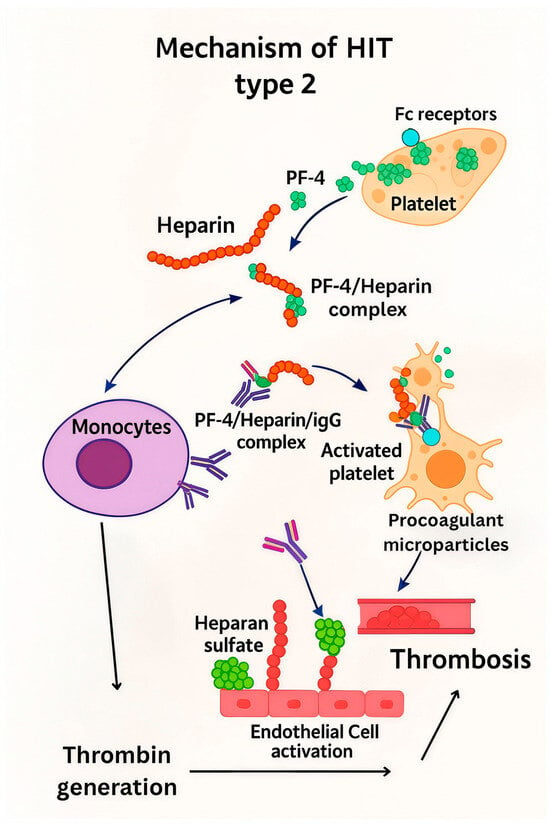
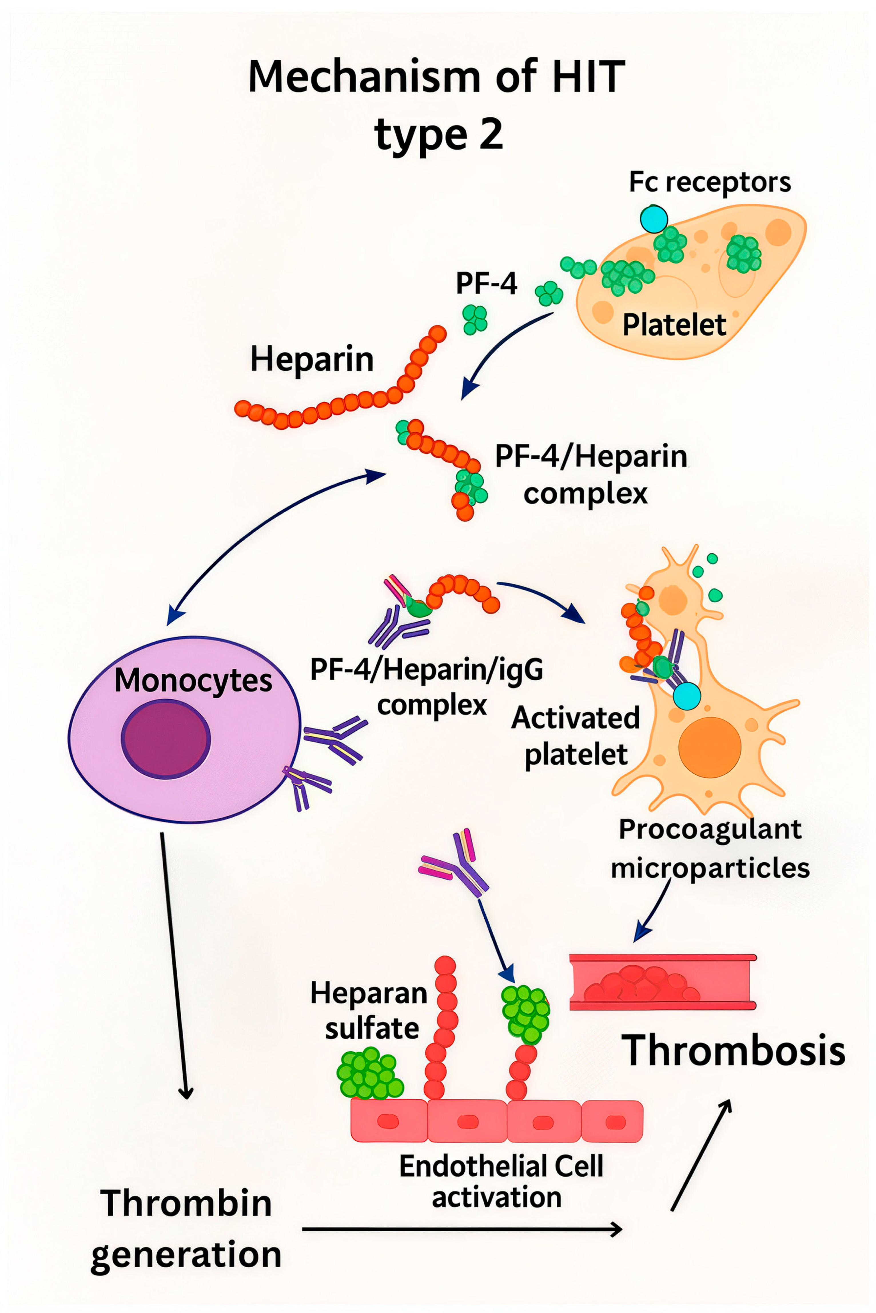
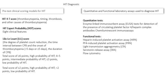
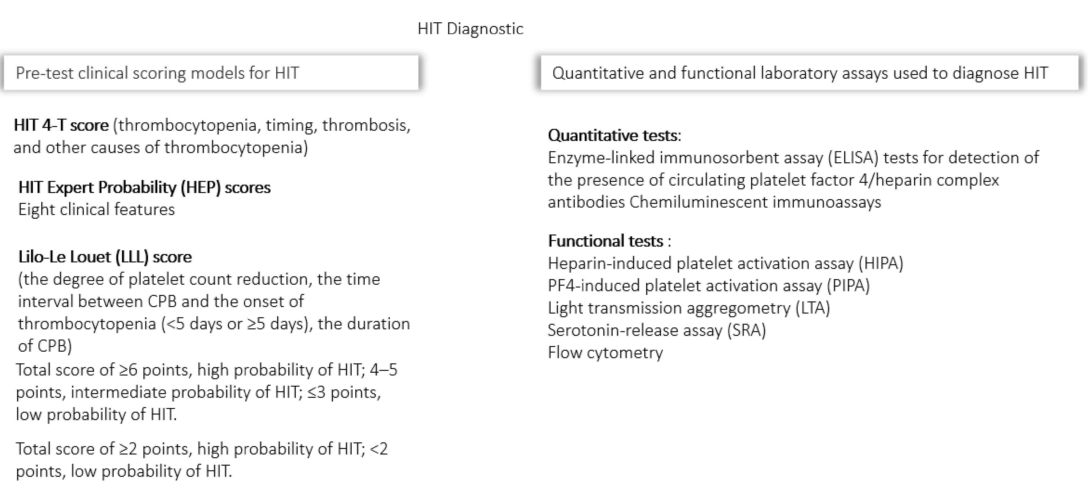
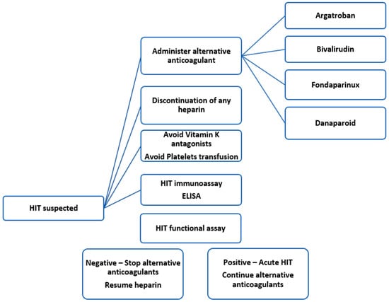
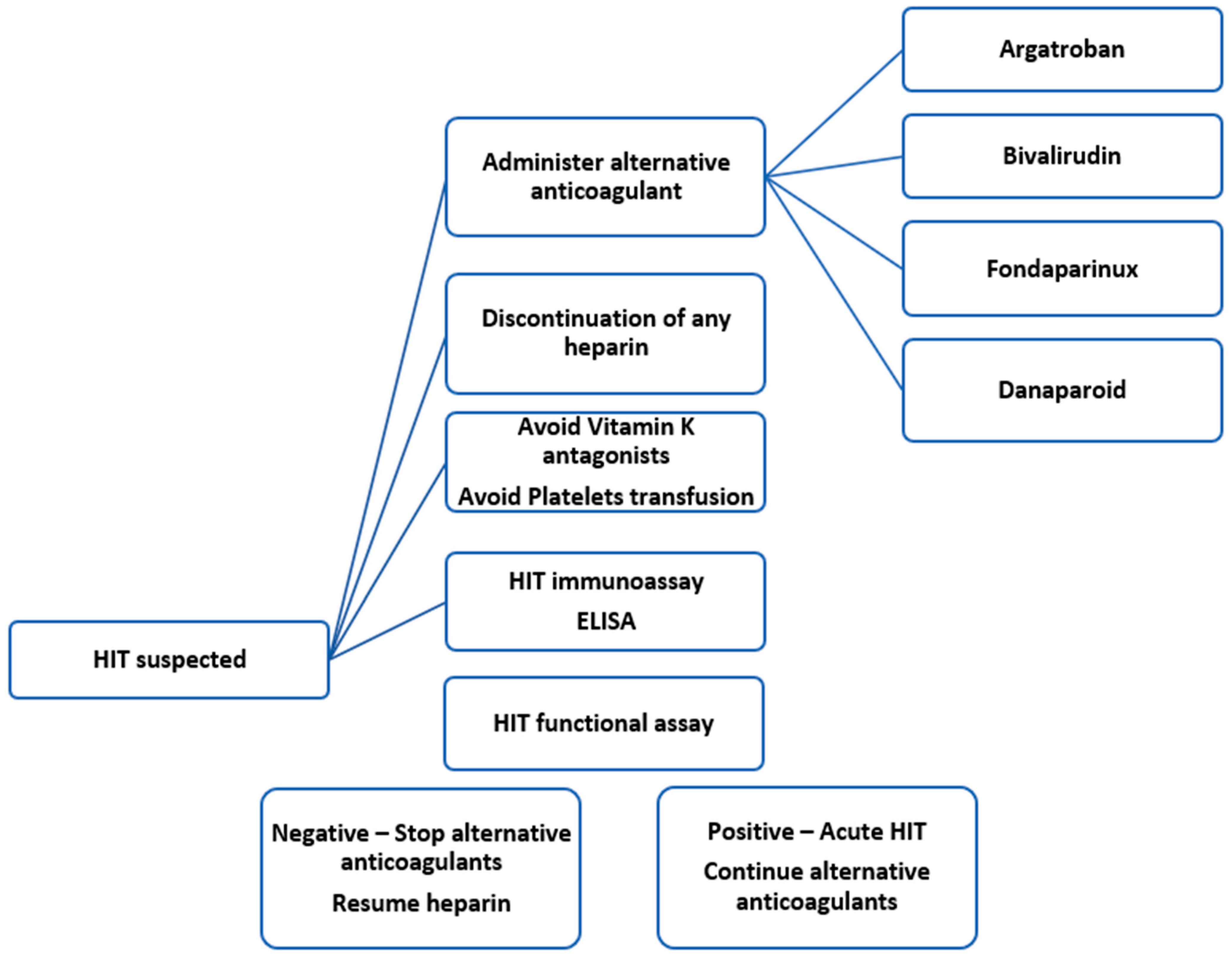

Disclaimer/Publisher’s Note: The statements, opinions and data contained in all publications are solely those of the individual author(s) and contributor(s) and not of MDPI and/or the editor(s). MDPI and/or the editor(s) disclaim responsibility for any injury to people or property resulting from any ideas, methods, instructions or products referred to in the content. |
© 2025 by the authors. Licensee MDPI, Basel, Switzerland. This article is an open access article distributed under the terms and conditions of the Creative Commons Attribution (CC BY) license (https://creativecommons.org/licenses/by/4.0/).