


Abstract
Body mass index (BMI) is widely used in clinical practice, but it does not directly reflect fat distribution, ectopic adiposity, or adipose tissue function. Individuals with a normal or near-normal BMI may still have excess visceral fat. The Thin Outside, Fat Inside (TOFI) phenotype describes this mismatch between external body size and internal adiposity. This structured narrative review examined TOFI and related normal-weight phenotypes, focusing on visceral and ectopic fat, adipose tissue dysfunction, molecular mechanisms, imaging, and organ-specific risk. A literature search was conducted in PubMed/MEDLINE, Scopus, and Web of Science for English-language studies published mainly between 2021 and 2026. Earlier seminal and directly relevant studies were also included. Evidence from related human phenotypes and experimental models suggests that impaired adipose tissue expandability, visceral and ectopic fat accumulation, inflammation, mitochondrial stress, and lipotoxicity may help explain metabolic dysfunction despite a normal BMI. These mechanisms remain incompletely characterized in populations explicitly defined as TOFI. Preclinical models and imaging methods may help characterize TOFI-like features. Diagnostic criteria are still heterogeneous, and TOFI-specific longitudinal and intervention data remain limited. TOFI may serve as a useful translational framework for identifying hidden adiposity and metabolic risk in individuals with a normal BMI.
1. Introduction
Body mass index (BMI) remains widely used in clinical practice and epidemiological research because it is simple, inexpensive, and easy to apply. BMI is only an indirect measure of adiposity. It does not directly assess body composition, fat distribution, or adipose tissue function. This limitation is clinically relevant because abdominal and visceral adiposity provide information that BMI alone cannot capture. The International Atherosclerosis Society (IAS) and International Chair on Cardiometabolic Risk (ICCR) Working Group on Visceral Obesity stated that BMI alone is not sufficient for assessing cardiometabolic risk related to increased adiposity and recommended waist circumference as a routine clinical measurement alongside BMI [1].
This limitation is now formally recognized in updated clinical guidance. The American Diabetes Association (ADA) Standards of Care in Overweight and Obesity—2026 guidelines describe obesity as a heterogeneous chronic disease in which excess adiposity increases morbidity and mortality risk. The ADA not only recommends screening adults for excess adiposity at least annually using BMI but also emphasizes that BMI may overestimate adiposity in muscular individuals and underestimate adiposity in some populations, including Asian adults and older adults. The same document supports the use of race- and ethnicity-specific BMI thresholds and additional waist circumference-based measures in selected individuals to reduce underdiagnosis. It also recognizes that waist-to-height ratio, bioelectrical impedance analysis, and dual-energy X-ray absorptiometry (DXA) may help confirm adiposity when BMI and physical examination are ambiguous [2].
The European Association for the Study of Obesity proposed a complementary framework that aligns obesity with the concept of an adiposity-based chronic disease. In this framework, diagnosis should not rely only on abnormal or excessive fat accumulation but should also consider the present or potential effects of adiposity on health [3].
These developments are highly relevant for individuals with a normal BMI. Some individuals fall within the conventional normal BMI range but have excess body fat, central adiposity, visceral adiposity, or metabolic abnormalities. Normal-weight obesity (NWO) illustrates this limitation. It is generally defined as normal BMI with increased body fat percentage. In a systematic review and meta-analysis including 25 studies and 177,792 participants, NWO was associated with higher odds of hyperglycemia, high triglycerides, low HDL cholesterol, diabetes, dyslipidemia, hypertension, and metabolic syndrome compared with normal-weight non-obese individuals [4].
Normal BMI with abdominal obesity is also clinically relevant at the population level. In a large cross-sectional study using WHO STEPS data from 91 countries, normal-weight abdominal obesity was defined as normal BMI with high waist circumference. More than one in five adults with a normal BMI had abdominal obesity, and this phenotype was associated with hypertension, diabetes, high total cholesterol, and high triglycerides [5].
This phenotype is not limited to older adults. In young Colombian adults, NWO was defined as normal BMI with excess body fat measured by bioelectrical impedance analysis. This phenotype was associated with increased cardiometabolic risk, low HDL cholesterol, abdominal obesity, and low muscular strength [6].
NWO may also involve biological features beyond classical cardiometabolic risk factors. In two independent cohorts, NWO was defined as normal BMI with an excess percentage of body fat measured by DXA. Individuals with NWO showed distinct gut microbiota profiles, and the NWO-related gut microbiome was associated with hypertension, dyslipidemia, and metabolic syndrome [7].
Visceral adipose tissue is central to the biological interpretation of these findings. BMI is accessible, but it does not capture differences in body composition, fat distribution, or ectopic fat burden. Visceral adipose tissue has been described as a major driver of cardiometabolic consequences and a potential therapeutic target for preventing progression within the cardiovascular–kidney–metabolic risk continuum [8].
The term “Thin Outside, Fat Inside” (TOFI) describes a mismatch between apparently normal body size and disproportionate internal adiposity. Thomas et al. characterized this subphenotype using whole-body MRI and magnetic resonance spectroscopy in 477 white volunteers. They assessed abdominal adipose tissue distribution and hepatic and intramyocellular lipid content, providing reference ranges and identifying individuals whose internal fat burden was not adequately reflected by conventional anthropometric measures. Their proposed TOFI classification used the ratio of intra-abdominal to abdominal subcutaneous adipose tissue, providing an imaging-based foundation for the concept [9].
This lack of standardization is an important gap. Some studies define the phenotype by body fat percentage. Others use waist circumference, waist-to-height ratio, abdominal adiposity, visceral adiposity, metabolic syndrome, insulin resistance, or other cardiometabolic markers. As a result, similar individuals may be classified differently across studies.
In the context of emerging adiposity-based frameworks, TOFI should not be considered only a descriptive label. It represents a clinically relevant phenotype that exposes a blind spot in BMI-based medicine. A normal BMI does not exclude increased body fat, central or visceral adiposity, ectopic fat deposition, metabolic dysfunction, or cardiometabolic and vascular risk.
This narrative review examines clinical, mechanistic, and imaging evidence relevant to the TOFI phenotype. It clarifies the relationship between TOFI and related normal-weight adiposity phenotypes and distinguishes findings from studies explicitly characterizing TOFI, evidence from related human populations, and experimental observations. The synthesis covers visceral and ectopic fat, adipose tissue dysfunction, potential mechanisms of metabolic injury, imaging approaches, and organ-specific outcomes. Its main purpose is to identify what is established, what remains extrapolated from broader obesity research, and where TOFI-specific studies are needed. Attention is given to factors that may explain internal fat accumulation despite a normal BMI and to the limitations of applying existing diagnostic and therapeutic evidence to this population.
In this review, we propose the Hidden Adiposity–Lipotoxicity–Organ Vulnerability Axis as a hypothesis-generating framework for the TOFI phenotype. The framework integrates impaired subcutaneous adipose tissue expandability, preferential visceral and ectopic lipid deposition, adipose tissue dysfunction, inflammatory and mitochondrial stress, bioactive lipid intermediates, insulin resistance, and organ-specific vulnerability. Importantly, these mechanisms have not been demonstrated as a unified causal pathway in individuals with rigorously defined TOFI. The framework therefore distinguishes direct evidence obtained in normal BMI adiposity phenotypes from indirect human mechanistic evidence and experimental or preclinical findings.
A further objective was to evaluate how current preclinical models and imaging approaches can reproduce or quantify TOFI-like features and support translation from mechanistic adiposity research to clinical phenotyping. The proposed framework is illustrated in Figure 1.
Figure 1.
Proposed Hidden Adiposity–Lipotoxicity–Organ Vulnerability Axis relevant to the TOFI phenotype. The framework brings together adiposity patterns, molecular mechanisms, and potential organ vulnerability. Dashed connectors indicate proposed associations, not established causal relationships in TOFI. T, studies explicitly characterizing TOFI; H, studies in related human populations; E, experimental studies. Labels indicate evidence sources, not evidence quality, and do not imply that every listed feature has been demonstrated in TOFI. Created in BioRender. Ispas, S. (2026) https://BioRender.com/xb8ed7d, accessed on 16 October 2026. Abbreviations: AKT, protein kinase B; BMI, body mass index; GLUT4, glucose transporter type 4; HIF-1α, hypoxia-inducible factor 1 alpha; IL-1β, interleukin-1 beta; IRS, insulin receptor substrate; MASLD, metabolic dysfunction-associated steatotic liver disease; MONW, metabolically obese normal weight; mtROS, mitochondrial reactive oxygen species; NF-κB, nuclear factor kappa B; NLRP3, NOD-like receptor family pyrin domain-containing 3; NWO, normal-weight obesity; PI3K, phosphoinositide 3-kinase; RBP4, retinol-binding protein 4; TLR4, Toll-like receptor 4; TOFI, Thin Outside, Fat Inside. Evidence labels indicate the populations and study types supporting each component; they do not imply that every listed feature has been demonstrated in TOFI. Solid arrows illustrate mechanistic processes within the proposed framework. Upward and downward arrows indicate increases and decreases, respectively. Green, blue, and pink shading distinguish the phenotypic, mechanistic, and or-gan-vulnerability domains, respectively.
2. Materials and Methods
2.1. Review Design and Reporting Framework
This article was designed as a structured narrative review examining the Thin Outside, Fat Inside (TOFI) phenotype as a model of hidden visceral and ectopic adiposity in individuals with normal or near-normal body mass index (BMI). This review integrates clinical, molecular, anatomical, imaging, preclinical, and translational evidence relevant to TOFI and related normal-weight adiposity phenotypes.
Attention was given to distinguishing evidence obtained directly in TOFI, normal-weight obesity, metabolically obese normal weight, normal-weight central obesity, and normal-weight visceral obesity from evidence derived from broader studies of visceral adiposity, ectopic fat, obesity, metabolic disease, and experimental models.
The reporting approach was guided by the Scale for the Assessment of Narrative Review Articles (SANRA), with attention to the review rationale, formulation of the objective, description of the literature search, appropriate citation of the literature, scientific reasoning, and balanced interpretation of the available evidence [10].
This review was not conducted as a systematic review or meta-analysis. No Preferred Reporting Items for Systematic Reviews and Meta-Analyses (PRISMA) flow diagram, formal risk-of-bias assessment, protocol registration, or quantitative synthesis was performed.
2.2. Search Strategy
Structured searches were conducted in PubMed/MEDLINE, Scopus, and Web of Science for English-language publications mainly from 2021 to 2026. The initial database searches were conducted on 19 August 2026. The TOFI-specific searches reported in blocks A9, B9 and C9 were conducted on 13 October 2026. Earlier studies were included when they provided foundational information on adipose tissue distribution, visceral adiposity, ectopic fat, insulin resistance, vascular dysfunction, body-composition assessment, or preclinical imaging. Reference lists of selected publications were also screened to identify relevant earlier studies.
PubMed/MEDLINE was used as the primary database for the literature search. Scopus and Web of Science were searched to cross-check and expand reference retrieval.
Terms related to visceral and ectopic adiposity included “visceral adipose tissue”, “fatty pancreas”, “intrapancreatic fat”, “pancreatic steatosis”, “epicardial adipose tissue”, “epicardial fat”, “perivascular adipose tissue”, and “myosteatosis”.
Mechanistic searches included terms related to “adipose tissue expansion”, “adipose tissue plasticity”, “lipodystrophy syndromes”, “hypoxia”, “adiponectin”, “leptin resistance”, “adipose tissue dysfunction”, “mitochondrial dysfunction”, “ceramides”, and “insulin resistance”.
Terms related to the TOFI phenotype included “TOFI”, “thin outside fat inside”, “thin on the outside fat on the inside”, “normal-weight visceral obesity”, and “normal weight visceral obesity”.
Additional searches addressed vascular and cardiometabolic risk; preclinical models of visceral and ectopic adiposity; micro-CT and MRI; body-composition imaging; and interventions targeting visceral or ectopic fat.
Thematic searches were supplemented by reference list checking and targeted retrieval of known articles using distinctive title terms. These title-based searches served to locate specific publications.
Search terms were combined using the Boolean operators “AND” and “OR”, according to the requirements of each database.
The complete database-specific search strategies are provided in the Supplementary Materials (Table S1: PubMed/MEDLINE; Table S2: Web of Science; Table S3: Scopus).
The numbers in Supplementary Tables S1–S3 refer to individual searches. The same publication may appear in several queries or databases, so these counts cannot be added to obtain the number of unique records assessed. Publications were evaluated for relevance to the predefined review domains using the criteria described in Section 2.3 and Section 2.4. The tables document the searches performed but do not provide a complete numerical account of study selection.
The predefined inclusion and exclusion criteria are summarized in Supplementary Table S4.
2.3. Selection Criteria
Human, animal, experimental, translational, imaging, anatomical, observational, and clinical studies were considered, together with systematic reviews, meta-analyses, narrative reviews, guidelines, consensus statements, and expert reports.
Studies were considered relevant when they addressed at least one of the main domains of the review: TOFI or related normal BMI phenotypes; visceral adiposity; ectopic fat deposition; adipose tissue dysfunction; insulin resistance; molecular mechanisms of lipotoxicity and inflammation; vascular, renal, or retinal outcomes; preclinical models; diagnostic imaging; body-composition assessment; or interventions affecting visceral or ectopic fat.
Studies focused only on BMI-defined obesity without information on body composition, fat distribution, visceral adiposity, ectopic fat, metabolic dysfunction, or related mechanisms were not prioritized. Priority was given to studies with clearly described phenotype definitions, objective measures of adiposity, and outcomes relevant to this review. Studies in explicitly characterized TOFI or related normal BMI populations were considered separately from broader human and experimental evidence. Foundational studies were retained when they established a phenotype definition or a concept needed for interpretation.
Conference abstracts without accessible full text, letters, comments, and editorials without relevant mechanistic or clinical content were excluded. Non-English publications were excluded. Studies without a clear connection to the predefined TOFI-related domains were not used for detailed interpretation.
2.4. Study Selection and Author Involvement
The literature search and initial organization of the retrieved material were coordinated by S.I., A.G., and M.R.
Titles and abstracts were screened for thematic relevance by S.I., D.M., A.C.F.F.S., M.D.-G., S.K., S.A.R., J.L., D.H.H.S., V.V.-R., and Z.A.M.C.
Potentially relevant publications were assessed in full text and allocated to the predefined domains of this review. Evidence related to TOFI, normal-weight obesity, metabolically obese normal weight, and other BMI-discordant adiposity phenotypes was reviewed by S.I., A.G., D.M., A.C.F.F.S., M.D.-G., and Z.A.M.C.
The molecular and metabolic literature addressing adipose tissue expandability, inflammation, adipokines, mitochondrial dysfunction, lipotoxicity, and insulin resistance was evaluated by S.I., S.K., I.R.R., S.A.R., and J.L.
The anatomical, body-composition, visceral adiposity, and imaging literature was reviewed by S.I., V.I., D.H.H.S., and V.V.-R.
The cardiovascular, renal, retinal, metabolic, and translational implications were evaluated by A.G., I.R.R., M.R., S.K., and S.I.
Preclinical models and imaging approaches were reviewed by S.I., I.R.R., S.A.R., J.L., V.I., and M.R.
Uncertainties regarding article relevance, thematic classification, or interpretation were resolved through discussion among the authors. Overall scientific review and oversight were provided by S.I., A.G., and M.R.
2.5. Evidence Synthesis
The selected literature was synthesized narratively and organized into thematic sections covering TOFI and related normal-weight phenotypes, visceral and ectopic adipose depots, molecular mechanisms, vascular and microvascular vulnerability, experimental models, imaging, and therapeutic implications.
Direct evidence obtained in TOFI or closely related normal BMI adiposity phenotypes was distinguished from indirect evidence derived from broader studies of visceral adiposity, obesity, type 2 diabetes, MASLD, cardiovascular disease, or experimental models.
Mechanistic plausibility was separated from direct clinical evidence. Findings from animal models, cellular studies, and broader obesity research were not interpreted as proof of TOFI-specific causality.
The strength of the evidence was considered according to its source. Prospective clinical studies, systematic reviews, meta-analyses, and consensus documents were used primarily for clinical and translational interpretation, while experimental and mechanistic studies were used to support biological plausibility.
The proposed Hidden Adiposity–Lipotoxicity–Organ Vulnerability Axis was used as a hypothesis-generating framework to integrate the available evidence; it was not considered an established causal model.
The descriptions in the evidence synthesis table reflect the authors’ narrative interpretation of the populations studied, study designs, measurements, and consistency of findings. They are not formal evidence grades. Human observational associations, experimental mechanisms, and findings in explicitly characterized TOFI populations were considered separately; evidence from one category was not taken as direct confirmation in another.
3. Definitions and Phenotypic Boundaries: TOFI, NWO, MONW, and Related States
3.1. TOFI: A Clinical Concept Without a Standardized Definition
The term “Thin Outside, Fat Inside” (TOFI) describes a phenotype in which apparently normal body size conceals disproportionate internal adiposity. Thomas et al. characterized this subphenotype using whole-body MRI and magnetic resonance spectroscopy, providing an imaging-based foundation for the concept [9]. Müller et al. subsequently discussed TOFI within the broader limitations of BMI-based assessment [11].
The TOFI phenotype is also consistent with the broader concept of “obesities”, which recognizes obesity-related conditions as heterogeneous phenotypes and not a single entity defined only by BMI [12].
TOFI is not yet a standardized clinical diagnosis. The lack of universally shared definitions and diagnostic criteria contributes to variability in the identification of obesity-related phenotypes [12].
In this review, TOFI is used as a descriptive framework for disproportionate internal adiposity despite an apparently normal body size. The original imaging-based characterization emphasized abdominal fat distribution [9]. Isolated ectopic fat accumulation is relevant to this framework, but its sufficiency for classifying an individual as TOFI has not been established. Metabolic abnormalities are not treated as a prerequisite: adiposity and metabolic health are assessed separately.
BMI thresholds should be reported explicitly for each study, with normal-weight and near-normal-weight populations kept distinct. Population-specific thresholds also require attention: A BMI below 27 kg/m2, as used in ReTUNE, should not be equated with the conventional normal-weight range [13]. Age, sex, and ancestry may influence body composition and fat distribution; thus, imaging measurements should be interpreted against appropriate reference populations whenever available [14]. These considerations inform phenotyping but do not provide validated TOFI-specific diagnostic cut-offs.
Several related normal BMI phenotypes are used in the literature. Their definitions overlap, but they are not interchangeable. These differences are summarized in Table 1.
3.2. Normal-Weight Obesity
Normal-weight obesity (NWO) is generally defined as normal BMI combined with an increased body fat percentage [4]. This phenotype illustrates that BMI-defined normal weight does not necessarily indicate normal body composition. Romero-Corral et al. showed that normal BMI combined with high body fat percentage was associated with cardiometabolic abnormalities, and in women, it was associated with increased cardiovascular mortality [15]. A systematic review and meta-analysis showed that NWO is associated with higher odds of hyperglycemia, elevated triglycerides, low HDL cholesterol, diabetes, dyslipidemia, hypertension, and metabolic syndrome [4].
Body-composition studies further support the biological relevance of NWO. Dojo et al. reported differences in anthropometric traits, metabolic profiles, visceral and subcutaneous fat areas, and brown-adipose-tissue-related parameters in women with NWO [16].
NWO may also be associated with systemic inflammation. Liu-Galvin et al. found increased odds of inflammation in individuals with a normal BMI but a high body fat percentage [17].
DXA and bioelectrical impedance analysis are commonly used to identify NWO, while BMI alone remains an imperfect surrogate for adiposity [18].
NWO should not be considered synonymous with TOFI. NWO is defined mainly by increased total body fat, whereas TOFI emphasizes hidden internal adiposity, particularly visceral or ectopic fat.
3.3. Metabolically Obese Normal Weight
Ruderman et al. introduced the concept of the “metabolically obese, normal-weight” individual in 1981, describing metabolic disorders associated with obesity in individuals who were not obese according to conventional weight criteria [19].
The concept of metabolically obese normal weight (MONW) describes individuals whose BMI remains within the normal range but whose metabolic profile resembles that usually observed in obesity. These individuals may present with insulin resistance, elevated triglycerides, low HDL cholesterol, impaired glucose regulation, increased blood pressure, chronic low-grade inflammation, or components of the metabolic syndrome [20].
Several terms are used for this phenotype. Metabolically obese normal weight and metabolically unhealthy normal weight (MUNW) are closely related, but they are not always defined identically. Pluta et al. emphasized that MONW is associated with increased risks of type 2 diabetes and cardiovascular disease, while diagnostic criteria differ considerably across studies [20].
Some studies define MONW through insulin resistance, often using HOMA-IR. Others use metabolic syndrome components, lipid abnormalities, blood pressure, fasting glucose, inflammatory markers, or combinations of these variables [20]. This heterogeneity limits comparisons across studies and may lead to different classifications of the same individual.
Population-based data support the clinical relevance of MUNW. López-Herrera et al. identified metabolically unhealthy normal-weight individuals within a classification based on weight status and metabolic health [21]. MUNW was defined by the presence of at least one metabolic syndrome component in individuals with a normal BMI [21]. This approach uses readily available clinical variables, including blood pressure, glucose, triglycerides, HDL cholesterol, and abdominal obesity-related criteria.
The phenotype may also change over time. Suarez-Ortegón et al. examined the prevalence, incidence, and six-year stability of MUNW in U.S. Hispanic/Latino adults with a normal BMI and no cardiometabolic disease at baseline [22]. Metabolic abnormalities developed in some individuals who remained within the normal BMI range, supporting the dynamic nature of this phenotype [22].
MONW and MUNW are relevant to the TOFI framework, but they are not equivalent to TOFI. MONW and MUNW are primarily defined by metabolic abnormalities, whereas TOFI emphasizes hidden internal adiposity, particularly visceral or ectopic fat. Metabolic dysfunction may therefore occur without documented visceral adiposity, while visceral or ectopic fat may be present before overt metabolic abnormalities develop.
The phenotypes can overlap. Their overlap may identify individuals with a normal BMI, hidden adiposity, and measurable metabolic dysfunction.
3.4. Normal-Weight Central and Visceral Obesity
Normal-weight central obesity describes individuals with a normal BMI but increased abdominal adiposity, usually identified by waist circumference, waist-to-height ratio, or waist-to-hip ratio. In a global analysis of WHO STEPS data from 91 countries, normal-weight abdominal obesity was defined using normal BMI together with elevated waist circumference. More than one in five adults with a normal BMI had abdominal obesity, and this phenotype was associated with hypertension, diabetes, hypercholesterolemia, and hypertriglyceridemia [5].
National data from Türkiye provide similar evidence. Bağcı et al. reported that 25.2% of individuals with a normal BMI had central obesity, with a higher prevalence among women. Individuals with this phenotype also had a higher prevalence of hypertension, diabetes, and hypercholesterolemia than normal-weight adults without central obesity [23].
Longitudinal evidence supports the prognostic relevance of this phenotype. In a Chinese population-based cohort, Ren et al. defined normal-weight central obesity as a BMI of <24.0 kg/m2 combined with a waist circumference of >85 cm in men or >80 cm in women. During seven years of follow-up, this phenotype was associated with incident cardiovascular disease [24]. Participants who transitioned from normal weight without central obesity to normal-weight central obesity also exhibited higher cardiovascular risk [24].
Normal-weight visceral obesity represents a more specific phenotype because it refers directly to excess visceral adipose tissue in individuals with a normal BMI. Waist-based measures are practical but cannot distinguish visceral from abdominal subcutaneous fat. Direct assessment of visceral adiposity therefore requires imaging-based methods such as CT or MRI.
Table 1.
Distinguishing TOFI from related normal BMI adiposity phenotypes.
The phenotypes listed here were defined using different measures of adiposity and metabolic health. They are not interchangeable with TOFI. The cited studies used their own diagnostic criteria, which should be considered when comparing findings across populations.
The available evidence supporting the TOFI framework is heterogeneous and derives from several related phenotypes. To clarify the strength and source of this evidence, Table 2 summarizes the findings obtained directly from studies of TOFI or normal BMI adiposity phenotypes and distinguishes them from broader human and preclinical evidence.
Table 2.
Evidence supporting the TOFI framework.
4. From BMI to Adiposity-Based Disease: TOFI in the New Obesity Paradigm
Obesity as Excess or Dysfunctional Adiposity
BMI remains useful for population screening. It is simple, inexpensive, and reproducible, yet BMI measures body size, not adipose tissue distribution or function [1].
The limitation is clinically relevant. Individuals with the same BMI may differ in visceral fat, ectopic fat, muscle mass, and cardiometabolic risk [11].
Recent obesity frameworks have moved beyond BMI alone. The ADA Standards of Care in Overweight and Obesity—2026 guidelines define obesity as a heterogeneous chronic disease related to excess adiposity [2]. ADA still recommends annual BMI screening, but it also supports waist-based measures and body-composition tools when BMI is insufficient [2].
EASO also supports an adiposity-based framework. Its 2024 proposal states that diagnosis and staging should consider fat accumulation, fat distribution, and health effects, not BMI alone [3].
Waist circumference adds practical information to BMI. A recent review emphasized that BMI combined with waist circumference helps capture obesity heterogeneity and identify higher-risk phenotypes [53].
Population-level analyses support the same shift. Al-Roub et al. used BMI together with waist circumference, waist-to-hip ratio, and waist-to-height ratio to estimate obesity prevalence under newer anthropometric criteria [54].
A U.S. analysis applying the Lancet Commission framework incorporated BMI, waist circumference, waist-to-hip ratio, waist-to-height ratio, and body fat percentage to identify excess adiposity; then, it classified obesity as preclinical or clinical according to organ dysfunction and activity limitations [55].
5. Anatomical and Imaging Substrates of Hidden Adiposity
5.1. Visceral Adipose Tissue: The Core Depot of TOFI
Visceral adipose tissue is a central anatomical substrate of the TOFI phenotype. Beyond energy storage, it functions as an endocrine and immunometabolic tissue with effects on glucose, lipid, hepatic, and vascular homeostasis. Compared with subcutaneous adipose tissue, visceral adipose tissue is more strongly associated with insulin resistance, type 2 diabetes, dyslipidemia, metabolic syndrome, and cardiovascular disease [27].
The joint IAS/ICCR position statement identifies visceral adiposity and ectopic fat as important markers of cardiometabolic risk and emphasizes their assessment beyond BMI alone [56]. This provides a clinical foundation for considering internal fat distribution in apparently lean individuals, although the statement does not establish TOFI-specific diagnostic criteria.
Portal drainage is particularly important. Visceral adipose tissue releases fatty acids, glycerol, adipokines, and inflammatory mediators into the portal circulation, increasing hepatic metabolic and inflammatory exposure [27]. This pathway provides a mechanistic link between visceral adiposity, hepatic insulin resistance, and steatotic liver disease.
Adipose depots differ substantially in cellular composition, vascularization, inflammatory profile, lipid handling, and endocrine activity. These depot-specific characteristics help explain why individuals with similar BMI and total fat mass may have markedly different metabolic risk [57].
Recent evidence further supports adipose tissue distribution as an important determinant of metabolic disease, with visceral and ectopic fat linked to insulin resistance, type 2 diabetes, metabolic syndrome, cardiovascular disease, and organ-specific injury [58].
Ectopic fat extends this concept beyond the abdomen and includes lipid accumulation in organs such as the liver, pancreas, skeletal muscle, and heart. Such deposition may contribute to insulin resistance, β-cell dysfunction, and metabolic syndrome [28].
In TOFI, clinical relevance lies in the possibility that visceral and ectopic fat accumulation may occur despite a normal BMI and remain undetected by routine anthropometric assessment.
5.2. Hepatic Fat and Lean MASLD
Hepatic fat is a major ectopic fat depot within the TOFI framework and may be present in individuals without BMI-defined obesity. MASLD is defined by hepatic steatosis in the presence of at least one cardiometabolic risk factor, after exclusion of other specific causes of steatotic liver disease. The 2024 EASL–EASD–EASO guidelines recommend case-finding for MASLD and advanced fibrosis in individuals with cardiometabolic risk factors, abnormal liver enzymes, or imaging evidence of steatosis [59].
Lean MASLD is particularly relevant to TOFI because hepatic steatosis may occur below conventional BMI thresholds for overweight. A meta-analysis comparing lean and non-lean MASLD found lower odds of overall cardiovascular disease in lean MASLD but higher cardiovascular mortality, indicating that this phenotype should not be considered metabolically benign [29].
Recent Eastern European data similarly suggest that lean MASLD may represent a clinically significant condition in individuals with a normal BMI and may be missed by BMI-centered screening [60]. Normal-weight MASLD also presents classification challenges, as reclassification between NAFLD and MASLD may be less straightforward in individuals with a lower cardiometabolic risk burden [61].
Visceral adiposity may promote hepatic fat accumulation through increased portal delivery of free fatty acids, glycerol, adipokines, and inflammatory mediators. This metabolic flux may contribute to hepatic insulin resistance, increased glucose production, and very-low-density lipoprotein (VLDL) secretion [27].
Within the TOFI framework, hepatic steatosis represents an important form of hidden ectopic adiposity that may coexist with normal BMI and cardiometabolic dysfunction.
5.3. Intrapancreatic Fat and β-Cell Vulnerability
Intrapancreatic fat deposition represents an important ectopic fat depot within the TOFI framework. Excess pancreatic fat has been associated with insulin resistance, metabolic syndrome, type 2 diabetes, and broader metabolic dysfunction, although the underlying mechanisms remain incompletely defined [30].
Pancreatic fat may contribute to metabolic dysfunction through local lipotoxicity, inflammatory signaling, altered adipokine activity, oxidative stress, and impaired β-cell function. These relationships are complex, and pancreatic fat content alone should not be interpreted as a direct marker of β-cell failure [30].
Imaging is central to the non-invasive assessment of intrapancreatic fat. Chemical shift-encoded MRI and MRI-derived proton density fat fraction provide quantitative estimates of pancreatic fat and show strong agreement with histological fat measurements [62].
Recent multidisciplinary consensus guidance identifies MRI as the preferred modality for quantitative assessments of pancreatic fat, while magnetic resonance spectroscopy and chemical-shift techniques may provide complementary information [63].
5.4. Epicardial and Perivascular Fat: Local Vascular Signaling
Epicardial adipose tissue is a visceral fat depot located between the myocardium and the visceral pericardium. Its anatomical proximity to the myocardium and coronary arteries establishes its direct paracrine and vasocrine relevance [31].
EAT is a biologically active depot involved in fatty acid handling, adipokine secretion, inflammation, oxidative stress, and local immune signaling [31]. Under physiological conditions, epicardial fat may provide mechanical protection and local energy supply for the myocardium [31]. In cardiometabolic disease, it may become dysfunctional and pro-inflammatory [64].
Pericoronary adipose tissue surrounds the coronary arteries and participates in bidirectional crosstalk with the vascular wall [31]. Computed tomography can assess epicardial fat volume and attenuation, and EAT attenuation has been proposed as an imaging marker of local inflammation and cardiovascular risk [65].
Perivascular adipose tissue surrounds blood vessels and contributes to vascular homeostasis [66]. In metabolic disease, PVAT may lose its protective vascular effects and promote oxidative stress, endothelial dysfunction, vascular inflammation, and atherosclerosis [66].
PVAT interacts with immune cells, endothelial cells, smooth muscle cells, and vascular inflammatory pathways, contributing to the complex relationship between adiposity, diabetes, and cardiovascular disease [67].
TOFI-specific outcome studies of epicardial and perivascular adipose tissue remain limited.
5.5. Intramuscular Fat and Skeletal Muscle Quality
Myosteatosis refers to pathological fat accumulation within skeletal muscle and is increasingly recognized as a marker of reduced muscle quality [32]. It may occur despite preserved muscle mass and is associated with lower muscle strength, impaired mobility, and poorer muscle function [32].
Fat infiltration in skeletal muscle has been linked to insulin resistance, metabolic dysfunction, and type 2 diabetes risk [68].
Skeletal muscle is a major site of insulin-mediated glucose disposal. Excess lipid accumulation may impair insulin signaling and metabolic flexibility [68].
Intramyocellular triglycerides may be increased in endurance-trained individuals despite preserved insulin sensitivity, a phenomenon known as the athlete’s paradox [69].
Lipid localization, lipid composition, mitochondrial capacity, and physical fitness influence the metabolic consequences of intramuscular lipid accumulation [69].
CT-derived muscle attenuation, MRI, magnetic resonance spectroscopy, and ultrasound-based approaches can be used to assess muscle quality and myosteatosis [32].
A normal BMI may coexist with visceral adiposity, ectopic fat accumulation, myosteatosis, and reduced muscle metabolic quality.
6. Molecular Mechanisms Linking Hidden Adiposity to Organ Risk
6.1. Adipose Tissue Expandability Failure and Ectopic Lipid Overflow
Adipose tissue can protect metabolism by storing excess energy safely through adipocyte recruitment, extracellular matrix remodeling, vascular adaptation, and adequate lipid-buffering capacity [33]. When subcutaneous adipose tissue cannot expand appropriately, lipids may be redirected toward visceral and ectopic depots, a process described by the adipose tissue expandability hypothesis [33].
Human genetic evidence supports this hypothesis. Lotta et al. linked genetic susceptibility to insulin resistance with reduced peripheral adipose storage capacity, supporting a role for limited lipid storage in cardiometabolic disease [37]. These findings are relevant to the dissociation between body size and metabolic risk described by TOFI, although the study did not specifically recruit an imaging-defined TOFI population. Limited peripheral adipose storage offers a possible explanation for metabolic dysfunction at a lower body mass. Its role in determining the TOFI phenotype requires direct investigation.
Limited capacity to store fat in lower body subcutaneous depots may contribute to an adverse metabolic profile. Stefan’s review discusses this limitation alongside visceral and ectopic fat accumulation, emphasizing the importance of fat distribution in cardiometabolic risk assessment [70].
The “Y-Y paradox” illustrates how BMI can conceal differences in body composition. Yajnik and Yudkin had nearly identical BMI, but their DXA scans showed markedly different body fat levels. This comparison illustrates the South Asian “thin–fat” phenotype and highlights why similar BMI values may conceal differences in body composition across populations [71]. It does not, however, establish the prevalence of imaging-defined TOFI.
Menopausal status is another factor to consider. Lovejoy et al. observed increased visceral fat during the menopausal transition, together with changes in energy expenditure [72]. These findings suggest that menopausal changes may contribute to internal fat accumulation, although the study did not specifically examine women with TOFI. Age-related changes in muscle composition may also be relevant. In the Health ABC cohort, Delmonico et al. documented declining muscle strength and quality alongside increased intermuscular fat over five years [73]. These findings show that changes in tissue composition can occur despite stable body weight. They provide supporting evidence from older adults but do not establish that muscle loss causes TOFI.
Early-life exposures may also influence later adipose tissue function. In a rodent model described by Sousa et al., perinatal exposure to obesogenic conditions altered adipose tissue and hepatic energy-balance pathways [74]. This provides experimental support for a developmental contribution to TOFI-like features, but its relevance to human TOFI remains uncertain.
Physical activity may help explain differences in metabolic health among individuals with similar body sizes. Pesta et al. found higher muscle insulin sensitivity in endurance-trained individuals than in sedentary normal-weight participants [69]. This supports considering activity levels when assessing metabolic risk at normal BMI, although it does not establish physical inactivity as a cause of TOFI.
Impaired adipose expansion may promote lipid accumulation in the liver, pancreas, skeletal muscle, heart, and other non-adipose tissues [28]. Adipose tissue dysfunction is characterized by adipocyte hypertrophy, impaired subcutaneous expandability, hypoxia, inflammation, fibrosis, stress signaling, and release of pro-inflammatory and diabetogenic mediators [34].
Limited capacity for metabolically safe lipid storage may therefore favor visceral and ectopic fat deposition despite normal body size [33]. Depot-specific adipose biology and fat distribution further influence metabolic risk [58]. Visceral and ectopic fat distribution, together with depot-specific adipose dysfunction, may be more informative for metabolic risk than total adiposity alone [27].
6.2. Adipocyte Hypertrophy, Hypoxia, and HIF-1α Signaling
Adipocyte hypertrophy is a major feature of maladaptive adipose tissue expansion. Enlargement of adipocytes increases diffusion distances and may create local oxygen stress when vascular adaptation is insufficient [33]. Reduced oxygen availability activates hypoxia-inducible signaling and may alter adipocyte metabolism, immune-cell recruitment, extracellular matrix remodeling, and insulin sensitivity [58].
HIF-1α is a central mediator of the hypoxic response in adipose tissue. Chronic HIF-1α activation has been associated with inflammation, macrophage infiltration, fibrosis, and insulin resistance [38]. Persistent hypoxic signaling may shift adipose tissue from flexible lipid storage toward a fibrotic and metabolically dysfunctional state [38]. Adipose tissue dysfunction has been described as a triad involving inflammation, fibrosis, and impaired angiogenesis, with fibrosis limiting tissue plasticity and impaired angiogenesis further worsening local hypoxia [39].
Human data also support associations among adipose hypoxia, impaired glucose transporter type 4 (GLUT4)-mediated glucose transport, altered insulin signaling, and inflammatory activation [40].
6.3. Macrophage Infiltration, Toll-like Receptor 4 (TLR4)/Nuclear Factor Kappa B (NF-κB) Activation, and Metaflammation
Adipose tissue macrophages are key regulators of local inflammation and insulin resistance [41]. During adipose tissue dysfunction, macrophage numbers and phenotypes change, while lipid overload favors pro-inflammatory activation associated with insulin resistance and fibrosis [75]. The classical M1/M2 framework is incomplete, as adipose tissue macrophages include more diverse populations, including lipid-associated and metabolically activated macrophages [76].
TLR4 signaling provides a molecular link between nutrient excess and inflammation. Saturated fatty acids and lipopolysaccharide-related signals may activate TLR4 pathways in macrophages and adipocytes [43]. Downstream nuclear factor kappa B (NF-κB) activation increases the production of pro-inflammatory mediators, including tumor necrosis factor alpha (TNF-α), interleukin-6 (IL-6), interleukin-1 beta (IL-1β), and monocyte chemoattractant protein-1 (MCP-1) [43]. MCP-1 promotes monocyte recruitment into adipose tissue and can amplify macrophage accumulation and local inflammation [41].
Metaflammation describes chronic low-grade inflammation driven by metabolic stress and sustained by adipose tissue dysfunction, immune-cell infiltration, nutrient excess, mitochondrial stress, and inflammatory signaling [42]. Macrophages are major contributors to this process in adipose tissue and liver [77].
Direct TOFI-specific macrophage data remain limited, and most mechanistic evidence derives from obesity, metabolic syndrome, diabetes, and MASLD.
6.4. Adipokine Imbalance: Low Adiponectin, Leptin Resistance, and Pro-Inflammatory Adipokines
Adipose tissue acts as an endocrine organ. It secretes adipokines that influence insulin sensitivity, inflammation, lipid metabolism, vascular function, hepatic metabolism, and pancreatic β-cell function [78]. Healthy adipose tissue releases adipokines with protective metabolic effects. Adiponectin and adipsin are commonly described as antidiabetic or insulin-sensitizing adipose-derived signals [78].
Adipose tissue dysfunction changes this secretory profile. Dysfunctional adipose tissue shows reduced secretion of protective adipokines and increased secretion of pro-inflammatory or prodiabetic mediators such as resistin and RBP4 [78].
Adiponectin is especially relevant. It improves insulin sensitivity, supports fatty acid oxidation, and has anti-inflammatory and vascular-protective effects [79]. Low adiponectin levels are associated with insulin resistance, type 2 diabetes, MASLD, and cardiovascular risk [79].
Leptin has a different profile. It is produced mainly by white adipose tissue and regulates appetite, energy expenditure, neuroendocrine function, and immune responses [80]. In adipose tissue dysfunction, leptin levels may rise, while leptin signaling becomes impaired. This state is described as leptin resistance [80]. Leptin resistance may involve impaired blood–brain barrier transport, altered Janus kinase 2 (JAK2)–signal transducer and activator of transcription 3 (STAT3) signaling, suppressor of cytokine signaling 3 (SOCS3) activation, endoplasmic reticulum stress, inflammation, and peripheral leptin resistance [80].
The leptin-to-adiponectin ratio has been proposed as a marker of adipose tissue dysfunction and insulin resistance. A recent review linked adipose tissue dysregulation in type 2 diabetes to increased leptin, reduced adiponectin, chronic inflammation, and impaired glucose metabolism [81]. Resistin and RBP4 are also relevant in dysfunctional adipose tissue. These adipokines have been associated with inflammation, insulin resistance, and cardiometabolic risk [81]. Adipokine imbalance provides a molecular link between hidden adiposity and distant organ dysfunction. Altered adipose-derived signals can affect the liver, pancreas, skeletal muscle, myocardium, and vascular wall [81].
This mechanism is relevant to TOFI. A normal BMI may coexist with adipose tissue dysfunction and abnormal adipokine signaling. The external phenotype may appear reassuring, while endocrine adipose communication is already altered.
Direct TOFI-specific adipokine studies remain limited. For this review, adipokine imbalance is interpreted as a plausible mechanism linking visceral and ectopic adiposity to insulin resistance, metabolic inflammation, and vascular risk.
6.5. Mitochondrial Stress, mtROS, and Inflammatory Amplification
Mitochondrial dysfunction should be interpreted as a molecular amplifier of hidden adiposity, not only as a marker of metabolic disease. In obesity-related metabolic dysfunction, hypertrophied adipose tissue releases pro-inflammatory cytokines and increases circulating free fatty acids, promoting systemic insulin resistance and ectopic lipid deposition [82]. Persistent lipid overload may impair mitochondrial homeostasis. Reduced oxidative phosphorylation, excessive reactive oxygen species production, and mitochondrial DNA damage can intensify metabolic stress and weaken insulin signaling [82]. Mitochondrial ROS may also activate inflammatory pathways. Mitochondrial dysfunction and mitochondrial DNA damage can stimulate the NLRP3 inflammasome, creating a feedback loop between oxidative stress, inflammation, and metabolic dysfunction [82]. This mechanism is relevant to TOFI because normal BMI does not exclude visceral or ectopic adiposity. A patient may appear lean externally while hidden fat depots generate excess lipid flux, oxidative stress, and inflammatory signaling.
Mitophagy adds another layer to this framework. In adipose tissue macrophages, nutritional excess can alter mitochondrial quality control and contribute to obesity-induced inflammation and insulin resistance [83]. Kim et al. showed that BNIP3-mediated mitophagy regulates macrophage polarization in response to obesity. In their model, macrophage-specific BNIP3 deletion reduced high-fat-diet-induced adipose tissue inflammation and improved glucose tolerance and insulin sensitivity [83]. This is important for a molecular TOFI framework. It suggests that hidden adiposity may not only affect adipocytes but may also reshape immune-cell metabolism within adipose tissue. Through HIF1A–BNIP3 signaling, hypoxia and mitophagy may contribute to pro-inflammatory macrophage activation [83].
Skeletal muscle is another key target of lipid overflow. Fat infiltration in skeletal muscle can alter muscle structure, metabolism, and signaling pathways, reducing muscle quality and physical performance [84]. Intracellular lipid droplets may disrupt muscle-cell physiology, but their effect depends on quantity, size, location, and biochemical characteristics. This explains why intramuscular lipid accumulation does not always have the same metabolic meaning [84]. The athlete’s paradox illustrates this distinction. Highly trained individuals may remain insulin sensitive despite increased muscular fat, while individuals with obesity or type 2 diabetes tend to accumulate larger lipid droplets in metabolically less favorable locations [84]. Excess nutrient intake and obesity may also trigger mitochondrial dysfunction in skeletal muscle through inflammatory activation and increased reactive oxygen species production [84].
In the TOFI framework, this point is clinically important. A normal BMI individual may have preserved external body size but increased visceral fat, ectopic lipid deposition, myosteatosis, reduced muscle quality, and impaired insulin sensitivity.
Epicardial adipose tissue provides an organ-specific example of cardio-lipotoxicity [85]. Under pathological conditions, epicardial fat may expose the myocardium to excessive lipid influx, inflammation, diacylglycerols, ceramides, mitochondrial dysfunction, and oxidative stress [85].
This supports the concept that hidden adiposity can have local organ effects. In TOFI-related phenotypes, ectopic fat depots may contribute to organ vulnerability even when BMI appears normal.
For this review, mitochondrial dysfunction is not presented as a TOFI-specific mechanism already proven in dedicated cohorts. It is interpreted as a biologically plausible mechanism derived from obesity-related adipose tissue dysfunction, ectopic fat research, skeletal muscle lipid biology, and cardio-lipotoxicity. Direct studies in strictly defined TOFI populations remain limited.
6.6. Lipotoxic Intermediates: Ceramides, DAGs, and Insulin Signaling
Lipotoxicity refers to the harmful accumulation of lipid species in non-adipose tissues. It is not only triglyceride storage that matters but also the formation of bioactive lipid intermediates [84].
Diacylglycerols and ceramides are among the most studied lipotoxic mediators. They can interfere with insulin signaling in the liver, skeletal muscle, adipose tissue, and pancreatic β-cells [85].
In the liver, DAG accumulation is linked to protein kinase C epsilon (PKCε) activation. This may impair insulin receptor signaling and contribute to hepatic insulin resistance [86].
Ceramides affect insulin signaling through a different molecular route. They can inhibit protein kinase B (AKT) activity through protein phosphatase 2A and atypical PKC-related mechanisms [44].
AKT inhibition is metabolically important. In muscle and adipose tissue, AKT is required for insulin-stimulated GLUT4 translocation and glucose uptake [44].
Ceramides may also contribute to mitochondrial dysfunction, oxidative stress, endoplasmic reticulum stress, inflammation, β-cell dysfunction, and apoptosis [44].
Not all ceramides have the same biological effect. Chain length, tissue location, and subcellular compartment influence ceramide action [45].
This is important for TOFI. A normal BMI may coexist with ectopic lipid deposition, but the metabolic risk depends on lipid species and intracellular localization, not only on total fat quantity.
Recent human data support this concept. Muscle very-long-chain ceramides were associated with insulin resistance independently of obesity [46].
This finding is relevant for BMI-discordant phenotypes. It suggests that lipidomic signatures may identify metabolic risk even when body size is not elevated [46].
Ceramides are increasingly recognized as circulating biomarkers of cardiometabolic and cardiovascular risk [46].
For TOFI, lipotoxicity offers a molecular explanation for hidden risk. Visceral or ectopic fat may generate lipid intermediates that impair insulin signaling before overt obesity is present.
This pathway should be presented cautiously. Most evidence comes from obesity, type 2 diabetes, MASLD, and mechanistic lipid biology. Direct lipidomic studies in well-defined TOFI cohorts remain limited.
6.7. Insulin Resistance as the Systemic Readout of TOFI Biology
Insulin resistance integrates several mechanisms associated with hidden adiposity, including impaired adipose tissue expandability, visceral and ectopic fat accumulation, hypoxia, macrophage-driven inflammation, adipokine imbalance, mitochondrial stress, and lipotoxic signaling.
At the cellular level, insulin action depends on the coordinated activation of the insulin receptor, insulin receptor substrate (IRS) proteins, phosphoinositide 3-kinase (PI3K), 3-phosphoinositide-dependent protein kinase-1 (PDK1), and AKT. In skeletal muscle, AKT-mediated GLUT4 translocation is required for insulin-stimulated glucose uptake, and disruption of IRS–PI3K–AKT signaling reduces glucose disposal [87].
Ectopic lipid accumulation can impair insulin signaling through DAG-related PKC activation, ceramide-mediated AKT inhibition, inflammatory kinases, and mitochondrial stress [82]. In the liver, insulin resistance is characterized by impaired suppression of hepatic glucose production, reduced glycogen storage, altered lipid metabolism, and increased VLDL secretion [47].
Adipose tissue dysfunction further promotes hepatic insulin resistance by increasing lipolysis and non-esterified fatty acid flux to the liver [47]. Adipose-derived cytokines, adipokines, lipids, metabolites, and extracellular vesicles also contribute to inter-organ metabolic communication [48].
Skeletal muscle, liver, adipose tissue, and pancreatic β-cells therefore contribute to a multi-organ insulin-resistant state, with reduced peripheral glucose disposal, increased hepatic glucose output, dyslipidemia, lipid overflow, inflammatory signaling, compensatory hyperinsulinemia, and eventual β-cell dysfunction [49].
Direct insulin-clamp studies in rigorously defined TOFI populations remain limited.
7. Vascular and Microvascular Consequences of TOFI Biology
7.1. Endothelial Dysfunction and Early Vascular Injury
Adipose tissue distribution may influence endothelial function independently of overall adiposity. In the Framingham cohorts, CT-assessed visceral adipose tissue, but not subcutaneous adipose tissue, was inversely associated with brachial artery flow-mediated dilation after adjustment for cardiovascular risk factors [88].
Perivascular adipose tissue (PVAT) provides a local mechanism linking adipose dysfunction to the vascular wall. Under physiological conditions, PVAT exerts anti-contractile and vasoprotective effects, whereas hypoxia, inflammation, and oxidative stress can impair these properties and promote vascular dysfunction [89].
Experimental metabolic syndrome models indicate that increased O-GlcNAcylation of endothelial nitric oxide synthase may impair PVAT-derived nitric oxide signaling and reduce its anti-contractile effects [90].
Additional mechanistic evidence links adiposity-related vascular dysfunction to reduced nitric oxide bioavailability and inflammatory signaling. Increased endothelial arginase activity may reduce L-arginine availability for endothelial nitric oxide synthase and impair vascular function [91]. In human obesity, Wnt family member 5A (WNT5A)–c-Jun N-terminal kinase (JNK) signaling has been implicated in vascular insulin resistance and impaired insulin-mediated vasodilation [92].
Direct studies evaluating endothelial function in rigorously defined TOFI populations remain limited.
7.2. Arterial Stiffness and Subclinical Atherosclerosis
Central adiposity may identify vascular risk that is not apparent from BMI alone. Among normal-weight Chinese adults with hypertension, central obesity was associated with higher brachial-ankle pulse-wave velocity and increased odds of arterial stiffness compared with normal-weight individuals without central obesity [50].
Large-scale imaging data also support the relevance of visceral adiposity across BMI categories. Using UK Biobank imaging data, Makhmudova et al. reported associations between MRI- or DXA-derived visceral adiposity, reduced aortic distensibility, and atherosclerotic cardiovascular risk across BMI strata [93].
Composite measures of visceral adiposity show similar associations. The EVasCu study and accompanying meta-analysis identified a consistent association between the visceral adiposity index (VAI) and arterial stiffness, although VAI is an indirect surrogate and not a direct measure of visceral fat [94].
A systematic review of the visceral-to-subcutaneous abdominal fat ratio found that visceral adiposity was more consistently associated with cardiovascular disease risk than subcutaneous adiposity, independently of waist circumference [95].
Mechanistic evidence also suggests that visceral adipose dysfunction may contribute to vascular remodeling. In patients with severe obesity, visceral adipose tissue amine oxidase copper-containing 3 (AOC3) expression and circulating soluble vascular adhesion protein-1/semicarbazide-sensitive amine oxidase activity were associated with arterial stiffness assessed by the cardio-ankle vascular index [96].
Most available studies assess visceral adiposity, central obesity, or obesity. Prospective studies combining direct visceral-fat assessment with vascular phenotyping in normal BMI populations are still needed.
7.3. Renal Microvascular Susceptibility
Visceral adiposity may contribute to kidney injury through chronic inflammation, oxidative stress, endothelial dysfunction, hemodynamic stress, and accumulation of kidney-related adipose depots such as perirenal fat.
In an NHANES-based cross-sectional analysis, visceral fat areas measured by DXA were independently associated with albuminuria. A significant additive interaction between high visceral fat and diabetes was also observed in relation to albuminuria [51].
Longitudinal evidence further supports an association between visceral adiposity and renal risk. In a population-based cohort, a higher visceral adiposity index was independently associated with increased urinary albumin excretion and chronic kidney disease during a mean follow-up of 3.6 years, with risk increasing across VAI quartiles [97].
Kidney-related adiposity has also been linked to renal sinus and perirenal fat accumulation, altered adipokine signaling, inflammation, oxidative stress, endothelial dysfunction, and hemodynamic stress [98].
Direct prospective studies combining visceral-fat quantification with renal phenotyping in normal BMI or TOFI populations remain limited.
7.4. Retinal Microvascular Vulnerability
The retina provides a directly visualizable window into systemic microvascular health. In a large prospective UK Biobank cohort of individuals with diabetes who were free of diabetic retinopathy at baseline, body roundness index, lipid accumulation product, and visceral adiposity index were longitudinally associated with incident diabetic retinopathy [99].
Prospective evidence from the Guangzhou Diabetic Eye Study also showed that visceral obesity indices were independently associated with incident diabetic retinopathy after adjustments for relevant metabolic and clinical factors, including glycemic control [52].
In a cross-sectional NHANES analysis of 5572 U.S. adults, a higher weight-adjusted waist index was associated with greater odds of retinopathy. Mediation analysis suggested that glycated hemoglobin (HbA1c) accounted for approximately 30% of this association, indicating that dysglycemia may explain part, but not all, of the relationship between central adiposity and retinal abnormalities [100].
Direct studies using imaging-based visceral fat quantification in normal BMI or rigorously defined TOFI populations remain limited.
7.5. TOFI as a Systemic Cardio–Renal–Retinal Risk Phenotype
The cardiovascular–kidney–metabolic (CKM) framework provides a useful context for hidden adiposity. The American Heart Association defines CKM syndrome as a systemic disorder reflecting the interrelationships among metabolic risk factors, chronic kidney disease, and cardiovascular disease [101].
Visceral and ectopic fat may coexist with insulin resistance, dysglycemia, MASLD, vascular dysfunction, albuminuria, and other early cardiometabolic abnormalities despite a BMI value within the conventional normal range [102].
In a cross-sectional analysis of 22,019 adults from NHANES 1999–2018, visceral adiposity-related indices, including VAI, metabolic score for visceral fat (METS-VF), body roundness index (BRI), and weight-adjusted waist index (WWI), were significantly associated with advanced CKM syndrome [103].
Longitudinal evidence also supports the association between visceral adiposity and cardiovascular outcomes. In a prospective cohort of 6715 Chinese adults with CKM stages 0–3, visceral fat accumulation indices were independently associated with incident cardiovascular disease during 9 years of follow-up. Arterial stiffness partially mediated these associations [104].
Retinal and renal microvascular injury share several biological pathways, including endothelial dysfunction, inflammation, oxidative stress, and metabolic dysregulation, although organ-specific responses remain important.
A unified cardio–renal–retinal TOFI phenotype has not yet been prospectively validated. Future studies should combine predefined TOFI criteria; direct visceral-fat assessment; and simultaneous cardiovascular, renal, and retinal phenotyping.
The outcome evidence differs in both design and population. Prospective studies of normal-weight central obesity assessed cardiovascular outcomes [24], while a prospective retinopathy study recruited patients with diabetes and used visceral obesity indices [52]. The arterial stiffness and albuminuria studies provide observational associations in their respective populations [50,51]. These findings should not be combined as proof that imaging-defined TOFI causes multi-organ complications.
8. Experimental and Translational Models of TOFI-like Biology
8.1. Adipose Expandability and Lipodystrophy Models
Impaired subcutaneous adipose tissue expandability can redirect lipids toward visceral and ectopic depots, contributing to insulin resistance and type 2 diabetes [33].
Lipodystrophy provides an extreme model of defective adipose storage capacity. Loss or insufficiency of adipose tissue is associated with ectopic lipid deposition, hypertriglyceridemia, hepatic steatosis, insulin resistance, and diabetes [35].
Adipose tissue plasticity also influences the capacity to accommodate excess lipid. Failure of adaptive adipose remodeling may promote lipid overflow toward the liver and skeletal muscle [36].
A rodent model provides direct continuity with the TOFI concept. Sousa et al. combined postnatal overfeeding with maternal exposure to glycotoxins and described a “thin-outside-fat-inside” phenotype characterized by impaired visceral adipose tissue expandability, reduced neuropeptide Y receptor Y2 (NPY2R) signaling, altered adipocyte morphology, and disrupted energy-balance pathways [74].
This model demonstrates that external body size and internal adipose dysfunction can become dissociated during metabolic programming.
Experimental models of TOFI are difficult to define because TOFI is not yet a standardized diagnostic category. Most available models do not reproduce the clinical phenotype of a lean adult with hidden visceral or ectopic fat. They model specific biological components of TOFI.
8.2. Diet-Induced Models of Visceral and Ectopic Fat Accumulation
Diet-induced obesity models are widely used to investigate adipose inflammation, insulin resistance, hepatic steatosis, and ectopic lipid deposition [105].
In outbred CD-1 mice, high-fat feeding increased fat mass, adipocyte hypertrophy, adipose inflammatory gene expression, hepatic steatosis, and hyperinsulinemia [98].
In high-fat-diet-fed rats, aerobic exercise reduced intrahepatic and intramyocellular lipids, visceral and subcutaneous fat, leptin, triglycerides, and HOMA-IR despite relatively modest changes in body weight [106].
Scaffold implantation into epididymal adipose tissue in high-fat-diet-fed animals reduced ectopic lipid accumulation in the liver and skeletal muscle and improved hyperinsulinemia [107].
These models reproduce selected components of TOFI-related biology, particularly visceral and ectopic lipid accumulation, adipose dysfunction, and insulin resistance, but they do not fully reproduce the normal-weight human phenotype.
8.3. Translational Limits of Current Models
No current animal model fully reproduces human TOFI. Species differences in fat distribution, brown adipose tissue activity, diet exposure, adipose depot biology, pancreatic lipid handling, and physical activity limit direct translation.
Rodent visceral fat depots are not identical to human visceral adipose tissue, and imaging thresholds established in humans cannot be directly applied to rodents.
Diet-induced obesity models are valuable for mechanism discovery but are less suitable for defining clinical thresholds in normal BMI individuals.
Lipodystrophy models are useful because they isolate adipose storage failure, but they are not equivalent to common TOFI, which is likely to represent a milder and multifactorial phenotype.
Preclinical models should be interpreted as mechanistic tools. They help explain lipid overflow, adipose dysfunction, ectopic fat accumulation, inflammation, and insulin resistance, but they do not establish a standardized TOFI phenotype.
8.4. Preclinical Imaging of Visceral and Ectopic Fat
Preclinical imaging is essential for studying TOFI-like biology because it can quantify fat distribution in vivo and allows repeated assessments of visceral, subcutaneous, hepatic, muscular, cardiac, and brown adipose depots during disease progression or intervention.
Micro-CT scans quantify total, visceral, and subcutaneous adipose tissue in small rodents and provide high spatial resolution, allowing separation of adipose compartments based on X-ray attenuation characteristics [108].
In vivo micro-CT has been validated against harvested fat-pad mass in mice, with strong correlations between imaging-derived measurements and direct measurements of visceral and subcutaneous fat [109].
Micro-CT is therefore useful when regional adipose distribution is the main outcome and can generate three-dimensional maps of adipose tissue compartments [108].
MRI provides complementary information. It can quantify adipose tissue without ionizing radiation and distinguish fat and water signals using chemical shift-based methods [110].
Preclinical MRI can also quantify organ-specific fat. Relaxation-compensated fat-fraction MRI has been used to assess adipose tissue volume and hepatic fat fraction in rodent models of obesity and fatty liver disease [111].
DXA and quantitative magnetic resonance can estimate total fat and lean mass in living rodents. These techniques are useful for longitudinal body-composition monitoring, although they provide less spatial information than CT or MRI [112].
Automated segmentation methods may further improve reproducibility in body-composition imaging. The authors of [113] reviewed automated approaches for extracting visceral adipose tissue, subcutaneous adipose tissue, and skeletal muscle metrics from CT and MRI datasets.
These methods are particularly relevant to TOFI because body weight alone cannot capture visceral adiposity, hepatic fat, pancreatic fat, epicardial fat, or myosteatosis.
In preclinical TOFI-like studies, imaging should therefore be paired with metabolic and functional outcomes, including glucose tolerance, insulin sensitivity, fasting insulin, hepatic triglycerides, adipose inflammation, lipotoxic intermediates, and vascular or renal markers.
8.5. From Model to Clinic
Clinical translation requires concordance between experimental and human measures of fat distribution. CT and MRI provide direct assessments of regional and organ-specific adipose depots, including visceral and ectopic fat [114]. DXA and bioelectrical impedance analysis are more accessible for body-composition assessment but provide less direct information on individual ectopic fat depots [114].
Automated image-analysis approaches can quantify visceral adipose tissue, ectopic fat, and skeletal muscle from existing CT or MRI datasets [115]. Such methods may enable opportunistic identification of hidden adiposity on imaging performed for other clinical indications [115].
Integration of body composition, organ-specific adiposity, and metabolic or vascular biomarkers may support reproducible phenotyping across experimental and clinical studies.
8.6. Toward a Reproducible Preclinical Model of TOFI
A reproducible preclinical model of TOFI should reproduce the mismatch between normal or near-normal external body size and increased internal adiposity. Core features should include increased visceral or ectopic fat, limited subcutaneous adipose tissue expandability, and measurable metabolic dysfunction.
The rodent “thin-outside-fat-inside” phenotype described by Sousa et al. provides an important experimental precedent. In this model, perinatal exposure to obesogenic conditions was associated with impaired visceral adipose tissue expandability, altered adipocyte morphology, reduced NPY2R signaling, and changes in energy-balance pathways [74].
Longitudinal phenotyping should include imaging-based assessment of adipose tissue distribution to distinguish visceral from subcutaneous fat and to quantify ectopic fat accumulation. Micro-CT and MRI are particularly suitable for this purpose because they allow repeated evaluation of regional adiposity in vivo [108]. MRI may also provide quantitative information on hepatic fat accumulation in experimental models [111].
Molecular characterization should extend beyond total fat mass. Relevant endpoints include adipose inflammation, hypoxia-related signaling, adipokine imbalance, mitochondrial dysfunction, and lipotoxic intermediates. Diacylglycerols and ceramides are particularly relevant because they can impair insulin signaling and contribute to lipotoxicity [44]. Muscle very-long-chain ceramides have also been associated with insulin resistance independently of obesity [46].
Organ-specific outcomes should include hepatic steatosis, pancreatic fat, myosteatosis, vascular dysfunction, and renal or retinal abnormalities. These endpoints may help characterize the transition from hidden adiposity to multi-organ metabolic vulnerability.
No current animal model fully reproduces the human TOFI phenotype. Validation against well-phenotyped human cohorts is therefore still required.
9. Diagnostic Translation: From Anthropometry to Imaging Biomarkers
Clinical identification of TOFI requires assessment beyond body mass index (BMI). BMI does not distinguish fat mass from lean mass and does not identify visceral or organ-specific ectopic fat. In TOFI, apparently normal body size may therefore coexist with increased visceral, hepatic, pancreatic, epicardial, or intramuscular fat.
Anthropometric measures such as waist circumference and waist-to-height ratio provide useful information on central adiposity but remain indirect surrogates of visceral fat. Body-composition methods can refine phenotyping, whereas computed tomography (CT) and magnetic resonance imaging (MRI) allow more direct assessment of visceral and ectopic fat depots.
No universally validated TOFI-specific screening algorithm is currently available. The assessment domains shown in Figure 2 are complementary and should be selected according to the clinical question; they do not represent a mandatory diagnostic sequence.
Figure 2.
Complementary assessment domains relevant to TOFI phenotyping. Anthropometry, body composition, imaging, biomarkers, and AI-assisted analysis provide complementary information on adiposity and metabolic health. These domains do not constitute a validated TOFI-specific screening algorithm. Adiponectin, leptin, and lipidomic markers are presented as research measures. T denotes studies explicitly characterizing TOFI; H denotes studies in related human populations. Labels indicate evidence sources, not evidence quality, and do not imply that every listed measure has been validated in TOFI. Created in BioRender. Ispas, S. (2026) https://BioRender.com/75vomfa, accessed on 14 October 2026. Abbreviations: AI, artificial intelligence; ALT, alanine aminotransferase; apoB, apolipoprotein B; BIA, bioelectrical impedance analysis; BMI, body mass index; CT, computed tomography; DXA, dual-energy X-ray absorptiometry; GGT, gamma-glutamyl transferase; HbA1c, glycated hemoglobin; hsCRP, high-sensitivity C-reactive protein; MRI, magnetic resonance imaging; MRI-PDFF, magnetic resonance imaging–proton density fat fraction; MRS, magnetic resonance spectroscopy; TOFI, Thin Outside, Fat Inside.
Further assessment may be warranted when normal BMI coexists with increased waist measurements, metabolic abnormalities, or suspected steatotic liver disease [116]. Anthropometry and metabolic evaluation can help characterize this discordance. Imaging should address a specific clinical question and be considered when its findings could influence management [56].
Anthropometric and Body-Composition Assessment Beyond BMI
Waist circumference adds clinically useful information to BMI and is recommended as part of cardiometabolic risk assessment [1].
Its main limitation is that it cannot distinguish visceral adipose tissue from abdominal subcutaneous fat [1].
The waist-to-height ratio may provide a more standardized marker of central adiposity and has shown stronger associations with cardiometabolic risk than BMI in meta-analytic evidence [117].
In older adults, the waist-to-height ratio also shows diagnostic value for metabolic syndrome and its components, although thresholds may vary according to age, sex, ethnicity, and clinical context [118].
DXA can also estimate visceral adipose tissue using dedicated software. The IAS/ICCR position statement reports validation against CT and MRI while noting that DXA estimates visceral fat indirectly and may underestimate it in normal-weight individuals [56]. These estimates require cautious interpretation when assessing a possible TOFI phenotype.
CT and MRI are more appropriate when direct quantification of visceral or organ-specific ectopic fat is required [114].
The choice of imaging method also depends on radiation exposure, costs, and availability. CT involves ionizing radiation, whereas MRI avoids radiation but is generally more costly and less accessible. Both may reveal incidental findings requiring further assessment. Imaging should address a defined clinical question, with consideration of whether the result would change management [56].
MRI proton density fat fraction (MRI-PDFF) quantifies hepatic fat. The 2023 AASLD guidance lists MRI-PDFF ≥ 5% as a threshold for identifying hepatic steatosis [119]. This threshold concerns liver fat and should not be interpreted as a diagnostic cut-off for TOFI.
A visceral fat area of 100 cm2, measured by CT at the umbilical level, was proposed as a threshold for obesity-related risk in Japanese adults [120]. It was derived from a population spanning a wide BMI range and should not be applied as a universal TOFI cut-off.
Thomas et al. used MRI and MRS to characterize adipose tissue distribution and ectopic fat in 477 white UK volunteers. They proposed an intra-abdominal to abdominal subcutaneous adipose tissue ratio above 1.0 in men and 0.45 in women to identify TOFI among normal-weight participants [9]. These thresholds were derived within that cohort and require validation in other populations.
The main imaging methods for assessing visceral and ectopic fat, their limitations, and the populations supporting their use are summarized in Table 3.
Table 3.
Imaging methods for assessing visceral and ectopic fat: applications and limitations relevant to TOFI.
10. Therapeutic Implications: Targeting Fat Distribution, Not Weight Alone
10.1. Why Weight Loss Alone Is an Incomplete Target
TOFI challenges a weight-centered therapeutic model. The clinical target is not only body-weight reduction but also improvement in visceral fat, ectopic fat, insulin sensitivity, inflammatory tone, and muscle quality.
A patient with a normal BMI may not require substantial weight loss. The therapeutic aim may be to redistribute risks: lower visceral fat, less hepatic or pancreatic fat, preserved lean mass, and improved metabolic flexibility.
The personal fat threshold hypothesis proposed by Taylor and Holman offers a complementary perspective. It suggests that individuals differ in the amount of body fat they can tolerate before developing metabolic dysfunction; thus, a person may exceed this threshold while remaining within the normal BMI range [121]. This concept is relevant to TOFI, but it does not provide a validated diagnostic threshold for the phenotype.
The ReTUNE study tested this hypothesis in 20 individuals with type 2 diabetes and BMI below 27 kg/m2. Participants underwent sequential 5% weight-loss cycles, with magnetic resonance assessments of liver and pancreatic fat. Diabetes remission was achieved in 70% of participants, supporting the relevance of reducing ectopic fat in selected individuals with normal or near-normal BMI [13]. This study did not recruit participants using a formal TOFI definition and does not establish a general weight-loss recommendation for all normal BMI individuals with internal adiposity.
Dietary pattern may also influence internal fat reduction. In the 18-month DIRECT PLUS randomized trial, a polyphenol-enriched green Mediterranean diet produced a greater reduction in visceral adipose tissue compared to a Mediterranean diet or healthy dietary guidance (14.1%, 6.0%, and 4.2%, respectively). All groups received physical activity recommendations. Participants had abdominal obesity or dyslipidemia and were not selected using TOFI criteria; thus, these results provide indirect evidence for this phenotype [122].
In the CENTRAL randomized trial, a Mediterranean/low-carbohydrate diet supplemented with walnuts reduced hepatic, intrapericardial, and pancreatic fat more than a low-fat diet. Participants had abdominal obesity or dyslipidemia and were not recruited using TOFI criteria. The intervention combined several dietary changes; thus, the findings cannot be attributed to carbohydrate restriction alone [123].
In a systematic review and meta-analysis of randomized trials involving adults with overweight or obesity, both exercise and caloric restriction reduced visceral fat, while a dose–response relationship was observed only for exercise [124]. These findings support the potential value of exercise, but their applicability to normal BMI individuals with TOFI remains uncertain.
These studies support considering both energy intake and dietary pattern when evaluating changes in visceral and ectopic fat. They do not identify a preferred diet for TOFI. ReTUNE provides evidence for structured energy restriction in selected patients with type 2 diabetes and a BMI below 27 kg/m2, whereas CENTRAL and DIRECT PLUS inform comparisons of dietary patterns in populations with abdominal obesity or dyslipidemia [13,122,123].
10.2. Lifestyle Medicine Pillars Targeting Hidden Adiposity
Lifestyle medicine addresses several modifiable determinants associated with visceral adiposity, ectopic lipid deposition, insulin resistance, inflammation, sleep–circadian disruption, psychosocial stress, and behavioral risk factors. Diet, physical activity, sleep, stress management, avoidance of risky substances, and social connection constitute the six core pillars of this approach [125].
Circadian timing may also influence cardiometabolic regulation and therapeutic response, supporting an emerging role for chronobiology and chronopharmacology in metabolic disease management [126].
A systematic review and meta-analysis in adults with overweight or obesity found that exercise reduced visceral adipose tissue, with high-intensity interval training ranking highest among the modalities examined [127]. These results support exercise as a means of reducing visceral fat, but they do not establish the optimal exercise type or dose for normal-weight individuals with TOFI.
Sustained physical activity also depends on the opportunities available in daily life. In a cross-sectional survey of 585 adults in Bangkok, park visitation was associated with higher odds of meeting physical activity recommendations [128]. The study did not assess visceral fat or TOFI. It nevertheless highlights a practical consideration for future interventions: participants need accessible settings in which they can maintain the prescribed activity.
Dietary interventions may influence visceral and ectopic adiposity independently of major changes in body weight. A 2025 systematic review and meta-analysis found that predominantly plant-based dietary patterns were associated with lower visceral fat and improved metabolic health, although the certainty of evidence was very low [129].
Short sleep duration has been associated with a higher risk of central obesity in prospective cohort studies [130].
Higher perceived stress may be associated with insulin resistance assessed by HOMA-IR, although associations with BMI, waist circumference, fasting glucose, and blood pressure have been less consistent [131].
Prospective evidence directly linking social connection to obesity outcomes remains limited, with available longitudinal data mainly related to marital transitions [132].
Alcohol consumption has shown a positive association with metabolic syndrome and may add to hepatic and cardiometabolic risk in individuals with visceral or ectopic adiposity [133].
Lifestyle measures may help address metabolic risk in individuals with a normal BMI and excess internal fat. However, much of the intervention evidence comes from populations with overweight or obesity, and benefits in explicitly defined TOFI cohorts remain to be established. Management should therefore consider metabolic abnormalities, physical activity, diet and preservation of lean mass [134].
10.3. Treating Metabolic Disease and Internal Adiposity at Normal BMI
Normal BMI should not exclude a patient from assessment or treatment when excess internal adiposity accompanies metabolic abnormalities or organ disease. In a population-based cohort, normal-weight central obesity was associated with increased mortality, although central adiposity was defined by waist-to-hip ratios and not by imaging [25]. More directly relevant to ectopic fat, the AGA guidance for lean NAFLD recommends assessments of diabetes, dyslipidemia, and hypertension, together with fibrosis risk stratification and lifestyle intervention [116]. These findings support a clinical approach in which treatment need is determined by the abnormalities identified, with BMI providing only part of the assessment.
Evidence that intervention can benefit patients below conventional obesity thresholds comes from liver and diabetes studies. In a randomized lifestyle trial, the subgroup with NAFLD and a BMI < 25 kg/m2 achieved disease remission—assessed by proton magnetic resonance spectroscopy—in 67% of intervention participants compared with 18% receiving standard care [135]. This subgroup was classified as non-obese using an Asian BMI threshold and was not selected according to TOFI criteria. In ReTUNE, 14 of 20 participants with type 2 diabetes and a BMI < 27 kg/m2 achieved sustained diabetes remission after structured weight loss, accompanied by normalization of liver fat [13]. Although small and uncontrolled, ReTUNE provides evidence that concealed ectopic fat can represent a modifiable contributor to metabolic disease at normal or near-normal BMI. Neither study supports unrestricted weight loss in all normal-weight individuals.
GLP-1 receptor agonist trials provide additional evidence that visceral and ectopic fat respond to pharmacological intervention. Neeland et al. randomized 185 adults with overweight or obesity but without diabetes to receive liraglutide or placebo alongside lifestyle intervention. Among 128 participants included in the final imaging analysis, visceral adipose tissue decreased by 12.49% with liraglutide and 1.63% with placebo [14]. In a separate trial involving participants with NAFLD and a BMI of 25–40 kg/m2, semaglutide reduced hepatic fat measured by MRI-PDFF but did not significantly improve liver stiffness measured by magnetic resonance elastography [136]. These trials demonstrate effects on specific fat depots, but their populations differ from normal-weight TOFI.
Dual agonism and sodium–glucose cotransporter 2 (SGLT2) inhibition have also been investigated. In SURPASS-3 MRI, tirzepatide reduced hepatic fat and visceral and abdominal subcutaneous adipose tissue volumes compared with insulin degludec in participants with type 2 diabetes [137]. A meta-analysis of randomized trials similarly reported reductions in visceral, subcutaneous, and hepatic fat with SGLT2 inhibitors in type 2 diabetes [138]. These findings may inform treatment selection when an established clinical indication is present; they do not establish an indication based solely on imaging-defined internal adiposity.
Pioglitazone illustrates how metabolic improvement may involve changes in fat distribution. In a randomized trial of 41 women with obesity, treatment increased femoral subcutaneous adipogenesis, reduced the proportion of abdominal fat located viscerally, and improved insulin sensitivity [139]. These findings support the relevance of adipose storage capacity with respect to metabolic health, but they do not establish efficacy in normal-weight TOFI. The AGA guidance allows consideration of pioglitazone in selected lean patients with biopsy-confirmed NASH without cirrhosis [116].
The therapeutic evidence discussed here does not establish specific treatment recommendations for TOFI. It shows that visceral and ectopic fat can respond to intervention in related populations, while exposing a gap in the management of patients with normal BMI and excess internal adiposity. Metabolic abnormalities and organ disease should be addressed when present, but TOFI-specific trials are needed to define treatment eligibility, appropriate interventions, and clinically meaningful outcomes.
11. Research Gaps
Several Gaps Remain
First, TOFI does not have a standardized diagnostic definition. Studies use different criteria, including visceral fat area, waist circumference, waist-to-height ratio, body fat percentage, insulin resistance, metabolic syndrome, or imaging-based ectopic fat.
Second, TOFI-specific longitudinal data are limited. Most evidence comes from related phenotypes, including NWO, MONW, normal-weight central obesity, MASLD, visceral adiposity, and ectopic fat research.
Third, intervention studies specifically recruiting participants using an explicit TOFI definition remain scarce. ReTUNE provides relevant evidence in individuals with type 2 diabetes and a BMI below 27 kg/m2, showing that weight loss can reduce ectopic fat and achieve diabetes remission in selected individuals [13]. This population is not equivalent to an imaging-defined TOFI cohort. Therapeutic evidence for TOFI therefore remains largely indirect.
Fourth, there are no universally accepted cut-offs for visceral or ectopic fat in normal BMI adults. This limits clinical translation.
Cardio–renal–retinal outcomes are still underexplored in TOFI. The evidence supports biological plausibility, but direct prospective studies are needed.
Future research should combine anthropometry, body composition, imaging, lipidomics, inflammatory biomarkers, insulin-resistance assessment, and organ-specific outcomes. This integrated approach is essential to advance TOFI from a descriptive concept toward a measurable clinical-translational phenotype.
12. Limitations
Several limitations should be acknowledged. TOFI does not have a universally accepted diagnostic definition or validated clinical thresholds. Current studies use heterogeneous criteria based on BMI, waist circumference, body fat percentage, visceral adiposity, metabolic abnormalities, or imaging-derived ectopic fat, which limits comparability across studies.
Current international frameworks recognize the limitations of BMI as a standalone measure of adiposity. The IAS/ICCR consensus recommends assessment of waist circumference in addition to BMI because BMI alone does not adequately characterize cardiometabolic risk related to adiposity [1]. The EASO framework also emphasizes fat accumulation, distribution, and health effects in obesity assessment [3]. None of these frameworks provides standardized diagnostic criteria specifically for TOFI.
Direct evidence in rigorously defined TOFI populations remains limited. A substantial proportion of the mechanistic and clinical evidence included in this review derives from related phenotypes, such as normal-weight obesity, metabolically obese normal weight, normal-weight central obesity, visceral adiposity, MASLD, or broader obesity and metabolic-disease populations. These findings support biological plausibility but cannot be considered direct evidence of TOFI-specific causality.
Much of the evidence linking visceral and ectopic adiposity to vascular, renal, retinal, and metabolic outcomes is observational. Residual confounding and reverse causation therefore cannot be excluded.
Mechanistic evidence regarding adipose tissue hypoxia, macrophage activation, mitochondrial dysfunction, lipotoxic intermediates, and insulin resistance is largely derived from obesity-related human studies and experimental models. Translation of these mechanisms to individuals with a normal BMI and rigorously defined TOFI remains incomplete.
This is a structured narrative review and not a systematic review or meta-analysis. No formal risk-of-bias assessment or quantitative synthesis was performed, and study selection was based on thematic relevance and methodological contribution.
13. Future Directions
Future research should first establish a reproducible definition of TOFI based on normal or near-normal BMI combined with objective evidence of visceral or ectopic adiposity. Imaging-based quantification of visceral adipose tissue should be prioritized, while waist-based indices and body-composition methods may serve as more accessible screening tools.
Prospective longitudinal cohorts are needed to determine whether visceral adiposity in normal BMI individuals predicts incident diabetes, cardiovascular disease, chronic kidney disease, retinal microvascular complications, and mortality independently of BMI and conventional cardiometabolic risk factors.
Studies should also distinguish TOFI from normal-weight obesity, metabolically obese normal weight, normal-weight central obesity, and normal-weight visceral obesity using harmonized phenotyping criteria. This would reduce the current overlap between partially related but non-identical phenotypes.
Mechanistic studies should integrate adipose tissue biology with imaging, lipidomics, metabolomics, inflammatory biomarkers, adipokines, mitochondrial markers, and measures of insulin sensitivity. Attention should be given to the relationship between visceral fat, ectopic lipid deposition, ceramides, diacylglycerols, adipose hypoxia, and organ-specific vulnerability.
Preclinical models should reproduce the dissociation between external body size and internal adiposity and should include longitudinal imaging together with molecular and organ-level endpoints. Validation against deeply phenotyped human cohorts will be necessary before such models can be considered representative of TOFI.
Intervention trials should recruit participants using predefined TOFI criteria and assess changes in visceral and ectopic fat alongside metabolic and clinical outcomes. They should also evaluate preservation of lean mass, adverse effects, and adherence. Lifestyle trials should document participants’ achieved physical activity levels and the barriers to sustaining them. Pharmacological trials should establish which patients benefit, particularly when a normal BMI complicates treatment eligibility.
14. Discussion
The evidence reviewed here supports considering fat distribution when assessing metabolic health in individuals with a normal BMI. However, the populations described as TOFI, normal-weight obesity, MONW, and normal-weight central obesity are not interchangeable. MRI-defined internal adiposity, increased total body fat, and waist-defined central obesity capture distinct features, even when they coexist [9,25,56]. This review brings these findings together while explicitly highlighting their relevance to TOFI.
A central question is why some individuals accumulate visceral or ectopic fat despite having a body size conventionally considered normal. Limited subcutaneous storage capacity offers a plausible explanation, supported by broader research on adipose tissue expansion and plasticity [33,36]. The mechanisms summarized in this review may help explain metabolic susceptibility, but they have not been demonstrated as a single causal sequence in TOFI. The proposed framework should therefore guide the investigation of these relationships, with conclusions tied to the populations and methods from which the evidence was obtained.
The proposed axis organizes established concepts of adipose expandability, ectopic lipid deposition, and lipotoxicity around the mismatch between body size and internal adiposity. It connects these concepts with phenotyping and organ-specific outcomes while identifying where their application to TOFI remains untested. It does not introduce a newly demonstrated TOFI-specific mechanism. The framework instead highlights questions for future studies, including whether limited peripheral storage precedes ectopic fat accumulation and whether changes in these depots predict metabolic or organ outcomes independently of BMI.
The clinical concern is that a normal BMI may delay recognition of a need for intervention. Guidance for lean NAFLD already supports assessment of metabolic comorbidities and liver fibrosis, while intervention studies show that hepatic fat and metabolic dysfunction can improve below conventional obesity thresholds [13,116,135]. These findings support treating identified disease and modifiable risk factors despite a normal BMI. They do not establish that every person with increased internal fat requires weight loss or pharmacotherapy.
Pharmacological studies further show that visceral and ectopic fat are modifiable, although most participants had overweight, obesity, or type 2 diabetes [14,136,137,138]. This creates a specific research gap: whether selected normal-weight individuals with excess internal adiposity benefit from these interventions, and which outcomes should determine treatment success. Changes in fat depots should be evaluated alongside metabolic control, organ function, adverse effects, and lean mass preservation. Until such evidence is available, the clinical value of TOFI lies in prompting a more complete assessment and preventing a normal BMI from becoming a reason to overlook established metabolic disease.
15. Conclusions and Translational Perspective
TOFI describes a clinically relevant mismatch between apparently normal body size and increased internal adiposity. The current evidence indicates that visceral adipose tissue and ectopic fat depots may contribute to metabolic and organ-specific risk that is not captured by BMI alone.
Evidence from broader studies of obesity, visceral adiposity, metabolic disease, and experimental models identifies impaired adipose tissue expandability, ectopic lipid accumulation, inflammation, mitochondrial stress, and insulin resistance as plausible mechanisms relevant to TOFI. Their contribution to rigorously defined TOFI populations remains insufficiently established, and they have not been demonstrated as a unified causal pathway in this phenotype. This narrative review therefore distinguishes direct observations from mechanistic extrapolation and identifies the gaps that require targeted human studies.
TOFI should therefore not yet be regarded as a separate disease entity. Its main value lies in highlighting the limitations of BMI-centered risk assessment and the importance of fat distribution, adipose tissue function, and organ-specific fat deposition.
A translational approach combining anthropometry, body composition, imaging, molecular biomarkers, and organ-specific assessment may improve the identification of normal BMI individuals who have hidden metabolic risk. Standardized diagnostic criteria, prospective cohorts, and TOFI-specific intervention trials are now required to determine whether this framework can be translated into routine clinical practice.
Supplementary Materials
The following supporting information can be downloaded at https://www.mdpi.com/article/10.3390/biom16111543/s1, Table S1: PubMed/MEDLINE search strategy; Table S2: Web of Science search strategy; Table S3: Scopus search strategy; Table S4: Inclusion and exclusion criteria.
Author Contributions
Conceptualization, S.I. and A.G.; methodology, S.I., A.G., D.M., A.C.F.d.F.S., M.D.-G., S.K., I.R.R. and M.R.; validation, S.I., A.G., S.K., I.R.R., S.A.R., J.L., D.H.H.S., V.V.-R., Z.A.M.C., V.I. and M.R.; investigation, S.I., D.M., A.C.F.d.F.S., M.D.-G., S.K., S.A.R., J.L., D.H.H.S., V.V.-R., Z.A.M.C., V.I. and I.R.R.; data curation, S.I. and A.G.; writing—original draft preparation, S.I.; writing—review and editing, A.G. and M.R.; visualization, S.I. and V.I.; supervision, S.I., A.G. and M.R.; project administration, S.I. and A.G. All authors have read and agreed to the published version of the manuscript.
Funding
This research received no external funding.
Institutional Review Board Statement
Not applicable.
Informed Consent Statement
Not applicable.
Data Availability Statement
No new data were generated or analyzed in this review. Data sharing is not applicable to this article.
Acknowledgments
During the preparation of this manuscript, the authors used GPT-5.6 (OpenAI, San Francisco, CA, USA) for language editing and grammar correction. The authors reviewed and edited the output and take full responsibility for the content of this publication.
Conflicts of Interest
The authors declare no conflicts of interest.
Abbreviations
The following abbreviations are used in this manuscript:
| ADA | American Diabetes Association |
| AKT | Protein kinase B |
| ALT | Alanine aminotransferase |
| AOC3 | Amine oxidase copper-containing 3 |
| ApoB | Apolipoprotein B |
| BIA | Bioelectrical impedance analysis |
| BMI | Body mass index |
| BNIP3 | BCL2/adenovirus E1B 19 kDa protein-interacting protein 3 |
| BRI | Body roundness index |
| CKM | Cardiovascular–kidney–metabolic |
| CT | Computed tomography |
| DAG | Diacylglycerol |
| DXA | Dual-energy X-ray absorptiometry |
| EASD | European Association for the Study of Diabetes |
| EASL | European Association for the Study of the Liver |
| EASO | European Association for the Study of Obesity |
| EAT | Epicardial adipose tissue |
| GLP-1 | Glucagon-like peptide-1 |
| GLUT4 | Glucose transporter type 4 |
| HbA1c | Glycated hemoglobin |
| HDL | High-density lipoprotein |
| HIF-1α | Hypoxia-inducible factor 1 alpha |
| HOMA-IR | Homeostatic model assessment of insulin resistance |
| IAS | International Atherosclerosis Society |
| ICCR | International Chair on Cardiometabolic Risk |
| IL-1β | Interleukin-1 beta |
| IL-6 | Interleukin-6 |
| IRS | Insulin receptor substrate |
| JAK2 | Janus kinase 2 |
| JNK | c-Jun N-terminal kinase |
| MASLD | Metabolic dysfunction-associated steatotic liver disease |
| MCP-1 | Monocyte chemoattractant protein-1 |
| METS-VF | Metabolic score for visceral fat |
| MONW | Metabolically obese normal weight |
| MRI | Magnetic resonance imaging |
| mtROS | Mitochondrial reactive oxygen species |
| MUNW | Metabolically unhealthy normal weight |
| NF-κB | Nuclear factor kappa B |
| NLRP3 | NOD-like receptor family pyrin domain-containing 3 |
| NPY2R | Neuropeptide Y receptor Y2 |
| NWO | Normal-weight obesity |
| PDK1 | 3-phosphoinositide-dependent protein kinase-1 |
| PI3K | Phosphoinositide 3-kinase |
| PKC | Protein kinase C |
| PKCε | Protein kinase C epsilon |
| PRISMA | Preferred Reporting Items for Systematic Reviews and Meta-Analyses |
| PVAT | Perivascular adipose tissue |
| RBP4 | Retinol-binding protein 4 |
| SANRA | Scale for the Assessment of Narrative Review Articles |
| SGLT2 | Sodium–glucose cotransporter 2 |
| SOCS3 | Suppressor of cytokine signaling 3 |
| STAT3 | Signal transducer and activator of transcription 3 |
| T2D | Type 2 diabetes |
| TLR4 | Toll-like receptor 4 |
| TNF-α | Tumor necrosis factor alpha |
| TOFI | Thin Outside, Fat Inside |
| VAI | Visceral adiposity index |
| VAT | Visceral adipose tissue |
| VLDL | Very-low-density lipoprotein |
| WHO | World Health Organization |
| WHR | Waist-to-hip ratio |
| WHtR | Waist-to-height ratio |
| WNT5A | Wnt family member 5A |
| WWI | Weight-adjusted waist index |
References
- Ross, R.; Neeland, I.J.; Yamashita, S.; Shai, I.; Seidell, J.; Magni, P.; Santos, R.D.; Arsenault, B.; Cuevas, A.; Hu, F.B.; et al. Waist Circumference as a Vital Sign in Clinical Practice: A Consensus Statement from the IAS and ICCR Working Group on Visceral Obesity. Nat. Rev. Endocrinol. 2020, 16, 177–189. [Google Scholar] [CrossRef] [Scilit]
- American Diabetes Association Professional Practice Committee for Obesity; Gudzune, K.A.; Perreault, L.; Apovian, C.M.; Aroda, V.R.; Aronne, L.J.; Fitch, A.; Fitzpatrick, S.L.; Garvey, W.T.; Kahan, S.; et al. Screening, Diagnosis, Evaluation, and Staging of Obesity in Adults: Standards of Care in Overweight and Obesity—2026. Diabetes Obes. Cardiometabolic CARE 2026, 1, 536–571. [Google Scholar] [CrossRef] [Scilit]
- Busetto, L.; Dicker, D.; Frühbeck, G.; Halford, J.C.G.; Sbraccia, P.; Yumuk, V.; Goossens, G.H. A New Framework for the Diagnosis, Staging and Management of Obesity in Adults. Nat. Med. 2024, 30, 2395–2399. [Google Scholar] [CrossRef] [Scilit]
- Mohammadian Khonsari, N.; Khashayar, P.; Shahrestanaki, E.; Kelishadi, R.; Mohammadpoor Nami, S.; Heidari-Beni, M.; Esmaeili Abdar, Z.; Tabatabaei-Malazy, O.; Qorbani, M. Normal Weight Obesity and Cardiometabolic Risk Factors: A Systematic Review and Meta-Analysis. Front. Endocrinol. 2022, 13, 857930. [Google Scholar] [CrossRef] [Scilit]
- Ahmed, K.Y.; Aychiluhm, S.B.; Thapa, S.; Tegegne, T.K.; Ketema, D.B.; Kassa, Z.Y.; Kibret, G.D.; Duko, B.; Shifti, D.M.; Bore, M.G.; et al. Cardiometabolic Outcomes Among Adults with Abdominal Obesity and Normal Body Mass Index. JAMA Netw. Open 2025, 8, e2537942. [Google Scholar] [CrossRef] [Scilit]
- Correa-Rodríguez, M.; González-Ruíz, K.; Rincón-Pabón, D.; Izquierdo, M.; García-Hermoso, A.; Agostinis-Sobrinho, C.; Sánchez-Capacho, N.; Roa-Cubaque, M.A.; Ramírez-Vélez, R. Normal-Weight Obesity Is Associated with Increased Cardiometabolic Risk in Young Adults. Nutrients 2020, 12, 1106. [Google Scholar] [CrossRef] [Scilit]
- Wang, W.; Wang, F.; Li, Y.; Shi, Y.; Wang, X.; Chen, X.; Zheng, W.; Hsing, J.C.; Lu, Y.; Yi-Shuan, W.; et al. Distinct Gut Microbiota Profiles in Normal Weight Obesity and Their Association with Cardiometabolic Diseases: Results from Two Independent Cohort Studies. J. Cachexia Sarcopenia Muscle 2025, 16, e13644. [Google Scholar] [CrossRef] [Scilit]
- Khawaja, T.; Nied, M.; Wilgor, A.; Neeland, I.J. Impact of Visceral and Hepatic Fat on Cardiometabolic Health. Curr. Cardiol. Rep. 2024, 26, 1297–1307. [Google Scholar] [CrossRef] [Scilit]
- Thomas, E.L.; Parkinson, J.R.; Frost, G.S.; Goldstone, A.P.; Doré, C.J.; McCarthy, J.P.; Collins, A.L.; Fitzpatrick, J.A.; Durighel, G.; Taylor-Robinson, S.D.; et al. The Missing Risk: MRI and MRS Phenotyping of Abdominal Adiposity and Ectopic Fat. Obesity 2012, 20, 76–87. [Google Scholar] [CrossRef] [Scilit]
- Baethge, C.; Goldbeck-Wood, S.; Mertens, S. SANRA—A Scale for the Quality Assessment of Narrative Review Articles. Res. Integr. Peer Rev. 2019, 4, 5. [Google Scholar] [CrossRef] [Scilit]
- Müller, M.J.; Braun, W.; Enderle, J.; Bosy-Westphal, A. Beyond BMI: Conceptual Issues Related to Overweight and Obese Patients. Obes. Facts 2016, 9, 193–205. [Google Scholar] [CrossRef] [Scilit]
- Yárnoz-Esquiroz, P.; Olazarán, L.; Aguas-Ayesa, M.; Perdomo, C.M.; García-Goñi, M.; Silva, C.; Fernández-Formoso, J.A.; Escalada, J.; Montecucco, F.; Portincasa, P.; et al. ‘Obesities’: Position Statement on a Complex Disease Entity with Multifaceted Drivers. Eur. J. Clin. Investig. 2022, 52, e13811. [Google Scholar] [CrossRef] [Scilit]
- Taylor, R.; Barnes, A.C.; Hollingsworth, K.G.; Irvine, K.M.; Solovyova, A.S.; Clark, L.; Kelly, T.; Martin-Ruiz, C.; Romeres, D.; Koulman, A.; et al. Aetiology of Type 2 Diabetes in People with a ‘Normal’ Body Mass Index: Testing the Personal Fat Threshold Hypothesis. Clin. Sci. 2023, 137, 1333–1346. [Google Scholar] [CrossRef] [Scilit]
- Neeland, I.J.; Marso, S.P.; Ayers, C.R.; Lewis, B.; Oslica, R.; Francis, W.; Rodder, S.; Pandey, A.; Joshi, P.H. Effects of Liraglutide on Visceral and Ectopic Fat in Adults with Overweight and Obesity at High Cardiovascular Risk: A Randomised, Double-Blind, Placebo-Controlled, Clinical Trial. Lancet Diabetes Endocrinol. 2021, 9, 595–605. [Google Scholar] [CrossRef] [Scilit]
- Romero-Corral, A.; Somers, V.K.; Sierra-Johnson, J.; Korenfeld, Y.; Boarin, S.; Korinek, J.; Jensen, M.D.; Parati, G.; Lopez-Jimenez, F. Normal Weight Obesity: A Risk Factor for Cardiometabolic Dysregulation and Cardiovascular Mortality. Eur. Heart J. 2010, 31, 737–746. [Google Scholar] [CrossRef] [Scilit]
- Dojo, K.; Ishida, K.; Fuse-Hamaoka, S.; Aoki, M.; Tsunoda, M.; Yamamoto, M.; Ota, N.; Hamaoka, T. Brown Adipose Tissue, Anthropometric Traits, and Metabolic Profiles in Women with Normal Weight Obesity. Sci. Rep. 2025, 15, 24194. [Google Scholar] [CrossRef] [Scilit]
- Liu-Galvin, R.; Orlando, F.A.; Saguil, A.A.; Jo, A.; Smith, K.B.; Miller, A.M.; Nelson, D.S.; Sanders, E.C.; Mainous, A.G. More Evidence of the Health Risks of Normal Weight Obesity: The Association with Systemic Inflammation. Front. Med. 2025, 12, 1695935. [Google Scholar] [CrossRef] [Scilit]
- Potter, A.W.; Chin, G.C.; Looney, D.P.; Friedl, K.E. Defining Overweight and Obesity by Percent Body Fat Instead of Body Mass Index. J. Clin. Endocrinol. Metab. 2025, 110, e1103–e1107. [Google Scholar] [CrossRef] [Scilit]
- Ruderman, N.B.; Schneider, S.H.; Berchtold, P. The “Metabolically-Obese,” Normal-Weight Individual. Am. J. Clin. Nutr. 1981, 34, 1617–1621. [Google Scholar] [CrossRef] [Scilit]
- Pluta, W.; Dudzińska, W.; Lubkowska, A. Metabolic Obesity in People with Normal Body Weight (MONW)—Review of Diagnostic Criteria. Int. J. Environ. Res. Public Health 2022, 19, 624. [Google Scholar] [CrossRef] [Scilit]
- López-Herrera, J.A.; Castillo, A.; Ordoñez-Betancourth, J.; Martínez Quiroz, W.D.J.; Higuita-Gutiérrez, L.F.; Suarez-Ortegon, M. Metabolically Unhealthy Normal Weight: Prevalence and Associated Factors in an Adult Population from Northwest Colombia. Diabetes Metab. Syndr. Obes. 2024, 17, 1337–1357. [Google Scholar] [CrossRef] [Scilit]
- Suarez-Ortegón, M.F.; Filigrana, P.; Ma, W.; Pirzada, A.; Daviglus, M.; Krueger, M.; Cordero, C.; Sotres-Alvarez, D.; Siega-Riz, A.M.; Gallo, L.C.; et al. Prevalence, Incidence, and 6-Years Stability of Metabolically Unhealthy Normal Weighťs Phenotype in U.S. Hispanic/Latino Adults. Diabetes Res. Clin. Pract. 2026, 239, 113410. [Google Scholar] [CrossRef] [Scilit]
- Bağcı, H.H.; Yurdakul, Ş.; Gökler, M.E. Hidden Cardiometabolic Risk: Normal Weight Central Obesity in a Nationally Representative Study from Türkiye. BMC Public Health 2026, 26, 2197. [Google Scholar] [CrossRef] [Scilit]
- Ren, Z.; Sun, W.; Wang, S.; Ying, J.; Liu, W.; Fan, L.; Zhao, Y.; Wu, C.; Song, P. Status and Transition of Normal-Weight Central Obesity and the Risk of Cardiovascular Diseases: A Population-Based Cohort Study in China. Nutr. Metab. Cardiovasc. Dis. 2022, 32, 2794–2802. [Google Scholar] [CrossRef] [Scilit]
- Sahakyan, K.R.; Somers, V.K.; Rodriguez-Escudero, J.P.; Hodge, D.O.; Carter, R.E.; Sochor, O.; Coutinho, T.; Jensen, M.D.; Roger, V.L.; Singh, P.; et al. Normal-Weight Central Obesity: Implications for Total and Cardiovascular Mortality. Ann. Intern. Med. 2015, 163, 827–835. [Google Scholar] [CrossRef] [Scilit]
- Zheng, J.; Hu, Y.; Xu, H.; Lei, Y.; Zhang, J.; Zheng, Q.; Li, L.; Tu, W.; Chen, R.; Guo, Q.; et al. Normal-Weight Visceral Obesity Promotes a Higher 10-Year Atherosclerotic Cardiovascular Disease Risk in Patients with Type 2 Diabetes Mellitus–a Multicenter Study in China. Cardiovasc. Diabetol. 2023, 22, 137. [Google Scholar] [CrossRef] [Scilit]
- Lee, M.-J.; Kim, J. The Pathophysiology of Visceral Adipose Tissues in Cardiometabolic Diseases. Biochem. Pharmacol. 2024, 222, 116116. [Google Scholar] [CrossRef] [Scilit]
- Janssen, J.A.M.J.L. The Causal Role of Ectopic Fat Deposition in the Pathogenesis of Metabolic Syndrome. Int. J. Mol. Sci. 2024, 25, 13238. [Google Scholar] [CrossRef] [Scilit]
- Nso, N.; Mergen, D.; Ikram, M.; Macrinici, V.; Hussain, K.; Lee, K.; Ugwendum, D.; Trimingham, M.; Balasubramanian, S.; Sam, R.; et al. Cardiovascular Morbidity and Mortality in Lean vs. Non-Lean MASLD: A Comprehensive Meta-Analysis. Curr. Probl. Cardiol. 2024, 49, 102569. [Google Scholar] [CrossRef] [Scilit]
- Pagkali, A.; Makris, A.; Brofidi, K.; Agouridis, A.; Filippatos, T. Pathophysiological Mechanisms and Clinical Associations of Non-Alcoholic Fatty Pancreas Disease. Diabetes Metab. Syndr. Obes. 2024, 17, 283–294. [Google Scholar] [CrossRef] [Scilit]
- Trimarchi, G.; Carerj, M.L.; Zito, C.; Bella, G.D.; Taverna, G.; Cusmà Piccione, M.; Crea, P.; Lo Giudice, S.; Buonpane, A.; Bonanni, M.; et al. Epicardial Adipose Tissue: A Multimodal Imaging Diagnostic Perspective. Medicina 2025, 61, 961. [Google Scholar] [CrossRef] [Scilit]
- Garcia-Diez, A.I.; Porta-Vilaro, M.; Isern-Kebschull, J.; Naude, N.; Guggenberger, R.; Brugnara, L.; Milinkovic, A.; Bartolome-Solanas, A.; Soler-Perromat, J.C.; Del Amo, M.; et al. Myosteatosis: Diagnostic Significance and Assessment by Imaging Approaches. Quant. Imaging Med. Surg. 2024, 14, 7937–7957. [Google Scholar] [CrossRef] [Scilit]
- White, U. Adipose Tissue Expansion in Obesity, Health, and Disease. Front. Cell Dev. Biol. 2023, 11, 1188844. [Google Scholar] [CrossRef] [Scilit]
- Blüher, M. Understanding Adipose Tissue Dysfunction. J. Obes. Metab. Syndr. 2024, 33, 275–288. [Google Scholar] [CrossRef] [Scilit]
- Sabaratnam, R.; Højlund, K. Lipodystrophy Syndromes: When the Absence of Adipose Tissue Rewires Whole-Body Metabolism. Trends Mol. Med. 2026, 32, 834–835. [Google Scholar] [CrossRef] [Scilit]
- Sakers, A.; De Siqueira, M.K.; Seale, P.; Villanueva, C.J. Adipose-Tissue Plasticity in Health and Disease. Cell 2022, 185, 419–446. [Google Scholar] [CrossRef] [Scilit]
- EPIC-InterAct Consortium; Cambridge FPLD1 Consortium; Lotta, L.A.; Gulati, P.; Day, F.R.; Payne, F.; Ongen, H.; Van De Bunt, M.; Gaulton, K.J.; Eicher, J.D.; et al. Integrative Genomic Analysis Implicates Limited Peripheral Adipose Storage Capacity in the Pathogenesis of Human Insulin Resistance. Nat. Genet. 2017, 49, 17–26. [Google Scholar] [CrossRef] [Scilit]
- Huynh, P.M.; Wang, F.; An, Y.A. Hypoxia Signaling in the Adipose Tissue. J. Mol. Cell Biol. 2024, 16, mjae039. [Google Scholar] [CrossRef] [Scilit]
- Crewe, C.; An, Y.A.; Scherer, P.E. The Ominous Triad of Adipose Tissue Dysfunction: Inflammation, Fibrosis, and Impaired Angiogenesis. J. Clin. Investig. 2017, 127, 74–82. [Google Scholar] [CrossRef] [Scilit]
- Mirabelli, M.; Misiti, R.; Sicilia, L.; Brunetti, F.S.; Chiefari, E.; Brunetti, A.; Foti, D.P. Hypoxia in Human Obesity: New Insights from Inflammation towards Insulin Resistance—A Narrative Review. Int. J. Mol. Sci. 2024, 25, 9802. [Google Scholar] [CrossRef] [Scilit]
- Guria, S.; Hoory, A.; Das, S.; Chattopadhyay, D.; Mukherjee, S. Adipose Tissue Macrophages and Their Role in Obesity-Associated Insulin Resistance: An Overview of the Complex Dynamics at Play. Biosci. Rep. 2023, 43, BSR20220200. [Google Scholar] [CrossRef] [Scilit]
- Crasan, I.-M.; Tanase, M.; Delia, C.E.; Gradisteanu-Pircalabioru, G.; Cimpean, A.; Ionica, E. Metaflammation’s Role in Systemic Dysfunction in Obesity: A Comprehensive Review. Int. J. Mol. Sci. 2025, 26, 10445. [Google Scholar] [CrossRef] [Scilit]
- Dumesic, P.A. Innate Immune Signalling in the Adipocyte. J. Biochem. 2026, 179, 249–258. [Google Scholar] [CrossRef] [Scilit]
- Elkanawati, R.; Sumiwi, S.; Levita, J. Impact of Lipids on Insulin Resistance: Insights from Human and Animal Studies. Drug Des. Devel. Ther. 2024, 18, 3337–3360. [Google Scholar] [CrossRef] [Scilit]
- Sergi, D.; Zauli, E.; Celeghini, C.; Previati, M.; Zauli, G. Ceramides as the Molecular Link between Impaired Lipid Metabolism, Saturated Fatty Acid Intake and Insulin Resistance: Are All Saturated Fatty Acids to Be Blamed for Ceramide-Mediated Lipotoxicity? Nutr. Res. Rev. 2025, 38, 256–266. [Google Scholar] [CrossRef] [Scilit]
- Madsen, S.; Cutler, H.B.; Cooke, K.C.; Potter, M.; Khor, J.; Rau, C.D.; Stewart, W.M.; Howell, A.; Modrusan, Z.; Don, A.S.; et al. Muscle Very Long-Chain Ceramides Associate with Insulin Resistance Independently of Obesity. Mol. Metab. 2025, 99, 102212. [Google Scholar] [CrossRef] [Scilit]
- Sancar, G.; Birkenfeld, A.L. The Role of Adipose Tissue Dysfunction in Hepatic Insulin Resistance and T2D. J. Endocrinol. 2024, 262, e240115. [Google Scholar] [CrossRef] [Scilit]
- Funcke, J.-B.; Scherer, P.E. Beyond Adiponectin and Leptin: Adipose Tissue-Derived Mediators of Inter-Organ Communication. J. Lipid Res. 2019, 60, 1648–1697. [Google Scholar] [CrossRef] [Scilit]
- Lim, S.; Lee, S.-H.; Eckel, R.H. Revisiting Insulin Resistance in the Pathophysiology of Type 2 Diabetes Mellitus: A Multi-Organ Perspective. Diabetes Metab. J. 2026, 50, 641–665. [Google Scholar] [CrossRef] [Scilit]
- Xie, Y.; Yu, C.; Zhou, W.; Zhu, L.; Wang, T.; Bao, H.; Cheng, X. Relationship between Normal Weight Central Obesity and Arterial Stiffness in Chinese Adults with Hypertension. Nutr. Metab. Cardiovasc. Dis. 2024, 34, 343–352. [Google Scholar] [CrossRef] [Scilit]
- Liu, Y.; Zhao, D.; Chai, S.; Zhang, X. Association of Visceral Adipose Tissue with Albuminuria and Interaction between Visceral Adiposity and Diabetes on Albuminuria. Acta Diabetol. 2024, 61, 909–916. [Google Scholar] [CrossRef] [Scilit]
- Chen, J.; Li, Y.T.; Niu, Z.; He, Z.; Xie, Y.J.; Hernandez, J.; Huang, W.; Wang, H.H.X. Guangzhou Diabetic Eye Study Group. Association of Visceral Obesity Indices with Incident Diabetic Retinopathy in Patients with Diabetes: Prospective Cohort Study. JMIR Public Health Surveill. 2024, 10, e48120. [Google Scholar] [CrossRef] [Scilit]
- Jaggers, J.R.; Kindred, M.; Porter, R.; Sui, X.; Lavie, C.J.; Prasad, V.K. Invited State of the Art Review for MCP Rethinking Obesity Beyond Body Mass Index: A Review of Body Composition and Chronic Disease Risk. Mayo Clin. Proc. 2026, S0025619626185796. [Google Scholar] [CrossRef] [Scilit]
- Al-Roub, N.M.; Malik, D.; Essa, M.; Lu, Y.; Yang, H.; Spatz, E.S.; Krumholz, H.M.; Faridi, K.F. Body Mass Index and Anthropometric Criteria to Assess Obesity. JAMA Netw. Open 2025, 8, e2549124. [Google Scholar] [CrossRef] [Scilit]
- Hwang, J.H.; Laiteerapong, N. Beyond BMI: The Impact of the New Lancet Commission Diagnostic Criteria on Prevalence of Obesity in the United States. Obesity 2026, 34, 29–42. [Google Scholar] [CrossRef] [Scilit]
- Neeland, I.J.; Ross, R.; Després, J.-P.; Matsuzawa, Y.; Yamashita, S.; Shai, I.; Seidell, J.; Magni, P.; Santos, R.D.; Arsenault, B.; et al. Visceral and Ectopic Fat, Atherosclerosis, and Cardiometabolic Disease: A Position Statement. Lancet Diabetes Endocrinol. 2019, 7, 715–725. [Google Scholar] [CrossRef] [Scilit]
- Ziegler, A.K.; Scheele, C. Human Adipose Depots’ Diverse Functions and Dysregulations during Cardiometabolic Disease. npj Metab. Health Dis. 2024, 2, 34. [Google Scholar] [CrossRef] [Scilit]
- LiuLi, A.; Zhang, T.; Feng, Z. Adipose Tissue Distribution in Metabolic Disease: Depot-Specific Biology, Clinical Assessment, and Therapeutic Remodeling. Front. Endocrinol. 2026, 17, 1822659. [Google Scholar] [CrossRef] [Scilit]
- Tacke, F.; Horn, P.; Vincent Wai-Sun, W.; Ratziu, V.; Bugianesi, E.; Francque, S.; Zelber-Sagi, S.; Valenti, L.; Roden, M.; Schick, F.; et al. EASL–EASD–EASO Clinical Practice Guidelines on the Management of Metabolic Dysfunction-Associated Steatotic Liver Disease (MASLD). J. Hepatol. 2024, 81, 492–542. [Google Scholar] [CrossRef] [Scilit]
- Rotaru, A.; Stratina, E.; Huiban, L.; Girleanu, I.; Minea, H.; Sfarti, C.; Chiriac, S.; Trifan, A.V. Beyond BMI: Revealing Metabolic Risk in Lean MASLD. Arch. Clin. Cases 2025, 12, 110–118. [Google Scholar] [CrossRef] [Scilit]
- Song, S.J.; Yoon, E.L.; Wong, V.W.-S.; Jo, A.J.; Wong, G.L.-H.; Lai, J.C.-T.; Jun, D.W.; Yip, T.C.-F. Normal-Weight Metabolic Dysfunction-Associated Steatotic Liver Disease: Reclassification, Characteristics, and Adverse Liver Outcomes across Diverse Populations. Clin. Mol. Hepatol. 2026, 32, 646–660. [Google Scholar] [CrossRef] [Scilit]
- Zhang, J.; Liu, Y.; Petrov, M.S. Validation of Magnetic Resonance Imaging for Quantification of Intrapancreatic Fat Deposition Using Phantom and Histologic Comparators: A Systematic Review and Meta-Analysis. Eur. Radiol. 2026, 36, 6546–6558. [Google Scholar] [CrossRef] [Scilit]
- Vujasinovic, M.; Demir, I.E.; Marchegiani, G.; Hegyi, P.; Archibugi, L.; Valente, R.; Capurso, G.; Witt, H.; Bonovas, S.; Piovani, D.; et al. International Multidisciplinary Consensus Report on Definitions, Diagnostic Criteria, and Management of Fatty Pancreas: A Joint Statement Endorsed by EPC, APA, EASD, EASL, ESGAR, ESGE, ESP, ESPCG, ESPEN, ESPGHAN, IAP, JPS, KPBA, LAPSG, and UEG. United Eur. Gastroenterol. J. 2026, 14, e70185. [Google Scholar] [CrossRef] [Scilit]
- Raggi, P.; Stillman, A.E. Clinical Role of Epicardial Adipose Tissue. Can. J. Cardiol. 2025, 41, 1753–1763. [Google Scholar] [CrossRef] [Scilit]
- Abbas, N.; Budoff, M.; Lakshmanan, S. Imaging Inflammation: Epicardial Fat Density as an Imaging Marker of Cardiovascular Risk. Heart Int. 2025, 19, 29–34. [Google Scholar] [CrossRef] [Scilit]
- Muffová, B.; Králová Lesná, I.; Poledne, R. Physiology and Pathobiology of Perivascular Adipose Tissue: Inflammation-Based Atherogenesis. Physiol. Res. 2024, 73, 929–941. [Google Scholar] [CrossRef] [Scilit]
- Queiroz, M.; Sena, C.M. Perivascular Adipose Tissue: A Central Player in the Triad of Diabetes, Obesity, and Cardiovascular Health. Cardiovasc. Diabetol. 2024, 23, 455. [Google Scholar] [CrossRef] [Scilit]
- Wang, L.; Valencak, T.G.; Shan, T. Fat Infiltration in Skeletal Muscle: Influential Triggers and Regulatory Mechanism. iScience 2024, 27, 109221. [Google Scholar] [CrossRef] [Scilit]
- Pesta, D.; Anadol-Schmitz, E.; Sarabhai, T.; Op Den Kamp, Y.; Gancheva, S.; Trinks, N.; Zaharia, O.-P.; Mastrototaro, L.; Lyu, K.; Habets, I.; et al. Determinants of Increased Muscle Insulin Sensitivity of Exercise-Trained versus Sedentary Normal Weight and Overweight Individuals. Sci. Adv. 2025, 11, eadr8849. [Google Scholar] [CrossRef] [Scilit]
- Stefan, N. Causes, Consequences, and Treatment of Metabolically Unhealthy Fat Distribution. Lancet Diabetes Endocrinol. 2020, 8, 616–627. [Google Scholar] [CrossRef] [Scilit]
- Yajnik, C.S.; Yudkin, J.S. The Y-Y Paradox. Lancet 2004, 363, 163. [Google Scholar] [CrossRef] [Scilit]
- Lovejoy, J.C.; Champagne, C.M.; De Jonge, L.; Xie, H.; Smith, S.R. Increased Visceral Fat and Decreased Energy Expenditure during the Menopausal Transition. Int. J. Obes. 2008, 32, 949–958. [Google Scholar] [CrossRef] [Scilit]
- Delmonico, M.J.; Harris, T.B.; Visser, M.; Park, S.W.; Conroy, M.B.; Velasquez-Mieyer, P.; Boudreau, R.; Manini, T.M.; Nevitt, M.; Newman, A.B.; et al. Longitudinal Study of Muscle Strength, Quality, and Adipose Tissue Infiltration. Am. J. Clin. Nutr. 2009, 90, 1579–1585. [Google Scholar] [CrossRef] [Scilit]
- Sousa, D.; Rocha, M.; Amaro, A.; Ferreira-Junior, M.D.; Cavalcante, K.V.N.; Monteiro-Alfredo, T.; Barra, C.; Rosendo-Silva, D.; Saavedra, L.P.J.; Magalhães, J.; et al. Exposure to Obesogenic Environments during Perinatal Development Modulates Offspring Energy Balance Pathways in Adipose Tissue and Liver of Rodent Models. Nutrients 2023, 15, 1281. [Google Scholar] [CrossRef] [Scilit]
- Hinojosa Vera, K.F.; Hemakumar, C.; Bilachi, R.S.; Ramirez, D.C.; Gomez Mejiba, S.E. Macrophage Phenotypic Switch and Obesity-Associated Metabolic Risk: Mechanisms and Targets. Oxidative Med. Cell. Longev. 2025, 2025, 6710641. [Google Scholar] [CrossRef] [Scilit]
- Soedono, S.; Julietta, V.; Nawaz, H.; Cho, K.W. Dynamic Roles and Expanding Diversity of Adipose Tissue Macrophages in Obesity. J. Obes. Metab. Syndr. 2024, 33, 193–212. [Google Scholar] [CrossRef] [Scilit]
- Kardinal, R.; Wachten, D. Macrophages in Metaflammation—Fueling Chronic Inflammation in Metabolic Disease. Pflug. Arch.-Eur. J. Physiol. 2026, 478, 10. [Google Scholar] [CrossRef] [Scilit]
- Gilani, A.; Stoll, L.; Homan, E.A.; Lo, J.C. Adipose Signals Regulating Distal Organ Health and Disease. Diabetes 2024, 73, 169–177. [Google Scholar] [CrossRef] [Scilit]
- Baldelli, S.; Aiello, G.; Mansilla Di Martino, E.; Campaci, D.; Muthanna, F.M.S.; Lombardo, M. The Role of Adipose Tissue and Nutrition in the Regulation of Adiponectin. Nutrients 2024, 16, 2436. [Google Scholar] [CrossRef] [Scilit]
- Hu, W.; Zhu, H.; Gong, F. Leptin and Leptin Resistance in Obesity: Current Evidence, Mechanisms and Future Directions. Endocr. Connect. 2025, 14, e250521. [Google Scholar] [CrossRef] [Scilit]
- Rausch, J.; Horne, K.E.; Marquez, L. The Effects of Adipose Tissue Dysregulation on Type 2 Diabetes Mellitus. Biomedicines 2025, 13, 1770. [Google Scholar] [CrossRef] [Scilit]
- Choi, W.; Woo, G.H.; Kwon, T.-H.; Jeon, J.-H. Obesity-Driven Metabolic Disorders: The Interplay of Inflammation and Mitochondrial Dysfunction. Int. J. Mol. Sci. 2025, 26, 9715. [Google Scholar] [CrossRef] [Scilit]
- Kim, S.; Choi, C.; Son, Y.; Lee, J.; Joo, S.; Lee, Y.-H. BNIP3-Mediated Mitophagy in Macrophages Regulates Obesity-Induced Adipose Tissue Metaflammation. Autophagy 2025, 21, 2009–2027. [Google Scholar] [CrossRef] [Scilit]
- Jang, S.Y.; Choi, K.M. Impact of Adipose Tissue and Lipids on Skeletal Muscle in Sarcopenia. J. Cachexia Sarcopenia Muscle 2025, 16, e70000. [Google Scholar] [CrossRef] [Scilit]
- Bodenstab, M.L.; Varghese, R.T.; Iacobellis, G. Cardio-Lipotoxicity of Epicardial Adipose Tissue. Biomolecules 2024, 14, 1465. [Google Scholar] [CrossRef] [Scilit]
- Janssen, J.A.M.J.L. Hypothesis: Nutrient Off-Loading and Ectopic Fat Reduction Reverse Insulin Resistance and Improve Cardiovascular Outcomes in Type 2 Diabetes-A Narrative Review. Int. J. Mol. Sci. 2026, 27, 2150. [Google Scholar] [CrossRef] [Scilit]
- Choo, Y.N.; Ravi, R.N.; Subramaniyan, V. Insulin Resistance Induced by Obesity: Mechanisms, Metabolic Implications and Therapeutic Approaches. Mol. Biol. Rep. 2026, 53, 357. [Google Scholar] [CrossRef] [Scilit]
- Parikh, N.I.; Keyes, M.J.; Larson, M.G.; Pou, K.M.; Hamburg, N.M.; Vita, J.A.; O’Donnell, C.J.; Vasan, R.S.; Mitchell, G.F.; Hoffmann, U.; et al. Visceral and Subcutaneous Adiposity and Brachial Artery Vasodilator Function. Obesity 2009, 17, 2054–2059. [Google Scholar] [CrossRef] [Scilit]
- Xia, N.; Li, H. The Role of Perivascular Adipose Tissue in Obesity-induced Vascular Dysfunction. Br. J. Pharmacol. 2017, 174, 3425–3442. [Google Scholar] [CrossRef] [Scilit]
- Da Costa, R.M.; Silva, J.F.D.; Alves, J.V.; Dias, T.B.; Rassi, D.M.; Garcia, L.V.; Lobato, N.D.S.; Tostes, R.C. Increased O-GlcNAcylation of Endothelial Nitric Oxide Synthase Compromises the Anti-Contractile Properties of Perivascular Adipose Tissue in Metabolic Syndrome. Front. Physiol. 2018, 9, 341. [Google Scholar] [CrossRef] [Scilit]
- Yao, L.; Bhatta, A.; Xu, Z.; Chen, J.; Toque, H.A.; Chen, Y.; Xu, Y.; Bagi, Z.; Lucas, R.; Huo, Y.; et al. Obesity-Induced Vascular Inflammation Involves Elevated Arginase Activity. Am. J. Physiol.-Regul. Integr. Comp. Physiol. 2017, 313, R560–R571. [Google Scholar] [CrossRef] [Scilit]
- Farb, M.G.; Karki, S.; Park, S.-Y.; Saggese, S.M.; Carmine, B.; Hess, D.T.; Apovian, C.; Fetterman, J.L.; Bretón-Romero, R.; Hamburg, N.M.; et al. WNT5A-JNK Regulation of Vascular Insulin Resistance in Human Obesity. Vasc. Med. 2016, 21, 489–496. [Google Scholar] [CrossRef] [Scilit]
- Makhmudova, U.; Wild, B.; Williamson, A.; Steinhagen-Thiessen, E.; Langenberg, C.; Eils, R.; Landmesser, U.; Sannino, A. Visceral Adipose Tissue, Aortic Distensibility and Atherosclerotic Cardiovascular Risk across Body Mass Index Categories. Eur. J. Prev. Cardiol. 2026, 33, 2557–2566. [Google Scholar] [CrossRef] [Scilit]
- Rescalvo-Fernández, E.; Cavero-Redondo, I.; Medrano, M.; Martínez-García, I.; Lever-Megina, C.G.; Fenoll-Morante, M.; Saz-Lara, A. Association Between the Visceral Adiposity Index and Arterial Stiffness: Results of the EVasCu Study and a Meta-Analysis Including EVasCu Data and Prior Studies. Metabolites 2026, 16, 20. [Google Scholar] [CrossRef] [Scilit]
- Emamat, H.; Jamshidi, A.; Farhadi, A.; Ghalandari, H.; Ghasemi, M.; Tangestani, H. The Association between the Visceral to Subcutaneous Abdominal Fat Ratio and the Risk of Cardiovascular Diseases: A Systematic Review. BMC Public Health 2024, 24, 1827. [Google Scholar] [CrossRef] [Scilit]
- Horikawa, O.; Ikeda, M.; Togawa, M.; Koshikawa, Y.; Watanabe, Y.; Takahashi, Y.; Nabekura, T.; Nagayama, D.; Shirai, K.; Saiki, A. Association of AOC3 Expression in Visceral Adipose Tissue and Circulating Soluble Vascular Adhesion Protein-1 with Arterial Stiffness Assessed by Cardio-Ankle Vascular Index in Japanese Patients with Severe Obesity. Obes. Facts 2026, 19, 59–68. [Google Scholar] [CrossRef] [Scilit]
- Yu, J.; Huang, C.; Lin, J.; Lin, D.; Li, F.; Qi, Y.; Liang, Y.; Wang, C.; Cai, L.; Feng, W.; et al. Visceral Adiposity Index Is Associated with the Development of Increased Urinary Albumin Excretion and Chronic Kidney Disease: A Population-Based Study. Int. J. Endocrinol. 2025, 2025, 1173231. [Google Scholar] [CrossRef] [Scilit]
- Jang, K.W.; Hur, J.; Lee, D.W.; Kim, S.R. Metabolic Syndrome, Kidney-Related Adiposity, and Kidney Microcirculation: Unraveling the Damage. Biomedicines 2024, 12, 2706. [Google Scholar] [CrossRef] [Scilit]
- He, Y.; Zhou, J.; Wang, J.; Huang, S.; Li, H.; Cao, J.; Ye, J. Novel Visceral Obesity Indicators and Associated Metabolic Fingerprint in Incident Diabetic Retinopathy. Investig. Ophthalmol. Vis. Sci. 2025, 66, 17. [Google Scholar] [CrossRef] [Scilit]
- Li, J.; Yin, Q.; Hao, J.; Zhu, R.; Zhang, J.; Zhang, Y.; Gu, X.; Wu, Z.; Yang, L. Association between Weight-Adjusted-Waist Index and Retinopathy among American Adults: A Cross-Sectional Study and Mediation Analysis. Front. Nutr. 2025, 12, 1556065. [Google Scholar] [CrossRef] [Scilit]
- Ndumele, C.E.; Neeland, I.J.; Tuttle, K.R.; Chow, S.L.; Mathew, R.O.; Khan, S.S.; Coresh, J.; Baker-Smith, C.M.; Carnethon, M.R.; Després, J.-P.; et al. A Synopsis of the Evidence for the Science and Clinical Management of Cardiovascular-Kidney-Metabolic (CKM) Syndrome: A Scientific Statement from the American Heart Association. Circulation 2023, 148, 1636–1664. [Google Scholar] [CrossRef] [Scilit]
- Acierno, C.; Orio, M.; Schiavo, L.; Saracino, A.; Stabile, E. From Metabolic Syndrome to Cardiovascular–Kidney–Metabolic Syndrome (CKM): A Clinical and Pathophysiological Continuum. Biomedicines 2026, 14, 790. [Google Scholar] [CrossRef] [Scilit]
- Fang, Z.; Yuan, J.; Qiu, J.; Liu, Q.; He, R.; Zheng, D.; Jin, J.; He, Q. Association between Visceral Lipid Accumulation Indicators and Advanced Cardiovascular-Kidney-Metabolic Syndrome: A Cross-Sectional Study Based on NHANES 1999–2018. Cardiorenal Med. 2025, 15, 572–586. [Google Scholar] [CrossRef] [Scilit]
- Wang, K.; Luo, X.; Wan, D.; Su, B.; Li, Y. Associations of Visceral Fat Accumulation with Cardiovascular Disease across Cardiovascular-Kidney-Metabolic Syndrome Stages 0–3: Mediation Effects of Arterial Stiffness in a Prospective Cohort Study. BMC Cardiovasc. Disord. 2026, 26, 387. [Google Scholar] [CrossRef] [Scilit]
- De Moura E Dias, M.; Dos Reis, S.A.; Da Conceição, L.L.; Sediyama, C.M.N.D.O.; Pereira, S.S.; De Oliveira, L.L.; Gouveia Peluzio, M.D.C.; Martinez, J.A.; Milagro, F.I. Diet-Induced Obesity in Animal Models: Points to Consider and Influence on Metabolic Markers. Diabetol. Metab. Syndr. 2021, 13, 32. [Google Scholar] [CrossRef] [Scilit]
- Gopalan, V.; Yaligar, J.; Michael, N.; Kaur, K.; Anantharaj, R.; Verma, S.K.; Sadananthan, S.A.; Le, G.T.T.; Goh, J.; Velan, S.S. A 12-Week Aerobic Exercise Intervention Results in Improved Metabolic Function and Lower Adipose Tissue and Ectopic Fat in High-Fat Diet Fed Rats. Biosci. Rep. 2021, 41, BSR20201707. [Google Scholar] [CrossRef] [Scilit]
- Hendley, M.A.; Isely, C.; Murphy, K.P.; Hall, H.E.; Annamalai, P.; Gower, R.M. Scaffold Implant Into the Epididymal Adipose Tissue Protects Mice from High Fat Diet Induced Ectopic Lipid Accumulation and Hyperinsulinemia. Front. Bioeng. Biotechnol. 2020, 8, 562. [Google Scholar] [CrossRef] [Scilit]
- Luu, Y.K.; Lublinsky, S.; Ozcivici, E.; Capilla, E.; Pessin, J.E.; Rubin, C.T.; Judex, S. In Vivo Quantification of Subcutaneous and Visceral Adiposity by Micro-Computed Tomography in a Small Animal Model. Med. Eng. Phys. 2009, 31, 34–41. [Google Scholar] [CrossRef] [Scilit]
- Judex, S.; Luu, Y.K.; Ozcivici, E.; Adler, B.; Lublinsky, S.; Rubin, C.T. Quantification of Adiposity in Small Rodents Using Micro-CT. Methods 2010, 50, 14–19. [Google Scholar] [CrossRef] [Scilit]
- Marzola, P.; Boschi, F.; Moneta, F.; Sbarbati, A.; Zancanaro, C. Preclinical In Vivo Imaging for Fat Tissue Identification, Quantification, and Functional Characterization. Front. Pharmacol. 2016, 7, 336. [Google Scholar] [CrossRef] [Scilit]
- Johnson, D.H.; Narayan, S.; Wilson, D.L.; Flask, C.A. Body Composition Analysis of Obesity and Hepatic Steatosis in Mice by Relaxation Compensated Fat Fraction (RCFF) MRI. J. Magn. Reson. Imaging. 2012, 35, 837–843. [Google Scholar] [CrossRef] [Scilit]
- Metzinger, M.; Miramontes, B.; Zhou, P.; Liu, Y.; Chapman, S.; Sun, L.; Sasser, T.; Duffield, G.; Stack, M.; Leevy, W. Correlation of X-Ray Computed Tomography with Quantitative Nuclear Magnetic Resonance Methods for Pre-Clinical Measurement of Adipose and Lean Tissues in Living Mice. Sensors 2014, 14, 18526–18542. [Google Scholar] [CrossRef] [Scilit]
- Winder, C.; Clark, M.; Frood, R.; Smith, L.; Bulpitt, A.; Cook, G.; Scarsbrook, A. Automated Extraction of Body Composition Metrics from Abdominal CT or MR Imaging: A Scoping Review. Eur. J. Radiol. 2024, 181, 111764. [Google Scholar] [CrossRef] [Scilit]
- Mouchti, S.; Orliacq, J.; Reeves, G.; Chen, Z. Assessment of Correlation between Conventional Anthropometric and Imaging-Derived Measures of Body Fat Composition: A Systematic Literature Review and Meta-Analysis of Observational Studies. BMC Med. Imaging 2023, 23, 127. [Google Scholar] [CrossRef] [Scilit]
- Kandi, S.R.; Khera, R.; Rajagopalan, S.; Neeland, I.J. AI in Adipose Imaging: Revolutionizing Visceral Adipose Tissue, Ectopic Fat, and Cardiovascular Risk Assessment. Curr. Atheroscler. Rep. 2025, 27, 101. [Google Scholar] [CrossRef] [Scilit]
- Long, M.T.; Noureddin, M.; Lim, J.K. AGA Clinical Practice Update: Diagnosis and Management of Nonalcoholic Fatty Liver Disease in Lean Individuals: Expert Review. Gastroenterology 2022, 163, 764–774.e1. [Google Scholar] [CrossRef] [Scilit]
- Savva, S.; Lamnisos, D.; Kafatos, A. Predicting Cardiometabolic Risk: Waist-to-Height Ratio or BMI. A Meta-Analysis. Diabetes Metab. Syndr. Obes. 2013, 6, 403–419. [Google Scholar] [CrossRef] [Scilit]
- Chan, V.; Cao, L.; Wong, M.M.H.; Lo, K.; Tam, W. Diagnostic Accuracy of Waist-to-Height Ratio, Waist Circumference, and Body Mass Index in Identifying Metabolic Syndrome and Its Components in Older Adults: A Systematic Review and Meta-Analysis. Curr. Dev. Nutr. 2024, 8, 102061. [Google Scholar] [CrossRef] [Scilit]
- Rinella, M.E.; Neuschwander-Tetri, B.A.; Siddiqui, M.S.; Abdelmalek, M.F.; Caldwell, S.; Barb, D.; Kleiner, D.E.; Loomba, R. AASLD Practice Guidance on the Clinical Assessment and Management of Nonalcoholic Fatty Liver Disease. Hepatology 2023, 77, 1797–1835. [Google Scholar] [CrossRef] [Scilit]
- The Examination Committee of Criteria for ‘Obesity Disease’ in Japan, Japan Society for the Study of Obesity. New Criteria for ‘Obesity Disease’ in Japan. Circ. J. 2002, 66, 987–992. [Google Scholar] [CrossRef] [Scilit]
- Taylor, R.; Holman, R.R. Normal Weight Individuals Who Develop Type 2 Diabetes: The Personal Fat Threshold. Clin. Sci. 2015, 128, 405–410. [Google Scholar] [CrossRef] [Scilit]
- Zelicha, H.; Kloting, N.; Kaplan, A.; Yaskolka Meir, A.; Rinott, E.; Tsaban, G.; Chassidim, Y.; Bluher, M.; Ceglarek, U.; Isermann, B.; et al. The Effect of High-Polyphenol Mediterranean Diet on Visceral Adiposity: The DIRECT PLUS Randomized Controlled Trial. BMC Med. 2022, 20, 327. [Google Scholar] [CrossRef] [Scilit]
- Gepner, Y.; Shelef, I.; Schwarzfuchs, D.; Zelicha, H.; Tene, L.; Yaskolka Meir, A.; Tsaban, G.; Cohen, N.; Bril, N.; Rein, M.; et al. Effect of Distinct Lifestyle Interventions on Mobilization of Fat Storage Pools: CENTRAL Magnetic Resonance Imaging Randomized Controlled Trial. Circulation 2018, 137, 1143–1157. [Google Scholar] [CrossRef] [Scilit]
- Recchia, F.; Leung, C.K.; Yu, A.P.; Leung, W.; Yu, D.J.; Fong, D.Y.; Montero, D.; Lee, C.-H.; Wong, S.H.S.; Siu, P.M. Dose–Response Effects of Exercise and Caloric Restriction on Visceral Adiposity in Overweight and Obese Adults: A Systematic Review and Meta-Analysis of Randomised Controlled Trials. Br. J. Sports Med. 2023, 57, 1035–1041. [Google Scholar] [CrossRef] [Scilit]
- Sannidhi, D.; Abeles, R.; Andrew, W.; Bonnet, J.P.; Vitale, K.; Niranjan, V.; Gulati, M.; Pauly, K.; Moran, R.; Alexander, L.; et al. Lifestyle Medicine for Obesity in the Era of Highly Effective Anti-Obesity Treatment. Nutrients 2025, 17, 2382. [Google Scholar] [CrossRef] [Scilit]
- Satyam, S.M.; Prabhakar, S.; El-Tanani, M.; Bhongade, B.; Wali, A.F.; Rangraze, I.R.; Matalka, I.I.A.; El-Tanani, Y.; Rizzo, M.; Ispas, S.; et al. Chronopharmacology-Driven Precision Therapies for Time-Optimized Cardiometabolic Disease Management. Biology 2026, 15, 241. [Google Scholar] [CrossRef] [Scilit]
- Chang, Y.; Wang, L.; Wang, H.; Zhan, E. Unveiling the Perfect Workout: Exercise Modalities and Dosages to Combat Visceral Adipose Tissue in Individuals with Overweight and Obesity—A Systematic Review with Pairwise, Network, and Dose-Response Meta-Analyses. Nutr. Metab. 2026, 23, 75. [Google Scholar] [CrossRef] [Scilit]
- Arifwidodo, S.D.; Chandrasiri, O.; Rasri, N.; Sirawarong, W.; Rattanawichit, P.; Sangyuan, N. Association between Park Visitation and Physical Activity among Adults in Bangkok, Thailand. Sustainability 2022, 14, 12938. [Google Scholar] [CrossRef] [Scilit]
- Vij, V.; Deshmukh, K.; Vijayageetha, M.; Goyal, C.; Gumashta, J.; Gandhi, A.P. Effect of Predominantly Plant-Based Diets on Visceral Fat: A Systematic Review and Meta-Analysis. J. Hum. Nutr. Diet. 2025, 38, e70055. [Google Scholar] [CrossRef] [Scilit]
- Kohanmoo, A.; Akhlaghi, M.; Sasani, N.; Nouripour, F.; Lombardo, C.; Kazemi, A. Short Sleep Duration Is Associated with Higher Risk of Central Obesity in Adults: A Systematic Review and Meta-analysis of Prospective Cohort Studies. Obes. Sci. Pract. 2024, 10, e772. [Google Scholar] [CrossRef] [Scilit]
- Garcia-Ravelo, K.F.; Martagon, A.J.; Galán-Ramírez, G.A.; Morales-Esponda, M.; Delgado-Magaña, G.; Portillo-Martínez, I.; Farina, M.; Muñoz, E.; Sanchez, C.; Soto-Mota, A. Association between Perceived Stress and Metabolic Parameters: A Systematic Review and Meta-Analysis. Diabetol. Metab. Syndr. 2026, 18, 202. [Google Scholar] [CrossRef] [Scilit]
- Madani Civi, R.; Mehranfar, S.; Plunkett, R.; Veenstra, G.; Conklin, A.I. A Systematic Review of Social Connections as Determinants of Obesity: Longitudinal Evidence Limited to Marital Transitions. Obes. Rev. 2024, 25, e13819. [Google Scholar] [CrossRef] [Scilit]
- Arshadi, M.; Yekta, E.M.; Kazemi, M.; Khaledi, E.; Beygi, Z.; Azizi, H.; Khodamoradi, F. The Association between Alcohol and Metabolic Syndrome: A Systematic Review and Meta-Analysis. Diabetol. Metab. Syndr. 2025, 17, 250. [Google Scholar] [CrossRef] [Scilit]
- Papadakis, Z. Translational Lifestyle Medicine Approaches to Cardiovascular–Kidney–Metabolic Syndrome. Healthcare 2026, 14, 51. [Google Scholar] [CrossRef] [Scilit]
- Wong, V.W.-S.; Wong, G.L.-H.; Chan, R.S.-M.; Shu, S.S.-T.; Cheung, B.H.-K.; Li, L.S.; Chim, A.M.-L.; Chan, C.K.-M.; Leung, J.K.-Y.; Chu, W.C.-W.; et al. Beneficial Effects of Lifestyle Intervention in Non-Obese Patients with Non-Alcoholic Fatty Liver Disease. J. Hepatol. 2018, 69, 1349–1356. [Google Scholar] [CrossRef] [Scilit]
- Flint, A.; Andersen, G.; Hockings, P.; Johansson, L.; Morsing, A.; Sundby Palle, M.; Vogl, T.; Loomba, R.; Plum-Mörschel, L. Randomised Clinical Trial: Semaglutide versus Placebo Reduced Liver Steatosis but Not Liver Stiffness in Subjects with Non-alcoholic Fatty Liver Disease Assessed by Magnetic Resonance Imaging. Aliment. Pharmacol. Ther. 2021, 54, 1150–1161. [Google Scholar] [CrossRef] [Scilit]
- Gastaldelli, A.; Cusi, K.; Fernández Landó, L.; Bray, R.; Brouwers, B.; Rodríguez, Á. Effect of Tirzepatide versus Insulin Degludec on Liver Fat Content and Abdominal Adipose Tissue in People with Type 2 Diabetes (SURPASS-3 MRI): A Substudy of the Randomised, Open-Label, Parallel-Group, Phase 3 SURPASS-3 Trial. Lancet Diabetes Endocrinol. 2022, 10, 393–406. [Google Scholar] [CrossRef] [Scilit]
- Wang, X.; Wu, N.; Sun, C.; Jin, D.; Lu, H. Effects of SGLT-2 Inhibitors on Adipose Tissue Distribution in Patients with Type 2 Diabetes Mellitus: A Systematic Review and Meta-Analysis of Randomized Controlled Trials. Diabetol. Metab. Syndr. 2023, 15, 113. [Google Scholar] [CrossRef] [Scilit]
- White, U.; Fitch, M.D.; Beyl, R.A.; Hellerstein, M.K.; Ravussin, E. Adipose Depot-Specific Effects of 16 Weeks of Pioglitazone on In Vivo Adipogenesis in Women with Obesity: A Randomised Controlled Trial. Diabetologia 2021, 64, 159–167. [Google Scholar] [CrossRef] [Scilit]
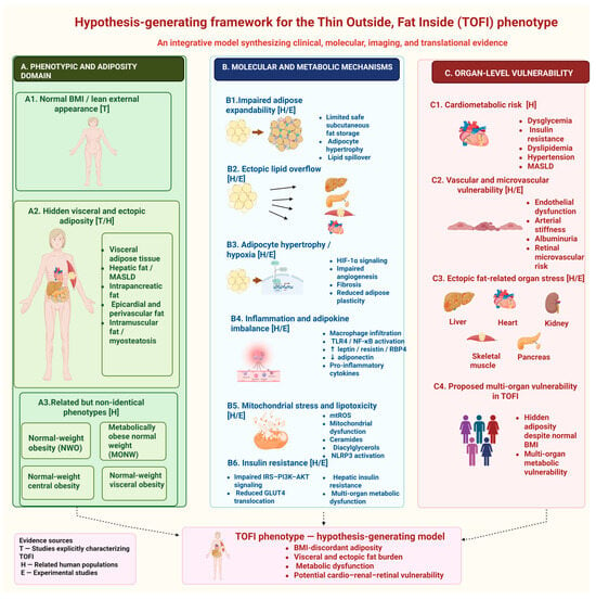
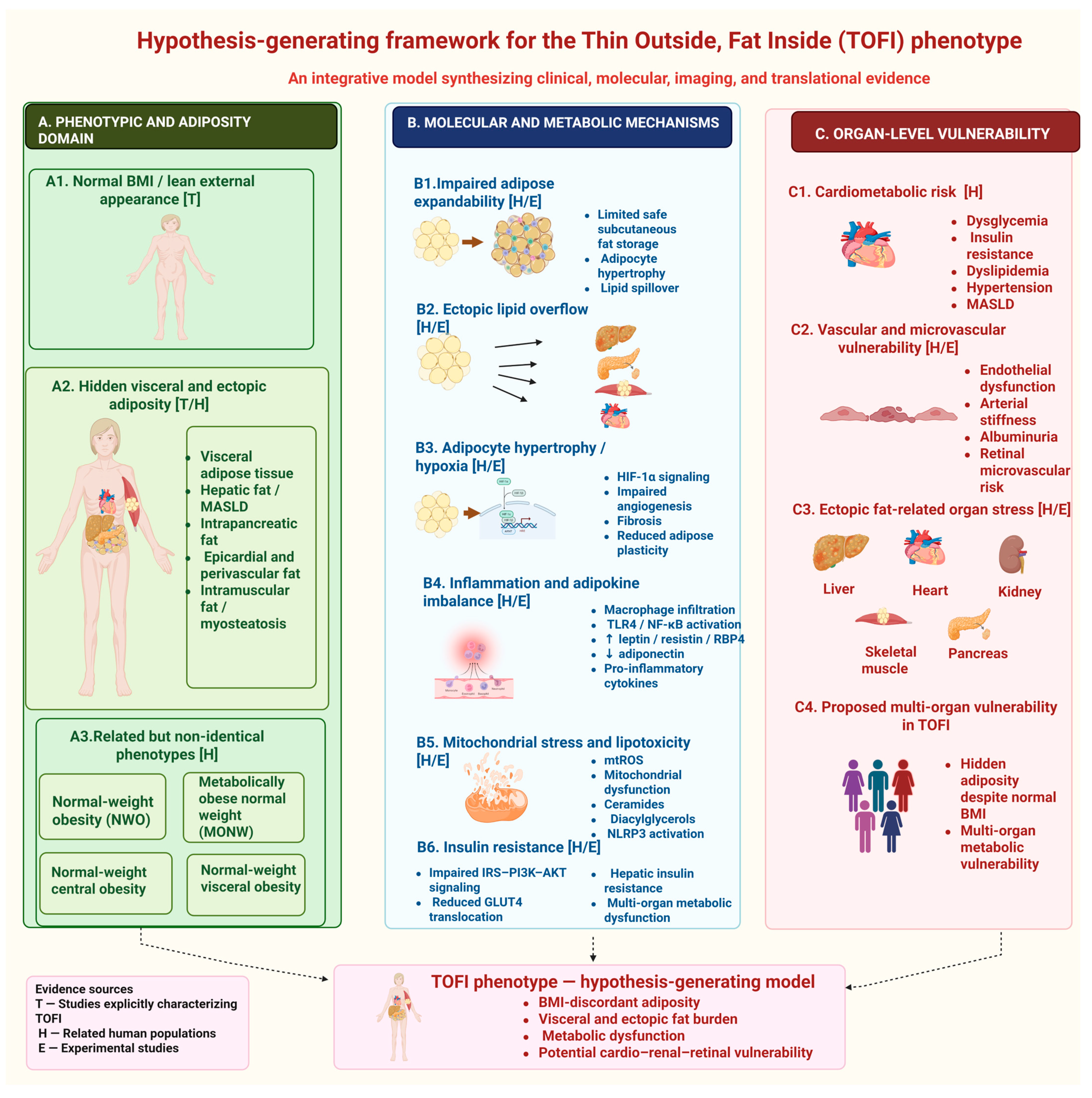
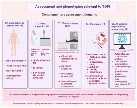
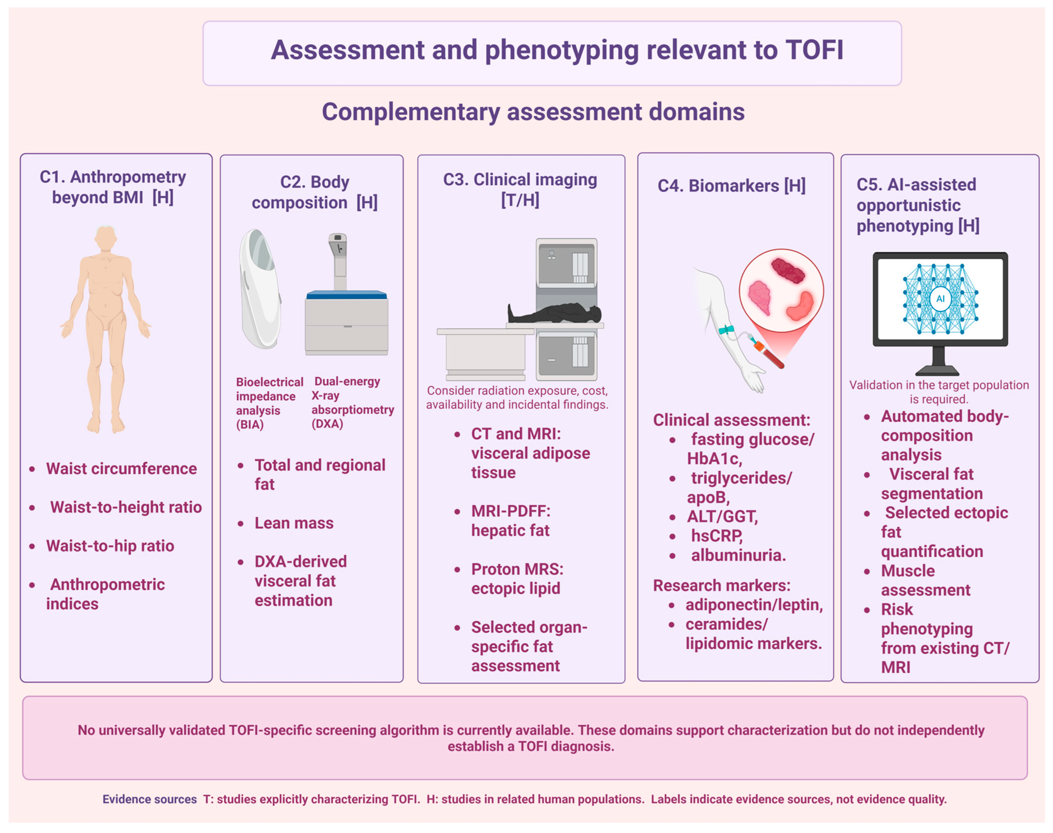

Disclaimer/Publisher’s Note: The statements, opinions and data contained in all publications are solely those of the individual author(s) and contributor(s) and not of MDPI and/or the editor(s). MDPI and/or the editor(s) disclaim responsibility for any injury to people or property resulting from any ideas, methods, instructions or products referred to in the content. |
© 2026 by the authors. Licensee MDPI, Basel, Switzerland. This article is an open access article distributed under the terms and conditions of the Creative Commons Attribution (CC BY) license.