


Abstract
Background/Objectives: Coronary artery disease (CAD) remains one of the leading cardiovascular diseases worldwide. While obstructive CAD is well characterized and managed, identification of patients with non-obstructive CAD (NOCAD) remains challenging. Unlike the coronary vasculature, the eye’s microcirculation can be easily and non-invasively assessed. Therefore, this systematic review summarized the ophthalmological diagnostic methods used to assess microvascular alterations associated with coronary microvascular dysfunction (CMD), angina with non-obstructive coronary arteries (ANOCA), or ischemia with non-obstructive coronary arteries (INOCA). Methods: According to PRISMA guidelines, PubMed/MEDLINE and Embase databases were screened by two independent reviewers from inception to 25 November 2025. Original articles that examined ophthalmological microvascular changes by any method in adults with CMD or its subtypes were included. The quality of the studies was assessed using the JBI Critical Appraisal Checklist. Results: Of 101 identified articles, nine studies met the inclusion criteria, comprising 1894 patients. Optical coherence tomography angiography was the most frequently used imaging modality, followed by optical coherence tomography, slit-lamp smartphone imaging, and fundus photography. Five investigations employed blinded image analysis, three did not, and one study used it partially. Four studies used semi-automated measurements, four employed fully automated methods, and one study applied manual and automated measurements for different parameters. Conclusions: Despite a limited number of studies, retinal and conjunctival microvascular alterations helped differentiate CAD subtypes and may reflect systemic microcirculatory impairment among patients with ANOCA/INOCA. Ophthalmological imaging techniques have the potential to serve as non-invasive tools for detecting microvascular alterations associated with CMD in ANOCA and INOCA patients. PROSPERO Registration Number: CRD420251239875
1. Introduction
Despite continuous advances in prevention, diagnostics, and therapy over recent decades, coronary artery disease (CAD) remains a leading cause of morbidity and mortality worldwide [1]. The traditional diagnosis of CAD is based on typical clinical symptoms, predominantly chest pain, evidence of myocardial ischemia on stress testing, and the presence of significant coronary artery stenoses demonstrated by coronary computed tomography angiography (CCTA) or invasive coronary angiography (ICA). However, in clinical practice, a substantial proportion of patients presenting with anginal symptoms or evidence of myocardial ischemia on functional testing do not exhibit such obstructive lesions in the coronary arteries [2].
The term non-obstructive coronary artery disease (NOCAD) is the broadest concept and refers to the presence of symptoms suggestive of myocardial ischemia, with or without accompanying ischemic changes in additional diagnostic tests, in patients without significant stenoses in the epicardial coronary arteries. One of the most important underlying causes of NOCAD is coronary microvascular dysfunction (CMD). According to the most recent European Society of Cardiology (ESC) Guidelines from 2024, NOCAD patients suffering from angina or its equivalents should be stratified as angina with non-obstructive coronary arteries (ANOCA) or ischemia with non-obstructive coronary arteries (INOCA) when myocardial ischemia is documented. The term microvascular angina (MVA) represents one of the two major endotypes of ANOCA/INOCA, alongside vasospastic angina (VSA) [2].
The exact prevalence of ANOCA/INOCA remains challenging to quantify due to heterogeneity in diagnostic criteria and under-recognition in clinical practice. It is estimated that up to 60% of women and 30% of men referred for ICA due to symptoms suggestive of CAD have no significant atherosclerotic narrowing in the epicardial coronary arteries [2]. Historically, ANOCA and INOCA were often considered benign or predominantly functional conditions, leading to their underdiagnosis and marginalization in clinical guidelines. However, an increasing body of evidence indicates that patients with ANOCA/INOCA are at elevated risk of major adverse cardiovascular events, including myocardial infarction, heart failure, and cardiac death. Moreover, these patients frequently experience substantially impaired quality of life [3,4,5].
The first step in diagnosis is estimating the pre-test likelihood of obstructive CAD (OCAD) with the risk-factor-weighted clinical likelihood (RF-CL) model to guide diagnostic testing and avoid unnecessary procedures in low-risk patients. Although RF-CL demonstrates higher sensitivity than previous models in identifying patients with a very low likelihood (≤5%), particularly when combined with the coronary artery calcium score, there is currently no validated model capable of estimating the probability of ANOCA/INOCA [2,6].
The diagnostic workup of ANOCA/INOCA remains complex, requiring a multimodal approach [2]. As CMD may arise from distinct pathophysiological mechanisms, current evidence supports the use of multiple, mechanism-oriented diagnostic pathways integrating invasive and non-invasive tests [7]. The availability of non-invasive techniques remains limited and insufficient to meet the clinical demand arising from the growing prevalence of ANOCA/INOCA. Moreover, the invasive techniques are associated with a risk of potential adverse events, necessitating careful patient selection [2]. Consequently, there is an ongoing search for simple, non-invasive, and widely applicable diagnostic methods that would enable the identification of patients with ANOCA/INOCA.
The prognosis of patients with ANOCA/INOCA depends on the underlying pathophysiological mechanism and treatment effectiveness. Management focuses on cardiovascular risk-factor control, lifestyle modification, strict control of comorbidities (e.g., diabetes, dyslipidaemia), avoidance of vasospasm-inducing substances, and endotype-specific pharmacotherapy [2].
There is growing interest in ophthalmological possibilities for assessing the microcirculation of the eye as a window into coronary microcirculation. In contrast to the coronary vasculature, the ocular microcirculation can be easily and non-invasively evaluated due to its accessible location [8,9]. This provides a unique opportunity to directly assess microcirculation in vivo, in real time, and repeatedly, without the need for invasive procedures. Retinal arterioles share several key physiological and anatomical features with coronary microvessels [8]. Additionally, both retinal and coronary microcirculation are influenced by the same major cardiovascular risk factors, including hypertension, diabetes, and dyslipidemia [10,11,12]. Therefore, changes in the retinal microvasculature may serve as non-invasive indicators of systemic microvascular dysfunction relevant to coronary disease [8]. The potential clinical relevance of retinal microvascular changes has been widely investigated. Previous studies have found that a range of retinal microvascular alterations, such as generalized and focal arteriolar narrowing, arteriovenous nicking, and retinopathy, may serve as markers for cardiovascular disease [8]. Moreover, retinal imaging has been proposed as a non-invasive tool for cardiovascular risk stratification [13,14].
Previous reviews primarily concentrate on OCAD and are often limited to one specific imaging modality. While prior reviews have occasionally included ANOCA/INOCA patients as part of broader study groups, they have not made this subgroup a central focus, and data specific to these patients are often not extractable. Numerous studies have been conducted to establish the correlation between retinal changes and CAD; however, few have examined microvascular alterations in patients with ANOCA/INOCA disease [9,15].
The patients with NOCAD, including those with ANOCA/INOCA, are often underdiagnosed and clinically challenging. Therefore, this systematic review aimed to investigate the potential of ophthalmological diagnostic methods in identifying patients with ANOCA/INOCA disease.
2. Materials and Methods
2.1. Searching Strategy
This systematic review was conducted in accordance with the PRISMA guidelines [16] (PRISMA checklist in Supplementary Materials, Table S1). PubMed/MEDLINE and Embase databases were searched from their inception up to 25 November 2025. The protocol was registered in PROSPERO (ID: CRD420251239875). The following searching strategy was used for both databases: (“ischaemia with non-obstructive coronary arteries” OR “angina with non-obstructive coronary arteries” OR “coronary microvascular dysfunction” OR “microvascular angina” OR “microvascular coronary disease” OR “cardiac syndrome X” OR “myocardial infarction with non-obstructive coronary arteries” OR “non-obstructive coronary artery disease” OR “normal coronary angiography” OR “normal angiographic”) AND (“eye” OR “retina” OR “choroid” OR “retinal vessels” OR “retinal microvasculature” OR “retinal microcirculation” OR “chorioretinal microvasculature” OR “choroidal microvasculature” OR “conjuntival microvasculature”).
2.2. Inclusion and Exclusion Criteria
Inclusion criteria were determined based on PICOS principles (population, intervention, comparison, outcome, study type). Therefore, the population was defined as adults (≥18 years old) with NOCAD or its subtypes, as explicitly defined by the authors or as identifiable based on the cardiological criteria provided. Eligible patients were those who had undergone ICA or CCTA to rule out obstructive coronary disease and who presented with angina symptoms, angina equivalents, and/or objective evidence of myocardial ischemia on non-invasive testing. Intervention was defined as any kind of ophthalmologic assessment of the microvasculature (optical coherence tomography angiography (OCTA), slit-lamp smartphone imaging, fundus photography, etc.). Comparison was defined as healthy controls, OCAD, or other NOCAD subtypes. Studies without control groups were also considered if they met other inclusion criteria. Outcome was defined as any available result of microvascular assessments (e.g., vessel density (VD), choroidal thickness, etc.). Only original articles were included. All other publication types, such as reviews, editorials, letters to the editor, short communications, conference abstracts, etc., were not considered. There were no language limitations. As the terminology used to describe ischemia and angina in the absence of obstructive coronary artery disease has evolved, studies have used a range of historical terms. The ANOCA and INOCA concepts were formalized only recently and were not uniformly applied in earlier literature. To ensure consistent classification, all cohorts were mapped to ANOCA/INOCA phenotypes whenever sufficient clinical information was available.
Although our primary focus was chronic non-obstructive ischemic syndromes (ANOCA/INOCA), we also permitted inclusion of studies enrolling patients with myocardial infarction with non-obstructive coronary arteries (MINOCA), provided that obstructive epicardial CAD was excluded and ocular microvascular assessment was performed. MINOCA was treated as a distinct clinical entity, and its findings were described separately to avoid conflating post-infarction mechanisms with chronic ischemic syndromes.
Exclusion criteria were determined as follows: studies in which NOCAD patients were part of a broader population and their data could not be analyzed separately, studies in which NOCAD was defined solely by angiographic stenosis severity (e.g., Gensini score thresholds) without accompanying clinical criteria (angina or documented ischemia) or without any diagnostic evaluation of coronary microvascular function, studies in which coronary anatomy was not confirmed by ICA or CCTA, and studies that did not report any ophthalmologic microvascular outcomes. In addition, studies were excluded if the coronary slow-flow phenomenon (CSFP) was used as the sole criterion for defining microvascular dysfunction in the absence of mechanism-oriented diagnostic testing.
2.3. Used Terminology
In this review, we use ANOCA/INOCA to describe patients with angina and/or objective ischemia with non-obstructive epicardial coronary arteries, in line with contemporary ESC nomenclature. We use NOCAD only as a broader descriptive umbrella term to reflect the wording of some original studies and/or the overarching concept of non-obstructive epicardial CAD. When applicable, study populations were reclassified into ANOCA/INOCA (and related subgroups such as CMD) based on the information provided by the authors.
2.4. Screening and Data Extraction
Firstly, the identified records were uploaded into EndNote (version 21.5; Clarivate Analytics, Philadelphia, PA, USA), and duplicates were removed. Then, two authors (MRA and PK) independently screened the title and abstract. Subsequently, those two authors independently extracted the data from the included articles. The extracted data were study type, usage of blinded image analysis, measurement method, imaging method, characteristics of the NOCAD subgroup, characteristics of the comparison group, number of participants (if available, performed separately for NOCAD subgroup and comparison group), analyzed parameters, key findings, and confounders (if available, stratified for only measured, measured and statistically adjusted, and excluded). Any discrepancies were resolved through the discussion.
2.5. Risk of Bias Assessment
All identified articles appear to be cross-sectional studies. Therefore, the JBI Critical Appraisal Checklist for Analytical Cross-Sectional Studies was used [17]. Two reviewers (MRA and PK) independently graded the selected articles in an 8-item list. Each item was graded as Yes, No, Unclear, or Not Applicable. Discrepancies were resolved through discussion.
2.6. Use of Artificial Intelligence Tools
ChatGPT (OpenAI, GPT-5.2) was used to assist with English language editing of the manuscript.
3. Results
3.1. Characteristics of Included Studies
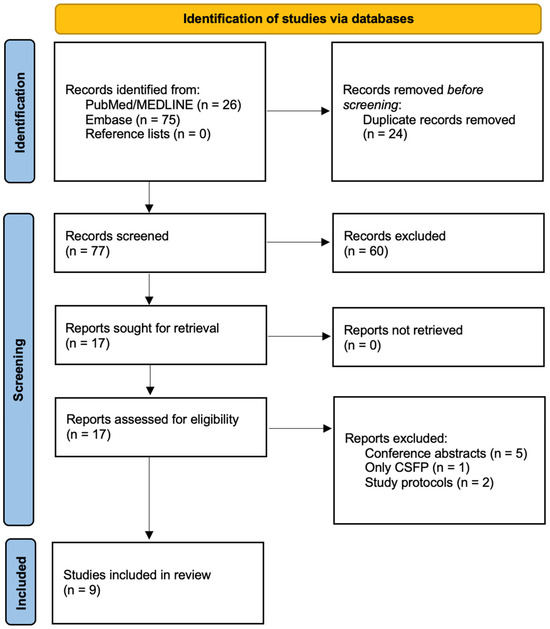
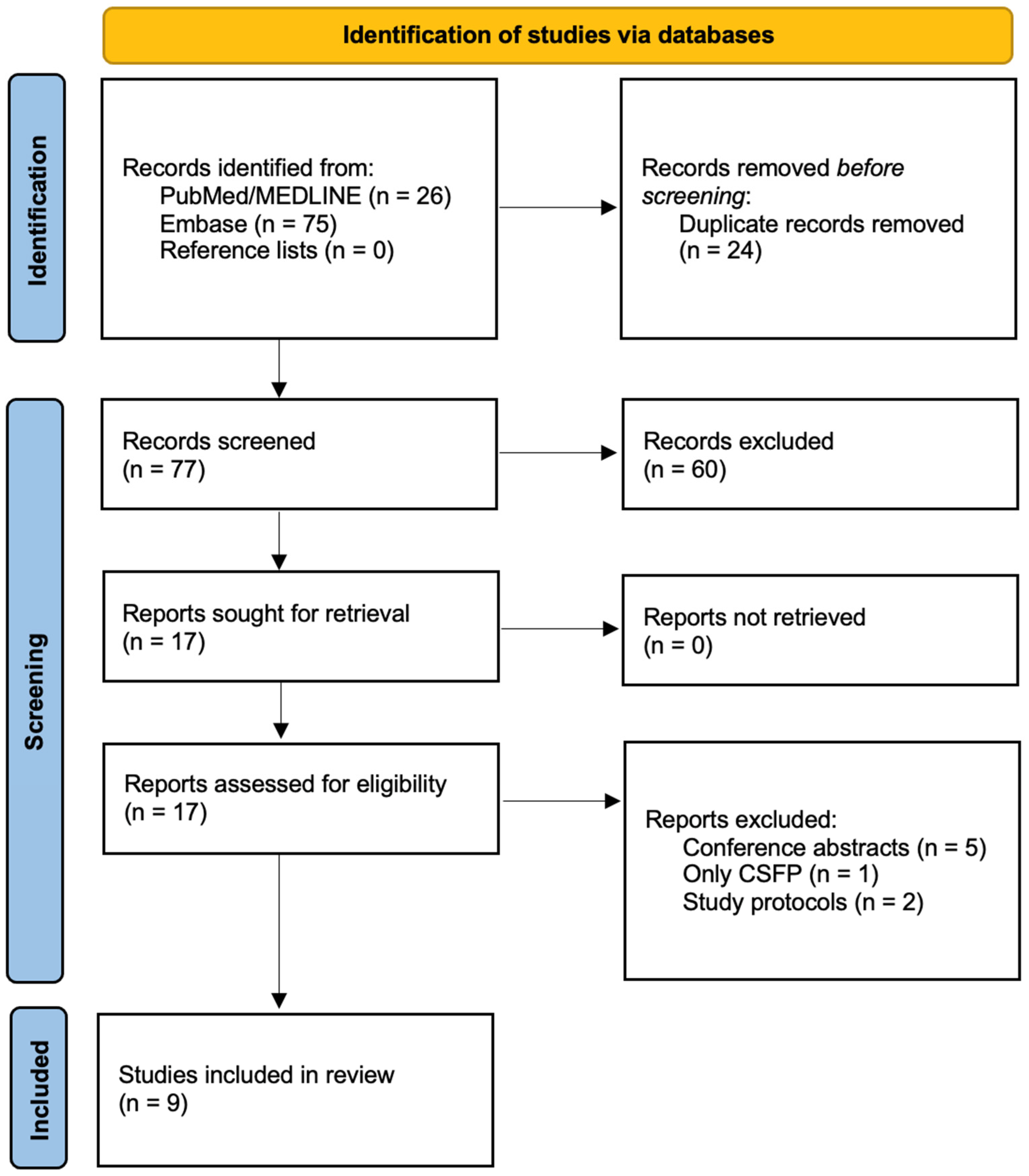
Initially, 26 articles were identified in PubMed/MEDLINE and 75 articles in Embase. Of 101 articles, 24 were duplicates. Of 77 articles, 60 were excluded after screening their title and abstracts. Then, five conference abstracts [18,19,20,21,22], two study protocols [23,24], and one investigation with CSFP [25] were excluded. A precise screening protocol is presented in Figure 1.
Figure 1.
Flowchart for screening protocol. Abbreviations: CSFP, coronary slow-flow phenomenon.
Finally, nine articles were included. Studies vary in the characteristics of their study groups. Three studies defined the study group as MVA [26,27,28], two as NOCAD [29,30], three as INOCA [31,32,33], and one as a normal angiographic patient with typical chest pain or anginal equivalents [34]. Moreover, the definition of NOCAD/MVA/ANOCA/INOCA subgroups varies across studies. Liew et al. [26] defined MVA as chest pain with less than 25% stenosis in all coronary epicardial arteries, whereas in the study by Xu et al. [27], MVA patients had either normal coronary angiography or stenosis < 50% in primary branches and <75% in secondary branches. In a study by Kanar et al. [28], patients with MVA were reported to have normal epicardial arteries, but no information was provided regarding the degree of stenosis. In Table 1, the study groups were reclassified according to the new nomenclature regarding the 2024 ESC Guidelines for the management of chronic coronary syndromes and presented with the original terminology used in the individual studies.
Table 1.
Summary of studies evaluating ophthalmological imaging in patients with non-obstructive coronary artery disease.
In all studies, adult patients presenting with cardiovascular symptoms underwent ICA to rule out the presence of OCAD, except for one study [29], where CCTA was used. Additionally, two patients with CMD assessed by positron emission tomography were included in Shiromani et al.’s study [32]. Nearly all studies involve comparisons between ANOCA/INOCA and other groups (e.g., OCAD, other subgroups of NOCAD, or healthy controls). In only one investigation [34] is there no dedicated control group, and this study compares its findings to normative values for normal populations.
Various ophthalmological imaging methods were used to assess microcirculation in the eye. The most common methods were OCTA (four studies), followed by spectral-domain optical coherence tomography (SD-OCT) (two studies), slit-lamp and smartphone imaging (two studies), and fundus photography (one study).
3.2. Quality of Included Studies
Qualitative assessment of the included studies is presented in Table 2. All studies were assessed using the JBI Critical Appraisal Tool, and a final decision was made on their inclusion. The inclusion criteria and study settings were clearly explained in the selected articles. Moreover, the objective criteria were used for measurements, and outcomes were measured reliably. Imaging and analysis protocols were generally reported in sufficient detail to ensure reproducibility. Moreover, several studies applied masked assessment and one study dual-observer evaluation with inter-observer agreement procedures, reducing operator-dependent bias and strengthening the reliability of outcome measurements. There were no studies with inappropriate statistical methods used. The majority of the included studies identified the confounding factors and coped well with them. However, Xu et al. [27], Agca et al. [31], and Eslami et al. [34] did not clearly describe strategies to address potential confounders. In these studies, confounders were listed in baseline characteristics, but the statistical analyses relied mainly on univariate comparisons, without multivariable adjustment, despite only partial control provided by group matching and exclusion criteria.
Table 2.
Quality of the included studies.
3.3. Results of Included Studies
Four studies assessed retinal microvascular parameters using OCTA and reported retinal microvascular alterations in ANOCA/INOCA populations. VD in the superficial capillary plexus (SCP) was assessed in all four studies. In two studies, VD in the SCP was reduced when NOCAD/INOCA patients were compared with healthy controls [29,31] and in one study when women with INOCA and CMD were compared with those without CMD [32]. Among three studies assessing VD in the deep capillary plexus (DCP), two reported a decrease in NOCAD/INOCA patients when compared with healthy controls [29,31]. One study found that 45% of normal angiographic patients had abnormal VD in SCP, and 8,3% had abnormal VD in DCP [34]. In one study, women with INOCA and CMD showed lower perfusion density in SCP when compared with women without CMD [32].
Findings for the peripapillary circulation were inconsistent. One study reported that VD in the radial peripapillary capillary (RPC) network did not differ between NOCAD and healthy controls, with reductions observed only in OCAD [29], whereas another study found reduced VD in RPC in both INOCA and OCAD compared with healthy controls [31].
Foveal avascular zone (FAZ) parameters were evaluated in three studies and showed no significant differences [31,32,34]. In one study, the foveal density in a 300 μm region around the FAZ (FD 300) was reduced in NOCAD compared to healthy controls, while retinal nerve fiber layer (RNFL) thickness did not differ between NOCAD and healthy controls [29].
Based on single studies, SD-OCT retinal vessel caliber parameters assessed with the full-width half-maximum (FWHM) method (retinal arterial lumen diameter (RALD), retinal arterial outer diameter (RAOD), arteriovenous ratio (AVR)) differentiated MVA from ischemic heart disease and healthy controls [27], whereas structural OCT parameters (subfoveal choroidal thickness (SFCT) and peripapillary retinal nerve fiber layer thickness (pRNFLT)) showed no discriminatory value for MVA [28].
In both studies evaluating conjunctival microvasculature, all examined flow parameters (axial velocity, cross-sectional velocity, and blood flow rate) differed significantly between the comparison groups (INOCA with CMD vs. control and NOCAD vs. MI), and the greatest alterations were most pronounced in smaller vessels [30,33].
One included study enrolled a MINOCA cohort; therefore, we report it as a separate subgroup from the chronic ANOCA/INOCA/NOCAD cohorts. In the single study using fundus photography, in women, a smaller retinal venular caliber was more frequently observed in MVA than in OCAD [26].
4. Discussion
To our knowledge, this is the first systematic review that attempts to gather findings from ophthalmological imaging in patients with ANOCA/INOCA. Only a few studies have specifically investigated ocular vascular changes in this group. This reflects the novelty of the topic and the fact that reliable diagnostic methods confirming the presence of CMD have only recently become available. The key insights are as follows. Firstly, ophthalmological imaging techniques show emerging potential for the non-invasive assessment of microvascular alterations in patients with ANOCA/INOCA. Secondly, the observed ocular microvascular changes are consistent with the concept that CMD is a part of a broader systemic microvascular disorder. Thirdly, current studies were conducted on relatively small cohorts, and further investigations are required to validate these observations.
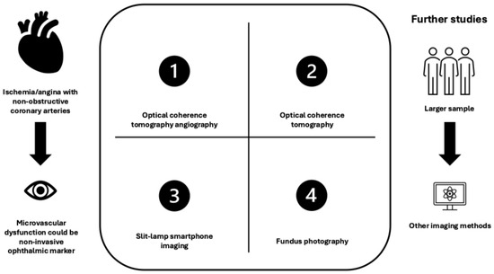
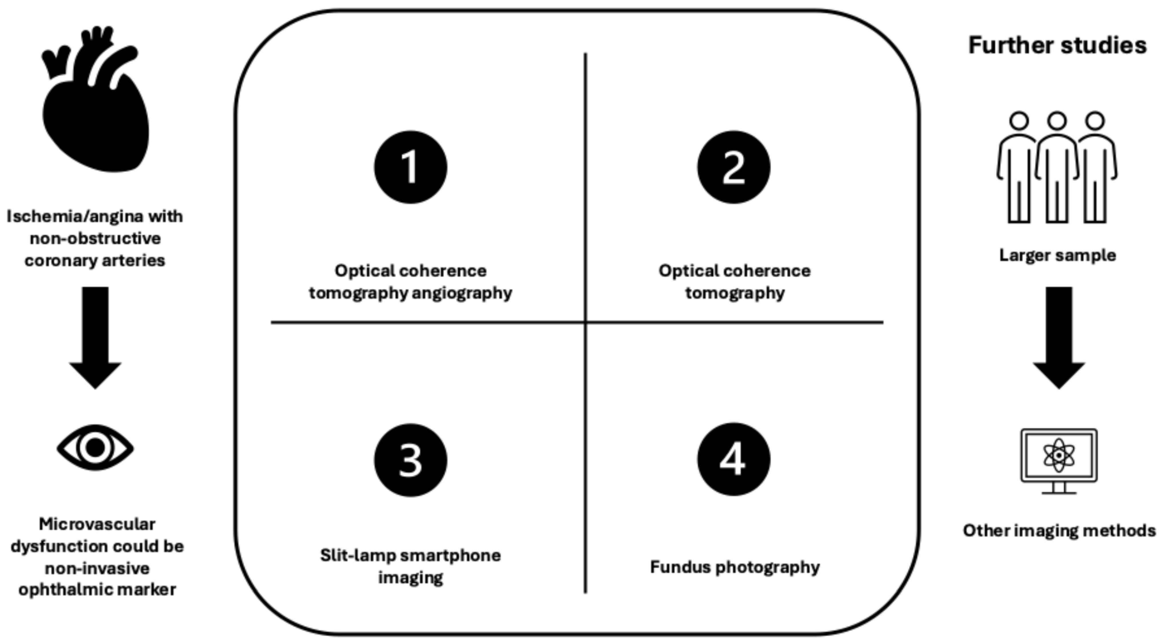
Figure 2 presents a comparison of ocular imaging modalities and their relevance to coronary microvascular dysfunction.
Figure 2.
Comparison of ocular imaging modalities and their relevance to coronary microvascular dysfunction.
Patients with ANOCA/INOCA represent a challenging and often overlooked group. Although these patients do not have significant coronary artery stenosis, they remain at elevated risk for adverse cardiovascular events, including myocardial infarction and heart failure. Quality of life is often significantly impaired due to persistent anginal symptoms that can be challenging to manage [2]. The complicated diagnostic pathway in patients with ANOCA/INOCA often leads to multiple hospitalizations, repeated invasive procedures such as coronary angiography, and high healthcare costs [2,35]. Therefore, there is an urgent need for new diagnostic tools that are ideally non-invasive, repeatable, and easily performed on an outpatient basis. Hence, cardiologists have a significant interest in collaborating with ophthalmologists and analyzing microcirculatory disorders in the eye in the course of various cardiovascular diseases.
Among all described studies, the study group investigated by Shiromani et al. [32] and Mailey et al. [33] were the best characterized. Unlike other studies, they assessed patients for CMD as the underlying mechanism of ANOCA/INOCA. These patients underwent a complete diagnostic assessment, including both anatomical evaluation by ICA (similarly to other studies) and additionally functional testing of the coronary microcirculation. This dual approach allowed for the precise identification of patients with CMD, resulting in a more homogeneous study population. This approach was not utilized in earlier studies, likely because they were conducted before the 2024 ESC Guidelines, which strengthened and clarified recommendations for invasive coronary functional testing for CMD [2]. In addition, the limited availability of this method further restricted its use in clinical research [2,35]. Consequently, former studies may have included heterogeneous patient populations with different underlying etiologies of ANOCA/INOCA. Because the current gold-standard diagnostic methods for CMD are invasive and limited in availability, the implementation of non-invasive and easily accessible imaging tools could potentially offer substantial clinical benefit by enhancing patient comfort and enabling faster and more accurate diagnosis.
Shiromani et al. demonstrated that reduced retinal vessel and perfusion densities in the SCP, assessed by OCTA, were significantly associated with higher odds of CMD among women with INOCA, even after adjustment for major cardiovascular risk factors. These findings suggest that retinal microvascular metrics may serve as potential non-invasive markers of CMD [32]. However, the study was a prospective analysis with a homogeneous cohort; nonetheless, the small sample size, absence of healthy controls, and inclusion of only women limit the generalizability of the findings, which should be validated in larger, mixed-gender populations.
Similar to the findings of Shiromani et al., all other studies using OCTA detected a reduction in VD within the SCP in ANOCA/INOCA population, making it the most extensively investigated vascular parameter across the studies included in this review [29,31,32,34]. The consistent finding of reduced VD in the SCP in studies indicates a reproducible pattern of retinal microvascular changes in patients with ANOCA/INOCA. However, as Eslami et al. found reduced VD in approximately 45% of ANOCA/INOCA patients [34], this suggests limited specificity, which might also result from the lack of detailed evaluation of NOCAD etiology, including CMD.
None of the studies found significant differences in FAZ parameters in OCTA among the study groups. Most former studies found increased FAZ area in patients with CAD compared to controls [9]. Interestingly, Jeremic et al. introduced a novel ellipse-based FAZ analysis that revealed significant differences in FAZ morphology across all CAD severity groups. This relationship was observed only for the left eye. The authors suggest that this asymmetry may be explained by differences in blood flow between the carotid arteries [36]. Zhou et al. found that the left eye shows greater sensitivity to CAD-related changes detected by OCTA than the right eye, suggesting it could be a better indicator for early CAD detection [37]. Among the included studies, the right eye was preferred [26,27,31], the results from both eyes were averaged for further analysis [32], one eye was selected at random [29], or eye selection was not reported [30,33]. Future research should account for possible inter-eye microvascular asymmetry, as single-eye assessment may miss clinically relevant changes in CAD patients.
Choroidal thickness (ChT) measurement has not, to date, entered routine clinical practice. ChT fluctuates during the day and is sensitive to systemic and ocular factors such as age or axial length. This variability, together with the lack of standardization and the inconsistent results across studies investigating ChT in systemic diseases, limits its clinical utility [38,39]. Furthermore, manual caliper measurements are time-consuming and observer-dependent, as the choroid boundary must be identified manually. Kanar et al. evaluated SFCT using SD-OCT and found no significant difference between patients with MVA (ANOCA) and healthy controls [28]. This lack of difference may be related to the relatively small sample size and the reliance on manual measurements.
The assessment of conjunctival circulation using a smartphone combined with a slit-lamp biomicroscope remains a relatively new and unfamiliar technique to most clinicians, and to date, there are no universally standardized measurement protocols or widely established reference norms for bulbar conjunctival microcirculation measurements. The conjunctival microcirculation is directly exposed to environmental and inflammatory factors, which may cause variability and nonspecific changes in vascular parameters [40,41]. In contrast to other imaging modalities, the quality of videos or images is not affected by media opacities; therefore, this technique could be potentially beneficial for individuals where fundus visualization is challenging or impossible due to opaque optical media.
The FWHM method applied to SD-OCT images provides a more objective and reproducible approach for assessing retinal vessel morphology than traditional manual measurements. In this technique, the boundaries of the vessel wall are determined automatically by the algorithm, based on the intensity profile across the vessel [42].
Color fundus photography remains the oldest and most widely used modality for imaging retinal vasculature [43,44]. In Liew et al.’s study, significant differences in retinal vasculature were observed only in venules and only within the female MVA cohort [26]. The authors hypothesized that their findings might reflect better cardiovascular health in women rather than pathological microvascular changes associated with CAD. This interpretation appears plausible and is supported by previous findings showing that narrower retinal arterioles and wider venules are markers of more severe CAD in women [45], and that decreasing arteriolar diameters are associated with an increased risk of CHD in women [46]. Nevertheless, this interpretation remains speculative, as the study did not include a healthy control group for comparison. Moreover, given that this cohort comprised MINOCA patients, these observations should be interpreted as hypothesis-generating and not directly comparable with findings from chronic ANOCA/INOCA cohorts.
Alterations in microvasculature in the eye should not be interpreted in isolation, as multiple cardiovascular risk factors, including hypertension, diabetes, obesity, and dyslipidemia, affect both the retinal and coronary microcirculatory systems similarly [10,11,12]. Retinal venular diameter tends to narrow with age, while wider retinal venular caliber is associated with endothelial dysfunction and inflammation caused by, e.g., smoking, obesity, and higher glycosylated hemoglobin. High blood pressure is associated with narrower arteriolar caliber and wider venular diameter. In contrast to retinal arteriolar caliber, men have wider retinal venular diameters than women [47,48]. Hypertension and diabetes are both associated with reduced retinal vascular density [9]. Smoking, chronic heart failure, and hypertension have been associated with significant choroidal thinning, while choroidal thickness naturally decreases with age. It was found that patients with obstructive CAD have significantly lower choroidal thickness [39], reduced VD (especially in SCP), and enlarged FAZ when compared to healthy controls [9].
In the studies by Kanar, Shiromani, Ren, Liew, and Mailey, multivariable analyses were performed, and the reported associations remained significant even after adjustment for multiple comorbidities [26,28,29,30,32]. Other studies relied on univariate comparisons, group matching, and exclusion criteria rather than multivariable adjustment; therefore, their findings remain more susceptible to residual confounding by prevalent cardiovascular and metabolic comorbidities. The weakest control was observed in Eslami’s study, where high rates of hypertension, diabetes, and hyperlipidemia were present, making it difficult to distinguish ANOCA/INOCA microvascular changes from those driven by underlying systemic disease. Notably, in the Agca et al. study, all patients suffered from obstructive sleep apnea syndrome, a condition associated with reduced retinal perfusion and FAZ alterations. Therefore, the retinal microvascular abnormalities observed in this study cannot be interpreted as arising solely from coronary pathology.
Across all included studies, imaging modalities were non-invasive and enabled rapid image acquisition, but differed substantially in anatomical coverage, processing time, and susceptibility to observer-related variability. Fundus photography and SD-OCT FWHM methods focused on the retinal vessel caliber in Zone B, SD-OCT was applied to choroidal thickness assessment, and the slit-lamp smartphone technique evaluated conjunctival microvasculature, whereas OCTA enabled layer-specific analysis of SCP, DCP, RPC, FD-300, and FAZ. Across all methods, measurements were influenced by patient cooperation, particularly regarding motion and fixation stability.
The most promising imaging modality for microvascular assessment in ANOCA/INOCA is OCTA. This relatively new modality, available since 2016, has been most frequently employed in described studies, likely because it is widely accessible and very simple to operate, even by clinicians without ophthalmologic training. Notably, unlike the other imaging modalities described in this review, OCTA provides fully automated quantification of VD parameters using built-in software algorithms, which substantially reduces observer bias and enhances reproducibility. Unlike other modalities that require manual or semi-manual post-processing or export to external software, OCTA provides immediate quantitative results. The main limitation of OCTA is its small field of view, which may miss peripheral vascular abnormalities. However, this limitation is not unique to OCTA, as vessel assessment in SD-OCT and fundus photography is typically confined to the posterior pole as well [11,49,50,51]. Examples of OCTA imaging could be found in the Agca et al. and Ren et al. studies [29,31] (Table 3).
Table 3.
Overview of ocular imaging modalities for retinal microcirculation and their potential relevance to coronary microvascular dysfunction.
4.1. Future Directions
Across studies evaluating structural, layer-based OCT and OCTA parameters (SFCT, pRNFLT, and RNFL thickness in the RPC) showed inconsistent and limited discriminatory value [28,29]. Among structural biomarkers, retinal ischemic perivascular lesions (RIPLs) are considered the earliest subclinical sign of retinal ischemic infarction. Although RIPLs were not assessed in ANOCA/INOCA patients in the included studies, their evaluation may represent a valuable future direction for detecting subtle ischemic damage in ANOCA/INOCA. RIPLs are defined based on SD-OCT images as a permanent focal thinning of the inner nuclear layer of the retina, resulting from hypoperfusion in the DCP [53,54]. Although nearly half of patients with cardiovascular disease show no RIPLs, the presence of two or more lesions demonstrates high specificity (≈90%). Thus, despite limited sensitivity, RIPLs may serve as a rule-in marker to identify individuals who may benefit from further targeted cardiovascular evaluation [53]. Supportive imaging for RIPLs could be found in a study by Bousquet et al. [55].
To the best of our knowledge, there are still a few other ophthalmological imaging modalities that have not yet been employed for retinal vessel assessment in patients with ANOCA/INOCA. In contrast to fundus photography and the SD-OCT FWHM method, adaptive optics (AO) techniques enable direct visualization of the vessel wall structure due to the very high resolution of approximately 2 µm. Previous studies have demonstrated its capability to detect early microvascular alterations in conditions such as glaucoma, hypertension, and diabetic retinopathy [56,57,58]. In the study by Aschauer et al., patients with coronary heart disease (CHD) exhibited a high prevalence of retinal microvascular abnormalities, including arteriovenous nicking, focal arteriolar narrowing, and microaneurysms, as well as asymmetric vessel wall thickening and hyperreflective intraluminal material. However, no statistically significant differences in quantitative vessel parameters were observed between patients with and without CHD, underscoring the need for larger studies to better establish the role of AO-derived retinal biomarkers in cardiovascular risk assessment [59]. Evaluation of retinal arteriolar morphology with adaptive optics camera (Rtx-1, Imagine Eyes, Orsay, France) was presented by Szewczuk et al. [57]
4.2. Limitations
Some limitations are inherited in this systematic review, both from the included studies and from the methodological protocol. Only two publicly available databases (PubMed and Embase) were screened. Updated systematic reviews in the future should screen Web of Science, Google Scholar, and Scopus, too. Moreover, the analysis of ophthalmologic microvascular changes associated with ANOCA/INOCA is a relatively new and emerging concept, and all the included studies were from the last 6 years. Therefore, the number of studies screened and included was relatively small in comparison to systematic reviews on more common topics. The included studies were relatively small, and only one investigation had NOCAD and comparison groups > 100 subjects.
Included articles presented a very heterogeneous methodology and classification system, with only a few fulfilling the exact definition of ESC. In particular, the lack of standardized diagnostic criteria for ANOCA/INOCA across the included studies (e.g., varying stenosis thresholds), together with the fact that only two studies confirmed CMD using invasive coronary functional testing, may have contributed to patient misclassification and influenced the reported associations. Finally, inclusion of a MINOCA cohort—although methodologically valuable—adds clinical heterogeneity. Therefore, this heterogeneity limits the comparability and synthesis of findings across studies. Consequently, updated systematic reviews based on contemporary terminology will be essential in the coming years.
5. Conclusions
Ophthalmological diagnostic methods, particularly retinal OCTA, represent promising non-invasive tools for detecting microvascular alterations as a biomarker for evaluating systemic microcirculation abnormalities in ANOCA and INOCA patients. Their advantages include accessibility, rapid acquisition, and quantitative biomarker potential. However, current evidence is limited by small sample sizes, heterogeneity, and a lack of standardized diagnostic thresholds. Further, adequately powered investigations are required to validate these observations, and underexplored modalities such as adaptive optics imaging may provide additional insights into coronary microvascular dysfunction.
Supplementary Materials
The following supporting information can be downloaded at https://www.mdpi.com/article/10.3390/jcm15041344/s1. Table S1: PRISMA checklist.
Author Contributions
Conceptualization, M.R.-A. and A.Z.-Ż.; Methodology, M.R.-A. and M.J.; Original Draft Preparation, M.R.-A. and M.J.; Writing—Review and Editing, M.R.-A., M.J., A.Z.-Ż., M.T., P.K. and J.P.S.; Supervision, J.P.S., A.Z.-Ż. and M.T. All authors have read and agreed to the published version of the manuscript.
Funding
This research received no external funding.
Institutional Review Board Statement
Not applicable.
Informed Consent Statement
Not applicable.
Data Availability Statement
The raw data supporting the conclusions will be made available on a reasonable request to the corresponding author.
Acknowledgments
During the preparation of this manuscript, the authors used ChatGPT (OpenAI, GPT-5.2) for the purposes of English language editing and grammatical improvement. The authors reviewed and edited the output and take full responsibility for the content of this publication. Przemysław Kasiak has been supported by the START Scholarship from the Foundation for Polish Science for outstanding young researchers under 30 years old.
Conflicts of Interest
The authors declare no conflicts of interest.
Abbreviations
The following abbreviations are used in this manuscript:
| ANOCA | Angina with non-obstructive coronary arteries |
| AO | Adaptive optics |
| AWT | Arterial wall thickness |
| AVR | Arteriovenous ratio |
| CAD | Coronary artery disease |
| CCTA | Coronary computed tomography angiography |
| CHD | Coronary heart disease |
| CHT | Choroidal thickness |
| CMD | Coronary microvascular dysfunction |
| CSFP | Coronary slow-flow phenomenon |
| DCP | Deep capillary plexus |
| ESC | European Society of Cardiology |
| FAZ | Foveal avascular zone |
| FWHM | Full-width half-maximum |
| ICA | Invasive coronary angiography |
| INOCA | Ischemia with non-obstructive coronary arteries |
| MINOCA | Myocardial infarction with non-obstructive coronary arteries |
| MVA | Microvascular angina |
| NOCAD | Non-obstructive coronary artery disease |
| OCAD | Obstructive coronary artery disease |
| OCT | Optical coherence tomography |
| OCTA | Optical coherence tomography angiography |
| OSAS | Obstructive sleep apnea syndrome |
| pRNFLT | Peripapillary retinal nerve fiber layer thickness |
| RALD | Retinal arterial lumen diameter |
| RAOD | Retinal arterial outer diameter |
| RF-CL | Risk-factor-weighted clinical likelihood |
| RIPLs | Retinal ischemic perivascular lesions |
| RNFL | Retinal nerve fiber layer |
| RPC | Radial peripapillary capillaries |
| RVLD | Retinal venular lumen diameter |
| RVOD | Retinal venular outer diameter |
| SCP | Superficial capillary plexus |
| SD-OCT | Spectral domain optical coherence tomography |
| SFCT | Subfoveal choroidal thickness |
| VD | Vessel density |
| VSA | Vasospastic angina |
| VWT | Venous wall thickness |
References
- GBD 2019 Diseases and Injuries Collaborators. Global burden of 369 diseases and injuries in 204 countries and territories, 1990–2019: A systematic analysis for the Global Burden of Disease Study 2019. Lancet 2020, 396, 1204–1222. [Google Scholar] [CrossRef] [PubMed]
- Vrints, C.; Andreotti, F.; Koskinas, K.C.; Rossello, X.; Adamo, M.; Ainslie, J.; Banning, A.P.; Budaj, A.; Buechel, R.R.; Chiariello, G.A.; et al. 2024 ESC Guidelines for the management of chronic coronary syndromes. Eur. Heart J. 2024, 45, 3415–3537. [Google Scholar] [CrossRef] [Scilit] [PubMed]
- Gdowski, M.A.; Murthy, V.L.; Doering, M.; Monroy-Gonzalez, A.G.; Slart, R.; Brown, D.L. Association of Isolated Coronary Microvascular Dysfunction With Mortality and Major Adverse Cardiac Events: A Systematic Review and Meta-Analysis of Aggregate Data. J. Am. Heart Assoc. 2020, 9, e014954. [Google Scholar] [CrossRef] [Scilit] [PubMed]
- Jespersen, L.; Hvelplund, A.; Abildstrom, S.Z.; Pedersen, F.; Galatius, S.; Madsen, J.K.; Jorgensen, E.; Kelbaek, H.; Prescott, E. Stable angina pectoris with no obstructive coronary artery disease is associated with increased risks of major adverse cardiovascular events. Eur. Heart J. 2012, 33, 734–744. [Google Scholar] [CrossRef] [Scilit]
- Jespersen, L.; Abildstrom, S.Z.; Hvelplund, A.; Prescott, E. Persistent angina: Highly prevalent and associated with long-term anxiety, depression, low physical functioning, and quality of life in stable angina pectoris. Clin. Res. Cardiol. 2013, 102, 571–581. [Google Scholar] [CrossRef] [Scilit]
- Winther, S.; Schmidt, S.E.; Mayrhofer, T.; Botker, H.E.; Hoffmann, U.; Douglas, P.S.; Wijns, W.; Bax, J.; Nissen, L.; Lynggaard, V.; et al. Incorporating Coronary Calcification Into Pre-Test Assessment of the Likelihood of Coronary Artery Disease. J. Am. Coll. Cardiol. 2020, 76, 2421–2432. [Google Scholar] [CrossRef] [Scilit]
- Lanza, G.A.; Morrone, D.; Pizzi, C.; Tritto, I.; Bergamaschi, L.; De Vita, A.; Villano, A.; Crea, F. Diagnostic approach for coronary microvascular dysfunction in patients with chest pain and no obstructive coronary artery disease. Trends Cardiovasc. Med. 2022, 32, 448–453. [Google Scholar] [CrossRef] [Scilit]
- Wong, T.Y.; Klein, R.; Klein, B.E.; Tielsch, J.M.; Hubbard, L.; Nieto, F.J. Retinal microvascular abnormalities and their relationship with hypertension, cardiovascular disease, and mortality. Surv. Ophthalmol. 2001, 46, 59–80. [Google Scholar] [CrossRef] [Scilit]
- Rusu, A.C.; Horvath, K.U.; Tinica, G.; Chistol, R.O.; Bulgaru-Iliescu, A.I.; Todosia, E.T.; Brinzaniuc, K. Retinal Structural and Vascular Changes in Patients with Coronary Artery Disease: A Systematic Review and Meta-Analysis. Life 2024, 14, 448. [Google Scholar] [CrossRef] [Scilit]
- Liew, G.; Wang, J.J.; Mitchell, P.; Wong, T.Y. Retinal vascular imaging: A new tool in microvascular disease research. Circ. Cardiovasc. Imaging 2008, 1, 156–161. [Google Scholar] [CrossRef] [Scilit]
- de Carlo, T.E.; Romano, A.; Waheed, N.K.; Duker, J.S. A review of optical coherence tomography angiography (OCTA). Int. J. Retin. Vitr. 2015, 1, 5. [Google Scholar] [CrossRef] [Scilit] [PubMed]
- Podoleanu, A.G. Optical coherence tomography. J. Microsc. 2012, 247, 209–219. [Google Scholar] [CrossRef] [Scilit] [PubMed]
- Liew, G.; Wang, J.J. Retinal vascular signs: A window to the heart? Rev. Esp. Cardiol. 2011, 64, 515–521. [Google Scholar] [CrossRef] [Scilit] [PubMed]
- Rim, T.H.; Lee, C.J.; Tham, Y.C.; Cheung, N.; Yu, M.; Lee, G.; Kim, Y.; Ting, D.S.W.; Chong, C.C.Y.; Choi, Y.S.; et al. Deep-learning-based cardiovascular risk stratification using coronary artery calcium scores predicted from retinal photographs. Lancet Digit. Health 2021, 3, e306–e316. [Google Scholar] [CrossRef] [Scilit]
- Sakuno, G.; Sarraf, D.; Sadda, S.R.; Preti, R.C.; Oliveira, B.P.M.; Damico, F.M. Coronary artery and retinal vascularization by optical coherence tomography angiography: Are eyes the window to the heart? Graefes Arch. Clin. Exp. Ophthalmol. 2025, 263, 1503–1513. [Google Scholar] [CrossRef] [Scilit]
- Page, M.J.; McKenzie, J.E.; Bossuyt, P.M.; Boutron, I.; Hoffmann, T.C.; Mulrow, C.D.; Shamseer, L.; Tetzlaff, J.M.; Akl, E.A.; Brennan, S.E.; et al. The PRISMA 2020 statement: An updated guideline for reporting systematic reviews. BMJ 2021, 372, n71. [Google Scholar] [CrossRef] [Scilit]
- Munn, Z.; Barker, T.H.; Moola, S.; Tufanaru, C.; Stern, C.; McArthur, A.; Stephenson, M.; Aromataris, E. Methodological quality of case series studies: An introduction to the JBI critical appraisal tool. JBI Evid. Synth. 2020, 18, 2127–2133. [Google Scholar] [CrossRef] [Scilit]
- Bressa, S.; Rosei, C.A.; Bevacqua, C.; Malerba, P.; Lemoli, M.; Baresi, M.; Salvotti, F.; Cattaneo, C.; Rizzoni, D.; De Ciuceis, C.; et al. Retinal Wall to Lumen Ratio in patients with angina and no coronary artery disease. Clin. Sci. 2024, 137, A17. [Google Scholar] [CrossRef] [Scilit]
- Hampilos, K.; Merz, C.N.B.; Gulati, M.; Wei, J.; Cook-Wiens, G.; Dumitrascu, O. Relationships between retinal microvasculature and coronary microvascular function in women with ischemia and no obstructive coronary artery disease. Investig. Ophthalmol. Vis. Sci. 2024, 65, 307. [Google Scholar]
- Muiesan, M.L.; Paini, A.; De Ciuceis, C.; Rosei, C.A.; Guarinoni, V.; Rossini, C.; Salvetti, M.; Bertacchini, F.; Aggiusti, C.; Rizzoni, D.; et al. Retinal wall to lumen ratio in patients with angina and no coronary artery disease. An eye to the heart. J. Hypertens. 2021, 39, e326. [Google Scholar] [CrossRef] [Scilit]
- Shiromani, S.; Al-Badri, A.; Lindeke-Myers, A.; Schwartz, A.; Vatsa, N.; Dave, E.; Rashid, F.; Jain, N.; Mehta, P. Abstract 4141680: Reduced Retinal Microvascular Density in Women with Coronary Microvascular Dysfunction: A Pilot Study. Circulation 2024, 150, A4141680. [Google Scholar] [CrossRef] [Scilit]
- Mailey, J.; Moore, J.S.; Brennan, P.F.; Jing, M.; Awuah, A.; McLaughlin, J.A.D.; Nesbit, M.A.; Moore, C.B.T.; Spence, M.S. INOCA affects more than the coronaries. Eur. Heart J. 2022, 43, ehac544.1192. [Google Scholar] [CrossRef] [Scilit]
- Centre Hospitalier Universitaire Dijon. The Value of Measuring Retinal Vascular Density by Optical Coherence Tomography-Angiography (OCT-A) in Patients with Microvascular Angina Confirmed by Myocardial Microcirculatory Resistance Index (MRI). Optical Coherence Tomography News, 19 November 2024.
- Alberti, M. Retinal OCTA for Microvascular Dysfunction Evaluation and Outcome Prediction in MINOCA Patients; University of Pisa: Pisa, Italy, 2025. [Google Scholar]
- MozafaryBazargany, M.; Khalili Pour, E.; Shamsedini, A.; Banifatemeh, S.A.; Azimi, A.; Manavishad, A.; Sadr, S.; Sadeghipour, P.; Firouzi, A.; Elahifar, A.; et al. Chorioretinal microvascular changes in slow flow coronary phenomenon: A multi-center study. Sci. Rep. 2024, 14, 31090. [Google Scholar] [CrossRef] [Scilit] [PubMed]
- Liew, G.; Mitchell, P.; Chiha, J.; Plant, A.J.H.; White, A.; Joachim, N.; Wang, S.; Burlutsky, G.; Kovoor, P.; Thiagalingam, A.; et al. Retinal microvascular changes in microvascular angina: Findings from the Australian Heart Eye Study. Microcirculation 2019, 26, e12536. [Google Scholar] [CrossRef] [Scilit] [PubMed]
- Xu, B.L.; Zhou, W.L.; Zhu, T.P.; Cheng, K.Y.; Li, Y.J.; Zhan, H.J.; Jiang, L.G.; Tong, Y.H. A full-width half-maximum method to assess retinal vascular structural changes in patients with ischemic heart disease and microvascular anginga. Sci. Rep. 2019, 9, 11019. [Google Scholar] [CrossRef] [Scilit]
- Kanar, H.S.; Arsan, A.; Kup, A.; Kanar, B.G.; Tanyildiz, B.; Akaslan, D.; Uslu, A.; Sadic, B.O. Comparison of subfoveal choroidal thickness and retinal nerve fiber layer thickness in patients with coronary slow flow phenomenon and microvascular angina: Optical coherence tomography based study. Photodiagnosis Photodyn. Ther. 2021, 33, 102189. [Google Scholar] [CrossRef] [Scilit]
- Ren, Y.; Hu, Y.; Li, C.; Zhong, P.; Liu, H.; Wang, H.; Kuang, Y.; Fu, B.; Wang, Y.; Zhao, H.; et al. Impaired retinal microcirculation in patients with non-obstructive coronary artery disease. Microvasc. Res. 2023, 148, 104533. [Google Scholar] [CrossRef] [Scilit]
- Mailey, J.A.; Moore, J.S.; Brennan, P.F.; Jing, M.; Awuah, A.; Trucco, E.; McLaughlin, J.A.D.; Nesbit, M.A.; Moore, T.C.B.; Spence, M.S. Assessment of Indices of Conjunctival Microvascular Function in Patients With and Without Obstructive Coronary Artery Disease. Cardiovasc. Revascularization Med. 2023, 50, 26–33. [Google Scholar] [CrossRef] [Scilit]
- Agca, F.V.; Sensoy, B.; Aslanci, M.E.; Ulutas, H.G.; Gunes, A. Retinal microvascular changes in patients with coronary artery disease and apnea. Microvasc. Res. 2023, 148, 104514. [Google Scholar] [CrossRef] [Scilit]
- Shiromani, S.; AlBadri, A.; Lindeke-Myers, A.; Schwartz, A.; Vatsa, N.; Dave, E.; Rashid, F.; Jain, N.; Mehta, P.K. Reduced retinal microvascular density in women with coronary microvascular dysfunction: A pilot study. Am. Heart J. Plus 2025, 51, 100502. [Google Scholar] [CrossRef] [Scilit]
- Mailey, J.A.; Moore, J.S.; Brennan, P.F.; Jing, M.; Awuah, A.; McLaughlin, J.A.D.; Nesbit, M.A.; Moore, T.C.B.; Spence, M.S. Assessment of hemodynamic indices of conjunctival microvascular function in patients with coronary microvascular dysfunction. Microvasc. Res. 2023, 147, 104480. [Google Scholar] [CrossRef] [Scilit] [PubMed]
- Eslami, V.; Mojahedin, S.; Nourinia, R.; Tabary, M.; Khaheshi, I. Retinal changes in patients with angina pectoris and anginal equivalents: A study of patients with normal coronary angiography. Rom. J. Intern. Med. 2021, 59, 174–179. [Google Scholar] [CrossRef] [Scilit] [PubMed]
- Kunadian, V.; Chieffo, A.; Camici, P.G.; Berry, C.; Escaned, J.; Maas, A.; Prescott, E.; Karam, N.; Appelman, Y.; Fraccaro, C.; et al. An EAPCI Expert Consensus Document on Ischaemia with Non-Obstructive Coronary Arteries in Collaboration with European Society of Cardiology Working Group on Coronary Pathophysiology & Microcirculation Endorsed by Coronary Vasomotor Disorders International Study Group. Eur. Heart J. 2020, 41, 3504–3520. [Google Scholar] [CrossRef] [Scilit] [PubMed]
- Jeremic, N.; Pawloff, M.; Lachinov, D.; Rokitansky, S.; Hasun, M.; Weidinger, F.; Pollreisz, A.; Bogunovic, H.; Schmidt-Erfurth, U. Severity Stratification of Coronary Artery Disease Using Novel Inner Ellipse-Based Foveal Avascular Zone Biomarkers. Investig. Ophthalmol. Vis. Sci. 2024, 65, 15. [Google Scholar] [CrossRef] [Scilit]
- Zhou, S.; Yan, Q.; Guo, J.; He, W.; Zhao, Y. Early diagnosis of coronary heart disease based on retinal microvascular parameters of OCTA. Front. Cell Dev. Biol. 2025, 13, 1654159. [Google Scholar] [CrossRef] [Scilit]
- Yeung, S.C.; You, Y.; Howe, K.L.; Yan, P. Choroidal thickness in patients with cardiovascular disease: A review. Surv. Ophthalmol. 2020, 65, 473–486. [Google Scholar] [CrossRef] [Scilit]
- Ahmad, M.; Kaszubski, P.A.; Cobbs, L.; Reynolds, H.; Smith, R.T. Choroidal thickness in patients with coronary artery disease. PLoS ONE 2017, 12, e0175691. [Google Scholar] [CrossRef] [Scilit]
- Brennan, P.F.; McNeil, A.J.; Jing, M.; Awuah, A.; Finlay, D.D.; Blighe, K.; McLaughlin, J.A.D.; Wang, R.; Moore, J.; Nesbit, M.A.; et al. Quantitative assessment of the conjunctival microcirculation using a smartphone and slit-lamp biomicroscope. Microvasc. Res. 2019, 126, 103907. [Google Scholar] [CrossRef] [Scilit]
- Sun, Z.; Li, Y.; Liu, R.; Ma, B.; Zhou, Y.; Duan, H.; Bian, L.; Li, W.; Qi, H. Progress of Bulbar Conjunctival Microcirculation Alterations in the Diagnosis of Ocular Diseases. Dis. Markers 2022, 2022, 4046809. [Google Scholar] [CrossRef] [Scilit]
- Tong, Y.H.; Zhu, T.P.; Zhao, Z.L.; Zhan, H.J.; Jiang, F.Z.; Lian, H.L. Retinal Arteriolar Morphometry Based on Full Width at Half Maximum Analysis of Spectral-Domain Optical Coherence Tomography Images. PLoS ONE 2015, 10, e0144437. [Google Scholar] [CrossRef] [Scilit]
- Bhuiyan, A.; Kawasaki, R.; Lamoureux, E.; Ramamohanarao, K.; Wong, T.Y. Retinal artery-vein caliber grading using color fundus imaging. Comput. Methods Programs Biomed. 2013, 111, 104–114. [Google Scholar] [CrossRef] [Scilit] [PubMed]
- Knudtson, M.D.; Lee, K.E.; Hubbard, L.D.; Wong, T.Y.; Klein, R.; Klein, B.E. Revised formulas for summarizing retinal vessel diameters. Curr. Eye Res. 2003, 27, 143–149. [Google Scholar] [CrossRef] [Scilit] [PubMed]
- Gopinath, B.; Chiha, J.; Plant, A.J.; Thiagalingam, A.; Burlutsky, G.; Kovoor, P.; Liew, G.; Mitchell, P. Associations between retinal microvascular structure and the severity and extent of coronary artery disease. Atherosclerosis 2014, 236, 25–30. [Google Scholar] [CrossRef] [Scilit] [PubMed]
- Wong, T.Y.; Klein, R.; Sharrett, A.R.; Duncan, B.B.; Couper, D.J.; Tielsch, J.M.; Klein, B.E.; Hubbard, L.D. Retinal arteriolar narrowing and risk of coronary heart disease in men and women. The Atherosclerosis Risk in Communities Study. JAMA 2002, 287, 1153–1159. [Google Scholar] [CrossRef] [Scilit]
- Liew, G.; Sharrett, A.R.; Wang, J.J.; Klein, R.; Klein, B.E.; Mitchell, P.; Wong, T.Y. Relative importance of systemic determinants of retinal arteriolar and venular caliber: The atherosclerosis risk in communities study. Arch. Ophthalmol. 2008, 126, 1404–1410. [Google Scholar] [CrossRef] [Scilit]
- Myers, C.E.; Klein, R.; Knudtson, M.D.; Lee, K.E.; Gangnon, R.; Wong, T.Y.; Klein, B.E. Determinants of retinal venular diameter: The Beaver Dam Eye Study. Ophthalmology 2012, 119, 2563–2571. [Google Scholar] [CrossRef] [Scilit]
- Zaleska-Żmijewska, A.; Szewczuk, A.; Wawrzyniak, Z.M.; Żmijewska, M.; Szaflik, J.P. Assessment of Retinal Microcirculation in Primary Open-Angle Glaucoma Using Adaptive Optics and OCT Angiography: Correlation with Structural and Functional Damage. J. Clin. Med. 2025, 14, 4978. [Google Scholar] [CrossRef] [Scilit]
- Kashani, A.H.; Chen, C.L.; Gahm, J.K.; Zheng, F.; Richter, G.M.; Rosenfeld, P.J.; Shi, Y.; Wang, R.K. Optical coherence tomography angiography: A comprehensive review of current methods and clinical applications. Prog. Retin. Eye Res. 2017, 60, 66–100. [Google Scholar] [CrossRef] [Scilit]
- Kellner, R.L.; Harris, A.; Ciulla, L.; Guidoboni, G.; Verticchio Vercellin, A.; Oddone, F.; Carnevale, C.; Zaid, M.; Antman, G.; Kuvin, J.T.; et al. The Eye as the Window to the Heart: Optical Coherence Tomography Angiography Biomarkers as Indicators of Cardiovascular Disease. J. Clin. Med. 2024, 13, 829. [Google Scholar] [CrossRef] [Scilit]
- Wong, T.Y.; Cheung, N.; Islam, F.M.; Klein, R.; Criqui, M.H.; Cotch, M.F.; Carr, J.J.; Klein, B.E.; Sharrett, A.R. Relation of retinopathy to coronary artery calcification: The multi-ethnic study of atherosclerosis. Am. J. Epidemiol. 2008, 167, 51–58. [Google Scholar] [CrossRef] [Scilit]
- Long, C.P.; Chan, A.X.; Bakhoum, C.Y.; Toomey, C.B.; Madala, S.; Garg, A.K.; Freeman, W.R.; Goldbaum, M.H.; DeMaria, A.N.; Bakhoum, M.F. Prevalence of subclinical retinal ischemia in patients with cardiovascular disease—A hypothesis driven study. eClinicalMedicine 2021, 33, 100775. [Google Scholar] [CrossRef] [Scilit]
- Limoli, C.; Khalid, H.; Wagner, S.K.; Huemer, J. Retinal Ischemic Perivascular Lesions (RIPLs) as Potential Biomarkers for Systemic Vascular Diseases: A Narrative Review of the Literature. Ophthalmol. Ther. 2025, 14, 1183–1197. [Google Scholar] [CrossRef] [Scilit]
- Bousquet, E.; Santina, A.; Au, A.; Somisetty, S.; Abraham, N.; Voichanski, S.; Estawro, R.; Fouad, Y.A.; Romero-Morales, V.; Bakhoum, M.F.; et al. Retinal Ischemic Perivascular Lesions Are Associated With Myocardial Infarction in Patients With Coronary Artery Disease. Am. J. Ophthalmol. 2024, 264, 224–228. [Google Scholar] [CrossRef] [Scilit]
- Zaleska-Zmijewska, A.; Wawrzyniak, Z.M.; Dabrowska, A.; Szaflik, J.P. Adaptive Optics (rtx1) High-Resolution Imaging of Photoreceptors and Retinal Arteries in Patients with Diabetic Retinopathy. J. Diabetes Res. 2019, 2019, 9548324. [Google Scholar] [CrossRef] [Scilit]
- Szewczuk, A.; Zaleska-Zmijewska, A.; Dziedziak, J.; Szaflik, J.P. Clinical Application of Adaptive Optics Imaging in Diagnosis, Management, and Monitoring of Ophthalmological Diseases: A Narrative Review. Med. Sci. Monit. 2023, 29, e941926. [Google Scholar] [CrossRef] [Scilit]
- Lombardo, M.; Parravano, M.; Serrao, S.; Ducoli, P.; Stirpe, M.; Lombardo, G. Analysis of retinal capillaries in patients with type 1 diabetes and nonproliferative diabetic retinopathy using adaptive optics imaging. Retina 2013, 33, 1630–1639. [Google Scholar] [CrossRef] [Scilit]
- Aschauer, J.; Aschauer, S.; Pollreisz, A.; Datlinger, F.; Gatterer, C.; Mylonas, G.; Egner, B.; Hofer, D.; Steiner, I.; Hengstenberg, C.; et al. Identification of Subclinical Microvascular Biomarkers in Coronary Heart Disease in Retinal Imaging. Transl. Vis. Sci. Technol. 2021, 10, 24. [Google Scholar] [CrossRef] [Scilit] [PubMed]

Disclaimer/Publisher’s Note: The statements, opinions and data contained in all publications are solely those of the individual author(s) and contributor(s) and not of MDPI and/or the editor(s). MDPI and/or the editor(s) disclaim responsibility for any injury to people or property resulting from any ideas, methods, instructions or products referred to in the content. |
© 2026 by the authors. Licensee MDPI, Basel, Switzerland. This article is an open access article distributed under the terms and conditions of the Creative Commons Attribution (CC BY) license.