


Abstract
Background: Root canal sealers may come into direct contact with periapical tissues, particularly in cases of apical extrusion, potentially influencing periapical healing and treatment outcomes. Cytotoxicity assessment represents a clinically relevant parameter when selecting endodontic sealers. However, evidence derived from in vitro studies remains heterogeneous and challenging to interpret from a clinical perspective. Therefore, the aim of this systematic review was to critically evaluate the in vitro cytotoxicity of all root canal sealers that have been commercially marketed over the years, excluding experimental materials, and to contextualize the findings in relation to clinically relevant experimental conditions. Methods: This systematic review was conducted according to PRISMA guidelines and preregistered on the Open Science Framework. PubMed, Scopus, and the Cochrane Library were searched up to 30 November 2025. In vitro studies evaluating the cytotoxicity of commercially available root canal sealers using validated cell viability or proliferation assays were included. Data extraction focused on sealer composition, setting condition, extraction protocols, exposure parameters, and cytotoxic outcomes. Due to marked methodological heterogeneity, a qualitative synthesis was performed. Results: Ninety-eight in vitro studies were included. All categories of root canal sealers demonstrated some degree of cytotoxicity, particularly when tested in freshly mixed conditions, at higher extract concentrations, or after prolonged exposure. Bioactive and calcium silicate-based sealers generally showed a more favorable cytotoxicity profile compared with conventional materials, especially after complete setting and at diluted concentrations, although cytotoxic effects were reported under specific experimental conditions. Resin-based sealers, including AH Plus, exhibited condition-dependent cytotoxicity, while zinc oxide–eugenol and glass ionomer sealers tended to display higher cytotoxic potential. Conclusions: In vitro cytotoxicity of root canal sealers varies according to material composition and experimental conditions. Bioactive sealers generally exhibit a more favorable biological profile, which may be clinically relevant in situations involving sealer extrusion or prolonged tissue contact. Standardized testing protocols and further translational studies are required to support evidence-based clinical material selection.
1. Introduction
Root canal treatment aims to eliminate intracanal infection and to promote long-term periapical healing. In this context, root canal sealers play a crucial role in achieving an effective seal of the root canal system; however, these materials may come into direct contact with periapical tissues, mainly in cases of apical extrusion. Such extrusion may be favored by several factors, including anatomical complexity, iatrogenic damage to the apical region, and local inflammatory or resorptive processes. Therefore, the biological behavior of endodontic sealers represents a clinically relevant aspect of material selection, as adverse tissue reactions may potentially compromise healing outcomes. Among the biological properties of dental materials, cytotoxicity has been widely investigated as a surrogate marker of biocompatibility under controlled in vitro conditions [1].
The overarching goals of endodontic treatment are the reduction of intra-radicular microbial burden, the establishment of conditions favorable for periapical tissue repair, and the long-term prevention of reinfection through a hermetic three-dimensional sealing of the canal system [2].
Endodontic therapy involves several interconnected phases, including diagnosis and case difficulty assessment, access cavity preparation, chemo-mechanical shaping, irrigation, intracanal medication when indicated, root canal obturation, definitive coronal restoration, and clinical monitoring. When biological conditions allow, current guidelines support single-visit endodontic treatment, as this approach has been shown to reduce the risk of canal recontamination. Accordingly, intracanal medicaments should not be considered a routine phase of treatment, but rather an adjunct reserved for selected clinical situations in which obturation cannot be immediately achieved, such as persistent exudation or unresolved infection. Within this framework, mechanical shaping aims to create favorable conditions for effective irrigation and disinfection of the root canal system while preserving dentinal structure and minimizing the risk of procedural errors, particularly in anatomically complex canals, while obturation seals the root canal after disinfection, ideally preventing apical leakage and recurrence of infection [3].
Gutta-percha combined with a root canal sealer remains the most widely used obturation technique. Sealers exhibit considerable chemical diversity, encompassing zinc oxide–eugenol, epoxy resin, methacrylate resin, glass ionomer, silicone-based, calcium hydroxide, and bioactive formulations [4]. According to classical criteria, an ideal sealer should demonstrate adequate adhesion to canal walls, dimensional stability, radiopacity, insolubility in biological fluids, antimicrobial properties or at least non-supportive behavior toward microbial growth, retrievability when required, and critically biocompatibility [5].
Biocompatibility holds clinical relevance because sealers may come into direct contact with periapical tissues, especially in cases of unintentional extrusion [6]. Defined as the ability of a material to trigger an appropriate and non-harmful biological response, biocompatibility represents a prerequisite for promoting periapical healing [7]. Nonetheless, all sealers display some degree of cytotoxicity, predominantly in their freshly mixed state, with toxicity typically decreasing upon setting and stabilization of the material [8]. Consequently, minimizing apical extrusion remains a fundamental clinical principle.
In vitro assays are widely employed to investigate the biological response to dental materials, providing valuable insights into their potential cytotoxic effects [9]. Various methodological approaches exist, each probing distinct cellular processes [10].
Commonly used assays include tests based on metabolic activity (e.g., resazurin, tetrazolium salts, calcein-AM fluorescence, ATP quantification, and lactate dehydrogenase (LDH) assay release) [11,12,13,14], membrane integrity (e.g., trypan blue, eosin, and erythrosin B) [15,16], lysosomal uptake (e.g., Neutral Red) [17], and DNA synthesis (e.g., Tritium-labeled (3H) thymidine uptake and BrdU assay) [18]. Additional methods explore cell death pathways, such as apoptosis assays (e.g., Hoechst chromatin staining, Annexin V binding, caspase activation, DNA fragmentation, Comet, and TUNEL assays) [19,20] and necrosis assays (e.g., PI uptake) [21].
Despite their relevance, in vitro cytotoxicity assessment methods differ markedly in sensitivity, specificity, exposure protocols, and cell models, generating substantial heterogeneity across studies and complicating direct comparison and synthesis of findings. Moreover, the continuously expanding range of commercially available endodontic sealers further challenges the interpretation of isolated experimental results.
In this context, the novelty of the present systematic review lies in the comprehensive inclusion of all root canal sealers that have been commercially marketed over the years, excluding experimental materials, evaluated through validated in vitro assays, combined with a structured and comparative analysis of the methodological variables that most strongly influence cytotoxicity outcomes. Specifically, this review systematically examines the impact of material condition, extract concentration, extraction time, and cell exposure duration, factors that have often been addressed inconsistently or in isolation in previous reviews.
From a clinical perspective, this condition-oriented and integrative approach allows for a more meaningful interpretation of in vitro findings and provides clinicians with an up-to-date overview of the biocompatibility profiles of endodontic sealers, supporting informed material selection aimed at promoting periapical tissue repair and long-term treatment success.
2. Materials and Methods
2.1. Protocol, Registration, and Search Strategy
A systematic review was carried out up to 30 November 2025 in accordance with the Preferred Reporting Items for Systematic Reviews and Meta-Analyses (PRISMA) statement guidelines [22,23]. The PRISMA 2020 checklist is provided in the Supplementary Materials (Table S1) [24]. A comprehensive literature search was conducted across PubMed, Scopus, and the Cochrane Library. This systematic review has been preregistered on the Open Science Framework (OSF) under the registration DOI 10.17605/OSF.IO/VHR5W. The detailed search strategy is provided in Table 1.
Table 1.
Search strategy.
2.2. Study Design
The research question, together with the eligibility criteria, was delineated in accordance with the PICO (Population, Intervention, Comparison, and Outcomes) framework (Table 2) [25]. Accordingly, the overarching question guiding this review was articulated as follows: “How do endodontic root canal sealers perform in terms of cell viability and cytotoxicity in experimental cell?”.
Table 2.
Population, Intervention, Comparison and Outcome (PICO) strategy used for assessment of scientific literature.
2.3. Inclusion and Exclusion Criteria
Studies were eligible for inclusion if they consisted of in vitro investigations, published in English, that quantitatively evaluated the cytotoxicity of root canal sealers using validated assays measuring cell viability or proliferation. Only commercially marketed endodontic sealers were considered eligible, including materials that are no longer currently available but were previously commercialized and widely investigated in the literature. Studies were excluded if they adopted designs other than in vitro ones (e.g., in vivo or in silico designs); if data relevant to cytotoxicity assessment were incomplete, unreliable, or insufficiently reported; or if the methods used to assess cytotoxicity were unclear, inadequately described, or exclusively qualitative. Experimental or non-commercial sealers were explicitly excluded. Further exclusion criteria included studies not written in English, studies that did not employ specific viability- or proliferation-based assays, and those limited to evaluating alternative biological properties (such as antimicrobial activity), physicochemical characteristics (including bond strength, radiopacity, pH, solubility, setting or working times, and dimensional changes), or clinical performance parameters. Studies investigating materials intended for applications other than root canal obturation were not considered eligible, as such products do not fall within the definition of root canal sealers and are therefore inherently excluded by the PICO framework. This includes materials used for apexification, management of root resorption, pulpotomy, pulp capping, root-end filling, and perforation repair, as well as adhesive systems or prosthetic cementation materials.
2.4. Study Selection
The study selection process was conducted independently by two reviewers and structured in two sequential steps. In the first step, two authors (AA and MP) screened the titles and abstracts of all retrieved records using the Rayyan Software (web-based application, accessed in September 2025; https://www.rayyan.ai). Articles meeting the predefined inclusion criteria were marked for further assessment, whereas those not fulfilling the criteria were excluded. Any uncertainties or disagreements were resolved through consultation with a third reviewer (RG).
In the second step, the same eligibility criteria were applied to the full texts. Two authors (AA and MP) independently reviewed all articles available in full text to identify studies that specifically evaluated the in vitro cytotoxicity of root canal sealers using validated assays for cell viability or proliferation. All selected articles were subsequently subjected to a thorough and critical evaluation by three authors (AA, MP, and RG). Newly identified publications were also incorporated and assessed following the same procedure.
Any discrepancies arising during either phase of selection were resolved through collective consensus among the three reviewers. Ultimately, only full-text articles were included in the present systematic review.
2.5. Data Extraction
Data extraction was carried out initially by the first (AA) and second (MP) authors. All relevant information was systematically retrieved and organized into two structured tables (Table 3 and Table 4).
Table 3 encompasses the descriptive characteristics of the included studies, including first author and year of publication, sample size, method of sealer preparation, cell model employed, and the type of assay used for cytotoxicity assessment. Table 4 summarizes the primary and secondary outcomes of interest extracted from each study, specifically material conditions (setting time), extraction time, extract concentration, cell exposure duration, and the reported cytotoxic effects.
Subsequently, the third author (RG) conducted an independent verification of the compiled dataset to ensure its completeness and accuracy. Any discrepancies or uncertainties identified during this process were resolved through discussion and consensus among all authors. In instances where essential information was not clearly reported in a publication, attempts were made to contact the corresponding authors of the original studies, and any additional data obtained were incorporated into the extraction spreadsheet.
2.6. Quality Assessment and Critical Appraisal for the Systematic Review of Included Studies
The methodological quality of the included in vitro studies was assessed using the reporting guidelines for preclinical research on dental materials proposed by Faggion Jr. [26]. These guidelines are adapted from the original 25-item CONSORT statement, traditionally used for randomized clinical trials, to better reflect the characteristics of in vitro and preclinical experimental designs, and in the present systematic review, a subset of 15 items was applied, in line with previous methodological studies.
This checklist evaluated whether studies provided a structured abstract, a scientifically grounded introduction with clear objectives or hypotheses, and a methods section detailing the intervention, defined outcomes, sample size justification, randomization procedures, allocation concealment, blinding, and statistical analyses. It also assessed whether results were reported with effect estimates and precision, whether limitations and potential biases were discussed, and whether funding sources and protocol availability were disclosed.
For each checklist item, a Yes (Y) was recorded when the required information was explicitly and adequately reported. Items for which the information was entirely missing were scored as No (N), whereas those with incomplete, unclear, or insufficiently detailed reporting were classified as Unclear (U). Based on the total number of items satisfactorily fulfilled, studies were stratified into three categories of methodological risk: high risk (1–5 items), moderate risk (6–10 items), and low risk of bias (11–15 items).
3. Results
3.1. Search Results
The complete workflow for article retrieval, screening, and eligibility assessment is illustrated in Figure 1.
Figure 1.
Flow diagram depicting the identification and selection of studies included in this systematic review, structured according to the Preferred Reporting Items for Systematic Reviews and Meta-Analyses (PRISMA) guidelines.
The initial search across PubMed, Scopus, and the Cochrane Library yielded a total of 4056 records (1655 from PubMed, 2391 from Scopus, and 10 from the Cochrane Library). Before initiating the screening phase, 1345 duplicate records were identified and removed. No records were excluded through automation tools or for other reasons at this stage. Consequently, 2711 unique records proceeded to title and abstract screening.
During the screening step, 1980 records were excluded based on title and abstract evaluation because they clearly did not meet the pre-established eligibility criteria. The remaining 731 articles were retrieved for full-text assessment, and all were successfully obtained.
Full-text evaluation led to the exclusion of 633 articles for specific reasons: 35 were written in languages other than English; 12 did not provide access to the full text; 434 investigated outcomes beyond the scope of this review (including studies on pulp capping, root repair, root-end filling materials, non-commercial or unavailable cements, or modified sealers intended for non-root canal filling applications); 50 were not in vitro investigations (e.g., in vivo, ex vivo, in silico, or review articles); and 102 either did not assess cytotoxicity or used inappropriate or incorrect methodologies for its evaluation.
After completion of the screening and eligibility assessment, a total of 98 studies [27,28,29,30,31,32,33,34,35,36,37,38,39,40,41,42,43,44,45,46,47,48,49,50,51,52,53,54,55,56,57,58,59,60,61,62,63,64,65,66,67,68,69,70,71,72,73,74,75,76,77,78,79,80,81,82,83,84,85,86,87,88,89,90,91,92,93,94,95,96,97,98,99,100,101,102,103,104,105,106,107,108,109,110,111,112,113,114,115,116,117,118,119,120,121,122,123,124] met all the inclusion criteria and were incorporated into the qualitative synthesis. None of the included studies provided data suitable for quantitative synthesis, and therefore no meta-analysis was performed.
The endodontic sealers tested in vitro in the studies included in this review are grouped into their respective categories in Table S2.
3.2. Summary of Methodological Characteristics of the Included Studies
The methodological characteristics of the included in vitro studies are presented in Table 3.
Table 3.
Overview of in vitro studies included in the systematic review evaluating the cytotoxicity of root canal sealers. The table summarizes, for each study, the year of publication, tested sealers and control groups, sample size and number of independent experiments, method of sealer preparation, cell models employed, and cytotoxicity assays used.
Among the 98 included studies [27,28,29,30,31,32,33,34,35,36,37,38,39,40,41,42,43,44,45,46,47,48,49,50,51,52,53,54,55,56,57,58,59,60,61,62,63,64,65,66,67,68,69,70,71,72,73,74,75,76,77,78,79,80,81,82,83,84,85,86,87,88,89,90,91,92,93,94,95,96,97,98,99,100,101,102,103,104,105,106,107,108,109,110,111,112,113,114,115,116,117,118,119,120,121,122,123,124], substantial methodological heterogeneity was observed in the experimental setup used to evaluate cytotoxicity. Most investigations employed tests on extracts, while a smaller proportion used tests by direct contact between cells and the material; specifically, tests by direct contact were adopted in 15 studies [45,49,51,74,82,83,86,87,100,104,111,115,116,122,124], while tests on extracts (eluates) represented the predominant approach, used in 71 studies [27,28,29,30,31,32,34,35,36,37,38,39,40,42,43,44,46,47,48,50,52,53,54,56,57,58,59,60,61,62,63,64,65,66,68,69,70,73,75,76,77,78,79,80,81,84,85,88,90,91,93,95,97,98,99,101,102,103,105,106,107,108,109,112,113,117,118,119,120,121,123]. Only nine studies used tests by indirect contact (filter diffusion) [33,55,67,71,72,89,92,94,96]. A few studies combined approaches to improve methodological robustness: one study employed both test by direct contact and test on extracts [41], one study used both test on extracts and test by indirect contact (filter diffusion) [110], while another one combined test by direct contact with test by indirect contact (filter diffusion) [114].
Regarding the cellular models used for the assessment of cell viability, a broad range of human and animal cell lines were used, reflecting the diversity of biological environments potentially affected by root canal sealers. These included stem cells, fibroblasts, osteoblastic lineages, cancer-derived lines, and immune cells, such as human dental pulp stem cells (hDPSCs) [30,45,67], human dental pulp cells (hDPCs) [33,70,84], human osteoblastic cells (SAOS-2) [31,33,36,41,57,92], HeLa human cervical cancer cells (HeLa) [114,115], human gingival fibroblasts (hGFs) [28,88,93,95,113,121], immortalized human bone marrow-derived mesenchymal stem cells (hMSCs) (68), human periodontal ligament fibroblasts (hPDLFs) [37,41,50,51,52,54,55,58,61,71,100,119], human osteoblasts (hOBs) [50,57,69,81,82], dental follicle mesenchymal stem cells (DF-MSCs) (82), human periodontal ligament cells (hPDLCs) [62,72,86,99], human periodontal ligament stem cells (hPDLSCs) [32,35,38,39,46,48,49,56,59,60,64,65,66,76,77,79], human apical papilla stem cells (hSCAPs) [43,44], human tooth germ stem cells (hTGSCs) [89], a human oral cancer cell line (OC2) [117], THP-1 human monocytic cells (THP-1) [41,105], the U2OS human osteoblastic cell line (U2OS) [101,108,109], human osteoblast-like cells (MG63) [94,103], and bone marrow-derived monocyte–macrophages (BMMs) [27,34,47]. Several studies used rodent-derived or immortalized lines, including rat apical papilla stem cells (rSCAPs) [40], RAW 264.7 mouse macrophages (RAW 264.7), [42] L929 mouse fibroblasts (L929) [53,74,75,87,97,106,110,113,123], MC3T3-E1 mouse osteoblast-like cells (MC3T3-E1) [29,63,78,90,96,102], NIH/3T3 mouse fibroblasts (NIH/3T3) [73], Balb/c 3T3 fibroblasts [80,98,116], A4 pulp cells [83], V79 Chinese hamster fibroblasts (V79) [85,91], ROS 17/12.8 rat osteosarcoma cells (ROS 17/12.8) [104,111], 3T3 fibroblasts [107,112,119], rat cerebral astrocytes [118], rat hepatocytes [120,122], and BHK21/C13 baby hamster kidney cells (BHK21/C13) [124]. This variability in cellular models contributes to differences in cytotoxicity outcomes across studies.
As regards, on the other hand, the techniques used to assess cell viability, most studies relied on metabolic activity assays, particularly tetrazolium-based tests.
In particular, 61 studies used the 3-[4,5-dimethylthiazol-2-yl]-2,5-diphenyltetrazolium bromide (MTT) assay [28,30,31,32,33,35,37,38,39,41,42,45,46,47,48,49,51,54,56,57,59,60,61,62,64,65,67,69,70,71,74,75,76,77,79,80,84,85,86,87,90,91,92,93,94,96,97,98,99,102,103,104,106,110,111,112,113,116,117,118,123]. A total of four studies used the 2,3-bis-(2-methoxy-4-nitro-5-sulfophenyl)-2H-tetrazolium-5-carboxanilide (XTT) assay [52,81,95,119]; six used the Alamar Blue assay [43,53,68,82,100,101]; eight used the Cell Counting Kit 8 (CCK-8/WST-8) assay [27,29,34,40,44,58,78,88]; two used the Water-Soluble Tetrazolium Salt-1 (WST-1) assay [50,63]; and one study used the 3-(4,5-dimethylthiazol-2-yl)-5-(3-carboxymethoxyphenyl)-2-(4-sulfophenyl)-2H-tetrazolium (MTS) assay [89]. Other studies used the Sulforhodamine B assay, Trypan Blue dye exclusion, Neutral Red uptake, and additional tests [36,45,49,57,72,73,81,83,84,87,89,105,107,108,109,114,115,121,124]. Moreover, eight studies employed more than one technique for cell viability assessment [45,49,57,62,69,81,84,110].
Furthermore, data showed that the cytotoxicity of root canal sealers varied significantly depending on their state of polymerization or setting state. Only fresh materials were analyzed in 18 studies [61,68,74,80,87,90,93,95,98,100,102,105,109,112,118,120,121,122], while only set materials after different incubation periods were evaluated in 57 studies [27,28,29,31,32,33,34,36,39,40,41,42,43,44,45,46,47,48,49,50,51,56,57,58,59,60,63,65,66,67,70,73,75,76,77,78,79,82,83,84,86,89,92,94,96,99,101,103,104,107,108,111,115,123,124]. Among these, 24 studies used a standardized 24 h setting period. Finally, both fresh and set conditions were tested in 22 studies [30,35,52,53,54,55,62,64,71,72,81,85,88,91,97,106,110,113,114,116,117,119]. This allowed several studies to directly compare how setting influences cytotoxicity.
Among studies using the extract method, extraction time was a key variable. The 24 h extraction period was the most common, used in 62 studies [12,28,30,31,32,35,36,37,38,39,41,42,43,44,47,48,50,52,53,54,57,58,59,60,61,62,64,65,66,68,69,70,73,76,77,79,80,81,84,85,88,90,91,93,95,97,101,102,103,105,106,107,108,109,110,112,113,117,118,119,120,121]. Multiple extraction times were evaluated in 11 studies [36,50,54,64,73,88,93,97,102,106,121], enabling assessment of time-dependent elution patterns.
To determine dose–response relationships, 58 studies tested more than one extract concentration [27,28,29,30,31,32,34,35,37,38,39,40,41,42,43,44,46,47,48,50,52,56,57,58,59,60,62,63,65,68,69,70,73,75,76,77,78,79,84,85,88,91,95,97,98,99,101,103,105,106,108,109,112,117,118,120,122,123]. This allowed for the identification of concentration-dependent cytotoxic effects, which are critical for interpreting material behavior in clinical contexts.
In conclusion, moreover, most studies analyzed the effect of different cell exposure times, reflecting the dynamic interaction between sealers and biological tissues. A total of 55 studies modulated exposure duration to evaluate its impact on cell viability [27,28,29,30,31,34,35,37,38,39,40,41,43,44,45,46,47,48,51,53,54,56,57,58,59,60,61,62,64,65,67,68,69,73,75,76,77,79,82,83,86,87,89,92,94,96,99,100,102,104,111,116,122,124]. Time-dependent variations were widely observed, reinforcing the importance of temporal factors in cytotoxicity outcomes.
3.3. In Vitro Cytotoxicity of Root Canal Sealers
In general, all categories of endodontic sealers showed some degree of toxicity (Table 4). To provide a more structured interpretation of these results, the cytotoxicity findings were organized according to the specific material class of each sealer, based on their intrinsic physicochemical characteristics, as detailed in Table S2.
Table 4.
Experimental conditions and main cytotoxic outcomes of the included in vitro studies. For each investigation, the table reports the tested sealers, material condition (fresh or set), setting time, extraction time, extract concentration, cell exposure duration, and the principal cytotoxic effects observed under the different experimental conditions.
3.3.1. Epoxy Resin-Based Sealers
The most extensively investigated sealer was the epoxy resin-based material AH Plus, evaluated in 53 studies. In the vast majority of these investigations, AH Plus exhibited cytotoxic effects under all tested conditions [28,29,32,37,41,43,48,51,52,53,54,60,62,63,64,65,66,72,74,75,76,79,80,82,88,90,91,93,97,98,100,105,112,113,117,118,119,120,121,122]. Only two studies reported AH Plus to be non-cytotoxic [50,57], while two others documented the absence of cytotoxicity exclusively at low concentrations [40,103]. Evidence regarding the temporal pattern of cytotoxicity was heterogeneous: one study showed complete cell death after just 24 h of exposure [69], whereas another observed the same effect after 3 days [67]. By contrast, one study reported that after 7 days of exposure, AH Plus did not differ significantly from the negative control [44], and another found a comparable outcome only after 3 weeks [96]. Two studies indicated that AH Plus was cytotoxic in its freshly mixed state but became non-cytotoxic after 7 days of setting [81,115]; conversely, two other studies found it to be non-cytotoxic after just 24 h of setting [110,114]. Additionally, one investigation reported that AH Plus remained consistently cytotoxic when tested undiluted but exhibited cytotoxicity only in its fresh condition when diluted [85].
The second most extensively studied epoxy resin-based sealer was AH 26, evaluated in 11 studies, all of which reported it as cytotoxic [99,101,102,106,108,109,118,120,121,122,123]. AH Plus Jet was investigated in 12 studies and was similarly found to be cytotoxic in most of them [32,35,41,68,71,87,89,95]. However, Santiago M.C. et al. and Chen J. H. et al. reported AH Plus Jet as biocompatible [31,36], and Martinho F. C. et al. indicated that it was non-cytotoxic when tested at low concentrations [70]. Pinna L. et al. found it to be non-cytotoxic after 3 weeks of exposure [111].
Other epoxy resin-based sealers, such as Acroseal, SimpliSeal, TopSeal, Sealer Plus, ThermaSeal Plus, Dia-Proseal, and AD Seal, also demonstrated varying degrees of cytotoxicity [53,64,73,75,82,93,113,116].
3.3.2. Methacrylate Resin-Based Sealers
All methacrylate resin-based sealers, EndoREZ, Epiphany, Epiphany SE, RealSeal, RealSeal SE, RealSeal XT, MetaSEAL, MetaSEAL Soft, and SuperBond Sealer, demonstrated some degree of cytotoxicity across the included studies [49,55,70,71,74,85,87,88,92,93,95,98,104,106,107,110,111,113,114]. However, several investigations also reported conditions under which these materials exhibited limited or no cytotoxic effects. Specifically, EndoREZ and RealSeal were found to be non-cytotoxic at low concentrations in one study [70]. Ames J.M. et al. documented that both RealSeal SE and MetaSEAL ceased to exhibit cytotoxicity after 5 weeks of continuous cell exposure [104]. Lodiene G. et al. indicated that EndoREZ was non-cytotoxic not only in its freshly mixed state but also after a 24 h setting period [110]. Similarly, one study showed that Epiphany was non-cytotoxic when freshly mixed and remained non-cytotoxic after 24 h of setting [114]. RealSeal XT was reported to be non-cytotoxic in one investigation [87], while Pinna L. et al. found that MetaSEAL no longer displayed cytotoxic effects after 3 weeks of exposure [111].
3.3.3. Glass Ionomer Cements
The glass ionomer cements Ketac Endo, Activ GP, Nishika Canal Sealer BG, and Endion were consistently reported as cytotoxic in all studies in which they were evaluated [49,106,119,123,124]. These findings indicate a uniform cytotoxic profile across this material class, with no study documenting conditions under which these sealers exhibited reduced or absent cytotoxicity.
3.3.4. Silicone Based Sealers
The silicone-based sealers evaluated in the included studies were GuttaFlow [71,93,97,100,113], GuttaFlow 2 [49,51,53,66,72,76,88], GuttaFlow Bioseal [50,66,76], RoekoSeal [85,113], and RoekoSeal Automix [55,110,115,116]. Overall, these materials demonstrated a favorable biocompatibility profile in most investigations. Zoufan K. et al. reported that GuttaFlow Bioseal exhibited significantly lower cytotoxicity compared with an epoxy resin-based sealer (AH Plus) and a zinc oxide–eugenol-based sealer (Tubli-Seal), while showing no statistically significant differences from a bioactive sealer (EndoSequence BC) under the tested conditions [97]. Ghanaati S. et al. observed that GuttaFlow did not differ significantly from the control during the first 24 h; however, after 48 and 72 h, it promoted a higher proliferation rate than the control and displayed significantly lower cytotoxicity compared with AH Plus [100]. In contrast, one investigation found that GuttaFlow 2 caused a significant reduction in cell viability after a 72 h elution period at both low and high concentrations, and after a 24 h elution period when tested at high concentrations [88]. Another study reported that GuttaFlow Bioseal exhibited cytotoxicity only during the first 24 h and exclusively at high concentrations, both in hPDLFs and hOBs [50]. Finally, Pawinska M. et al. documented that RoekoSeal Automix demonstrated cytotoxic effects [55].
3.3.5. Calcium Hydroxide-Based Sealers
Sealapex, Apexit, Apexit Plus, Sealer 26, L&C, and Calciobiotic Root Canal Sealer were reported to be cytotoxic in most of the studies in which they were evaluated [37,55,71,74,93,105,112,113,117,120,123]. Nonetheless, isolated findings suggest variability within this material class. Chang S.W. et al. documented a good biocompatibility profile for Sealapex [86], while two studies reported similarly favorable results for Apexit [70,119]. With respect to Sealer 26, the evidence was conflicting: Oliveira P. Y. et al. found it to be non-cytotoxic [45], whereas another study reported cytotoxic effects when the material was tested at high concentrations [112].
3.3.6. Zinc Oxide–Eugenol-Based Sealers
Among zinc oxide–eugenol-based sealers, Pulp Canal Sealer was the most extensively investigated, being evaluated in eight studies, and was found to be cytotoxic in most of them under all tested conditions [61,62,104,107,111,116]. Jung S. et al. reported cytotoxicity after 2 days of cell exposure, whereas another found the material to be non-cytotoxic when tested at low concentrations [69]. Pulp Canal Sealer Extended Working Time was assessed in four studies and was consistently cytotoxic in all of them [80,94,96,99]. N2, also reported in four studies, demonstrated cytotoxicity in every investigation [101,108,109,119]. Endofil was evaluated in four studies: two reported cytotoxicity under all tested conditions [75,92], one documented cytotoxicity only after 2 days of exposure [45], and another found the material to be non-cytotoxic at low concentrations [112]. Canals, likewise, analyzed in four studies, was consistently cytotoxic [101,108,109,120]. Endométhasone was also reported in four studies and demonstrated cytotoxicity in each of them [55,80,119,123]. Finally, Roth’s Sealer, Zinc Oxide–Eugenol, Tubli-Seal, Tubli-Seal Xpress, Pulpdent Root Canal Sealer, and FillCanal were cytotoxic in all studies in which they were evaluated [53,71,72,74,84,93,97,105,121].
3.3.7. Bioactive Sealers
Bioactive sealers were evaluated in 67 studies, making them the second most extensively investigated material category. Among them, MTA Fillapex was the most frequently studied and was consistently reported as cytotoxic across all available investigations [33,37,46,56,62,63,66,69,71,72,73,76,79,80,81,82,84,86,89,91,92]. Endoseal MTA also generally demonstrated a reduction in cell viability [59,64,77], although one study documented a favorable biocompatibility profile [67]. MTA Angelus and AGM MTA were reported to be cytotoxic at high concentrations in a single study [30], whereas another investigation found MTA Angelus to be cytocompatible in both fresh and set conditions [91], i-MTA SP demonstrated a dose-dependent response and a time-dependent increase in cytotoxicity [27]. NEOMTA Plus showed good biocompatibility in both pulp cells and osteoblasts [33]. BrightEndo MTA was cytotoxic during the first 24 h, but its cytotoxicity progressively decreased thereafter, ultimately promoting increased cell viability after 7 days [54]. MTA Plus was reported as non-cytotoxic [84]. ProRoot MTA generally showed a favorable cytocompatibility profile and even increased cell viability [78], although it was found to be cytotoxic at high concentrations in one study [88]. Apatite Root Sealer consistently reduced cell viability [86,94,99], except in one investigation where it demonstrated good biocompatibility [123].
BioRoot RCS exhibited good biocompatibility in most studies [36,42,61,62,67,68,69,72,73,77,83], although one study reported cytotoxicity [50]. Increased cell viability was observed at low concentrations in two studies [6,42] and even at higher concentrations in another [77]. Conversely, three studies reported cytotoxicity when the material was tested undiluted [62,68,69]. Endosequence BC was analyzed in 12 studies and was generally non-cytotoxic [35,47,48,59,60,67,68,72]. Increased cell viability was reported after 3 days at a 1:2 concentration in one study [59] and after 24 h at high concentrations in another [60], while Ye Y et al. showed a significant increase in cell viability after 3 days when analyzed at the 1:64 dilution compared with the negative control [35]. Two studies found it to be non-cytotoxic except at high concentrations [58,63], while one study reported cytotoxicity [96]. Endosequence BC HiFlow demonstrated increased cell viability in two studies [44,60], was cytotoxic in one, [31] and non-cytotoxic except at high concentrations in another [58].
TotalFill BC generally exhibited good biocompatibility [37,41,57,79]. In two studies, it was cytotoxic when undiluted but non-cytotoxic when diluted [38,65], and in one of these investigations dilution resulted in increased cell viability [65]. Wuersching S. N. et al. found TotalFill BC to be non-cytotoxic on hPDLFs but cytotoxic at high concentrations on hOBs [50]. IRoot SP displayed good cytocompatibility in most studies, both in undiluted and diluted forms [27,34,44,52,86,103]. Increased cell viability was observed at a 1:4 dilution after 7 days in one study [44] and Kwan DCY et al. reported significant cytotoxicity at the 1:1 dilution but also an increase in the viability of cells exposed for 3 days to the 1:10 dilution of the eluate [29], while Wang Z et al. showed an increase in cell viability after 24 h of exposure to the eluate [34]. However, Zhou G. et al. reported cytotoxicity at high concentrations [40], while Guven E.P. et al. observed cytotoxicity only after 5 days of exposure [89]. IRoot BP Plus and IRoot FS increased cell viability when tested in set conditions at both low and high concentrations after 24 h, 2 days, and 3 days [78]. Bio-C Sealer was reported as cytocompatible in three studies [33,56,65], with Pedrosa M. D. S. et al. showing a significant increase in cell viability after 3 days at a 1:16 concentration [56]. Ramos RF et al. showed a reduction in cell viability when Bio-C Sealer and Bio-C Sealer Ion+ were analyzed at the 1:1 dilution and reported good cytocompatibility at higher dilutions [32]. Two studies found it to be cytotoxic when undiluted but non-cytotoxic when diluted [42,46]. In contrast, one investigation reported cytotoxicity [31], and another found cytotoxicity after 2 days of exposure [47].
AH Plus Bioceramic was non-cytotoxic in four studies [35,36,38,41], while three studies found it to be cytotoxic when undiluted but non-cytotoxic when diluted [29,39,48]. Santiago M.C. et al. reported cell type-dependent effects, with cytotoxicity on hDPCs but not on SAOS-2 cells [33]. Sealer Plus Bioceramic reduced cell viability at 1:1 and 1:2 concentrations in two studies [42,57], was cytotoxic after 2 days in one investigation [47], and increased cell viability at a 1:5 concentration in another [42]. CeraSeal increased cell viability in three studies [43,54,59] and was non-cytotoxic in one [28]. Neosealer Flo was non-cytotoxic in one study [28], but cytotoxic at high concentrations in another [38]; Chen Z et al. reported good cytocompatibility and observed both a dose-dependent response and a time-dependent increase in cytotoxicity [27], while Wang Z et al. showed significantly enhanced cell proliferation compared with the control group when cells were analyzed at high dilutions after two days of exposure [34]. BioRoot Flow demonstrated good cytocompatibility in two studies [36,39]. CRoot SP showed good cytocompatibility [27,29,40] and Kwan DCY et al. reported an increase in the viability of cells exposed for 3 days to the 1:10 dilution of the CRoot eluate [29], while Zhou G. et al. reported cytotoxicity only at high concentrations and after 3–5 days of exposure [40]. Cimmo HP was cytotoxic when undiluted but demonstrated good cytocompatibility at low concentrations [46,56]; moreover, at a 1:16 dilution, it increased cell viability after 48 h in one study [56]. Endoseal TCS showed good cytocompatibility and increased cell viability after 7 days when tested fresh [54]. Wellroot ST demonstrated good biocompatibility and increased cell viability after 3 days [64]. Finally, Nano-Ceramic Sealer exhibited good cytocompatibility in one study [64] but was found to be cytotoxic in another [77].
3.4. Effect of Setting Time on Cytotoxicity
In studies that evaluated materials at different setting times, most reported that set sealers were less cytotoxic than freshly mixed ones [30,35,53,54,64,71,72,81,85,88,91,110,114,116,117,119]. Erdogan H. et al. specifically noted that setting time influenced the material’s cytotoxicity [52], while Pawinska M. et al. indicated that variations in setting affected cytotoxicity, although not in a statistically significant manner [55].
3.5. Effect of Extraction Time on Cytotoxicity
Among the studies that evaluated materials using tests on extracts obtained from eluates prepared at different extraction times, Chen J. H. et al. and Park M. G. et al. reported no influence of extraction time on material cytotoxicity [36,54]. Wuersching S. N. et al. found that materials exhibited a better biocompatibility profile after 7 days of extraction compared with 24 h [50], whereas Vouzara T. et al. reported higher cytotoxicity after 7 days relative to 24 h [73]. In addition, Lee J. K. et al. demonstrated that materials improved biocompatibility when evaluated after 3 days of extraction rather than after 2 days [64]. Three studies indicated that extraction time modulated cytotoxicity, either increasing or decreasing it depending on the conditions tested [88,97,106]. Another three studies observed a progressive improvement in biocompatibility as the eluate extraction time increased [93,102,121].
3.6. Effect of Extract Concentration on Cytotoxicity
In studies assessing the influence of different eluate concentrations, most findings indicated that higher concentrations were associated with increased cytotoxicity for several materials [27,28,29,30,31,32,34,35,37,40,41,42,46,48,50,52,57,58,59,63,68,70,75,84,85,88,91,95,98,101,103,105,106,108,109,112,117,118,120,122,123]. Manda P. et al. specifically showed a concentration-dependent reduction in biocompatibility for ProRoot MTA and GuttaFlow 2, whereas AH Plus and RealSeal remained consistently cytotoxic irrespective of concentration [88]. Two investigations found no correlation between eluate concentration and cytotoxicity [60,79]. Other studies reported that BioRoot, Sealer Plus Bioceramic, and TotalFill BC decreased cell viability at high concentrations but significantly increased viability at lower concentrations compared with the control [42,57,65]. Lee B. N. et al. found that AH Plus maintained high cytotoxicity regardless of dilution [63], whereas Lv F. et al. reported that undiluted eluates of iRoot FS, iRoot BP, and ProRoot MTA resulted in higher biocompatibility than their diluted forms [78]. In other studies, iRoot SP, CRoot, NeoSealer Flo, and EndoSequence BC Sealer induced a significant increase in cell viability compared with the negative control when analyzed at high dilutions [29,34,35].
3.7. Effect of Cell Exposure Time on Cytotoxicity
Among the studies that evaluated the cytotoxicity of materials at different cell exposure times, considerable heterogeneity emerged regarding the effect of exposure duration on cytotoxic behavior. Several studies reported that longer exposure to certain materials was associated with increased cytotoxicity [28,30,31,40,41,45,47,53,58,59,61,62,64,69,73,76,77,79,87,89,92,94,102,104,116,122]. Cotti et al. observed a progressive reduction in cell viability over time for AH Plus Jet, whereas the cytocompatibility of RealSeal XT appeared unaffected by exposure duration [87]. Salles et al. reported that Epiphany SE caused a time-dependent decrease in cell viability, while MTA Fillapex reduced viability during the first 3 days of exposure, followed by recovery at 7 days [92].
Ames et al. found a time-dependent reduction in cytotoxicity for MetaSEAL and RealSeal SE and reported that exposure time did not influence the cytotoxicity of Pulp Canal Sealer, EndoREZ, or RealSeal [104]. Pinna et al. similarly demonstrated a reduction in cytotoxicity over time for MetaSEAL and AH Plus Jet, but noted that the cytotoxicity of Pulp Canal Sealer was unaffected by exposure duration [111].
Seven studies showed that, for some sealers, longer exposure was associated with increased cell viability [54,61,62,65,69,96,100]. Park et al. observed that CeraSeal and EndoSeal TCS, which showed no significant differences compared with the control after 24 h, 2 days, and 3 days, produced a statistically significant increase in viability at 7 days [54]. Jeanneau et al. reported increased viability in cells exposed to BioRoot RCS after 6 and 9 days, while noting a significant reduction in viability for cells treated with Pulp Canal Sealer over the same periods [61].
Jung et al. found a time-dependent decrease in cell viability for MTA Fillapex, Pulp Canal Sealer, and AH Plus, but an increase for BioRoot RCS [62], with similar findings in another study by the same authors (excluding MTA Fillapex) [69]. Loushine et al. reported that EndoSequence Bioceramic Sealer and AH Plus were strongly cytotoxic during the first 24 h but showed reduced cytotoxicity over time; conversely, Pulp Canal Sealer EWT remained cytotoxic even after 6 weeks of exposure [96].
Four studies reported that exposure duration did not influence the cytotoxicity of the tested materials [39,51,60,86].
3.8. Risk of Bias Assessment of the Included Studies
A total of 98 in vitro studies were evaluated for methodological quality using the modified CONSORT-based checklist for preclinical dental materials research proposed by Faggion Jr. [26] The detailed scoring for each checklist item (Items 1–15) is reported in Table S3 and is summarized schematically in Figure 2.
Figure 2.
Assessment of the methodological quality of included in vitro studies.
Overall, reporting quality varied substantially across studies, with none of the included investigations fulfilling all 15 checklist items.
Across the dataset, items related to title/abstract structure (Item 1), scientific background (Item 2a), study objectives (Item 2b), intervention description (Item 3), outcome definition (Item 4), and sample size reporting (Item 5) were consistently well-reported, with many studies scoring “Yes” for these domains. By contrast, randomization procedures (Items 6–8) and blinding (Item 9) were almost universally absent, with many studies scoring “No,” reflecting the common lack of allocation procedures in in vitro experimental designs.
Statistical methods (Item 10) and outcome reporting with effect estimates (Item 11) were generally described, although the level of detail varied. Items related to limitations (Item 12) and funding disclosures (Item 13) were moderately reported, whereas protocol availability (Item 14) was seldom included, indicating limited transparency regarding experimental preregistration or methodological protocols.
Based on the total number of checklist items adequately fulfilled, studies were stratified into three methodological quality categories: 91 studies demonstrated a moderate risk of bias, fulfilling between 6 and 10 items; 2 studies satisfied 11 or more items and were therefore classified as having a low risk of bias; and 5 studies reported fewer than six items and were rated as having a high risk of bias. These findings reflect substantial heterogeneity in adherence to recommended methodological standards for in vitro research on dental materials.
4. Discussion
The present systematic review provides a comprehensive, updated, and structured synthesis of the available in vitro evidence concerning the cytotoxicity of root canal sealers that have been commercially marketed over the years. By including 98 in vitro studies published over more than two decades, this review highlights both the extensive scientific interest in the biological behavior of endodontic sealers and the substantial methodological heterogeneity that currently limits data comparability and precludes quantitative synthesis.
Across the included studies, all categories of root canal sealers demonstrated some degree of cytotoxicity, particularly when tested in their freshly mixed state, at higher extract concentrations, or after prolonged exposure times. This finding was consistently reported for epoxy resin-based, zinc oxide–eugenol-based, glass ionomer, silicone-based, calcium hydroxide-based, methacrylate resin-based, and bioactive sealers, confirming that no endodontic material can be considered biologically inert under in vitro conditions [28,29,32,37,40,45,61,69,80,119]. These observations are consistent with the broader literature on dental biomaterials, which emphasizes that cytotoxic effects are frequently detected when materials are evaluated under simplified experimental conditions [125,126]. From a clinical perspective, these findings reinforce the fundamental principle that apical extrusion of sealers should be minimized, as direct or indirect contact with periapical tissues may elicit unfavorable biological responses.
Despite this general observation, relevant differences emerged among material classes. In several studies included in this review, bioactive sealers generally demonstrated a more favorable cytotoxicity profile, in several but not all experimental conditions, when compared with conventional resin-based or zinc oxide–eugenol-based sealers [27,28,29,31,32,33,34,35,36,38,39,40,41,42,43,44,47,50,57,60,62,65,66,67,68,69,72,73,77]. Calcium silicate-based materials such as BioRoot RCS, EndoSequence BC, iRoot SP, TotalFill BC, CeraSeal, Bio-C Sealer, and related formulations frequently showed cell viability values comparable to negative controls, particularly when tested after complete setting and at diluted extract concentrations across different cellular models, including periodontal ligament fibroblasts, osteoblasts, and mesenchymal stem cells [36,42,50,61,67,68,69]. These findings agree with previous reports suggesting improved biocompatibility of calcium silicate-based sealers compared with traditional endodontic materials [127,128,129].
However, the biological behavior of bioactive sealers was not uniformly non-cytotoxic. Several studies included in the present review reported measurable cytotoxic effects associated with specific bioceramic formulations, especially under unfavorable experimental conditions such as testing freshly mixed materials, using undiluted extracts, or evaluating early exposure time points [31,32,33,46,56,59,71,76]. These discrepancies likely reflect differences in material composition, setting reactions, ion release kinetics, alkalinity, and experimental protocols rather than true contradictions in biological behavior. Consequently, although bioactive sealers generally exhibit a more favorable cytotoxicity profile, they should not be indiscriminately considered biologically inert.
A particularly relevant finding with potential clinical implications is the time-dependent improvement in cell viability observed for several bioactive sealers. Multiple studies reported increased cell viability over time for calcium silicate-based materials compared with traditional sealers, in some cases approaching or even exceeding control values [28,29,30,34,37,50,57,65]. This phenomenon has been attributed to the gradual release of calcium and hydroxyl ions, which may enhance cellular metabolic activity, promote proliferation, and create an alkaline microenvironment favorable to cell survival and mineralization-related processes [130,131]. In this context, recent in vitro investigations have demonstrated that ion release from bioactive dental materials is strongly influenced by environmental conditions such as pH and temperature, which in turn may modulate cellular responses and biological performance over time [132]. Similar findings have been reported for fluoride- and calcium-releasing restorative materials, further supporting the concept that ionic exchange plays a key role in the biological behavior of bioactive dental materials under laboratory conditions [133,134]. Nevertheless, these findings were observed under specific in vitro conditions and should not be interpreted as direct evidence of clinical bioactivity.
In contrast, zinc oxide–eugenol-based sealers and glass ionomer sealers generally exhibited the highest cytotoxic potential among the materials analyzed [45,49,53,69,74,80,101,108,109,119,121,122,123,124]. The release of eugenol and acidic components has been proposed as a major contributor to reduced cell viability, oxidative stress, and membrane damage [133]. However, it must be acknowledged that these conclusions are based on a relatively limited number of studies compared with other material classes, representing an important limitation when interpreting the relative cytotoxic ranking of these sealers.
Among resin-based materials, AH Plus emerged as the most extensively investigated sealer, being included in the majority of the analyzed studies [28,29,32,37,40,41,43,44,48,50,51,52,53,54,57,60,62,63,64,65,66,67,69,72,74,75,76,79,80,81,82,85,88,90,91,93,96,97,98,100,103,105,110,112,113,114,115,117,118,119,120,121,122]. While AH Plus frequently demonstrated acceptable biocompatibility after complete setting and at lower extract concentrations, most studies reported some degree of cytotoxicity under at least one experimental condition. Notably, only two studies included in this review described AH Plus as non-cytotoxic, underscoring the strong influence of experimental parameters such as setting time, dilution, and exposure duration on biological outcomes [29,90]. These findings are consistent with previous reviews highlighting the condition-dependent cytotoxicity of epoxy resin-based sealers [125,135].
4.1. Limitations of the Study
The findings of this systematic review should be interpreted in light of several limitations inherent to both the included studies and the review design itself. The absence of a quantitative meta-analysis represents a major methodological limitation of this study. The included studies exhibited pronounced heterogeneity with respect to cytotoxicity assays (e.g., MTT, XTT, Alamar Blue, CCK-8, Neutral Red, and LDH release), cell types (fibroblasts, osteoblasts, stem cells, immune cells, and cancer-derived lines), test procedures, setting times, extraction procedures, extract concentrations, and exposure durations. In addition to differences in biological testing protocols, variability in the physicochemical behavior of dental materials during setting and polymerization may also contribute to heterogeneous experimental outcomes, as demonstrated by computational studies highlighting material-dependent stress development and structural behavior under simulated conditions [136]. In this context, it is worth noting that the variability in cytotoxicity outcomes may also be partially attributed to differences among cell viability assays themselves. A systematic review by Pintor et al. demonstrated that tetrazolium salt-based assays, including MTT and XTT, generally show moderate to good concordance with other cell viability tests when evaluating root canal filling materials, although discrepancies may occur depending on the cell type and experimental conditions [137]. These findings support the reliability of tetrazolium-based assays for cytotoxic screening, while simultaneously reinforcing the need for multiparametric and standardized testing protocols to improve the comparability and translational relevance of in vitro data.
Furthermore, intrinsic differences in cellular sensitivity and metabolic activity among the tested cell lines likely contributed to the observed variability in cytotoxic responses. Similar methodological limitations have been reported in previous studies addressing the biological assessment of endodontic materials [126,138,139,140,141,142]. Collectively, these factors precluded meaningful quantitative synthesis and direct numerical comparison of outcomes across studies. A previous review of in vitro and in vivo studies explored related aspects of the biological behavior of endodontic sealers under controlled experimental conditions, further supporting the need for a comprehensive and systematic synthesis of the available evidence [143].
An additional methodological aspect that warrants careful consideration concerns the evaluation of freshly mixed sealers. According to ISO 10993-12, ISO 10993-6, and ISO 6876:2012, biological testing of endodontic sealers should ideally be performed on set materials under standardized conditions, as setting significantly influences material stability and biological behavior [144,145,146]. Nevertheless, a substantial number of in vitro studies included in this review investigated freshly mixed sealers, often with the aim of simulating early tissue exposure following accidental apical extrusion. While such data may provide insight into worst-case or early-exposure scenarios, results obtained from fresh materials should be interpreted with caution and cannot be considered fully representative of long-term clinical biocompatibility. This methodological heterogeneity further supports the need for standardized testing protocols and careful contextualization of in vitro findings.
From a clinical standpoint, the findings of this systematic review suggest that bioceramic and bioactive sealers present the most favorable in vitro cytotoxicity profiles among currently available materials, supporting their increasing use in contemporary endodontic practice, particularly in clinical scenarios where material extrusion may occur. It should also be acknowledged that periapical healing is a multifactorial process influenced not only by the biological properties of endodontic materials, but also by host-related and adjunctive biological factors, as highlighted in recent clinical systematic reviews on regenerative and platelet-based approaches in endodontic surgery [147,148]. Nevertheless, given the inherent limitations of in vitro models, these results should be interpreted with caution. Therefore, these results should be interpreted as supportive, rather than definitive, when guiding clinical material selection.
4.2. Future Research Directions
Future research should focus on the development and adoption of standardized in vitro testing protocols to improve the comparability and reproducibility of cytotoxicity assessments across different endodontic sealers. Greater consistency in cell models, assay selection, extract preparation, and exposure parameters would facilitate more robust synthesis of evidence.
Moreover, translational studies integrating advanced in vitro models, such as three-dimensional cell cultures or co-culture systems, as well as in vivo and clinical investigations, are needed to better elucidate the biological behavior of sealers under clinically relevant conditions. Long-term studies evaluating chronic exposure, material degradation, and inflammatory responses following sealer extrusion would further enhance clinical applicability. Finally, future investigations should aim to correlate cytotoxicity findings with clinical outcomes, supporting evidence-based material selection and optimizing patient-centered endodontic care.
5. Conclusions
This systematic review demonstrates that all commercially available root canal sealers exhibit some degree of cytotoxicity in vitro, with considerable variability depending on material composition, experimental design, and testing conditions. Bioactive and bioceramic sealers generally showed a more favorable cytotoxicity profile compared with traditional materials, particularly after complete setting and at diluted extract concentrations. Several studies also reported time-dependent improvements in cell viability for calcium silicate-based sealers, suggesting a potentially more supportive biological behavior under specific in vitro conditions.
However, these findings were not consistent across all experimental settings, and measurable cytotoxic effects were also reported for certain bioceramic formulations. Zinc oxide–eugenol-based and glass ionomer sealers tended to display higher cytotoxic potential, although the limited number of available studies warrants cautious interpretation. Resin-based sealers, particularly AH Plus, remain extensively studied and clinically successful, yet their biological behavior appears to be strongly influenced by experimental variables.
The marked heterogeneity among the included studies precluded quantitative synthesis and underscores the urgent need for standardized in vitro protocols for cytotoxicity assessment of endodontic sealers. From a clinical perspective, cytotoxicity should be considered a relevant parameter when selecting endodontic sealers, especially in situations where material extrusion or prolonged tissue contact may occur. Further standardized in vitro, in vivo, and clinical studies are required to confirm the translational relevance of these results and to support evidence-based decision-making in endodontic practice.
Supplementary Materials
The following supporting information can be downloaded at https://www.mdpi.com/article/10.3390/jcm15030973/s1: Table S1: PRISMA 2020 checklist; Table S2: Commercially available root canal sealers used in the studies included in this systematic review; Table S3: Results of risk of bias assessment of in vitro studies according to the guidelines for reporting of preclinical studies on dental materials by Faggion Jr. [26].
Author Contributions
Conceptualization, M.P. and A.A.; methodology, A.A.; software, R.G.; validation, G.S., N.C., and P.A.; formal analysis, M.P.; investigation, A.A.; resources, R.G.; data curation, M.P.; writing—original draft preparation, A.A.; writing—review and editing, M.P. and A.A.; visualization, G.S.; supervision, P.A.; project administration, N.C.; funding acquisition, N.C. All authors have read and agreed to the published version of the manuscript.
Funding
This research received no external funding.
Institutional Review Board Statement
Not applicable.
Informed Consent Statement
Not applicable.
Data Availability Statement
The original contributions presented in this study are included in the article/Supplementary Materials. Further inquiries can be directed to the corresponding authors.
Conflicts of Interest
The authors declare no conflicts of interest.
References
- Karamifar, K.; Tondari, A.; Saghiri, M.A. Endodontic Periapical Lesion: An Overview on the Etiology, Diagnosis and Current Treatment Modalities. Eur. Endod. J. 2020, 5, 54–67. [Google Scholar] [CrossRef] [Scilit]
- Fransson, H.; Dawson, V. Tooth survival after endodontic treatment. Int. Endod. J. 2023, 56, 140–153. [Google Scholar] [CrossRef] [Scilit]
- Peters, O.A.; Rossi-Fedele, G.; George, R.; Kumar, K.; Timmerman, A.; Wright, P.P. Guidelines for non-surgical root canal treatment. Aust. Endod. J. 2024, 50, 202–214. [Google Scholar] [CrossRef] [Scilit]
- Komabayashi, T.; Colmenar, D.; Cvach, N.; Bhat, A.; Primus, C.; Imai, Y. Comprehensive review of current endodontic sealers. Dent. Mater. J. 2020, 39, 703–720. [Google Scholar] [CrossRef] [Scilit] [PubMed]
- Ortega, M.A.; Rios, L.; Fraile-Martinez, O.; Liviu Boaru, D.; De Leon-Oliva, D.; Barrena-Blázquez, S.; Pereda-Cerquella, C.; Garrido-Gil, M.J.; Manteca, L.; Buján, J.; et al. Bioceramic versus traditional biomaterials for endodontic sealers according to the ideal properties. Histol. Histopathol. 2024, 39, 279–292. [Google Scholar] [PubMed]
- Hegde, V.R.; Hegde, S.R.; Fanibunda, U.E.; Vartak, M.A. Comparative evaluation of postoperative pain and healing following root canal obturation with calcium silicate and bioactive glass-based sealers to epoxy resin-based sealers: A systematic review and meta-analysis. J. Conserv. Dent. Endod. 2025, 28, 211–221. [Google Scholar] [CrossRef] [Scilit] [PubMed]
- Franca, C.M.; Balbinot, G.S.; Cunha, D.; Saboia, V.P.A.; Ferracane, J.; Bertassoni, L.E. In-vitro models of biocompatibility testing for restorative dental materials: From 2D cultures to organs on-a-chip. Acta Biomater. 2022, 150, 58–66. [Google Scholar] [CrossRef] [Scilit]
- Kaur, A.; Shah, N.; Logani, A.; Mishra, N. Biotoxicity of commonly used root canal sealers: A meta-analysis. J. Conserv. Dent. 2015, 18, 83–88. [Google Scholar] [CrossRef] [Scilit]
- Shahi, S.; Özcan, M.; Maleki Dizaj, S.; Sharifi, S.; Al-Haj Husain, N.; Eftekhari, A.; Ahmadian, E. A review on potential toxicity of dental material and screening their biocompatibility. Toxicol. Mech. Methods 2019, 29, 368–377. [Google Scholar] [CrossRef] [Scilit]
- Khalef, L.; Lydia, R.; Filicia, K.; Moussa, B. Cell viability and cytotoxicity assays: Biochemical elements and cellular compartments. Cell Biochem. Funct. 2024, 42, E4007. [Google Scholar] [CrossRef] [Scilit]
- Kumar, P.; Nagarajan, A.; Uchil, P.D. Analysis of Cell Viability by the alamarBlue Assay. Cold Spring Harb. Protoc. 2018, 1, 462–464. [Google Scholar] [CrossRef] [Scilit]
- Grela, E.; Kozłowska, J.; Grabowiecka, A. Current methodology of MTT assay in bacteria—A review. Acta Histochem. 2018, 120, 303–311. [Google Scholar] [CrossRef] [Scilit] [PubMed]
- Uggeri, J.; Gatti, R.; Belletti, S.; Scandroglio, R.; Corradini, R.; Rotoli, B.M.; Orlandini, G. Calcein-AM is a detector of intracellular oxidative activity. Histochem. Cell Biol. 2004, 122, 499–505. [Google Scholar] [CrossRef] [Scilit] [PubMed]
- Yang, Y.; Lu, Y.; Wu, Q.Y.; Hu, H.Y.; Chen, Y.H.; Liu, W.L. Evidence of ATP assay as an appropriate alternative of MTT assay for cytotoxicity of secondary effluents from WWTPs. Ecotoxicol. Environ. Saf. 2015, 122, 490–496. [Google Scholar] [CrossRef] [Scilit] [PubMed]
- Crowley, L.C.; Marfell, B.J.; Christensen, M.E.; Waterhouse, N.J. Measuring Cell Death by Trypan Blue Uptake and Light Microscopy. Cold Spring Harb. Protoc. 2016, 1, 643–646. [Google Scholar] [CrossRef] [Scilit]
- Chaudhry, G.; Zeenia Safdar, N.; Begum, S.; Akim, A.M.; Sung, Y.Y.; Muhammad, T.S.T. Cytotoxicity assays for cancer drug screening: Methodological insights and considerations for reliable assessment in drug discovery. Braz. J. Biol. 2024, 84, E284409. [Google Scholar] [CrossRef] [Scilit]
- Ates, G.; Vanhaecke, T.; Rogiers, V.; Rodrigues, R.M. Assaying Cellular Viability Using the Neutral Red Uptake Assay. Methods Mol. Biol. 2017, 1601, 19–26. [Google Scholar]
- Halim, A.B. Do We have a Satisfactory Cell Viability Assay? Review of the Currently Commercially Available Assays. Curr. Drug Discov. Technol. 2020, 17, 2–22. [Google Scholar] [CrossRef] [Scilit]
- Kabakov, A.E.; Gabai, V.L. Cell Death and Survival Assays. Methods Mol. Biol. 2018, 1709, 107–127. [Google Scholar]
- Kyrylkova, K.; Kyryachenko, S.; Leid, M.; Kioussi, C. Detection of apoptosis by TUNEL assay. Methods Mol. Biol. 2012, 887, 41–47. [Google Scholar]
- Crowley, L.C.; Marfell, B.J.; Scott, A.P.; Waterhouse, N.J. Quantitation of Apoptosis and Necrosis by Annexin V Binding, Propidium Iodide Uptake, and Flow Cytometry. Cold Spring Harb. Protoc. 2016, 1, 953–957. [Google Scholar] [CrossRef] [Scilit]
- Moher, D.; Liberati, A.; Tetzlaff, J.; Altman, D.G.; PRISMA Group. Preferred reporting items for systematic reviews and meta-analyses: The PRISMA statement. PLoS Med. 2009, 6, E1000097. [Google Scholar] [CrossRef] [Scilit]
- Moher, D.; Shamseer, L.; Clarke, M.; Ghersi, D.; Liberati, A.; Petticrew, M.; Shekelle, P.; Stewart, L.A.; PRISMA-P Group. Preferred reporting items for systematic review and meta-analysis protocols (PRISMA-P) 2015 statement. Syst Rev. 2015, 4, 1. [Google Scholar] [CrossRef]
- Page, M.J.; McKenzie, J.E.; Bossuyt, P.M.; Boutron, I.; Hoffmann, T.C.; Mulrow, C.D.; Shamseer, L.; Tetzlaff, J.M.; Akl, E.A.; Brennan, S.E.; et al. The PRISMA 2020 statement: An updated guideline for reporting systematic reviews. BMJ 2021, 372, n71. [Google Scholar] [CrossRef] [Scilit] [PubMed]
- Riva, J.J.; Malik, K.M.P.; Burnie, S.J.; Endicott, A.R.; Busse, J.W. What is your research question? An introduction to the PICOT format for clinicians. J. Can. Chiropr. Assoc. 2012, 56, 167–171. [Google Scholar] [PubMed]
- Faggion, C.M. Guidelines for Reporting Pre-clinical In Vitro Studies on Dental Materials. J. Evid. Based Dent. Pract. 2012, 12, 182–189. [Google Scholar] [CrossRef] [Scilit]
- Chen, Z.; Sun, X.; Wang, Z.; Chang, Z.; Zhu, L.; Wang, L. In vitro Comparison of Cytotoxicity and Osteoclastogenesis of Four Bioceramic Sealers in Murine Bone Marrow Cells. Int. Dent. J. 2025, 75, 103959. [Google Scholar] [CrossRef] [Scilit]
- Gaafar, S.S.; El Mekkawi, A.R.O.; Farag, R.A.; Gadelmawla, M.H.A.; Hussein, A.M.H.M.; Sayed, M.; Rayyan, M.; Basta, D.G.A. Comparative analysis of the inflammatory response of human gingival fibroblasts to NeoSEALER Flo and CeraSeal bioceramic sealers: An in vitro study. BMC Oral. Health 2025, 25, 395. [Google Scholar] [CrossRef] [Scilit]
- Kwan, D.C.Y.; Hu, M.; Liu, J.; Zhang, C.; Lee, A.H.C.; Chang, J.W.W. Evaluation of Cytotoxicity and Osteogenic Potential of Strontium Silicate and Calcium Silicate-Based Sealers. Int. Dent. J. 2025, 75, 103869. [Google Scholar] [CrossRef] [Scilit]
- Nashibi, S.; Amdjadi, P.; Ahmadi, S.; Hekmatian, S.; Torshabi, M. Physical, chemical and biological properties of MTA Angelus and novel AGM MTA: An in vitro analysis. BMC Oral. Health 2025, 25, 145. [Google Scholar] [CrossRef] [Scilit]
- Pitzschk, M.A.L.R.; Bueno, C.E.D.S.; Fontana, C.E.; Martin, A.S.; Rocha, D.G.P.; Stringheta, C.P.; Limoeiro, A.G.D.S.; Braga, V.M.B.; Guimaraes, C.C.; Marceliano-Alves, M.F.V.; et al. In vitro Evaluation of Cytotoxicity, Biocompatibility, and Osteogenic Effect of Two Calcium Silicate Sealers Compared with AH Plus Jet. Eur. J. Dent. 2025, 19, 617–623. [Google Scholar] [CrossRef] [Scilit]
- Ramos, R.F.; da Silva, J.C.; de Melo, M.E.F.; de Sousa, L.B.; de Oliveira, M.A.; Bispo, E.C.I.; Costa, R.A.; Martins, D.C.M.; Silva-Carvalho, A.É.; Guerra, E.N.S.; et al. Antimicrobial, toxicity, and cellular interactions of bioceramic sealers in periodontal ligament: In vitro insights. Clin. Oral. Investig. 2025, 29, 505. [Google Scholar] [CrossRef] [Scilit]
- Santiago, M.C.; Henrique de Oliveira Salles, G.; Gomes de Lima, G.; Alves de Oliveira, L.; Salles, L.P. Comparative analysis of viability, proliferation, and mineralization potential of human pulp and osteoblastic cells exposed to different bioceramic endodontic sealers. J. Oral. Biol. Craniofac Res. 2025, 15, 191–198. [Google Scholar] [CrossRef] [Scilit] [PubMed]
- Wang, Z.; Sun, X.; Chen, Z.; Yu, J.; Chen, Z.; Chang, Z.; Zhu, L.; Wang, L. Evaluation of a novel bioceramic sealer NeoSEALER Flo on the activation and polarization of macrophage. Odontology 2025, 7. [Google Scholar] [CrossRef] [Scilit]
- Ye, Y.; Hosseinpour, S.; Wen, J.; Peters, O.A. In Vitro Bioactivity and Cytotoxicity Assessment of Two Root Canal Sealers. Materials 2025, 18, 3717. [Google Scholar] [CrossRef] [Scilit]
- Chen, J.H.; Raman, V.; Kuehne, S.A.; Camilleri, J.; Hirschfeld, J. Chemical, Antibacterial, and Cytotoxic Properties of Four Different Endodontic Sealer Leachates Over Time. J. Endod. 2024, 50, 1612–1621. [Google Scholar] [CrossRef] [Scilit]
- Çelebi Keskin, İ.S.; Kabadayı, H.; Vatansever, H.S.; Erişen, F.R. Impact of various endodontic sealers on HPDLF Cell viability and apoptosis. Sci. Rep. 2024, 14, 27270. [Google Scholar] [CrossRef] [Scilit]
- López-García, S.; Sánchez-Bautista, S.; García-Bernal, D.; Lozano, A.; Forner, L.; Sanz, J.L.; Murcia, L.; Rodríguez-Lozano, F.J.; Oñate-Sánchez, R.E. Premixed calcium silicate-based ceramic sealers promote osteogenic/cementogenic differentiation of human periodontal ligament stem cells: A microscopy study. Microsc. Res. Tech. 2024, 87, 1584–1597. [Google Scholar] [CrossRef] [Scilit]
- Sanz, J.L.; López-García, S.; García-Bernal, D.; Rodríguez-Lozano, F.J.; Forner, L.; Lozano, A.; Murcia, L. Comparative bioactivity and immunomodulatory potential of the new Bioroot Flow and AH Plus Bioceramic sealer: An in vitro study on hPDLSCs. Clin. Oral. Investig. 2024, 28, 195. [Google Scholar] [CrossRef] [Scilit] [PubMed]
- Zhou, G.; Zhao, Y.; Cai, L.; Liu, L.; Li, X.; Sun, L.; Deng, J. Cytotoxicity and cell migration evaluation of a strontium silicate-based root canal sealer on stem cells from rat apical papilla: An in vitro study. BMC Oral. Health 2024, 24, 1023. [Google Scholar] [CrossRef] [Scilit] [PubMed]
- Kandemir Demirci, G.; Çöven, F.O.; Güneri, P.; Karavana, S.Y.; Nalbantsoy, A.; Köse, T.; Kaval, M.E. The solubility, pH value, chemical structure, radiopacity, and cytotoxicity of four different root canal sealers: An in vitro study. Clin. Oral. Investig. 2023, 27, 5413–5425. [Google Scholar] [CrossRef] [Scilit]
- Melo, A.P.; de Rosatto, C.M.P.; Ferraz, D.C.; de Souza, G.L.; Moura, C.C.G. Evaluation of Cytotoxicity, Cell Attachment, and Elemental Characterization of Three Calcium Silicate-Based Sealers. Materials 2023, 16, 6705. [Google Scholar] [CrossRef] [Scilit] [PubMed]
- Nguyen, L.C.H.; Pham, L.N.G.; Van Pham, K.; Nguyen, M.D.; Ngo, L.T.Q. Cytocompatibility and cell migration evaluation of calcium silicate-based root canal sealer compared to epoxide-amine resin sealer in stem cells from human apical papilla: An in-vitro study. Aust. Endod. J. 2023, 49, 554–562. [Google Scholar] [CrossRef] [Scilit]
- Yan, Y.; Li, Y.; Chi, Y.; Ji, M.; Shen, Y.; Zou, L. A comparative study of biological properties of three root canal sealers. Clin. Oral. Investig. 2023, 28, 11, Erratum in Clin. Oral. Investig. 2024, 28, 96. [Google Scholar] [CrossRef] [Scilit]
- Oliveira, P.Y.; Lacerda, M.F.L.S.; Maranduba, C.M.D.C.; Rettore, J.V.P.; Vieira, L.Q.; Ribeiro Sobrinho, A.P. The response of Mesenchymal Stem Cells to endodontic materials. Braz. Dent. J. 2022, 33, 33–43. [Google Scholar] [CrossRef] [Scilit]
- Pedrosa, M.D.S.; Vilela, H.D.S.; Rahhal, J.G.; Bueno, N.P.; Lima, F.S.; Nogueira, F.N.; Sipert, C.R. Response of periodontal ligament stem cells to lipopolysaccharide and calcium silicate-based materials. Braz. Dent. J. 2022, 33, 73–82. [Google Scholar] [CrossRef] [Scilit] [PubMed]
- Rosatto, C.M.P.; Souza, G.L.; Ferraz, D.C.; Silva, M.J.B.; Tanomaru Filho, M.; Moura, C.C.G. Physicochemical properties and osteoclastogenesis for three premixed calcium silicate-based sealers post set. Braz. Oral. Res. 2022, 36, E065. [Google Scholar] [CrossRef] [Scilit]
- Sanz, J.L.; López-García, S.; Rodríguez-Lozano, F.J.; Melo, M.; Lozano, A.; Llena, C.; Forner, L. Cytocompatibility and bioactive potential of AH Plus Bioceramic Sealer: An in-vitro study. Int. Endod. J. 2022, 55, 1066–1080. [Google Scholar] [CrossRef] [Scilit] [PubMed]
- Tomokiyo, A.; Hasegawa, D.; Ono, T.; Nagano, R.; Ipposhi, K.; Yamashita, K.; Alhasan, M.A.; Maeda, H. Characterization of a clonal human periodontal ligament stem cell line exposed to methacrylate resin-, bioactive glass-, or silicon-based root canal sealers. Odontology 2022, 110, 127–137. [Google Scholar] [CrossRef] [Scilit] [PubMed]
- Wuersching, S.N.; Diegritz, C.; Hickel, R.; Huth, K.C.; Kollmuss, M. A comprehensive in vitro comparison of the biological and physicochemical properties of bioactive root canal sealers. Clin. Oral. Investig. 2022, 26, 6209–6222. [Google Scholar] [CrossRef] [Scilit]
- Dhopavkar, V.V.; Shivanand, S.S.; Bhat, K.; Patil, A.C.; Doddwad, P.K.; Godbole, N.J. Comparative Evaluation of Cytotoxic and Genotoxic Effects of Three Resin-Based Sealers by 3, (4,5-Dimethylthiazol-2-yl)-2,5-Diphenyltetrazolium Bromide Assay and Comet Assay—An In Vitro Study. Contemp. Clin. Dent. 2021, 12, 376–382. [Google Scholar] [CrossRef] [Scilit]
- Erdogan, H.; Yildirim, S.; Cobankara, F.K. Cytotoxicity and genotoxicity of salicylate- and calcium silicate-based root canal sealers on primer human periodontal ligament fibroblasts. Aust. Endod. J. 2021, 47, 645–653. [Google Scholar] [CrossRef] [Scilit] [PubMed]
- Khan, M.T.; Moeen, F.; Safi, S.Z.; Said, F.; Mansoor, A.; Khan, A. The Structural, Physical, and In Vitro Biological Performance of Freshly Mixed and Set Endodontic Sealers. Eur. Endod. J. 2021, 6, 98–109. [Google Scholar] [CrossRef] [Scilit] [PubMed]
- Park, M.G.; Kim, I.R.; Kim, H.J.; Kwak, S.W.; Kim, H.C. Physicochemical properties and cytocompatibility of newly developed calcium silicate-based sealers. Aust. Endod. J. 2021, 47, 512–519. [Google Scholar] [CrossRef] [Scilit]
- Pawinska, M.; Szczurko, G.; Luczaj-Cepowicz, E.; Marczuk-Kolada, G.; Holownia, A. Cytotoxicity and Oxidative Stress Induction by Root Canal Sealers in Human Periodontal Ligament Fibroblasts: An in vitro Study. Iran. Endod. J. 2021, 16, 164–175. [Google Scholar]
- Pedrosa, M.D.S.; Alves, T.; Nogueira, F.N.; Holzhausen, M.; Sipert, C.R. Cytotoxicity and cytokine production by calcium silicate-based materials on periodontal ligament stem cells. Braz. Dent. J. 2021, 32, 65–74. [Google Scholar] [CrossRef] [Scilit]
- Zordan-Bronzel, C.L.; Tanomaru-Filho, M.; Torres, F.F.E.; Chávez-Andrade, G.M.; Rodrigues, E.M.; Guerreiro-Tanomaru, J.M. Physicochemical Properties, Cytocompatibility and Antibiofilm Activity of a New Calcium Silicate Sealer. Braz. Dent. J. 2021, 32, 8–18. [Google Scholar] [CrossRef] [Scilit] [PubMed]
- Chen, B.; Haapasalo, M.; Mobuchon, C.; Li, X.; Ma, J.; Shen, Y. Cytotoxicity and the Effect of Temperature on Physical Properties and Chemical Composition of a New Calcium Silicate-based Root Canal Sealer. J. Endod. 2020, 46, 531–538. [Google Scholar] [CrossRef] [Scilit]
- López-García, S.; Myong-Hyun, B.; Lozano, A.; García-Bernal, D.; Forner, L.; Llena, C.; Guerrero-Gironés, J.; Murcia, L.; Rodríguez-Lozano, F.J. Cytocompatibility, bioactivity potential, and ion release of three premixed calcium silicate-based sealers. Clin. Oral. Investig. 2020, 24, 1749–1759. [Google Scholar] [CrossRef] [Scilit]
- Rodríguez-Lozano, F.J.; López-García, S.; García-Bernal, D.; Tomás-Catalá, C.J.; Santos, J.M.; Llena, C.; Lozano, A.; Murcia, L.; Forner, L. Chemical composition and bioactivity potential of the new Endosequence BC Sealer formulation HiFlow. Int. Endod. J. 2020, 53, 1216–1228. [Google Scholar] [CrossRef] [Scilit]
- Jeanneau, C.; Giraud, T.; Laurent, P.; About, I. BioRoot RCS Extracts Modulate the Early Mechanisms of Periodontal Inflammation and Regeneration. J. Endod. 2019, 45, 1016–1023. [Google Scholar] [CrossRef] [Scilit] [PubMed]
- Jung, S.; Libricht, V.; Sielker, S.; Hanisch, M.R.; Schäfer, E.; Dammaschke, T. Evaluation of the biocompatibility of root canal sealers on human periodontal ligament cells ex vivo. Odontology 2019, 107, 54–63. [Google Scholar] [CrossRef] [Scilit] [PubMed]
- Lee, B.N.; Hong, J.U.; Kim, S.M.; Jang, J.H.; Chang, H.S.; Hwang, Y.C.; Hwang, I.N.; Oh, W.M. Anti-inflammatory and Osteogenic Effects of Calcium Silicate-based Root Canal Sealers. J. Endod. 2019, 45, 73–78. [Google Scholar] [CrossRef] [Scilit] [PubMed]
- Lee, J.K.; Kim, S.; Lee, S.; Kim, H.C.; Kim, E. In Vitro Comparison of Biocompatibility of Calcium Silicate-Based Root Canal Sealers. Materials 2019, 12, 2411. [Google Scholar] [CrossRef] [Scilit]
- López-García, S.; Pecci-Lloret, M.R.; Guerrero-Gironés, J.; Pecci-Lloret, M.P.; Lozano, A.; Llena, C.; Rodríguez-Lozano, F.J.; Forner, L. Comparative Cytocompatibility and Mineralization Potential of Bio-C Sealer and TotalFill BC Sealer. Materials 2019, 12, 3087. [Google Scholar] [CrossRef] [Scilit]
- Rodríguez-Lozano, F.J.; Collado-González, M.; Tomás-Catalá, C.J.; García-Bernal, D.; López, S.; Oñate-Sánchez, R.E.; Moraleda, J.M.; Murcia, L. GuttaFlow Bioseal promotes spontaneous differentiation of human periodontal ligament stem cells into cementoblast-like cells. Dent. Mater. 2019, 35, 114–124. [Google Scholar] [CrossRef] [Scilit]
- Seo, D.G.; Lee, D.; Kim, Y.M.; Song, D.; Kim, S.Y. Biocompatibility and Mineralization Activity of Three Calcium Silicate-Based Root Canal Sealers Compared to Conventional Resin-Based Sealer in Human Dental Pulp Stem Cells. Materials 2019, 12, 2482. [Google Scholar] [CrossRef] [Scilit]
- Alsubait, S.A.; Al Ajlan, R.; Mitwalli, H.; Aburaisi, N.; Mahmood, A.; Muthurangan, M.; Almadhri, R.; Alfayez, M.; Anil, S. Cytotoxicity of Different Concentrations of Three Root Canal Sealers on Human Mesenchymal Stem Cells. Biomolecules 2018, 8, 68. [Google Scholar] [CrossRef] [Scilit]
- Jung, S.; Sielker, S.; Hanisch, M.R.; Libricht, V.; Schäfer, E.; Dammaschke, T. Cytotoxic effects of four different root canal sealers on human osteoblasts. PLoS ONE 2018, 13, E0194467. [Google Scholar] [CrossRef] [Scilit]
- Martinho, F.C.; Camargo, S.E.A.; Fernandes, A.M.M.; Campos, M.S.; Prado, R.F.; Camargo, C.H.R.; Valera, M.C. Comparison of cytotoxicity, genotoxicity and immunological inflammatory biomarker activity of several endodontic sealers against immortalized human pulp cells. Int. Endod. J. 2018, 51, 41–57. [Google Scholar] [CrossRef] [Scilit]
- Szczurko, G.; Pawińska, M.; Łuczaj-Cepowicz, E.; Kierklo, A.; Marczuk-Kolada, G.; Hołownia, A. Effect of root canal sealers on human periodontal ligament fibroblast viability: Ex vivo study. Odontology 2018, 106, 245–256. [Google Scholar] [CrossRef] [Scilit]
- Taraslia, V.; Anastasiadou, E.; Lignou, C.; Keratiotis, G.; Agrafioti, A.; Kontakiotis, E.G. Assessment of cell viability in four novel endodontic sealers. Eur. J. Dent. 2018, 12, 287–291. [Google Scholar] [CrossRef] [Scilit]
- Vouzara, T.; Dimosiari, G.; Koulaouzidou, E.A.; Economides, N. Cytotoxicity of a New Calcium Silicate Endodontic Sealer. J. Endod. 2018, 44, 849–852. [Google Scholar] [CrossRef] [Scilit] [PubMed]
- Arun, S.; Sampath, V.; Mahalaxmi, S.; Rajkumar, K. A Comparative Evaluation of the Effect of the Addition of Pachymic Acid on the Cytotoxicity of 4 Different Root Canal Sealers-An In Vitro Study. J. Endod. 2017, 43, 96–99. [Google Scholar] [CrossRef] [Scilit]
- Cintra, L.T.A.; Benetti, F.; de Azevedo Queiroz, Í.O.; Ferreira, L.L.; Massunari, L.; Bueno, C.R.E.; de Oliveira, S.H.P.; Gomes-Filho, J.E. Evaluation of the Cytotoxicity and Biocompatibility of New Resin Epoxy-based Endodontic Sealer Containing Calcium Hydroxide. J. Endod. 2017, 43, 2088–2092. [Google Scholar] [CrossRef] [Scilit] [PubMed]
- Collado-González, M.; Tomás-Catalá, C.J.; Oñate-Sánchez, R.E.; Moraleda, J.M.; Rodríguez-Lozano, F.J. Cytotoxicity of GuttaFlow Bioseal, GuttaFlow2, MTA Fillapex, and AH Plus on Human Periodontal Ligament Stem Cells. J. Endod. 2017, 43, 816–822. [Google Scholar] [CrossRef] [Scilit] [PubMed]
- Collado-González, M.; García-Bernal, D.; Oñate-Sánchez, R.E.; Ortolani-Seltenerich, P.S.; Lozano, A.; Forner, L.; Llena, C.; Rodríguez-Lozano, F.J. Biocompatibility of three new calcium silicate-based endodontic sealers on human periodontal ligament stem cells. Int. Endod. J. 2017, 50, 875–884. [Google Scholar] [CrossRef] [Scilit]
- Lv, F.; Zhu, L.; Zhang, J.; Yu, J.; Cheng, X.; Peng, B. Evaluation of the in vitro biocompatibility of a new fast-setting ready-to-use root filling and repair material. Int. Endod. J. 2017, 50, 540–548. [Google Scholar] [CrossRef] [Scilit]
- Rodríguez-Lozano, F.J.; García-Bernal, D.; Oñate-Sánchez, R.E.; Ortolani-Seltenerich, P.S.; Forner, L.; Moraleda, J.M. Evaluation of cytocompatibility of calcium silicate-based endodontic sealers and their effects on the biological responses of mesenchymal dental stem cells. Int. Endod. J. 2017, 50, 67–76. [Google Scholar] [CrossRef] [Scilit]
- Silva, E.J.; Carvalho, N.K.; Ronconi, C.T.; De-Deus, G.; Zuolo, M.L.; Zaia, A.A. Cytotoxicity Profile of Endodontic Sealers Provided by 3D Cell Culture Experimental Model. Braz. Dent. J. 2016, 27, 652–656. [Google Scholar] [CrossRef] [Scilit]
- Silva, E.J.; Accorsi-Mendonça, T.; Pedrosa, A.C.; Granjeiro, J.M.; Zaia, A.A. Long-Term Cytotoxicity, pH and Dissolution Rate of AH Plus and MTA Fillapex. Braz. Dent. J. 2016, 27, 419–423. [Google Scholar] [CrossRef] [Scilit]
- Suciu, I.; Soritau, O.; Gheorghe, I.; Lazăr, V.; Bodnar, D.C.; Delean, A.G.; Scărlătescu, S.A.; Stanciu, R.; Burlibasa, M.; Dimitriu, B. Biocompatibility testing on cell culture of some root canal sealers used in endodontics. Rom. Biotechnol. Lett. 2016, 21, 11543–11549. [Google Scholar]
- Dimitrova-Nakov, S.; Uzunoglu, E.; Ardila-Osorio, H.; Baudry, A.; Richard, G.; Kellermann, O.; Goldberg, M. In vitro bioactivity of Bioroot™ RCS, via A4 mouse pulpal stem cells. Dent. Mater. 2015, 31, 1290–1297. [Google Scholar] [CrossRef] [Scilit]
- Mestieri, L.B.; Gomes-Cornélio, A.L.; Rodrigues, E.M.; Salles, L.P.; Bosso-Martelo, R.; Guerreiro-Tanomaru, J.M.; Tanomaru-Filho, M. Biocompatibility and bioactivity of calcium silicate-based endodontic sealers in human dental pulp cells. J. Appl. Oral. Sci. 2015, 23, 467–471. [Google Scholar] [CrossRef] [Scilit] [PubMed]
- Camargo, C.H.; Oliveira, T.R.; Silva, G.O.; Rabelo, S.B.; Valera, M.C.; Cavalcanti, B.N. Setting time affects in vitro biological properties of root canal sealers. J. Endod. 2014, 40, 530–533. [Google Scholar] [CrossRef] [Scilit] [PubMed]
- Chang, S.W.; Lee, S.Y.; Kang, S.K.; Kum, K.Y.; Kim, E.C. In vitro biocompatibility, inflammatory response, and osteogenic potential of 4 root canal sealers: Sealapex, Sankin apatite root sealer, MTA Fillapex, and iRoot SP root canal sealer. J. Endod. 2014, 40, 1642–1648. [Google Scholar] [CrossRef] [Scilit]
- Cotti, E.; Petreucic, V.; Re, D.; Simbula, G. Cytotoxicity evaluation of a new resin-based hybrid root canal sealer: An in vitro study. J. Endod. 2014, 40, 124–128. [Google Scholar] [CrossRef] [Scilit]
- Mandal, P.; Zhao, J.; Sah, S.K.; Huang, Y.; Liu, J. In vitro cytotoxicity of guttaflow 2 on human gingival fibroblasts. J. Endod. 2014, 40, 1156–1159. [Google Scholar] [CrossRef] [Scilit]
- Güven, E.P.; Yalvaç, M.E.; Kayahan, M.B.; Sunay, H.; Şahın, F.; Bayirli, G. Human tooth germ stem cell response to calcium-silicate based endodontic cements. J. Appl. Oral. Sci. 2013, 21, 351–357. [Google Scholar] [CrossRef] [Scilit]
- Kim, T.G.; Lee, Y.H.; Lee, N.H.; Bhattarai, G.; Lee, I.K.; Yun, B.S.; Yi, H.K. The antioxidant property of pachymic acid improves bone disturbance against AH plus-induced inflammation in MC-3T3 E1 cells. J. Endod. 2013, 39, 461–466. [Google Scholar] [CrossRef] [Scilit]
- Bin, C.V.; Valera, M.C.; Camargo, S.E.; Rabelo, S.B.; Silva, G.O.; Balducci, I.; Camargo, C.H. Cytotoxicity and genotoxicity of root canal sealers based on mineral trioxide aggregate. J. Endod. 2012, 38, 495–500. [Google Scholar] [CrossRef] [Scilit]
- Salles, L.P.; Gomes-Cornélio, A.L.; Guimarães, F.C.; Herrera, B.S.; Bao, S.N.; Rossa-Junior, C.; Guerreiro-Tanomaru, J.M.; Tanomaru-Filho, M. Mineral trioxide aggregate-based endodontic sealer stimulates hydroxyapatite nucleation in human osteoblast-like cell culture. J. Endod. 2012, 38, 971–976. [Google Scholar] [CrossRef] [Scilit] [PubMed]
- Scelza, M.Z.; Coil, J.; Alves, G.G. Effect of time of extraction on the biocompatibility of endodontic sealers with primary human fibroblasts. Braz. Oral. Res. 2012, 26, 424–430. [Google Scholar] [CrossRef] [Scilit] [PubMed]
- Shon, W.J.; Bae, K.S.; Baek, S.H.; Kum, K.Y.; Han, A.R.; Lee, W.C. Effects of calcium phosphate endodontic sealers on the behavior of human periodontal ligament fibroblasts and MG63 osteoblast-like cells. J. Biomed. Mater. Res. B Appl. Biomater. 2012, 100, 2141–2147. [Google Scholar] [CrossRef] [Scilit]
- Van Landuyt, K.L.; Geebelen, B.; Shehata, M.; Furche, S.L.; Durner, J.; Van Meerbeek, B.; Hickel, R.; Reichl, F.X. No evidence for DNA double-strand breaks caused by endodontic sealers. J. Endod. 2012, 38, 636–641. [Google Scholar] [CrossRef] [Scilit] [PubMed]
- Loushine, B.A.; Bryan, T.E.; Looney, S.W.; Gillen, B.M.; Loushine, R.J.; Weller, R.N.; Pashley, D.H.; Tay, F.R. Setting properties and cytotoxicity evaluation of a premixed bioceramic root canal sealer. J. Endod. 2011, 37, 673–677. [Google Scholar] [CrossRef] [Scilit]
- Zoufan, K.; Jiang, J.; Komabayashi, T.; Wang, Y.H.; Safavi, K.E.; Zhu, Q. Cytotoxicity evaluation of Gutta Flow and Endo Sequence BC sealers. Oral. Surg. Oral. Med. Oral. Pathol. Oral. Radiol. Endod. 2011, 112, 657–661. [Google Scholar] [CrossRef] [Scilit]
- Al-Hiyasat, A.S.; Tayyar, M.; Darmani, H. Cytotoxicity evaluation of various resin based root canal sealers. Int. Endod. J. 2010, 43, 148–153. [Google Scholar] [CrossRef] [Scilit]
- Bae, W.J.; Chang, S.W.; Lee, S.I.; Kum, K.Y.; Bae, K.S.; Kim, E.C. Human periodontal ligament cell response to a newly developed calcium phosphate-based root canal sealer. J. Endod. 2010, 36, 1658–1663. [Google Scholar] [CrossRef] [Scilit]
- Ghanaati, S.; Willershausen, I.; Barbeck, M.; Unger, R.E.; Joergens, M.; Sader, R.A.; Kirkpatrick, C.J.; Willershausen, B. Tissue reaction to sealing materials: Different view at biocompatibility. Eur. J. Med. Res. 2010, 15, 483–492. [Google Scholar] [CrossRef] [Scilit]
- Huang, F.M.; Yang, S.F.; Chang, Y.C. Effects of root canal sealers on alkaline phosphatase in human osteoblastic cells. J. Endod. 2010, 36, 1230–1233. [Google Scholar] [CrossRef] [Scilit]
- Yu, M.K.; Lee, Y.H.; Yoon, M.R.; Bhattarai, G.; Lee, N.H.; Kim, T.G.; Jhee, E.C.; Yi, H.K. Attenuation of AH26-induced apoptosis by inhibition of SAPK/JNK pathway in MC-3T3 E1 cells. J. Endod. 2010, 36, 1967–1971. [Google Scholar] [CrossRef] [Scilit] [PubMed]
- Zhang, W.; Li, Z.; Peng, B. Effects of iRoot SP on mineralization-related genes expression in MG63 cells. J. Endod. 2010, 36, 1978–1982. [Google Scholar] [CrossRef] [Scilit]
- Ames, J.M.; Loushine, R.J.; Babb, B.R.; Bryan, T.E.; Lockwood, P.E.; Sui, M.; Roberts, S.; Weller, R.N.; Pashley, D.H.; Tay, F.R. Contemporary methacrylate resin-based root canal sealers exhibit different degrees of ex vivo cytotoxicity when cured in their self-cured mode. J. Endod. 2009, 35, 225–228. [Google Scholar] [CrossRef] [Scilit]
- Correa, G.T.; Veranio, G.A.; Silva, L.E.; Hirata Junior, R.; Coil, J.M.; Scelza, M.F. Cytotoxicity evaluation of two root canal sealers and a commercial calcium hydroxide paste on THP1 cell line by Trypan Blue assay. J. Appl. Oral. Sci. 2009, 17, 457–461. [Google Scholar] [CrossRef] [Scilit]
- Donadio, M.; Jiang, J.; He, J.; Wang, Y.H.; Safavi, K.E.; Zhu, Q. Cytotoxicity evaluation of Activ GP and Resilon sealers in vitro. Oral. Surg. Oral. Med. Oral. Pathol. Oral. Radiol. Endod. 2009, 107, E74–E78. [Google Scholar] [CrossRef] [Scilit] [PubMed]
- Gambarini, G.; Romeo, U.; Tucci, E.; Gerosa, R.; Nocca, G.; Lupi, A.; De Luca, M.; Quaranta, M.; Gagliani, M.; Testarelli, L. Cytotoxicity of epiphany SE endodontic sealer: A comparative in vitro study. Med. Sci. Monit. 2009, 15, PI15–PI18. [Google Scholar]
- Huang, F.M.; Lee, S.S.; Yang, S.F.; Chang, Y.C. Up-regulation of receptor activator nuclear factor-kappaB ligand expression by root canal sealers in human osteoblastic cells. J. Endod. 2009, 35, 363–366. [Google Scholar] [CrossRef] [Scilit] [PubMed]
- Huang, F.M.; Yang, S.F.; Chang, Y.C. Up-regulation of gelatinases and tissue type plasminogen activator by root canal sealers in human osteoblastic cells. J. Endod. 2008, 34, 291–294. [Google Scholar] [CrossRef] [Scilit]
- Lodiene, G.; Morisbak, E.; Bruzell, E.; Ørstavik, D. Toxicity evaluation of root canal sealers in vitro. Int. Endod. J. 2008, 41, 72–77. [Google Scholar] [CrossRef] [Scilit]
- Pinna, L.; Brackett, M.G.; Lockwood, P.E.; Huffman, B.P.; Mai, S.; Cotti, E.; Dettori, C.; Pashley, D.H.; Tay, F.R. In vitro cytotoxicity evaluation of a self-adhesive, methacrylate resin-based root canal sealer. J. Endod. 2008, 34, 1085–1088. [Google Scholar] [CrossRef] [Scilit] [PubMed]
- Valois, C.R.; Azevedo, R.B. Cell-cycle deregulation induced by three different root canal sealers in vitro. Oral. Surg. Oral. Med. Oral. Pathol. Oral. Radiol. Endod. 2008, 106, 763–767. [Google Scholar] [CrossRef] [Scilit]
- Eldeniz, A.U.; Mustafa, K.; Ørstavik, D.; Dahl, J.E. Cytotoxicity of new resin-, calcium hydroxide- and silicone-based root canal sealers on fibroblasts derived from human gingiva and L929 cell lines. Int. Endod. J. 2007, 40, 329–337. [Google Scholar] [CrossRef] [Scilit] [PubMed]
- Merdad, K.; Pascon, A.E.; Kulkarni, G.; Santerre, P.; Friedman, S. Short-term cytotoxicity assessment of components of the epiphany resin-percha obturating system by indirect and direct contact millipore filter assays. J. Endod. 2007, 33, 24–27. [Google Scholar] [CrossRef] [Scilit]
- Miletić, I.; Devcić, N.; Anić, I.; Borcić, J.; Karlović, Z.; Osmak, M. The cytotoxicity of RoekoSeal and AH plus compared during different setting periods. J. Endod. 2005, 31, 307–309. [Google Scholar] [CrossRef] [Scilit] [PubMed]
- Bouillaguet, S.; Wataha, J.C.; Lockwood, P.E.; Galgano, C.; Golay, A.; Krejci, I. Cytotoxicity and sealing properties of four classes of endodontic sealers evaluated by succinic dehydrogenase activity and confocal laser scanning microscopy. Eur. J. Oral. Sci. 2004, 112, 182–187. [Google Scholar] [CrossRef] [Scilit]
- Huang, T.H.; Ding, S.J.; Hsu, T.Z.; Lee, Z.D.; Kao, C.T. Root canal sealers induce cytotoxicity and necrosis. J. Mater. Sci. Mater. Med. 2004, 15, 767–771. [Google Scholar] [CrossRef] [Scilit]
- Huang, T.H.; Yang, J.J.; Li, H.; Kao, C.T. The biocompatibility evaluation of epoxy resin-based root canal sealers in vitro. Biomaterials 2002, 23, 77–83. [Google Scholar] [CrossRef] [Scilit]
- Schwarze, T.; Fiedler, I.; Leyhausen, G.; Geurtsen, W. The cellular compatibility of five endodontic sealers during the setting period. J. Endod. 2002, 28, 784–786. [Google Scholar] [CrossRef] [Scilit]
- Hunag, T.H.; Lii, C.K.; Kao, C.T. Root canal sealers cause cytotoxicity and oxidative damage in hepatocytes. J. Biomed. Mater. Res. 2001, 54, 390–395. [Google Scholar] [CrossRef] [Scilit]
- Azar, N.G.; Heidari, M.; Bahrami, Z.S.; Shokri, F. In vitro cytotoxicity of a new epoxy resin root canal sealer. J. Endod. 2000, 26, 462–465. [Google Scholar] [CrossRef] [Scilit]
- Huang, T.H.; Lii, C.K.; Chou, M.Y.; Kao, C.T. Lactate dehydrogenase leakage of hepatocytes with AH26 and AH Plus sealer treatments. J. Endod. 2000, 26, 509–511. [Google Scholar] [CrossRef] [Scilit] [PubMed]
- Telli, C.; Serper, A.; Dogan, A.L.; Guc, D. Evaluation of the cytotoxicity of calcium phosphate root canal sealers by MTT assay. J. Endod. 1999, 25, 811–813. [Google Scholar] [CrossRef] [Scilit]
- Beltes, P.; Koulaouzidou, E.; Kolokuris, I.; Kortsaris, A.H. In vitro evaluation of the cytotoxicity of two glass-ionomer root canal sealers. J. Endod. 1997, 23, 572–574. [Google Scholar] [CrossRef] [Scilit] [PubMed]
- Camps, J.; About, I. Cytotoxicity testing of endodontic sealers: A new method. J. Endod. 2003, 29, 583–586. [Google Scholar] [CrossRef] [Scilit]
- Schmalz, G. Concepts in biocompatibility testing of dental restorative materials. Clin. Oral. Investig. 1997, 1, 154–162. [Google Scholar] [CrossRef] [Scilit]
- Błaszczyk-Pośpiech, A.; Struzik, N.; Szymonowicz, M.; Sareło, P.; Wiśniewska-Wrona, M.; Wiśniewska, K.; Dobrzyński, M.; Wawrzyńska, M. Endodontic Sealers and Innovations to Enhance Their Properties: A Current Review. Materials 2025, 18, 4259. [Google Scholar] [CrossRef] [Scilit]
- Estrela, C.; Cintra, L.T.A.; Duarte, M.A.H.; Rossi-Fedele, G.; Gavini, G.; Sousa-Neto, M.D. Mechanism of action of Bioactive Endodontic Materials. Braz. Dent. J. 2023, 34, 1–11. [Google Scholar] [CrossRef] [Scilit]
- Zordan-Bronzel, C.L.; Esteves Torres, F.F.; Tanomaru-Filho, M.; Chávez-Andrade, G.M.; Bosso-Martelo, R.; Guerreiro-Tanomaru, J.M. Evaluation of Physicochemical Properties of a New Calcium Silicate-based Sealer, Bio-C Sealer. J. Endod. 2019, 45, 1248–1252. [Google Scholar] [CrossRef] [Scilit] [PubMed]
- Koutroulis, A.; Kuehne, S.A.; Cooper, P.R.; Camilleri, J. The role of calcium ion release on biocompatibility and antimicrobial properties of hydraulic cements. Sci. Rep. 2019, 9, 19019. [Google Scholar] [CrossRef] [Scilit]
- Chang, S.W.; Lee, S.Y.; Ann, H.J.; Kum, K.Y.; Kim, E.C. Effects of calcium silicate endodontic cements on biocompatibility and mineralization-inducing potentials in human dental pulp cells. J. Endod. 2014, 40, 1194–1200. [Google Scholar] [CrossRef] [Scilit]
- Aliberti, A.; Di Duca, F.; Triassi, M.; Montuori, P.; Scippa, S.; Piscopo, M.; Ausiello, P. The Effect of Different pH and Temperature Values on Ca2+, F−, PO43−, OH−, Si, and Sr2+ Release from Different Bioactive Restorative Dental Materials: An In Vitro Study. Polymers 2025, 17, 640. [Google Scholar] [CrossRef] [Scilit]
- Aliberti, A.; Gasparro, R.; Triassi, M.; Piscopo, M.; Ausiello, P.; Tribst, J.P.M. Fluoride Release from Pediatric Dental Restorative Materials: A Laboratory Investigation. Dent. J. 2025, 13, 224. [Google Scholar] [CrossRef] [Scilit] [PubMed]
- Aliberti, A.; Garcia-Godoy, F.; Borges, A.L.S.; Tribst, J.P.M.; Gasparro, R.; Mariniello, M.; Ausiello, P. Calcium, phosphate and fluoride ionic release from dental restorative materials for elderly population: An in vitro analysis. Front. Oral. Health 2025, 6, 1609502. [Google Scholar] [CrossRef] [Scilit] [PubMed]
- Mutoh, N.; Satoh, T.; Watabe, H.; Tani-Ishii, N. Evaluation of the biocompatibility of resin-based root canal sealers in rat periapical tissue. Dent. Mater. J. 2013, 32, 413–419. [Google Scholar] [CrossRef] [Scilit]
- Grassi, E.D.A.; de Andrade, G.S.; de Carvalho, A.B.G.; Gasparro, R.; Mariniello, M.; Aliberti, A.; Ausiello, P.; Borges, A.L.S. Evaluation of Internal and Marginal Shrinkage Stress in Adhesive Class III Cavities Restored with Different Resin Composite Combinations-A 3D-FEA Study. Dent. J. 2025, 13, 367. [Google Scholar] [CrossRef] [Scilit] [PubMed]
- Pintor, A.V.B.; Queiroz, L.D.; Barcelos, R.; Primo, L.S.G.; Maia, L.C.; Alves, G.G. MTT versus other cell viability assays to evaluate the biocompatibility of root canal filling materials: A systematic review. Int. Endod. J. 2020, 53, 1348–1373. [Google Scholar] [CrossRef] [Scilit]
- Benetti, F.; de Azevedo Queiroz, Í.O.; Oliveira, P.H.C.; Conti, L.C.; Azuma, M.M.; Oliveira, S.H.P.; Cintra, L.T.A. Cytotoxicity and biocompatibility of a new bioceramic endodontic sealer containing calcium hydroxide. Braz. Oral. Res. 2019, 33, E042. [Google Scholar] [CrossRef] [Scilit]
- Malta, C.P.; Santi, S.S.; Barcelos, R.C.S.; Zanatta, F.B.; Bier, C.A.S.; Morgental, R.D. Premixed calcium silicate-based root canal sealers have better biological properties than AH Plus: A systematic review and meta-analysis of in vivo animal studies and in vitro laboratory studies. J. Conserv. Dent. Endod. 2024, 27, 345–359. [Google Scholar] [CrossRef] [Scilit]
- Boreak, N.M.; Qadi, M.A.; Khormi, F.H.; Faqiri, L.M.; Zaylai, S.O.; Jad, Y.A.; Hamdi, B.A.; Juraybi, A. Evaluation of Cytotoxicity of Calcium Silicate-based Mineral Trioxide Aggregate Sealers: A Systematic Review of In Vitro Studies. J. Contemp. Dent. Pract. 2023, 24, 610–619. [Google Scholar] [CrossRef] [Scilit]
- de Oliveira, N.G.; de Souza Araújo, P.R.; da Silveira, M.T.; Sobral, A.P.V.; Carvalho, M.V. Comparison of the biocompatibility of calcium silicate-based materials to mineral trioxide aggregate: Systematic review. Eur. J. Dent. 2018, 12, 317–326. [Google Scholar] [CrossRef] [Scilit] [PubMed]
- Özdemir, O.; Kopac, T. Cytotoxicity and biocompatibility of root canal sealers: A review on recent studies. J. Appl. Biomater. Funct. Mater. 2022, 20, 22808000221076325. [Google Scholar] [CrossRef] [Scilit] [PubMed]
- Fonseca, D.A.; Paula, A.B.; Marto, C.M.; Coelho, A.; Paulo, S.; Martinho, J.P.; Carrilho, E.; Ferreira, M.M. Biocompatibility of Root Canal Sealers: A Systematic Review of In Vitro and In Vivo Studies. Materials 2019, 12, 4113. [Google Scholar] [CrossRef] [Scilit] [PubMed]
- International Organization for Standardization (ISO). ISO 10993-12:2012; Biological Evaluation of Medical Devices—Part 12: Sample Preparation and Reference Materials. ISO: Geneva, Switzerland, 2012.
- International Organization for Standardization (ISO). ISO 10993-6:2016; Biological Evaluation of Medical Devices—Part 6: Tests for Local Effects after Implantation. ISO: Geneva, Switzerland, 2016.
- International Organization for Standardization (ISO). ISO 6876:2012; Dentistry—Root Canal Sealing Materials. ISO: Geneva, Switzerland, 2012.
- di Lauro, A.E.; Valletta, A.; Aliberti, A.; Cangiano, M.; Dolce, P.; Sammartino, G.; Gasparro, R. The Effectiveness of Autologous Platelet Concentrates in the Clinical and Radiographic Healing after Endodontic Surgery: A Systematic Review. Materials 2023, 16, 7187. [Google Scholar] [CrossRef] [Scilit]
- Kawthar, B.S.; Fatma, B.; Hanen, B.; Imen, G.; Souha, B.Y. Regenerative Endodontics in the Treatment of Periapical Lesions of Endodontic Origin: A Systematic Review of Randomized Controlled Trials. J. Contemp. Dent. Pract. 2025, 26, 623–631. [Google Scholar] [CrossRef] [Scilit]
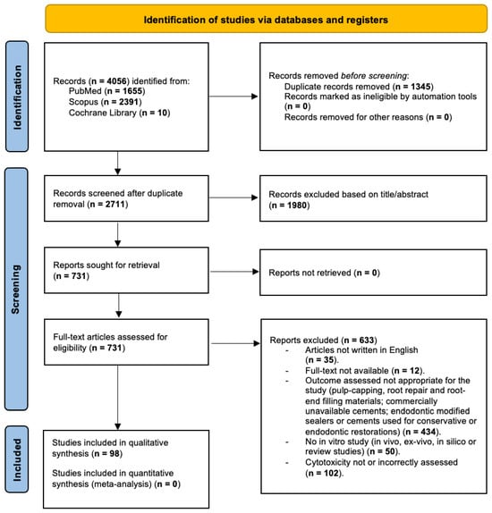
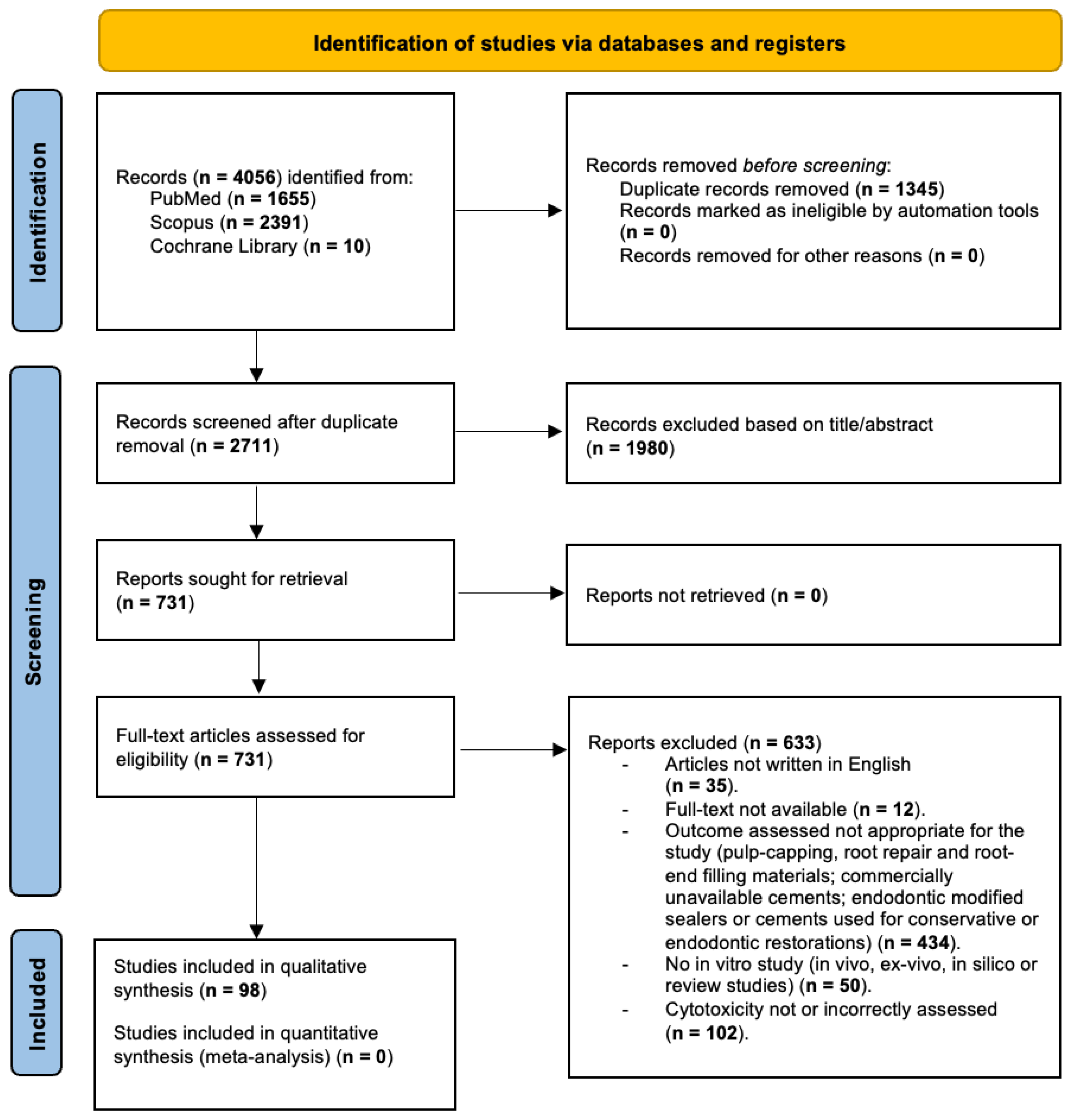
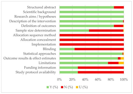
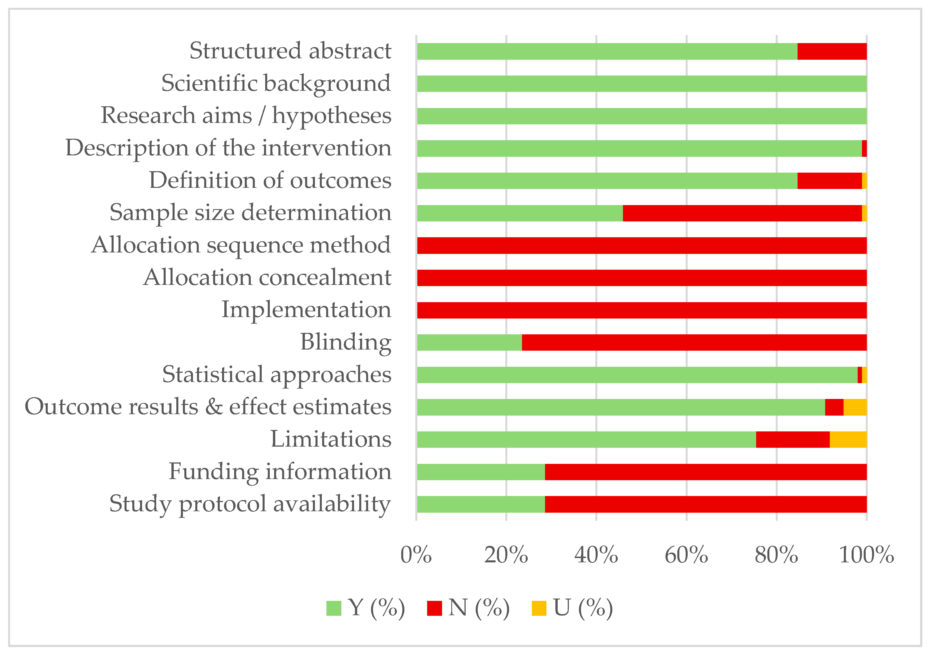

Disclaimer/Publisher’s Note: The statements, opinions and data contained in all publications are solely those of the individual author(s) and contributor(s) and not of MDPI and/or the editor(s). MDPI and/or the editor(s) disclaim responsibility for any injury to people or property resulting from any ideas, methods, instructions or products referred to in the content. |
© 2026 by the authors. Licensee MDPI, Basel, Switzerland. This article is an open access article distributed under the terms and conditions of the Creative Commons Attribution (CC BY) license.