


Abstract
Regenerative endodontic treatment (RET) has been considered a reliable procedure to treat immature necrotic teeth; however, the effect of dental trauma on the prognosis of RET is questionable. This systematic review aimed to evaluate the current level of evidence for revascularization techniques (the RET) in the management of traumatized necrotic immature permanent teeth with or without periapical radiolucent areas. Four electronic databases—PubMed, Web of Science, Scopus, and Embase—were searched until November 2022. Only randomized clinical trials, cohort studies, and case-control studies with a minimum of 10 cases and 12 months of follow-ups were included. The search identified 363 preliminary results. After discarding the duplicates and screening the titles, abstracts, and full texts, 13 articles were considered eligible. The results showed that RET techniques seemed to have high survival and success rates, 93.8% and 88.3%, respectively, in the treatment of traumatized necrotic immature permanent teeth. Root maturation with RET techniques seemed to be lower in traumatized teeth. Future studies are needed to evaluate root maturation in traumatized teeth using 3-dimensional radiographic evaluations. In addition, the lack of literature on the studies comparing RET and apexification (calcium hydroxide or an MTA) in the treatment of traumatized necrotic immature teeth highlights the necessity for high-level clinical studies comparing these treatment modalities.
1. Introduction
Traumatic dental injuries mainly occur in children and teenagers, and the loss of permanent teeth has lifetime consequences. Therefore, efforts should be made to preserve pulp vitality to promote continued root development and apex formation [1]. Several studies have demonstrated that pulp necrosis is the most common posttraumatic complication of all types of dental trauma [2,3]. Kaba A.D. et al. [4] found that 12% of enamel–dentin fractures produced pulp necrosis. Hecova H. et al. [3] observed that dental pulp necrosis was present in 27% of 889 permanent teeth with traumatic injuries, where the maxillary central incisors were the most frequently affected teeth; 62% of other studies showed figures close to 80% [4,5].
Pulp necrosis in immature permanent teeth leads to the discontinuation of root development, leaving the tooth with an open apex, thin dentinal walls, and short roots, making root canal debridement difficult, presenting an unfavourable crown-to-root ratio, and increasing the risk of future root fracture under occlusal forces [6,7,8]. Therefore, the optimal goal of treating these teeth is to prevent or resolve apical periodontitis, achieve continuous root development, and regenerate a dentin–pulp complex that restores the functional properties of this tissue [8].
There are three main treatment options to manage traumatized immature permanent teeth with infected root canal systems: apexification with calcium hydroxide, apexification with a mineral trioxide aggregate (MTA), and regenerative endodontic procedures [9].
The traditional approach for treating traumatized permanent teeth with open apices is apexification using calcium hydroxide (Ca(OH)2) dressings. However, it is a long treatment with multiple visits, which may reduce patient compliance, complicate follow-ups, and increase the vulnerability of coronal restoration [6]. In addition, Ca(OH)2 dressings have a significant negative effect on root strength and increase the risk of cervical fractures [10,11,12].
Recently, apexification has been performed with a mineral trioxide aggregate (MTA), also known as apexification with an MTA or an MTA apical plug [13]. An MTA is biocompatible, has the ability to set in the presence of blood, induces hard tissue formation, and has antibacterial effects due to its high pH [14,15]. In addition, MTA apexification can be performed in one or two visits, increasing patient compliance, improving the healing rate, and reducing the risk of subsequent tooth fractures [11,16]. Both apexification techniques induce the formation of an apical hard tissue barrier and promote periapical healing and the disappearance of clinical symptoms. While Ca(OH)2 apexification is still used by some practitioners, studies have shown that MTA apexification provides more advantages [17,18,19]. Nevertheless, it is important to highlight that apexification by either Ca(OH)2 or an MTA does not strengthen the root or foster further root development in terms of increasing the root length or width [11,20].
Replacing damaged root structures, as well as the pulp–dentin complex, is an optimal approach for treating traumatized immature permanent teeth with a necrotic pulp. Therefore, regenerative endodontic treatment (RET) has recently been introduced as an alternative to conventional treatment [21]. RET in immature permanent teeth has been highlighted in studies of dental trauma, with evidence that the dental pulp often remains vital despite substantial traumatic injuries such as intrusions and avulsions [22,23]. It has been radiographically proven that regeneration was possible in re-implanted teeth after intentional or traumatically related avulsion [24]. Therefore, if a favourable environment is established, the same can be accomplished in an infected tooth [25].
RET is defined as “biologically based procedures designed to replace damaged structures, including dentin and root structures, as well as cells of the pulp-dentin complex” [26]. RET is used in pain and inflammation resolution, the healing of periapical lesions, and increasing root length and thickness (maturogenesis) [27,28]. It is considered by the American Association of Endodontics [29], the European Academy of Paediatric Dentistry [30], and the European Society of Endodontics [31] as an accepted procedure to treat immature necrotic permanent teeth.
RET procedures include the following key points: (a) minimal or no instrumentation of the dentinal walls, (b) disinfection with irrigants, (c) the application of an intracanal medicament, (d) provocation of bleeding into the canal and creation of a blood clot, (e) capping with a hydraulic silicate cement, and (f) an effective coronal seal [31].
In 2011, Lovelace T.W. et al. [32] demonstrated that the evoked-bleeding act in regenerative endodontic procedures results in clinically significant delivery of mesenchymal stem cells into root canal systems, as periapical tissues in immature permanent teeth are rich in blood supply and contain stem cells that have the potential for tissue regeneration. Other studies [23,33] suggest that dental trauma injuries damage the periapical vasculature and Hertwig’s epithelial root sheath (HERS), even when the coronal dentin and enamel remain intact. These studies stated that partial removal of the HERS may compromise root development and lead to PDL and bone invasion into the pulp canal [23,33].
All previous systematic reviews conducted to evaluate the efficacy of RETs in the management of immature necrotic teeth included studies of both traumatized and non-traumatized immature necrotic teeth [34,35,36]. To date, no systematic review with meta-analysis has been conducted on the effectiveness of the RET technique in the management of traumatized immature necrotic permanent teeth. Therefore, this systematic review and meta-analysis aimed to evaluate the level of evidence of RET in the treatment of traumatized immature necrotic permanent teeth with or without periapical lesions after a minimum follow-up of 12 months and to conduct a meta-analysis on the survival and success rates of the treated teeth. The secondary objectives were to evaluate root development, apex formation, tooth vitality, and crown discoloration in the treated teeth.
2. Materials and Methods
2.1. Protocol and Registration
The present work followed the guidelines recommended by the PRISMA Statement (Preferred Reporting Items for Systematic Reviews and Meta-analysis) [37] and was performed in accordance with the current recommendations regarding endodontic systematic reviews and meta-analyses [38]. The systematic review protocol was previously registered in the Prospective Register of Systematic Reviews (PROSPERO) of the University of York with the registration number CRD42021261774.
2.2. Eligibility Criteria
A research question was developed in accordance with the Patient, Intervention, Comparison, and Outcome (PICO) method: “What is the current scientific evidence for pulp revascularization in the management of traumatized necrotic immature permanent anterior teeth?”, where (P): patients with traumatized necrotic immature permanent anterior teeth with or without periapical radiolucency, (I): different protocols of pulp revascularization, including blood clots, platelet-rich plasma [PRP], platelet-rich fibrin [PRF], and the platelet pellet [PP], used in the treatment of traumatized necrotic immature permanent anterior teeth, (C): traumatized necrotic immature permanent anterior teeth treated with conventional treatment, which is apexification [MTA apexification or Ca(OH)2 apexification], (O): the survival, clinical, and radiographic success of pulp revascularization in the treatment of traumatized necrotic immature permanent anterior teeth.
2.3. Outcome Measures
The primary outcomes were survival, clinical, and radiographic success. Survival was defined as a tooth that remained in the oral cavity after the follow-up. Clinical success was achieved with the absence of clinical symptoms (i.e., tenderness to percussion or palpation, swelling or fistulas, or spontaneous pain). Radiographic success was obtained when a reduction in the size of the periapical area was observed, with the absence or no increase in the size of the adverse events, such as root resorption and ankylosis, after the follow-up. Secondary outcomes were: continuation of root development, pulp vitality, and crown discoloration, where the continuation of root development had 3 aspects: increased root length, increased root width, and reduced apical diameter.
2.4. Study Selection Criteria
The inclusion criteria were as follows: randomized clinical trials (RCTs), retrospective and prospective cohort studies, and case-control studies. This review only included studies with a follow-up of a minimum of 12 months and studies with a minimum of 10 cases. Only articles that studied pulp revascularization (the RET) procedures or compared RET with other techniques (i.e., apexification) in traumatized necrotic immature teeth were included. Studies were not restricted by language or year of publication.
2.5. Databases and Search Strategy
The database search, study selection, and data extraction were performed by two independent examiners (M.S.; I.A.). In the case of any discrepancy between them, a third author was consulted (V.F.L.).
A systematic advanced electronic search was performed in PubMed, the Web of Science, Scopus, and Embase on the 21st of June 2021 and later updated on 28 November 2022, without any date or language restrictions. The search was conducted using boolean operators ‘AND’ and ‘OR’ to annex the terms and develop the search strategy. The search terms were structured as follows: (regenerative endodontic treatment OR regenerative* OR endodontic regeneration OR regenerative endodontics OR Regenerative Approach OR pulp revascularization OR revascularization* OR revitalisation* OR revitalise* OR blood clot OR platelet-rich fibrin OR platelet-rich plasma) AND (immature teeth OR Open apex OR immature dentition) AND (necrosis OR necrot* OR (non-vital AND tooth*) OR pulpless) AND (Dental trauma* OR Traumatized* OR Traumatic* OR tooth Injuries) (Supplementary Table S1). In addition, reference lists of all selected articles were screened to identify additional studies.
2.6. Study Selection Process
Titles and abstracts of all studies were assessed independently by two reviewers (M.S. and I.A.). When no sufficient information was provided by screening the abstract, the full article was reviewed before making the final decision. Similarly, the two examiners extracted all data regarding the relevant variables. A researcher who had not participated in the selection process (J.M.M.-C.) performed the subsequent meta-analysis.
2.7. Data Extraction
The variables extracted from each included article were: author and year of publication, study type, sample size, number of groups, demographic variables (gender and age), follow-up in months, loss to follow-up, type of trauma, irrigant agents used and their concentrations, root canal dressing, duration of root canal dressing in weeks, scaffold type with or without a matrix, type of coronal seal, survival rate, success rate, failure, changes in root length and width, changes in apical diameter, adverse events or effects, crown discoloration, and tooth.
2.8. Methodological Quality Assessment
Two researchers assessed the risk of bias in all selected studies (M.S. and J.M.M.-C.) using a Cochrane risk-of-bias tool, RoB 2.0 [39], to evaluate randomized clinical trials and the ONS (Newcastle–Ottawa scale) [40] to evaluate non-randomized studies, including case-control and cohort studies.
The NOS includes 8 items with a potential score of 9. Three main domains are considered: patient selection, comparability of the study groups, and results or outcome. Articles were classified as being of ‘high’, ‘moderate’, or ‘low quality’, where high-quality articles scored more than 6 points.
The Cochrane RoB 2.0 methodology assessment [39] consists of five domains that evaluate: the randomization process, deviations from intended interventions, missing outcome data, outcome measurement, and selection of reported outcomes. Producing three levels of bias: ‘low risk of bias’, ‘some concerns’, or ‘high risk of bias’.
2.9. Quantitative Synthesis—Meta-Analysis
All studies included in the meta-analysis were combined using a random-effects model. The estimated effect size was the event rate, odd ratios, means, and different means. The 95% confidence intervals were calculated for all estimated variables.
Heterogeneity among the combined studies was assessed using a Q test (p-value < 0.05) and quantified with the I2, considering slight heterogeneity if it was 25–50%, moderate if 50–75%, and high heterogeneity if > 75%. Statistical significance was tested using a Z test (p-value < 0.05). The meta-analysis has been represented with a forest plot, and the publication bias was assessed using an Egger’s test that indicates the existence of possible publication bias when the p-value is less than 0.05 (indicating significant asymmetry).
3. Results
3.1. Study Selection
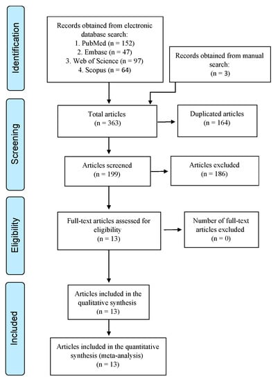
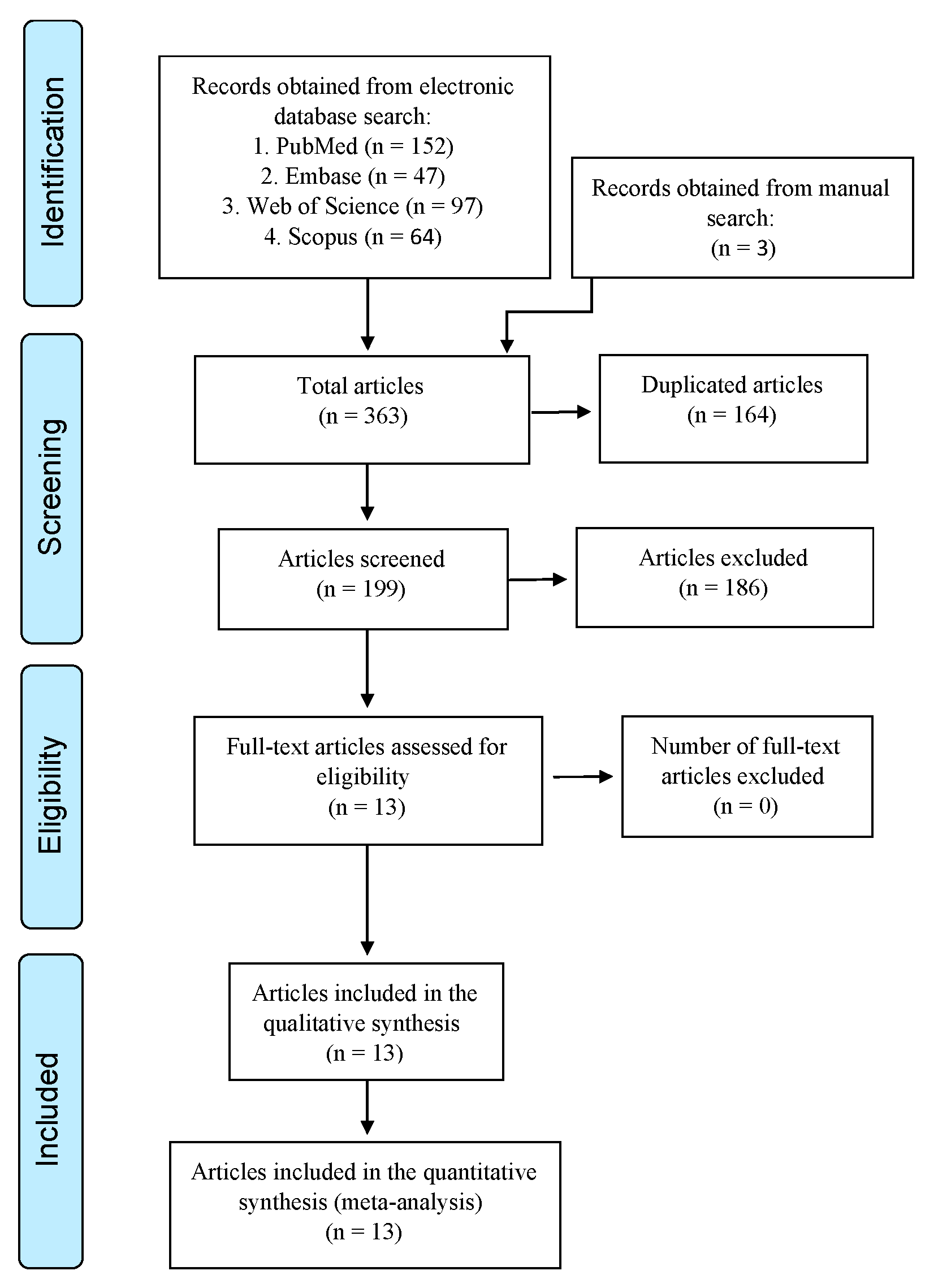
The search identified a total of three hundred and sixty-three preliminary results, where one hundred and fifty-two were found in PubMed, forty-seven in Embase, ninety-seven in the Web of Science, sixty-four in Scopus, and three from a manual search of reference lists. The duplicates were manually removed using Mendeley reference management software, and a total of 199 studies remained. After the title and abstract screening, 186 articles were excluded. A total of 13 studies were eligible for full-text reading, and all were selected for qualitative and quantitative synthesis (Figure 1).
Figure 1.
Preferred Reporting Items for Systematic Reviews and Meta-Analyses (PRISMA) flow diagram.
3.2. Characteristics of the Studies
The sample size of the selected studies ranged from 15 [41] to 118 cases [42]. The patients’ gender was mentioned in most studies, except in three [42,43,44]. The minimum follow-up time was 12 months [42,43,45,46,47], and the maximum follow-up time was “43.42” months [41]. As for dropouts, five studies had no dropouts [44,45,48,49,50], and the other six studies [41,42,43,51,52,53] had dropouts that ranged from 1 [51] to 16 [47]. The results are presented in Table 1 and Supplementary Table S2.
Table 1.
Methodological characteristics of the included studies.
3.3. Assessment of Risk of Bias
Six RCTs were identified, and the Cochrane RoB 2 tool was used for quality assessment. Only one study [46] was considered as a ‘low risk of bias’, four studies were considered as ‘Some concerns’ [42,43,44,53], and one study had a ‘high risk of bias’ [48] (Table 2). The quality was mainly affected by non-compliance with the items related to the blinding of patients or evaluators. As for the rest of the studies, NOS was used for the quality assessment, and one case-control study was identified with ‘moderate’ methodological quality [a score of 5] [49], four cohort studies with ‘moderate’ methodological quality [a score of 5–6] [41,45,50,51], and two cohort studies with ‘high’ methodological quality [a score of 7] [47,52] (Table 3).
Table 2.
Cochrane RoB 2 tool for randomized clinical trials.
Table 3.
Newcastle–Ottawa scale for cohort and case-control studies.
The articles were classified as being of ‘high’, ‘moderate’, or ‘low’ quality, where high-quality articles scored more than six points.
3.4. Quantitative Analysis Results
3.4.1. Survival and Success Rate
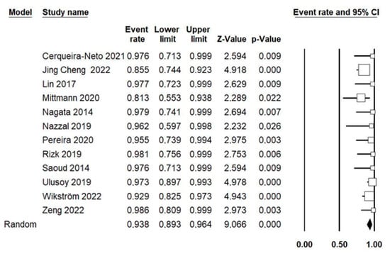
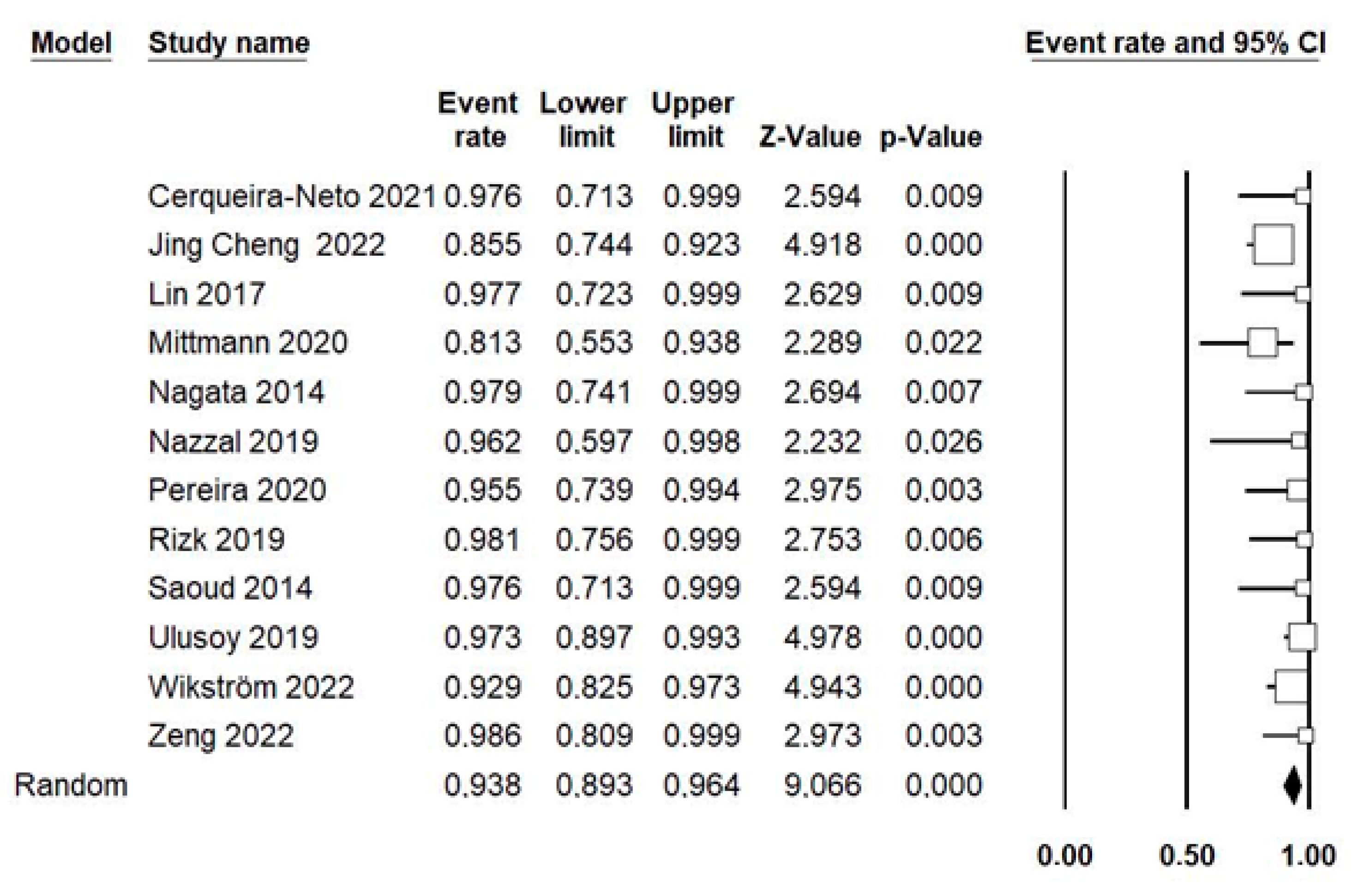
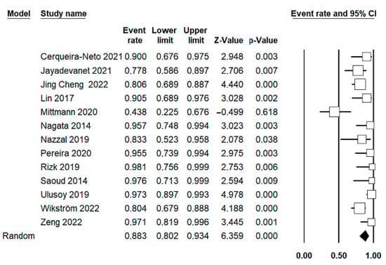
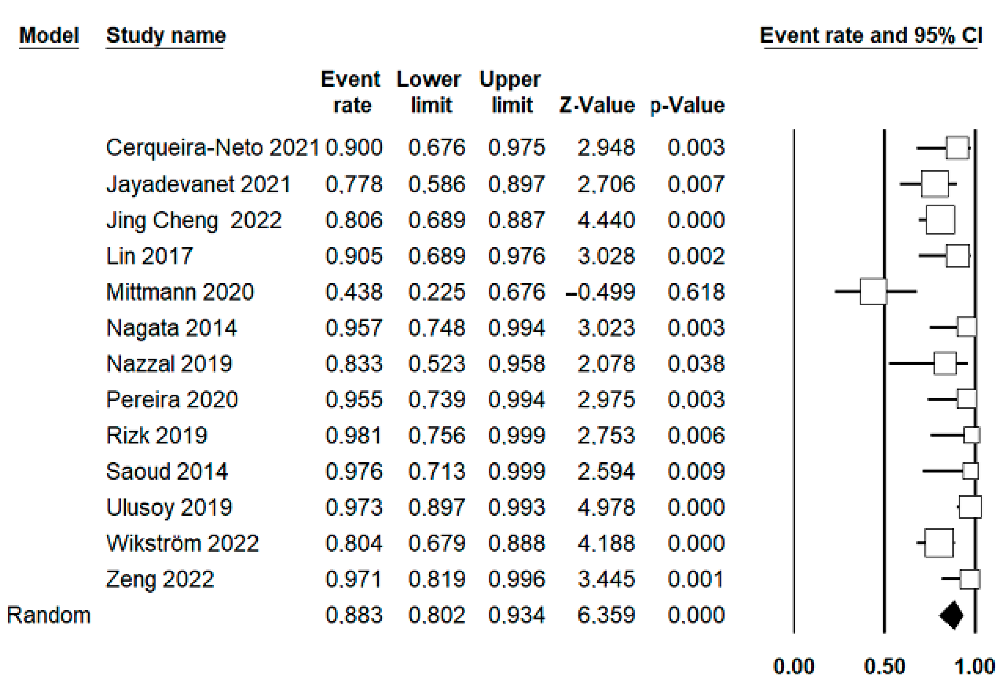
Using the random-effects model, 12 studies, including 384 cases, were combined to estimate the survival rate, and 13 studies, including 411 cases, were combined for the success rate. The estimated survival rate for traumatized teeth treated with RET was 93.8% (95% CI; 89.3–96.4%), with a slight heterogeneity between the combined studies (p = 0.183; I2 = 26.6%; Q-value = 14.9) (Figure 2). The estimated success rate was 88.3% (95% CI; 80.2–93.4%), with a moderate heterogeneity between the combined studies (p < 0.001; I2 = 66.7%; Q-value = 36) (Figure 3).
Figure 2.
Forest plot of the estimated survival rate of traumatized teeth treated with pulpal revascularization among the selected studies. Using a random-effects model where 12 studies, including 384 cases, were combined to estimate the survival rate of RET [42,44,45,46,47,48,49,50,51,52,53,54].
Figure 3.
Forest plot of the estimated success rate of traumatized teeth treated with pulpal revascularization among the selected studies. Using a random-effects model where 13 studies, including 411 cases, were combined to estimate the success rate of RET [42,43,44,45,46,47,48,49,50,51,52,53,54].
3.4.2. Root Development, Apex Formation, Tooth Vitality, and Crown Discoloration
The results of the meta-analysis, with the number of combined studies, samples, and heterogeneity for signs of apical closure (partial or complete), the decrease in apical diameter, increase in root length and width, regain of vitality, and crown discoloration, are listed in Table 4.
Table 4.
Results of meta-analysis for all studied variables.
3.4.3. Apexification and RET Comparison
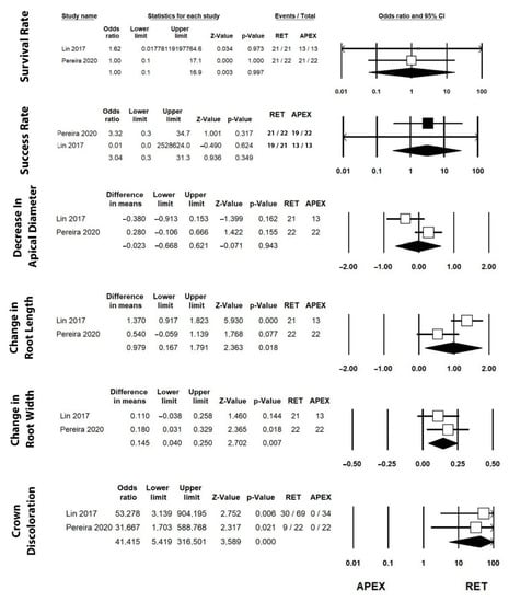
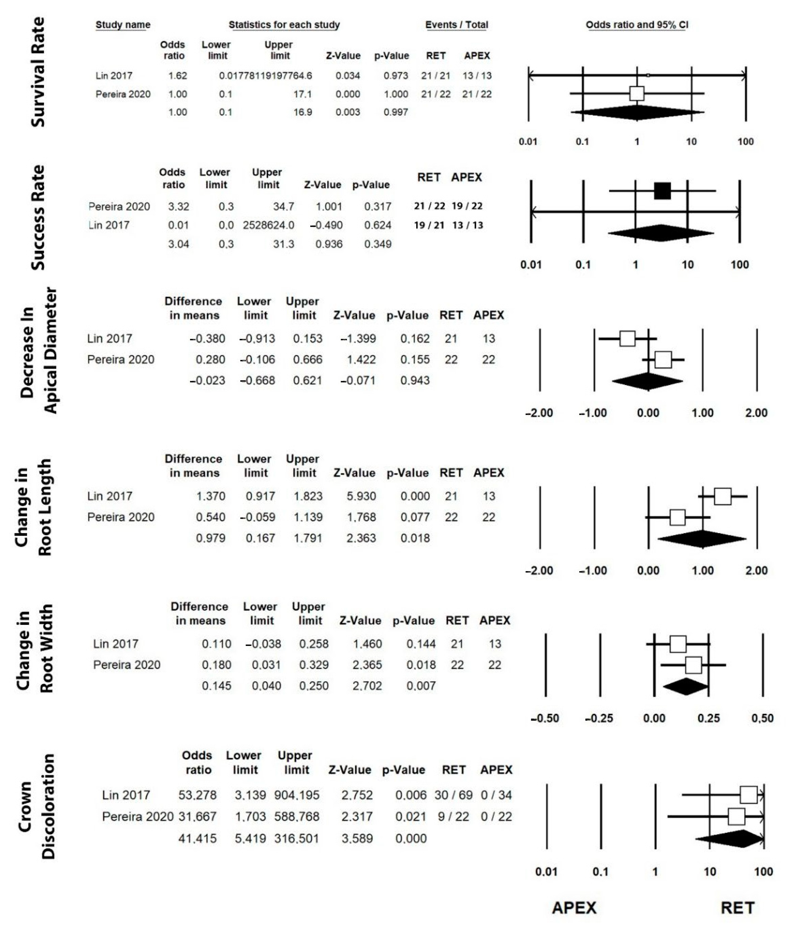
Only two studies [42,49] compared both the apexification and RET techniques. The odds ratios and differences in means for survival and success rate, increase in root length and width, apex formation, and crown discoloration are presented in Figure 4.
Figure 4.
Forest plot of estimated odds ratio and difference in means for the studied variables comparing RET and apexification techniques. Results of two studies that compared the apexification and RET techniques [42,49].
3.5. Publication Bias
The results of the Egger test used to assess the publication bias are presented in Supplementary Table S3.
4. Discussion
Previous studies reported that the success rate of RET is lower in traumatized immature teeth [51,54,55] in comparison to RET in non-traumatic dental injuries, such as caries or developmental anomalies [20,27,42]; however, a recent systematic review and meta-analysis demonstrated that there is no relationship between the aetiology of pulp necrosis in immature teeth and the success rate after RET [35], which is aligned with the results of a study conducted by Linsuwanont et al. [56]. Among the included studies in our review, Mittmann et al. [51] reported the lowest survival and success rates of RETs (81.3% and 44%, respectively); this could be associated with the severity of the trauma as 75% of the cases were serious avulsion, 19% were luxation, and 6% were intrusion cases. Similarly, Jing Cheng et al. [50] reported that failure of RET occurred in avulsed teeth more than in other types of traumatic teeth. In addition, Wikström et al. [52] stated that the failed 11 cases were characterized by severe trauma. Hence, severe trauma might influence the outcome of RET when compared with mild-to-moderate trauma as, according to some studies and depending on the type of dental trauma, irreversible damage to the HERS may be present in traumatized immature teeth affected by different degrees of traumatic injuries [22,23]. The quantitative analysis in our review estimated that the survival and success rates of necrotic traumatized immature permanent teeth treated with RET and followed up after 12–48 months were 93.8% and 88.3%, respectively. Our results are slightly lower than the findings of Torabinejad et al. [57], who previously reported a survival rate of 97.8% and a success rate of 91.3%; the slight difference in the results may be due to the fact that the aforementioned study included all teeth treated with RET regardless of the aetiology of the pulp necrosis.
In this systematic review, to quantify the continuation of root development, most studies measured, over time, the radiographic changes in the root after RET. Specifically, the prognosis of root maturation was evaluated according to the following parameters: the number or percentage of cases with signs of root maturation, the total mean percentage of root maturation after the follow-up, and the total mean of root development in millimetres. Kim S.G. et al. [58] suggested that traumatic injuries can negatively affect the success of root maturation after RET due to the interaction between the HERS and mesenchymal stem cells in the dental follicle. Similarly, Zeng et al. [47] demonstrated that the aetiology of pulp necrosis (dens evaginatus or trauma) has a significant effect on root development after RETs and that traumatized immature teeth have a lower root development success rate. In a recent review [34], immature necrotic teeth treated with RET, regardless of the aetiology of the pulp necrosis, reported an 80% success rate of dentin thickening and lengthening; however, in our review, all the included studies reported a lower success rate of root development, and the results of root maturation among the studies varied, as six studies [42,46,48,49,51,52] exhibited more increase in the root length over the root width, and seven studies [41,43,44,45,47,50,53] reported more increase in the root width over the root length. Nevertheless, only six of the included studies [41,42,48,49,52] quantified the continuation of root development in millimetres, and our quantitative analysis of these studies estimated a 1.26 mm mean increase in root length and a 0.52 mm mean increase in root width.
The variation in the results could be attributed to the heterogeneity among the studies, which could be caused by the variability in the participants, studied interventions and outcomes, outcome measurement tools, and risk of bias [59]. Moreover, 11 out of 13 included studies [41,43,44,45,48,49,50,51,52,53] used 2-dimensional radiographs (periapical radiographs) to evaluate the outcome of root development, and only two studies used a cone-beam computed tomography (CBCT) scan [42,47]. Taking into consideration the limitations of 2-dimensional radiographs, since the root development occurs in a 3-dimensional (3D) pattern, 3D imaging is the most accurate method for the assessment of root development and patterns of hard tissue formation [60]. Although Flake N.M. et al. [61] recently introduced a standardized method to measure radiographic root changes in 2-dimensional radiographs after RET, it has been shown that dental trauma can be better assessed in three dimensions [62]. CBCT has been demonstrated to be a more powerful diagnostic tool for evaluating the anatomical characteristics in a three-dimensional pattern to know, in advance, the measurements of the last apical millimetre of the roots. [63].
Previous studies [21,32] demonstrated that stem cells from the apical papilla (SCAP) are essential in regenerative endodontic procedures, and their conservation should be a priority for clinicians. Martin D.E. et al. [64] reported that different concentrations of NaOCl can influence the survival rate of stem cells and recommended a concentration of NaOCl between 1.5 and 3% during RET. The included studies in the present systematic review used different NaOCl concentrations: 0.5% [41,52], 1% [51,53], 1.5% [42,43,47,50], 2% [46,49], 2.5% [45], 5% [44], and 6% [48]; therefore, lower or higher concentrations of NaOCl may have negatively influenced the outcome of the RET. Previous studies recommended the use of ethylenediaminetetraacetic acid (EDTA) in the second follow-up appointment of RET [29,31,64,65], as it reduces the undesirable effect of NaOCl on stem cell viability and releases growth factors. Although, another study by Farhad Mollashahi et al. [66] demonstrated that EDTA had higher cytotoxicity than NaOCl on the survival of stem cells of the human apical papilla and that CHX had less cytotoxicity compared with NaOCl or EDTA. In addition, a recent systematic review reported that there were no failed cases when EDTA was not used as an irrigation solution during RET procedures [35]. In the present review, two of the included studies [41,45] did not irrigate with EDTA and still had similar results to the other included studies. Further investigations regarding the use of EDTA in RET are required to better understand its impact on such treatments.
Adequate disinfection of the root canal system is an essential step in RET [67]. Triple antibiotic paste (TAP) has been highlighted as an effective antimicrobial intracanal medicament in RET procedures, but it has been shown to be cytotoxic to the stem cells of the apical papilla at clinical concentrations [65,68]. Berkhoff et al. [67] reported that a TAP presented high diffusion and retention within the dentin and that 80% of this intracanal agent cannot be removed in the second appointment of RET, regardless of the irrigation agent used. Therefore, current guidelines recommend the use of low-concentration antibiotic mixtures or calcium hydroxide [29,31], as Nagata et al. [44] made a comparison between TAP and Ca(OH)2 and reported that there were no clinical or radiographic differences between the two aforementioned protocols. In the present review, all of the studies, except for two [48,52], used TAP as the intracanal medicament for RET for a dressing time from 7 days [51] to 4 weeks [43,47,53] and had a similar outcome to the rest of the studies, although the number of studies is inadequate to make a clear conclusion regarding this parameter.
Tooth discoloration after RET was reported in seven of the included studies [41,42,44,48,50,51,52], and our analysis estimated that this event was presented in 37.9% of traumatized immature necrotic teeth treated with RET. Crown discoloration may be attributed to the use of TAP as the intracanal medicament [69,70,71], as tetracycline-based antibiotics (minocycline or doxycycline) can result in higher crown discoloration [71,72], which is why some studies [70,71] suggest the use of amoxicillin-based antibiotics or double antibiotic paste (DAP), as it induces less coronal discoloration. On the other hand, the bismuth oxide content in an MTA is also associated with coronal discoloration [72,73]. Therefore, some studies [74,75] suggested the use of dentin bonding agents and a flowable composite in the coronal pulpal chamber; nevertheless, the efficacy of this technique has not been confirmed yet.
The results of the present review estimated that 16.2% of the traumatized necrotic immature teeth would respond to sensibility after the RET. All of the studies in this review used cold and electric pulp sensibility tests, while only two studies used thermal tests [45,46]. Ulusoy et al. [53] reported that a “blood clot” study group had a significantly slower initial response to sensitivity tests and suggested that the higher platelet level of the biological scaffolds may have stimulated the regeneration process of the sensory fibres; these results were in agreement with two previous studies [73,76]. However, Rizk et al. [46] and Jayadevan et al. [43] used PRP and PRF scaffolds and reported no response to pulp sensibility tests in all of the cases, which agrees with Torabinejad et al. [77]. The negative responses might have arisen for different reasons: First, in the children population, it was difficult to obtain accurate pulp testing scores. Second, the thick three-layered coronal seal over the “blood clot” scaffold. Third, dentin sensitivity in the natural pulp–dentin complex is related to the hydrodynamic activity of dentin tubules in association with A-β sensory fibres, whereas newly regenerated, mineralized root canal tissues do not appear to have well-organized dentin tubules and, thus, may not have exhibited the same sensitivity as natural tissue [78]. Therefore, the results of the sensibility tests should be interpreted with caution.
This review reports the necessity to carry out more comparative studies between the apexification technique (with Ca(OH)2 or an MTA) and RET procedures for the treatment of necrotic traumatized immature permanent teeth. To date, there are only two studies [42,49] that compared these two techniques, and one of them is not randomized. The lack of randomized clinical trials comparing these two treatment modalities makes the comparisons less conclusive.
Within the limitations of our study, most of the included randomized studies have a risk of bias of “some concerns”, and seven out of thirteen studies were non-randomized with moderate or low quality and an unclear risk of bias. In addition, there is no standardized protocol for RET used in all of the included studies, which causes heterogeneity among the studies. Moreover, there were variations in the assessment criteria and interpretation of the results among the studies, and most of the studies have a small sample size and lack long-term follow-ups, which are important to address an accurate assessment of the RET outcome. Therefore, the result of this review must be interpreted with caution. Additional long-term, high-quality clinical studies with larger sample sizes, standardized protocols, and accurate radiographic evaluations, such as by CBCT scans, are needed to evaluate the success of RET techniques in the management of necrotic traumatized immature teeth and root maturation. The lack of literature on studies comparing RET and apexification (Ca(OH)2 or an MTA) in the treatment of necrotic traumatized immature teeth addresses the necessity for high-level clinical studies comparing these two treatment modalities.
5. Conclusions
Based on the present review and meta-analysis, RET techniques seem to have high survival and success rates in the treatment of traumatized necrotic immature permanent teeth, and they appear to be similar to those immature teeth affected by caries or dental anomalies. However, severe trauma may have a negative effect on the results of RET treatment, and further future studies are needed to support this hypothesis.
Root maturation (the lengthening and thickening of the dental walls) with RET techniques seems to be less in traumatized teeth. Nevertheless, future studies are needed to evaluate root maturation in traumatized teeth using 3-dimensional radiographic evaluations.
Coronal discoloration is an adverse event in most traumatized teeth treated with RET, so internal bleaching might be indicated at the end of the treatment, and future clinical studies are needed to overcome this issue.
The recuperation of sensitivity after RET is not clear due to difficulties in counting pulp vitality tests as an accurate method for evaluation.
Supplementary Materials
The following supporting information can be downloaded at: https://www.mdpi.com/article/10.3390/jcm12072656/s1, Table S1: search strategies for each database; Table S2: results of RET in each included study; Table S3: results of the Egger test of each RET meta-analysis performed.
Author Contributions
M.S.: Conceptualization, Investigation, and Methodology; I.F.-M.: Writing–Review & Editing; Á.Z.-M.: Writing–Review & Editing; I.A.: Investigation and Writing–Original Draft; V.F.-M.: Validation; C.B.-A.: Writing—Review & Editing; J.E.I.-C.: Writing—Review & Editing; J.M.M.-C.: Project Administration, Resources, Supervision, and Formal Analysis. All authors have read and agreed to the published version of the manuscript. All authors have read and approved the final version.
Funding
This research received no external funding.
Institutional Review Board Statement
Not applicable.
Informed Consent Statement
Not applicable.
Data Availability Statement
The data presented in this study are available in the article and Supplementary Material.
Conflicts of Interest
The authors declare no conflict of interest.
References
- DiAngelis, A.J.; Andreasen, J.O.; Ebeleseder, K.A.; Kenny, D.J.; Trope, M.; Sigurdsson, A.; Andersson, L.; Bourguignon, C.; Flores, M.T.; Hicks, M.L.; et al. International Association of Dental Traumatology guidelines for the management of traumatic dental injuries: 1. Fractures and luxations of permanent teeth. Dent. Traumatol. 2012, 28, 2–12. [Google Scholar] [CrossRef] [Scilit]
- Kallel, I.; Douki, N.; Amaidi, S.; Ben Amor, F. The Incidence of Complications of Dental Trauma and Associated Factors: A Retrospective Study. Int. J. Dent. 2020, 26, 2968174–2968178. [Google Scholar] [CrossRef] [Scilit]
- Hecova, H.; Tzigkounakis, V.; Merglova, V.; Netolicky, J. A retrospective study of 889 injured permanent teeth. Dent. Traumatol. 2010, 26, 466–475. [Google Scholar] [CrossRef] [Scilit] [PubMed]
- Kaba, A.D.; Maréchaux, S.C. A fourteen-year follow-up study of traumatic injuries to the permanent dentition. ASDC J. Dent. Child. 1989, 56, 417–425. [Google Scholar] [PubMed]
- Borum, M.K.; Andreasen, J.O. Therapeutic and economic implications of traumatic dental injuries in Denmark: An estimate based on 7549 patients treated at a major trauma centre. Int. J. Paediatr. Dent. 2001, 11, 249–258. [Google Scholar] [CrossRef] [Scilit]
- Mohammadi, Z. Strategies to manage permanent non-vital teeth with open apices: A clinical update. Int. Dent. J. 2011, 61, 25–30. [Google Scholar] [CrossRef] [Scilit]
- Schilder, H. Filling Root Canals in Three Dimensions. J. Endod. 2006, 32, 281–290. [Google Scholar] [CrossRef] [Scilit] [PubMed]
- Hargreaves, K.M.; Geisler, T.; Henry, M.; Wang, Y. Regeneration Potential of the Young Permanent Tooth: What Does the Future Hold? J. Endod. 2008, 34, S51–S56. [Google Scholar] [CrossRef] [Scilit]
- Flanagan, T.A. What can cause the pulps of immature, permanent teeth with open apices to become necrotic and what treatment options are available for these teeth. Aust. Endod. J. 2014, 40, 95–100. [Google Scholar] [CrossRef] [Scilit]
- Andreasen, J.O.; Farik, B.; Munksgaard, E.C. Long-term calcium hydroxide as a root canal dressing may increase risk of root fracture. Dent. Traumatol. 2002, 18, 134–137. [Google Scholar] [CrossRef] [Scilit]
- Jeeruphan, T.; Jantarat, J.; Yanpiset, K.; Suwannapan, L.; Khewsawai, P.; Hargreaves, K.M. Mahidol Study 1: Comparison of Radiographic and Survival Outcomes of Immature Teeth Treated with Either Regenerative Endodontic or Apexification Methods: A Retrospective Study. J. Endod. 2012, 38, 1330–1336. [Google Scholar] [CrossRef] [Scilit]
- American Academy of Pediatric Dentistry. Guideline on Pulp Therapy for Primary and Immature Permanent Teeth. Pediat. Dent. 2016, 38, 280–288. [Google Scholar]
- Rafter, M. Apexification: A review. Dent. Traumatol. 2005, 21, 1–8. [Google Scholar] [CrossRef] [Scilit]
- Mitchell, P.J.C.; Pitt Ford, T.R.; Torabinejad, M.; McDonald, F. Osteoblast biocompatibility of mineral trioxide aggregate. Biomaterials 1999, 20, 167–173. [Google Scholar] [CrossRef] [Scilit]
- Economides, N.; Pantelidou, O.; Kokkas, A.; Tziafas, D. Short-term periradicular tissue response to mineral trioxide aggregate (MTA) as root-end filling material. Int. Endod. J. 2003, 36, 44–48. [Google Scholar] [CrossRef] [Scilit]
- Damle, S.G.; Bhattal, H.; Loomba, A. Apexification of anterior teeth: A comparative evaluation of mineral trioxide aggregate and calcium hydroxide paste. J. Clin. Pediatr. Dent. 2012, 36, 263–268. [Google Scholar] [CrossRef] [Scilit] [PubMed]
- Chala, S.; Abouqal, R.; Rida, S. Apexification of immature teeth with calcium hydroxide or mineral trioxide aggregate: Systematic review and meta-analysis. Oral Surg. Oral Med. Oral Pathol. Oral Radiol. Endod. 2011, 112, e36–e42. [Google Scholar] [CrossRef] [Scilit] [PubMed]
- Bakland, L.K.; Andreasen, J.O. Will mineral trioxide aggregate replace calcium hydroxide in treating pulpal and periodontal healing complications subsequent to dental trauma? A review. Dent. Traumatol. 2012, 28, 25–32. [Google Scholar] [CrossRef] [Scilit]
- Witherspoon, D.E.; Small, J.C.; Regan, J.D.; Nunn, M. Retrospective Analysis of Open Apex Teeth Obturated with Mineral Trioxide Aggregate. J. Endod. 2008, 34, 1171–1176. [Google Scholar] [CrossRef] [Scilit]
- Bose, R.; Nummikoski, P.; Hargreaves, K. A Retrospective Evaluation of Radiographic Outcomes in Immature Teeth with Necrotic Root Canal Systems Treated With Regenerative Endodontic Procedures. J. Endod. 2009, 35, 1343–1349. [Google Scholar] [CrossRef] [Scilit]
- Diogenes, A.; Henry, M.A.; Teixeira, F.B.; Hargreaves, K.M. An update on clinical regenerative endodontics. Endod. Top. 2013, 28, 2–23. [Google Scholar] [CrossRef] [Scilit]
- Andreasen, J.O.; Borum, M.K.; Jacobsen, H.L.; Andreasen, F.M. Replantation of 400 avulsed permanent incisors. 2. Factors related to pulpal healing. Dent. Traumatol. 1995, 11, 59–68. [Google Scholar] [CrossRef] [Scilit] [PubMed]
- Andreasen, J.O.; Borum, M.K.; Andreasen, F.M. Replantation of 400 avulsed permanent incisors. 3. Factors related to root growth. Dent. Traumatol. 1995, 11, 69–75. [Google Scholar] [CrossRef] [Scilit] [PubMed]
- Cvek, M.; Cleaton-Jones, P.; Austin, J.; Lownie, J.; Kling, M.; Fatti, P. Pulp revascularization in reimplanted immature monkey incisors—Predictability and the effect of antibiotic systemic prophylaxis. Dent. Traumatol. 1990, 6, 157–165. [Google Scholar] [CrossRef] [Scilit] [PubMed]
- Banchs, F.; Trope, M. Revascularization of Immature Permanent Teeth with Apical Periodontitis: New Treatment Protocol? J. Endod. 2004, 30, 196–200. [Google Scholar] [CrossRef] [Scilit] [PubMed]
- Murray, P.E.; Garcia-Godoy, F.; Hargreaves, K.M. Regenerative Endodontics: A Review of Current Status and a Call for Action. J. Endod. 2007, 33, 377–390. [Google Scholar] [CrossRef] [Scilit] [PubMed]
- Hargreaves, K.M.; Diogenes, A.; Teixeira, F.B. Treatment Options: Biological Basis of Regenerative Endodontic Procedures. J. Endod. 2013, 39, S30–S43. [Google Scholar] [CrossRef] [Scilit]
- Wigler, R.; Kaufman, A.Y.; Lin, S.; Steinbock, N.; Hazan-Molina, H.; Torneck, C.D. Revascularization: A Treatment for Permanent Teeth with Necrotic Pulp and Incomplete Root Development. J. Endod. 2013, 39, 319–326. [Google Scholar] [CrossRef] [Scilit]
- AAE. AAE Clinical Considerations for a Regenerative Procedure. Available online: www.aae.org/uploadedfiles/publications_and_research/research/currentregenerativeendodonticconsiderations.pdf (accessed on 3 July 2021).
- Duggal, M.; Tong, H.J.; Al-Ansary, M.; Twati, W.; Day, P.F.; Nazzal, H. Interventions for the endodontic management of non-vital traumatised immature permanent anterior teeth in children and adolescents: A systematic review of the evidence and guidelines of the European Academy of Paediatric Dentistry. Eur. Arch. Paediatr. Dent. 2017, 18, 139–151. [Google Scholar] [CrossRef] [Scilit]
- Galler, K.M.; Krastl, G.; Simon, S.; Van Gorp, G.; Meschi, N.; Vahedi, B.; Lambrechts, P. European Society of Endodontology position statement: Revitalization procedures. Int. Endod. J. 2016, 49, 717–723. [Google Scholar] [CrossRef] [Scilit]
- Lovelace, T.W.; Henry, M.A.; Hargreaves, K.M.; Diogenes, A. Evaluation of the Delivery of Mesenchymal Stem Cells into the Root Canal Space of Necrotic Immature Teeth after Clinical Regenerative Endodontic Procedure. J. Endod. 2011, 37, 133–138. [Google Scholar] [CrossRef] [Scilit] [PubMed]
- Andreasen, J.O. Pulp and periodontal tissue repair–regeneration or tissue metaplasia after dental trauma. A review. Dent. Traumatol. 2012, 28, 19–24. [Google Scholar] [CrossRef] [Scilit] [PubMed]
- Tong, H.J.; Rajan, S.; Bhujel, N.; Kang, J.; Duggal, M.; Nazzal, H. Regenerative Endodontic Therapy in the Management of Nonvital Immature Permanent Teeth: A Systematic Review—Outcome Evaluation and Meta-analysis. J. Endod. 2017, 43, 1453–1464. [Google Scholar] [CrossRef] [Scilit] [PubMed]
- Koç, S.; Del Fabbro, M. Does the Etiology of Pulp Necrosis Affect Regenerative Endodontic Treatment Outcomes? A Systematic Review and Meta-analyses. J. Evid. -Based Dent. Pract. 2020, 20, 101400. [Google Scholar] [CrossRef] [Scilit] [PubMed]
- Wikström, A.; Brundin, M.; Lopes, M.F.; El Sayed, M.; Tsilingaridis, G. What is the best long-term treatment modality for immature permanent teeth with pulp necrosis and apical periodontitis? Eur. Arch. Paediatr. Dent. 2021, 22, 311–340. [Google Scholar] [CrossRef] [Scilit]
- Liberati, A.; Altman, D.G.; Tetzlaff, J.; Mulrow, C.; Gotzsche, P.C.; Ioannidis, J.P.A.; Clarke, M.; Devereaux, P.J.; Kleijnen, J.; Moher, D. The PRISMA statement for reporting systematic reviews and meta-analyses of studies that evaluate healthcare interventions: Explanation and Elaboration. BMJ 2009, 339, b2700. [Google Scholar] [CrossRef] [Scilit]
- Nagendrababu, V.; Duncan, H.F.; Tsesis, I.; Sathorn, C.; Pulikkotil, S.J.; Dharmarajan, L.; Dummer, P.M.H. Preferred reporting items for systematic reviews and meta-analyses for abstracts: Best practice for reporting abstracts of systematic reviews in Endodontology. Int. Endod. J. 2019, 52, 1096–1107. [Google Scholar] [CrossRef] [Scilit]
- Sterne, J.A.C.; Savović, J.; Page, M.J.; Elbers, R.G.; Blencowe, N.S.; Boutron, I.; Cates, C.J.; Cheng, H.; Corbett, M.S.; Eldridge, S.M.; et al. RoB 2: A revised tool for assessing risk of bias in randomised trials. BMJ 2019, 366, l4898. [Google Scholar] [CrossRef] [Scilit]
- Wells, G.A.; Seha, B.; O’Connell, D.; Peterson, J.; Welch, V.; Losos, M.; Tugwell, P. The Newcastle Ottawa Scale (NOS) for Assesing the Quality of Non-Randomized Studies in Meta-Analysis. Available online: https://www.ohri.ca/programs/clinical_epidemiology/oxford.asp (accessed on 14 July 2021).
- Nazzal, H.; Ainscough, S.; Kang, J.; Duggal, M.S. Revitalisation endodontic treatment of traumatised immature teeth: A prospective long-term clinical study. Eur. Arch. Paediatr. Dent. 2020, 21, 587–596. [Google Scholar] [CrossRef] [Scilit]
- Lin, J.; Zeng, Q.; Wei, X.; Zhao, W.; Cui, M.; Gu, J.; Lu, J.; Yang, M.; Ling, J. Regenerative Endodontics Versus Apexification in Immature Permanent Teeth with Apical Periodontitis: A Prospective Randomized Controlled Study. J. Endod. 2017, 43, 1821–1827. [Google Scholar] [CrossRef] [Scilit]
- Jayadevan, V.; Gehlot, P.; Manjunath, V.; Madhunapantula, S.V.; Lakshmikanth, J. A comparative evaluation of Advanced Platelet-Rich Fibrin (A-PRF) and Platelet-Rich Fibrin (PRF) as a Scaffold in Regenerative Endodontic Treatment of Traumatized Immature Non-vital permanent anterior teeth: A Prospective clinical study. J. Clin. Exp. Dent. 2021, 13, e463–e472. [Google Scholar] [CrossRef] [Scilit] [PubMed]
- Nagata, J.Y.; de Almeida Gomes, B.P.F.; Lima, T.F.R.; Murakami, L.S.; de Faria, D.E.; Campos, G.R.; de Souza-Filho, F.J.; de Jesus Soares, A. Traumatized Immature Teeth Treated with 2 Protocols of Pulp Revascularization. J. Endod. 2014, 40, 606–612. [Google Scholar] [CrossRef] [Scilit] [PubMed]
- Saoud, T.M.A.; Zaazou, A.; Nabil, A.; Moussa, S.; Lin, L.M.; Gibbs, J.L. Clinical and Radiographic Outcomes of Traumatized Immature Permanent Necrotic Teeth after Revascularization/Revitalization Therapy. J. Endod. 2014, 40, 1946–1952. [Google Scholar] [CrossRef] [Scilit] [PubMed]
- Rizk, H.M.; Al-Deen, M.S.M.S.; Emam, A.A. Comparative evaluation of Platelet Rich Plasma (PRP) versus Platelet Rich Fibrin (PRF) scaffolds in regenerative endodontic treatment of immature necrotic permanent maxillary central incisors: A double blinded randomized controlled trial. Saudi Dent. J. 2020, 32, 224–231. [Google Scholar] [CrossRef] [Scilit] [PubMed]
- Zeng, Q.; Zhang, J.; Guo, J.; Liu, S.; Yang, M.; Lin, J. Preoperative factors analysis on root development after regenerative endodontic procedures: A retrospective study. BMC Oral Health 2022, 22, 374. [Google Scholar] [CrossRef] [Scilit]
- Cerqueira-Neto, A.C.C.L.; Prado, M.C.; Pereira, A.C.; Oliveira, M.L.; Vargas-Neto, J.; Gomes, B.P.F.A.; Ferraz, C.C.R.; Almeida, J.F.A.; de-Jesus-Soares, A. Clinical and Radiographic Outcomes of Regenerative Endodontic Procedures in Traumatized Immature Permanent Teeth: Interappointment Dressing or Single-Visit? J. Endod. 2021, 47, 1598–1608. [Google Scholar] [CrossRef] [Scilit]
- Pereira, A.C.; Oliveira, M.L.; Cerqueira-Neto, A.C.C.; Vargas-Neto, J.; Nagata, J.Y.; Gomes, B.P.F.A.; Ferraz, C.C.R.; Almeida, J.F.A.; de-Jesus-Soares, A. Outcomes of traumatised immature teeth treated with apexification or regenerative endodontic procedure: A retrospective study. Aust. Endod. J. 2021, 47, 178–187. [Google Scholar] [CrossRef] [Scilit]
- Cheng, J.; Yang, F.; Li, J.; Hua, F.; He, M.; Song, G. Treatment Outcomes of Regenerative Endodontic Procedures in Traumatized Immature Permanent Necrotic Teeth: A Retrospective Study. J. Endod. 2022, 48, 1129–1136. [Google Scholar] [CrossRef] [Scilit]
- Mittmann, C.W.; Kostka, E.; Ballout, H.; Preus, M.; Preissner, R.; Karaman, M.; Preissner, S. Outcome of revascularization therapy in traumatized immature incisors. BMC Oral Health 2020, 20, 207. [Google Scholar] [CrossRef] [Scilit]
- Wikström, A.; Brundin, M.; Romani Vestman, N.; Rakhimova, O.; Tsilingaridis, G. Endodontic pulp revitalization in traumatized necrotic immature permanent incisors: Early failures and long-term outcomes—A longitudinal cohort study. Int. Endod. J. 2022, 55, 630–645. [Google Scholar] [CrossRef] [Scilit]
- Ulusoy, A.T.; Turedi, I.; Cimen, M.; Cehreli, Z.C. Evaluation of Blood Clot, Platelet-rich Plasma, Platelet-rich Fibrin, and Platelet Pellet as Scaffolds in Regenerative Endodontic Treatment: A Prospective Randomized Trial. J. Endod. 2019, 45, 560–566. [Google Scholar] [CrossRef] [Scilit] [PubMed]
- Nazzal, H.; Tong, H.; Nixon, P.; Duggal, M. Regenerative endodontic therapy for managing immature non-vital teeth: A national survey of UK paediatric dental specialists and trainees. Br. Dent. J. 2018, 224, 247–254. [Google Scholar] [CrossRef] [Scilit] [PubMed]
- Nazzal, H.; Duggal, M.S. Regenerative endodontics: A true paradigm shift or a bandwagon about to be derailed? Eur. Arch. Paediatr. Dent. 2017, 18, 3–15. [Google Scholar] [CrossRef] [Scilit]
- Linsuwanont, P.; Sinpitaksakul, P.; Lertsakchai, T. Evaluation of root maturation after revitalization in immature permanent teeth with nonvital pulps by cone beam computed tomography and conventional radiographs. Int. Endod. J. 2017, 50, 836–846. [Google Scholar] [CrossRef] [Scilit] [PubMed]
- Torabinejad, M.; Nosrat, A.; Verma, P.; Udochukwu, O. Regenerative Endodontic Treatment or Mineral Trioxide Aggregate Apical Plug in Teeth with Necrotic Pulps and Open Apices: A Systematic Review and Meta-analysis. J. Endod. 2017, 43, 1806–1820. [Google Scholar] [CrossRef] [Scilit]
- Kim, S.G.; Malek, M.; Sigurdsson, A.; Lin, L.M.; Kahler, B. Regenerative endodontics: A comprehensive review. Int. Endod. J. 2018, 51, 1367–1388. [Google Scholar] [CrossRef] [Scilit]
- Higgins, J.P.T.; Thomas, J.; Chandler, J.; Cumpston, M.; Li, T.; Page, M.J.; Welch, V.A. Cochrane Handbook for Systematic Reviews of Interventions; Wiley Cochrane Series; John Wiley & Sons, Ltd.: Newark, NJ, USA, 2019. [Google Scholar]
- Ezeldeen, M.; Van Gorp, G.; Van Dessel, J.; Vandermeulen, D.; Jacobs, R. 3-dimensional Analysis of Regenerative Endodontic Treatment Outcome. J. Endod. 2015, 41, 317–324. [Google Scholar] [CrossRef] [Scilit]
- Flake, N.M.; Gibbs, J.L.; Diogenes, A.; Hargreaves, K.M.; Khan, A.A. A Standardized Novel Method to Measure Radiographic Root Changes after Endodontic Therapy in Immature Teeth. J. Endod. 2014, 40, 46–50. [Google Scholar] [CrossRef] [Scilit]
- Patel, S. New dimensions in endodontic imaging: Part 2. Cone beam computed tomography. Int. Endod. J. 2009, 42, 463–475. [Google Scholar] [CrossRef] [Scilit]
- Iandolo, A.; Pisano, M.; Scelza, G.; Abdellatif, D.; Martina, S. Three-Dimensional Evaluation of the Root Apex of Permanent Maxillary Premolars: A Multicentric Study. Appl. Sci. 2022, 12, 6159. [Google Scholar] [CrossRef] [Scilit]
- Martin, D.E.; De Almeida, J.F.; Henry, M.A.; Khaing, Z.Z.; Schmidt, C.E.; Teixeira, F.B.; Diogenes, A. Concentration-dependent Effect of Sodium Hypochlorite on Stem Cells of Apical Papilla Survival and Differentiation. J. Endod. 2014, 40, 51–55. [Google Scholar] [CrossRef] [Scilit] [PubMed]
- Diogenes, A.R.; Ruparel, N.B.; Teixeira, F.B.; Hargreaves, K.M. Translational Science in Disinfection for Regenerative Endodontics. J. Endod. 2014, 40, S52–S57. [Google Scholar] [CrossRef] [Scilit] [PubMed]
- Farhad Mollashahi, N.; Saberi, E.; Karkehabadi, H. Evaluation of Cytotoxic Effects of Various Endodontic Irrigation Solutions on the Survival of Stem Cell of Human Apical Papilla. Iran. Endod. J. 2016, 11, 293–297. [Google Scholar] [CrossRef] [Scilit] [PubMed]
- Berkhoff, J.A.; Chen, P.B.; Teixeira, F.B.; Diogenes, A. Evaluation of Triple Antibiotic Paste Removal by Different Irrigation Procedures. J. Endod. 2014, 40, 1172–1177. [Google Scholar] [CrossRef] [Scilit] [PubMed]
- Ruparel, N.B.; Teixeira, F.B.; Ferraz, C.C.R.; Diogenes, A. Direct Effect of Intracanal Medicaments on Survival of Stem Cells of the Apical Papilla. J. Endod. 2012, 38, 1372–1375. [Google Scholar] [CrossRef] [Scilit]
- Kohli, M.R.; Yamaguchi, M.; Setzer, F.C.; Karabucak, B. Spectrophotometric Analysis of Coronal Tooth Discoloration Induced by Various Bioceramic Cements and Other Endodontic Materials. J. Endod. 2015, 41, 1862–1866. [Google Scholar] [CrossRef] [Scilit]
- Arslan, H.; Ahmed, H.M.A.; Şahin, Y.; Doğanay Yıldız, E.; Gündoğdu, E.C.; Güven, Y.; Khalilov, R. Regenerative Endodontic Procedures in Necrotic Mature Teeth with Periapical Radiolucencies: A Preliminary Randomized Clinical Study. J. Endod. 2019, 45, 863–872. [Google Scholar] [CrossRef] [Scilit]
- Akcay, M.; Arslan, H.; Yasa, B.; Kavrık, F.; Yasa, E. Spectrophotometric Analysis of Crown Discoloration Induced by Various Antibiotic Pastes Used in Revascularization. J. Endod. 2014, 40, 845–848. [Google Scholar] [CrossRef] [Scilit]
- Krastl, G.; Allgayer, N.; Lenherr, P.; Filippi, A.; Taneja, P.; Weiger, R. Tooth discoloration induced by endodontic materials: A literature review. Dent. Traumatol. 2013, 29, 2–7. [Google Scholar] [CrossRef] [Scilit]
- Lenherr, P.; Allgayer, N.; Weiger, R.; Filippi, A.; Attin, T.; Krastl, G. Tooth discoloration induced by endodontic materials: A laboratory study. Int. Endod. J. 2012, 45, 942–949. [Google Scholar] [CrossRef] [Scilit]
- Bezgin, T.; Yilmaz, A.D.; Celik, B.N.; Kolsuz, M.E.; Sonmez, H. Efficacy of Platelet-rich Plasma as a Scaffold in Regenerative Endodontic Treatment. J. Endod. 2015, 41, 36–44. [Google Scholar] [CrossRef] [Scilit]
- Reynolds, K.; Johnson, J.D.; Cohenca, N. Pulp revascularization of necrotic bilateral bicuspids using a modified novel technique to eliminate potential coronal discolouration: A case report. Int. Endod. J. 2009, 42, 84–92. [Google Scholar] [CrossRef] [Scilit]
- Yamauchi, N.; Yamauchi, S.; Nagaoka, H.; Duggan, D.; Zhong, S.; Lee, S.M.; Teixeira, F.B.; Yamauchi, M. DDS Tissue Engineering Strategies for Immature Teeth with Apical Periodontitis. J. Endod. 2011, 37, 390–397. [Google Scholar] [CrossRef] [Scilit] [PubMed]
- Torabinejad, M.; Faras, H. A Clinical and Histological Report of a Tooth with an Open Apex Treated with Regenerative Endodontics Using Platelet-rich Plasma. J. Endod. 2012, 38, 864–868. [Google Scholar] [CrossRef] [Scilit] [PubMed]
- Huang, G.T. Dental pulp and dentin tissue engineering and regeneration advancement and challenge. Front. Biosci. -Elite 2011, 3, 788–800. [Google Scholar] [CrossRef] [Scilit] [PubMed]



Disclaimer/Publisher’s Note: The statements, opinions and data contained in all publications are solely those of the individual author(s) and contributor(s) and not of MDPI and/or the editor(s). MDPI and/or the editor(s) disclaim responsibility for any injury to people or property resulting from any ideas, methods, instructions or products referred to in the content. |
© 2023 by the authors. Licensee MDPI, Basel, Switzerland. This article is an open access article distributed under the terms and conditions of the Creative Commons Attribution (CC BY) license (https://creativecommons.org/licenses/by/4.0/).