


Abstract
Deep learning is increasingly integrated into oral rehabilitation workflows, particularly in implant planning, prosthodontic design automation, and peri-implant diagnosis. However, reported performance is heterogeneous and difficult to compare across tasks, modalities, and validation designs. The goal of this study was to critically analyze deep learning architecture families applied to oral rehabilitation and to provide task-driven selection guidance supported by an evidence table reporting dataset characteristics, validation strategy, and performance metrics. A focused narrative review was conducted using transparent, database-specific search criteria (final n = 10 included studies), emphasizing implant planning (cone–beam computed tomography [CBCT]-based segmentation), prosthodontic design (intraoral scan [IOS]/mesh inputs), and peri-implant diagnosis (periapical/panoramic radiographs). Evidence certainty for each clinical task was assessed using GRADE-informed ratings (High/Moderate/Low/Very Low). Extracted variables included clinical task, imaging modality, dataset size, architecture, validation strategy (internal vs. internal + external), split level, ground truth protocol, and performance metrics. A structured computational and hardware feasibility analysis was conducted for each architecture family to support real-world deployment planning. Encoder–decoder networks (U-Net/nnU-Net) dominate CBCT segmentation for implant planning, while detection architectures (Faster R-CNN, YOLO) support implant localization and peri-implant assessment on radiographs. Generative models (3D GANs, transformer-based point-to-mesh networks) enable crown design from three-dimensional scans. Hybrid CNN–Transformer architectures show promise for multimodal CBCT–IOS fusion, though direct evidence from the included studies remains limited to a single study. External validation remains uncommon yet essential given the risk of domain shift. In conclusion, architecture selection should be anchored to task geometry (2D vs. 3D), artifact burden, and required clinical output type. Reporting standards should prioritize dataset transparency, validation rigor, multi-center external testing, and uncertainty-aware outputs.
1. Introduction
Awareness of artificial intelligence (AI) is increasing among dentists and dental students [1]. Deep learning systems are now being integrated into oral rehabilitation workflows, addressing tasks from radiographic interpretation to automated prosthetic design [2,3]. Contemporary oral rehabilitation workflows generate heterogeneous data streams—two-dimensional (2D) radiographs, cone–beam computed tomography (CBCT) volumes, intraoral surface scans, jaw-motion trajectories, and clinical photographs—that must be synthesized into coherent treatment plans [4]. Studies in dental imaging have documented substantial inter-observer variability for complex diagnostic tasks, underscoring the potential value of standardized AI-assisted interpretation [5,6,7]. For peri-implant defect assessment specifically, projection-dependent visibility remains a recognized challenge [8].
The role of AI in dentistry extends beyond the architectural domains examined in this review, encompassing a broad spectrum of clinical decision support applications. In diagnostic support, AI models have demonstrated performance approaching a specialist level in identifying common periodontal conditions and restorative pathologies on periapical radiographs [9]. For prognostic assessment, machine learning and deep learning models applied to panoramic radiographs can predict impacted tooth eruption outcomes, thereby supporting early intervention decisions [10]. In treatment planning specifically relevant to oral rehabilitation, AI has been deployed to guide dental implant treatment planning, with published learning curve analyses describing how model performance changes as training data increase [11]. Beyond imaging, large language model-based AI is now being evaluated for pharmacological decision support, including detection of clinically relevant drug–drug interactions in dental therapy [12]. These parallel developments underscore the transformative potential of AI across the full continuum of oral healthcare and provide the broader context within which the present architecture-focused review is situated.
However, the term “artificial intelligence” encompasses fundamentally different model families—rule-based systems, classical machine learning, and deep learning. In oral rehabilitation, these distinctions matter because clinical deliverables—implant planning segmentations, prosthodontic design outputs, and peri-implant diagnostic decisions—depend strongly on the underlying architecture’s inductive biases, data requirements, and characteristic failure modes. Throughout this review, “peri-implant diagnosis” is used as an umbrella term for three clinically distinct yet technically related tasks: (a) marginal bone level measurement (regression of crestal bone height relative to implant reference points on periapical radiographs); (b) peri-implant bone loss grading (categorical classification of severity, e.g., mild/moderate/severe); and (c) peri-implantitis diagnosis (binary or multi-class classification of peri-implantitis versus peri-implant health/mucositis, typically requiring correlation with clinical parameters beyond radiographic data alone). Included studies that addressed only one of these endpoints were also identified as such.
Previous reviews have catalogued AI applications in dentistry by reporting achieved performance across studies without systematically analyzing the architectural mechanisms that determine why specific models succeed or fail for defined clinical tasks and without providing task-driven selection guidance for practitioners [13,14]. This review addresses these specific gaps by: (a) examining the technical foundations and inductive biases of each architecture family as they apply to oral rehabilitation tasks; (b) providing an evidence-anchored task-to-architecture selection framework; and (c) presenting a structured risk-of-bias appraisal adapted for deep learning dental imaging workflows—contributions that distinguish this work from prior cataloguing reviews. The overarching goal is to provide clinicians and researchers with the conceptual framework needed to critically evaluate AI tools and to understand why certain architectures succeed or fail for specific clinical problems.
2. Materials and Methods
2.1. Search Strategy and Inclusion Criteria
Databases and sources. PubMed/MEDLINE, Embase, IEEE Xplore, and Google Scholar were searched for peer-reviewed articles and conference proceedings published from January 2015 to October 2025. All four databases were searched using tailored, database-specific query strings; complete strings—including exact syntax, field tags, Boolean structure, date filters, and search execution dates—are provided in Supplementary Materials S1 to ensure full reproducibility. The representative PubMed/MEDLINE string was (“dental” OR “oral” OR “implant” OR “prosthodont*” OR “peri-implant*” OR “CBCT” OR “cone-beam” OR “intraoral scan”) AND (“deep learning” OR “convolutional” OR “U-Net” OR “nnU-Net” OR “transformer” OR “Vision Transformer” OR “YOLO” OR “Faster R-CNN” OR “GAN” OR “diffusion”) AND (“segmentation” OR “detection” OR “planning” OR “bone loss” OR “crown” OR “design” OR “mesh”), with English-language and date filters applied. Equivalent controlled-vocabulary and free-text strategies were deployed in Embase, IEEE Xplore (title/abstract/index terms), and Google Scholar (first 200 results sorted by relevance); see Supplementary Materials S1 for each full string.
Eligibility criteria. Included studies (i) addressed oral rehabilitation-specific outcomes (implant planning, prosthodontic restoration/design, peri-implant diagnosis, or segmentation directly supporting rehabilitation planning); (ii) used CBCT, panoramic/periapical radiographs, and/or IOS meshes; (iii) reported quantitative performance and described the architecture used; and (iv) specified the validation strategy (internal split/cross-validation and, when available, external testing).
Excluded studies were those that: (i) were not directly related to oral rehabilitation deliverables as defined a priori; (ii) focused exclusively on orthodontics/cephalometrics or on general dental tasks (e.g., caries detection) without an explicit linkage to oral rehabilitation workflows; (iii) constituted purely technical or engineering contributions that did not define a clinically meaningful task or did not report task-level quantitative evaluation; (iv) lacked sufficient, extractable performance metrics (including missing validation description); (v) were not available as full texts in English; or (vi) were duplicates or secondary reports.
Study selection and inter-rater agreement. Two reviewers (H.D. and A.R.G.C.) independently screened all retrieved records at the title/abstract stage against the pre-specified eligibility criteria. Agreement between reviewers during title/abstract screening was Cohen’s κ = 0.81 (overall agreement 94.3%, across n = 715 unique records). Discrepancies were resolved by consensus discussion; cases unresolved by consensus were adjudicated by a third reviewer (C.A. or J.M.M.). Full texts were retrieved for all records judged potentially eligible by either reviewer and independently assessed against all eligibility criteria. Records excluded at full-text review were logged with the primary reason for exclusion (not within oral rehabilitation scope; not deep learning or no qualifying architecture; insufficient or non-extractable metrics; validation not described; non-English; duplicate or secondary report).
Study selection yield. The search retrieved n = 1182 records across the four databases: PubMed/MEDLINE (n = 423), Embase (n = 318), IEEE Xplore (n = 241), and Google Scholar (n = 200, first 200 by relevance). After deduplication, n = 715 unique records remained. Title/abstract screening excluded n = 661 records. Full-text assessment was conducted for n = 54 articles, of which n = 44 were excluded with reasons documented in Supplementary Materials S1; the PRISMA 2020-compatible study selection flow diagram is presented as Figure S1 in Supplementary Materials S1; and the corresponding record counts at each selection stage are tabulated in Table S1 therein. The final synthesis included n = 10 studies, constituting the structured evidence base. This sample size reflects the application of eligibility criteria designed for specificity rather than breadth. This focused review intentionally prioritizes methodological comparability and task-level evidence quality over exhaustive enumeration. As is appropriate for a focused narrative review, the 10 studies serve as illustrative anchors for architecture–performance claims; broader architectural recommendations are explicitly identified as expert-extrapolated in Section 4.2.
Data extraction. Two reviewers independently extracted: clinical task, imaging modality, dataset origin and size (single-center vs. multi-source), data-split level (patient-level vs. scan/image-level vs. unclear), ground truth protocol (multi-rater vs. single-rater vs. unclear), architecture family and specific model, validation strategy (internal split/cross-validation; internal + external), and primary performance metrics. Class imbalance handling was reviewed when reported, but this variable was not described consistently enough across the included studies to be coded reliably as a study-level field; it is, therefore, stated transparently as unavailable rather than inferred. Extracted data were cross-checked by a third reviewer; discrepancies were resolved by reference to the source publication. Synthesis was narrative; quantitative performance claims are intentionally restricted to individual study-level data to avoid aggregating metrics across heterogeneous tasks, modalities, and reference standards.
Risk-of-bias appraisal (PROBAST-informed) and quality standards. We followed SANRA guidance [15] for narrative review quality (Table 1). Each included study was appraised using five PROBAST-informed signaling domains adapted for deep learning imaging workflows: (i) split integrity/leakage risk (low: patient-level split confirmed; unclear: split method not reported or ambiguous; high: image-level split confirmed or strong leakage suspicion); (ii) dataset representativeness/spectrum bias (whether challenging cases—severe metal artifacts, multi-scanner variability, unusual anatomy—were included or systematically excluded); (iii) reference standard quality (multi-rater annotation with reported inter-rater reliability = low concern; single-rater or unreported = unclear or high); (iv) external validation (yes: independent dataset from a different institution or scanner; no: internal only); and (v) reporting completeness (adequate: dataset provenance, split method, architecture, and metrics all stated; partial: one element missing; poor: two or more missing). Study-level judgments across all five domains are summarized in Table 2 and Table S2 (Supplementary Materials S1). Studies rated overall low concern were used to anchor quantitative performance claims in Table 3; studies rated unclear or high were discussed qualitatively only. This review was also designed to align with CLAIM (Checklist for Artificial Intelligence in Medical Imaging) reporting standards. Key finding from Table S2: only one of the ten included studies (Dot et al., 2024 [16]) was rated overall low concern; the remaining nine were rated unclear (n = 7) or high (n = 2), primarily due to small single-center datasets, incomplete split reporting, and absence of external validation. This pattern directly motivates the methodological priority statements in Section 8 and Section 9.
Table 1.
CLAIM/SANRA reporting compliance checklist.
Table 2.
GRADE-informed summary of evidence certainty by clinical task. Certainty grades: High = multiple studies with low risk of bias and consistent results; Moderate = mostly low-risk studies or consistent results with some limitations; Low = few studies, unclear risk of bias, or inconsistent results; Very Low = very few studies, high or unclear bias across all domains. Ratings are adapted from GRADE principles for this focused narrative review and reflect the available evidence base (n = 10 included studies); they do not constitute a formal GRADE systematic review assessment.
Table 3.
Evidence table of deep learning studies reporting oral rehabilitation-specific performance. ‘Split Level’ indicates data-splitting granularity (Pt = patient-level; scan = scan/image-level; Unk = unknown). ‘GT Protocol’ indicates ground truth annotation approach (MR = multi-rater; SR = single-rater; Unk = unknown). ‘Ext.’ in the validation column denotes an independent external test set. ‘Hard Cases Included?’ (Y/N/Unk) reflects whether the study explicitly included challenging cases such as severe metal artifacts, unusual anatomy, or poor image quality (derived from domain ii of Table S2). Class imbalance handling was not reported or not extractable consistently enough across the included studies to be coded reliably as a study-level table column and is, therefore, stated transparently as unavailable rather than inferred. Full per-study risk-of-bias detail is provided in Table S2 (Supplementary Materials S1). Key finding from Table S2: only Dot et al. 2024 [16] achieved overall low concern; all remaining nine studies were rated unclear or high.
3. Architectural Foundations and Technical Mechanisms
3.1. Convolutional Neural Networks: Hierarchical Feature Learning
3.1.1. Mathematical Foundation
Convolutional neural networks (CNNs) remain the foundational architecture for dental image analysis [26,27]. The core operation is discrete convolution: for input I and kernel K, the output feature map is computed as shown in Equation (1):
(I * K)[i, j] = Σm Σn I[i + m, j + n] · K[m, n]
Note: Equation (1) implements cross-correlation, which is the operation deployed in all standard deep learning frameworks such as PyTorch, version 2.9.0 (PyTorch Foundation, San Francisco, CA, USA) and TensorFlow, version 2.20 (Google LLC, Mountain View, CA, USA), under the name “convolution,” because the kernel is not flipped relative to strict signal processing convention—a standard terminological practice in the deep learning literature [26].
This operation extracts local patterns (edges, textures, shapes) while maintaining spatial relationships. Non-linear activations are applied after each convolutional layer; the rectified linear unit (ReLU) is the most widely used:
f(x) = max(0, x)
Stacking multiple convolutional layers with ReLU activations enables hierarchical representation learning—early layers detect low-level features, while deeper layers compose these into increasingly abstract representations [28].
Modern CNN variants address specific architectural limitations. Residual Network (ResNet) introduces residual connections:
where F(x) represents the learned residual mapping. This formulation enables gradient flow through very deep networks (50–152 layers) by providing an identity shortcut, critical for learning subtle radiographic features that require deep representations [29]. Densely Connected Network (DenseNet) connects each layer to all preceding layers:
maximizing feature reuse while reducing the total parameter count. EfficientNet employs compound scaling, jointly optimizing network depth (d), width (w), and resolution (r) using:
y = F(x) + x
xl = Hl([x0, x1, …, xl − 1])
d = αφ, w = βφ, r = γφ, subject to α · β2 · γ2 ≈ 2
3.1.2. Performance in Dental Applications
For oral rehabilitation, CNN classifiers are most commonly applied to peri-implant diagnosis on periapical or panoramic radiographs, where transfer learning can be effective because features such as implant threads, crest contours, and radiopaque restorative materials are consistent across many datasets. Recent frameworks combine region localization (object detection) with classification or measurement pipelines to support clinically meaningful outputs such as severity categories and time-efficient second opinion support.
In CBCT-based implant planning, CNN-based components are often embedded within broader pipelines (e.g., segmentation of the edentulous mandible, canal/sinus localization, or detection of candidate implant sites). Where quantitative claims are required, this review relies on the task-level evidence summarized in Table 3 rather than reporting aggregate ranges across heterogeneous studies.
3.1.3. Limitations and Failure Modes
The fundamental limitation of CNNs is their reliance on local receptive fields, which can miss long-range anatomical dependencies. In oral rehabilitation, this may manifest as inconsistent labeling across distant regions of a CBCT volume (e.g., canal continuity) or as sensitivity to acquisition differences across scanners and sites. Mitigation typically requires explicit context modeling (3D context, attention/hybrid blocks) and evaluation under domain shift with external datasets.
3.2. Object Detection Architectures
Object detection extends classification to localization, predicting both the identity and the spatial location of objects via bounding boxes. Two paradigms dominate dental applications [30,31,32].
Two-stage detectors (Faster R-CNN) employ a Region Proposal Network (RPN) that generates candidate bounding boxes, followed by a classification network that refines proposals and assigns class labels. The RPN predicts objectness scores and bounding box regressions at each spatial location. This two-stage approach achieves high accuracy but at the cost of longer inference times relative to single-stage alternatives [30], and it has been applied to periapical radiographic detection of apical lesions in dental imaging [31].
Single-stage detectors (You Only Look Once [YOLO]) reformulate detection as a regression problem, predicting bounding boxes and class probabilities directly from image features in a single forward pass. The image is divided into an S × S grid; each cell predicts B bounding boxes with confidence scores and C class probabilities (Equation (6)):
P(Class|Object) × P(Object) × IoU
This formulation enables inference times of 5–30 ms, suitable for real-time chairside assistance [32,33].
For tooth detection and numbering on panoramic radiographs, YOLO family architectures achieve mean average precision (mAP) values of 0.85–0.94 with inference times enabling interactive applications [33]. Failure modes include small lesions below effective anchor sizes, overlapping teeth where bounding boxes compete, and non-standard radiographic positioning that differs from training distributions.
3.3. Encoder–Decoder Networks for Semantic Segmentation
3.3.1. U-Net Architecture and Skip Connections
Segmentation requires pixel-wise or voxel-wise classification, predicting the anatomical class of every spatial location. U-Net, originally introduced for biomedical image segmentation, remains the dominant architecture for this task [34].
The architecture comprises an encoder (contracting path) that progressively reduces spatial resolution through convolution and pooling while increasing feature channels, capturing increasingly abstract representations. The decoder (expanding path) recovers spatial resolution through transposed convolutions or bilinear upsampling. The critical innovation is skip connections that directly concatenate encoder feature maps to corresponding decoder layers: decoder_features = concat(upsampled_features, encoder_features). This preserves fine-grained spatial information lost during encoding—essential for precise boundary delineation [34]. Mathematically, the segmentation output is:
where the skip connections ensure that high-resolution spatial details from early encoder layers inform the final pixel-wise predictions.
p(class|x) = softmax(f_decoder(concat(f_encoder(x), skip_features)))
3.3.2. 3D Extensions and Self-Configuring Frameworks
3D U-Net extends the architecture to volumetric data by replacing 2D convolutions with 3D convolutions that capture spatial context across all three dimensions—essential for CBCT analysis where anatomical structures span multiple slices [35].
nnU-Net (no-new-Net) represents a self-configuring framework that automatically determines optimal preprocessing (resampling, normalization, intensity clipping), architecture parameters (network depth, feature channels, patch size), and training settings (batch size, learning rate, loss function, data augmentation) based on dataset fingerprinting [36]. This eliminates extensive manual hyperparameter tuning and consistently achieves state-of-the-art results across diverse medical imaging benchmarks.
DentalSegmentator, an open-source nnU-Net implementation for CBCT, demonstrates multi-structure segmentation (maxilla, mandible, individual teeth, mandibular canal, maxillary sinus) with Dice coefficients >0.90 and 95th percentile Hausdorff distance (HD95) <1.5 mm across multi-center datasets [16]. This framework reduces manual segmentation from 30–45 min to minutes of clinician review.
3.3.3. Domain-Specific Failure Modes
Metal artifacts from restorations and implants create streaking and beam hardening in CBCT, obscuring boundaries relevant to rehabilitation planning (cortical bone, canal walls, sinus floor) and biasing intensity-based features [37]. Reported mitigation strategies include artifact-aware preprocessing, robust loss functions (e.g., Dice/focal variants), targeted sampling of artifact-heavy cases, and training with scanner-diverse datasets.
3.4. Transformer Architectures: Global Context Through Self-Attention
3.4.1. Self-Attention Mechanism
Transformers, originally developed for natural language processing, have been adapted for computer vision through the Vision Transformer (ViT) and subsequent variants [38]. Unlike CNNs with local receptive fields, transformers employ self-attention mechanisms that compute relationships between all spatial locations simultaneously.
Self-attention operates by computing query (Q), key (K), and value (V) representations for each image patch through learned linear projections: Q = XWQ, K = XWK, and V = XWV. Attention weights are computed as:
where dk is the key dimension [39]. The scaling factor √dk prevents attention weights from becoming excessively peaked for large dimensions. This formulation allows each image patch to “attend to” every other patch, capturing global relationships regardless of spatial distance.
Attention(Q, K, V) = softmax(QKT/√dk)V
Multi-head attention runs h parallel attention operations with different learned projections, enabling the model to jointly attend to information from different representation subspaces: MultiHead(Q, K, V) = Concat(head1, …, headh)WO.
3.4.2. Application to Implant Planning, Landmarking, and Multimodal Reconstruction
For implant planning, landmark-type tasks are better framed as the localization of critical anatomical risk structures and reference points (e.g., mandibular canal trajectory, mental foramen, maxillary sinus floor, and crestal bone landmarks) in CBCT. Transformers may offer advantages when long-range context is needed to follow tubular structures (e.g., the mandibular canal) across slices or to integrate bilateral anatomy, but their benefits depend on training data scale and careful handling of metal artifacts.
Practical implementations often hybridize local CNN feature extraction with global self-attention to reduce computational cost while preserving contextual richness. In the oral rehabilitation literature, transformer modules are increasingly reported for 3D reconstruction and multimodal fusion (e.g., CBCT + intraoral mesh integration for tooth–bone models), where global correspondence is central to downstream prosthodontic planning.
3.4.3. Data Requirements and Limitations
Computational cost remains substantial. Self-attention has O(n2) complexity with respect to sequence length, making processing of high-resolution images memory intensive. Efficient variants (Swin Transformer, Pyramid Vision Transformer) mitigate this but remain more computationally demanding than CNNs [40].
Transformers are also data-hungry. Without pre-training, ViT typically requires >10,000 images to match CNN performance on dental tasks. The absence of inductive biases (translation equivariance, locality) that CNNs inherit from convolution means that transformers must learn these properties from data. Pre-training on large medical imaging corpora followed by task-specific fine-tuning enables effective learning from smaller dental datasets [41,42].
3.5. Hybrid CNN–Transformer Architectures
Hybrid architectures combine CNN encoders for local feature extraction with transformer blocks for long-range contextual reasoning, aiming to mitigate CNN locality limitations while avoiding the full data and computational burden of pure transformers [43].
Common configurations include: (1) a CNN encoder with a transformer bottleneck for global context; (2) hierarchical windowed attention for 3D volumes; and (3) alternating CNN–attention blocks that progressively mix local texture with global shape priors.
In oral rehabilitation, hybrid architectures are most compelling for tasks requiring both fine boundary delineation and global anatomical consistency—such as tooth–bone reconstruction, CBCT–IOS fusion, and segmentation in the presence of metal artifacts and complex spatial relationships that confound purely local filters. The key principle is that local CNN processing supports crisp margins (e.g., bone crest and implant thread interfaces), while attention mechanisms maintain anatomical plausibility across slices and support multimodal correspondence.
3.6. Generative Architectures for Design Synthesis
3.6.1. Generative Adversarial Networks
Generative adversarial networks (GANs) comprise a generator network G that synthesizes samples and a discriminator network D that distinguishes real from generated samples [44]. Training proceeds adversarially through the minimax objective, minG maxD V(D, G) = Ex[log D(x)] + Ez[log(1 − D(G(z)))], where x represents real data and z represents random noise.
For dental prosthetic design, conditional GANs (cGANs) generate outputs conditioned on clinical inputs—synthesizing crown morphology given the prepared tooth geometry and antagonist occlusion. The conditioning mechanism guides generation, G(z, c), where c encodes the clinical context [45]. Three-dimensional deep convolutional GANs (3D-DCGANs) extend this approach to volumetric crown generation using 3D transposed convolutions, achieving surface deviations of 0.15–0.25 mm (root mean square) compared to technician-designed restorations [21]. Biomechanical simulation of the generated crowns—including stress distribution under occlusal loading—was reported as part of that work; however, as noted in Table 2, formal marginal fit and occlusal constraint evaluation were not conducted, representing a gap for future studies.
Point-to-mesh completion networks offer an alternative pathway for CAD/CAM integration. Rather than voxel-based generation, these architectures operate directly on point clouds or mesh representations, completing missing tooth structure from partial preparations, thereby reducing format conversion losses inherent in voxel-to-mesh pipelines [22].
3.6.2. Diffusion Models
Diffusion models generate samples through iterative denoising [46]. The forward process gradually adds Gaussian noise to data over T timesteps: q(xt|xt − 1) = N(xt; √(1 − βt)xt − 1, βtI). A neural network εθ then learns the reverse process, predicting the noise added at each step.
Compared to GANs, diffusion models offer more stable training (no adversarial dynamics, no mode collapse), better mode coverage, and natural uncertainty quantification—multiple samples drawn from the same conditioning reveal generation uncertainty [47]. For constraint-aware prosthetic design, diffusion enables the incorporation of geometric constraints (minimal thickness, occlusal clearance, contact positions) as guidance signals during generation.
The primary limitation of diffusion models is inference speed. Generation requires 50–1000 denoising steps, each involving a neural network forward pass, resulting in generation times of seconds to minutes versus milliseconds for GANs—currently precluding real-time chairside use. Accelerated sampling methods, including denoising diffusion implicit models (DDIMs, reducing steps to 10–50) and consistency distillation (enabling single-step generation), are active areas of research [48].
4. Clinical Task–Architecture Mapping and Selection Criteria
Architecture selection should be driven by task characteristics, deployment constraints, and acceptable failure modes rather than by architectural novelty alone. In the subsections below, guidance labeled evidence-anchored is directly supported by the 10 studies summarized in Table 3; guidance labeled expert-extrapolated is derived from established architectural principles and the broader medical imaging literature and should be interpreted as informed expert consensus rather than direct empirical evidence from the included studies.
4.1. Selection Framework
The selection process should consider: (1) task type, classification, detection, segmentation, landmark localization, or generation; (2) data availability, volume of labeled data, annotation quality, and multi-center versus single-center origin; (3) computational constraints, inference time requirements and deployment environment (edge versus cloud); (4) interpretability requirements, regulatory context, clinical trust, and error analysis needs; and (5) failure mode tolerance, which types of errors are clinically acceptable given the intended use case.
4.2. Task-Specific Recommendations
A schematic overview of the architecture selection process is presented in Figure 1.
Figure 1.
Deep learning architecture selection workflow for oral rehabilitation.
4.2.1. Peri-Implant Diagnosis on Radiographs
For peri-implant diagnosis on radiographs (bone loss measurement or severity grading) [23,24,25], pipelines typically benefit from a two-stage design: (i) object detection to localize the implant/peri-implant region of interest (ROI) (YOLO or Faster R-CNN) followed by (ii) classification/regression or crest localization. Architecture selection should be based on the required inference speed and interpretability requirements, and evaluation should be conducted on clinically realistic radiographs, including common confounders (restorations, overlapping anatomy).
4.2.2. Implant Planning on CBCT
For implant planning on CBCT, segmentation is often the critical enabling step (teeth/edentulous ridge, mandibular canal, maxillary sinus, cortical boundaries) [17,18,19]. U-Net/nnU-Net families are recommended as starting points for 3D segmentation because they are data-efficient and benefit from well-established augmentation and loss configurations. Where multi-structure segmentation is needed, self-configuring frameworks (e.g., nnU-Net derivatives, such as DentalSegmentator [16]) can substantially reduce engineering burden.
4.2.3. CBCT and Intraoral Scan Integration
For CBCT + IOS integration (tooth–bone reconstruction or virtual patient models) [20], hybrid CNN–Transformer models and dedicated multimodal fusion networks are promising because they can align global correspondence while preserving local surface detail [expert-extrapolated for cross-device generalization]. Validation should explicitly test cross-device generalization across different CBCT scanners and IOS systems.
4.2.4. Prosthodontic Design Automation
For prosthodontic design automation (crowns/inlays) [21,22], generative models should be selected based on the output representation required by the downstream CAD/CAM workflow (mesh vs. voxel vs. point cloud). Point-to-mesh completion networks can directly output meshes compatible with milling software [22], while 3D GANs can learn realistic morphology when paired with clinically relevant geometric evaluation [21].
Across all tasks, preference should be given to studies with clear dataset provenance, patient-level splits, and external validation (Table 3). When external validation is absent, domain shift risk should be explicitly reported, and uncertainty estimation or output calibration should be considered before clinical deployment. Table 3 summarizes representative deep learning studies by clinical task, input modality, and dataset characteristics (including single-center vs. multi-source origin), alongside the reported architecture, validation approach, split level, ground truth protocol, hard-case inclusion indicator, and primary performance metrics. Class imbalance handling was not reported or not extractable consistently enough in the included studies to be coded reliably as a study-level table field and is, therefore, stated transparently as unavailable rather than inferred. Because tasks and metrics differ across studies, performance values should be interpreted within the same task/metric family rather than compared directly across rows [21,22].
4.2.5. Clinically Meaningful Constraints and Evaluation Criteria for AI-Generated Prosthodontic Outputs
AI-generated crown or inlay designs must satisfy constraints beyond geometric accuracy to be clinically acceptable and CAD/CAM-manufacturable. Minimum clinically meaningful evaluation criteria include: (i) marginal fit, which is assessed by cross-sectional or replica-based analysis of the margin–finish line interface; (ii) occlusal clearance, where material- and indication-specific clearance should be verified against antagonist models and restorative requirements; (iii) proximal contact adequacy, where contact adequacy should be checked on articulated models so that proximal contacts are present but not excessive; (iv) minimum wall thickness, where minimum wall thickness should remain consistent with restorative material requirements and manufacturer specifications, assessed via mesh thickness mapping; (v) CAD/CAM tool access, where geometry must remain manufacturable within the intended CAD/CAM workflow, with undercuts, internal angles, and bur access verified by milling simulation or equivalent tool-path analysis; and (vi) biomechanical plausibility, where stress distribution under simulated occlusal loading should remain biomechanically plausible. These items are presented as clinically motivated design checks rather than as ISO-defined thresholds. Studies in the current evidence base (Table 3; refs [21,22]) evaluated geometric deviation (RMS, Chamfer Distance) but did not report constraint-level clinical evaluation—a gap that future work should address.
Beyond purely geometric constraints, comprehensive AI-driven prosthodontic design must incorporate patient-specific facial and skeletal parameters that directly condition the esthetic and functional targets of the restoration. Upper lip length determines the amount of anterior tooth display at rest and during function, establishing the incisal edge position that an AI design algorithm must respect in order to achieve natural esthetics [49]; a crown designed without this biometric input risks producing incorrect tooth proportions regardless of marginal fit accuracy. Equally, the patient’s existing angle class/inter-arch relationship constrains the achievable occlusal scheme: fixed restorations should harmonize with the prevailing occlusal pattern rather than defaulting to an idealized Class I template, because protrusive and posterior contact relationships differ across Class I, II, and III occlusions [50]. These parameters represent inputs that should be encoded in AI training data and explicitly reported in study methods (Table 4). Reporting of AI prosthodontic design studies should explicitly state which geometric and patient-specific clinical constraints were evaluated and whether final outputs were clinically verified against these criteria.
Table 4.
Oral rehabilitation task-to-architecture selection guide. ‘Domain Shift Risk’: High = well-documented; Moderate = plausible based on modality diversity; Low = less sensitive due to standardized inputs. ‘Evidence Support’ indicates the number of Table 3 studies and metric range for each task. In the CBCT + IOS fusion row, the cross-device generalization statement is expert-extrapolated beyond the single included study.
The corresponding clinical AI pipeline for all three task domains—mapping modality-specific data inputs through architecture families to clinical outputs—is illustrated in Figure 2.
Figure 2.
Clinical AI pipeline for oral rehabilitation: modality-specific data inputs, architecture families, and corresponding clinical outputs.
5. Computational Requirements and Deployment Considerations
Clinical deployment requires consideration of computational constraints that laboratory research may not fully address. Training computational demands vary substantially: CNN classification with transfer learning requires hours on a single graphics processing unit (GPU); 3D segmentation requires days on high-memory GPUs (≥24 GB VRAM); transformer pre-training requires very large datasets and extended training periods; and GANs require careful hyperparameter tuning and monitoring for training stability.
Inference requirements determine deployment feasibility. CNNs and YOLO enable real-time inference (<50 ms) on modest hardware, supporting chairside integration and edge deployment on clinical workstations. 3D segmentation requires approximately 1–5 s per CBCT volume, which is acceptable for planning workflows but unsuitable for real-time guidance. Diffusion models’ inference times of seconds to minutes currently preclude chairside applications.
Deployment strategies include: (i) edge deployment (local GPU or optimized central processing unit [CPU] inference) for real-time applications with data privacy requirements; (ii) cloud deployment for computationally intensive tasks where latency is acceptable; and (iii) hybrid approaches combining lightweight local models for screening with cloud-based models for detailed analysis (Table 5).
Table 5.
Computational requirements by architecture. Inference time estimates are based on benchmarks using an NVIDIA RTX 4090 (24 GB VRAM, 2024–2025). TensorRT-optimized inference (FP16/INT8 quantization) typically reduces inference time 2–4× on clinical-grade workstations (e.g., NVIDIA RTX A4000, 16 GB VRAM); mobile/edge CPU inference (e.g., Intel Core i9 + OpenVINO) adds 5–10× latency relative to GPU baselines for CNN/YOLO architectures. All values are approximate and should be treated as indicative order of magnitude estimates only; actual performance depends on model complexity, input resolution, hardware configuration, and software stack. These values have not been independently verified in a systematic benchmark study and are provided solely to assist with deployment planning.
Model optimization techniques-knowledge distillation (transferring knowledge from large to compact models), quantization (reducing numeric precision from 32-bit to 8-bit), and pruning (removing redundant network connections) can reduce inference time 2–10× with minimal accuracy loss, enabling deployment of sophisticated architectures on clinical hardware.
6. Domain-Specific Failure Modes and Mitigation Strategies
Understanding architecture-specific failure modes is critical for safe clinical deployment. Each architecture family exhibits characteristic failure patterns in dental applications that must be anticipated and actively mitigated (Table 6).
Table 6.
Architecture-specific failure modes and mitigation strategies.
Systematic Analysis of Failure Patterns
For peri-implant diagnosis, CNN-based classifiers and regression models are sensitive to radiographic projection geometry, exposure settings, and confounding radiopacities (abutments, crowns, graft materials). These factors can bias crest localization and severity grading unless training data span common clinical acquisition variability.
Object detectors used to localize implants or peri-implant regions can fail in cases involving overlapping structures, partial field of view, or multiple adjacent implants. Failure is often driven by small target size in panoramic views and by confusing radiopaque artifacts; multi-scale feature pyramids and targeted data augmentation can improve robustness.
CBCT segmentation models are particularly vulnerable to metal artifacts from restorations and implants, which can obscure the cortical boundary, canal walls, and sinus floor. Artifact-aware preprocessing, robust loss functions, and training with multi-scanner datasets are frequently recommended mitigation strategies.
Transformer-based or hybrid models can propagate errors when global attention locks onto artifact patterns or when training data are insufficient. When used for multimodal fusion, registration errors between CBCT and IOS meshes can cascade into downstream prosthodontic design or surgical guide planning.
Generative models for crown or prosthetic design raise distinct safety concerns: plausible-looking outputs may still carry clinically unacceptable marginal fit, proximal contacts, or occlusal morphology. Robust geometric constraints, explicit uncertainty signaling, and clinician-in-the-loop verification are necessary before routine deployment.
7. Uncertainty Quantification and Clinical Reliability
A pervasive limitation across all architecture families is inadequate uncertainty quantification. Most dental AI systems output point predictions—class labels, bounding boxes, segmentation masks, or generated designs—without accompanying confidence estimates. This is problematic for clinical decision-making, where distinguishing confident from uncertain predictions is essential for determining the appropriate clinical action.
7.1. Approaches to Uncertainty Estimation
Epistemic uncertainty (model uncertainty arising from limited training data) and aleatoric uncertainty (inherent data uncertainty from noise and ambiguity) require different estimation approaches.
Monte Carlo (MC) dropout approximates Bayesian inference by performing multiple forward passes with dropout active at test time; prediction variance across passes estimates epistemic uncertainty. Implementation is straightforward—requiring only modification of inference code—but estimates may be poorly calibrated for dental applications without careful dropout rate tuning.
Deep ensembles train multiple models with different initializations or data subsets; prediction variance across ensemble members provides uncertainty estimates. This approach is computationally expensive (training and inference costs scale with ensemble size, typically 5–10 members) but produces well-calibrated uncertainties.
Evidential deep learning directly predicts parameters of probability distributions over outputs, providing uncertainty estimates in a single forward pass. For classification, the network outputs Dirichlet concentration parameters; for regression, normal-inverse-gamma parameters. This approach adds minimal computational cost but requires modified loss functions and architectural changes.
7.2. Clinical Utility of Uncertainty
Calibrated uncertainty enables risk-stratified clinical application: high-confidence predictions can proceed with minimal review; uncertain cases can be flagged for expert evaluation; and systematically uncertain cases can identify deployment limitations. For segmentation, voxel-wise uncertainty maps can highlight regions requiring manual correction. For generative design, sampling variance reveals areas where multiple plausible designs exist versus areas uniquely constrained by the input geometry.
The current dental AI literature rarely implements or reports uncertainty quantification. This represents a critical gap that limits safe clinical deployment and informed decision-making.
8. Discussion
The evidence base for dental AI architectures suffers from methodological limitations that likely inflate reported performance and restrict generalizability.
8.1. Methodological Concerns
Data splitting violations are common. Eight of ten included studies had unclear or unconfirmed split level reporting (domain i, Table S2), and at least two studies are suspected to have used image-level rather than patient-level splitting (the split method was not reported in a way that excluded this possibility)—creating potential for data leakage when multiple images per patient exist, as lesions or anatomical features from the same patient may appear in both training and test sets, thereby artificially inflating performance. Studies with confirmed patient-level splitting (Dot et al., 2024 [16]; Liu et al., 2023 [20]) consistently report lower but more realistic performance estimates.
Internal validation predominates. Nine of ten included studies relied exclusively on internal validation (Table S2); only Dot et al., 2024 [16] included an independent external test set. This predominance of single-center, single-scanner evaluation may not reflect performance under real-world domain shift. External testing, where reported (Table 3), provides higher-value evidence for deployment decisions and should be prioritized in future oral rehabilitation research—specifically through multi-center CBCT and radiograph cohorts and scanner-diverse IOS datasets.
Case spectrum limitations affect clinical applicability. Two of ten studies were rated high concern for dataset representativeness (domain ii, Table S2)—Kurt Bayrakdar & Orhan [17] and Moufti et al. [18]—due to very small single-center datasets where challenging cases (severe artifacts, unusual anatomy, poor image quality) were unlikely to be systematically represented. Performance on curated datasets may, therefore, not reflect performance on consecutive clinical cases.
Reference standard variability introduces noise into performance estimates. For oral rehabilitation tasks, segmentation ground truth (e.g., mandibular canal borders, sinus floor, crestal bone landmarks) can vary across annotators and software tools, while radiographic bone level measurements may depend on projection geometry and calibration. When the reference standard is uncertain, models may be penalized for plausible predictions or may appear to underperform relative to an imperfect standard.
8.2. Reporting Gaps
Computational requirements are rarely reported comprehensively. Training time, GPU memory requirements, inference latency, and model size are essential for assessing deployment feasibility but are inconsistently documented across the literature.
Failure cases are rarely analyzed systematically. Understanding when and why models fail is as important as understanding when they succeed, but most publications report only aggregate metrics without failure mode characterization.
Confidence intervals and statistical tests are frequently omitted. Point estimates without uncertainty ranges prevent assessment of whether observed performance differences are statistically meaningful or practically significant.
9. Future Directions and Research Priorities
9.1. Foundation Models for Dentistry
Foundation models—large models pre-trained on diverse data and adaptable to multiple downstream tasks—represent a promising direction for the field [51]. A dental foundation model pre-trained on heterogeneous dental imaging data (radiographs, CBCT, clinical photographs, IOS) through self-supervised learning could enable few-shot adaptation to specific clinical tasks. This would substantially reduce data requirements for individual applications and improve generalization across institutions and imaging systems. Initial work on medical imaging foundation models demonstrates the feasibility of this approach [42,52].
9.2. Multimodal Integration
Clinical decision-making integrates information across modalities—radiographs, clinical photographs, patient history, and functional recordings. Multimodal architectures that jointly process diverse inputs more closely mirror clinical reasoning. Early work demonstrates CBCT–IOS fusion for improved bone–tooth relationship analysis [20]. Extension to include clinical parameters, patient preferences, and longitudinal outcome data represents a natural evolution toward comprehensive treatment planning support.
9.3. Efficient and Uncertainty-Aware Architectures
Clinical deployment requires efficient inference. Research priorities include: knowledge distillation protocols optimized for dental imaging tasks; neural architecture search targeting inference speed on clinical hardware; and quantization-aware training that maintains diagnostic accuracy at reduced numeric precision. For diffusion models, accelerated sampling methods (DDIMs [48], consistency distillation) may enable interactive prosthetic design.
Integrating uncertainty quantification into standard architectures—not as an afterthought but as a core design requirement—should be prioritized. Evidential deep learning offers a promising path to single forward pass uncertainty estimation without ensemble computational costs.
9.4. Rigorous Validation Standards and the Evolving Role of AI in Evidence Synthesis
The field requires consensus validation standards, including mandatory patient-level data splitting; external validation on independent datasets before clinical claims are made; prospective validation in real clinical workflows; standardized reporting of computational requirements; systematic failure mode analysis; and explicit uncertainty quantification. Adoption of established reporting guidelines—TRIPOD for prediction models [53] and CLAIM for imaging AI [54]—would substantially improve evidence quality and cross-study comparability.
More broadly, the scientific community is actively examining how AI agent-based reasoning and automated synthesis may reshape review methodology itself [55]. As AI tools become integrated into evidence synthesis workflows—not only as subjects of review but as contributors to the review process—the dental AI research community will need to develop standards for transparent reporting of AI-assisted review processes. This emerging challenge is an extension of, rather than a departure from, the methodological rigor described throughout this review: the same principles of transparency, reproducibility, and bias control that govern primary research must be applied when AI contributes to secondary synthesis.
10. Limitations
This narrative review has several limitations. First, although we applied transparent search terms and screened major biomedical and technical databases, our retrieval strategy (PubMed/MEDLINE, Embase, IEEE Xplore, and Google Scholar; January 2015–October 2025) may have missed relevant studies indexed elsewhere or published outside the pre-specified time window. The database searches were executed in October 2025; the manuscript was submitted in March 2026, creating an approximate five-month gap during which new literature may have emerged. No supplementary update search was conducted to cover this interval; readers should be aware that studies published between November 2025 and submission may not be reflected in the evidence base. Second, eligibility restrictions (e.g., excluding non-English articles and studies without evaluable metrics) may introduce language and reporting bias, potentially over-representing well-documented models and under-representing negative or less mature evidence. Third, because included studies are heterogeneous in clinical tasks, datasets, reference standards, and metrics, meta-analytic pooling was not performed and is not appropriate: the 10 included studies differ across clinical task definitions (segmentation, detection, generation), imaging modalities (CBCT volumes, periapical radiographs, IOS meshes), reference standards (voxel-level annotations, geometric deviations, radiographic bone-level measurements), and performance metrics (Dice, AP, RMS, OKS)—precluding meaningful numeric aggregation. Consequently, all quantitative statements are restricted to individual study-level data as reported in Table 3, and cross-study comparisons are qualitative. Readers should interpret architecture recommendations in Section 4 as evidence-informed guidance rather than meta-analytically derived estimates. Fourth, we conducted a per-study risk-of-bias appraisal using five PROBAST-informed signaling domains (Table S2), but this appraisal is adapted for deep learning imaging workflows rather than constituting a full formal PROBAST application; it does not include all PROBAST sub-questions, and several domains are rated unclear owing to incomplete reporting in the primary studies rather than confirmed bias. Conclusions, therefore, emphasize architecture–task fit and evidence quality signals (validation strategy, dataset provenance) rather than definitive clinical effectiveness. Finally, most available evidence remains based on retrospective internal validation, and many reports incompletely describe deployment-relevant factors (failure modes, uncertainty reporting), which limits inference about real-world clinical utility and generalizability.
11. Conclusions
AI architectures for oral rehabilitation have demonstrated meaningful technical capability for specific, well-defined tasks when appropriately selected and deployed, although evidence quality remains predominantly low and external validation is rare. The key insight from this review is that architecture selection must be task-driven, with explicit consideration of failure modes and deployment constraints.
CNNs remain well-suited for classification tasks on 2D radiographs (e.g., peri-implant bone loss grading on periapical/panoramic images), offering transfer learning efficiency, interpretability through activation mapping, and real-time inference capability. Their limitation to local context is acceptable for focal lesion detection but problematic for tasks requiring global anatomical relationships across a 3D volume.
Encoder–decoder networks (U-Net family, especially nnU-Net) provide the most robust available solution for anatomical segmentation of 3D CBCT data (edentulous ridge, mandibular canal, maxillary sinus, multi-structure models), with self-configuring frameworks reducing implementation barriers. Metal artifact vulnerability and scanner dependency require explicit attention in deployment planning; the DentalSegmentator multi-center study [16] represents the highest-quality evidence currently available in this domain.
Transformers and hybrid CNN–Transformer architectures are suited to tasks requiring global anatomical context—including multimodal CBCT–IOS fusion for tooth–bone reconstruction—but data requirements and computational costs necessitate either large training datasets or effective pre-training strategies. Validated evidence in oral rehabilitation remains limited; these architectures are primarily expert-extrapolated for this domain.
Generative models enable design automation, with 3D GANs [21] and transformer-based point-to-mesh networks [22] demonstrated for crown generation from clinical datasets. Neither architecture provides inherent uncertainty quantification, requiring external geometric and clinical constraint validation of generated outputs before deployment.
Critical gaps persist across all architectures: uncertainty quantification remains inadequate; external validation is rare (only one of ten included studies achieved an overall low risk-of-bias concern, Table S2 (Supplementary Materials S1)); failure modes are insufficiently characterized; and computational requirements are inconsistently reported. Addressing these gaps is essential for translating technical capability into clinical benefit.
The path forward requires moving beyond architectural innovation toward rigorous clinical validation, uncertainty-aware deployment, and honest characterization of limitations. As these technologies mature, emphasis must shift from demonstrating what AI can do to rigorously establishing what AI should do in specific clinical contexts. The following roadmap summarizes the key priorities for advancing dental AI toward safe and meaningful clinical impact:
- Standardize datasets, benchmarks, and external validation protocols to improve comparability and clinical reproducibility across dental AI studies.
- Prioritize prospective, multi-center, and real-world studies to strengthen generalizability beyond retrospective single-center research.
- Incorporate transparent reporting, GRADE-based certainty assessment, and clinically meaningful outcome measures to improve evidence quality.
- Evaluate computational efficiency, hardware requirements, and deployment feasibility alongside model accuracy to support real-world clinical adoption.
- Address explainability, bias, robustness, and failure mode analysis to improve safety, trust, and regulatory readiness.
- Promote interdisciplinary collaboration among dental clinicians, AI researchers, engineers, and policymakers to guide clinically relevant and ethically responsible implementation.
Supplementary Materials
The following supporting information can be downloaded at: https://www.mdpi.com/article/10.3390/app16083739/s1. Sections S1.1–S1.4: Complete database-specific search strings (PubMed/MEDLINE, Embase, IEEE Xplore, and Google Scholar), including exact syntax, field tags, Boolean structure, date filters, and search execution dates, provided to ensure full reproducibility. Figure S1: PRISMA 2020-compatible study selection flow diagram summarizing identification, screening, eligibility assessment, and final inclusion (n = 10 studies). Table S1: PRISMA 2020-compatible study selection flow counts at each selection stage (records identified, deduplicated, screened, excluded at title/abstract, assessed at full-text with primary exclusion reasons, and included in final synthesis). Table S2: Per-study risk-of-bias appraisal across five PROBAST-informed signaling domains for all 10 included studies (split integrity/leakage risk; dataset representativeness/spectrum bias; reference standard quality; external validation; reporting completeness), with overall concern ratings and domain-level justifications.
Author Contributions
Conceptualization, H.D.; methodology, H.D. and A.R.G.C.; investigation, H.D., C.A., A.S.V., J.M.M. and A.R.G.C.; writing—original draft preparation, H.D.; writing—review and editing, H.D., C.A., A.S.V., J.M.M. and A.R.G.C.; supervision, C.A., J.M.M. and A.R.G.C. All authors have read and agreed to the published version of the manuscript.
Funding
This research received no external funding.
Institutional Review Board Statement
Not applicable.
Informed Consent Statement
Not applicable.
Data Availability Statement
No new data were created or analyzed in this study. Data sharing is not applicable to this article.
Acknowledgments
Figure 1 and Figure 2 were originally drafted using the AI-assisted tool Microsoft Copilot (https://copilot.microsoft.com/, Microsoft Corporation, Redmond, WA, USA) during manuscript development. For this revision, both flowcharts have been manually redrawn using vector graphics software Adobe Illustrator, version 30.0 (Adobe Inc., San Jose, CA, USA) and are submitted as separate high-resolution files (minimum 300 DPI) to ensure crisp, print-quality text legibility, in accordance with journal production standards. The redrawn figures were reviewed, verified, and approved by all authors. AI was not used to generate or substantially draft the manuscript text; all written content was produced and reviewed by the authors.
Conflicts of Interest
The authors declare no conflict of interest.
Abbreviations
The following abbreviations are used in this manuscript:
| 3D-DCGAN | 3D Deep Convolutional Generative Adversarial Network |
| AI | Artificial Intelligence |
| AP | Average Precision |
| CAD/CAM | Computer-Aided Design/Computer-Aided Manufacturing |
| CBCT | Cone–Beam Computed Tomography |
| CD | Chamfer Distance |
| cGAN | Conditional Generative Adversarial Network |
| CLAIM | Checklist for Artificial Intelligence in Medical Imaging |
| CNN | Convolutional Neural Network |
| CPU | Central Processing Unit |
| CV | Cross-Validation |
| DDIM | Denoising Diffusion Implicit Models |
| DDPM | Denoising Diffusion Probabilistic Models |
| DenseNet | Densely Connected Network |
| F1 | F1 Score (harmonic mean of precision and recall) |
| FP16 | 16-bit Floating Point Precision |
| FPN | Feature Pyramid Network |
| GAN | Generative Adversarial Network |
| GRADE | Grading of Recommendations, Assessment, Development, and Evaluations |
| GPU | Graphics Processing Unit |
| HD95 | 95th Percentile Hausdorff Distance |
| INT8 | 8-bit Integer Quantization |
| IOS | Intraoral Scan |
| IoU | Intersection over Union |
| MAE | Mean Absolute Error |
| mAP | Mean Average Precision |
| MAR | Metal Artifact Reduction |
| MC | Monte Carlo |
| OKS | Object Keypoint Similarity |
| R2 | Coefficient of Determination |
| PROBAST | Prediction model Risk Of Bias ASsessment Tool |
| ReLU | Rectified Linear Unit |
| ResNet | Residual Network |
| RMS | Root Mean Square |
| ROI | Region of Interest |
| RPN | Region Proposal Network |
| SANRA | Scale for the Quality Assessment of Narrative Review Articles |
| TRIPOD | Transparent Reporting of a multivariable prediction model for Individual Prognosis Or Diagnosis |
| ViT | Vision Transformer |
| YOLO | You Only Look Once |
References
- Hegde, S.; Nanayakkara, S.; Jordan, A.; Jeha, O.; Patel, U.; Luu, V.; Gao, J. Attitudes and perceptions of Australian dentists and dental students towards applications of artificial intelligence in dentistry: A survey. Eur. J. Dent. Educ. 2025, 29, 9–18. [Google Scholar] [CrossRef] [Scilit] [PubMed]
- Mohammad-Rahimi, H.; Motamedian, S.R.; Rohban, M.H.; Krois, J.; Uribe, S.E.; Mahmoudinia, E.; Rokhshad, R.; Nadimi, M.; Schwendicke, F. Deep learning for caries detection: A systematic review. J. Dent. 2022, 122, 104115. [Google Scholar] [CrossRef] [Scilit] [PubMed]
- Revilla-León, M.; Zeitler, J.M.; Kois, J.C. Digital maxillomandibular relationship recording. J. Prosthet. Dent. 2024, 132, 14–19. [Google Scholar] [CrossRef] [Scilit] [PubMed]
- Academy of Prosthodontics. The glossary of prosthodontic terms: Ninth edition. J. Prosthet. Dent. 2017, 117, e1–e105. [Google Scholar] [CrossRef] [Scilit]
- Wenzel, A. Bitewing radiography for detection of caries lesions. J. Dent. Res. 2004, 83, C72–C75. [Google Scholar] [CrossRef] [Scilit]
- Mol, A. Imaging methods in periodontology. Periodontology 2000 2004, 34, 34–48. [Google Scholar] [CrossRef] [Scilit]
- Naitoh, M.; Yuasa, H.; Toyama, M.; Shiojima, M.; Nakamura, M.; Ushida, M.; Iida, H.; Hayashi, M.; Ariji, E. Observer agreement in the detection of proximal caries with direct digital intraoral radiography. Oral Surg. Oral Med. Oral Pathol. Oral Radiol. Endod. 1998, 85, 107–112. [Google Scholar] [CrossRef] [Scilit]
- Oshima, T.; Asaumi, R.; Ogura, S.; Kawai, T. Challenges in detecting various peri-implant bone defects on modified intraoral oblique radiographic projections: Evaluation of an artificial mandibular model. Oral Radiol. 2025, 41, 248–259. [Google Scholar] [CrossRef] [Scilit]
- Ibraheem, W.I.; Jain, S.; Ayoub, M.N.; Namazi, M.A.; Alfaqih, A.I.; Aggarwal, A.; Meshni, A.A.; Almarghlani, A.; Alhumaidan, A.A. Assessment of the diagnostic accuracy of artificial intelligence software in identifying common periodontal and restorative dental conditions in intraoral periapical radiographs. Diagnostics 2025, 15, 1432. [Google Scholar] [CrossRef] [Scilit]
- Guldiken, I.N.; Tekin, A.; Kanbak, T.; Kahraman, E.N.; Özcan, M. Prognostic evaluation of lower third molar eruption status from panoramic radiographs using artificial intelligence-supported machine and deep learning models. Bioengineering 2025, 12, 1176. [Google Scholar] [CrossRef] [Scilit]
- Roongruangsilp, P.; Khongkhunthian, P. The learning curve of artificial intelligence for dental implant treatment planning: A descriptive study. Appl. Sci. 2021, 11, 10159. [Google Scholar] [CrossRef] [Scilit]
- Tayeb, S.; Barausse, C.; Pellegrino, G.; Sansavini, M.; Pistilli, R.; Felice, P. Comparing artificial intelligence (ChatGPT, Gemini, DeepSeek) and oral surgeons in detecting clinically relevant drug–drug interactions in dental therapy. Appl. Sci. 2025, 15, 12851. [Google Scholar] [CrossRef] [Scilit]
- Hung, K.; Montalvao, C.; Tanaka, R.; Kawai, T.; Bornstein, M.M. The use and performance of artificial intelligence applications in dental and maxillofacial radiology: A systematic review. Dentomaxillofac. Radiol. 2020, 49, 20190107. [Google Scholar] [CrossRef] [Scilit] [PubMed]
- Minoo, S.; Ghasemi, F. A narrative review: Dental radiology with deep learning. Int. Res. Med. Health Sci. 2024, 7, 23–36. [Google Scholar] [CrossRef]
- Baethge, C.; Goldbeck-Wood, S.; Mertens, S. SANRA—A scale for the quality assessment of narrative review articles. Res. Integr. Peer Rev. 2019, 4, 5. [Google Scholar] [CrossRef] [Scilit]
- Dot, G.; Chaurasia, A.; Dubois, G.; Savoldelli, C.; Haghighat, S.; Azimian, S.; Taramsari, A.R.; Sivaramakrishnan, G.; Issa, J.; Dubey, A.; et al. DentalSegmentator: Robust open-source deep learning-based CT and CBCT image segmentation. J. Dent. 2024, 147, 105130. [Google Scholar] [CrossRef] [Scilit]
- Kurt Bayrakdar, S.; Orhan, K. A deep learning approach for dental implant planning in cone-beam computed tomography images. BMC Med. Imaging 2021, 21, 86. [Google Scholar] [CrossRef] [Scilit]
- Moufti, M.A.; Trabulsi, N.; Ghousheh, M.; Fattal, T.; Ashira, A.; Danishvar, S. Developing an Artificial Intelligence Solution to Autosegment the Edentulous Mandibular Bone for Implant Planning. Eur. J. Dent. 2023, 17, 1330–1337. [Google Scholar] [CrossRef] [Scilit]
- do Nascimento, E.H.L.; Gaêta-Araujo, H.; Oliveira-Santos, C.; Freitas, D.Q.; Haiter-Neto, F.; Wenzel, A. Automated detection and labelling of teeth and small edentulous regions on cone-beam computed tomography using deep learning. J. Dent. 2022, 122, 104139. [Google Scholar] [CrossRef] [Scilit]
- Liu, J.; Hao, J.; Lin, H.; Pan, W.; Yang, J.; Feng, Y.; Wang, G.; Li, J.; Jin, Z.; Zhao, Z.; et al. Deep learning-enabled 3D multimodal fusion of cone-beam CT and intraoral mesh scans for clinically applicable tooth–bone reconstruction. Patterns 2023, 4, 100825. [Google Scholar] [CrossRef] [Scilit]
- Ding, H.; Cui, Z.; Maghami, E. Morphology and mechanical performance of dental crown designed by 3D-DCGAN. Dent. Mater. 2023, 39, 320–332. [Google Scholar] [CrossRef] [Scilit]
- Hosseinimanesh, G.; Alsheghri, A.; Keren, J.; Cheriet, F.; Guibault, F. Personalized dental crown design: A point-to-mesh completion network. Med. Image Anal. 2025, 101, 103439. [Google Scholar] [CrossRef] [Scilit] [PubMed]
- Jang, B.; Kim, S.; Yun, P.S.; Jang, H.S.; Seong, Y.W.; Yang, H.S.; Chang, J.S. Accurate detection and classification of dental implant and peri-implant tissue using Faster R-CNN. BMC Oral Health 2022, 22, 591. [Google Scholar] [CrossRef] [Scilit] [PubMed]
- Cha, J.-Y.; Yoon, H.-I.; Yeo, I.-S.; Huh, K.-H.; Han, J.-S. Peri-implant bone loss measurement using a region-based convolutional neural network on dental Periapical Radiographs. J. Clin. Med. 2021, 10, 1009. [Google Scholar] [CrossRef] [Scilit] [PubMed]
- Mao, Y.-C.; Chen, C.-A.; Lin, Y.-J.; Chang, Y.-J.; Wei, S.-T.; Chen, S.-L.; Chen, T.-Y.; Li, K.-C.; Tu, W.-C.; Abu, P.A.R. Automated assessment of peri-implant disease severity by deep learning and image processing in periapical radiographs. J. Dent. Sci. 2025; in press. [CrossRef] [Scilit]
- LeCun, Y.; Bengio, Y.; Hinton, G. Deep learning. Nature 2015, 521, 436–444. [Google Scholar] [CrossRef] [Scilit]
- Lee, J.-H.; Kim, D.-H.; Jeong, S.-N.; Choi, S.-H. Detection and diagnosis of dental caries using a deep learning-based convolutional neural network algorithm. J. Dent. 2018, 77, 106–111. [Google Scholar] [CrossRef] [Scilit]
- Krizhevsky, A.; Sutskever, I.; Hinton, G.E. ImageNet classification with deep convolutional neural networks. In Advances in Neural Information Processing Systems 25; Curran Associates, Inc.: Red Hook, NY, USA, 2012; pp. 1097–1105. Available online: https://proceedings.neurips.cc/paper/2012/hash/c399862d3b9d6b76c8436e924a68c45b-Abstract.html (accessed on 25 January 2026).
- He, K.; Zhang, X.; Ren, S.; Sun, J. Deep residual learning for image recognition. In Proceedings of the IEEE Conference on Computer Vision and Pattern Recognition (CVPR 2016), Las Vegas, NV, USA, 27–30 June 2016; pp. 770–778. [Google Scholar] [CrossRef] [Scilit]
- Ren, S.; He, K.; Girshick, R.; Sun, J. Faster R-CNN: Towards real-time object detection with region proposal networks. IEEE Trans. Pattern Anal. Mach. Intell. 2017, 39, 1137–1149. [Google Scholar] [CrossRef] [Scilit]
- Ekert, T.; Krois, J.; Meinhold, L.; Elhennawy, K.; Emara, R.; Golla, T.; Schwendicke, F. Deep learning for the radiographic detection of apical lesions. J. Endod. 2019, 45, 917–922. [Google Scholar] [CrossRef] [Scilit]
- Redmon, J.; Divvala, S.; Girshick, R.; Farhadi, A. You only look once: Unified, real-time object detection. In Proceedings of the IEEE Conference on Computer Vision and Pattern Recognition (CVPR 2016), Las Vegas, NV, USA, 27–30 June 2016; pp. 779–788. [Google Scholar] [CrossRef] [Scilit]
- Tuzoff, D.V.; Tuzova, L.N.; Bornstein, M.M.; Krasnov, A.S.; Kharchenko, M.A.; Nikolenko, S.I.; Sveshnikov, M.M.; Bednenko, G.B. Tooth detection and numbering in panoramic radiographs using convolutional neural networks. Dentomaxillofac. Radiol. 2019, 48, 20180051. [Google Scholar] [CrossRef] [Scilit]
- Ronneberger, O.; Fischer, P.; Brox, T. U-Net: Convolutional networks for biomedical image segmentation. In Medical Image Computing and Computer-Assisted Intervention—MICCAI 2015; Springer: Cham, Switzerland, 2015; pp. 234–241. [Google Scholar] [CrossRef] [Scilit]
- Çiçek, Ö.; Abdulkadir, A.; Lienkamp, S.S.; Brox, T.; Ronneberger, O. 3D U-Net: Learning dense volumetric segmentation from sparse annotation. In Medical Image Computing and Computer-Assisted Intervention—MICCAI 2016; Springer: Cham, Switzerland, 2016; pp. 424–432. [Google Scholar] [CrossRef] [Scilit]
- Isensee, F.; Jaeger, P.F.; Kohl, S.A.A.; Petersen, J.; Maier-Hein, K.H. nnU-Net: A self-configuring method for deep learning-based biomedical image segmentation. Nat. Methods 2021, 18, 203–211. [Google Scholar] [CrossRef] [Scilit]
- Bayrak, S.; Orhan, K.; Kursun Çakmak, E.S.; Görürgöz, C.; Odabaşı, O.; Yilmaz, D.; Atakan, C. Evaluation of a metal artifact reduction algorithm and an optimization filter in CBCT. Oral Surg. Oral Med. Oral Pathol. Oral Radiol. 2020, 130, 209–216. [Google Scholar] [CrossRef] [Scilit] [PubMed]
- Dosovitskiy, A.; Beyer, L.; Kolesnikov, A.; Weissenborn, D.; Zhai, X.; Unterthiner, T.; Dehghani, M.; Minderer, M.; Heigold, G.; Gelly, S.; et al. An image is worth 16 × 16 words: Transformers for image recognition at scale. In Proceedings of the 9th International Conference on Learning Representations (ICLR 2021), Virtual Event, 3–7 May 2021; Available online: https://openreview.net/forum?id=YicbFdNTTy (accessed on 25 January 2026).
- Vaswani, A.; Shazeer, N.; Parmar, N.; Uszkoreit, J.; Jones, L.; Gomez, A.N. Attention is All You Need. (NeurIPS 2017); Curran Associates, Inc.: Red Hook, NY, USA, 2017; pp. 5998–6008. Available online: https://arxiv.org/abs/1706.03762 (accessed on 25 January 2026).
- Liu, Z.; Lin, Y.; Cao, Y.; Hu, H.; Wei, Y.; Zhang, Z.; Lin, S.; Guo, B. Swin Transformer: Hierarchical vision transformer using shifted windows. In Proceedings of the IEEE/CVF International Conference on Computer Vision (ICCV 2021), Montreal, QC, Canada, 11–17 October 2021; pp. 10012–10022. [Google Scholar] [CrossRef] [Scilit]
- Chen, T.; Kornblith, S.; Norouzi, M.; Hinton, G. A simple framework for contrastive learning of visual representations. In Proceedings of the 37th International Conference on Machine Learning (ICML 2020), Virtual Event, 13–18 July 2020; Available online: http://proceedings.mlr.press/v119/chen20j.html (accessed on 25 January 2026).
- AzAzizi, S.; Culp, L.; Freyberg, J.; Mustafa, B.; Baur, S.; Kornblith, S.; Chen, T.; Tomasev, N.; Mitrović, J.; Strachan, P.; et al. Robust and data-efficient generalization of self-supervised machine learning for diagnostic imaging. Nat. Biomed. Eng. 2023, 7, 756–779. [Google Scholar] [CrossRef] [Scilit] [PubMed]
- Chen, J.; Mei, J.; Li, X.; Lu, Y.; Yu, Q.; Wei, Q.; Luo, X.; Xie, Y.; Adeli, E.; Wang, Y.; et al. TransUNet: Rethinking the U-Net architecture design for medical image segmentation through the lens of transformers. Med. Image Anal. 2024, 97, 103280. [Google Scholar] [CrossRef] [Scilit] [PubMed]
- Goodfellow, I.; Pouget-Abadie, J.; Mirza, M.; Xu, B.; Warde-Farley, D.; Ozair, S.; Courville, A.; Bengio, Y. Generative adversarial nets. In Advances in Neural Information Processing Systems 27 (NIPS 2014); Curran Associates, Inc.: Red Hook, NY, USA, 2014; pp. 2672–2680. Available online: https://arxiv.org/abs/1406.2661 (accessed on 25 January 2026).
- Mirza, M.; Osindero, S. Conditional generative adversarial nets. arXiv 2014, arXiv:1411.1784. [Google Scholar] [CrossRef] [Scilit]
- Ho, J.; Jain, A.; Abbeel, P. Denoising diffusion probabilistic models. In Advances in Neural Information Processing Systems 33 (NeurIPS 2020); Curran Associates, Inc.: Red Hook, NY, USA, 2020; pp. 6840–6851. Available online: https://proceedings.neurips.cc/paper/2020/hash/4c5bcfec8584af0d967f1ab10179ca4b-Abstract.html (accessed on 25 January 2026).
- Dhariwal, P.; Nichol, A. Diffusion models beat GANs on image synthesis. In Advances in Neural Information Processing Systems 34 (NeurIPS 2021); Curran Associates, Inc.: Red Hook, NY, USA, 2021; pp. 8780–8794. Available online: https://proceedings.neurips.cc/paper/2021/hash/49ad23d1ec9fa4bd8d77d02681df5cfa-Abstract.html (accessed on 25 January 2026).
- Song, J.; Meng, C.; Ermon, S. Denoising diffusion implicit models. In Proceedings of the 9th International Conference on Learning Representations (ICLR 2021), Virtual Event, 3–7 May 2021; Available online: https://openreview.net/forum?id=St1giarCHLP (accessed on 25 January 2026).
- Negrutiu, B.M.; Vaida, L.L.; Todor, B.I.; Judea, A.S.; Lile, I.E.; Moca, A.E.; Judea-Pusta, C.T. An important morphological feature of the face: Upper lip length. Rom. J. Morphol. Embryol. 2019, 60, 537–541. [Google Scholar]
- Goodacre, C.; Goldstein, G. Occlusal scheme for tooth-supported fixed partial dentures. J. Prosthodont. 2025, 34, 291–301. [Google Scholar] [CrossRef] [Scilit]
- Moor, M.; Banerjee, O.; Abad, Z.S.H.; Krumholz, H.M.; Leskovec, J.; Topol, E.J.; Rajpurkar, P. Foundation models for generalist medical artificial intelligence. Nature 2023, 616, 259–265. [Google Scholar] [CrossRef] [Scilit]
- Huang, X.; Xiao, F.; He, D.; Gao, A.; Li, D.; Zhang, X.; Zhang, S.; Wang, X. Towards generalist intelligence in dentistry: Vision foundation models for oral and maxillofacial radiology. arXiv 2025, arXiv:2510.14532v1. [Google Scholar] [CrossRef] [Scilit]
- Collins, G.S.; Reitsma, J.B.; Altman, D.G.; Moons, K.G.M. Transparent reporting of a multivariable prediction model for individual prognosis or diagnosis (TRIPOD): The TRIPOD statement. Circulation 2015, 131, 211–219. [Google Scholar] [CrossRef] [Scilit]
- Mongan, J.; Moy, L.; Kahn, C.E., Jr. Checklist for Artificial Intelligence in Medical Imaging (CLAIM): A guide for authors and reviewers. Radiol. Artif. Intell. 2020, 2, e200029. [Google Scholar] [CrossRef] [Scilit]
- Thurzo, A.; Varga, I. Revisiting the role of review articles in the age of AI-agents: Integrating AI-reasoning and AI-synthesis reshaping the future of scientific publishing. Bratisl. Med. J. 2025, 126, 381–393. [Google Scholar] [CrossRef] [Scilit]
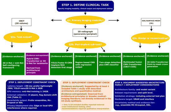
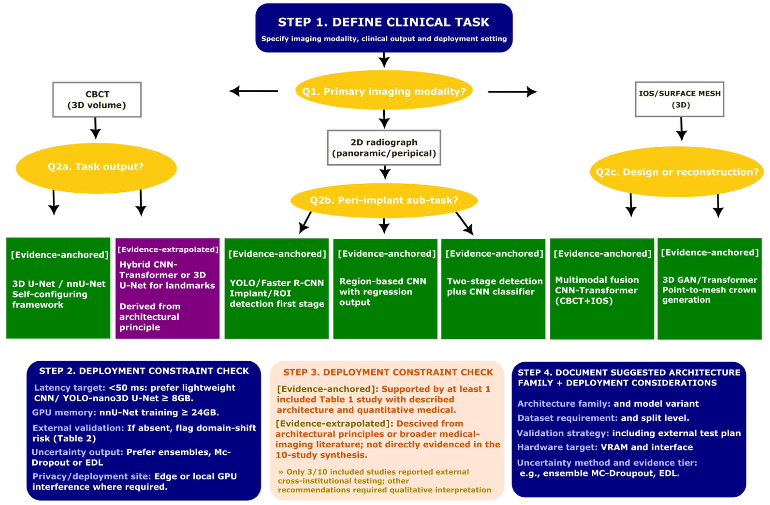
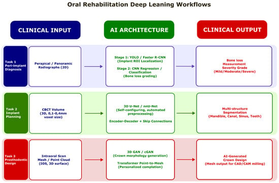
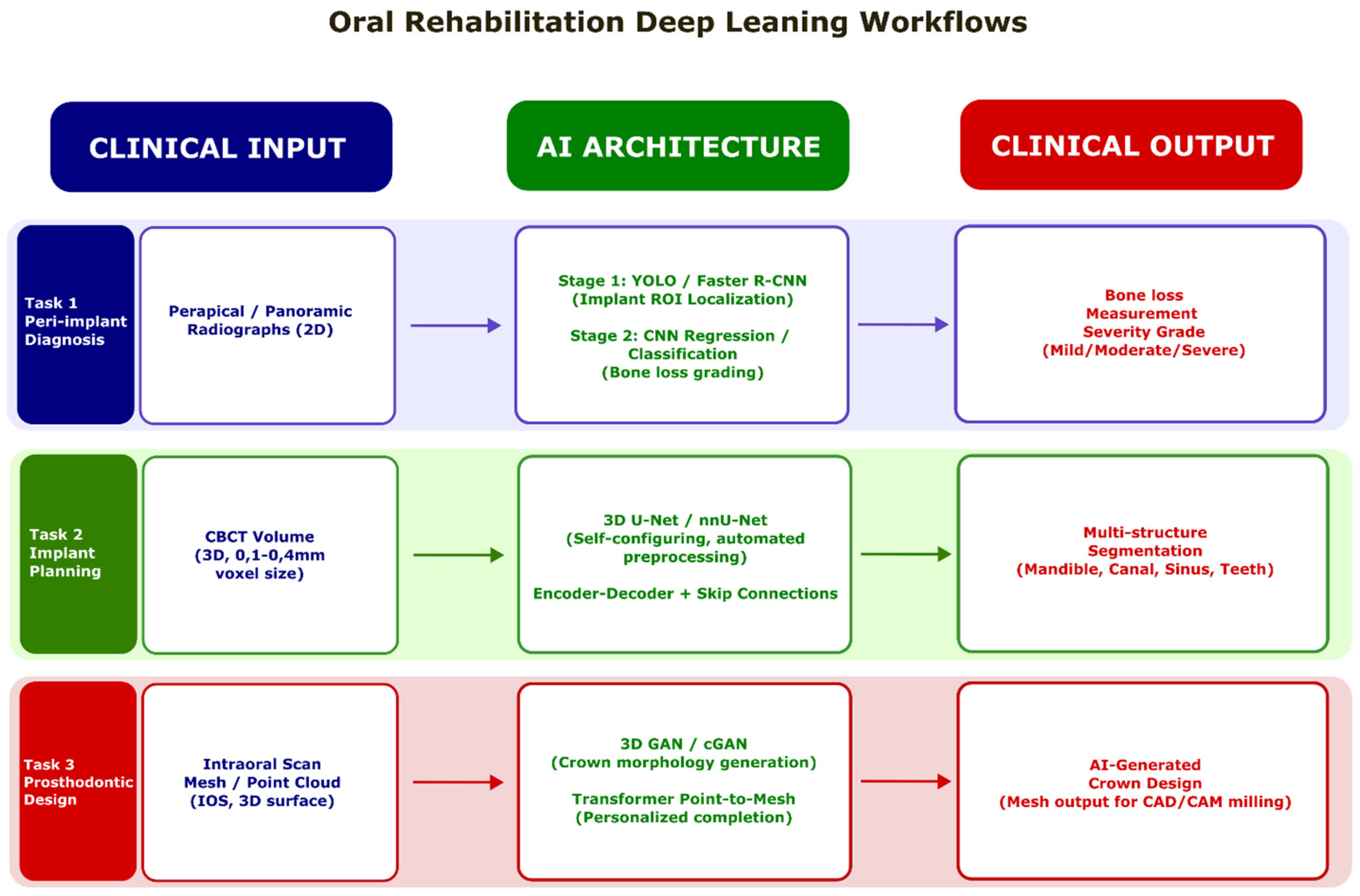

Disclaimer/Publisher’s Note: The statements, opinions and data contained in all publications are solely those of the individual author(s) and contributor(s) and not of MDPI and/or the editor(s). MDPI and/or the editor(s) disclaim responsibility for any injury to people or property resulting from any ideas, methods, instructions or products referred to in the content. |
© 2026 by the authors. Licensee MDPI, Basel, Switzerland. This article is an open access article distributed under the terms and conditions of the Creative Commons Attribution (CC BY) license.