


Abstract
The long-term antifouling performance of cholinium-based ionic liquid (IL) coatings applied to marble and tufa stone surfaces for cultural heritage protection was studied. A bilayer system consisting of a nano-silica consolidant (NanoEstel®, NE) and ILs was evaluated over a five-year period (2019–2024) under controlled environmental conditions. The coatings’ ability to prevent spontaneous microbial colonization was assessed through macroscopic observation, ImageJ analysis, light microscopy (LM), and field-emission scanning electron microscopy (FE-SEM). The results demonstrated that specific ILs, particularly 3 and 3a, exhibited sustained antifouling effectiveness, maintaining low surface coverage after five years (as low as 4.5% on tufa and 2.1% on marble). Microbiological analysis confirmed minimal fungal structures and the absence of culturable organisms on the most effective coatings. These findings highlight a clear relationship between IL lipophilicity and antifouling performance, with more lipophilic species providing better protection. Overall, these results confirm that NE/IL-based coatings offer a durable, environmentally friendly, and effective strategy for long-term protection of stone materials, supporting their application in the sustainable conservation of cultural heritage. A coating based on a cholinium IL formulation was also tested for in situ application over five years (2021–2026).
1. Introduction
Biological colonization of stone artifacts is closely related to several factors, including environmental conditions and the intrinsic properties of the surface that control its primary and secondary bioreceptivity [1]. This latter implies that an untreated stone surface will react differently from those treated with any products applied during conservation procedures when exposed to the same environmental conditions. Therefore, any specific treatment, be it biocide, water repellent, consolidant, or antifouling coating, will influence the speed and rate of the recolonization process. This aspect should always be taken into consideration, not only for protection but also to be cost-effective and avoid short-term intervention [2].
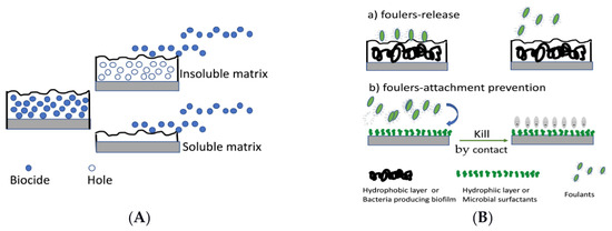
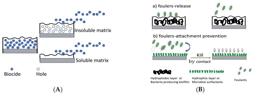
Biofilms and microbial colonization, once formed and stabilized on stone surfaces, not only affect the esthetic and structural integrity of the materials but also pose significant challenges for sustainable conservation practices [3,4,5,6]. The long-term preservation of cultural heritage, especially stone-built monuments, is a pressing problem. Antifouling coatings on inorganic surfaces represent the most challenging means for preventing the colonization process [7]. Two main classes, namely, (1) biocide-release antifouling coatings and (2) non-biocide coatings are currently known. The biocide-release coatings are based on the presence of molecules of biocides embedded in an insoluble or soluble matrix; they have, in general, a short duration (12–18 months) due to the loss of biocides released into the environment. The non-biocide-release coatings are achieved by applying chemicals, biofilm or surfactant produced by microorganisms; they can act as fouler-release by preventing the attachment of foulers or by killing the cells by contact. Their durability depends on the stability of the coating or the surfactant present on the treated surface. A schematic summary is shown in Figure 1.
Figure 1.
Graphical representation of the two main classes of antifouling coatings: (A) biocide-release coating; (B) non-biocide-release coatings.
The non-biocide-release coatings are very attractive. These are not based on biocidal properties that could selectively act on the recolonization process, but on the prevention of the attachment of colonizers on stone surfaces, inhibiting further development and diffusion [3,4].
In this context, surface-active ionic liquid (SAIL)-based coatings emerged as new materials offering environmentally friendly, durable, and effective antifouling features. ILs are low-melting-point organic salts showing a variety of tunable properties such as low vapor pressure, high thermal and chemical stability, recyclability, and so on [8]. The chemical versatility of ILs stems from their ionic nature, which allows synthetic control on their physicochemical and biological properties, and functions, through an appropriate choice of the anion/cation couple, creating theoretically infinite possibilities [3]. Although still little exploited in the context of CH stone conservation, the few reported studies show their high potential for application in this field. Eyssautier-Chuine and coworkers and Misra and coworkers [9,10] reported on ammonium-POM SAILs (POM = [α-SiW11O39]8) as antimicrobial and anticorrosive stone coatings. Recently, our group reported on the bioactivity of a series of cholinium SAILs containing a dodecylbenzenesulfonate (DBS) and/or bromide as anions [4,11,12]. Cholinium ILs were functionalized with alkyl chains of moderate lengths (specifically, 7 and 12 carbon atoms) to ensure a balance between toxicity and antimicrobial activity, also investigating the role of the anions. When applied on tufa and marble probes, previously treated with NanoEstel® (NE) as a consolidant [4], the DBS/bromide cholinium ILs demonstrated effective antibiofilm and preventive effects against new colonization over 90 days and also in a submerged environment for over 12 months of exposure [12]. The SAIL coatings have been proven to be compatible with conservation criteria, as they do not alter the colorimetric or water absorption properties of marble and limestone probes [11,12]. Furthermore, during UV weathering, the combination of nano-silica-based consolidants with ionic liquids has particularly effective results in improving coating adherence and endurance [12].
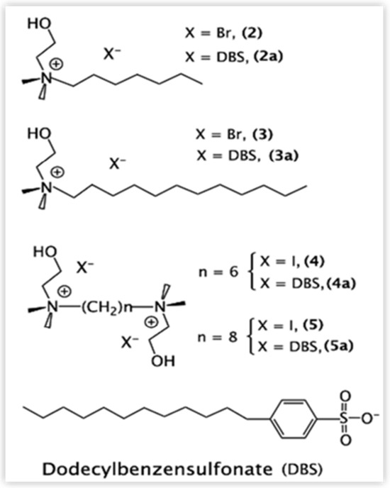
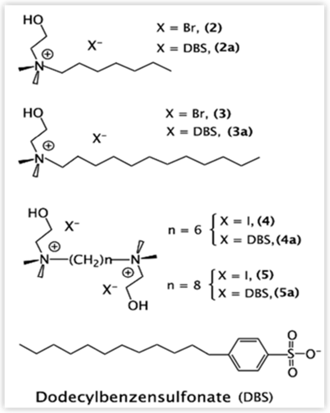
To highlight the IL-structure/bioactivity relationship of ILs, a series of mono- and di-cation cholinium ILs (Figure 2) were designed following eco-sustainable criteria [3] and their potential antifouling behavior was investigated to 90 days [4].
Figure 2.
Schematic representation of cholinium@halides (2, 3, 4 and 5) and cholinium@DBS ILs (2a, 3a, 4a and 5a), as reported by [4].
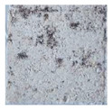
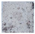
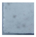
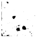
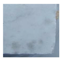
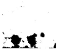
The results obtained after 90 days of exposure showed unambiguously that the mono-cholinium ILs, 2, 2a and 3, 3a featuring a medium and long chain at 7 and 12 C atoms, respectively, experienced promising AF capabilities, while the dication ones, 4a and 5a, did not [4]. It was hypothesized that a relationship exists between lipophilicity and antifouling activity, with the most lipophilic ILs, 3 and 3a, showing the best performance. In particular, 3a, featuring DBS as an anion, also exhibited a preventive function against new colonization on tufa and marble probes. The present work deals with data obtained by extending the period of exposure to spontaneous colonization up to 5 years, on the same probes described by De Leo et al. [4]. This monitoring times are considered compatible with the average time of a conservative intervention [2].
In addition to laboratory tests, we also report a five-year preliminary in situ trial on the marble part of Fontana Falconieri, in Messina, Italy. At this aim an appropriate formulation based on 3, as the most effective antimicrobial IL, and 3a, which showed a peculiar preventive antibiofilm action, was exploited as active IL.
2. Materials and Methods
2.1. Chemicals
NanoEstel®, Estel 1000, and CTS Silo111 were purchased from CTS S.r.l, Altavilla Vicentina, Italy. All the chemicals and solvents employed, unless otherwise stated, were purchased from Sigma-Aldrich Merck KGaA (Darmstadt, Germany), used as supplied. The synthesis and characterization of cholinium ionic liquids (ILs), as shown in Figure 1, namely N-(2-hydroxyethyl)-N,N-dimethyl-1-heptanaminium bromide (2), N-(2-Hydroxyethyl)-N,N-dimethyl-1-dodecanaminium bromide (3), N,N’-tetramethyl-N,N’-(dihydroxyethyl)-1,6-hexanediaminium dibromide (4) and N,N’-tetramethyl-N,N’-(dihydroxyethyl)-1,8-octanediaminium diiodide (5) and their corresponding dodecylbenzenesulfonate (DBS) derivatives, labeled with the letter a, were already reported [4].
For in situ application, a 3:3a (molar ratio 3:1) formulation at a concentration of 37.5 μmol/mL was used (see below). The synthesis was already reported in our previous work [11].
2.2. Probe Selection and Treatment
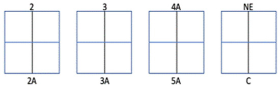
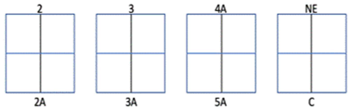
The marble (5 × 5 × 1.5 cm) and tufa (4.7 × 4.7 × 2 cm) stone probes coming from the previous experiments [4] were analyzed over 5 years (2019–2024). Briefly, coatings were prepared using a bilayer coating procedure consisting of different steps: (1) sterilized in autoclave, (2) pretreated with absolute ethanol, (3) treated with NanoEstel diluted 1:5 in sterile distilled H2O by using a sterile sponge, (4) allowed to polymerize for 3–4 days at 20 °C, as indicated in the technical data sheet of the producer (CTS S.r.l, Altavilla Vicentina, Italy), (5) sterilized by UV for 2 h and then (6) treated with ILs, 2, 2a, 3, 3a, 4a, 5a at concentration 37.5 µmol/mL in Me-OH, using the same sponge technique. Il-treated probes were compared with uncoated control probes as well as with treated with NanoEstel alone (Figure 3).
Figure 3.
Schematic representation of the distribution of the treatments on the tufa and marble stone probes divided in 4 sectors and the type of treatment is indicated.
2.3. Laboratory Test Monitoring
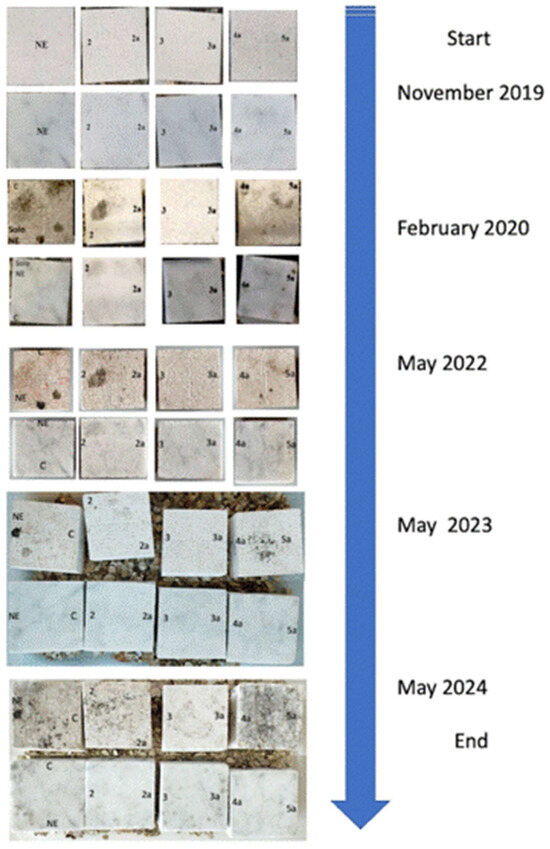
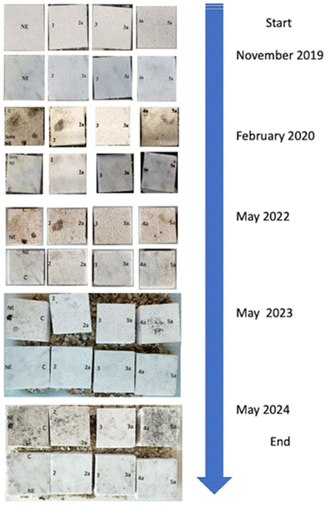
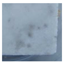
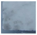
Laboratory-controlled daylight exposure of the probes was performed in a series of alternating humid and dry conditions at the temperature range of 18 °C to 30 °C. Probe surfaces were periodically monitored and images taken in February 2020, May 2022, May 2023, and May 2024 (Figure 4). Finally, the stone probes were properly treated and analyzed through light and SEM microscopy, microbiological culture and image analysis, as described below.
Figure 4.
Timeline of tufa and marble probes exposure under laboratory conditions.
2.4. Surface Colonization Analysis and Microbial Characterization
The extent of colonization was assessed by direct analysis of the probe surfaces using a portable USB Digital Microscope DM022C (Maditec, Milan, Italy) and photographic documentation. To further investigate surface colonization, samples were collected using the adhesive tape sampling method with Fungi-Tape™ (Scientific Device Laboratory Inc., Des Plaines, IL, USA). Small tape squares (5 × 5 mm) were cut and used both for direct microscopic examination and fungal isolation. For microscopic observation, tape was placed on microscope slides, and a drop of lactophenol was added and examined under a light microscope (Leica DMRE, Leica Microsystems GmbH, Heerbrugg, Switzerland) using different objectives (25×, 40×), as previously described [13]. For fungal isolation, equivalent tape squares were cut and streaked directly onto the surface of Petri dishes containing DRBC agar (Dichloran Rose Bengal Chloramphenicol, Condalab, Madrid, Spain) in duplicate. Plates were incubated at 25 °C and periodically inspected for fungal growth for up to one month. Fungal specimens were isolated by transferring them in Petri dishes containing DRBC agar (Dichloran Rose Bengal Chloramphenicol, Condalab, Madrid, Spain). Plates were incubated at 25 °C for up to one month and periodically inspected for fungal growth. Representative fungal isolates were subcultured on Potato Dextrose Agar (PDA, Condalab, Madrid, Spain) and identified based on colony macromorphology and microscopic examination of reproductive structures using both conventional microscopic preparations and slide culture techniques. Typology and diversity were determined at least to the genus level using standard taxonomic keys [14,15,16].
2.5. Image Analysis
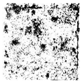
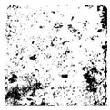
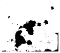
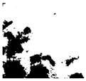
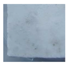
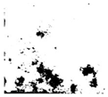
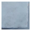
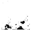
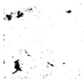
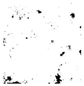
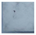
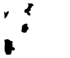
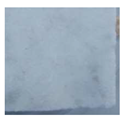
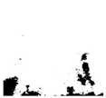
Detailed visualization and quantification of the visible colonization patterns and percentage coverage on the coated and uncoated surfaces were taken by using a portable USB Digital Microscope DM022C (Maditec, MI, Italy) taking images of dimension of 2.5 × 2.5 cm and 2.35 × 2.35 cm for tufa and marble, respectively), corresponding to the area of each treatment, as shown in Figure 4. The images obtained were analyzed by ImageJ 1.46r software, NIH, Bethesda, MD, USA.
2.6. Field-Emission Scanning Electron Microscopy (FE-SEM)
The probes collected in May 2024 were cut in small portions (less than 0.5 cm per side) and prepared for SEM analysis by gently brushing the surface to remove debris from the cutting procedure. The probes were kept in glutaraldehyde phosphate buffer 0.2 M (pH 7.2) for 12 h and then dehydrated by treatment in a series of water/alcohol solutions (30–50–70%-absolute). Finally, the samples were coated with graphite and observed under a Zeiss Merlin Field FE-SEM (Carl Zeiss AG, Oberkochen, Germany) at the Microanalysis laboratory of the MIFT department of the University of Messina.
2.7. In Situ Application
Our previous studies [4] showed that, while the 3 IL, N-(2-HYdroxyethyl)-N,N-Dimethyl-1-DodecanaminiumBromide, was the most effective antimicrobial IL, 3a, featuring the lipophilic dodecyl benzensulfonate (DBS) as an anion, also displayed a preventive antibiofilm action. These findings suggested to us that a combination of 3 and 3a, coupling the antibacterial properties of 3 to the antibiolfilm activity of 3a, could exert a double function when combined.
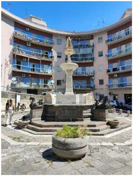
With this in mind, before the trial application on the fountain, a possible combined action of the two ionic liquids 3 and 3a was evaluated. The antimicrobial activity was tested against a representative panel of strains isolated from biodeteriorated surfaces: two bacterial strains (Gram+ and Gram-), a hyphomycete, a yeast, and a eukaryotic unicellular alga using the agar diffusion assay (methodology and results are reported in the Supplemental File S1). Based on MBC results, the 3:3a (3:1 molar ratio) IL combination at a concentration of 37.5 μmol/mL was chosen for in situ tests. These were performed on specific areas of Fontana Falconieri in Messina, Italy (Figure 5 and Figure 6).
Figure 5.
Falconieri fountain after restoration in 2021.
Figure 6.
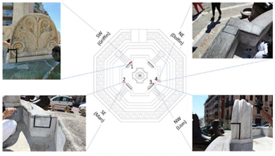
The formulation IL 3/3a was applied in small areas delimited by black tape. The stone surface was treated with the consolidant Estel 1000 on the outer part and with Silo 111 for the inner part of the fountain in contact with water.
The fountain was built in 1843 by the architect Carlo Falconieri for the town of Messina. In 2021, it was restored under the supervision of Dr. Luigi Giacobbe of Superindence of Messina, Italy.
As reported by the restorers of Project srl (Brolo, Messina), stone consolidation was carried out by application of CTS Estel 1000 (in the outer part of the fountain) and by CTS Silo111 (in the inner part of the fountain in contact with water). In agreement with the restorers, four areas of the fountain were chosen, as shown in Figure 6. A specific formulation of 3 and 3a, in a 3:1 molar ratio [8], was then applied by brush.
2.8. In Situ Monitoring
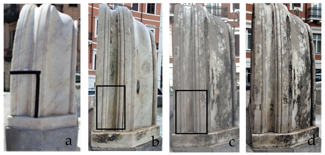
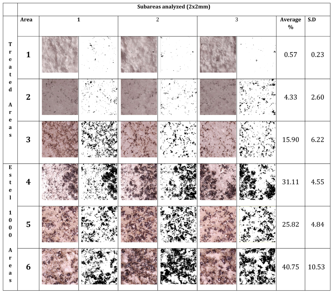
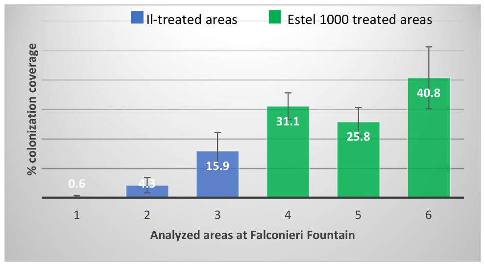
The treated areas were monitored and photographic documentation was taken in June 2023 and June 2026. In 2026, quantification of colonization coverage on the marble item under observation (Figure 6, NW) of the Falconieri Fountain (Messina) was done by using a portable USB Digital Microscope DM022C (Maditec, MI, Italy). Six areas were analyzed (Figure 7). One to three were representative of the IL-treated areas, while four to six represented the areas treated only with Estel 1000. For each area, squares (2 × 2 mm) in triplicate were considered and analyzed via ImageJ (version 1.54), to visualize and calculate the percentage area of coverage. The dataset was processed in Microsoft Excel (Microsoft Corp., Redmond, WA, USA) to determine the arithmetic mean and sample standard deviation (SD) for each site. Data distribution was then visualized using a clustered column histogram, where the statistical variability across the zones was illustrated by standard deviation error bars (±S.D) to each mean value.
Figure 7.
IL-treated areas of the Falconieri fountain (north–east/lion) are indicated by the rectangular forms. (a) Immediately after the treatment in July 2021; (b) after 2 years (June 2023); (c) after 5 years (June 2026); (d) areas examined with a USB Digital Microscope DM022C (Maditec, MI, Italy): 1 to 3 (treated with Ils); 4 to 6 treated only with Estel 1000). The choice of the areas, close to each other, was determined in order to reduce other side-effects due to a different material (such, e.g., marble versus Pietra serena stone) or orientation (such as wind direction, water and sun exposure).
3. Results and Discussion
3.1. Monitoring of Tufa and Marble Stone Probes
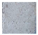
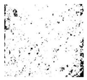
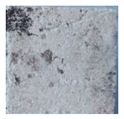
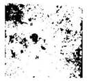
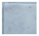
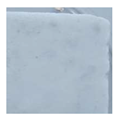
After 5 years of exposure, the macroscopic examination (Figure 4) of the stone probes revealed a similar pattern of colonization to that observed after 90 days (2019 to 2020). Samples 3 and 3a, along the years of monitoring, showed a better performance and resilience against spontaneous colonization as compared to 2 and 2a, 4a and 5a, untreated probes, and those coated with the consolidant only, exposed in the same environmental controlled conditions. Hence, the different pattern observed between untreated and treated surfaces is attributable to the different surface bioreceptivity and not to the environmental conditions. Again, the tufa stone surfaces resulted in being more affected to spontaneous colonization than marble ones (Figure 4 and Table 1 and Table 2).
Table 1.
Image J analysis carried out on tufa probes subjected to a spontaneous colonization after 5 years of exposure (May 2024). The percentage of coverage, the average and the standard deviation (SD) are reported.
Table 2.
Image J analysis carried out on marble probes subjected to a spontaneous colonization after 5 years of exposure (May 2024). The percentage of coverage, the average and the standard deviation (SD) are reported.
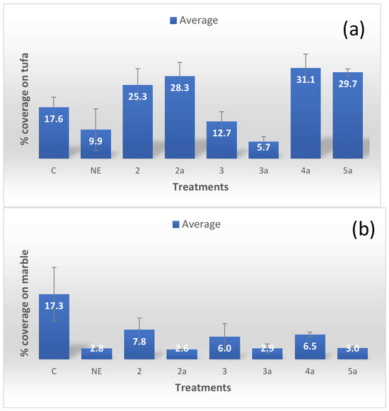
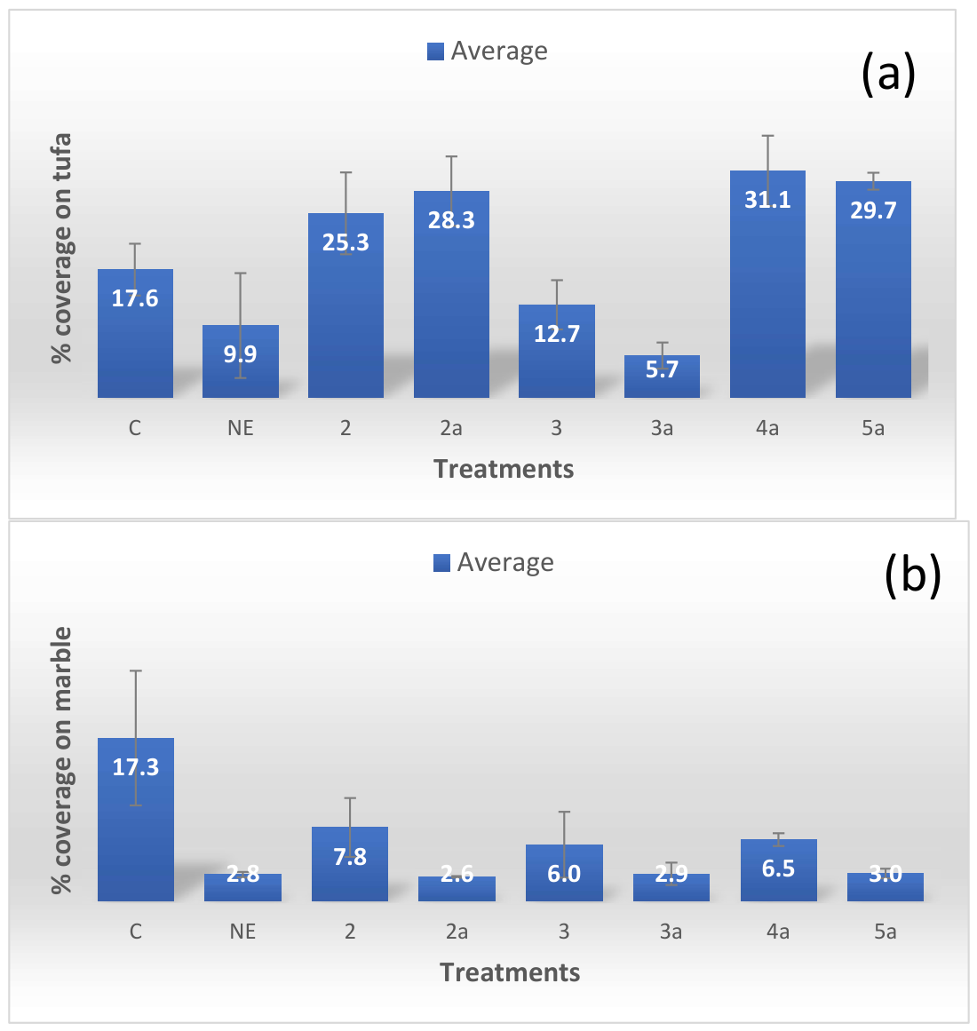
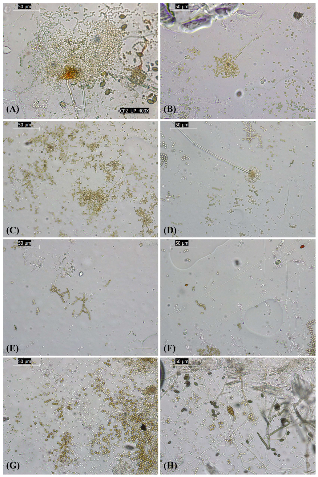
The probe treated with IL 3 displayed a degree of coverage similar to the untreated probes (C) (15.1 and 12,7% against 15 and 17.6, respectively) (Table 1 and Figure 8a), but although the coverage looks similar, the microscopic analysis shows significant difference between only spores in the 3-treated probes (Figure 9E), while an active growth is visible for untreated probes (Figure 9A). Probe IL 3a -coated tufa probes displayed a remarkable reduction in colonization, showing relatively clean surfaces (Figure 9F), while for 2 and 2a, it is interesting to note that, up to June 2023, their performance was comparable to those of 3 and 3a; then, in 2024, a widespread colonization was observed (Figure 4 and Figure 9C,D) with a similar coverage percentage (Table 1 and Figure 8a), even higher (21.3 and 25.3%) than the control (C). It is noteworthy that 4a and 5a (average 31.1 and 29.7%) exhibited an even higher bioreceptivity than the untreated tufa samples (average 17.6). Although less evident, the same trend was observed on marble probes (Table 2 and Figure 8b and Figure 10), with the IL products, 3 and 3a, preventing colonization more effectively than 2 and 2a.
Figure 8.
Graphical representation of the behavior of ILs on tufa (a) and on marble (b) as compared to the controls (C) and treated with only consolidant NE.
Figure 9.
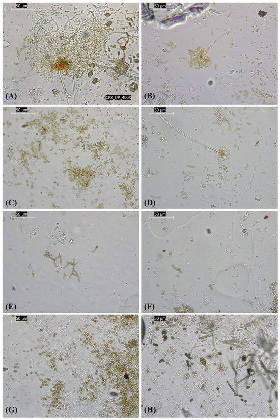
Fungal colonization of the tufa probe surfaces seen under light microscopy; adhesive tape samples offer the exact mirror image of the sampled surface as described by Urzì and Albertano [13]. Bar is 50 μm (A) C, untreated control; (B) NE, NanoEstel-treated control; (C) IL 2; (D) IL 2a; (E) IL 3; (F) IL 3a; (G) IL 4a; (H) IL 5a.Bar is 50 µm.
Figure 10.
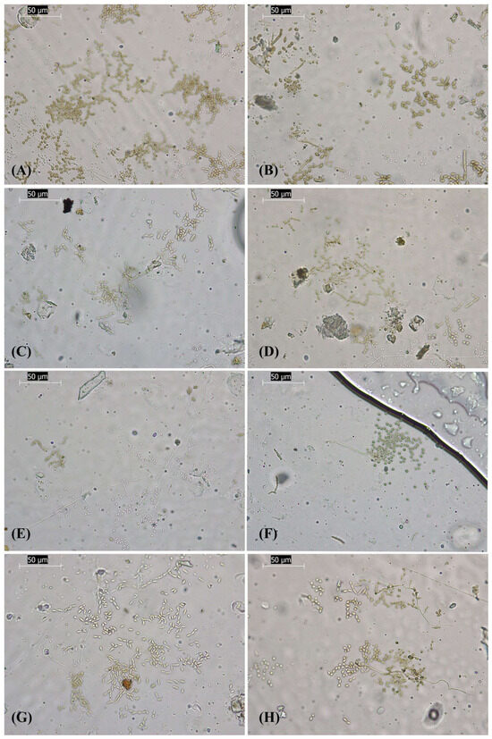
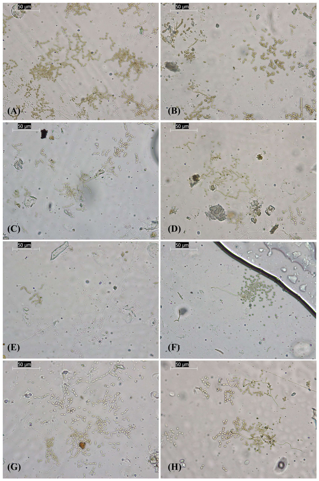
Fungal colonization of the marble probe surfaces seen under light microscopy; adhesive tape samples offer the exact mirror image of the sampled surface as described by Urzì and Albertano [13]. Bar is 50 μm. (A) C, untreated control; (B) NE, NanoEstel-treated control; (C) IL 2; (D) IL 2a; (E) IL 3; (F) IL 3a; (G) IL 4a; (H) IL 5a. Bar is 50 µm.
The results clearly demonstrate that the IL-based coatings, particularly 3 and 3a, were effective in preventing spontaneous microbial colonization over the five-year period, highlighting the potential of IL-based coatings as a long-term solution for protecting stone surfaces from microbial deterioration (Table 1, Figure 9A). It is worth noting that, for tufa probes treated with Il 2 and 2a, during the different years of monitoring (2020 to 2023), their macroscopic appearance was quite similar to the probes treated with IL 3 and 3a; in 2024, however, a high % of coverage was observed macroscopically and confirmed microscopically (Figure 4, Figure 9 and Figure 10, Table 1). Our hypothesis is that the IL 2 and 2a have a shorter performance time than 3 and 3a. As regarding marble probes, due to the lower porosity respect tufa, we observed a very scarce visible colonization, mainly in localized spots. This is particularly evident in the untreated marble surfaces, while only NanoEstel coatings behaved similarly to the surface treated with 2a, 3a, and 5a. Thus, for marble, we did not see any significantly better behavior of IL 3a than consolidant, IL 2a and even 5a. However, bare marble surfaces without any treatment (Table 2 and Figure 8b) showed a more marked colonization than the other marble probes.
3.2. Microbiological Survey
Microscopic investigations and cultural analysis of samples obtained by adhesive tape technique showed that fungi tend to colonize all the surfaces examined. Utilizing a multistep approach based on microscopy (portable microscope and light microscopy) and cultural analysis, it provided coherent complementary information on the occurrence of fungi on the stone surfaces. Fungal colonization was exclusive on probes kept in laboratory conditions, where no phototrophic microorganisms were observed. Bacteria probably present were not considered relevant to the survey.
Based on LM observations, several fungal structures were observed on the colonized probes. As shown in Figure 9, adhesive tape samples collected from tufa probes revealed a higher abundance of fungal elements than those collected from marble probes (Figure 10), confirming the greater susceptibility of tufa to microbial colonization. In the tufa samples, C and NE probes (Figure 9A,B) displayed abundant fungal material, including hyphal fragments, different types of conidia and conidiophore-like structures. Tufa probes treated with ILs 2 and 2a (Figure 9C,D) contained fewer fungal elements than the controls, although fungal propagules and occasional reproductive structures were still present. Notably, an Aspergillus-like conidiophore was observed in the probe treated with IL 2a (Figure 9D). Similar to C and NE controls, tufa probes treated with ILs 4a and 5a (Figure 9G,H) were characterized by abundant fungal propagules and reproductive structures. In particular, sample 5a (Figure 9H) contained pigmented muriform conidia resembling Alternaria-type spores. In contrast, treatments 3 and 3a (Figure 9E,F) contained only occasional fungal structures and fragmented fungal elements, while most of the observed material consisted of dirt debris and environmental particles.
A similar pattern of colonization was observed on marble probes (Figure 10), although fungal elements were generally less abundant than on tufa. C and NE probes (Figure 10A,B) were characterized by the presence of hyphal fragments and melanized ovoid conidia morphologically compatible with Cladosporium-like fungi. Marble probes treated with ILs 2 and 2a (Figure 10C,D) contained only sparse fungal propagules. Similar fungal elements were also present on marble probes treated with ILs 4a and 5a (Figure 10G,H). In contrast, treatments 3 and 3a (Figure 10E,F) contained only occasional fungal structures and fragmented fungal elements, while most of the observed material consisted of environmental debris particles.
The observation of occasional fungal structures in 3 and 3a samples, despite the absence of fungal growth in culture, suggests that these elements may represent non-viable fungal remnants, dormant propagules, or structures not recoverable under the culture conditions adopted [14].
Cultural analysis of adhesive tapes taken from the controls (C and NE) revealed the presence of species belonging to genera of Alternaria, Cladosporium, Penicillium, Aspergillus, as well as not yet identified hyaline and black melanized fungi.
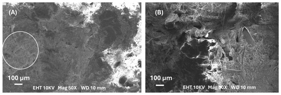
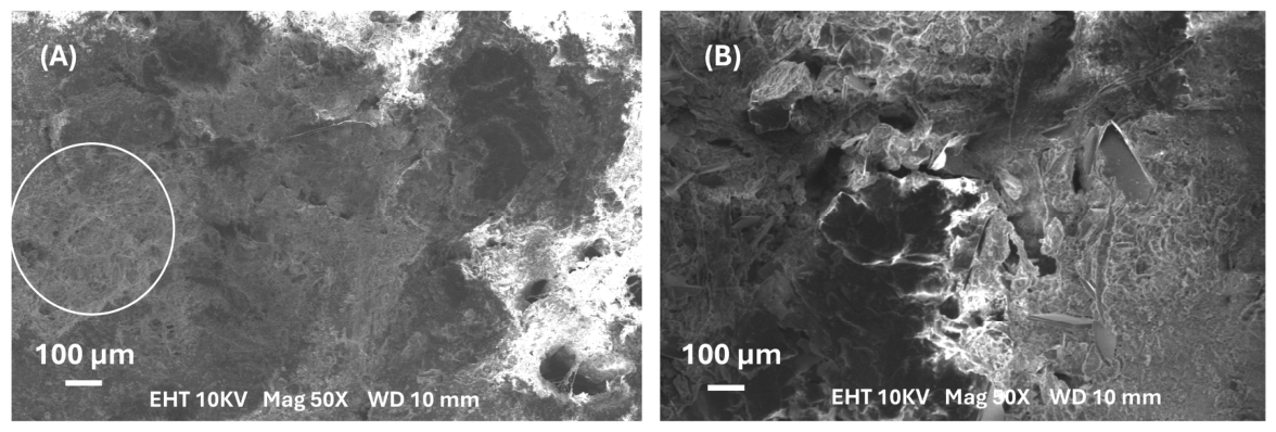
The macro- and microscopic observations were confirmed also by SEM analysis, as shown in Figure 11, where NE-treated tufa probes resulted in being heavily colonized (Figure 11A), while probes treated with the bilayer NE plus Il 3a did not show any evident microbial structure (Figure 11B).
Figure 11.
Comparative behavior of NE-treated tufa surface versus that treated with NE/3a observed by SEM microscopy. On the left, (A) a network of hyphae (as indicated by the white circle) is visible across most of the examined surface, while on the right, (B) (bilayer NE and 3a), no microbial structures are evidenced on the probe surface.
3.3. In Situ Performance
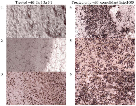
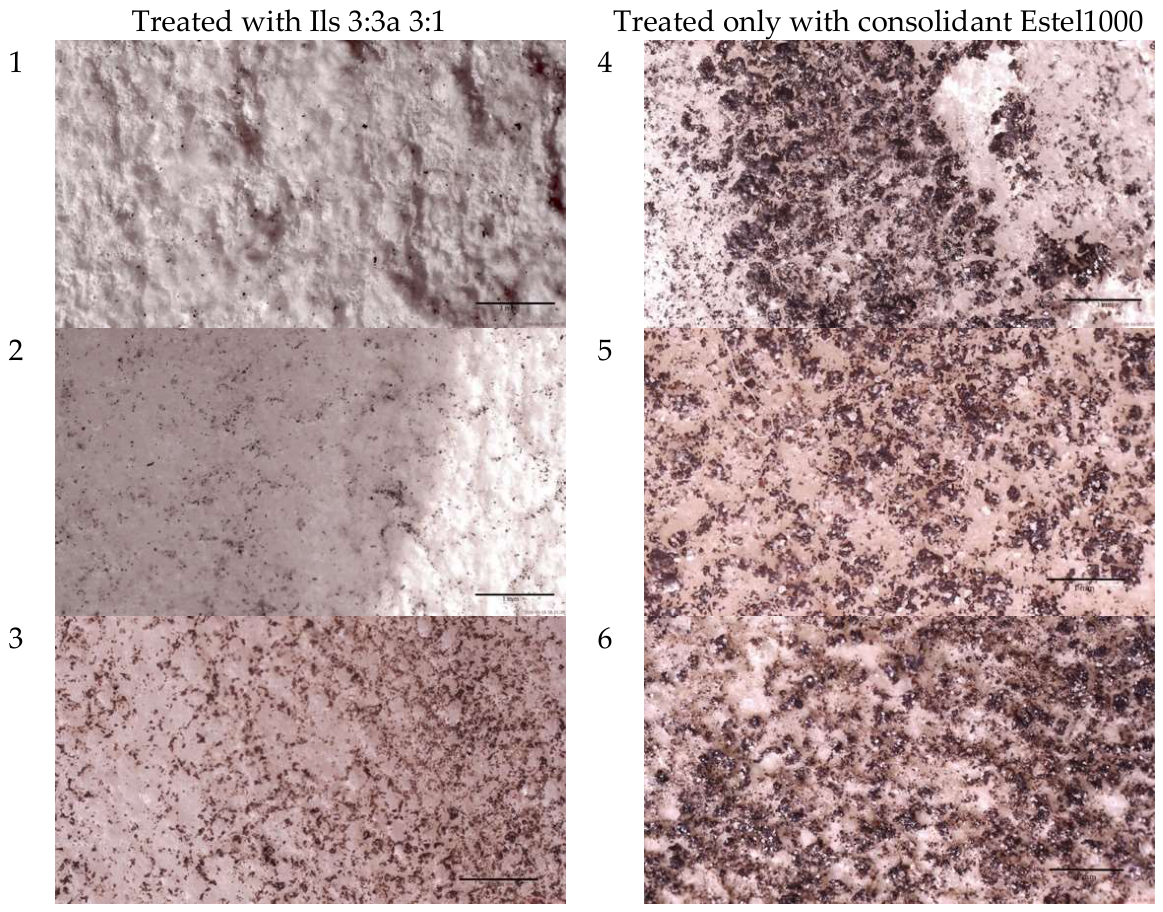
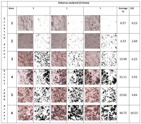
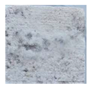
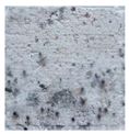
In situ monitoring was carried out over 5 years (July 2021–June 2023–June 2026) with a special focus on the marble item situated on the north–west side of the fountain. In June 2023, no visible colonization was observed (Figure 7b), while in June 2026, colonization was evidenced in specific areas, consistent with those treated with only the consolidant, while in the areas treated with ILs, the colonization was significantly reduced. Observation via a portable digital microscope (Figure 12) and ImageJ analysis (Figure 13 and Figure 14) clearly showed a great difference existing between the two groups of areas. It is noteworthy that, in the areas where the restoration intervention was carried out with only a cleaning procedure followed by the application of Estel 1000 alone as consolidant, a heavy black colonization was observed (Figure 7d) in sampling areas 4 to 6 (Figure 12 and Figure 13).
Figure 12.
Images taken with a digital microscope at 1× magnification. Treated areas: 1, 2, 3. Areas treated only with consolidant: 4, 5, 6. Bar is 1 mm.
Figure 13.
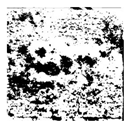
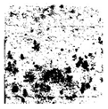
The average value of coverage obtained by Image J analysis of three subareas for each area considered shown in Figure 12; areas 1 to 3 treated with Ils 3:3a and areas 4 to 6 treated only with Estel 1000, as well standard deviation (SD) of each subarea.
Figure 14.
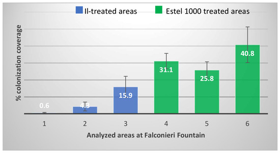
Graphical representation of the behavior after 5 years from restoration of IL-treated areas compared to those treated with Estel 100 alone on the marble relief of Falconieri fountain.
Both microscopy images (Figure 12) and ImageJ analysis and graphical representation (Figure 13 and Figure 14) show a marked difference in colonization coverage between the two groups.
The graphical representation (Figure 14) gives a clear idea of the differences observed in the two groups examined (Il-treated versus Estel 1000 alone).
Visive analysis should be carried out as well as a continuous monitoring. However, from these preliminary in situ results, we can affirm that SA-cholinium ILs were able to reduce the rate of disfigurement of the surface due to the growth of microorganisms. Our data, focused on a small part of the fountain, confirm antimicrobial/AF effectiveness, even in a real-world environment such as a fountain, where the stones are also subjected to the continuous action of diversified environmental factors such as water, rain, wind, sun, etc.
4. Conclusions
The long-term antifouling performance of bilayer NanoEstel/IL-based coatings was assessed for a period of 5 years in laboratory conditions, being a continuation of the conditions that were established for the 90-day monitoring, as previously published [4].
At that time, we evaluated the role of surfactant ionic liquids designed to be Safe by Design materials and matched the green conservation requirements. Therefore, they can be safely used in the field of conservation of cultural heritage. We applied them as a bilayer in combination with a commercial consolidant, NanoEstel, commonly used for conservation purposes. In other studies [11,12], we demonstrate that IL solution could be applied in combination with other consolidants or water repellents without losing their antifouling characteristics. In this study, we could confirm that the mixture of IL 3 and 3a provides long-term performance both in laboratory as well as in situ conditions.
The methodology adopted to evaluate the long-term performance of these coatings to prevent colonization and consequent biodeterioration has mainly involved (i) stone characterization and properties, as previously reported by [11,12], (ii) regular monitoring of the spontaneous colonization at short term (3 months) and long term (5 years) through visual and microscopic analysis, and (iii) a conventional microscopic and cultural characterization (macromorphological and micromorphological characteristics of the main colonizers and their reproductive structures, typology and diversity). This approach may be considered an easy protocol to be applied even in in situ monitoring and could be implemented with other analysis, as suggested by [17]. After this period of exposure, the 3 and 3a IL-based coatings displayed effective antifouling properties against spontaneous colonization, implying good potential for long-lasting application [18]. It is noteworthy that, in laboratory conditions, 3 and 3a avoided the growth of fungi, contrasting their settlement and propagation on the surface. It is well known that fungi, especially in dry environments, act as pioneering colonizers of stone substrates [19,20,21], due to their oligotrophic lifestyle and remarkable resistance to environmental stress. Their settlement may promote both esthetic and structural deterioration phenomena through discoloration, hyphal penetration into the substrate, and secretion of organic acids capable of inducing mineral dissolution and progressive stone decay.
Ultimately, it is to underline that these coatings were designed according to safe criteria [4], as they do not contain harmful components, are easy to apply, and do not affect the stone surface integrity [22]. Therefore, they are safe for operators and the environment and may represent a promising alternative AF sustainable solution for stone conservation.
The results reported here allow us to assert that the NE/ILs bilayer coatings have a longer durability than the consolidant applied alone, both in the laboratory and the in situ trial, maintaining reliable protection against biofouling for more than five years. Despite their inherently uneconomical manufacturing method, the continuous effectiveness of IL-based coatings makes them durable and unique for long-term stone protection within heritage conservation and construction applications. The resulting extended durability and improved antifouling characteristics indicate that cholinium IL coatings can be considered an ideal coating option for protecting porous substrates, such as tufa and marble, over long periods of time, preventing biological growth and degradation.
Five-year in situ test findings support that surfactant cholinium ILs preserve their antimicrobial/AF activity even under real and stressful environmental conditions, allowing for IL technology to envisage a great potential even in the context of stone asset restoration. By exploiting the large library of surfactant cations and anions available, it is possible to design a large variety of IL couples and formulations which meet eco-sustainable criteria. We believe that our pioneering studies may be stimulating for further research on the application of ILs in the field of stone conservation.
Supplementary Materials
The following supporting information can be downloaded at: https://www.mdpi.com/article/10.3390/app16146945/s1, File S1. Evaluation of antimicrobial action of 3 and 3a IL mixtures.
Author Contributions
Conceptualization, S.L.S. and C.E.U.; methodology, S.L.S., C.E.U. and F.D.L.; software, G.A. and R.H.A.; validation, S.L.S. and C.E.U.; formal analysis, S.L.S., E.F. and C.E.U.; investigation, R.H.A. and G.A.; resources, F.D.L., C.E.U. and S.L.S.; data curation, F.D.L., C.E.U., S.L.S. and E.F.; writing—original draft preparation, R.H.A., C.E.U. and S.L.S.; writing—review and editing, R.H.A., C.E.U. and S.L.S.; visualization, S.L.S. and C.E.U.; supervision, C.E.U.; project administration, C.E.U., S.L.S. and F.D.L.; funding acquisition, F.D.L. All authors have read and agreed to the published version of the manuscript.
Funding
This research received partial support from the BIO4ART Project (CUP B53C22004010006) Project PE0000020 CHANGES (CUP B53C22003780006), NRP Mission 4 Component 2 Investment 1.3, funded by the European Union–NextGenerationEU.
Institutional Review Board Statement
Not applicable.
Informed Consent Statement
Not applicable.
Data Availability Statement
The datasets generated and analyzed during the current study are available from the corresponding authors on reasonable request.
Acknowledgments
The authors are grateful to Rosamaria Soldano and Alessio Lena (holders of BIO4ART scholarship) and Angelo Cunto for their valuable technical support. The director of restoration work of the Falconieri fountain dott Luigi Giacobbe of Superintendence of Messina, Italy and the restorer team of Project s.r.l., Brolo, Messina are deeply thanked. The authors also acknowledge the BIO4ART Project (CUP B53C22004010006) Project PE0000020 CHANGES (CUP B53C22003780006), NRP Mission 4 Component 2 Investment 1.3, funded by the European Union–NextGeneration EU. R.H.A. carried out his doctorate work with a scholarship in the frame of RISORSE PON “RICERCA E INNOVAZIONE” 2014–2020.
Conflicts of Interest
The authors declare no conflicts of interest.
Abbreviations
The following abbreviations are used in this manuscript:
| ILs | Ionic liquids |
| SAILs | Surface-active ionic liquids |
| NE | NanoEstel® (Nano-silica consolidant) |
| DBS | Dodecylbenzenesulfonate |
| AF | Antifouling |
References
- Guillitte, O. Bioreceptivity: A new concept for building ecology studies. Sci. Total Environ. 1995, 167, 215–220. [Google Scholar] [CrossRef] [Scilit]
- Pinna, D. Coping with Biological Growth on Stone Heritage Objects: Methods, Products, Applications, and Perspectives; Apple Academic Press: Toronto, ON, Canada, 2017. [Google Scholar] [CrossRef] [Scilit]
- Lo Schiavo, S.; De Leo, F.; Urzì, C. Present and future perspectives for biocides and antifouling products for stone-built cultural heritage: Ionic liquids as a challenging alternative. Appl. Sci. 2020, 10, 6568. [Google Scholar] [CrossRef] [Scilit]
- De Leo, F.; Marchetta, A.; Capillo, G.; Germanà, A.; Primerano, P.; Schiavo, S.L.; Urzì, C. Surface active ionic liquids based coatings as subaerial anti-biofilms for stone built cultural heritage. Coatings 2021, 11, 26. [Google Scholar] [CrossRef] [Scilit]
- Liu, X.; Koestler, R.J.; Warscheid, T.; Katayama, Y.; Gu, J.D. Microbial deterioration and sustainable conservation of stone monuments and buildings. Nat. Sustain. 2020, 3, 991–1004. [Google Scholar] [CrossRef] [Scilit]
- Ruffolo, S.A.; La Russa, M.F. Nanostructured coatings for stone protection: An overview. Front. Mater. 2019, 6, 147. [Google Scholar] [CrossRef] [Scilit]
- Shi, Q.; Zhao, T.; Yang, T.; Liao, L.; Dai, W.; Zhou, X.; Qin, X.; Dong, Y.; Han, W. Recent Advances in the Research and Application of Protective Composites for Stone Surfaces. Materials 2026, 19, 1545. [Google Scholar] [CrossRef] [Scilit] [PubMed]
- Welton, T. Ionic liquids: A brief history. Biophys. Rev. 2018, 10, 691–706. [Google Scholar] [CrossRef] [Scilit] [PubMed]
- Eyssautier-Chuine, S.; Franco-Castillo, I.; Misra, A.; Hubert, J.; Vaillant-Gaveau, N.; Streb, C.; Mitchell, S.G. Evaluating the durability and performance of polyoxometalate-ionic liquid coatings on calcareous stones: Preventing biocolonisation in outdoor environments. Sci. Total Environ. 2023, 884, 163739. [Google Scholar] [CrossRef] [Scilit] [PubMed]
- Misra, A.; Franco-Castillo, I.; Müller, D.P.; González, C.; Eyssautier-Chuine, S.; Ziegler, A.; de la Fuente, J.M.; Mitchell, S.G.; Streb, C. Polyoxometalate-ionic liquids (POM-ILs) as anticorrosion and antibacterial coatings for natural stones. Angew. Chem. Int. Ed. 2018, 57, 14926–14930. [Google Scholar] [CrossRef] [Scilit] [PubMed]
- Luci, M.; De Leo, F.; De Pascale, D.; Galasso, C.; La Russa, M.F.; Lo Schiavo, S.; Ricca, M.; Ruffolo, S.A.; Ruocco, N.; Urzì, C. Surface-active ionic-liquid-based coatings as anti-biofilms for stone: An evaluation of their physical properties. Coatings 2023, 13, 1669. [Google Scholar] [CrossRef] [Scilit]
- Luci, M.; De Leo, F.; Mutalipassi, M.; Romeo, T.; Greco, S.; Giommi, C.; Evola, L.; La Russa, M.F.; Ricca, M.; De Pascale, D.; et al. Underwater performance of eco-friendly choline-based ionic liquid coatings applied on stone surfaces. Coatings 2026, 16, 136. [Google Scholar] [CrossRef] [Scilit]
- Urzì, C.; Albertano, P. Studying phototrophic and heterotrophic microbial communities on stone monuments. In Methods in Enzymology; Academic Press: San Diego, CA, USA, 2001; Volume 336, pp. 340–355. [Google Scholar] [CrossRef] [Scilit] [PubMed]
- Mueller, G.M.; Bills, G.F.; Foster, M.S. Biodiversity of Fungi: Inventory and Monitoring Methods; Elsevier Academic Press: Burlington, MA, USA, 2004. [Google Scholar] [CrossRef] [Scilit]
- Samson, R.A.; Houbraken, J.; Thrane, U.; Frisvad, J.C.; Andersen, B. Food and Indoor Fungi; CBS-KNAW Fungal Biodiversity Centre: Utrecht, The Netherlands, 2010; ISBN 9789070351823. Available online: https://books.google.it/books?id=Az6-ZwEACAAJ&redir_esc=y (accessed on 22 June 2026).
- Pitt, J.I.; Hocking, A.D. Fungi and Food Spoilage, 3rd ed.; Springer: New York, NY, USA, 2009; Available online: https://link.springer.com/book/10.1007/978-3-030-85640-3 (accessed on 22 June 2026).
- Szulc, J.; Komar, M.; Kata, I.; Szafran, K.; Gutarowska, B. Novel method for assessing the protection lifetime of building coatings against fungi. Coatings 2023, 13, 2026. [Google Scholar] [CrossRef] [Scilit]
- Bartoli, F.; Hosseini, Z.; Graziani, V.; Zuena, M.; Venettacci, C.; Della Ventura, G.; Tortora, L.; Sodo, A.; Caneva, G. In situ evaluation of new silica nanosystems as long-lasting methods to prevent stone monument biodeterioration. Coatings 2024, 14, 163. [Google Scholar] [CrossRef] [Scilit]
- De Leo, F.; Marchetta, A.; Urzì, C. Black fungi on stone-built heritage: Current knowledge and future outlook. Appl. Sci. 2022, 12, 3969. [Google Scholar] [CrossRef] [Scilit]
- Watanabe, T. Pictorial Atlas of Soil and Seed Fungi: Morphologies of Cultured Fungi and Key to Species, 3rd ed.; CRC Press: Boca Raton, FL, USA, 2010. [Google Scholar] [CrossRef] [Scilit]
- Gadd, G.M.; Fomina, M.; Pinzari, F. Fungal biodeterioration and preservation of cultural heritage, artwork, and historical artifacts: Extremophily and adaptation. Microbiol. Mol. Biol. Rev. 2024, 88, e00200-22. [Google Scholar] [CrossRef] [Scilit] [PubMed]
- Ou, Y.X.; Wang, H.Q.; Ouyang, X.; Zhao, Y.Y.; Zhou, Q.; Luo, C.W.; Zhang, S. Recent advances and strategies for high-performance coatings. Prog. Mater. Sci. 2023, 136, 101125. [Google Scholar] [CrossRef] [Scilit]

Disclaimer/Publisher’s Note: The statements, opinions and data contained in all publications are solely those of the individual author(s) and contributor(s) and not of MDPI and/or the editor(s). MDPI and/or the editor(s) disclaim responsibility for any injury to people or property resulting from any ideas, methods, instructions or products referred to in the content. |
© 2026 by the authors. Licensee MDPI, Basel, Switzerland. This article is an open access article distributed under the terms and conditions of the Creative Commons Attribution (CC BY) license.