Abstract
Humans spend a substantial proportion of their time indoors, where exposure to environmental pollutants such as radon gas and particulate contaminants in household dust is common. While radon is a well-established risk factor for lung cancer, household dust may serve as a reservoir for a complex mixture of indoor and outdoor pollutants. However, the biological effects of such exposures, particularly under sequential conditions, remain incompletely understood. This study aimed to investigate the cytotoxic and genotoxic effects of sequential exposure to household dust extract followed by indoor radon using human lung adenocarcinoma (A549) cells as an in vitro model. Household dust samples from upper northern Thailand were extracted and applied to cells, followed by controlled radon exposure. Cellular responses were evaluated using cell viability assays, cytokinesis-block micronucleus (MN) formation assays, and Western blot analysis of oxidative stress-related (Nrf2/HO-1), DNA damage-related (γ-H2AX), autophagy-related (LC3), and inflammatory-related (IL-6) protein expression. Exposure to household dust extract was associated with reduced cell viability and increased MN formation, while radon exposure alone produced relatively modest effects under the present conditions. Sequential exposure to household dust extract followed by indoor radon was associated with increased oxidative stress-related responses and elevated DNA damage than either treatment alone under the present experimental conditions. A trend toward autophagy-related responses was also observed, and the overall findings may indicate possible combined biological responses under sequential exposure conditions. These findings suggest that sequential exposure may be associated with changes in oxidative stress-related pathways, DNA damage responses, and autophagy-related processes in this in vitro model. However, the results should be interpreted with caution as they are derived from a single cancer cell line and there are limitations to the in vitro exposure model. Further studies using additional cell models and in vivo systems are warranted to further clarify the potential biological and human health relevance of these findings.
1. Introduction
Humans spend a substantial proportion of their time in indoor environments, often exceeding 80–90% of the day, particularly during daily living and occupational activities [1]. Therefore, exposure to indoor environmental pollutants, such as radon and household dust, may have important implications for human health, including an increased risk of lung cancer (LC), particularly in upper northern Thailand (UNT) where severe air pollution and high indoor radon levels have frequently been reported [2,3,4].
Radon (222Rn) is a naturally occurring radioactive gas formed during the radioactive decay of uranium (238U) in soil and rocks and is widely recognized as the second leading cause of LC after cigarette smoking [5]. It can accumulate in enclosed indoor environments, particularly in lower-level areas such as underground rooms, ground-floor premises, and bathrooms, due to its relatively high density and continuous entry from soil and groundwater. Its colorless and odorless nature makes it difficult to detect, potentially leading to prolonged exposure among residents [6]. Previous studies have indicated that indoor radon activity concentrations in UNT range from 11 to 405 Bq/m3 [3], with some measurements exceeding the reference level of 100 Bq/m3 recommended by the World Health Organization (WHO) [2]. Numerous epidemiological studies have demonstrated that long-term exposure to elevated indoor radon activity concentrations is associated with an increased risk of LC [2,7,8].
Household dust represents an important indoor environmental pollutant that may influence human health in the UNT. It is a complex mixture of organic and inorganic components associated with particulate matter (PM), including heavy metals, polychlorinated biphenyls (PCBs), polycyclic aromatic hydrocarbons (PAHs), synthetic organic compounds, microorganisms, and other chemical contaminants originating from both indoor and outdoor sources [9,10,11]. In addition, volatile organic compounds (VOCs), such as formaldehyde, may also be present in the indoor environment [12]. Outdoor particulate matter can infiltrate indoor environments and contribute to the accumulation of household dust on indoor surfaces. These particles may subsequently become resuspended into the indoor air through daily activities, resulting in inhalation exposure among residents. Because humans spend the majority of their time indoors, exposure to household dust may represent a significant and sometimes greater health concern than outdoor PM. Previous studies have suggested that long-term exposure to household dust may contribute to the development and progression of LC [13,14]. These adverse effects are thought to be mediated through oxidative stress, inflammatory responses, and deoxyribonucleic acid (DNA) damage induced by particulate contaminants present in indoor dust [15].
Radon decay products are electrically charged and readily attach to airborne aerosols and indoor dust particles. These particle-bound radon progeny can be inhaled and deposited in the respiratory tract, particularly in the bronchial epithelium, where emitted alpha particles may induce DNA damage and contribute to lung carcinogenesis [2,16,17]. Alpha radiation is considered one of the most harmful types of ionizing radiation due to its high linear energy transfer (LET), which results in dense ionization along its track and increases the likelihood of severe biological damage [16,18]. Indoor dust may therefore serve as a carrier for radon progeny, potentially enhancing localized radiation exposure in lung tissues. However, the present study does not directly reproduce this particle-bound radon progeny condition. Instead, radon gas exposure and household dust extract treatment were applied independently and in a sequential manner as a simplified in vitro model to evaluate their potential biological effects under controlled conditions. To date, the biological effects of sequential exposure to household dust followed by indoor radon in human-relevant contexts remain poorly understood. While epidemiological studies provide important evidence regarding population-level health risks, such approaches cannot easily distinguish the individual or sequential effects of these exposures at the cellular level. Therefore, the present study focuses on assessing cytotoxic and genotoxic responses in a controlled cell model, rather than directly addressing human health risk or disease mechanisms.
In this study, we investigated biological alterations in LC cells following exposure to indoor environmental pollutants, including radon gas and filtered aqueous extract of household dust (hereafter referred to as “household dust extract”) collected from UNT. Specifically, we aimed to evaluate the cytotoxic and genotoxic effects of these exposures and to explore potential additive effects following sequential exposure, rather than inferring direct interaction effects. This study provides evidence of cellular responses to sequential exposure in a controlled in vitro model, which may contribute to our understanding of the potential biological effects.
2. Materials and Methods
2.1. Sampling Sites for Household Dust
Household dust samples were collected in Lampang Province, UNT, an area known for significant problems related to fine particulate matter (PM2.5) [11,19]. Sampling sites (n = 22) were randomly selected across several districts of Lampang Province between January and April 2025 during the high air pollution season (Figure 1). The sampling frame consisted of accessible, occupied residential households within the study area. Households were included if they had been continuously occupied for at least 10 years and had not undergone renovation or major cleaning within the preceding two weeks. Houses that were vacant, under construction or renovation, located near industrial facilities, or situated in areas with evident contamination sources were excluded. The sampled residences primarily consisted of low-rise residential houses with similar construction characteristics (e.g., wood, mixed wood–brick, or concrete structures). Most households relied on natural ventilation through windows and doors and were not equipped with centralized mechanical ventilation systems. The sampling locations were distributed within the approximate geographic range of 17.4–19.0° N and 99.1–100.0° E. Indoor dust samples were collected from residential environments, targeting surfaces where dust typically accumulates, such as living rooms, bedrooms, and dining rooms. These surfaces included bookshelves, document shelves, chairs, tables, and door and window frames. Dust deposited on household furniture surfaces was also collected. To ensure consistency, dust was collected from comparable indoor locations and surface heights using a standardized brushing method, and multiple sampling points within each household were combined to obtain a representative composite sample. The accumulated dust was gently brushed and transferred into sealed clean polyethylene bags, which were subsequently wrapped in aluminum foil to prevent potential degradation from sunlight. The collected samples were dried overnight in an oven at 105 °C to remove moisture. This drying step was performed to standardize sample weight and reduce moisture content prior to sieving and extraction, following commonly used environmental dust and soil preparation protocols [20]. It is acknowledged that drying at this temperature may result in the loss of some semivolatile or thermally labile compounds, which represents a limitation of the present study. However, because the present study focused primarily on particle-bound components in the fine dust fraction, this preparation step was considered acceptable for comparative purposes, and the same procedure was consistently applied to all samples to ensure comparability. After drying, the dust samples were sieved through a stainless-steel sieve with a mesh size of <63 µm, and the fine fractions were stored in desiccators until further analysis [20].
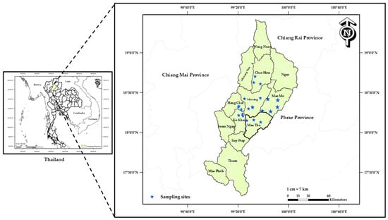
Figure 1.
Map showing the locations of household dust sampling sites in Lampang Province, Upper Northern Thailand.
2.2. Household Dust Extraction
Household dust samples collected from 22 sampling sites were pooled and subsequently separated into two extraction groups. In the first group, dust was extracted using a methanol-deionized water mixture (1:1, v/v), whereas in the second group, deionized water was used as the extraction solvent. For both extraction groups, an identical mass of sieved dust (10 g per sample) was used and combined with 150 mL of the respective solvent, either deionized water or a methanol-deionized water mixture (1:1, v/v), resulting in a concentration of 66.7 mg/mL to ensure comparability between extraction conditions. The mixtures were vigorously shaken for 1 min, and this procedure was repeated five times. The mixtures were then sonicated for 30 min in three successive cycles to facilitate the release of soluble components. Following the extraction process, the suspensions were centrifuged at 5000 rpm for 10 min to separate dust samples. The resulting supernatants were collected and passed through a 0.22 µm membrane filter to remove any remaining particles. The filtrates from the aqueous extraction group were directly subjected to freeze-drying (lyophilization) under controlled conditions to obtain dry extracts. In contrast, the filtrates from the methanol-water extraction group were first evaporated to remove methanol, followed by freeze-drying to achieve complete solvent removal and obtain dry extracts. The resulting filtered extracts represent the soluble (extractable) fraction of household dust and do not include intact particulate matter. These extracts were used for all subsequent in vitro experiments and are hereafter referred to as “household dust extracts.” The total mass of dried extract obtained was 0.735 g for the aqueous extraction group and 0.324 g for the methanol–water (1:1, v/v) extraction group. All procedures were performed under identical conditions for both extraction groups. The extraction yield was estimated to be approximately 7.35% for the aqueous extraction and 3.24% for the methanol–water extraction. Recovery was not determined due to the absence of specific target analytes; therefore, the reported yields reflect overall extract mass rather than compound-specific recovery. Nevertheless, identical protocols were applied to ensure consistency across samples.
2.3. Cell Culture
Human lung adenocarcinoma A549 cells were obtained from the RIKEN Bioresource Center Cell Bank (Tsukuba, Japan). They were maintained in Dulbecco’s Modified Eagle Medium (DMEM) supplemented with 10% fetal bovine serum (FBS), along with 100 U/mL penicillin and 100 µg/mL streptomycin. Cultures were incubated at 37 °C in a humidified atmosphere with 5% CO2. For the experiments, A549 cells were seeded at densities of 1 × 10⁵ cells per well in 6-well plates or 2 × 10⁵ cells per T25 culture flask 24 h prior to treatment. All experimental groups were processed in parallel within each independent experiment. Endpoint measurements were performed on the same day within each experiment to minimize potential batch effects and temporal variability. Independent experiments were conducted on different days, and the results represent pooled data from multiple biological replicates (n ≥ 3).
2.4. Radon Exposure
A radon exposure system was custom-built for this study at the Institute of Radiation Emergency Medicine, Hirosaki University (Aomori, Japan). Natural uranium rocks were used as the radon source to generate radon gas within the system (Figure 2). The produced radon was delivered into an acrylic exposure chamber (0.012 m3) through a larger acrylic chamber (0.54 m3) at a constant airflow rate of 10 L/min to maintain a stable radon activity concentration. The schematic design and operational details of this system have been described in our previous study [21]. The radiation dose delivered to the cells was estimated based on the radon activity concentration and exposure duration using a dose conversion coefficient of 9 nSv h−1 per Bq m−3, which is commonly applied for human exposure scenarios [16]. Under the present experimental conditions, A549 cells were exposed to radon at an average concentration of 1142.8 ± 48.3 Bq m−3 for 45 min per day over two consecutive days, corresponding to an estimated effective dose (based on environmental conversion factors) of approximately 15.4 µSv. It should be noted that this value represents an approximate effective dose equivalent derived from human-based conversion factors and may not directly reflect the actual absorbed dose at the cellular level under in vitro conditions. Control cells were maintained under similar experimental conditions without radon exposure (Figure 2b). It should be noted that the radon exposure system was operated at room temperature (approximately 22–25 °C) and did not include CO2 control comparable to that of a standard cell culture incubator. To assess the potential impact on culture conditions, the pH of the culture medium was measured immediately after the 45 min exposure and was found to be approximately 8.1. After returning the cells to a standard incubator (37 °C, 5% CO2), the pH recovered to approximately 7.4 within 3–4 h. These findings indicate that the observed pH change was transient and largely restored under standard incubation conditions. Given the relatively short exposure duration, such fluctuations are unlikely to have a substantial impact on cellular responses under the present experimental conditions.
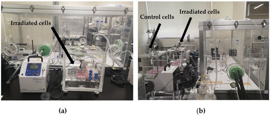
Figure 2.
Schematic representation of the in-house radon exposure system used for cell exposure experiments. (a) Acrylic chamber containing A549 cells exposed to radon gas (irradiated cells). (b) Experimental setup showing both radon-exposed cells and control cells maintained in a separate chamber under similar conditions without radon exposure. The system includes a radon source chamber connected to the exposure chamber via controlled airflow, facilitating controlled radon delivery and minimizing cross-contamination between irradiated and control conditions [21].
2.5. Cytotoxicity
Cell viability was determined using the MTT colorimetric assay [3-(4,5-dimethylthiazol-2-yl)-2,5-diphenyltetrazolium bromide; Life Technologies Corporation, Eugene, OR, USA] [22]. Briefly, A549 cells were seeded in 96-well plates at a density of 1 × 10⁴ cells per well and incubated for 24 h at 37 °C in a humidified atmosphere containing 5% CO2 to allow cell attachment and growth. After this period, the cells were treated with filtered aqueous and methanol–water (1:1, v/v) extracts of household dust samples at different concentrations. All dust extracts were dried and reconstituted in sterile deionized water prior to exposure. The cells were then incubated for 24 h under the same conditions. MTT solution (5 mg/mL in PBS) was then added to each well and the plates were incubated for 4 h under standard culture conditions. The culture medium was subsequently removed and replaced with 100 µL of dimethyl sulfoxide (DMSO) per well to dissolve the formazan crystals, followed by incubation for 15 min. The absorbance (A) of the solubilized formazan was measured at 540 and 630 nm using an H4 Hybrid Microplate Reader (BioTek, Winooski, VT, USA). Cell viability was calculated using Equation (1).
where Asample represents the absorbance of A549 cells exposed to household dust, while Acontrol represents the absorbance of untreated control cells. The inhibitory concentrations required to reduce cell viability by 25% and 50% (IC25 and IC50) were estimated from the dose–response curves fitted to the MTT assay data [23].
Cell viability (%) = (Asample/Acontrol) × 100
2.6. Cell Morphology
Morphological alterations in A549 cells following exposure to radon, household dust extract, or sequential exposure to household dust extract followed by radon were examined using an inverted phase-contrast microscope (Olympus IX71, Olympus, Tokyo, Japan). Representative images were captured with a 10× objective lens.
2.7. Determination of Cell Number and Viability
Cell numbers were determined using an automated cell counter (Countess™ 3, Invitrogen, Thermo Fisher Scientific, Waltham, MA, USA). Prior to counting, cell suspensions were mixed with trypan blue to distinguish viable (live) cells from non-viable (dead) cells. Each experimental condition was analyzed in at least three technical replicates, and the results were averaged for analysis. Before counting, cell suspensions were gently resuspended by pipetting to minimize cell aggregation, and only single, well-dispersed cells were included based on the instrument’s default gating criteria. Cell concentrations were subsequently calculated according to the manufacturer’s instructions. To minimize potential interference from filtered aqueous extracts of household dust extract, samples were appropriately diluted and visually inspected to ensure accurate discrimination between cells and debris. In addition, trypan blue staining facilitated the distinction between viable cells and non-cellular particles. No significant interference with cell counting was observed under the experimental conditions.
2.8. Micronucleus Formation
DNA damage was assessed by measuring micronucleus (MN) formation using the cytokinesis-block micronucleus (CBMN) assay as previously described [24]. Briefly, approximately 5 × 10⁴ cells were seeded into 4-well chamber slides and allowed to attach for 4 h. The cells were then exposed to the indicated treatment conditions. Following treatment, cytochalasin B (2 µg/mL, Sigma-Aldrich, St. Louis, MO, USA) was added to the culture medium to block cytokinesis and cells were incubated for 48 h to allow the formation of binucleated cells. Following incubation, the culture medium was removed and the cells were gently rinsed with phosphate-buffered saline (PBS) before fixation with 99.5% ethanol. The fixed cells were subsequently stained with 1 µg/mL Hoechst 33342 (Sigma-Aldrich, St. Louis, MO, USA) and observed under a fluorescence microscope. For each sample, at least 1000 binucleated cells were scored, and only micronuclei present in binucleated cells were included in the analysis.
2.9. Western Blot
Primary antibodies against γ-H2AX, Nrf2, and GAPDH were obtained from Merck Millipore (Darmstadt, Hessen, Germany). Antibodies against HO-1 were purchased from Santa Cruz Biotechnology (Dallas, TX, USA), IL-6 antibodies were purchased from Affinity Biosciences (Cincinnati, OH, USA), and LC3A/B antibodies were purchased from Cell Signaling Technology (Danvers, MA, USA). Catalog numbers are as follows: 07-627 (γ-H2AX), SAB4501984 (Nrf2), G9545 (GAPDH), SC-136960 (HO-1), DF6087 (IL-6), and 4108S (LC3A/B). Horseradish peroxidase (HRP)-conjugated secondary antibodies (catalog numbers: 1706515 and 1706516) were purchased from Bio-Rad Laboratories (Hercules, CA, USA). Cells were washed twice with ice-cold phosphate-buffered saline (PBS) and lysed in RIPA buffer (Sigma-Aldrich, St. Louis, MO, USA) to extract total cellular proteins. Equal amounts of protein (20 μg) were separated by 10% sodium dodecyl sulfate–polyacrylamide gel electrophoresis (SDS-PAGE) and then transferred onto polyvinylidene fluoride (PVDF) membranes. The membranes were incubated with the corresponding primary antibodies at the following dilutions: γ-H2AX (1:500), Nrf2 (1:500), HO-1 (1:500), LC3 A/B (1:1000), IL-6 (1:2000), and GAPDH (1:10,000); this was followed by incubation with HRP-conjugated secondary antibodies diluted 1:2000–1:10,000. GAPDH served as the loading control. Protein bands were detected and quantified using an iBright™ FL1500 Imaging System (Version 5.4.0. Invitrogen, Carlsbad, CA, USA). All experiments were conducted in biological triplicate (n ≥ 3).
2.10. Statistical Analysis
Data are expressed as the mean ± standard error (SE). Statistical analyses were performed using SigmaPlot version 10 (Systat Software Inc., San Jose, CA, USA). All analyses were based on results obtained from at least three independent experiments performed on separate days. Dose–response curves were fitted using a nonlinear regression model to estimate IC25 and IC50 values. For experiments involving multiple groups, statistical comparisons were performed using one-way or two-way analysis of variance (ANOVA), as appropriate, followed by Tukey’s post hoc test for multiple comparisons. For MN count data, results were expressed as frequencies or proportions, and statistical comparisons were performed using appropriate methods for count/proportion data. A p value of <0.05 was considered statistically significant.
3. Results
3.1. Cytotoxicity of Filtered Aqueous and Methanol–Water (1:1, v/v) Extracts of Household Dust
Cell viability in A549 cells decreased in a dose-dependent manner following exposure to increasing concentrations of filtered aqueous and methanol–water (1:1, v/v) extracts of household dust (Figure 3). The methanol–water extract showed greater cytotoxicity than the water extract. Based on the dose–response curves, the estimated IC25 and IC50 values for the water extract were approximately 7500 and 12,955 µg/mL, respectively (Figure 3a), whereas the corresponding values for the methanol–water extract were approximately 104 and 122 µg/mL (Figure 3b).
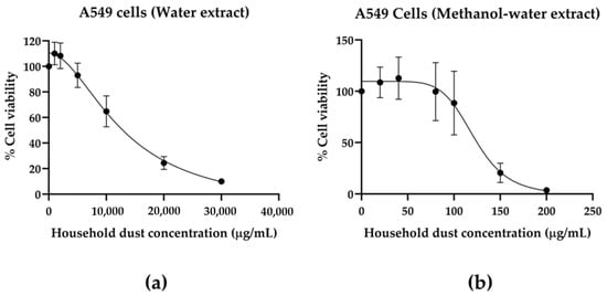
Figure 3.
Dose–response effects of household dust extracts on A549 cell viability. Cells were treated with increasing concentrations of household dust extracts prepared using water (left panel, (a)) or methanol-water (1:1, v/v) (right panel, (b)) for 24 h. Cell viability was measured using the MTT assay and expressed as a percentage relative to untreated control cells. Data are presented as mean ± SE from four independent experiments. Dose–response curves were fitted using nonlinear regression analysis to estimate IC25 and IC50 values.
Based on these results, the IC25 concentration of the filtered aqueous extract of household dust (7500 μg/mL) was selected for subsequent experiments to investigate the effects of indoor radon exposure following prior treatment with household dust extract on A549 cells. This concentration produced limited cytotoxicity while enabling detection of cellular responses associated with subsequent radon exposure in vitro. The aqueous extract represents soluble dust components that could potentially interact with airway lining fluid. However, this simplified model does not reflect real inhalation exposure involving whole particles and biological defenses. Therefore, the findings should be interpreted as reflecting cellular responses to household dust extracts under simplified in vitro conditions rather than physiological inhalation exposure conditions.
3.2. Effects of Sequential Exposure to Household Dust Extract and Indoor Radon on Cell Morphology, Proliferation, and DNA Damage in A549 Cells
The experimental design for household dust extract and radon exposure is illustrated in Figure 4a–d. A549 cells were divided into four groups: control, radon exposure (2 days, ~1140 Bq/m3, 45 min/day), treatment with household dust extract for 24 h (7500 μg/mL (IC25), and sequential exposure to household dust extract followed by radon. Representative phase-contrast images of A549 cells under different experimental conditions are shown in Figure 4e–h. Control and radon-exposed cells generally maintained a typical epithelial-like morphology with relatively dense cell attachment. In contrast, cells exposed to household dust extract showed apparent morphological changes, including reduced cell density and irregular cell shapes. These observations are qualitative and intended to provide supportive visual evidence of cellular responses under the different exposure conditions. These morphological changes were more apparent in the sequential exposure group (Figure 4h) and were consistent with the quantitative reduction in cell number observed in Figure 4i. Quantitative analysis demonstrated that exposure to household dust extract significantly reduced the number of viable cells compared with the control group (p = 0.03). A similar reduction was observed when compared with the radon group (p = 0.033). Furthermore, sequential exposure to household dust extract followed by radon resulted in a further decrease in cell numbers compared with the radon group (p = 0.003) and the control group (p = 0.002), suggesting an additional reduction in cell number under the present sequential exposure conditions.
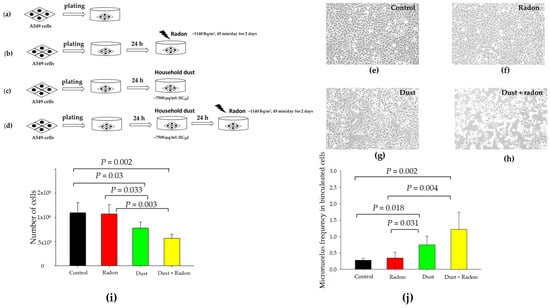
Figure 4.
Effects of radon exposure and household dust extract on A549 cell morphology, cell proliferation, and micronucleus formation under sequential exposure conditions. (a–d) Schematic representation of the experimental design. A549 cells were divided into four groups: (a) control, (b) radon exposure (2 days, ~1140 Bq/m3, 45 min/day), (c) household dust extract treatment for 24 h, and (d) sequential exposure to household dust extract for 24 h followed by radon exposure (2 days, ~1140 Bq/m3, 45 min/day). (e–h) Representative phase-contrast microscopy images showing the morphology of A549 cells in the control, radon, dust, and sequential exposure (dust extract followed by radon) groups, respectively. (i) Total cell number following different treatments. (j) Micronucleus frequency in binucleated cells. Data are presented as mean ± SE from independent experiments (n = 4). Statistical significance is indicated in the figure.
MN formation was evaluated using the CBMN assay. As shown in Figure 4j, the MN frequency in binucleated cells was significantly increased in cells treated with household dust extract compared with the control group (p = 0.018) and the radon group (p = 0.031). The sequential exposure group (household dust extract followed by radon) showed a further increase in MN frequency compared with the control (p = 0.002) and radon groups (p = 0.004). Notably, the increased MN frequency corresponded with the morphological alterations observed in the phase-contrast images and the reduction in cell numbers, suggesting that DNA damage may be associated with altered cellular responses and reduced cell numbers under sequential exposure conditions.
3.3. Household Dust Extract and Indoor Radon Exposure Is Associated with Oxidative Stress Signaling, DNA Damage, Autophagy, and Inflammatory Responses in A549 Cells
Exposure to radon and household dust extract altered the expression levels of proteins related to oxidative stress, DNA damage, autophagy, and inflammation in A549 cells (Figure 5, Supplementary Figure S1). Western blot analysis demonstrated that the expression of nuclear factor erythroid 2-related factor 2 (Nrf2), a key regulator of oxidative stress responses [25], was significantly increased in the sequential exposure group (household dust extract followed by radon) compared with the control group (p = 0.028) (Figure 5b). Consistently, the downstream antioxidant enzyme heme oxygenase-1 (HO-1) was also significantly upregulated in the sequential exposure group compared with the control group (p = 0.048) (Figure 5c). An increasing trend in HO-1 expression was observed in the radon group; however, this difference was not statistically significant. These findings suggest possible involvement of oxidative stress-related signaling pathways rather than providing direct evidence of intracellular oxidative stress.
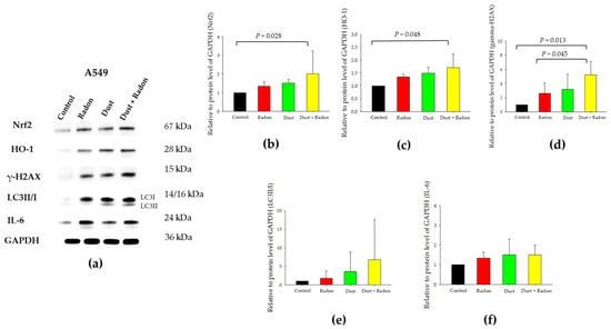
Figure 5.
Effects of sequential exposure to household dust extract followed by radon on oxidative stress-related, DNA damage-related, autophagy-related, and inflammatory-related protein expression in A549 cells. (a) Representative Western blot images showing the protein expression levels of Nrf2, HO-1, γ-H2AX, LC3 and IL-6 in A549 cells following exposure to radon, household dust extract, or sequential exposure (household dust extract followed by radon). Densitometric analyses of protein expression normalized to GAPDH are shown in (b–f): (b) Nrf2, (c) HO-1, (d) γ-H2AX, (e) LC3 II/I and (f) IL-6 ratio. Data are presented as mean ± SE from three independent experiments.
In addition, the DNA damage marker γ-H2AX [26] was significantly increased in the sequential exposure group (household dust extract followed by radon) compared with the control cells (p = 0.013) and the radon group (p = 0.045), suggesting an association with DNA damage-related responses rather than a specific type of DNA damage under sequential exposure conditions (Figure 5d). Densitometric analysis also showed an increasing trend in the microtubule-associated protein 1 light chain 3 II/I (LC3 II/I) ratio, a commonly used autophagy-related marker, particularly in the sequential exposure group (household dust extract followed by radon) (Figure 5e). However, this increase was not statistically significant and should be interpreted with caution. Alterations in autophagy-related markers may reflect cellular stress responses and cannot be directly linked to apoptosis or cytotoxicity under the present experimental conditions [27]. Similarly, the inflammatory-related marker interleukin-6 (IL-6) showed a modest increase following household dust extract and radon exposure [28], although the change was not statistically significant and therefore should not be interpreted as independent evidence of inflammation (Figure 5f).
These findings suggest that sequential exposure may be associated with activation of oxidative stress–related pathways involving Nrf2 and HO-1, accompanied by alterations in DNA damage responses (γ-H2AX), autophagy-related markers (LC3 II/I), and inflammatory-related markers (IL-6) in A549 cells, rather than demonstrating direct causal relationships among these processes.
4. Discussion
In this study, household dust samples were randomly collected from Lampang Province, one of the provinces in UNT, where high levels of PM pollution and indoor radon have been reported [3,11]. Dust samples were extracted with water and methanol–water, and cytotoxicity was assessed in A549 cells. The results showed that household dust extracts prepared with the methanol–water mixture showed greater cytotoxicity than those extracted with water alone (Figure 3), likely due to the enhanced extraction of hydrophobic organic contaminants present in household dust, including heavy metals, PAHs and other organic pollutants known to induce oxidative stress and toxicity [14,20,29]. These findings are consistent with previous studies, such as that of Pyambri et al., which reported that indoor dust extracts can induce ROS production and inflammatory responses in lung cell models, highlighting the potential biological impacts of complex chemical mixtures present in indoor dust that may be associated with cytotoxic effects in exposed lung cells [30]. Similarly, Naimabadi et al. reported that indoor dust contains various toxic components, including heavy metals such as zinc (Zn), copper (Cu), chromium (Cr), nickel (Ni), and lead (Pb), which have been associated with increased cytotoxicity and inflammatory responses in lung cell lines [31]. Furthermore, Huang et al., using A549 lung cells, demonstrated that exposure to water-soluble fractions of environmental dust and PM can induce cytotoxic effects, with toxicity potentially associated with soluble metal(loid)s and other chemical components present in dust particles [32]. However, it should be noted that no chemical characterization of the household dust extract was performed in the present study; therefore, these interpretations are based on previously reported findings rather than direct evidence from our data. For subsequent experiments, the water extract of household dust at the IC25 concentration was selected because it was intended to provide a simplified model of exposure conditions relevant to human inhalation of indoor dust. This concentration produced relatively low cytotoxicity while allowing for clearer observation of cellular responses following subsequent radon exposure.
Next, we investigated the effects of sequential exposure to household dust extract followed by indoor radon in A549 cells. Radon exposure was performed at approximately 1140 Bq/m3 for two consecutive days (45 min/day); this protocol was selected based on previous in vitro studies that showed that it can induce detectable cellular responses within a practical experimental time frame [21]. Our results demonstrated that sequential exposure to household dust extract followed by radon may be associated with greater cytotoxic and genotoxic responses compared with either treatment alone (Figure 4). Cells exposed to household dust extract showed reduced cell numbers and morphological alterations, which were more evident in the sequential exposure group (Figure 4e–h). Quantitative analysis further confirmed a significant reduction (p = 0.002 and p = 0.002, respectively) in cell number and an increase in MN frequency following sequential exposure, suggesting increased DNA damage-related responses (Figure 4i,j). Interestingly, radon exposure alone produced only minimal effects compared with dust treatment, which may be partly attributed to the relatively short exposure duration used in this study. Under these conditions, the effective radiation dose delivered to the cells may have been insufficient to induce substantial biological responses as the biological effects of radon are generally associated with cumulative dose and prolonged exposure duration rather than short-term exposure [7,33]. However, when radon exposure was preceded by household dust extract treatment, increased cellular responses were observed. This may be associated with the presence of particulate-derived components in household dust extract that could influence cellular susceptibility to radon exposure, rather than direct interactions with radon progeny under the present in vitro conditions. Moreover, both radon exposure and dust have been reported to induce oxidative stress and DNA damage, which may contribute to cumulative cellular responses under sequential exposure conditions [34,35,36,37].
In addition, oxidative stress has been reported to activate the Nrf2 signaling pathway, which regulates the expression of antioxidant enzymes such as HO-1 to maintain cellular redox homeostasis [25]. Although activation of this pathway generally represents a protective cellular response, excessive oxidative stress-related responses may be associated with impaired antioxidant balance and DNA damage-related responses (Figure 5b,c) [38,39]. In the present study, the increased γ-H2AX expression (Figure 5d) and MN formation (Figure 4j) observed following sequential exposure to household dust extract and radon suggest increased DNA damage-related responses in A549 cells. Furthermore, oxidative stress and DNA damage-related responses have been reported to be associated with autophagy- and inflammation-related pathways [27,28,40], which is partially consistent with the observed trends in LC3 II/I and IL-6 expression, although these changes were not statistically significant (Figure 5e,f). Taken together, these findings suggest that sequential exposure to household dust extract followed by radon may be associated with oxidative stress-related cellular responses, including changes in DNA damage-related, autophagy-related, and inflammatory-related markers. These associations should be interpreted cautiously and do not imply direct interactions or causal relationships between the pathways. Although LC3 II/I did not show a statistically significant change, its increasing trend was more evident than that of IL-6. Therefore, autophagy-related responses were retained in the hypothetical model (Figure 6) as a potential associated cellular response, whereas IL-6 was not included to avoid overinterpretation of a weak and non-significant inflammatory response. The hypothetical model summarizing the observed cellular responses under sequential exposure conditions is presented in Figure 6. Briefly, sequential exposure to household dust followed by radon may be associated with oxidative stress-related responses, which may be reflected by altered expression of Nrf2/HO-1-related proteins. These responses may also be associated with changes in DNA damage-related and autophagy-related markers. Collectively, these cellular responses may be associated with reduced cell viability under the present experimental conditions. This model is hypothetical and does not imply direct causal relationships.
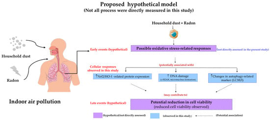
Figure 6.
Hypothetical model of cellular responses following sequential exposure to household dust extract followed by indoor radon in A549 cells. Sequential exposure to household dust extract followed by radon may be associated with oxidative stress-related responses, which is reflected by altered expression of Nrf2/HO-1-related proteins. These responses may be associated with DNA damage, as evidenced by increased γ-H2AX expression and micronucleus formation. These alterations may also be associated with changes in autophagy-related markers (LC3 II/I conversion). Collectively, these cellular responses may be associated with reduced cell viability under the present experimental conditions. Overall, this model is hypothetical and intended to illustrate potential relationships rather than confirmed causal mechanisms. Some elements of this figure were adapted from Servier Medical Art by Servier (https://smart.servier.com/ [accessed on 9 March 2026]), which was licensed under a Creative Commons Attribution 4.0 International License.
From an environmental health perspective, these findings emphasize the importance of considering multiple or sequential exposures to indoor environmental pollutants, rather than evaluating individual agents in isolation. In many residential settings, particularly in regions affected by seasonal air pollution such as UNT, indoor radon exposure may occur simultaneously with elevated levels of PM that accumulate as household dust. Recent evidence further suggests that indoor PM, household dust, and radon-related exposures may coexist in residential environments and may contribute to adverse respiratory health concerns [41,42,43,44,45]. The possible associated cellular responses observed in this study, characterized by oxidative stress-related responses, provide preliminary evidence suggesting an association between sequential exposure to household dust extract followed by radon and cytotoxic and genotoxic responses in A549 cells. Oxidative stress may be associated with reduced cell viability and increased DNA damage, as indicated by elevated γH2AX expression and MN formation. Potential involvement of oxidative stress-related pathways and associated cellular responses, including altered expression of Nrf2/HO-1-related proteins and changes in autophagy-related and inflammatory-related markers, further supports the possibility of oxidative stress-related cellular responses under sequential exposure conditions. These findings highlight the importance of integrated indoor air quality management strategies, including radon mitigation and effective control of indoor particulate accumulation, to reduce human exposure and potentially reduce exposure to indoor environmental pollutants associated with adverse respiratory health effects.
Despite providing preliminary insights into the cellular responses associated with sequential exposure to filtered aqueous extracts of household dust followed by radon, several limitations of the present study should be acknowledged.
First, this study was conducted using a single human lung adenocarcinoma cell line under controlled in vitro conditions, which may not fully recapitulate the complex biological responses of normal lung tissues or real-world exposure scenarios. Although A549 cells are widely used as an in vitro model for investigating lung epithelial responses to environmental pollutants, they may not fully represent the responses of normal lung cells or other cell types within the respiratory system. In addition, the acute exposure conditions used in this study differ from the chronic, low-level exposure typically experienced in indoor environments, and caution is required when extrapolating these findings to human health. Future studies using normal bronchial epithelial cells (e.g., BEAS-2B) and more physiologically relevant exposure conditions would be valuable.
Second, uncertainties related to the dose–response relationship and in vitro radon dosimetry should be considered. The dose–response relationship showed a non-monotonic trend at low concentrations, which may reflect experimental variability or possible non-linear cellular responses, although these changes were not statistically significant. Furthermore, radon dose estimation was based on environmental conversion factors rather than direct cellular dosimetry, which may limit the accuracy of exposure assessment.
Third, several methodological and statistical limitations may influence data interpretation. In particular, the absence of a vehicle control may affect the interpretation of the observed effects of the household dust extracts. In addition, no formal statistical interaction analysis was performed; therefore, potential synergistic interactions between exposures cannot be determined, and the observed effects should be interpreted as cumulative cellular responses rather than synergistic. The lack of mycoplasma testing represents an additional limitation that may affect the reliability and reproducibility of the experimental data.
Fourth, mechanistic interpretation of the observed cellular responses remains limited. Although oxidative stress-related responses were discussed, these were inferred based on downstream molecular markers, and direct measurement of intracellular ROS was not performed. Increased Nrf2 and HO-1 expression was interpreted as indicative of oxidative stress-related signaling responses rather than direct evidence of oxidative status. Similarly, γ-H2AX and micronucleus formation were used as general indicators of DNA damage, and morphological observations were qualitative and considered supportive rather than quantitative evidence. In addition, autophagy was inferred from the LC3 II/I ratio without assessment of autophagic flux and therefore cannot be conclusively interpreted as definitive evidence of functional autophagy. Additional assays (e.g., apoptosis, cell cycle or direct ROS measurement) would be required to strengthen mechanistic conclusions.
Finally, the representation of household dust exposure and the sequential exposure design are limited. Household dust samples were processed as filtered aqueous extracts; therefore, the experimental system only reflects the soluble fraction rather than whole particles. In addition, no detailed chemical characterization of the dust extracts was performed, limiting attribution of the observed cellular responses to specific constituents. Furthermore, the sequential exposure design does not fully represent real-world simultaneous exposure conditions, where radon progeny interact dynamically with airborne particles under varying environmental factors. Therefore, further studies using more physiologically relevant models, such as three-dimensional (3D) cultures, co-culture systems, or in vivo models, are warranted.
5. Conclusions
This study suggests that sequential exposure to household dust extract followed by indoor radon may be associated with cytotoxic and genotoxic responses in human lung adenocarcinoma cells under the present experimental conditions. Sequential exposure was associated with changes in oxidative stress-related responses, including altered expression of the Nrf2/HO-1 pathway and DNA damage markers, such as γ-H2AX expression and micronucleus formation. Changes in autophagy-related markers were also observed, which may reflect alterations in autophagy-related response, while IL-6 showed a non-significant increase and should be interpreted cautiously. Overall, these findings suggest that oxidative stress-related pathways may be involved in the observed cellular responses associated with sequential exposure conditions. However, the results should be interpreted cautiously due to the use of a single cancer cell line and the limitations inherent to the in vitro exposure model.
To the best of our knowledge, this study is among the first to examine the biological effects of sequential exposure to household dust extract followed by indoor radon using an in vitro lung cancer cell model. The findings provide preliminary evidence of associated cellular responses and highlight the potential relevance of multiple indoor environmental exposures. Further studies using additional cell models, normal lung epithelial cells, and in vivo systems are warranted to clarify the biological mechanisms and potential human health relevance of these findings.
Supplementary Materials
The following supporting information can be downloaded at https://www.mdpi.com/article/10.3390/environments13050280/s1: Figure S1: Original uncropped Western blot images.
Author Contributions
Conceptualization, N.A.; methodology, N.A., C.K., T.M. and S.T.; validation, N.A.; formal analysis, N.A. and A.D.; investigation, N.A., A.D., S.K., S.S., C.K., D.A., Y.F. and T.M.; resources, N.A., S.K., S.B., S.S., C.K., T.M. and S.T.; data curation, N.A.; writing—original draft preparation, N.A.; writing—review and editing, N.A., A.D., S.K., S.B., S.S., C.K., D.A., Y.F., T.M. and S.T.; visualization, N.A., A.D. and S.K.; project administration, N.A.; funding acquisition, N.A., C.K. and S.T. All authors have read and agreed to the published version of the manuscript.
Funding
This research was funded by the Environmental Radioactivity Network (ERAN) (grant number I-25-16).
Data Availability Statement
The data presented in this study are available upon request from the corresponding author.
Acknowledgments
The authors gratefully acknowledge the technical assistance provided by the staff of the Institute of Radiation Emergency Medicine (IREM), Hirosaki University; the Research Institute for Health Sciences (RIHES), Chiang Mai University; the Department of Biochemistry, Faculty of Medicine, Chiang Mai University; and the Department of Environmental Science and Technology, Faculty of Science, Lampang Rajabhat University. The authors also thank Ariyaphong Wongnoppavich, Pilaiporn Thippraphan and Nipattha Srioun for their valuable technical assistance.
Conflicts of Interest
The authors declare no conflicts of interest.
Abbreviations
| DNA | Deoxyribonucleic acid |
| HO-1 | Heme oxygenase-1 |
| IC | Inhibitory concentration |
| IL-6 | Interleukin-6 |
| LC | Lung cancer |
| LC3 | Microtubule-associated protein 1 light chain 3 |
| LET | Linear energy transfer |
| MN | Micronucleus |
| Nrf2 | Nuclear factor erythroid 2-related factor 2 |
| PM | Particulate matter |
| ROS | Reactive oxygen species |
| UNT | Upper northern Thailand |
| VOCs | Volatile organic compounds |
References
- USEPA. The Inside Story: A Guide to Indoor Air Quality. Available online: https://www.epa.gov/indoor-air-quality-iaq/inside-story-guide-indoor-air-quality (accessed on 9 March 2026).
- World Health Organization. Handbook on Indoor Radon; World Health Organization: Geneva, Switzerland, 2009. [Google Scholar]
- Somsunun, K.; Prapamontol, T.; Pothirat, C.; Liwsrisakun, C.; Pomhnikorn, D.; Fongmoon, D.; Chantata, S.; Wongpoomchai, R.; Naksen, W.; Autsavapromporn, N.; et al. Estimation of lung cancer deaths attributable to indoor radon exposure in Upper Northern Thailand. Sci. Rep. 2022, 12, 5169. [Google Scholar] [CrossRef] [PubMed]
- Autsavapromporn, N.; Krandrod, C.; Klunklin, P.; Kritsananuwat, R.; Jaikang, C.; Kittidachanan, K.; Chitapanarux, I.; Fugkeaw, S.; Hosoda, M.; Tokonami, S. Health effects of natural environmental radiation during burning season in Chiang Mai, Thailand. Life 2022, 12, 853. [Google Scholar] [CrossRef] [PubMed]
- National Research Council. Biological Effects of Ionizing Radiation (BEIR) VI Report: The Health Effects of Exposure to Radon; National Academy Press: Washington, DC, USA, 1999.
- United States Environmental Protection Agency (EPA). A Citizen’s Guide to Radon: The Guide to Protecting Yourself and Your Family from Radon; EPA: Washington, DC, USA, 2016. Available online: https://www.epa.gov/sites/default/files/2016-12/documents/2016_a_citizens_guide_to_radon.pdf (accessed on 9 March 2026).
- Darby, S.; Hill, D.; Auvinen, A.; Barros-Dios, J.M.; Baysson, H.; Bochicchio, F.; Deo, H.; Falk, R.; Forastiere, F.; Hakama, M.; et al. Radon in homes and risk of lung cancer: Collaborative analysis of individual data from 13 European case-control studies. BMJ 2005, 330, 223. [Google Scholar] [CrossRef] [PubMed]
- Krewski, D.; Lubin, J.H.; Zielinski, J.M.; Alavanja, M.; Catalan, V.S.; Field, R.W.; Klotz, J.B.; Létourneau, E.G.; Lynch, C.F.; Lyon, J.I.; et al. Residential radon and risk of lung cancer: A combined analysis of 7 North American case-control studies. Epidemiology 2005, 16, 137–145. [Google Scholar] [CrossRef]
- Weschler, C.J.; Nazaroff, W.W. Semivolatile organic compounds in indoor environments. Atmos. Environ. 2008, 42, 9018–9040. [Google Scholar] [CrossRef]
- Somsunun, K.; Prapamontol, T.; Kuanpan, T.; Santijitpakdee, T.; Kohsuwan, K.; Jeytawan, N.; Thongjan, N. Health risk assessment of heavy metals in indoor household dust in urban and rural areas of Chiang Mai and Lamphun Provinces, Thailand. Toxics 2023, 11, 1018. [Google Scholar] [CrossRef]
- Somsunun, K.; Santijitpakdee, T.; Kohsuwan, K.; Jeytawan, N.; Kirtsaeng, S.; Norbäck, D.; Prapamontol, T. Contamination and health risk assessment of potentially toxic elements in household dust across the haze season in upper northern Thailand. Toxics 2025, 13, 1008. [Google Scholar] [CrossRef]
- World Health Organization (WHO). WHO Guidelines for Indoor Air Quality: Selected Pollutants; WHO Regional Office for Europe: Copenhagen, Denmark, 2010; Available online: https://www.who.int/publications/i/item/9789289002134 (accessed on 20 April 2026).
- Thatcher, T.L.; Layton, D.W. Deposition, resuspension, and penetration of particles within a residence. Atmos. Environ. 1995, 29, 1487–1497. [Google Scholar] [CrossRef]
- Whitehead, T.; Metayer, C.; Buffler, P.; Rappaport, S.M. Estimating exposures to indoor contaminants using residential dust. J. Expo. Sci. Environ. Epidemiol. 2011, 21, 549–564. [Google Scholar] [CrossRef]
- Xiang, P.; Liu, R.-Y.; Sun, H.-J.; Han, Y.-H.; He, R.-W.; Cui, X.-Y.; Ma, L.Q. Molecular mechanisms of dust-induced toxicity in human corneal epithelial cells: Water and organic extract of office and house dust. Environ. Int. 2016, 92–93, 348–356. [Google Scholar] [CrossRef]
- United Nations Scientific Committee on the Effect of Atomic Radiation (UNSCEAR). Sources and Effects on Ionizing Radiation; United Nation: New York, NY, USA, 2000. [Google Scholar]
- Porstendörfer, J. Properties and behaviour of radon and thoron and their decay products in the air. J. Aerosol Sci. 1994, 25, 219–263. [Google Scholar] [CrossRef]
- International Commission on Radiological Protection (ICRP). The 2007 Recommendations of the International Commission on Radiological Protection; ICRP Publication 103; International Commission on Radiological Protection: Ottawa, ON, Canada, 2007; Volume 37, pp. 1–332. [Google Scholar]
- Suriyawong, P.; Chuetor, S.; Samae, H.; Piriyakarnsakul, S.; Amin, M.; Furuuchi, M.; Hata, M.; Inerb, M.; Phairuang, W. Airborne particulate matter from biomass burning in Thailand: Recent issues, challenges, and options. Heliyon 2023, 9, e14261. [Google Scholar] [CrossRef] [PubMed]
- Bootdee, S.; Sillapapiromsuk, S.; Kawichai, S. Trace Elements in Indoor Dust Exposure from Child Development Centers and Health Risk Assessment in Haze and Industrial Areas, Thailand. Toxics 2025, 13, 547. [Google Scholar] [CrossRef]
- Subsomwong, P.; Kranrod, C.; Sakai, Y.; Asano, K.; Nakane, A.; Tokonami, S. Impact of intermittent high-dose radon exposures on lung epithelial cells: Proteomic analysis and biomarker identification. J. Radiat. Res. 2025, 66, 107–114. [Google Scholar] [CrossRef] [PubMed]
- Mosmann, T. Rapid colorimetric assay for cellular growth and survival: Application to proliferation and cytotoxicity assays. J. Immunol. Methods 1983, 65, 55–63. [Google Scholar] [CrossRef]
- Sebaugh, J.L. Guidelines for accurate EC50/IC50 estimation. Pharm. Stat. 2011, 10, 128–134. [Google Scholar] [CrossRef]
- Fenech, M. The in vitro micronucleus technique. Mutat. Res. 2000, 455, 81–95. [Google Scholar] [CrossRef]
- Ma, Q. Role of Nrf2 in oxidative stress and toxicity. Annu. Rev. Pharmacol. Toxicol. 2013, 53, 401–426. [Google Scholar] [CrossRef]
- Rogakou, E.P.; Pilch, D.R.; Orr, A.H.; Ivanova, V.S.; Bonner, W.M. DNA double-stranded breaks induce histone H2AX phosphorylation on serine 139. J. Biol. Chem. 1998, 273, 5858–5868. [Google Scholar] [CrossRef] [PubMed]
- Scherz-Shouval, R.; Elazar, Z. Regulation of autophagy by reactive oxygen species (ROS): Implications for oxidative stress. Trends Biochem. Sci. 2011, 36, 30–38. [Google Scholar] [CrossRef]
- Kishimoto, T. IL-6: From its discovery to clinical applications. Int. Immunol. 2010, 22, 347–352. [Google Scholar] [CrossRef]
- Xia, T.; Kovochich, M.; Nel, A. The role of reactive oxygen species and oxidative stress in mediating particulate matter injury. Clin. Occup. Environ. Med. 2006, 5, 817–836. [Google Scholar] [CrossRef]
- Pyambri, M.; Lacorte, S.; Jaumot, J.; Bedia, C. Effects of indoor dust exposure on lung cells: Association of chemical compo-sition with phenotypic and lipid changes in a 3D lung cancer cell model. Environ. Sci. Technol. 2023, 57, 20532–20541. [Google Scholar] [CrossRef]
- Naimabadi, A.; Ghasemi, A.; Mohtashami, M.; Saeidi, J.; Bakaeian, M.; Haddad Mashadrizeh, A.; Azimi-Nezhad, M.; Mo-hammadi, A.A. Heavy metal analysis of indoor and outdoor dust extracts and cytotoxicity evaluation and inflammation factors on lung, gastric and skin cell lines. Heliyon 2022, 8, e12414. [Google Scholar] [CrossRef] [PubMed]
- Huang, M.; Kang, Y.; Wang, W.; Chan, C.Y.; Wang, X.; Wong, M.H. Potential cytotoxicity of water-soluble fraction of dust and particulate matters and relation to metal(loid)s based on three human cell lines. Chemosphere 2015, 135, 61–66. [Google Scholar] [CrossRef] [PubMed]
- Little, M.P. Risks associated with ionizing radiation. Br. Med. Bull. 2003, 68, 259–275. [Google Scholar] [CrossRef] [PubMed]
- Vilas-Boas, V.; Chatterjee, N.; Carvalho, A.; Alfaro-Moreno, E. Particulate matter-induced oxidative stress—Mechanistic insights and antioxidant approaches reported in in vitro studies. Environ. Toxicol. Pharmacol. 2024, 110, 104529. [Google Scholar] [CrossRef]
- Sultani, M.A.; Bulko, M.; Müllerová, M.; Masarik, J.; Eckertová, T. Interaction between radon progeny and particulate matter in an urban environment. J. Environ. Radioact. 2026, 294, 107918. [Google Scholar] [CrossRef]
- Romero-Gutierrez, C.; Koutrakis, P.; Liu, M.; Vieira, C.L.Z.; Coull, B.A.; Maher, E.F.; Zhang, J.; Garshick, E. Radon decay product particle radioactivity and oxidative stress biomarkers in patients with COPD. Environ. Res. 2023, 240, 117505. [Google Scholar] [CrossRef]
- Lee, H.; Hanson, H.A.; Logan, J.; Maguire, D.; Kapadia, A.; Dewji, S.; Agasthya, G. Evaluating county-level lung cancer incidence from environmental radiation exposure, PM2.5, and other exposures with regression and machine learning models. Environ. Geochem. Health 2024, 46, 82. [Google Scholar] [CrossRef]
- Jiang, X.Y.; Guo, Q.Q.; Wang, S.S.; Guo, R.; Zou, Y.; Liu, J.W.; Feng, Y.L.; Guo, Y.; Li, Y.H.; Liu, X.Y.; et al. DNA damage response pathway regulates Nrf2 in response to oxidative stress. Sci. Adv. 2025, 11, eadu9555. [Google Scholar] [CrossRef]
- Valko, M.; Leibfritz, D.; Moncol, J.; Cronin, M.T.D.; Mazur, M.; Telser, J. Free radicals and antioxidants in normal physiological functions and human disease. Int. J. Biochem. Cell Biol. 2007, 39, 44–84. [Google Scholar] [CrossRef]
- Filomeni, G.; De Zio, D.; Cecconi, F. Oxidative stress and autophagy: The clash between damage and metabolic needs. Cell Death Dis. 2015, 6, e1972. [Google Scholar] [CrossRef] [PubMed]
- Lee, D.H. Is indoor air pollution the missing link between radon and lung cancer? Rethinking epidemiological support for the LNT model. Dose Response 2025, 23, 15593258251399780. [Google Scholar] [CrossRef] [PubMed]
- Morantes, G.; Jones, B.; Molina, C.; Sherman, M.H. Harm from residential indoor air contaminants. Environ. Sci. Technol. 2024, 58, 1262–1274. [Google Scholar] [CrossRef] [PubMed]
- Autsavapromporn, N.; Kranrod, C.; Kritsananuwat, R.; Sola, P.; Jaikang, C.; Monum, T.; Kawichai, S.; Klunklin, P.; Chitapanarux, I.; Hosoda, M.; et al. Health impacts of natural background radiation in high air pollution area of Thailand. Toxics 2024, 12, 428. [Google Scholar] [CrossRef]
- Radbel, J.; Nethery, R.C.; Gaffin, J.M. Indoor air pollution and airway health. J. Allergy Clin. Immunol. 2024, 154, 1123–1135. [Google Scholar] [CrossRef]
- Autsavapromporn, N.; Duangya, A.; Klunklin, P.; Chitapanarux, I.; Kranrod, C.; Jaikang, C.; Monum, T.; Paemanee, A.; Tokonami, S. Serum metabolomics study to screen potential biomarkers of lung cancer risk in high natural background radiation areas of Thailand: A pilot study. Cancers 2024, 16, 4182. [Google Scholar] [CrossRef]


Disclaimer/Publisher’s Note: The statements, opinions and data contained in all publications are solely those of the individual author(s) and contributor(s) and not of MDPI and/or the editor(s). MDPI and/or the editor(s) disclaim responsibility for any injury to people or property resulting from any ideas, methods, instructions or products referred to in the content. |
© 2026 by the authors. Licensee MDPI, Basel, Switzerland. This article is an open access article distributed under the terms and conditions of the Creative Commons Attribution (CC BY) license.