


Abstract
Background: Comprehensive molecular profiling is increasingly important in advanced non-small cell lung cancer (NSCLC), but the integration of tissue- and plasma-based next-generation sequencing (NGS) and its real-world therapeutic impact remain uncertain. Methods: We conducted a retrospective single-center study of 205 patients with advanced NSCLC treated between January 2016 and January 2019 who underwent routine molecular testing and tissue- and/or plasma-based NGS. Concordance was assessed for clinically relevant genomic alterations, and the impact of NGS on treatment decisions was evaluated. Results: A total of 280 NGS assays were performed. NGS identified potentially actionable genomic alterations in 88 patients (42.9%); in 50 patients (24.4%), potentially actionable genomic alterations were identified by NGS relative to conventional molecular testing. Among 64 patients who underwent both tissue- and plasma-based NGS, overall agreement was 78.1%, with moderate concordance (κ = 0.533). NGS results led to a direct treatment modification in 23 patients (11.2%), including clinical trial enrollment, standard targeted therapy, or compassionate-use treatment. Conclusions: Tissue- and plasma-based NGS provided complementary rather than interchangeable genomic information. The difference between theoretical actionability and actual therapeutic impact highlights the importance of clinical context, timing of testing, and treatment availability when interpreting comprehensive genomic profiling in advanced NSCLC.
1. Introduction
Lung cancer remains one of the most frequently diagnosed malignancies worldwide and the leading cause of cancer-related mortality. According to the most recent GLOBOCAN estimates, approximately 2.5 million new cases are diagnosed worldwide, accounting for 12.4% of all cancer diagnoses, with nearly 1.8 million deaths annually [1]. In Spain, lung cancer continues to represent a major public health burden and remains the leading cause of cancer-related death [2]. Non-small cell lung cancer (NSCLC) represents the majority of lung cancer cases and comprises several histological entities, most commonly adenocarcinoma, squamous cell carcinoma and large cell carcinoma, as reflected in current pathological classifications [3]. Its epidemiology is strongly linked to tobacco exposure, although a clinically relevant proportion of patients, particularly those with adenocarcinoma, develop the disease in the absence of active smoking history [4]. Large real-world registries have further illustrated the clinical heterogeneity of NSCLC and the prognostic relevance of stage, histology and patient-related factors [5]. In parallel, lung cancer screening studies have highlighted the importance of early detection and radiological characterization of pulmonary nodules, although most patients worldwide continue to be diagnosed with advanced disease [6].
Over the past two decades, the treatment paradigm of advanced NSCLC has undergone a profound transformation driven by advances in molecular oncology. The identification of oncogenic driver alterations and the development of targeted therapies have shifted clinical decision-making from a predominantly histology-based approach to a biomarker-driven strategy. Genomic alterations in genes such as EGFR, ALK, ROS1, BRAF, KRAS, MET, RET, ERBB2/HER2 and NTRK are now recognized as clinically relevant therapeutic targets and are routinely assessed in patients with advanced non-squamous NSCLC [7,8,9,10,11,12].
International guidelines, including those from ESMO and SEOM/SEAP, recommend comprehensive molecular profiling in advanced NSCLC, particularly in non-squamous histology, to identify actionable alterations and guide treatment selection [9,10,11,12]. The clinical relevance of these alterations is further supported by evidence-based actionability frameworks such as ESCAT, which stratify genomic alterations according to their therapeutic implications [11,12].
Traditionally, molecular testing has relied on tumor tissue obtained through invasive diagnostic procedures such as bronchoscopic biopsy, transthoracic needle biopsy, surgical biopsy or cytological sampling. However, tissue biopsy has several limitations, including procedural risk, limited tumor material, variable tumor cellularity and the inability to fully capture spatial and temporal tumor heterogeneity. Repeated biopsies at disease progression are also frequently difficult to perform in routine clinical practice.
In this context, next-generation sequencing (NGS) has emerged as a key technology for comprehensive molecular profiling. NGS allows simultaneous analysis of multiple genomic alterations, including point mutations, insertions, deletions, copy number alterations and gene fusions, with high analytical sensitivity and specificity [13,14,15,16,17]. Compared with sequential single-gene testing, NGS may improve tissue preservation, reduce diagnostic fragmentation and provide broader genomic information, including subclonal alterations and co-occurring mutations that may influence prognosis, treatment response or resistance [18,19].
Parallel to tissue-based genomic profiling, liquid biopsy has emerged as a minimally invasive approach for tumor genotyping. Analysis of circulating tumor DNA (ctDNA) offers several practical advantages, including easier sample collection, reduced procedural risk, shorter turnaround time and the potential to capture tumor heterogeneity and dynamic molecular changes during treatment [20]. Clinical applications of plasma-based NGS include initial molecular profiling when tissue is insufficient, identification of resistance mechanisms at progression and longitudinal monitoring of tumor evolution. Prospective and real-world studies have shown that plasma-based NGS can identify actionable alterations in advanced NSCLC and may complement tissue-based testing, although sensitivity depends on tumor burden, metastatic distribution and ctDNA shedding [20,21,22].
Despite these advantages, plasma-based NGS is not a complete substitute for tissue-based molecular profiling. Negative plasma results do not reliably exclude the presence of actionable genomic alterations, particularly in patients with low-volume disease, limited metastatic spread or low ctDNA shedding. Moreover, several studies have reported variable concordance between tissue- and plasma-based NGS depending on tumor type, timing of sampling, genomic alteration, platform characteristics and technical factors [23,24,25,26,27,28].
In this context, real-world studies comparing routine molecular testing, tissue-based NGS and plasma-based NGS remain clinically relevant. They may help define how these strategies should be integrated into diagnostic workflows, identify potential sources of discordance and clarify the extent to which molecular findings translate into actual treatment modifications. The aim of this study was to evaluate the concordance between routine molecular testing and NGS, the agreement between tissue- and plasma-based NGS, and the clinical impact of NGS on treatment decision-making in patients with advanced NSCLC treated in routine practice.
2. Materials and Methods
2.1. Study Design and Patient Selection
This was a retrospective, single-center, real-world study including patients with advanced NSCLC treated at the Medical Oncology Department of Hospital Universitario Ramón y Cajal (Madrid, Spain) between January 2016 and January 2019. Eligible patients had unresectable locally advanced disease (stage III) or metastatic disease (stage IV), according to the 7th edition of the TNM classification until December 2017 and the 8th edition thereafter. In selected cases, patients initially diagnosed with early-stage disease (stage I–II) were also included if they subsequently developed advanced disease during the study period and underwent NGS at that time.
Adult patients (≥18 years) with histologically confirmed adenocarcinoma, large cell carcinoma, or mixed tumors with a predominant adenocarcinoma component were eligible for inclusion. All patients had undergone routine molecular assessment in clinical practice whenever feasible and additionally underwent genomic profiling using NGS in tumor tissue and/or peripheral blood within institutional research protocols. Patients were required to have received treatment and clinical follow-up at our institution.
Patients with squamous cell carcinoma, small cell lung cancer, sarcomatoid carcinoma, or undifferentiated carcinoma were excluded in order to maintain a clinically and molecularly homogeneous cohort focused on advanced non-squamous NSCLC. A total of 205 patients fulfilled the inclusion criteria and were included in the final analysis. For statistical purposes, tumors were classified into three histological subgroups: adenocarcinoma, large cell carcinoma, and mixed tumors with a predominant adenocarcinoma component.
2.2. Sample Collection and Routine Molecular Testing
All patients had a histological diagnosis established by the Department of Pathology at our institution. Tumor tissue samples were obtained through standard diagnostic procedures, including fiberoptic bronchoscopy, endobronchial ultrasound-guided biopsy, CT-guided fine-needle aspiration or core biopsy, and surgical procedures. According to sample type, specimens were classified as small biopsies, cell blocks, core needle biopsies, or surgical samples. Histopathological evaluation and routine molecular testing were performed by specialized pathologists according to institutional practice and contemporary recommendations.
During the study period, routine molecular testing in clinical practice was performed in patients with non-squamous NSCLC. Standard molecular assessment included EGFR, BRAF, and KRAS mutation analysis by real-time quantitative PCR, ALK rearrangement testing by immunohistochemistry using the D5F3 antibody with fluorescence in situ hybridization (FISH) confirmation in inconclusive cases, and ROS1 rearrangement testing initially by FISH and subsequently by immunohistochemistry with confirmatory FISH in positive cases.
For plasma-based NGS analyses, peripheral blood samples were collected in two 8.5 mL tubes. Whenever feasible, blood collection was performed in the absence of active systemic treatment. In patients receiving chemotherapy or radiotherapy, an interval of at least 15 days from the last treatment administration was recommended before sample collection in order to minimize potential interference with circulating tumor DNA analysis.
2.3. Next-Generation Sequencing Platforms and Testing Strategy
Patients underwent genomic profiling using one or more NGS platforms available at our institution during the study period, within the context of institutional research protocols. Tissue-based NGS was performed using FoundationOne® and Oncomine Focus Assay®, whereas plasma-based NGS was performed using FoundationOne Liquid®, Guardant360®, and NEOlung®. Platform selection was determined by temporal availability, protocol-specific inclusion criteria, prior routine molecular results and the clinical context at diagnosis or progression; allocation to platforms was neither random nor uniform. These platforms differed in gene content, sequencing depth, analytical methodology and reporting criteria. The Oncomine Focus Assay® was the only platform performed in the molecular biology laboratory of our institution, while all other assays were analyzed in external certified laboratories.
Interpretation of cross-platform comparisons: The assays were not analytically harmonized and did not necessarily interrogate identical genes or variant classes. Accordingly, comparisons across platforms are descriptive and cannot establish relative sensitivity, specificity, or assay superiority. Selection of platform was also linked to indication and time of testing, potentially introducing selection bias.
FoundationOne® was available from October 2016 to February 2018, Oncomine Focus Assay® from November 2017 to February 2019, FoundationOne Liquid® from September 2017 to February 2019, Guardant360® from February 2017 to October 2017, and NEOlung® from October 2016 to February 2018. Tissue-based NGS was generally performed either at diagnosis in treatment-naïve patients or in previously treated patients without known actionable alterations identified through routine molecular testing. FoundationOne Liquid® was used at diagnosis in treatment-naïve patients regardless of prior molecular testing results obtained from tissue. Guardant360® was used either at diagnosis in patients with insufficient tissue for complete molecular characterization or at disease progression in EGFR- or ALK-driven tumors to identify resistance mechanisms. NEOlung® was used at diagnosis in patients without known EGFR or KRAS mutations by PCR and at disease progression in patients with EGFR-mutated tumors to assess resistance-associated alterations.
Tissue and plasma samples were processed according to platform-specific manufacturer requirements. Tissue-based assays were performed on formalin-fixed paraffin-embedded specimens or other validated cytological preparations depending on the platform, whereas plasma-based assays were performed on circulating tumor DNA isolated from peripheral blood samples.
2.4. Molecular Variables and Definition of Actionable Alterations
The genes routinely assessed in clinical practice were EGFR, ALK, ROS1, KRAS, and BRAF. In addition, extended genomic profiling was performed using the different NGS platforms described above, each of which included a broader panel of genes depending on the specific assay.
Genomic alterations were individually reviewed and retrospectively assessed for therapeutic actionability using the ESMO Scale for Clinical Actionability of Molecular Targets (ESCAT), the OncoKB precision oncology knowledge base, and contemporary clinical guidelines for advanced NSCLC [9,10,11,12]. Classification considered the specific molecular alteration and its supporting evidence, rather than assigning a uniform evidence level to all variants within a gene. Retrospective molecular actionability was assessed independently of drug approval, reimbursement, or availability in Spain during the 2016–2019 study period. Alterations were classified as potentially actionable pathogenic alterations, non-actionable pathogenic alterations, or variants of uncertain significance. Supplementary Table S5 summarizes genomic findings and documented treatment changes without displaying variant-specific ESCAT or OncoKB levels.
2.5. Concordance Analysis and Clinical Impact
Two concordance analyses were performed. First, gene-level concordance was assessed between routine molecular testing and NGS results for the genes most frequently evaluated in clinical practice, namely EGFR, ALK, ROS1, KRAS, and BRAF. Second, global concordance was assessed between plasma-based NGS and tissue-based NGS for the detection of clinically actionable genomic alterations.
For the comparison between routine molecular testing and NGS, concordance was evaluated individually for each gene as a dichotomous variable according to the presence or absence of a genomic alteration. In the specific case of KRAS, concordance was established according to mutation status irrespective of actionability, since routine PCR testing identified KRAS-mutated cases without consistently distinguishing actionable from non-actionable variants.
For the comparison between plasma-based and tissue-based NGS, concordance was defined according to the detection of at least one clinically actionable genomic alteration by each approach. Cases were considered concordant when both analyses were either positive or negative for actionable alterations and discordant when an actionable alteration was identified by only one of the two approaches. This endpoint captures agreement in actionability status, not necessarily identification of the same gene or exact variant in both specimens.
The clinical relevance of NGS was evaluated using distinct endpoints. Potential molecular actionability was defined as the proportion of patients in whom NGS identified a potentially actionable genomic alteration on retrospective review, irrespective of whether that alteration had also been identified by routine testing. Additional NGS findings were those not previously documented by conventional molecular testing; they do not necessarily indicate false-negative conventional assays, given differences in gene coverage, methodology, and timing. Observed clinical impact was defined as a treatment-strategy modification directly attributable to NGS results, including standard targeted treatment, clinical trial enrollment, or compassionate-use treatment.
The denominator for both reported endpoints was the full 205-patient cohort. The 88/205 (42.9%) figure represents patients with alterations classified as potentially actionable following individual variant-level review using both ESCAT and OncoKB; it is not a matched-treatment rate. The 23/205 (11.2%) figure represents treatment-strategy changes attributable to NGS, verified using individual patient-level treatment-decision records (10 clinical trial enrollments, 9 standard targeted treatments, and 4 compassionate-use treatments).
2.6. Statistical Analysis
Descriptive statistics were used to summarize clinical, pathological, and molecular data. Categorical variables were reported as absolute and relative frequencies, whereas continuous variables were described using mean values and ranges or standard deviations, as appropriate. Clinical data were obtained from the hospital electronic medical records system and paper-based medical records when required, while molecular data were retrieved from the institutional database containing results from routine molecular testing and next-generation sequencing analyses.
Concordance between diagnostic techniques was assessed using contingency tables and the proportion of observed agreement. For the paired tissue–plasma analysis, 95% confidence intervals for overall agreement and positive and negative percent agreement were calculated using the Wilson score method; tissue was used as the comparator for the latter two measures, without assuming it to be a definitive reference standard. For qualitative variables, agreement beyond chance was evaluated using the kappa index. Kappa values were interpreted according to the Landis and Koch classification as follows: <0.00, no agreement; 0.00–0.20, slight agreement; 0.21–0.40, fair agreement; 0.41–0.60, moderate agreement; 0.61–0.80, substantial agreement; and 0.81–1.00, almost perfect agreement [27].
For the 2 × 2 tissue–plasma comparisons, Cohen’s kappa was calculated as κ = (Po − Pe)/(1 − Pe), where Po is observed agreement and Pe is chance-expected agreement based on the marginal proportions. The global counts (33 double-negative, 17 double-positive, 7 tissue-only positive, and 7 plasma-only positive) yield Po = 50/64 = 0.78125, Pe = 0.53125, and κ = 0.5333. These binary comparisons assessed detection of at least one alteration classified as actionable, rather than exact variant-by-variant agreement.
2.7. Ethics Approval and Informed Consent
This retrospective, observational, non-interventional study was approved by the Ethics Committee for Medicinal Research of Hospital Universitario Ramón y Cajal, Madrid, Spain (favorable opinion issued on 28 February 2023, Acta 449). The study was conducted in accordance with applicable institutional guidelines, the principles of the Declaration of Helsinki, and applicable data protection regulations.
NGS analyses were performed within institutional research projects or clinical protocols, each of which included its own informed consent process, previously reviewed and approved by the Ethics Committee. For the present retrospective analysis, the Ethics Committee formally accepted the proposed waiver of additional informed consent, as documented in its favorable opinion. The waiver was granted because the study was based on previously generated coded clinical and molecular data and did not involve any additional intervention or risk to the patients.
Clinical and molecular data were collected and managed in coded form in accordance with Spanish Organic Law 3/2018 of 5 December on Personal Data Protection and guarantee of digital rights and Regulation (EU) 2016/679 (General Data Protection Regulation). Investigators with access to confidential information were bound by confidentiality obligations.
3. Results
3.1. Patient Characteristics
A total of 205 patients with advanced NSCLC were included in the study. Baseline clinical and pathological characteristics are summarized in Table 1. The cohort was predominantly composed of patients with adenocarcinoma (n = 189, 92.2%) and metastatic disease at diagnosis (n = 184, 89.7%). The mean age at diagnosis was 62.4 years (range, 27–85), and 112 patients (54.7%) were male.
Table 1.
Baseline clinical and pathological characteristics of the study cohort (N = 205).
Tumor samples used for molecular diagnosis and/or NGS were obtained from the primary lung tumor in 120 patients (58.5%) and from metastatic sites in 85 patients (41.5%). Transbronchial biopsy and tru-cut/core needle biopsy were the most frequent sampling procedures. Among metastatic biopsy sites, lymph nodes, lung and pleura were the most common. The detailed distribution of metastatic biopsy sites is provided in Supplementary Table S1.
3.2. Routine Molecular Testing and Distribution of NGS Assays
Routine molecular testing and the distribution of NGS assays are summarized in Table 2. EGFR and ALK were the most frequently assessed genes by routine molecular techniques, evaluated in 164 (80.0%) and 161 (78.5%) patients, respectively. KRAS was analyzed in 144 patients (70.2%), whereas ROS1 and BRAF were assessed in 131 (63.9%) and 127 (62.0%) patients, respectively.
Table 2.
Molecular testing landscape in the study cohort.
Among patients tested by routine molecular techniques, KRAS was the most frequently altered gene, with 38 positive cases (26.4% of tested patients), followed by EGFR in 25 patients (15.2%), ALK in 13 patients (8.1%), ROS1 in 4 patients (3.1%) and BRAF in 3 patients (2.4%). Among the 25 EGFR-positive cases identified by routine testing, 19 were detected at diagnosis and 6 at disease progression.
Overall, 280 NGS assays were performed in the 205 patients included in the study, as some patients underwent more than one NGS assay. The most frequently used platforms were Oncomine Focus Assay and FoundationOne Liquid, performed in 109 (53.2%) and 90 (43.9%) patients, respectively. NEOlung was used in 43 patients (21.0%), Guardant360 in 25 patients (12.2%) and FoundationOne Tissue in 13 patients (6.3%).
3.3. Actionable Genomic Alterations Detected by NGS
The distribution of clinically relevant genomic alterations detected by NGS according to platform is summarized in Table 3, with additional platform-specific details provided in Supplementary Table S2. Established clinically relevant driver alterations included EGFR, ALK, ROS1, BRAF and KRAS, whereas emerging clinically relevant alterations included MET, RET, ERBB2/HER2, FGFR1/2 and NTRK.
Table 3.
Clinically relevant genomic alterations detected by NGS according to platform.
EGFR and KRAS were the most frequently detected alterations across NGS platforms. A higher proportion of EGFR alterations was observed in plasma-based assays used in selected clinical scenarios, particularly NEOlung and Guardant360, which were frequently performed at disease progression in patients with EGFR-mutated disease. KRAS alterations were detected across both tissue- and plasma-based platforms, with the highest absolute numbers observed with FoundationOne Liquid and Oncomine Focus Assay.
NGS also identified less frequent actionable alterations with potential therapeutic relevance, including MET, RET, HER2 and FGFR1/2. MET and HER2 alterations were detected mainly with Oncomine Focus Assay and FoundationOne Liquid, whereas RET alterations were predominantly identified in tissue-based assays. One NTRK fusion was identified by FoundationOne® tissue-based NGS following positive immunohistochemistry.
Overall, potentially actionable genomic alterations were identified in 88 patients (42.9% of the cohort) on retrospective assessment. This figure describes potential molecular actionability, not the proportion receiving matched treatment during 2016–2019. The genomic findings and their documented therapeutic impact are summarized in Supplementary Table S5. In particular, three FGFR1 amplifications were identified, and one NTRK fusion was confirmed by NGS after positive immunohistochemistry; the latter patient entered an entrectinib clinical trial.
3.4. Concordance Between Routine Molecular Testing and NGS
Concordance between routine molecular testing and NGS for the main clinically relevant driver genes is summarized in Table 4, with detailed platform-specific analyses provided in Supplementary Table S3. Overall, a high proportion of agreement was observed across platforms, particularly for EGFR, ALK, BRAF and KRAS, whereas interpretation for ROS1 was limited by the low number of positive cases.
Table 4.
Concordance between routine molecular testing and NGS for clinically relevant driver genes.
For EGFR, concordance between routine PCR testing and NGS ranged from 81.8% to 94.3%, with kappa values indicating moderate to substantial agreement. For ALK, agreement was very high in evaluable comparisons, reaching 97.7% with Oncomine Focus Assay and 98.5% with FoundationOne Liquid, with substantial to almost perfect agreement. For ROS1, meaningful kappa estimation was only possible with Oncomine Focus Assay, which showed 97.2% agreement and substantial agreement by kappa index.
For BRAF, observed agreement ranged from 98.6% to 100%, with substantial to almost perfect agreement in evaluable comparisons. For KRAS, concordance was assessed according to mutation status irrespective of actionability, since routine PCR testing did not consistently distinguish actionable from non-actionable variants. Agreement ranged from 92.6% to 100%, with substantial to almost perfect kappa values across evaluable platforms.
3.5. Concordance Between Plasma-Based and Tissue-Based NGS
Concordance between plasma-based and tissue-based NGS is summarized in Table 5, with detailed contingency tables provided in Supplementary Table S4. A total of 64 patients underwent both plasma-based and tissue-based NGS. In 55 cases, both analyses were performed at the same time point, whereas in 9 patients they were carried out at different stages of disease evolution.
Table 5.
Concordance between plasma-based and tissue-based NGS for actionable genomic alterations.
In the global analysis, overall agreement between plasma-based and tissue-based NGS was 78.1%, with concordant results in 50 of 64 patients. Both approaches were negative in 33 patients and positive in 17 patients. Discordant results were observed in 14 patients: 7 had actionable alterations detected only by tissue-based NGS, whereas 7 had actionable alterations detected only by plasma-based NGS. The kappa index was 0.533, indicating moderate agreement. Overall agreement was 78.1% (50/64; 95% CI, 66.6–86.5%). Using tissue as the comparator, positive percent agreement was 70.8% (17/24; 95% CI, 50.8–85.1%) and negative percent agreement was 82.5% (33/40; 95% CI, 68.1–91.3%). These figures describe agreement rather than diagnostic sensitivity or specificity.
Platform-specific analyses showed variable results. Among the 44 patients who underwent both FoundationOne Liquid and tissue-based NGS, overall agreement was 77.2%, with moderate agreement by kappa index (κ = 0.530). Among 13 patients analyzed with NEOlung and tissue-based NGS, agreement was 76.9%, although the kappa index indicated only fair agreement (κ = 0.264). In the 7 patients analyzed with Guardant360 and tissue-based NGS, agreement reached 85.7%, with substantial agreement by kappa index (κ = 0.720).
3.6. Discordant Cases
Discordant results between routine molecular testing and NGS were identified in 24 patients, in whom an actionable genomic alteration detected by routine molecular techniques was not confirmed by NGS. These discordant alterations involved EGFR in 10 patients, KRAS in 6 patients, ALK in 5 patients, ROS1 in 2 patients and BRAF in 1 patient.
A total of 27 NGS assays were performed in these 24 patients, as some individuals underwent more than one NGS test. Discordance was observed with both tissue-based and plasma-based platforms. Among the discordant NGS assays, 12 were tissue-based, including 11 Oncomine Focus Assay and 1 FoundationOne Tissue assay, whereas 15 were plasma-based, including 6 Guardant360, 6 NEOlung and 3 FoundationOne Liquid assays.
These findings indicate that discordance was not restricted to plasma-based NGS and may reflect a combination of preanalytical, technical and biological factors, including sample quality, tumor cellularity, assay-specific gene coverage, fusion detection limitations, temporal heterogeneity and clonal evolution. These explanations were not established for individual discordant patients, and discordance alone cannot be taken as proof of analytical failure of either method.
3.7. Clinical Impact of NGS
The clinical utility and therapeutic impact of NGS are summarized in Figure 1. NGS was performed at diagnosis in 127 patients (62.0%) and at disease progression in 78 patients (38.0%). In addition, 13 patients (6.3%) underwent upfront NGS at diagnosis without prior routine molecular testing. Among the 88 patients with actionable NGS findings, 47 corresponded to tests performed at diagnosis and 41 to tests performed at disease progression.
Figure 1.
Molecular findings and therapeutic impact of NGS in the study cohort. Among 205 patients with advanced NSCLC, 280 NGS assays were performed. Potentially actionable genomic findings were identified in 88 patients (42.9%). Fifty patients (24.4%) had additional findings identified by NGS relative to the preceding conventional molecular workup; this does not imply that conventional testing had assessed and missed the same alteration. NGS results led to treatment-strategy modifications in 23 patients (11.2%): 10 clinical trial enrollments, 9 standard targeted treatments, and 4 compassionate-use treatments. NGS, next-generation sequencing; NSCLC, non-small-cell lung cancer.
Overall, 88 patients (42.9%) had potentially actionable genomic findings on retrospective review. Fifty patients (24.4% of the total cohort) had additional NGS findings not previously documented by conventional molecular testing. Because routine assays differed in gene coverage, variant classes, and timing, these additional findings should not be interpreted as proof of superior analytical sensitivity or as conventional false-negative results. The descriptive gene-level counts in Supplementary Table S5 should not be treated as an independently validated patient-level cross-tabulation.
NGS-attributable treatment-strategy modifications were documented in 23 of 205 patients (11.2%): 10 entered clinical trials, 9 received standard targeted treatments, and 4 received treatments through compassionate-use programs. One patient with an NTRK fusion confirmed by FoundationOne® tissue-based NGS following positive immunohistochemistry entered an entrectinib trial. Two patients with KIT alterations received compassionate-use imatinib.
Among the 88 patients with potentially actionable findings, 23 (26.1%) had a documented NGS-attributable treatment modification. This descriptive proportion should not be interpreted as a rate of treatment eligibility or therapeutic response. Importantly, molecular actionability was assessed retrospectively, whereas treatment decisions were made between 2016 and 2019, when some targeted therapies currently available had not yet been approved. KRAS G12C inhibitors are a representative example, as they were not available during the study period.
The clinical impact of NGS varied according to molecular subgroup. In patients with EGFR-mutated NSCLC, NGS contributed to treatment modification in 12 cases, mainly through identification of resistance-associated alterations at disease progression. Treatment changes attributable to NGS were also observed in selected patients with ROS1, ALK, MET, RET, FGFR and HER2 alterations. No treatment modifications were observed in patients with BRAF alterations. KRAS G12C was identified in 18 of 35 KRAS-mutated tumors analyzed by NGS, although no patient received KRAS G12C-targeted therapy during the study period.
3.8. Resistance Profiling at Disease Progression
Among the 27 patients with EGFR-mutated NSCLC, 16 underwent molecular reassessment at disease progression to evaluate potential resistance mechanisms. Resistance-associated genomic alterations were identified in 10 patients (62.5%). All resistance alterations were detected by NGS, whereas 6 were also identified by routine molecular techniques. These included 5 cases with EGFR T790M and 1 case with a BRAF alteration.
Overall, 16 positive NGS results were recorded in this subgroup, reflecting the fact that some patients underwent more than one NGS assay at progression. In 6 patients, NGS confirmed the original activating EGFR alteration but did not identify an additional resistance-associated genomic alteration.
Resistance-associated alterations were detected exclusively by NGS in 4 patients and by both routine techniques and NGS in 6 patients. In the EGFR-mutated subgroup, NGS contributed to treatment modification in 12 patients, corresponding to a clinical impact of 44.4%. Among these, 4 patients received treatment within clinical trials, whereas the remaining patients received standard targeted therapies.
In addition, 3 patients with ALK-rearranged NSCLC underwent NGS at progression to evaluate potential resistance mechanisms. In this subgroup, KLC1–ALK fusion and FGFR2 alterations were identified as potential resistance-associated findings.
3.9. Co-Mutations
Co-mutations were assessed in patients analyzed with NGS platforms that included the corresponding genes in their panels. The most clinically relevant co-mutations involved TP53 and STK11, particularly in tumors harboring EGFR or KRAS alterations. TP53 co-mutations were observed in both EGFR-mutated and KRAS-mutated tumors, whereas STK11 alterations were detected in a limited number of KRAS-mutated cases. No KEAP1 co-mutations were identified in the study cohort.
These findings should be interpreted cautiously because co-mutation assessment was not uniform across all NGS platforms. TP53 and STK11 were not included in the Oncomine Focus Assay panel, and KEAP1 analysis was restricted to selected platforms. Therefore, the reported co-mutation frequencies reflect both the underlying tumor biology and the technical characteristics of the assays used.
4. Discussion
4.1. Principal Findings
In this real-world single-center study, we evaluated the clinical utility of NGS and the concordance between routine molecular testing, tissue-based NGS and plasma-based NGS in patients with advanced NSCLC. Three main findings emerged. First, NGS identified potentially actionable genomic alterations in a substantial proportion of patients, with a theoretical clinical utility of 42.9%, and provided additional molecular information beyond routine testing in 24.4% of the total cohort. Second, concordance between routine molecular testing and NGS was generally high for the main clinically relevant driver alterations, supporting the reliability of both approaches when adequate samples and validated assays are used. Third, concordance between plasma-based and tissue-based NGS was only moderate overall, indicating that these approaches should be considered complementary rather than interchangeable, in line with previous evidence supporting plasma genotyping as a clinically useful but imperfect substitute for tissue-based profiling [20,21,22,23,24,25,26,27,28].
Importantly, the real-world therapeutic impact of NGS was lower than its theoretical actionability. Although actionable alterations were identified in 88 patients, NGS results led to a direct treatment strategy modification in 23 patients (11.2%). This discrepancy reflects the complexity of implementing precision oncology in routine practice, where molecular actionability must be interpreted in the context of treatment availability, clinical trial access, regulatory approval, timing of testing and patient-related factors. Similar differences between molecular actionability and actual matched treatment implementation have been reported in previous real-world studies of comprehensive genomic profiling in lung cancer [29,30,31].
4.2. Concordance Between Routine Molecular Testing and NGS
The high concordance observed between routine molecular testing and NGS for the major clinically relevant driver genes supports the analytical reliability of both approaches in routine practice. This finding is particularly relevant because, during the study period, molecular characterization in advanced NSCLC was often performed using sequential single-gene or limited biomarker testing, whereas NGS enabled broader simultaneous profiling. In our cohort, agreement was generally high for EGFR, ALK, BRAF and KRAS, although kappa values varied according to the number of positive cases and the specific platform analyzed.
These results are consistent with previous studies showing that broad genomic profiling can reproduce conventional molecular findings while also identifying additional clinically relevant alterations not detected by routine approaches [28,32,33,34]. In a German study including patients with non-squamous NSCLC, routine testing identified genomic alterations in 53% of cases, compared with 66% using tissue-based NGS and 49% using plasma-based NGS [28]. Other studies have reported that NGS may identify clinically relevant rearrangements missed by conventional testing, including ALK and ROS1 rearrangements, with subsequent clinical benefit from matched targeted therapy in selected patients [32,33,34].
However, our findings also show that NGS should not be interpreted as universally superior to conventional testing in every case. We identified patients in whom alterations detected by routine molecular techniques were not confirmed by NGS, including EGFR, KRAS, ALK, ROS1 and BRAF alterations. Several factors may explain these discordances. Preanalytical variables such as sample age, fixation conditions, tumor cellularity, nucleic acid quality and the use of archival material may influence NGS performance. Technical issues are also relevant, particularly for fusion detection, where RNA quality and platform-specific assay design may affect sensitivity. Biological factors, including tumor heterogeneity, spatial differences between primary and metastatic samples, temporal evolution under treatment pressure, and subclonal architecture, may further contribute to discordant findings.
Therefore, discrepancies between routine testing and NGS should be interpreted in a multidisciplinary context rather than as simple evidence of superiority or failure of one method. From a practical perspective, these findings support an integrated molecular diagnostic workflow. Validated routine molecular techniques remain reliable for established biomarkers, whereas NGS provides broader genomic coverage and may increase the probability of identifying actionable alterations, resistance mechanisms or clinically relevant co-mutations. In current practice, where the number of actionable biomarkers in NSCLC continues to expand, comprehensive NGS has become increasingly important as an upfront diagnostic strategy, provided that sample quality and turnaround time are adequate [9,10,11,12].
4.3. Concordance Between Plasma-Based and Tissue-Based NGS
The concordance between plasma-based and tissue-based NGS was moderate in our cohort, with an overall agreement of 78.1% and a kappa index of 0.533. Platform-specific results showed some variability, with agreement ranging from 76.9% for NEOlung to 85.7% for Guardant360. These findings support the role of plasma-based NGS as a clinically useful complementary strategy, but not as a complete substitute for tissue-based testing.
Published studies evaluating concordance between tissue- and plasma-based NGS have reported heterogeneous results, with variability depending on tumor type, disease stage, timing of sampling, genomic alteration analyzed and platform characteristics [23,24,25,26,27,28]. In colorectal cancer, concordance between FoundationOne Liquid and tissue-based profiling has been reported to be high for KRAS/NRAS alterations but lower for BRAF, illustrating that agreement may differ by gene and alteration type [23]. Other studies comparing commercial plasma-based NGS platforms have shown relevant inter-platform discordance, highlighting the influence of assay design, gene coverage, sequencing depth and bioinformatic pipelines on molecular results [24]. Similar variability has also been reported in breast cancer and heterogeneous solid tumor cohorts, supporting the concept that plasma and tissue assays may provide overlapping but non-identical genomic information [25,26].
Several factors may explain discordance between plasma-based and tissue-based NGS. First, technical differences among platforms are relevant, as the number of genes analyzed, sequencing depth, variant calling algorithms and reporting thresholds vary substantially. Second, the time interval between tissue and plasma sampling may allow clonal evolution to occur, particularly in patients receiving systemic therapy between analyses. Third, ctDNA shedding is influenced by tumor burden, metastatic distribution, disease biology and treatment status, which may reduce the sensitivity of plasma-based assays in patients with low-volume disease or limited metastatic spread [20,21,22]. Fourth, tissue biopsy captures only a specific tumor region, whereas plasma may better reflect spatial heterogeneity but can also miss alterations when ctDNA levels are low.
An additional consideration in plasma genotyping is clonal hematopoiesis, which may generate variants in circulating cell-free DNA that do not originate from tumor cells. This phenomenon is particularly relevant for genes such as TP53, JAK2, DNMT3A, TET2, ASXL1 and, in selected contexts, KRAS, and may contribute to false-positive plasma findings if paired leukocyte sequencing or careful clinical interpretation is not performed [35,36,37]. Although clonal hematopoiesis was not specifically assessed in our study, it should be considered when interpreting discordant plasma results.
Taken together, our data reinforce a clinically important principle: a positive plasma NGS result may be actionable when consistent with the clinical context, but a negative plasma result should not be considered sufficient to rule out the presence of a targetable alteration. Whenever feasible, negative or inconclusive plasma findings should be followed by tissue-based testing, particularly in patients with advanced non-squamous NSCLC and no previously identified oncogenic driver.
4.4. Clinical Utility and Therapeutic Impact of NGS
The theoretical clinical utility of NGS in our cohort was 42.9%, corresponding to the proportion of patients in whom a potentially actionable genomic alteration was identified. However, this value should be interpreted in the context of the routine molecular testing already performed in many patients. When considering findings not previously documented through routine molecular testing, NGS identified potentially actionable genomic alterations in 50 patients, representing 24.4% of the total cohort. This additional molecular yield reflects the historical testing pathway rather than a head-to-head estimate of assay performance: conventional tests were not uniformly performed, and their genomic coverage differed from that of NGS platforms.
The retrospective molecular actionability rate of 42.9% should be distinguished from the 11.2% observed treatment impact during 2016–2019. Therapeutic options for several alterations, including KRAS G12C, RET, and MET, evolved substantially after the study period. Supplementary Table S5 reports the molecular findings and observed treatment changes without assigning a single ESCAT or OncoKB tier to an entire gene or implying historical regulatory approval or reimbursement.
More importantly, the real therapeutic impact of NGS was lower than its theoretical actionability. NGS results led to a direct change in treatment strategy in 23 patients (11.2%), including enrollment in clinical trials, use of standard targeted therapies and compassionate-use treatments. This distinction is clinically relevant because identifying an actionable alteration does not necessarily translate into treatment modification. Matched treatment implementation depends on drug availability, regulatory approval, reimbursement, clinical trial access, timing of disease progression, patient performance status, comorbidities and physician assessment.
Our findings are consistent with previous real-world studies showing variable clinical impact of comprehensive genomic profiling in advanced NSCLC [29,30,31,32]. Ali et al. showed that comprehensive genomic profiling could identify ALK-rearranged tumors not detected by conventional FISH testing, allowing selected patients to receive matched ALK inhibition [32]. Rozenblum et al. reported that hybrid capture-based NGS influenced treatment decisions in a clinically meaningful subset of patients with lung cancer [35]. Drilon et al. demonstrated that broad genomic profiling identified actionable genomic alterations in lung adenocarcinomas otherwise negative by standard testing approaches [30]. Similarly, DiBardino et al. reported measurable clinical utility of NGS in selected patients with lung adenocarcinoma [31].
The clinical impact observed in this historical cohort should be interpreted in light of subsequent developments in precision oncology. Therapeutic options targeting KRAS G12C, RET, and MET expanded after the study period. Consequently, the 11.2% rate of NGS-attributable treatment changes should not be extrapolated to contemporary treatment access. Importantly, retrospective molecular actionability does not establish that matched treatment was approved, reimbursed, or available to an individual patient during 2016–2019.
4.5. Biological and Clinical Relevance of Co-Mutations
Co-mutation analysis provided additional biological information beyond single-driver detection, although these findings should be considered exploratory because the relevant genes were not uniformly covered across all NGS platforms. In our cohort, the most clinically relevant co-mutations involved TP53 and STK11, particularly in tumors harboring EGFR or KRAS alterations. TP53 co-mutations were observed in both EGFR-mutated and KRAS-mutated tumors, whereas STK11 alterations were detected in a limited number of KRAS-mutated cases. No KEAP1 co-mutations were identified.
The biological and clinical relevance of co-mutations in NSCLC is increasingly being recognized. In KRAS-mutated NSCLC, co-occurring alterations in STK11, KEAP1 and TP53 define biologically distinct subgroups with different prognostic and predictive implications [38,39,40,41]. Several studies have associated STK11 and KEAP1 alterations with poorer outcomes and reduced sensitivity to immune checkpoint inhibitors, although the magnitude and independence of these effects may vary according to KRAS status, PD-L1 expression, tumor mutational burden and treatment context [38,39,40,41]. Conversely, TP53 co-mutation may identify a subgroup with more inflamed tumor biology and potentially different therapeutic sensitivity in selected contexts.
In EGFR-mutated NSCLC, TP53 co-mutations have been associated with poorer prognosis, shorter duration of response to EGFR tyrosine kinase inhibitors and greater genomic instability [42]. Although our study was not designed to evaluate the prognostic or predictive impact of co-mutations, these findings illustrate the added value of comprehensive NGS beyond the identification of a single actionable driver. As therapeutic strategies become increasingly biomarker-driven, co-mutation patterns may help refine risk stratification, treatment selection and interpretation of resistance mechanisms.
4.6. Clinical Implications for Current Practice
From a clinical perspective, our findings support the integration of comprehensive molecular profiling into the routine management of advanced NSCLC, particularly in non-squamous tumors. Current guideline recommendations emphasize the need to assess established and emerging actionable biomarkers, including EGFR, ALK, ROS1, BRAF, KRAS, MET, RET, ERBB2/HER2 and NTRK, because the identification of these alterations may directly influence first-line treatment selection, access to targeted therapies and clinical trial eligibility [9,10,11,12]. In parallel, the therapeutic landscape of NSCLC continues to expand beyond classical oncogenic drivers, including emerging biomarker-directed strategies such as antibody–drug conjugates, further increasing the need for robust molecular characterization [43].
Our results also support a complementary diagnostic strategy combining tissue-based and plasma-based approaches according to the clinical scenario. Tissue-based NGS remains essential when adequate tumor material is available, as it provides direct tumor characterization and allows integration with histopathological evaluation. Plasma-based NGS offers clear practical advantages when tissue is insufficient, inaccessible or unsuitable for repeat biopsy and may be particularly useful at disease progression to identify resistance mechanisms. However, given the moderate concordance observed in our cohort, plasma-based NGS should not be considered fully interchangeable with tissue-based profiling. In patients with negative or inconclusive plasma results, tissue testing should be pursued whenever feasible.
Implementation of comprehensive NGS in routine practice also requires attention to workflow, turnaround time, tissue stewardship, multidisciplinary coordination and equitable access. Molecular tumor boards or equivalent multidisciplinary structures may help interpret complex genomic results, prioritize therapeutic options and determine whether variants are clinically actionable in a specific patient. This is particularly relevant when alterations are rare, when evidence levels vary across biomarkers or when potential treatment access depends on clinical trial availability or compassionate-use programs.
Access to molecular testing and precision oncology therapies remains heterogeneous across healthcare systems. An ESMO survey across European countries documented substantial variability in the availability and accessibility of biomolecular technologies and matched treatments, with cost, reimbursement and infrastructure representing major barriers [44]. In Spain, cost-effectiveness analyses have suggested that NGS may improve molecular diagnostic efficiency compared with sequential single-gene testing, particularly as the number of actionable biomarkers increases [45]. These considerations support the progressive incorporation of NGS as an upfront diagnostic strategy in advanced NSCLC, provided that adequate quality standards, reporting procedures and clinical interpretation pathways are in place.
4.7. Limitations
This study has several limitations. First, its retrospective single-center design may introduce selection bias and limits the generalizability of the findings. However, this design also reflects real-world clinical practice, including heterogeneous sample availability, platform selection and timing of molecular testing.
Second, the study period, from 2016 to 2019, should be taken into account when interpreting clinical utility. During this period, several alterations currently considered actionable had limited therapeutic implications because targeted drugs were not yet approved, reimbursed or broadly available through clinical trials. Therefore, the real-world therapeutic impact observed in this study probably underestimates the current clinical value of comprehensive molecular profiling in advanced NSCLC.
Nine of the 64 tissue–plasma comparisons involved samples obtained at different disease time points, potentially reflecting temporal tumor evolution. Furthermore, differences in genomic coverage, analytical characteristics, and clinical indications for testing across NGS platforms should be considered when interpreting platform-specific concordance results.
Third, NGS testing was not performed using a single standardized platform. Platform selection depended on temporal availability, institutional protocols and clinical context. Consequently, different assays had different gene coverage, sequencing depth, bioinformatic pipelines and reporting criteria. This heterogeneity may have influenced both alteration detection rates and concordance estimates.
Fourth, tissue and plasma samples were not always obtained simultaneously. Although both analyses were performed at the same time point in most patients included in the tissue-plasma concordance analysis, a subset of cases had temporal separation between tests. This may have contributed to discordance through clonal evolution, treatment-related selective pressure or changes in ctDNA shedding.
Fifth, sample-related factors may have affected molecular results. Some analyses were performed on archival tissue samples, and variations in tumor cellularity, fixation, nucleic acid quality and RNA integrity may have influenced assay performance, particularly for fusion detection. Similarly, plasma-based NGS sensitivity may have been affected by tumor burden, metastatic distribution and ctDNA shedding.
Finally, co-mutation analysis was exploratory and limited by differences in gene coverage across platforms. Genes such as TP53, STK11 and KEAP1 were not uniformly included in all assays, preventing robust estimation of their prevalence or clinical impact in this cohort.
4.8. Future Perspectives
Comprehensive NGS is likely to become increasingly established as a standard upfront molecular diagnostic strategy in advanced NSCLC, particularly in non-squamous histologies. As the number of actionable biomarkers and targeted treatment options continues to expand, sequential single-gene testing is becoming progressively less efficient from both a clinical and tissue-stewardship perspective [9,10,11,12,45]. This is particularly relevant in patients with limited diagnostic material, where broad molecular profiling may reduce the need for repeated testing and shorten the time to treatment selection.
Plasma-based NGS will probably play an expanding role in several clinical scenarios, including molecular profiling when tissue is unavailable, identification of resistance mechanisms at progression and longitudinal monitoring of tumor evolution. However, its implementation should remain clinically contextualized. Negative plasma results require cautious interpretation, and tissue-based testing should still be pursued whenever feasible in patients without an identified actionable driver [20,21,22,23,24,25,26,27,28].
Future diagnostic workflows should integrate tissue-based NGS, plasma-based NGS, histopathological assessment, clinical context and multidisciplinary molecular interpretation. This approach will become increasingly relevant as therapeutic options expand beyond established tyrosine kinase inhibitors to include new targeted agents, combination strategies and biomarker-directed therapies such as antibody–drug conjugates [43]. In parallel, equitable access, reimbursement, turnaround time and quality standards will be essential to ensure that comprehensive genomic profiling translates into real clinical benefit across healthcare systems [44,45].
5. Conclusions
In this real-world cohort of patients with advanced NSCLC, NGS provided clinically relevant molecular information beyond conventional routine testing and identified potentially actionable genomic alterations in a substantial proportion of patients. Concordance between routine molecular testing and NGS was generally high for the main driver alterations, supporting the reliability of both approaches when performed on adequate samples using validated assays.
Agreement between plasma-based and tissue-based NGS was only moderate, reinforcing that these approaches should be considered complementary rather than interchangeable. Plasma-based NGS offers important practical advantages, particularly when tissue is insufficient or repeat biopsy is not feasible, but negative plasma results should not be considered sufficient to exclude the presence of an actionable alteration.
Although the theoretical clinical utility of NGS was 42.9%, its real-world therapeutic impact was lower, with treatment modification directly attributable to NGS results in 11.2% of patients. This discrepancy highlights the importance of interpreting molecular findings within the clinical context, including drug availability, timing of testing, patient fitness and access to clinical trials or compassionate-use programs.
Overall, these findings support the integration of comprehensive genomic profiling into routine management of advanced NSCLC and underline the need for coordinated tissue- and plasma-based molecular diagnostic strategies in precision oncology.
Supplementary Materials
The following supporting information can be downloaded at: https://www.mdpi.com/article/10.3390/medsci14070763/s1, Table S1. Distribution of metastatic biopsy sites. Table S2. Detailed distribution of established and emerging genomic alterations detected by NGS according to platform. Table S3. Detailed concordance between routine molecular testing and NGS according to gene and platform. Table S4. Detailed concordance between plasma-based and tissue-based NGS. Table S5. Potentially actionable molecular diagnoses identified by NGS, diagnoses established only by NGS, and NGS-attributable treatment changes.
Author Contributions
A.G.-R.: Conceptualization, Investigation, Data curation, Writing—review and editing. P.G.-L.: Conceptualization, Investigation, Data curation, Validation, Writing—review and editing. L.P.: Conceptualization, Methodology, Writing—original draft, Writing—review and editing. M.A.O.: Conceptualization, Supervision, Project administration, Writing—original draft, Writing—review and editing. E.D.R.-C.: Data interpretation, Methodology, Writing—review and editing. M.E.O.-G.: Investigation, Data curation, Writing—review and editing. Y.L.-A.: Investigation, Data curation, Writing—review and editing. M.G.P.D.S.: Investigation, Data curation, Writing—review and editing. J.C.-P.: Investigation, Data curation, Writing—review and editing. L.M.-M.: Investigation, Data curation, Writing—review and editing. A.B.-B.: Pathological assessment, Investigation, Resources, Writing—review and editing. A.S.: Pathological assessment, Investigation, Resources, Writing—review and editing. All authors have read and agreed to the published version of the manuscript.
Funding
This work was supported by grant P2022/BMD-7321 from Comunidad de Madrid, co-financed by the European Regional Development Fund (“A way to achieve Europe”). Additional institutional support was provided by ProA Capital, Halekulani S.L. and MJR. The funders had no role in the design of the study, in the collection, analysis, or interpretation of data, in the writing of the manuscript, or in the decision to publish the results.
Institutional Review Board Statement
This retrospective, observational, non-interventional study was approved by the Ethics Committee for Medicinal Research of Hospital Universitario Ramón y Cajal, Madrid, Spain (favorable opinion issued on 28 February 2023, Acta 449). The study was conducted in accordance with applicable institutional guidelines, the principles of the Declaration of Helsinki, and applicable data protection regulations. NGS analyses were performed within institutional research projects or clinical protocols, each of which included its own informed consent process, previously reviewed and approved by the Ethics Committee. For the present retrospective analysis, the Ethics Committee formally accepted the proposed waiver of additional informed consent, as documented in its favorable opinion. The waiver was granted because the study was based on previously generated coded clinical and molecular data and did not involve any additional intervention or risk to the patients. Clinical and molecular data were collected and managed in coded form in accordance with Spanish Organic Law 3/2018 of 5 December on Personal Data Protection and guarantee of digital rights and Regulation (EU) 2016/679 (General Data Protection Regulation). Investigators with access to confidential information were bound by confidentiality obligations.
Informed Consent Statement
Not applicable. The manuscript does not contain identifiable individual patient data or images.
Data Availability Statement
The data presented in this study are available on request from the corresponding author due to privacy and ethical restrictions. Individual-level clinical and genomic data are not publicly available because they may contain potentially identifiable information, and public sharing of these data was not covered by the consent procedures applicable at the time of data generation. Access is subject to institutional approval and applicable data-protection regulations.
Acknowledgments
The authors thank the patients included in this study and their families. The authors also acknowledge the contribution of the Medical Oncology Department and Pathology Department of Hospital Universitario Ramón y Cajal, as well as all professionals involved in molecular testing and clinical data collection. During manuscript preparation, AI-assisted tools were used solely to support language editing, structural refinement and improvement of readability. These tools were not used for data collection, data analysis, data interpretation, statistical analysis, generation of original research findings or production of figures. All content was critically reviewed, verified and approved by the authors, who take full responsibility for the accuracy, integrity and scientific content of the manuscript.
Conflicts of Interest
The authors declare that they have no competing interests directly related to the submitted work. Some authors have received honoraria for lectures, advisory activities and/or educational activities from pharmaceutical or diagnostic companies outside the submitted work. These relationships had no role in the design of the study, data collection, data analysis, data interpretation, manuscript preparation or the decision to submit the article for publication.
Abbreviations
| ALK | anaplastic lymphoma kinase |
| BRAF | B-Raf proto-oncogene |
| ctDNA | circulating tumor DNA |
| EGFR | epidermal growth factor receptor |
| ESCAT | ESMO Scale for Clinical Actionability of Molecular Targets |
| FISH | fluorescence in situ hybridization |
| IHC | immunohistochemistry |
| KRAS | Kirsten rat sarcoma viral oncogene homolog |
| NGS | next-generation sequencing |
| NSCLC | non-small cell lung cancer |
| PCR | polymerase chain reaction |
| RET | rearranged during transfection |
| ROS1 | ROS proto-oncogene 1 |
| VUS | variant of uncertain significance |
| CI | confidence interval |
| DNA | deoxyribonucleic acid |
| RNA | ribonucleic acid |
| MET | mesenchymal–epithelial transition |
| HER2 | human epidermal growth factor receptor 2 |
| FGFR1 | fibroblast growth factor receptor 1 |
| KIT | proto-oncogene, receptor tyrosine kinase |
| NTRK | neurotrophic tyrosine receptor kinase |
| ESMO | European Society for Medical Oncology |
| SEOM | Spanish Society of Medical Oncology |
| SEAP | Spanish Society of Pathology |
| OncoKB | Oncology Knowledge Base |
References
- Bray, F.; Laversanne, M.; Sung, H.; Ferlay, J.; Siegel, R.L.; Soerjomataram, I.; Jemal, A. Global cancer statistics 2022: GLOBOCAN estimates of incidence and mortality worldwide for 36 cancers in 185 countries. CA Cancer J. Clin. 2024, 74, 229–263. [Google Scholar] [CrossRef] [Scilit] [PubMed]
- Cayuela, L.; Gaeta, A.M.; Lopez-Campos, J.L.; Cayuela, A. Trends in lung cancer incidence in Spain (1990–2019): Insights from Global Burden of Diseases data. Clin. Transl. Oncol. 2025, 27, 189–195. [Google Scholar] [CrossRef] [Scilit] [PubMed]
- Travis, W.D.; Brambilla, E.; Burke, A.P.; Marx, A.; Nicholson, A.G. Introduction to The 2015 World Health Organization Classification of Tumors of the Lung, Pleura, Thymus, and Heart. J. Thorac. Oncol. 2015, 10, 1240–1242. [Google Scholar] [CrossRef] [Scilit] [PubMed]
- Alberg, A.J.; Samet, J.M. Epidemiology of Lung Cancer. Chest 2003, 123, 21S–49S. [Google Scholar] [CrossRef] [Scilit] [PubMed]
- Kocher, F.; Hilbe, W.; Seeber, A.; Pircher, A.; Schmid, T.; Greil, R.; Auberger, J.; Nevinny-Stickel, M.; Sterlacci, W.; Tzankov, A.; et al. Longitudinal analysis of 2293 NSCLC patients: A comprehensive study from the TYROL registry. Lung Cancer 2015, 87, 193–200. [Google Scholar] [CrossRef] [Scilit] [PubMed]
- Walter, J.E.; Heuvelmans, M.A.; de Bock, G.H.; Yousaf-Khan, U.; Groen, H.J.; van der Aalst, C.M.; Nackaerts, K.; van Ooijen, P.M.; de Koning, H.J.; Vliegenthart, R.; et al. Relationship between the number of new nodules and lung cancer probability in incidence screening rounds of CT lung cancer screening: The NELSON study. Lung Cancer 2018, 125, 103–108. [Google Scholar] [CrossRef] [Scilit] [PubMed]
- Kalemkerian, G.P.; Narula, N.; Kennedy, E.B.; Biermann, W.A.; Donington, J.; Leighl, N.B.; Lew, M.; Pantelas, J.; Ramalingam, S.S.; Reck, M.; et al. Molecular Testing Guideline for the Selection of Patients With Lung Cancer for Treatment With Targeted Tyrosine Kinase Inhibitors: American Society of Clinical Oncology Endorsement of the College of American Pathologists/International Association for the Study of Lung Cancer/Association for Molecular Pathology Clinical Practice Guideline Update. J. Clin. Oncol. 2018, 36, 911–919. [Google Scholar] [CrossRef] [Scilit] [PubMed]
- Kerr, K.M.; Bubendorf, L.; Edelman, M.J.; Marchetti, A.; Mok, T.; Novello, S.; O’Byrne, K.; Stahel, R.; Peters, S.; Felip, E.; et al. Second ESMO consensus conference on lung cancer: Pathology and molecular biomarkers for non-small-cell lung cancer. Ann. Oncol. 2014, 25, 1681–1690. [Google Scholar] [CrossRef] [Scilit] [PubMed]
- Garrido, P.; Conde, E.; de Castro, J.; Gómez-Román, J.J.; Felip, E.; Pijuan, L.; Isla, D.; Sanz, J.; Paz-Ares, L.; López-Ríos, F. Updated guidelines for predictive biomarker testing in advanced non-small-cell lung cancer: A National Consensus of the Spanish Society of Pathology and the Spanish Society of Medical Oncology. Clin. Transl. Oncol. 2020, 22, 989–1003. [Google Scholar] [CrossRef] [Scilit] [PubMed]
- Isla, D.; Lozano, M.D.; Paz-Ares, L.; Salas, C.; de Castro, J.; Conde, E.; Felip, E.; Gómez-Román, J.; Garrido, P.; Enguita, A.B. New update to the guidelines on testing predictive biomarkers in non-small-cell lung cancer: A National Consensus of the Spanish Society of Pathology and the Spanish Society of Medical Oncology. Clin. Transl. Oncol. 2023, 25, 1252–1267. [Google Scholar] [CrossRef] [Scilit] [PubMed]
- Mosele, F.; Remon, J.; Mateo, J.; Westphalen, C.; Barlesi, F.; Lolkema, M.; Normanno, N.; Scarpa, A.; Robson, M.; Meric-Bernstam, F.; et al. Recommendations for the use of next-generation sequencing (NGS) for patients with metastatic cancers: A report from the ESMO Precision Medicine Working Group. Ann. Oncol. 2020, 31, 1491–1505. [Google Scholar] [CrossRef] [Scilit] [PubMed]
- Hendriks, L.; Kerr, K.; Menis, J.; Mok, T.; Nestle, U.; Passaro, A.; Peters, S.; Planchard, D.; Smit, E.; Solomon, B.; et al. Oncogene-addicted metastatic non-small-cell lung cancer: ESMO Clinical Practice Guideline for diagnosis, treatment and follow-up. Ann. Oncol. 2023, 34, 339–357. [Google Scholar] [CrossRef] [Scilit] [PubMed]
- Oberndorfer, F.; Müllauer, L. Molecular pathology of lung cancer: Current status and perspectives. Curr. Opin. Oncol. 2018, 30, 69–76. [Google Scholar] [CrossRef] [Scilit] [PubMed]
- Behjati, S.; Tarpey, P.S. What is next generation sequencing? Arch. Dis. Child. Educ. Pract. Ed. 2013, 98, 236–238. [Google Scholar] [CrossRef] [Scilit] [PubMed]
- Levy, S.E.; Myers, R.M. Advancements in Next-Generation Sequencing. Annu. Rev. Genom. Hum. Genet. 2016, 17, 95–115. [Google Scholar] [CrossRef] [Scilit] [PubMed]
- Williams, H.L.; Walsh, K.; Diamond, A.; Oniscu, A.; Deans, Z.C. Validation of the Oncomine™ focus panel for next-generation sequencing of clinical tumour samples. Virchows Arch. 2018, 473, 489–503. [Google Scholar] [CrossRef] [Scilit] [PubMed]
- Frampton, G.M.; Fichtenholtz, A.; Otto, G.A.; Wang, K.; Downing, S.R.; He, J.; Schnall-Levin, M.; White, J.; Sanford, E.M.; An, P.; et al. Development and validation of a clinical cancer genomic profiling test based on massively parallel DNA sequencing. Nat. Biotechnol. 2013, 31, 1023–1031. [Google Scholar] [CrossRef] [Scilit] [PubMed]
- Facchinetti, F.; Bluthgen, M.V.; Tergemina-Clain, G.; Faivre, L.; Pignon, J.-P.; Planchard, D.; Remon, J.; Soria, J.-C.; Lacroix, L.; Besse, B. LKB1/STK11 mutations in non-small cell lung cancer patients: Descriptive analysis and prognostic value. Lung Cancer 2017, 112, 62–68. [Google Scholar] [CrossRef] [Scilit] [PubMed]
- Kurman, J.S.; Murgu, S.D. Hyperprogressive disease in patients with non-small cell lung cancer on immunotherapy. J. Thorac. Dis. 2018, 10, 1124–1128. [Google Scholar] [CrossRef] [Scilit] [PubMed]
- Zugazagoitia, J.; Ramos, I.; Trigo, J.; Palka, M.; Gómez-Rueda, A.; Jantus-Lewintre, E.; Camps, C.; Isla, D.; Iranzo, P.; Ponce-Aix, S.; et al. Clinical utility of plasma-based digital next-generation sequencing in patients with advance-stage lung adenocarcinomas with insufficient tumor samples for tissue genotyping. Ann. Oncol. 2019, 30, 290–296. [Google Scholar] [CrossRef] [Scilit] [PubMed]
- Kuderer, N.M.; Burton, K.A.; Blau, S.; Rose, A.L.; Parker, S.; Lyman, G.H.; Blau, C.A. Comparison of 2 Commercially Available Next-Generation Sequencing Platforms in Oncology. JAMA Oncol. 2017, 3, 996–998. [Google Scholar] [CrossRef] [Scilit] [PubMed]
- Kim, S.T.; Lee, W.-S.; Lanman, R.B.; Mortimer, S.; Zill, O.A.; Kim, K.-M.; Jang, K.T.; Kim, S.-H.; Park, S.H.; Park, J.O.; et al. Prospective blinded study of somatic mutation detection in cell-free DNA utilizing a targeted 54-gene next generation sequencing panel in metastatic solid tumor patients. Oncotarget 2015, 6, 40360–40369. [Google Scholar] [CrossRef] [Scilit] [PubMed]
- Gregg, J.P.; Li, G.; Pavlick, D.; Chung, J.; Cooke, M.; He, J.; Schrock, A.B.; Lipson, D.; Forcier, B.; Clark, T.A.; et al. Comprehensive genomic profiling of ctDNA in patients with colon cancer and its fidelity to the genomics of the tumor biopsy. J. Clin. Oncol. 2018, 36, 569. [Google Scholar] [CrossRef] [Scilit]
- Torga, G.; Pienta, K.J. Patient-Paired Sample Congruence Between 2 Commercial Liquid Biopsy Tests. JAMA Oncol. 2018, 4, 868–870. [Google Scholar] [CrossRef] [Scilit] [PubMed]
- Shatsky, R.; Parker, B.A.; Bui, N.Q.; Helsten, T.; Schwab, R.B.; Boles, S.G.; Kurzrock, R. Next-Generation Sequencing of Tissue and Circulating Tumor DNA: The UC San Diego Moores Center for Personalized Cancer Therapy Experience with Breast Malignancies. Mol. Cancer Ther. 2019, 18, 1001–1011. [Google Scholar] [CrossRef] [Scilit] [PubMed]
- Chae, Y.K.; Davis, A.A.; Carneiro, B.A.; Chandra, S.; Mohindra, N.; Kalyan, A.; Kaplan, J.; Matsangou, M.; Pai, S.; Costa, R.; et al. Concordance between genomic alterations assessed by next-generation sequencing in tumor tissue or circulating cell-free DNA. Oncotarget 2016, 7, 65364–65373. [Google Scholar] [CrossRef] [Scilit] [PubMed]
- Landis, J.R.; Koch, G.G. The Measurement of Observer Agreement for Categorical Data. Biometrics 1977, 33, 159–174. [Google Scholar] [CrossRef] [Scilit]
- Müller, J.N.; Falk, M.; Talwar, J.; Neemann, N.; Mariotti, E.; Bertrand, M.; Zacherle, T.; Lakis, S.; Menon, R.; Gloeckner, C.; et al. Concordance between Comprehensive Cancer Genome Profiling in Plasma and Tumor Specimens. J. Thorac. Oncol. 2017, 12, 1503–1511. [Google Scholar] [CrossRef] [Scilit] [PubMed]
- Rozenblum, A.B.; Ilouze, M.; Dudnik, E.; Dvir, A.; Soussan-Gutman, L.; Geva, S.; Peled, N. Clinical Impact of Hybrid Capture–Based Next-Generation Sequencing on Changes in Treatment Decisions in Lung Cancer. J. Thorac. Oncol. 2017, 12, 258–268. [Google Scholar] [CrossRef] [Scilit] [PubMed]
- Drilon, A.; Wang, L.; Arcila, M.E.; Balasubramanian, S.; Greenbowe, J.R.; Ross, J.S.; Stephens, P.; Lipson, D.; Miller, V.A.; Kris, M.G.; et al. Broad, Hybrid Capture–Based Next-Generation Sequencing Identifies Actionable Genomic Alterations in Lung Adenocarcinomas Otherwise Negative for Such Alterations by Other Genomic Testing Approaches. Clin. Cancer Res. 2015, 21, 3631–3639. [Google Scholar] [CrossRef] [Scilit] [PubMed]
- DiBardino, D.M.; Saqi, A.; Elvin, J.A.; Greenbowe, J.; Suh, J.H.; Miller, V.A.; Ali, S.M.; Stoopler, M.; Bulman, W.A. Yield and Clinical Utility of Next-Generation Sequencing in Selected Patients With Lung Adenocarcinoma. Clin. Lung Cancer. 2016, 17, 517–522.e3. [Google Scholar] [CrossRef] [Scilit] [PubMed]
- Ali, S.M.; Hensing, T.; Schrock, A.B.; Allen, J.; Sanford, E.; Gowen, K.; Kulkarni, A.; He, J.; Suh, J.H.; Lipson, D.; et al. Comprehensive Genomic Profiling Identifies a Subset of Crizotinib-Responsive ALK-Rearranged Non-Small Cell Lung Cancer Not Detected by Fluorescence In Situ Hybridization. Oncologist 2016, 21, 762–770. [Google Scholar] [CrossRef] [Scilit] [PubMed]
- Lim, S.M.; Kim, E.Y.; Kim, H.R.; Ali, S.M.; Greenbowe, J.R.; Shim, H.S.; Chang, H.; Lim, S.; Paik, S.; Cho, B.C. Genomic profiling of lung adenocarcinoma patients reveals therapeutic targets and confers clinical benefit when standard molecular testing is negative. Oncotarget 2016, 7, 24172–24178. [Google Scholar] [CrossRef] [Scilit] [PubMed]
- Pekar-Zlotin, M.; Hirsch, F.R.; Soussan-Gutman, L.; Ilouze, M.; Dvir, A.; Boyle, T.; Wynes, M.; Miller, V.A.; Lipson, D.; Palmer, G.A.; et al. Fluorescence In Situ Hybridization, Immunohistochemistry, and Next-Generation Sequencing for Detection of EML4-ALK Rearrangement in Lung Cancer. Oncologist 2015, 20, 316–322. [Google Scholar] [CrossRef] [Scilit] [PubMed]
- Genovese, G.; Kähler, A.K.; Handsaker, R.E.; Lindberg, J.; Rose, S.A.; Bakhoum, S.F.; Chambert, K.; Mick, E.; Neale, B.M.; Fromer, M.; et al. Clonal Hematopoiesis and Blood-Cancer Risk Inferred from Blood DNA Sequence. N. Engl. J. Med. 2014, 371, 2477–2487. [Google Scholar] [CrossRef] [Scilit] [PubMed]
- Coombs, C.C.; Zehir, A.; Devlin, S.M.; Kishtagari, A.; Syed, A.; Jonsson, P.; Hyman, D.M.; Solit, D.B.; Robson, M.E.; Baselga, J.; et al. Therapy-Related Clonal Hematopoiesis in Patients with Non-hematologic Cancers Is Common and Associated with Adverse Clinical Outcomes. Cell Stem Cell 2017, 21, 374–382.e4. [Google Scholar] [CrossRef] [Scilit] [PubMed]
- Hu, Y.; Ulrich, B.C.; Supplee, J.; Kuang, Y.; Lizotte, P.H.; Feeney, N.B.; Guibert, N.M.; Awad, M.M.; Wong, K.-K.; Jänne, P.A.; et al. False-Positive Plasma Genotyping Due to Clonal Hematopoiesis. Clin. Cancer Res. 2018, 24, 4437–4443. [Google Scholar] [CrossRef] [Scilit] [PubMed]
- Arbour, K.C.; Jordan, E.; Kim, H.R.; Dienstag, J.; Yu, H.A.; Sanchez-Vega, F.; Lito, P.; Berger, M.; Solit, D.B.; Hellmann, M.; et al. Effects of Co-occurring Genomic Alterations on Outcomes in Patients with KRAS -Mutant Non–Small Cell Lung Cancer. Clin. Cancer Res. 2018, 24, 334–340. [Google Scholar] [CrossRef] [Scilit] [PubMed]
- Cho, B.C.; Lopes, G.; Kowalski, D.M.; Kasahara, K.; Wu, Y.-L.; Castro, G.; Turna, H.Z.; Cristescu, R.; Aurora-Garg, D.; Loboda, A.; et al. Abstract CT084: Relationship between STK11 and KEAP1 mutational status and efficacy in KEYNOTE-042: Pembrolizumab monotherapy versus platinum-based chemotherapy as first-line therapy for PD-L1-positive advanced NSCLC. Cancer Res. 2020, 80, CT084. [Google Scholar] [CrossRef] [Scilit]
- West, H.J.; McCleland, M.; Cappuzzo, F.; Reck, M.; Mok, T.S.; Jotte, R.M.; Nishio, M.; Kim, E.; Morris, S.; Zou, W.; et al. Clinical efficacy of atezolizumab plus bevacizumab and chemotherapy in KRAS-mutated non-small cell lung cancer with STK11, KEAP1, or TP53 comutations: Subgroup results from the phase III IMpower150 trial. J. Immunother. Cancer 2022, 10, e003027. [Google Scholar] [CrossRef] [Scilit] [PubMed]
- Ricciuti, B.; Arbour, K.C.; Lin, J.J.; Vajdi, A.; Vokes, N.; Hong, L.; Zhang, J.; Tolstorukov, M.Y.; Li, Y.Y.; Spurr, L.F.; et al. Diminished Efficacy of Programmed Death-(Ligand)1 Inhibition in STK11- and KEAP1-Mutant Lung Adenocarcinoma Is Affected by KRAS Mutation Status. J. Thorac. Oncol. 2022, 17, 399–410. [Google Scholar] [CrossRef] [Scilit] [PubMed]
- Liu, S.; Yu, J.; Zhang, H.; Liu, J. TP53 Co-Mutations in Advanced EGFR-Mutated Non–Small Cell Lung Cancer: Prognosis and Therapeutic Strategy for Cancer Therapy. Front. Oncol. 2022, 12, 860563. [Google Scholar] [CrossRef] [Scilit] [PubMed]
- Zanchetta, C.; De Marchi, L.; Macerelli, M.; Pelizzari, G.; Costa, J.; Aprile, G.; Cortiula, F. Antibody–Drug Conjugates in Non-Small Cell Lung Cancer: State of the Art and Future Perspectives. Int. J. Mol. Sci. 2025, 26, 221. [Google Scholar] [CrossRef] [Scilit] [PubMed]
- Bayle, A.; Bonastre, J.; Chaltiel, D.; Latino, N.; Rouleau, E.; Peters, S.; Galotti, M.; Bricalli, G.; Besse, B.; Giuliani, R. ESMO study on the availability and accessibility of biomolecular technologies in oncology in Europe. Ann. Oncol. 2023, 34, 934–945. [Google Scholar] [CrossRef] [Scilit] [PubMed]
- Arriola, E.; Bernabé, R.; Campelo, R.G.; Biscuola, M.; Enguita, A.B.; López-Ríos, F.; Martínez, R.; Mezquita, L.; Palanca, S.; Pareja, M.J.; et al. Cost-Effectiveness of Next-Generation Sequencing Versus Single-Gene Testing for the Molecular Diagnosis of Patients With Metastatic Non–Small-Cell Lung Cancer From the Perspective of Spanish Reference Centers. JCO Precis. Oncol. 2023, 7, e2200546. [Google Scholar] [CrossRef] [Scilit] [PubMed]
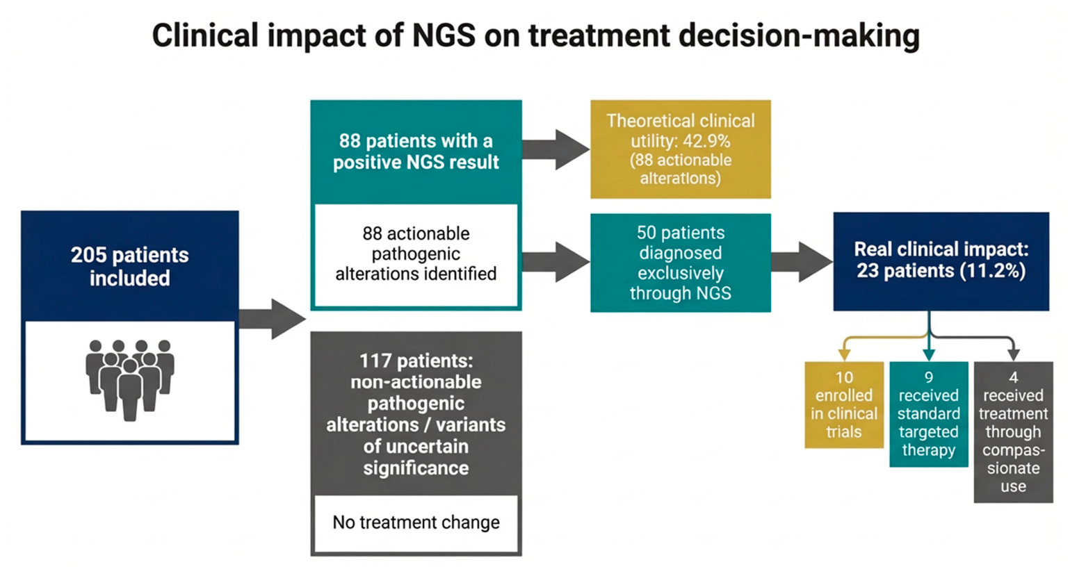

Disclaimer/Publisher’s Note: The statements, opinions and data contained in all publications are solely those of the individual author(s) and contributor(s) and not of MDPI and/or the editor(s). MDPI and/or the editor(s) disclaim responsibility for any injury to people or property resulting from any ideas, methods, instructions or products referred to in the content. |
© 2026 by the authors. Licensee MDPI, Basel, Switzerland. This article is an open access article distributed under the terms and conditions of the Creative Commons Attribution (CC BY) license.