


Abstract
Background: Metastatic-site frequencies reflect both how extensively a cancer spreads and how commonly an organ is involved across cancers. We assessed whether cancer–organ affinity beyond these overall effects is reproducible between autopsy and clinical cohorts. Methods: We analyzed 796,211 organ-involvement events from 332,195 Japanese malignant autopsy cases, covering 76 primary cancers and 48 target sites. We quantified observed-to-expected affinity with empirical-Bayes shrinkage and screened associations with multiple-testing correction. Robust associations underwent anatomical adjudication and fixed-margin testing. External validation compared 143 harmonized cancer–organ pairs (13 cancers, 11 targets) with an independently analyzed MSK-MET-derived clinical matrix. Results: We identified 176 robust enriched associations; 10 remained in a conservative analysis of patterns compatible with distant-organ spread. All 176 remained significant under exact fixed-margin tests. Among 5000 fixed-margin randomized matrices, none contained more than three robust associations, versus 176 observed (Monte Carlo p = 2.00 × 10⁻⁴). Conventional metastatic-site fractions were highly correlated across cohorts (Spearman ρ = 0.861). Residual affinities also showed concordance (Spearman ρ = 0.669; 95% bootstrap confidence interval, 0.416–0.839; Pearson r = 0.747), with matching enrichment or depletion directions in 76.9% of pairs. However, only three of the ten selected distant-organ associations could be compared individually across cohorts, and two showed enrichment in both datasets. Conclusions: Residual metastatic affinity captures partly reproducible population-level patterns beyond metastatic frequency. Validation was restricted to harmonizable categories; these aggregate findings neither estimate individual metastatic risk nor confirm lesion-level distant spread.
1. Introduction
Metastatic dissemination requires malignant cells to leave the primary tumor and establish lesions in permissive secondary tissues. Metastatic organotropism, the preferential involvement of particular organs, reflects anatomical and vascular accessibility, tumor-cell phenotype, host-tissue characteristics, immune interactions, and pre-metastatic niche formation [1,2,3]. Consequently, primary malignancies with similar overall metastatic burdens may nevertheless differ in their patterns of organ involvement.
Conventional metastatic-site fractions describe how frequently particular organs are involved by a given primary malignancy, but they do not distinguish source-specific metastatic preference from the overall abundance of metastases in commonly affected organs. Residual metastatic affinity addresses a different question: does a particular cancer involve a specific organ more frequently than would be expected after accounting for both the total metastatic output of that cancer and the overall frequency of involvement of the target organ? Throughout this study, “residual organotropism” refers to the organ-specific patterns captured by “residual metastatic affinity”, the quantitative measure of source–target enrichment or depletion relative to marginal-independence expectations. For example, the liver may receive numerous metastases because it is a common metastatic target across many cancers, without necessarily exhibiting a disproportionately strong association with every primary malignancy. Conversely, a relatively uncommon metastatic site may demonstrate substantial enrichment for a particular cancer despite having fewer recorded events. This distinction is clinically relevant because metastatic frequency should not automatically be interpreted as preferential organ involvement. Identifying source-specific residual affinities may generate hypotheses relevant to metastatic risk stratification, imaging strategies, and clinical trial design. However, these population-level associations cannot independently predict individual patient risk or justify changes in clinical management.
Autopsy datasets permit pathological assessment of multiple organs, including clinically occult metastatic involvement. Hara et al. analyzed 332,195 malignant autopsies from Japan (1993–2021), reporting 810,206 organ metastases across 76 primary malignancies and 48 target categories [4]. Using metastatic frequencies and proportions, hierarchical and K-means clustering identified seven broad metastatic-pattern groups, with lung, liver, bone, peritoneal, brain, and hematolymphoid involvement contributing prominently [4].
Previous network-based studies described source–target structure and specialist–generalist metastatic behavior [5]. Degree-preserving hypergraph analyses indicated that apparent metastatic hotspots may reflect differences in source and target connectivity rather than pair-specific affinity [6], whereas entropy-based analyses characterized metastatic breadth [7]. Conventional metastatic-site fractions have also shown reproducibility across independent clinical cohorts [8]. The MSK-MET resource [9] and its organotropism matrix distributed through Metient [10] allow us to test whether this reproducibility extends to residual source–target affinity.
Whether pair-specific residual affinity is reproducible across cohorts with different modes of metastatic ascertainment remains uncertain. The Japanese autopsy dataset provides a 76 × 48 matrix of 796,211 anatomically identified organ-involvement events but does not preserve patient-level multi-organ configurations. We therefore asked whether robust source–target enrichment could be identified beyond primary-cancer and target-organ marginal frequencies, and whether the resulting residual-affinity structure would be reproducible in an independently ascertained clinical cohort.
We addressed the first question using a residual-affinity framework in which every source–target edge was evaluated against its marginal-independence expectation. Pairwise enrichment was quantified using observed-to-expected ratios and adjusted residuals, unstable estimates were regularized using empirical-Bayes Gamma–Poisson shrinkage, and multiplicity was addressed by false-discovery-rate screening. An important anatomical limitation is that the source autopsy registry records organ involvement that may represent direct tumor invasion, locoregional dissemination, or distant metastasis. Consequently, involvement of a particular target organ does not necessarily establish a distant metastatic event, even when the corresponding source–target association shows strong statistical enrichment. To address this ambiguity, all statistically robust relationships underwent conservative rule-based anatomical adjudication, distinguishing potentially distant organ involvement from anatomically adjacent or locoregional spread, possible direct extension, and relationships of uncertain classification. Confidence in the distant-organ interpretation was assessed separately, and organ-level classification was not considered equivalent to histopathological confirmation of metastatic routes or individual clinical staging. We then addressed the second question by independently re-estimating the same residual-affinity framework in a conservatively harmonized MSK-MET-derived clinical organotropism matrix and comparing conventional metastatic-site fractions with marginally adjusted residual affinities across cohorts.
Accordingly, the study had five objectives: (i) to map residual metastatic affinity across 76 primary malignancies and 48 target sites; (ii) to distinguish overall organ involvement from preferential source–target association; (iii) to characterize metastatic burden and breadth; (iv) to identify robust associations compatible with distant-organ involvement following conservative anatomical assessment; and (v) to evaluate reproducibility of the residual-affinity structure in the independent MSK-MET cohort.
2. Materials and Methods
2.1. Study Design and Data Source
This study was a secondary analysis of publicly available aggregated data from the study by Hara et al. [4]. The source data were derived from the Annual of Pathological Autopsy Cases in Japan and included autopsies recorded from 1993 to 2021. Primary malignancies were mapped according to ICD-10 categories, and pathological involvement of metastatic organs was recorded on an organ-by-organ basis.
The source study included 332,195 malignant autopsy cases and reported 810,206 organ metastases overall. Metastatic records in which the organ could not be clearly identified were included in the source study’s overall metastatic count but excluded from its metastatic-pattern analysis [4]. The fully identified 48-target supplementary matrix analyzed in the present study contained 796,211 metastatic events.
The source study was approved by the Research Ethics Review Board of Toranomon Hospital (Approval No. 2451), with informed consent waived because of its retrospective design [4]. The present analysis used only published aggregate information and did not access individual-level patient records.
2.2. Data Reconstruction and Validation
The Supplementary Data consisted of one row for each of 76 primary malignancies and 48 metastatic target-site variables. For every source–target pair, the Supplementary File contained the metastatic count, the published metastatic proportion, and metastatic frequency.
The integer metastatic count was extracted separately from each of the 3648 cells, producing a 76 × 48 source–target count matrix. Published proportions and frequencies were retained independently and were not used to reconstruct the count matrix.
The reconstructed matrix was checked against the published numbers of primary malignancies (76), metastatic targets (48), and autopsy cases (332,195). Count-level checks matched the reported prostate-to-bone/bone marrow (n = 2993), liver/intrahepatic-bile-duct-to-trachea/bronchus/lung (n = 15,535), and parathyroid-to-trachea/bronchus/lung (n = 18) values. All 3648 source–target cells were successfully parsed and validated.
2.3. Expected Metastatic Counts Under Marginal Independence
Let Oij denote the observed number of metastatic events from primary malignancy i to target organ j. For each primary malignancy, Ri represented the total number of metastatic events across the 48 target categories; Cj represented the total number of metastatic events received by target j across all 76 primary malignancies; and N represented the total number of events in the 76 × 48 matrix.
The expected count under marginal independence was calculated as:
This null model preserves the total metastatic output of each primary malignancy and the overall metastatic ubiquity of each target while removing source-specific target preference. The pooled marginal-independence model does not explicitly account for sex-specific anatomical organ availability. Consequently, residual-affinity estimates involving reproductive organs may be affected by structural differences between primary-cancer populations. We therefore examined the influence of sex-specific target availability in a separate sensitivity analysis.
The unshrunk effect size was expressed as the observed-to-expected ratio:
Values greater than 1 indicate more metastatic events than expected from marginal frequencies alone, whereas values below 1 indicate relative depletion.
2.4. Residual Screening and Multiple Testing
Pearson residuals were calculated from observed and expected counts. To account for row and column marginal proportions, adjusted Pearson residuals were calculated as:
Two-sided screening p values were derived from the standard-normal approximation to the adjusted residuals. Because all 76 × 48 = 3648 primary–target relationships were examined, p values were adjusted using the Benjamini–Hochberg procedure [11].
The resulting p values and Benjamini–Hochberg q values were used exclusively as screening statistics for aggregated, weighted organ-involvement counts; they do not represent patient-level probabilities and must not be interpreted as estimates of individual metastatic risk or clinical risk prediction. Individual metastatic events arising within the same autopsy are not independent, and the aggregated Supplementary Data do not permit reconstruction of within-patient dependence.
2.5. Empirical-Bayes Estimation of Residual Affinity
Sparse source–target combinations can produce unstable raw observed-to-expected ratios when expected counts are small. We therefore applied an empirical-Bayes Gamma–Poisson model to stabilize affinity estimates [12].
For each primary–target pair, the observed count was modeled conditionally as:
where represents the underlying source-specific affinity relative to the independence expectation.
A Gamma prior centered at the null value of 1 was assumed:
where the Gamma distribution was parameterized by shape and rate, yielding prior mean of 1 and a variance of 1/k. The common prior-strength parameter k was estimated from all source–target pairs by marginal maximum likelihood.
Under this model, the posterior distribution was:
The empirical-Bayes affinity estimate was the posterior mean:
Posterior 95% credible intervals were calculated from the 2.5th and 97.5th percentiles of the posterior Gamma distribution. Effect sizes were additionally represented on a log2 scale. The estimated prior-strength parameter in the final model was approximately 0.76.
The reported 95% posterior credible intervals are model-conditional uncertainty measures derived under the assumed Gamma–Poisson framework. Specifically, they are conditional on the Poisson sampling assumption, the marginal-independence expected counts treated as fixed offsets, and the empirical-Bayes prior-strength parameter estimated from the observed data. They do not incorporate uncertainty in the estimated marginal expectations or prior hyperparameter, nor do they account for correlations among organ involvements within individual patients. Accordingly, these intervals quantify uncertainty in the model-based residual-affinity parameters and should not be interpreted as patient-level confidence intervals or estimates of individual metastatic risk.
2.6. Definition of a Robust Residual-Affinity Signal
To prevent sparse or clinically trivial source–target combinations from dominating the results, a relationship was considered a robust enriched affinity only when all of the following criteria were satisfied:
- Observed metastatic count ≥ 20;
- Expected metastatic count ≥ 5;
- At least 100 autopsy cases for the corresponding primary malignancy;
- Benjamini–Hochberg event-level q value < 0.05;
- Empirical-Bayes O/E ≥ 2.0;
- Lower bound of the 95% empirical-Bayes credible interval > 1.0.
These thresholds were pragmatic safeguards against sparse-data artifacts, not clinically validated cutoffs. Minimum observed and expected counts limited weakly supported estimates, while empirical-Bayes O/E ≥ 2.0 required at least twofold enrichment after shrinkage. Multiple-testing adjustment and the credible-interval condition provided further screening safeguards. Sensitivity results appear in Supplementary Methods S1 and Tables S1–S3.
Application of these predefined criteria produced 176 robust enriched source–target relationships.
Threshold sensitivity was evaluated using a 27-scenario factorial design and separate source-cohort support checks. Parameter combinations and retention rules for the strict distant-organ set are detailed in Supplementary Methods S1 and Tables S1–S3.
Detailed threshold definitions, the complete 27-scenario factorial sensitivity analysis, and source-cohort support analyses are provided in Supplementary Methods S1 and Supplementary Tables S1 and S2.
A separate post hoc sensitivity analysis excluded five sex-specific reproductive targets from the Japanese discovery matrix and re-estimated the residual-affinity model. Eligibility criteria, recalculation procedures, and results for the retained distant-organ relationships are documented in Supplementary Methods S1 and Supplementary Data S16.
2.7. Fixed-Margin Null-Model Robustness Analysis
Fixed-margin analyses tested whether pair-specific enrichment persisted after conditioning on the observed total metastatic output of each primary malignancy and the overall involvement of each target organ in the 76 × 48 matrix.
Each of the 3648 source–target pairs was examined with an exact two-sided Fisher test and, as a directional sensitivity check, a one-sided hypergeometric enrichment test. p values were adjusted across all pairs with the Benjamini–Hochberg procedure.
At the matrix level, 5000 randomized fixed-margin tables were evaluated using the original robust-affinity criteria, with the empirical-Bayes prior-strength parameter held fixed at its observed-data estimate. The full Monte Carlo procedure is described in Supplementary Methods S2 and Tables S5 and S6.
Detailed implementation of the fixed-margin analysis, including the exact conditional tests, Monte Carlo randomization procedure, and complete robustness outputs, is provided in Supplementary Methods S2 and Supplementary Tables S5 and S6.
Because individual-level multi-organ metastatic configurations were unavailable in the aggregated source dataset, this weighted fixed-margin contingency-table analysis was used as the structure-preserving null model appropriate to the available data rather than a patient-level hypergraph rewiring procedure.
2.8. Conservative Rule-Based Anatomical Adjudication and Distant-Organ Sensitivity Analysis
Statistical enrichment was not equated with distant metastatic spread because the autopsy registry includes organ involvement potentially attributable to direct invasion as well as metastasis [4]. Accordingly, all 176 robust associations underwent four-category rule-based anatomical classification. This adjudication was strictly descriptive and non-inferential, based on organ-level anatomical relationships rather than independently verified lesion-level evidence; consequently, it could neither confirm noncontiguous metastatic lesions nor establish individual routes of metastatic dissemination.
Each relationship was assigned to one of four categories: (i) distant, when involvement of an anatomically distinct target organ was compatible with nonlocal metastatic dissemination and direct extension was not the more plausible explanation; (ii) possible direct extension, when anatomical continuity made direct invasion plausible; (iii) locoregional/adjacent, when involvement was confined to the same organ system, regional anatomical compartment, or a pattern of local or intracavitary dissemination that did not permit separate-organ colonization to be distinguished from regional spread; and (iv) uncertain, when aggregate organ labels, histological heterogeneity, or anatomical ambiguity prevented reliable classification. Importantly, this classification was based on organ-level anatomical relationships rather than direct pathological evidence of discontinuity between individual lesions. The possible route of dissemination, including transcoelomic spread, was considered separately and did not automatically determine anatomical classification. Consequently, involvement of a distinct target organ could be retained as potentially distant with moderate confidence when alternative dissemination mechanisms could not be excluded.
A confidence level of high, moderate, or low was additionally assigned according to the anatomical specificity of the source–target relationship and the plausibility of competing mechanisms. The strict distant-organ sensitivity analysis retained only relationships classified as distant with high or moderate confidence.
Two relationships, appendix-to-ovaries and caecum-to-ovaries, were retained as potentially distant with moderate confidence because secondary ovarian involvement from these gastrointestinal primaries is compatible with metastatic dissemination, while transcoelomic spread and possible anatomical continuity cannot be excluded using aggregate organ-level data. To assess the dependence of the distant-organ interpretation on these anatomically ambiguous relationships, we additionally examined a more restrictive eight-relationship subset excluding both gastrointestinal-to-ovarian associations. This supplementary restriction did not involve re-estimation of the statistical model. To further assess the influence of anatomically ambiguous and potentially locoregional involvement on the marginal-independence reference model, we performed a separate post hoc sensitivity analysis using an anatomically restricted source–target matrix. Four non-organ-specific or hematolymphoid primary categories and 17 potentially ambiguous, compartmental, or sex-specific target categories were excluded. Additional source–target combinations belonging to the same broad anatomical system were treated as ineligible for this sensitivity analysis. Expected counts were re-estimated using a Poisson log-linear independence model with anatomical eligibility constraints, fitted by iterative proportional fitting. The empirical-Bayes Gamma–Poisson prior-strength parameter and posterior affinity estimates were subsequently recalculated using the restricted reference matrix. The analysis focused on the six high-confidence distant-organ associations whose target organs remained eligible. Complete eligibility rules, the source–target inclusion mask, and the resulting estimates are provided in Supplementary Data S15. The complete classification, confidence assignment, inclusion decision, and rationale for all 176 robust relationships are provided in Supplementary Methods S3 and Supplementary Table S7.
2.9. Specialist–Generalist Metastatic Architecture
For each primary malignancy, metastatic events were normalized across the 48 target categories to generate a source-specific metastatic distribution. If pij denotes the proportion of metastatic events from primary i involving target j, metastatic diversity was quantified using Shannon entropy [7,13]:
The corresponding effective number of metastatic targets was calculated as:
Lower values represent relatively specialized metastatic distributions concentrated in fewer organs, whereas higher values indicate more generalized dissemination across target categories.
Metastatic burden was characterized separately as the number of metastatic events represented in the 48-target matrix per autopsy case. This allowed metastatic breadth and metastatic intensity to be examined as distinct dimensions.
2.10. Target Ubiquity and Affinity
Target ubiquity was defined as the total number of metastatic events received by each target across all 76 primary malignancies.
Source-specific affinity was quantified independently using empirical-Bayes O/E estimates. For visualization of the distinction between ubiquity and affinity, target-site ubiquity was compared with the strongest adequately supported empirical-Bayes affinity involving that target.
This analysis was descriptive and was intended to illustrate that frequent involvement of an organ across cancers does not necessarily imply strong source-specific preference.
2.11. Independent Cross-Cohort Validation in MSK-MET
External validation used the publicly available cancer-type-by-metastatic-site matrix from the MSK-MET clinical cohort [9], distributed through the Metient framework [10]. The non-anatomical “Unspecified” target category was excluded. Each cell counts patients with recorded involvement at that site; a patient may contribute to more than one site count. No individual-level patient records were accessed.
Only primary-cancer and target-site categories with direct or near-direct anatomical correspondence were harmonized; heterogeneous categories requiring subjective aggregation were excluded. This yielded 13 primary cancers, 11 metastatic targets, and 143 matched relationships. The mapping was defined independently of observed concordance and is documented in Supplementary Table S8.
The marginal-independence expectations and empirical-Bayes prior parameters were estimated independently from the complete available source–target matrices of each cohort, before cross-cohort harmonization. Specifically, the Japanese discovery estimates were derived from the full 76 × 48 matrix, whereas the MSK-MET estimates were calculated from the 27 × 20 cancer-type-by-metastatic-site matrix after exclusion of the non-anatomical “Unspecified” target category. For each cohort, expected counts were calculated from its own complete row and column marginal totals, and the Gamma–Poisson prior-strength parameter was independently estimated by marginal maximum likelihood (Japan: k = 0.7606; MSK-MET: k = 1.0634). Harmonization was performed only after estimation, selecting 143 anatomically matched relationships across 13 primary-cancer and 11 metastatic-target categories. Neither the expected counts nor the prior parameters were transferred between cohorts, and the empirical-Bayes models were not re-estimated on the harmonized 13 × 11 submatrices.
For the 143 matched relationships, we compared conventional within-primary metastatic-site fractions, unshrunk observed-to-expected ratios, and empirical-Bayes observed-to-expected ratios. Validation used continuous effect estimates rather than requiring MSK-MET relationships to meet the Japanese discovery-stage thresholds, avoiding selection based on cohort-specific support or statistical power.
Cross-cohort concordance was assessed using Spearman and Pearson correlations of independently estimated empirical-Bayes residual affinities. Two-way source-and-target cluster bootstrap resampling (10,000 replicates) provided 95% confidence intervals; implementation details are given in Supplementary Methods S4.
A matrix-label permutation test (20,000 permutations) assessed whether the cross-cohort residual-affinity concordance exceeded arbitrary source–target label alignment (Supplementary Methods S4 and Supplementary Code S3).
Directional reproducibility was the proportion of relationships sharing the same enrichment (empirical-Bayes O/E > 1) or depletion (O/E < 1) direction across cohorts. Influence was assessed by leave-one-primary-out and leave-one-target-out correlations, together with descriptive per-primary analyses.
A post hoc structural sensitivity check excluded breast-to-breast, kidney-to-kidney, ovary-to-ovary, and the anatomically unavailable prostate-to-ovary relationship, leaving 139 matched relationships.
A separate post hoc analysis excluded sex-specific reproductive targets from both cohort matrices and re-estimated the models before comparing the 130 remaining harmonized relationships. Full exclusion rules, resampling procedures, and output are given in Supplementary Methods S4, Supplementary Data S16, and Supplementary Code S4.
2.12. Visualization
The residual organotropism atlas was based on empirical-Bayes log2(O/E) values for all 76 × 48 source–target pairs. Primary malignancies and metastatic targets were ordered using average-linkage hierarchical clustering with Euclidean distance for visualization only; no discrete biological cluster classification was inferred from this ordering.
The strict distant-organ sensitivity analysis was displayed using a forest plot of empirical-Bayes O/E estimates and corresponding 95% credible intervals. Moderate-confidence relationships were explicitly distinguished from high-confidence relationships.
The specialist–generalist analysis plotted the effective number of metastatic targets against metastatic events per autopsy case.
Cross-cohort reproducibility was visualized using paired scatter plots of the 143 harmonized source–target relationships. The first panel compared conventional within-primary metastatic-site fractions between cohorts, whereas the second compared independently estimated empirical-Bayes log2 observed-to-expected ratios. Horizontal and vertical reference lines at log2(O/E) = 0 indicated the marginal-independence expectation. Per-primary cross-cohort Spearman correlations were displayed separately as a Supplementary Figure.
2.13. Statistical Software and Reproducibility
Statistical analyses used Python 3 with pandas 2.2.3, NumPy 2.1.3, SciPy 1.16.3, and Matplotlib 3.10.0. The reproducibility package includes the complete pair-level statistics, threshold and fixed-margin analyses, anatomical adjudication, harmonization, cross-cohort validation, bootstrap and permutation procedures, influence and sensitivity analyses, and figure-generation code. Post hoc anatomical-reference and sex-specific analyses are documented in Supplementary Code S4 and its input CSV files.
All statistical tests were two-sided where applicable. Monte Carlo, bootstrap, and permutation procedures used fixed pseudorandom seeds to permit exact computational reproduction. No patient-level survival, temporal, causal, or multivariable clinical inference was attempted because both analyses relied on aggregate source–target information.
The principal dataset characteristics, analytical definitions, and predefined criteria used throughout the study are summarized in Table 1.
Table 1.
Dataset characteristics and analytical framework.
3. Results
3.1. Target-Site Ubiquity Versus Pair-Specific Residual Affinity
The validated metastatic matrix comprised 76 primary malignancies and 48 metastatic target sites derived from 332,195 autopsy cases. The 48-site matrix contained 796,211 recorded primary-to-target metastatic events. To distinguish metastatic affinity from simple target-site frequency, observed counts for each of the 3648 primary–target combinations were compared with counts expected under a marginal-independence model. Effect sizes were expressed as observed-to-expected ratios and further stabilized using an empirical-Bayes Gamma–Poisson model.
The analysis identified 176 robustly enriched primary–target relationships in the Japanese discovery cohort, of which 10 were retained after conservative anatomical adjudication for the strict distant-organ analysis; independent external validation subsequently evaluated 143 anatomically harmonized source–target relationships. These signals represented residual organotropism: combinations occurring more frequently than expected after accounting simultaneously for the overall metastatic burden of the primary malignancy and the overall ubiquity of the metastatic target.
The residual-affinity analysis therefore produced a markedly different perspective from simple metastatic frequency. Highly prevalent metastatic sites such as the lung and liver accounted for large numbers of recorded organ-involvement events, but the strongest source-specific affinities were not necessarily directed toward the most frequently involved organs. This distinction supports the central analytical concept that metastatic ubiquity and metastatic affinity represent related but non-equivalent properties. The relationship between overall target ubiquity and the strongest adequately supported source-specific residual affinity is shown in Figure 1.
Figure 1.
Target ubiquity does not imply source-specific metastatic affinity. Relationship between target-site ubiquity and the strongest adequately supported pair-specific residual affinity for each target organ. The x-axis represents the log10-transformed total number of metastatic events involving each target across all 76 primary malignancies and therefore quantifies target ubiquity. The y-axis represents the maximum empirical-Bayes log2 observed-to-expected ratio among source–target relationships meeting the minimum support criteria for that target. Selected target labels highlight illustrative cases of high target ubiquity and/or strong adequately supported residual affinity. These labels are provided for visual orientation and do not represent an additional statistical selection or ranking. The absence of a one-to-one relationship between the two axes illustrates the central distinction between metastatic frequency and source-specific residual affinity. “Brain N” is the original label for the brain-involvement target category in the Japanese autopsy source matrix. The label is retained verbatim for traceability and does not denote a separate statistical variable.
Four complementary descriptors are distinguished throughout the results: target-site ubiquity, defined as the total number of recorded organ-involvement events at each target across all primary malignancies; residual metastatic affinity, quantified by the pair-specific empirical-Bayes observed-to-expected ratio; metastatic burden, expressed as recorded organ-involvement events per autopsy case for each primary malignancy; and metastatic breadth, quantified by the effective number of metastatic target sites, exp(H). Target-site ubiquity and residual affinity are illustrated in Figure 1, Figure 2 and Figure 3, whereas metastatic burden and breadth are compared in Figure 4.
Figure 2.
Empirical-Bayes residual organotropism atlas across 76 primary malignancies and 48 metastatic targets. Heatmap of empirical-Bayes log2 observed-to-expected ratios for all 3648 primary–target combinations. Positive values indicate more metastatic events than expected after accounting for the overall metastatic output of the primary malignancy and the overall ubiquity of the target; negative values indicate relative depletion. Empirical-Bayes shrinkage reduces instability in sparse source–target combinations. For visualization only, log2(O/E) values below −3 are displayed at −3, and values above +3 are displayed at +3. This clipping affects only the heatmap color scale and does not alter the underlying affinity estimates or statistical analyses. Rows and columns are ordered by average-linkage hierarchical clustering using Euclidean distance on the shrunken affinity matrix. Clustering is used only to organize the heatmap and does not define discrete biological classes. The label “Brain N” identifies the brain-involvement target category as coded in the Japanese source matrix.
Figure 3.
Strict distant-organ residual-affinity sensitivity analysis. Forest plot showing empirical-Bayes observed-to-expected ratios and 95% posterior credible intervals for the 10 primary–target relationships retained after statistical filtering and conservative rule-based anatomical adjudication of all 176 robust affinity signals. The vertical reference line at O/E = 1 represents the marginal-independence expectation. Seven associations were classified as high-confidence distant-organ relationships. The dagger identifies the three moderate-confidence relationships—appendix-to-ovary, breast-to-uterus, and caecum-to-ovary—for which a metastatic interpretation was considered plausible but additional mechanistic or histological uncertainty remained. The distant-only set is a conservative sensitivity analysis and should not be interpreted as reconstruction of clinical M-stage. The label “Brain N” identifies the brain-involvement target category as coded in the Japanese source matrix.
Figure 4.
Specialist–generalist architecture of metastatic dissemination. Primary malignancies are positioned according to the breadth and intensity of their metastatic distributions. The x-axis represents the effective number of metastatic target sites, calculated as exp(H), where H is the Shannon entropy of the source-specific distribution across the 48 target categories. Lower values indicate metastatic specialization, whereas higher values indicate a more generalized metastatic distribution. The y-axis represents the number of metastatic events recorded across the 48 target categories per autopsy case for each primary malignancy. Selected extreme specialist, generalist, and high-burden malignancies are labeled. Metastatic breadth and burden describe complementary source-level properties of metastatic dissemination. Neither measure directly quantifies pair-specific residual affinity, and malignancies with similar breadth or burden may exhibit different organ-specific affinities. Residual affinity is evaluated separately in Figure 1, Figure 2 and Figure 3.
The complete empirical-Bayes residual-affinity landscape across all 76 primary malignancies and 48 metastatic targets is shown in Figure 2.
3.2. Rule-Based Anatomical Adjudication Separates Anatomical Extension from Distant Organotropism
A critical feature of the source autopsy registry is that organ involvement may represent either invasion or distant metastasis. Consequently, residual enrichment alone could not be interpreted as evidence of systemic metastatic organotropism. All 176 statistically robust affinity signals were therefore subjected to rule-based anatomical adjudication.
Four categories were used: distant metastasis, locoregional/anatomically adjacent involvement, possible direct extension, and uncertain mechanism. Of the 176 robust relationships, 14 were classified as potentially distant, 57 as locoregional or anatomically adjacent, 62 as compatible with direct extension, and 43 as uncertain. Four of the 14 potentially distant relationships were excluded because confidence in the metastatic interpretation was low. The strict distant-organ sensitivity analysis consequently retained 10 primary–target relationships: seven high-confidence and three moderate-confidence signals. The complete rule-based anatomical classification, confidence assignment, inclusion decision, and pair-specific rationale for all 176 robust relationships are provided in Supplementary Table S7.
This adjudication substantially altered the apparent ranking of residual affinities. Very large enrichments involving anatomically contiguous structures were removed from the distant analysis, preventing local extension or same-compartment dissemination from being misinterpreted as systemic organotropism.
3.3. Strict Distant-Organ Affinity Signals
All 10 retained distant-organ relationships remained enriched after empirical-Bayes shrinkage, with posterior observed-to-expected ratios ranging from 2.23 to 3.63 and 95% credible intervals entirely above unity.
The largest effect was observed for appendiceal malignancy involving the ovary (EB O/E 3.63, 95% CrI 2.73–4.65), although this relationship was classified as moderate confidence because transcoelomic dissemination represents a plausible mechanism. The strongest high-confidence relationship was prostate malignancy to bone/bone marrow (EB O/E 3.20, 95% CrI 3.09–3.32; observed 2993 versus expected 933.5).
Several breast-cancer associations remained prominent after adjustment for background target frequency. These included breast-to-ovary involvement (EB O/E 2.91, 95% CrI 2.67–3.15), breast-to-pituitary/cephalopharyngeal region involvement (2.85, 2.32–3.43), breast-to-uterus involvement (2.54, 2.30–2.79), breast-to-spinal meningeal involvement (2.29, 1.83–2.80), and breast-to-meningeal involvement (2.28, 2.01–2.57). The breast-to-uterus relationship was retained as a moderate-confidence signal because histological subtype information, particularly lobular versus ductal phenotype, was unavailable.
Additional distant signals included caecal malignancy to the ovary (EB O/E 2.53, 95% CrI 2.08–3.03), testicular malignancy to the brain (2.50, 1.93–3.15), and bronchus/lung malignancy to the brain (2.23, 2.18–2.29). All retained relationships showed strong event-level residual screening evidence after Benjamini–Hochberg correction.
The complete empirical-Bayes estimates and confidence classifications for the 10 strict distant-organ relationships are provided in Supplementary Table S4.
Across the 10 distant-organ relationships, 10,781 metastatic events were observed compared with 4304.9 expected under the independence model, corresponding to a descriptive aggregate observed-to-expected ratio of 2.50. Restriction to the seven high-confidence relationships yielded a nearly identical aggregate ratio of 2.50, indicating that the principal result was not driven by the three moderate-confidence relationships.
In a more restrictive anatomical-interpretation sensitivity analysis, appendix-to-ovaries and caecum-to-ovaries were excluded because the available aggregate data could not distinguish noncontiguous metastatic deposits from transcoelomic dissemination or possible direct extension. The remaining eight relationships comprised all seven high-confidence distant-organ associations and one moderate-confidence association (breast-to-uterus). All eight had satisfied the original predefined statistical robustness criteria. This restricted analysis demonstrates that the principal distant-organ findings do not depend on the inclusion of the two gastrointestinal-to-ovarian relationships, although the route of dissemination cannot be established from the available data. The post hoc anatomically restricted reference-matrix analysis retained 72 primary-cancer categories, 31 metastatic target categories, and 1972 eligible source–target combinations, comprising 389,845 recorded organ-involvement events. The empirical-Bayes prior-strength parameter was re-estimated as k = 1.6208. All six evaluable high-confidence distant-organ relationships retained empirical-Bayes observed-to-expected ratios above 2.0, ranging from 2.215 to 3.143, with the lower bounds of their 95% model-conditional credible intervals remaining above unity. These results indicate that the principal evaluable distant-organ enrichments persisted under a substantially more restrictive anatomical reference model. The complete eligibility mask and pair-specific estimates are presented in Supplementary Data S15. Because the restricted model involved post hoc anatomical eligibility constraints, these results were interpreted as an effect-size sensitivity analysis rather than as a repetition of the original event-level multiple-testing procedure.
The complete set of 10 relationships retained in the strict distant-organ sensitivity analysis, together with their observed and expected counts, empirical-Bayes effect estimates, credible intervals, and confidence classifications, is summarized in Table 2.
Table 2.
Residual affinity signals retained in the strict distant-organ sensitivity analysis.
The magnitude and uncertainty of the empirical-Bayes affinity estimates for the strict distant-organ set are visualized in Figure 3.
Key Findings: Biological Interpretation of the Ten Distant-Organ Signals
The ten retained associations represent four recognizable patterns of organ-specific involvement: preferential skeletal involvement in prostate cancer; brain and meningeal involvement associated with lung, testicular, and breast malignancies; ovarian, uterine, and pituitary-region involvement associated with breast cancer; and ovarian involvement associated with appendiceal and caecal malignancies. Their principal significance is that these source–target relationships remain disproportionately represented after accounting for both primary-cancer metastatic output and overall target-organ ubiquity. Seven associations were classified as high-confidence and three as moderate-confidence on anatomical grounds. The latter require particular caution because alternative dissemination mechanisms or histological heterogeneity cannot be resolved from aggregate data. These findings quantify population-level patterns consistent with recognized metastatic phenotypes; they do not establish lesion-level metastatic routes, causal biological mechanisms, or individual metastatic risk.
3.4. Metastatic Burden and Breadth: Specialist–Generalist Architecture
Beyond individual primary–target affinities, the 76 primary malignancies varied substantially in the breadth of their metastatic distributions. Entropy-based measures demonstrated a continuum between metastatic specialists, whose observed events were concentrated in relatively few target sites, and metastatic generalists, whose distributions were spread across a broader set of organs.
This source-level architecture complements the edge-level affinity analysis. A malignancy may exhibit broad metastatic dissemination while retaining specific target affinities, whereas another malignancy may show a narrow metastatic distribution dominated by one or a few strongly favored organs. These observations indicate that metastatic burden, metastatic breadth, and source–target affinity capture distinct components of metastatic behavior. The relationship between metastatic breadth, expressed as the effective number of metastatic targets, and metastatic burden, expressed as metastatic events per autopsy case, is shown in Figure 4.
3.5. Threshold-Sensitivity Analysis
The number of statistically robust residual-affinity relationships varied, as expected, with the stringency of the filtering criteria, ranging from 86 to 350 across the 27 factorial threshold combinations. The composition of the primary strict distant-organ set was insensitive to variation in the observed- and expected-count thresholds. All 10 relationships, including all seven high-confidence relationships, were retained in every scenario using minimum empirical-Bayes O/E thresholds of either 1.5 or 2.0, corresponding to 18 of the 27 evaluated scenarios.
Complete results across all 27 threshold combinations are reported in Supplementary Table S1, and sensitivity to minimum source-cohort size is summarized in Supplementary Table S2. The scenario-level retention of each of the 10 strict distant-organ relationships is shown in Supplementary Table S3 and Supplementary Figure S1.
Increasing the empirical-Bayes O/E threshold to 2.5 retained seven of the 10 strict distant-organ relationships and four of the seven high-confidence relationships. Three relationships fell below the more stringent empirical-Bayes O/E threshold of 2.5: breast-to-spinal-cord-membrane involvement (EB O/E = 2.29), breast-to-meningeal involvement (2.28), and bronchus/lung-to-brain involvement (2.23). Their exclusion reflected the imposed effect-size cutoff, not inadequate statistical support or credible-interval evidence.
Sensitivity to minimum source-cohort size was similarly limited. Thresholds of 50 and 100 autopsy cases each yielded 176 robust relationships, whereas a threshold of 200 cases yielded 173. All 10 strict distant-organ relationships and all seven high-confidence relationships were retained at each source-support threshold. These analyses indicate that the principal distant-organ findings were not materially dependent on the selected minimum observed-count, expected-count, or source-cohort support criteria.
The additional anatomical-availability sensitivity analysis excluded five sex-specific reproductive target categories representing 20,567 recorded organ-involvement events. The restricted Japanese matrix contained 76 primary malignancies, 43 metastatic targets, and 775,644 recorded events. Re-estimation yielded an empirical-Bayes prior-strength parameter of k = 0.7611, compared with k = 0.7606 in the complete matrix. Under the unchanged robustness criteria, 153 source–target relationships qualified as robust enrichments. Of the 151 originally robust relationships not involving an excluded target, 150 remained robust, while three additional relationships newly satisfied the criteria. All six strict distant-organ relationships involving retained target organs continued to meet every original robustness criterion. Their recalculated empirical-Bayes observed-to-expected ratios ranged from 2.192 to 3.172. Detailed results are provided in Supplementary Data S16.
3.6. Fixed-Margin Null-Model Robustness
The residual-affinity architecture remained strongly supported under an exact fixed-margin null model preserving the total metastatic output of every primary malignancy and the total number of recorded organ-involvement events at every target organ. In practical terms, we kept the overall number of recorded metastatic events for each primary cancer and each target organ unchanged, then asked whether specific cancer–organ combinations remained disproportionately represented beyond what these totals alone would predict. All 176 relationships identified by the primary robust-affinity criteria remained significant after exact two-sided fixed-margin testing and Benjamini–Hochberg correction (176/176; maximum fixed-margin q = 9.97 × 10⁻⁴). Complete fixed-margin results for the primary robust-affinity set are summarized in Supplementary Table S5.
The result was unchanged for the rule-based anatomically adjudicated distant-organ subset: all 10 strict distant-organ relationships, including all seven high-confidence relationships, remained significant under the fixed-margin analysis. Pair-specific fixed-margin statistics for these 10 relationships are provided in Supplementary Table S6.
Matrix-level randomization provided additional evidence that the observed residual-affinity architecture could not be explained by source and target marginals alone. Across 5000 randomized 76 × 48 contingency tables preserving the observed row and column totals exactly, 4473 simulations yielded no relationships satisfying the complete primary robust-affinity criteria, 503 yielded one relationship, 23 yielded two relationships, and one yielded three relationships. No randomized matrix yielded more than three robust relationships, compared with 176 in the observed matrix. None approached the observed result, corresponding to an add-one empirical Monte Carlo p = 2.00 × 10⁻⁴. The fixed-margin null distribution is shown in Supplementary Figure S2. The complete pair-level fixed-margin results are provided in Supplementary Data S6–S8, and the simulation-level Monte Carlo null distribution is provided in Supplementary Data S9.
Together, these analyses indicate that the observed residual-affinity architecture contains substantial pair-specific structure beyond that expected from the marginal metastatic activity of primary malignancies and the marginal ubiquity of metastatic target organs.
3.7. Cross-Cohort Reproducibility of Residual Metastatic Affinity
Independent external validation was performed using the MSK-MET-derived clinical organotropism matrix. Because the Japanese autopsy registry and MSK-MET employ different primary-cancer and metastatic-target taxonomies, harmonization was restricted to direct or near-direct anatomical correspondences, excluding broad mappings that would require aggregation of biologically heterogeneous categories (Supplementary Table S8). This conservative approach retained 13 primary-cancer categories and 11 metastatic targets, yielding 143 matched source–target relationships. These represented 3.9% of the 3648 relationships in the complete Japanese discovery matrix; the remaining 3505 relationships (96.1%) were outside the harmonized comparison because their primary-cancer categories, target categories, or both were not included in the restricted mapping. This restriction affected only the scope of cross-cohort comparison: marginal-independence expected counts and empirical-Bayes prior parameters were estimated independently from each cohort’s complete available matrix before harmonization. Within MSK-MET, the independently estimated prior-strength parameter was k = 1.063; neither the Japanese expected counts nor its shrinkage parameter was transferred to the validation dataset.
Conventional within-primary metastatic-site fractions showed strong agreement between the Japanese autopsy cohort and MSK-MET (Spearman ρ = 0.861; two-way bootstrap 95% CI, 0.643–0.939). Substantial concordance remained after removal of first-order source and target marginal effects. For unshrunk observed-to-expected ratios, the cross-cohort Spearman correlation was ρ = 0.621 (95% CI, 0.290–0.833). For empirical-Bayes residual affinities, the Spearman correlation was ρ = 0.669 (95% CI, 0.416–0.839), and the Pearson correlation was r = 0.747 (95% CI, 0.504–0.875). The comparison between conventional metastatic-site fractions and residual metastatic affinity is shown in Figure 5, and the three cross-cohort representations are summarized in Table 3.
Figure 5.
Cross-cohort reproducibility of conventional metastatic-site distributions and residual metastatic affinity. (A) Concordance of conventional within-primary metastatic-site fractions between the Japanese pathological autopsy cohort and the independent MSK-MET-derived clinical cohort across 143 harmonized source–target relationships spanning 13 primary cancers and 11 metastatic targets. Conventional metastatic-site fractions showed strong cross-cohort agreement (Spearman ρ = 0.861; two-way bootstrap 95% CI, 0.643–0.939). (B) Concordance of independently estimated empirical-Bayes residual affinities after simultaneous adjustment for primary-cancer metastatic output and target-site ubiquity. Residual affinities remained substantially correlated across cohorts (Spearman ρ = 0.669; two-way bootstrap 95% CI, 0.416–0.839; Pearson r = 0.747; 95% CI, 0.504–0.875). Enrichment-versus-depletion direction was concordant for 110 of 143 relationships (76.9%). Horizontal and vertical reference lines at log2(O/E) = 0 denote the marginal-independence expectation. The solid line in panel B represents the least-squares linear fit and is included for visualization only.
Table 3.
Cross-cohort benchmarking of conventional metastatic-site frequency and residual metastatic affinity.
Directional agreement provided complementary evidence of cross-cohort reproducibility. Of the 143 matched relationships, 110 (76.9%) retained the same enrichment-versus-depletion direction relative to the marginal-independence expectation in both cohorts. In a structure-preserving matrix-label permutation analysis, the observed residual-affinity concordance exceeded that obtained under arbitrary source and target label alignment (20,000 permutations; add-one empirical p = 5.0 × 10⁻⁵).
The overall result was not dependent on any single primary malignancy or metastatic target. Leave-one-primary-out Spearman correlations ranged from 0.639 to 0.715, whereas leave-one-target-out correlations ranged from 0.635 to 0.732. Per-primary concordance was heterogeneous across the 13 cancer types and is shown descriptively in Supplementary Figure S3, indicating that cross-cohort reproducibility was stronger for some primary malignancies than for others. Detailed per-primary concordance and influence-analysis results are provided in Supplementary Data S11–S13.
Finally, the post hoc structural sensitivity analysis excluded the three exact source–target self-organ combinations (breast-to-breast, kidney-to-kidney, and ovary-to-ovary) together with the anatomically unavailable prostate-to-ovary combination. Among the remaining 139 relationships, cross-cohort residual-affinity concordance remained substantial (Spearman ρ = 0.645; Pearson r = 0.677), with concordant enrichment-versus-depletion direction in 76.3% of relationships. Thus, the primary external-validation result was not materially driven by self-organ or structurally unavailable source–target combinations. The complete structural sensitivity results are provided in Supplementary Data S14.
In the additional sex-specific target-exclusion sensitivity analysis, the restricted MSK-MET matrix contained 27 primary-cancer categories and 17 metastatic targets, with an independently re-estimated empirical-Bayes prior-strength parameter of k = 1.4574. Across the 130 remaining harmonized source–target relationships, empirical-Bayes residual affinities remained substantially concordant between cohorts (Spearman ρ = 0.643; two-way bootstrap 95% CI, 0.382–0.819; Pearson r = 0.692). Enrichment-versus-depletion direction was concordant in 96 of 130 relationships (73.85%). The matrix-label permutation test remained significant (20,000 permutations; empirical p = 5.0 × 10⁻⁵). These findings indicate that cross-cohort residual-affinity structure was not solely attributable to the inclusion of sex-specific reproductive target categories. The complete recalculated discovery-cohort and cross-cohort sensitivity results are provided in Supplementary Data S16.
We additionally examined pair-specific cross-cohort concordance for the 10 relationships retained in the strict distant-organ analysis. Under the predefined conservative anatomical harmonization, only three of these relationships had direct source–target correspondences in MSK-MET. Appendix-to-ovary involvement showed enrichment in both cohorts (empirical-Bayes O/E: 3.628 in Japan versus 5.104 in MSK-MET), as did prostate-to-bone involvement (3.204 versus 2.376). In contrast, breast-to-ovary involvement showed substantial enrichment in the Japanese autopsy cohort (2.906) but an approximately null residual affinity in MSK-MET (0.985). Thus, two of the three evaluable distant-organ associations retained the same enrichment direction, whereas one did not. The remaining seven associations could not be evaluated individually without introducing broader or potentially heterogeneous anatomical mappings. These findings indicate that the overall cross-cohort residual-affinity concordance reflects reproducibility of the harmonized population-level architecture rather than uniform replication of every individual distant-organ association. Pair-specific estimates are provided in Supplementary Data S10.
4. Discussion
4.1. Metastatic Frequency and Metastatic Affinity Are Not Interchangeable
The central contribution of this study is the distinction between metastatic frequency and residual metastatic affinity. Previous proportion-based and clustering analyses, including the original Japanese autopsy study, characterized the frequency of organ involvement and identified groups of primary malignancies with similar metastatic distributions [4]. Although these approaches provide valuable descriptions of metastatic dissemination, they do not directly quantify whether a particular cancer–organ association is disproportionately represented after accounting for both the total metastatic output of the primary malignancy and the overall ubiquity of the target organ. In contrast, our approach evaluates pair-specific enrichment against marginal-independence expectations and stabilizes the resulting affinity estimates through empirical-Bayes shrinkage. Thus, residual metastatic affinity complements rather than replaces conventional proportion-based and clustering analyses.
This residual formulation changes the interpretation of metastatic pattern data. The lungs and liver are highly ubiquitous metastatic targets and therefore accumulate large absolute numbers of metastases from many primary malignancies. A high raw proportion involving these organs does not necessarily imply a uniquely strong biological affinity. Conversely, less ubiquitous organs may show marked enrichment for selected primary malignancies despite contributing relatively few metastases in absolute terms.
The distinction can be summarized as follows: ubiquity describes how often a target is colonized overall, whereas affinity describes whether a particular source colonizes that target more often than expected given the metastatic activity of both the source and target.
4.2. Consistency with Established Metastatic Patterns
The recovery of several well-established source–target associations after statistical shrinkage and conservative rule-based anatomical adjudication is consistent with known metastatic patterns but should not be regarded as independent biological validation. Throughout this study, the term “strict distant-organ” denotes a conservatively selected, organ-level sensitivity subset; it does not imply histopathological verification of distant metastasis or lesion-level evidence of noncontiguous spread.
Prostate-to-bone involvement was among the strongest high-confidence distant relationships, consistent with the well-established osteotropism of advanced prostate malignancy [14]. Bronchus/lung-to-brain involvement also remained strongly enriched despite the high overall frequency of both lung cancer and brain metastasis [15]. The persistence of this signal after adjustment for marginal frequencies indicates that the association cannot be explained solely by the abundance of lung primary tumors in the autopsy series.
The analysis similarly recovered several recognized patterns of breast-cancer dissemination. Meningeal and spinal-meningeal involvement is compatible with leptomeningeal dissemination [16], while pituitary metastasis represents an uncommon but well-documented distant manifestation [17]. Ovarian involvement is also a recognized metastatic phenotype of breast carcinoma and may be particularly relevant to invasive lobular histology [18,19]. The absence of histological subtype information in the source dataset prevents direct testing of this hypothesis and is especially relevant to interpretation of breast-to-uterus and breast-to-ovary affinities.
Ovarian involvement from caecal and appendiceal malignancies was also retained in the primary anatomical sensitivity analysis. Secondary ovarian tumors are well recognized in gastrointestinal malignancies, including colorectal and appendiceal neoplasms [20,21]. However, the classification of these relationships as potentially distant was based on the involvement of anatomically distinct recorded target organs rather than pathological confirmation of noncontiguous metastatic deposits. Transcoelomic/peritoneal dissemination represents a plausible mechanism, and direct extension cannot be definitively excluded in individual cases. Because the aggregate registry does not provide lesion-level information about anatomical continuity or dissemination routes, both relationships were assigned moderate rather than high confidence. To address this uncertainty, an additional, more restrictive anatomical sensitivity subset excluded both gastrointestinal-to-ovarian associations, retaining eight statistically robust distant-organ relationships, including all seven high-confidence associations. This restriction did not involve re-estimation of the statistical model. Furthermore, ovarian and uterine enrichment estimates require caution because the primary pooled reference model does not explicitly adjust for sex-specific organ availability. These associations remain descriptive findings of the complete-matrix analysis and should not be interpreted as sex-adjusted metastatic risks. Importantly, in the separate sex-specific target-exclusion sensitivity analysis, all six strict distant-organ associations involving non-reproductive target organs retained their statistical robustness after recalculation of the marginal expectations and empirical-Bayes affinity estimates. Together, these sensitivity analyses distinguish anatomical classification uncertainty from the influence of sex-specific organ availability, while supporting the robustness of the principal non-reproductive distant-organ findings. The additional anatomically restricted reference-matrix analysis further supported the stability of the principal evaluable distant-organ associations. Importantly, this analysis recalculated the marginal expectations and empirical-Bayes parameters after anatomical restriction, rather than merely excluding ambiguous relationships from the final list of findings. Nevertheless, because anatomical eligibility was determined from aggregate organ labels, the restricted reference model cannot establish lesion-level noncontiguity or definitively separate direct invasion from metastatic colonization.
Finally, the testis-to-brain signal is consistent with the established occurrence of brain metastases in disseminated germ-cell malignancies [22], further supporting the ability of the residual model to recover clinically credible source–target relationships.
The clinical relevance of these findings lies not in identifying previously unknown metastatic destinations, but in quantifying how strongly established source–target relationships remain enriched after accounting for the overall metastatic output of each primary malignancy and the general ubiquity of each target organ. For example, prostate-to-bone involvement retained an empirical-Bayes observed-to-expected ratio of 3.20, indicating substantially greater involvement than predicted by marginal metastatic frequencies alone. Similarly, lung-to-brain involvement remained enriched (EB O/E = 2.23), despite the high overall metastatic burden associated with lung cancer and the frequent involvement of the brain across malignancies. Breast-to-meningeal involvement also demonstrated residual enrichment (EB O/E = 2.28), highlighting a source-specific association that cannot be inferred from absolute metastatic frequency alone. Together, the 10 anatomically adjudicated relationships illustrate how residual affinity complements established clinical descriptions of metastatic dissemination by distinguishing frequent metastatic involvement from disproportionate source–target enrichment. Nevertheless, these findings represent population-level associations rather than individual metastatic-risk estimates. Their potential usefulness for imaging strategies, risk stratification, or clinical trial design requires prospective validation before clinical implementation.
4.3. Anatomical Adjudication Is Essential in Autopsy-Derived Metastatic Networks
The largest unfiltered residual enrichments were not necessarily distant metastatic relationships. Several involved anatomically contiguous structures, same-compartment spread, or relationships compatible with direct extension. This is not unexpected because the underlying registry explicitly allows organ involvement to represent invasion as well as distant metastasis.
Failure to account for this feature would produce a misleading biological interpretation. For example, a highly enriched relationship between two neighboring structures could reflect direct extension rather than preferential colonization of a distant metastatic niche. The present study therefore separated statistical enrichment from anatomical interpretation and applied the same conservative rule-based framework to every robust relationship before constructing the distant-organ sensitivity analysis.
This procedure reduced 176 robust statistical affinities to 10 conservatively defined distant relationships. The magnitude of this reduction is itself informative: statistical overrepresentation in an anatomical registry cannot automatically be equated with systemic metastatic organotropism.
4.4. Relationship to Network Models of Metastatic Specificity
Previous network-based analyses have conceptualized metastasis as a source–acceptor system exhibiting both generalist and specialist behavior. The current findings are compatible with this framework but extend it in a different direction by using a substantially larger autopsy-derived source–target matrix and by explicitly residualizing each edge against its marginal expectation.
The entropy analysis likewise demonstrated a specialist–generalist continuum among primary malignancies. Importantly, source-level metastatic breadth and edge-level organotropism need not coincide. A broadly disseminating malignancy can retain strong preference for particular target tissues, while a relatively specialized malignancy may generate fewer but more concentrated metastatic pathways.
This suggests a useful multidimensional description of metastatic phenotype incorporating at least three axes: metastatic burden, metastatic breadth, and residual target affinity. These dimensions are analytically distinct but are not assumed to be statistically independent. Their potential interrelationships require further investigation using patient-level data.
The biological relevance of this specialist–generalist framework becomes clearer when considered alongside established patterns of metastatic dissemination. Prostate-to-bone involvement provides a canonical example of pronounced organ-specific affinity, whereas bronchus/lung-to-brain involvement illustrates how a primary malignancy with potentially broad dissemination can nevertheless exhibit a preferential association with a particular metastatic target. In the present analysis, both relationships retained substantial residual enrichment after adjustment for primary-cancer metastatic output and target-organ ubiquity. Some hematologic and lymphoid malignancies may exhibit broader patterns of recorded organ involvement and substantial metastatic burden, providing a contrasting biological context for generalized dissemination. However, a strong individual source–target affinity should not be equated with low source-level entropy, nor should broad aggregate involvement be interpreted as evidence that individual patients necessarily develop simultaneous multi-organ metastases. Figure 4 therefore complements the pair-specific residual-affinity atlas by distinguishing the overall breadth of recorded dissemination from enrichment at individual metastatic targets.
The integration of metastatic breadth, event burden, and residual affinity may offer a useful framework for generating biological and clinical hypotheses. Strong residual affinities may motivate investigation of tumor–microenvironment compatibility, organ-specific vascular or immune interactions, and permissive metastatic niches, whereas generalized dissemination patterns may prompt investigation of broader tumor–host interactions. These complementary dimensions could also inform future research on cancer-specific imaging strategies and metastatic surveillance, particularly by identifying source–target relationships whose relative enrichment persists after adjustment for overall organ involvement. Nevertheless, the present findings are descriptive and population-level; they do not establish causal mechanisms, predict individual metastatic risk, or justify changes to clinical imaging or surveillance protocols without prospective validation using patient-level clinical and molecular data.
The fixed-margin analyses provide an additional test of whether the observed residual-affinity architecture could arise simply from heterogeneity in the metastatic activity of source malignancies or the overall susceptibility of target organs. Exact conditional testing retained all 176 primary robust-affinity relationships, while fixed-margin randomization generated at most three robust relationships in any of 5000 null matrices, compared with 176 in the observed matrix. Thus, first-order source and target marginals alone were insufficient to reproduce the observed pair-specific structure. This finding complements structure-aware network approaches that control for degree or strength heterogeneity [6]. Importantly, however, the present analysis operates on a weighted aggregate source–target matrix and is not equivalent to patient-level higher-order or hypergraph modeling, which would require individual multi-organ metastatic configurations unavailable in the source dataset.
Previous cross-cohort studies provide an important additional context for these findings. Jiang et al. demonstrated strong reproducibility of conventional metastatic-site fractions between independent clinical cohorts, establishing that broad organotropic patterns are not unique to a single dataset [8]. MSK-MET subsequently provided a large pan-cancer clinical resource for studying metastatic dissemination [9], and an aggregate cancer-type-by-metastatic-site matrix derived from this cohort has been made publicly available through the Metient framework [10]. These studies support the reproducibility and utility of metastatic-site distributions but address a different statistical level from the present analysis. Here, the external-validation question was whether source–target structure remains reproducible after both source metastatic output and target-site ubiquity have been explicitly removed and residual affinity has been estimated independently within each cohort.
4.5. Cross-Cohort Reproducibility Beyond Metastatic Frequency
The external-validation analysis provides a more stringent test of the residual-affinity concept than internal recovery of established metastatic routes alone. Conventional within-primary metastatic-site fractions were strongly concordant between the Japanese autopsy cohort and the MSK-MET-derived clinical cohort (Spearman ρ = 0.861), consistent with previous evidence that broad metastatic-site distributions are reproducible across independent populations. This high concordance is therefore not, by itself, the principal new observation of the present study.
More importantly, substantial concordance persisted after simultaneously removing the total metastatic output of each primary malignancy and the overall metastatic ubiquity of each target organ. Empirical-Bayes residual affinities remained correlated across cohorts (Spearman ρ = 0.669; Pearson r = 0.747), and 76.9% of matched relationships retained the same enrichment-versus-depletion direction. The observed matrix alignment was also greater than expected after structure-preserving permutation of source and target labels. These findings indicate that a substantial component of metastatic source–target organization persists beyond first-order differences in how extensively individual cancers disseminate and how frequently individual organs receive metastases.
The lower concordance of residual affinity compared with conventional metastatic fractions should not be interpreted as a failure of validation. Marginal adjustment deliberately removes shared first-order structure that contributes to the similarity of conventional metastatic distributions. The persistence of a correlation of approximately 0.67 after this removal therefore represents concordance of a more restricted quantity: pair-specific enrichment relative to each cohort’s own marginal metastatic architecture.
The observed cross-cohort concordance of residual metastatic affinity (Spearman ρ = 0.669) is substantial but imperfect. Such incomplete agreement is plausible given the pronounced differences between the two datasets in metastatic ascertainment, anatomical taxonomy, patient composition, and calendar period. The Japanese dataset reflects pathological organ involvement documented at autopsy between 1993 and 2021, disproportionately representing fatal and often advanced disease. In contrast, MSK-MET represents clinically ascertained metastatic disease, influenced by diagnostic imaging and biopsy practices, treatment exposure, referral patterns, disease stage, and contemporary cancer classification. Furthermore, although conservative anatomical harmonization restricted the comparison to 13 primary-cancer categories and 11 metastatic targets, differences in the original taxonomies and recording practices cannot be fully eliminated. These sources of heterogeneity may affect individual source–target effect sizes and limit their direct numerical comparability. Accordingly, the purpose of external validation was to assess whether the relative population-level residual-affinity architecture remains reproducible after independent adjustment for cohort-specific source and target marginal effects, rather than to establish identical observed-to-expected ratios or universal replication of every individual metastatic association. The correlation of approximately 0.67, together with 76.9% directional agreement, therefore supports partial structural reproducibility within the harmonizable subset while acknowledging meaningful cohort-dependent variation.
The robustness of the overall result to sequential omission of individual primary malignancies and metastatic targets further argues against domination by a small number of high-leverage categories. Likewise, exclusion of exact self-organ combinations and the anatomically unavailable prostate-to-ovary relationship produced only a modest reduction in concordance, indicating that the cross-cohort result was not generated by structurally trivial or unavailable source–target combinations.
Pair-specific examination of the strict distant-organ set further illustrates both the strengths and limitations of external reproducibility. Among the three anatomically harmonizable associations, appendix-to-ovary and prostate-to-bone involvement demonstrated residual enrichment in both cohorts, whereas the breast-to-ovary association was enriched in the Japanese autopsy dataset but approximately null in MSK-MET. The latter discrepancy may reflect differences in cohort composition, metastatic ascertainment, histological subtype distributions, treatment exposure, or sex-specific anatomical availability, although these explanations cannot be tested directly using the available aggregate data. Furthermore, seven of the ten strict distant-organ associations could not be individually evaluated under the conservative harmonization scheme. Consequently, the observed cross-cohort correlation should be interpreted as evidence of reproducible population-level residual-affinity structure within the harmonizable subset, not as evidence that all individual metastatic relationships are invariant across populations or clinical settings.
4.6. Statistical and Clinical Interpretation
The empirical-Bayes framework was introduced to reduce instability among rare primary–target combinations. Raw observed-to-expected ratios can become extremely large when expected counts are small, even when only a few metastatic events are observed. Shrinkage toward the null therefore provides a more conservative measure of affinity and allows strong associations supported by substantial counts to remain prominent while attenuating sparse extreme ratios.
The residual p values and Benjamini–Hochberg q values should be interpreted solely as screening statistics for aggregated organ-involvement events, not as patient-level probabilities, individualized metastatic risk estimates, or evidence of clinical predictive performance. The available Supplementary Dataset contains aggregated organ-level counts rather than patient-level metastatic profiles. Metastases occurring within the same autopsy are therefore not statistically independent observations, and within-patient correlation cannot be reconstructed. For this reason, significance testing was used as an event-level screening device, whereas the primary interpretation relies on effect sizes, empirical-Bayes shrinkage, support thresholds, credible intervals, and anatomical sensitivity analyses.
The external-validation analysis was therefore based primarily on continuous effect-size concordance rather than on replication of discovery-stage significance thresholds. This distinction is important because imposing the Japanese discovery thresholds on MSK-MET would conflate biological reproducibility with differences in cohort size, marginal distributions, and statistical power. Cross-cohort validation was instead interpreted as agreement in the relative residual-affinity architecture estimated independently within each dataset.
Implications and Non-Implications: At the population level, residual metastatic affinity provides a complementary framework for identifying cancer–organ associations that remain disproportionately represented after accounting for primary-cancer metastatic output and target-organ ubiquity. The partial reproducibility of these associations across independent cohorts supports their value for comparative epidemiological analyses and hypothesis generation concerning organ-specific dissemination. However, statistical enrichment and descriptive anatomical adjudication do not establish lesion-level metastatic routes, causal biological mechanisms, or individual metastatic risk. These findings should not be used to guide patient-specific prognosis, imaging surveillance, or treatment decisions without further validation using patient-level clinical and molecular data.
4.7. Limitations
This analysis inherits several limitations from the source registry. First, the study is based exclusively on autopsy cases and is therefore subject to selection toward fatal and potentially more advanced disease. The declining autopsy rate over the study period may introduce additional temporal selection effects.
Second, detailed patient-level variables were unavailable, including age, sex, treatment history, disease duration, molecular subtype, and histological phenotype. This limitation is particularly important for metastatic patterns known to depend on histology, including the distinctive dissemination patterns of invasive lobular breast carcinoma. The absence of individual-level sex information also prevents explicit adjustment of expected metastatic counts for sex-specific anatomical organ availability. Although the additional target-exclusion sensitivity analyses demonstrated persistence of the principal non-reproductive distant-organ findings and cross-cohort residual-affinity concordance, these analyses do not substitute for patient-level sex-stratified modeling. Residual-affinity estimates should therefore be interpreted as aggregate, model-dependent associations rather than individual metastatic risk estimates.
Third, the registry variable used for metastatic-organ involvement includes both invasion and distant metastasis. Although the conservative rule-based anatomical adjudication was applied to all robust associations, it cannot reproduce individual pathological staging. The distant-only analysis should therefore be regarded as a conservative sensitivity analysis rather than a definitive reconstruction of clinical M-stage disease.
Fourth, brain and bone metastases may be underestimated because brain examination was performed only in a subset of autopsies, and complete pathological evaluation of all skeletal sites was not systematic. An additional statistical limitation concerns the interpretation of empirical-Bayes posterior credible intervals. Although Gamma–Poisson shrinkage stabilizes sparse observed-to-expected ratios, the resulting credible intervals remain conditional on the assumed count model, plug-in marginal expectations, and estimated prior-strength parameter. Because multiple organ involvements may originate from the same patient, unmodeled within-patient dependence and uncertainty in the estimated model components may affect interval calibration. Consequently, the reported credible intervals should be regarded as measures of conditional statistical uncertainty rather than fully calibrated patient-level uncertainty. Their exclusion of the null value supports model-based enrichment but does not establish individual metastatic risk, causal preference, or clinical predictive validity.
4.7.1. Within-Patient Dependence and Multi-Organ Metastatic Patterns
The available datasets contain aggregated primary-cancer-by-target-organ counts rather than individual patient-level metastatic configurations. Because a single patient may contribute metastatic involvement to multiple target organs, within-patient dependence cannot be reconstructed or explicitly modeled. Our fixed-margin contingency-table analyses preserve aggregate source and target marginal totals but cannot recover the higher-order co-occurrence patterns represented by patient-level hypergraph models. Consequently, these methods should be regarded as aggregate approximations rather than substitutes for individual-level metastatic network analysis.
This limitation also constrains interpretation of the entropy-based specialist–generalist architecture. Shannon entropy and the effective number of metastatic targets characterize the population-level distribution of recorded organ involvement, rather than the number or diversity of metastatic organs affected within individual patients. Patients with extensive multi-organ dissemination may contribute disproportionately to aggregate organ-involvement counts, potentially influencing estimates of metastatic breadth and specialization. The direction and magnitude of such effects cannot be determined from the available data. Furthermore, patient-level survival, temporal metastatic progression, and causal relationships cannot be inferred. Accordingly, the specialist–generalist findings should be interpreted as descriptive properties of aggregate metastatic distributions, not as individual metastatic phenotypes or evidence of independent dissemination pathways.
4.7.2. Additional Anatomical and External-Validation Limitations
The anatomical adjudication was rule-based and deliberately conservative but was not independently replicated by multiple blinded clinical reviewers. Although the complete classification and rationale for all 176 robust relationships are provided to permit external scrutiny and reproducibility, some distinctions between distant metastasis, locoregional dissemination, and direct extension necessarily remain interpretative because the source registry contains aggregated organ-level data rather than patient-level anatomical trajectories.
The external-validation analysis introduces additional limitations. The MSK-MET-derived organotropism matrix is also an aggregate source–target dataset and therefore does not provide patient-level metastatic trajectories, treatment histories, temporal ordering, or within-patient dependence. Consequently, external validation was restricted to population-level source–target structure rather than individual-level metastatic behavior.
The anatomically restricted reference-matrix sensitivity analysis was post hoc and relied on conservative organ-system classification rules rather than individual pathological verification. These restrictions may exclude some genuine distant metastatic relationships and cannot guarantee the complete removal of direct invasion or locoregional involvement among retained observations. Accordingly, the analysis provides additional evidence for the stability of the principal residual-affinity effect sizes under an alternative reference model but does not establish a definitive distant-metastasis-only population or fully calibrated patient-level inference.
Taxonomic differences between the Japanese registry and MSK-MET required conservative harmonization. Only 13 primary-cancer categories and 11 metastatic target categories with direct or near-direct anatomical correspondence were retained, yielding 143 matched relationships. This restriction reduced the breadth of external validation but avoided potentially subjective aggregation of biologically heterogeneous categories. The validation therefore applies to the harmonizable portion of the residual-affinity architecture and should not be interpreted as complete replication of all 3648 Japanese source–target relationships.
Finally, individual residual affinities may differ across cohorts because of variation in population composition, histological and molecular subtype distributions, treatment exposure, metastatic ascertainment, disease stage, referral patterns, and temporal era. Accordingly, the external-validation result supports reproducibility of the overall residual-affinity architecture rather than universal equivalence of every pair-specific effect size. A post hoc structural sensitivity analysis excluding self-organ and anatomically unavailable relationships produced similar overall concordance, suggesting that these combinations did not materially determine the principal result.
5. Conclusions
Large-scale metastatic datasets can reveal not only where metastases occur, but also whether specific source–target relationships occur more frequently than expected after accounting for the overall metastatic activity of the primary malignancy and the general ubiquity of the target organ. In the Japanese pathological autopsy cohort, this residual-affinity framework identified a structured metastatic architecture that was not explained by source and target marginal frequencies alone and remained robust under empirical-Bayes shrinkage, threshold-sensitivity analysis, exact fixed-margin testing, and fixed-margin randomization.
Conservative anatomical adjudication further distinguished statistical enrichment from relationships potentially attributable to direct extension or locoregional spread and retained a restricted set of clinically credible distant-organ affinities, including canonical prostate-to-bone and lung-to-brain dissemination.
Importantly, independent analysis of an MSK-MET-derived clinical organotropism matrix showed that substantial cross-cohort concordance persisted after adjustment for both source metastatic output and target-site ubiquity. Although conventional metastatic-site fractions were more strongly correlated across cohorts, residual empirical-Bayes affinities remained substantially concordant, and most matched relationships preserved the same enrichment-versus-depletion direction. These findings indicate that residual metastatic affinity captures a reproducible component of source–target organization that is not reducible to metastatic frequency alone.
Residual affinity should nevertheless not be interpreted as a fixed property of individual cancer–organ pairs, because pair-specific effects may vary with cohort composition, metastatic ascertainment, treatment exposure, histological and molecular subtype, and disease stage. Taken together, metastatic burden, metastatic breadth, and residual affinity provide complementary dimensions for describing the organ-specific architecture of cancer dissemination.
Supplementary Materials
The following supporting information can be downloaded at: https://www.mdpi.com/article/10.3390/medsci14070742/s1, Supplementary Methods S1: Threshold-sensitivity analysis; Supplementary Methods S2: Structure-aware fixed-margin null-model robustness; Supplementary Methods S3: Conservative rule-based anatomical adjudication framework and audit trail; Supplementary Methods S4: Cross-Cohort Bootstrap and Matrix-Label Permutation Procedures. Figure S1: Retention of the 10 strict distant-organ relationships across 27 threshold scenarios; Figure S2: Fixed-margin null distribution of robust residual-affinity signals; Figure S3: Per-primary cross-cohort residual-affinity concordance. Table S1: Factorial sensitivity of robust residual-affinity signals to statistical support and effect-size thresholds; Table S2: Sensitivity to minimum source-cohort support; Table S3: Stability of the 10 strict distant-organ relationships across the 27 factorial threshold scenarios; Table S4: Statistical characteristics of the 10 strict distant-organ residual-affinity relationships; Table S5: Structure-aware fixed-margin null-model robustness summary; Table S6: Exact fixed-margin robustness of the 10 strict distant-organ relationships; Table S7: Conservative rule-based anatomical adjudication of all 176 robust residual-affinity relationships; Table S8: Conservative cross-cohort harmonization of Japanese and MSK-MET categories; Table S8A: Primary-cancer mappings; Table S8B: Metastatic-target mappings. Supplementary Data S1: Complete statistics for all 3648 primary malignancy–target combinations; Supplementary Data S2: The 176 robust enriched relationships identified using the primary statistical criteria; Supplementary Data S3: Detailed statistics for the 10 relationships retained in the strict distant-organ sensitivity analysis; Supplementary Data S4: Long-format retention status for each strict relationship across all 27 threshold scenarios; Supplementary Data S5: Binary retention matrix underlying Supplementary Figure S1; Supplementary Data S6: Exact fixed-margin p values and BH-adjusted q values for all 3648 source–target relationships; Supplementary Data S7: Fixed-margin robustness statistics for the 176 relationships retained by the primary analysis; Supplementary Data S8: Fixed-margin robustness statistics for the 10 strict distant-organ relationships; Supplementary Data S9: Robust-affinity counts for each of the 5000 fixed-margin randomized matrices; Supplementary Data S10: Matched cross-cohort source–target relationships and external-validation estimates; Supplementary Data S11: Per-primary cross-cohort concordance; Supplementary Data S12: Leave-one-primary-out influence analysis; Supplementary Data S13: Leave-one-target-out influence analysis; Supplementary Data S14: Structural sensitivity analysis excluding self-organ and anatomically unavailable source–target combinations; Supplementary Data S15: Anatomically restricted reference-matrix sensitivity analysis; Supplementary Data S16: Sex-specific reproductive target-exclusion sensitivity analysis. Supplementary Code S1: Reproducible primary filtering and threshold-sensitivity analyses; Supplementary Code S2: Reproducible exact fixed-margin testing and Monte Carlo null-model robustness analyses; Supplementary Code S3: Reproducible MSK-MET external-validation and cross-cohort concordance analyses; Supplementary Code S4: Reproducible post hoc anatomical-restriction and sex-specific target-exclusion sensitivity analyses.
Author Contributions
Conceptualization, M.R.O., C.G.B., S.R.V., M.A. and C.C.V.; methodology, C.G.B., M.R.O. and M.A.; software, C.G.B.; validation, M.R.O., S.R.V., D.I.R. and C.C.V.; formal analysis, C.G.B.; investigation, M.R.O., D.I.P., S.R.V. and D.I.R.; data curation, C.G.B. and M.R.O.; visualization, C.G.B.; writing—original draft preparation, M.R.O. and C.G.B.; writing—review and editing, M.R.O., C.G.B., D.I.P., S.R.V., M.A., D.I.R. and C.C.V.; supervision, S.R.V., M.A. and C.C.V.; project administration, M.R.O. and S.R.V. All authors have read and agreed to the published version of the manuscript.
Funding
This research received no external funding.
Institutional Review Board Statement
Ethical review and approval were not required for the present study because it was a secondary analysis of publicly available aggregate data and did not involve access to individual-level patient records. The source study was approved by the Research Ethics Review Board of Toranomon Hospital (Approval No. 2451), with informed consent waived because of its retrospective design.
Informed Consent Statement
Not applicable to the present secondary analysis because no individual-level patient data were accessed. Informed consent was waived in the source study because of its retrospective design.
Data Availability Statement
The Japanese discovery dataset analyzed in this study is publicly available as Supplementary Material to Hara et al. [4] (2025; doi:10.1159/000542684). The MSK-MET clinical cohort was described by Nguyen et al. [9], and the aggregate cancer-type-by-metastatic-site organotropism matrix used for external validation is publicly available through the Metient repository described by Koyyalagunta et al. [10]. Individual-level registry data were not accessed or redistributed; however, the supplementary package includes the analysis-ready aggregate inputs, including reconstructed Japanese source–target counts and the Metient-distributed MSK-MET organotropism matrix. Reproducibility materials for both the Japanese discovery analysis and the external-validation analysis, including derived source–target statistics, cross-cohort harmonization, threshold-sensitivity and fixed-margin analyses, influence analyses, structural sensitivity analyses, Supplementary Figures, and executable notebooks, are openly available at Zenodo: https://doi.org/10.5281/zenodo.23374494.
Conflicts of Interest
The authors declare no conflicts of interest.
References
- Fidler, I.J. The pathogenesis of cancer metastasis: The ‘seed and soil’ hypothesis revisited. Nat. Rev. Cancer 2003, 3, 453–458. [Google Scholar] [CrossRef] [Scilit] [PubMed]
- Carrolo, M.; Miranda, J.A.I.; Vilhais, G.; Quintela, A.; Fontes e Sousa, M.; Alpuim Costa, D.; Pinto, F.R. Metastatic organotropism: A brief overview. Front. Oncol. 2024, 14, 1358786. [Google Scholar] [CrossRef] [Scilit] [PubMed]
- Wang, Y.; Jia, J.; Wang, F.; Fang, Y.; Yang, Y.; Zhou, Q.; Yuan, W.; Gu, X.; Hu, J.; Yang, S. Pre-metastatic niche: Formation, characteristics and therapeutic implication. Signal Transduct. Target. Ther. 2024, 9, 236. [Google Scholar] [CrossRef] [Scilit] [PubMed]
- Hara, T.; Oka, S.; Ito, S.; Yamaguchi, T. Metastatic Patterns: Insights from Japanese Pathological Autopsy Registry Analysis. Oncology 2025, 103, 655–666. [Google Scholar] [CrossRef] [Scilit] [PubMed]
- Castillo, S.P.; Rebolledo, R.A.; Arim, M.; Hochberg, M.E.; Marquet, P.A. Metastatic cells exploit their stoichiometric niche in the network of cancer ecosystems. Sci. Adv. 2023, 9, eadi7902. [Google Scholar] [CrossRef] [Scilit]
- Margarit, D.H.; Reale, M.V.; Coolen, A.C.C. Higher-order architecture of metastatic networks: A statistical mechanics approach via validated hypergraphs. Nonlinear Sci. 2026, 8, 100155. [Google Scholar] [CrossRef] [Scilit]
- Newton, P.K.; Mason, J.; Hurt, B.; Bethel, K.; Bazhenova, L.; Nieva, J.; Kuhn, P. Entropy, complexity and Markov diagrams for random walk cancer models. Sci. Rep. 2014, 4, 7558. [Google Scholar] [CrossRef] [Scilit]
- Jiang, B.; Mu, Q.; Qiu, F.; Li, X.; Xu, W.; Yu, J.; Fu, W.; Cao, Y.; Wang, J. Machine learning of genomic features in organotropic metastases stratifies progression risk of primary tumors. Nat. Commun. 2021, 12, 6692. [Google Scholar] [CrossRef] [Scilit] [PubMed]
- Nguyen, B.; Fong, C.; Luthra, A.; Smith, S.A.; DiNatale, R.G.; Nandakumar, S.; Walch, H.; Chatila, W.K.; Madupuri, R.; Kundra, R.; et al. Genomic characterization of metastatic patterns from prospective clinical sequencing of 25,000 patients. Cell 2022, 185, 563–575.e11. [Google Scholar] [CrossRef] [Scilit]
- Koyyalagunta, D.; Ganesh, K.; Morris, Q. Inferring cancer type-specific patterns of metastatic spread using Metient. Nat. Methods 2026, 23, 574–584. [Google Scholar] [CrossRef] [Scilit] [PubMed]
- Benjamini, Y.; Hochberg, Y. Controlling the False Discovery Rate: A Practical and Powerful Approach to Multiple Testing. J. R. Stat. Soc. Ser. B 1995, 57, 289–300. [Google Scholar] [CrossRef] [Scilit]
- Morris, C.N. Parametric Empirical Bayes Inference: Theory and Applications. J. Am. Stat. Assoc. 1983, 78, 47–55. [Google Scholar] [CrossRef]
- Shannon, C.E. A Mathematical Theory of Communication. Bell Syst. Tech. J. 1948, 27, 379–423. [Google Scholar] [CrossRef] [Scilit]
- Guruvayurappan, G.K.; Frankenbach-Désor, T.; Laubach, M.; Klein, A.; von Bergwelt-Baildon, M.; Cusan, M.; Aszodi, A.; Holzapfel, B.M.; Böcker, W.; Mayer-Wagner, S. Clinical challenges in prostate cancer management: Metastatic bone-tropism and the role of circulating tumor cells. Cancer Lett. 2024, 606, 217310. [Google Scholar] [CrossRef] [Scilit]
- Wang, S.; Uriel, M.; Cheng, H. Lung Cancer with Brain Metastasis—Treatment Strategies and Molecular Characteristics. J. Clin. Med. 2024, 13, 7371. [Google Scholar] [CrossRef] [Scilit]
- Bartsch, R.; Jerzak, K.J.; Larrouquere, L.; Müller, V.; Le Rhun, E. Pharmacotherapy for leptomeningeal disease in breast cancer. Cancer Treat. Rev. 2024, 122, 102653. [Google Scholar] [CrossRef] [Scilit] [PubMed]
- Yang, K.; Begley, S.L.; Lynch, D.; Ye, V.; Saini, J.; Gutierrez, E.; Vialet, J.; Millar, B.-A.; Conrad, T.; Laperriere, N.; et al. Pituitary metastases: A case series and scoping review. Pituitary 2023, 26, 538–550. [Google Scholar] [CrossRef] [Scilit] [PubMed]
- Koufopoulos, N.I.; Pouliakis, A.; Samaras, M.G.; Skarentzos, K.; Nastos, T.; Boutas, I.; Kontogeorgi, A.; Zanelli, M.; Palicelli, A.; Zizzo, M.; et al. Invasive Lobular Carcinoma Metastasis to the Female Genital Tract: A Systematic Search and Review of Case Reports and Case Series. Diagnostics 2025, 15, 2356. [Google Scholar] [CrossRef] [Scilit] [PubMed]
- Mohammed, Y.M.; Hammood, Z.D.; Salih, A.M.; Yasseen, H.A.; Tahir, S.H.; Omar, S.S.; Ali, R.H.; Ali, R.M.; Radha, B.M.M.; Mustafa, A.M.; et al. Ovarian metastasis from lobular breast carcinoma: A case report with review of literature. Radiol. Case Rep. 2025, 20, 3099–3105. [Google Scholar] [CrossRef] [Scilit]
- Shi, J.; Huang, A.; Song, C.; Li, P.; Yang, Y.; Gao, Z.; Sun, F.; Gu, J. Effect of metastasectomy on the outcome of patients with ovarian metastasis of colorectal cancer: A systematic review and meta-analysis. Eur. J. Surg. Oncol. 2023, 49, 106961. [Google Scholar] [CrossRef] [Scilit] [PubMed]
- Kawecka, W.; Pasnik, I.; Adamiak-Godlewska, A.; Semczuk, M.; Tyczynska, M.; Semczuk, A. How to differentiate primary mucinous ovarian tumors from ovarian metastases originating from primary appendiceal mucinous neoplasms: A review. Pathol. Oncol. Res. 2025, 31, 1612066. [Google Scholar] [CrossRef] [Scilit] [PubMed]
- Le, A.; Arbab, M.; Adra, N.; Miller, J.C.; Watson, G.A.; Shiue, K. Literature review of management of brain metastases from germ cell tumors. Chin. Clin. Oncol. 2022, 11, 14. [Google Scholar] [CrossRef] [Scilit]
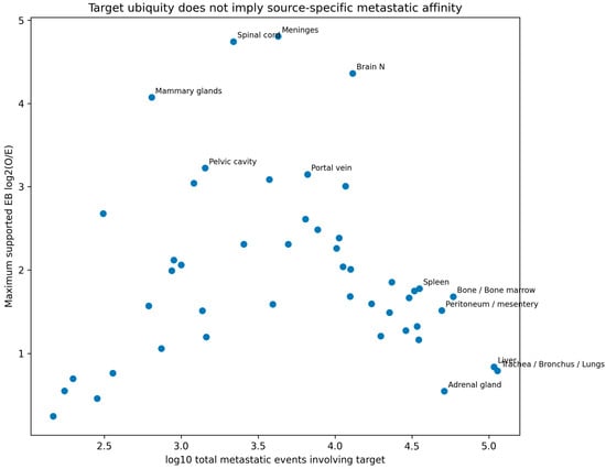
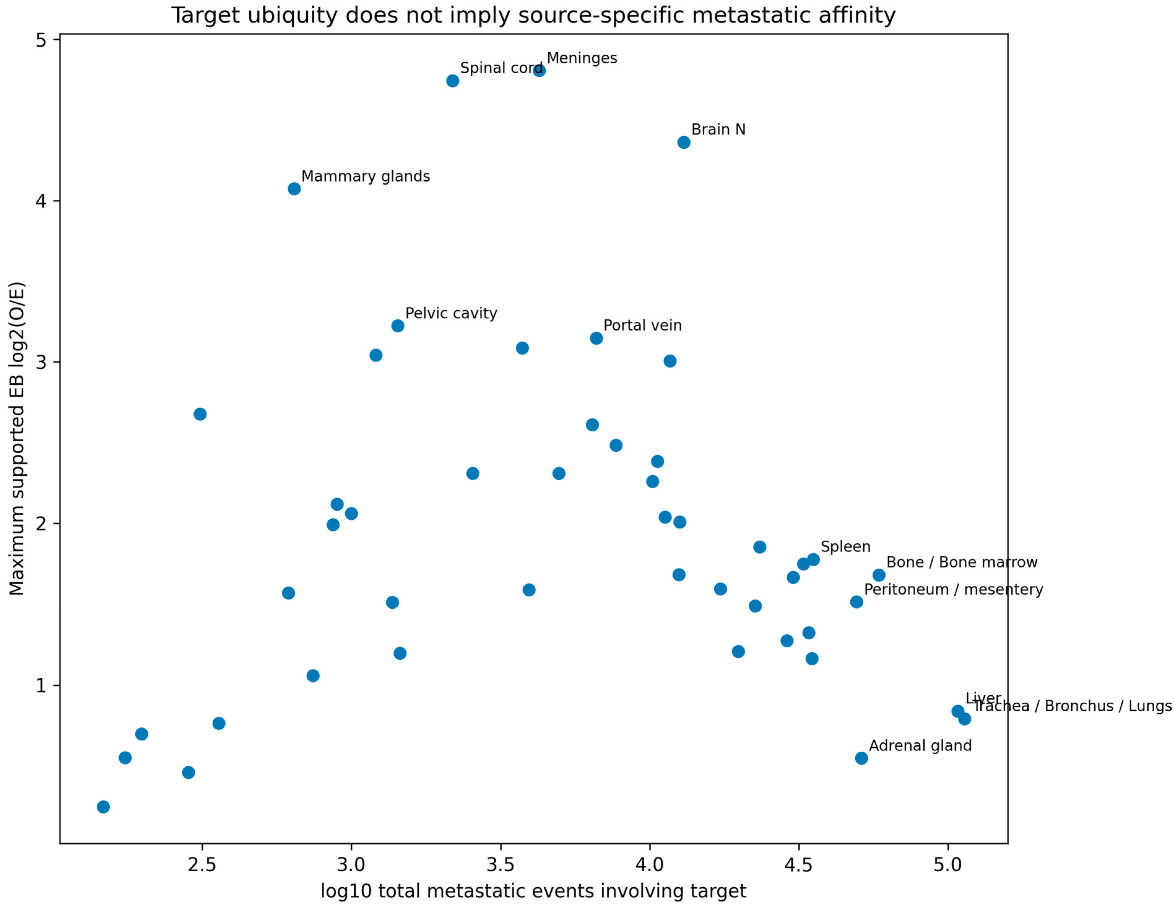
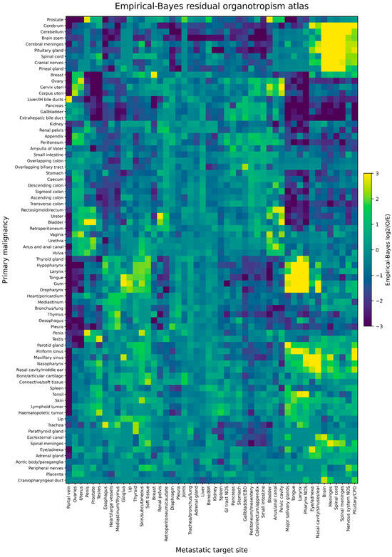
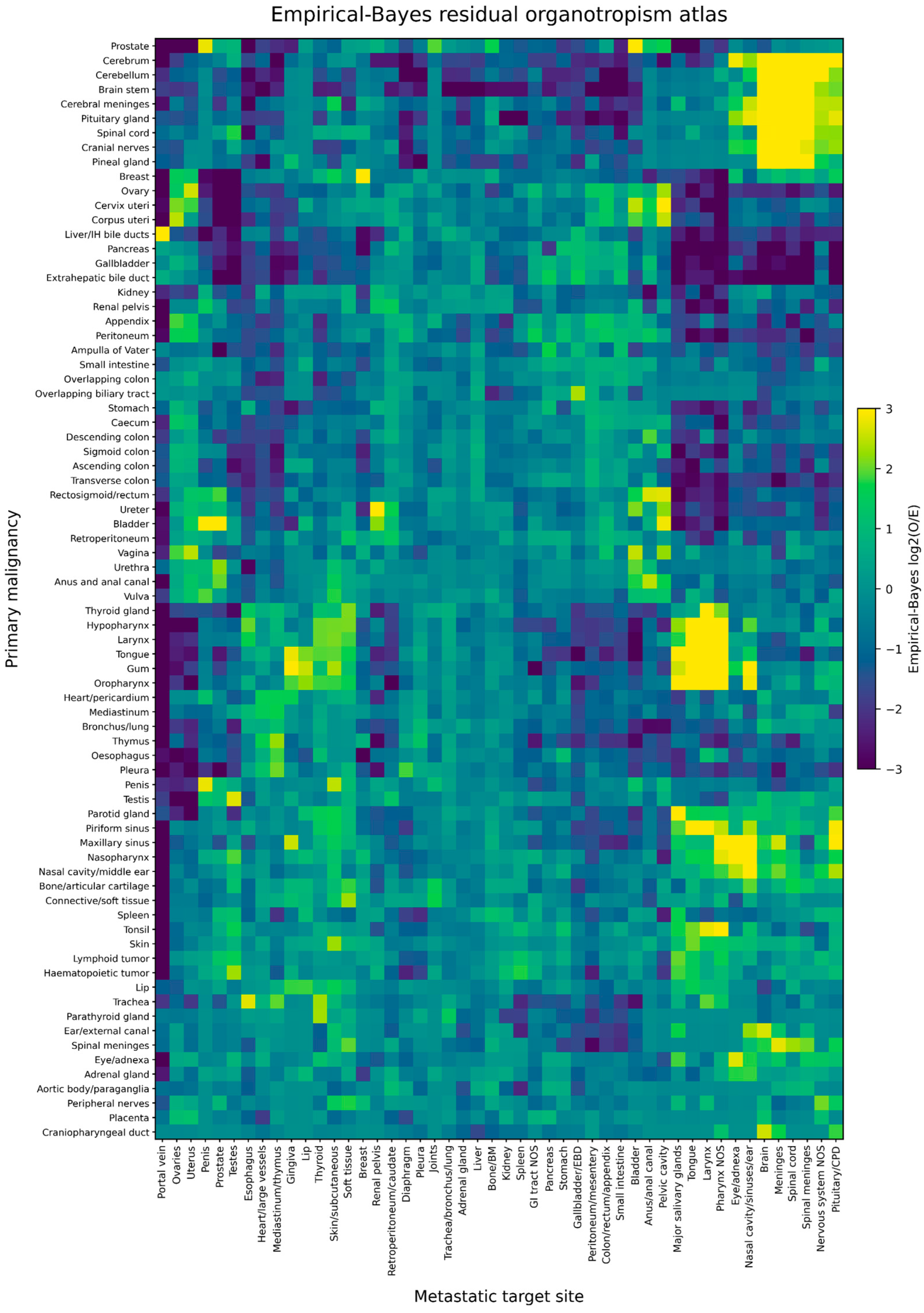
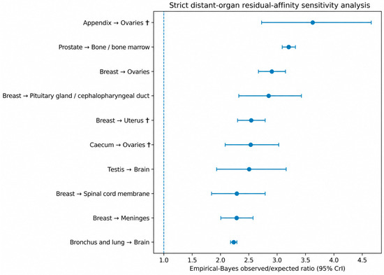
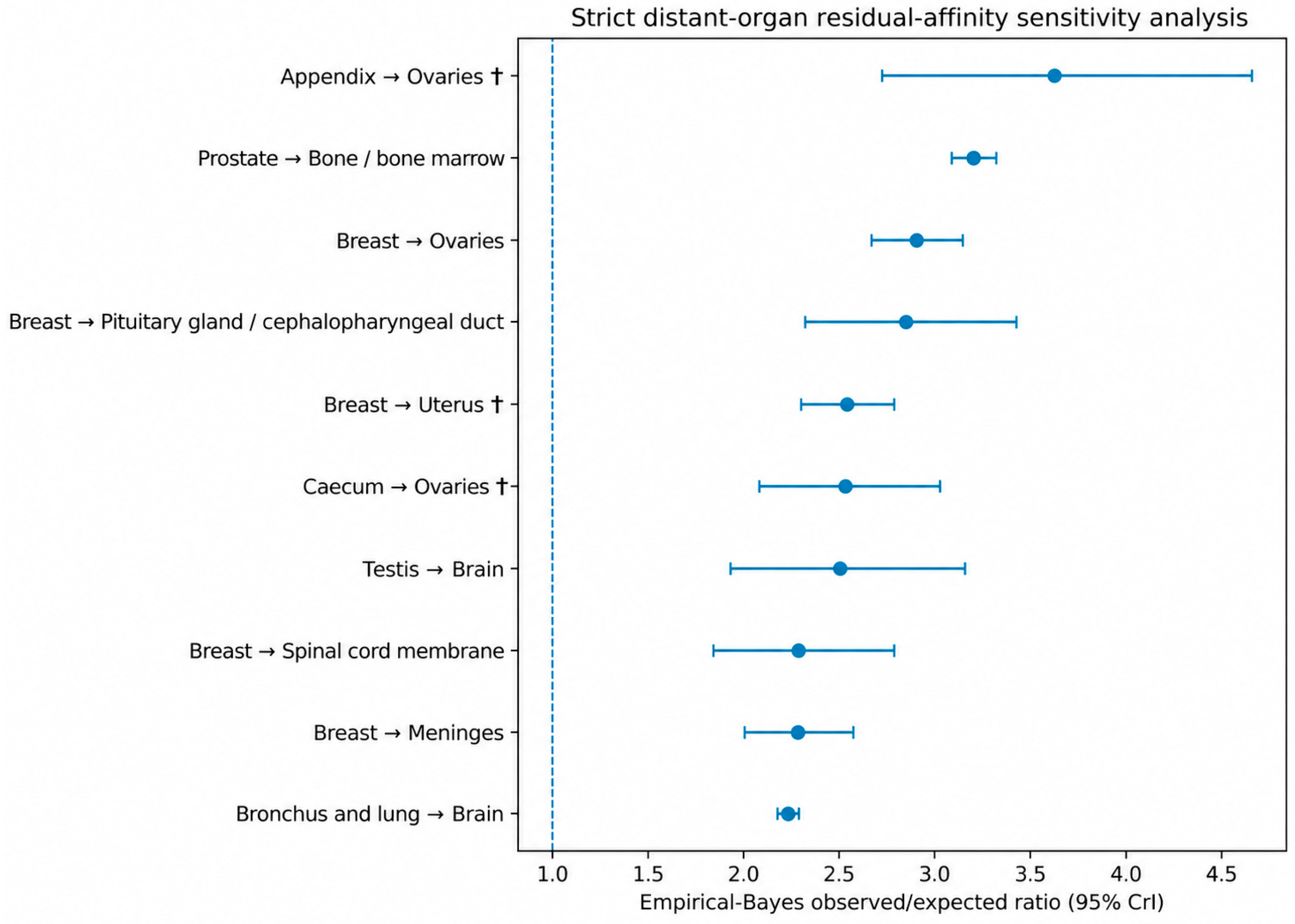
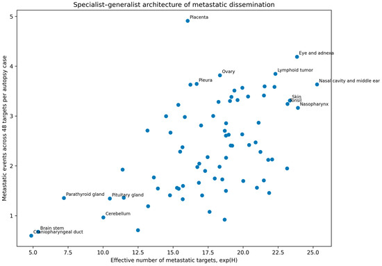
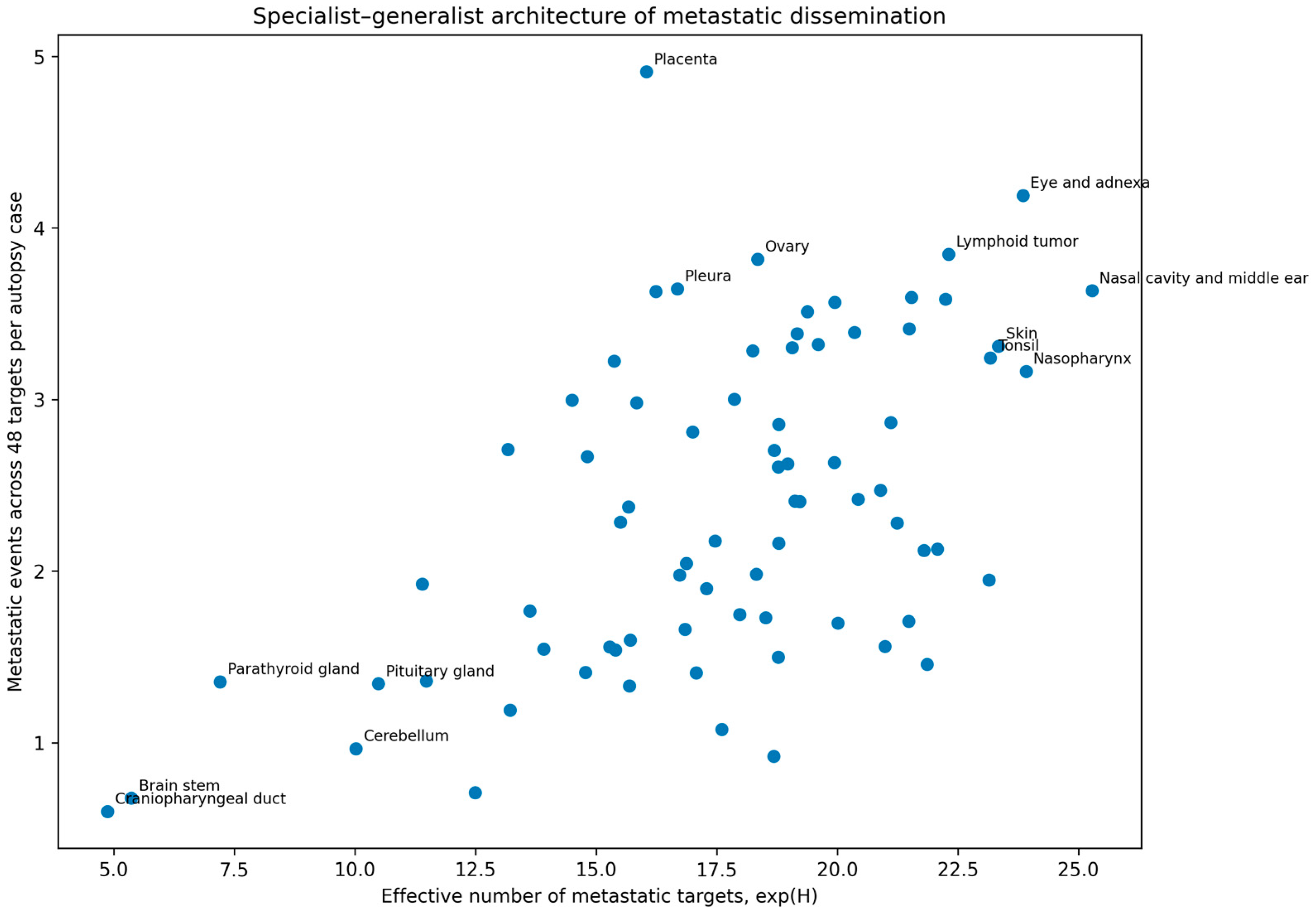
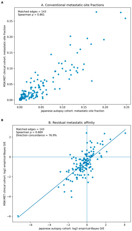
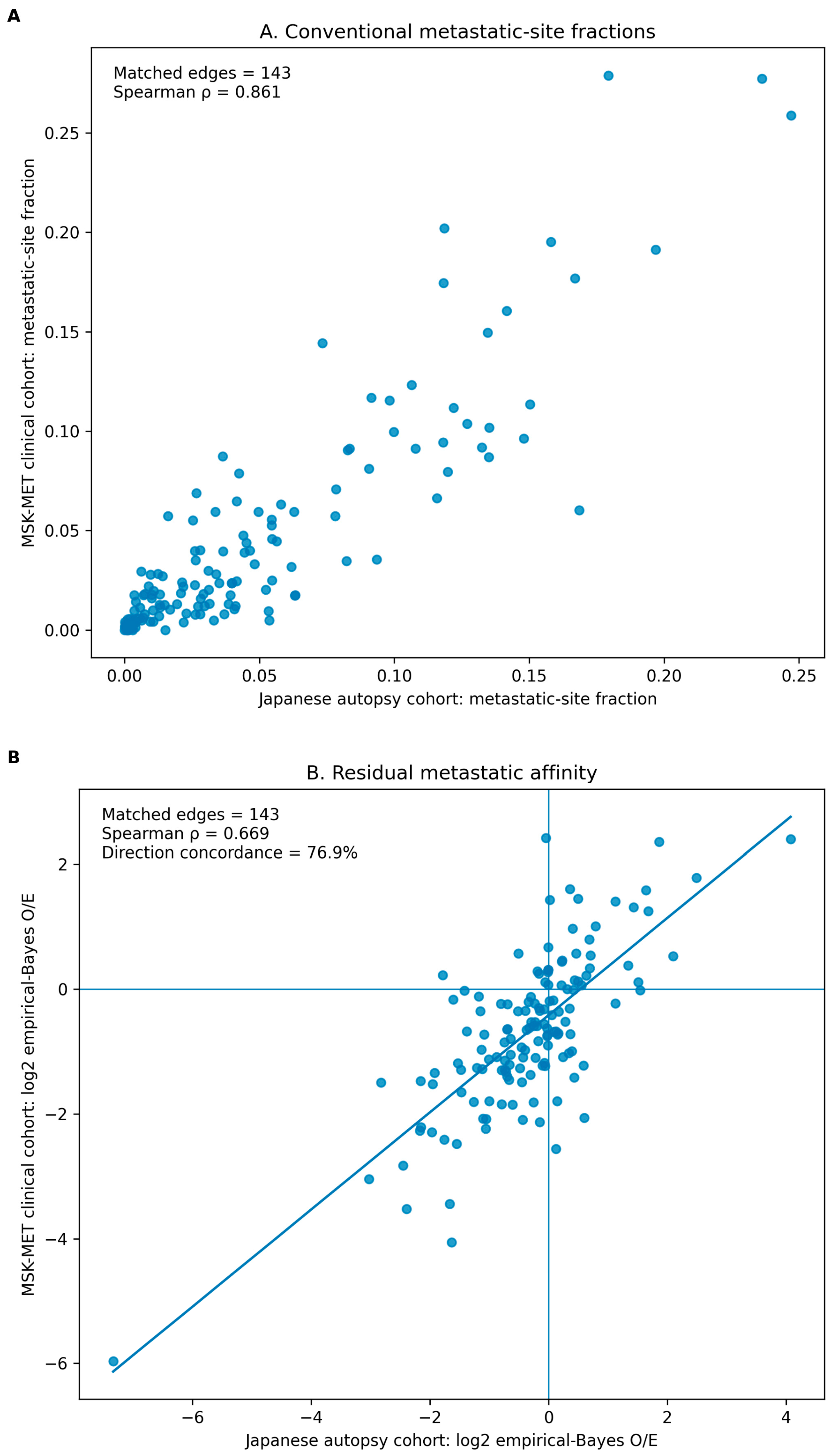

Disclaimer/Publisher’s Note: The statements, opinions and data contained in all publications are solely those of the individual author(s) and contributor(s) and not of MDPI and/or the editor(s). MDPI and/or the editor(s) disclaim responsibility for any injury to people or property resulting from any ideas, methods, instructions or products referred to in the content. |
© 2026 by the authors. Licensee MDPI, Basel, Switzerland. This article is an open access article distributed under the terms and conditions of the Creative Commons Attribution (CC BY) license.