


Abstract
Background/Objectives: Mucopolysaccharidosis IVA (MPS IVA) is a lysosomal disorder characterized by severe skeletal dysplasia caused by deficiency of N-acetylgalactosamine-6-sulfate sulfatase (GALNS). Although hematopoietic stem cell transplantation provides partial systemic benefit, its impact on skeletal disease remains limited. Rapidly expanding mesenchymal stem cell clones (REC-MSCs) may enhance skeletal-directed enzyme delivery while supporting hematopoietic cell recovery. This study evaluated lentiviral (LV) engineering of REC-MSCs and MPS IVA mouse hematopoietic stem cells (mHSCs) as an ex vivo strategy for enhancing GALNS expression. Methods: REC-MSCs were transduced with GALNS-expressing LV vectors driven by CBh, CD11b, COL2A1, or MND promoters and assessed for GALNS activity, vector copy number, viability, MSC phenotype, and chondrogenic differentiation potential. Functional interactions between engineered REC-MSCs and LV-transduced mHSCs were evaluated through enzyme transfer and co-culture recovery assays under regular, non-contact, and direct contact conditions. Results: LV engineering markedly increased intracellular and secreted GALNS activity in REC-MSCs, with CD11b and COL2A1 promoters providing the most favorable balance of expression and vector integration at the optimized MOI of 5. Transduced REC-MSCs maintained viability, MSC marker expression, colony-forming capacity, and early chondrogenic characteristics, including spheroid formation, Sox9 expression, and extracellular matrix deposition. GALNS activity and vector integration remained stable following chondrogenic differentiation. Conditioned media from engineered REC-MSCs enabled GALNS uptake by MPS IVA mHSCs, while direct contact REC-MSC/mHSC co-culture enhanced recovery, GALNS activity, and preservation of stem-like hematopoietic populations. Conclusions: These findings demonstrate that promoter-specific LV engineering of REC-MSCs enhances GALNS production while preserving fundamental MSC properties and early chondrogenic potential. In addition, REC-MSCs provide functional support for engineered mHSCs through enzyme transfer and direct stromal interactions. Together, these results establish a mechanistic foundation for future combined REC-MSC/HSC strategies targeting skeletal disease in MPS IVA.
1. Introduction
Mucopolysaccharidosis IVA (MPS IVA; Morquio A syndrome) is recognized as a lysosomal storage disorder resulting from a deficiency of N-acetylgalactosamine-6-sulfate sulfatase (GALNS). This deficiency leads to the accumulation of keratan sulfate (KS) and chondroitin-6-sulfate (C6S) within cartilage, bone, and other connective tissues [1,2]. Patients typically exhibit severe growth impairment, progressive skeletal abnormalities, and early morbidity associated with respiratory, cardiac, and cervical spine complications. Current therapeutic approaches, including enzyme replacement therapy (ERT) and hematopoietic stem cell transplantation (HSCT), offer partial systemic benefits but do not fully rectify the profound skeletal pathology characteristic of MPS IVA. ERT demonstrates limited penetration into avascular cartilage and is challenged by antibody formation, a short enzyme half-life, and inadequate clearance of GAGs in chondrocytes. While HSCT can improve several clinical parameters, its application is constrained by the availability of donors, the risk of graft-versus-host disease (GVHD), and limited evidence supporting its efficacy in correcting bone and cartilage lesions [2,3,4,5].
Because growth plate chondrocytes are among the most severely affected cells in MPS IVA, therapies that rely solely on HSCs or systemic enzyme delivery are unlikely to provide sufficient GALNS to skeletal tissues. Mesenchymal stem cells (MSCs), which give rise to chondrocytes, offer a promising alternative due to their multilineage differentiation capacity, immunomodulatory properties, and ability to migrate to sites of injury [6,7,8,9,10,11,12,13]. MSC-based approaches have shown therapeutic potential in hematologic, neurologic, cardiovascular, and joint disorders, including osteoarthritis and rheumatoid arthritis [6,14,15,16]. However, conventional bone marrow MSCs are heterogeneous and exhibit donor-dependent variability.
Rapidly expanding MSC clones (REC-MSCs), isolated from the CD90high/CD271high bone marrow population, yield a highly purified, clonally stable MSC population with consistent differentiation potential and reduced lot-to-lot variability [16]. Their robust chondrogenic capacity and paracrine support make them an attractive platform for skeletal applications. Supporting their translational relevance, a clinical trial evaluating REC-MSCs in combination with bone marrow transplantation for infants with severe hypophosphatasia has been approved in Japan and is scheduled to begin in 2026 (jRCT2053250167Q), and a separate trial investigating REC-MSC therapy combined with a bioresorbable gel for lumbar spinal canal stenosis is currently underway (jRCT2013210076). These advances support the rationale for exploring REC-MSCs as a skeletal-directed adjunct to HSC-based approaches in MPS IVA. Lentiviral (LV) engineering may further enhance their therapeutic utility by enabling stable, promoter-driven GALNS expression while preserving chondrogenic function. In parallel, REC-MSCs may improve the survival and recovery of engineered HSCs through stromal or contact-dependent support mechanisms; however, these interactions remain poorly defined in MPS IVA.
In this in vitro study, we evaluated promoter-specific LV engineering of REC-MSCs as a platform to enhance GALNS expression while preserving cell viability, vector stability, and chondrogenic differentiation. Additionally, we investigated whether REC-MSCs support the recovery of LV-transduced MPS IVA mouse hematopoietic stem cells (HSCs) through various cellular interactions. These mechanistic analyses collectively establish a foundation for future combined MSC–HSC strategies to improve skeletal correction in MPS IVA. Thus, the primary objective was to identify a lentiviral engineering condition that increased GALNS production in REC-MSCs while maintaining cell fitness and early chondrogenic potential. Secondary objectives included assessing extracellular GALNS uptake by MPS IVA hematopoietic cells and determining if exposure to REC-MSCs enhanced the recovery of LV-transduced hematopoietic progenitor cells.
2. Materials and Methods
2.1. Mouse Maintenance
In this study, MPS IVA (MKC2) knockout (KO; Galns–/– as recipients) and wild-type (Galns+/+ as donors) mice, both male and female, were utilized [17]. Animals were not subjected to any experimental procedures; they were used exclusively as tissue donors for isolation of primary cells. The control groups consisted of untreated, age-matched male and female wild-type mice. The colony was maintained in a pathogen-free environment under a 12 h light/dark cycle. All procedures concerning the cell collection procedures, care, and handling of the mice conformed to the guidelines established by the Institutional Animal Care and Use Committee (IACUC) of Nemours Children’s Health, Delaware Valley, under protocol RSP21-12482-001, titled “Ex vivo gene therapy with lentiviral vectors” (Latest approval date: 10 February 2025).
2.2. LV Vector Construction
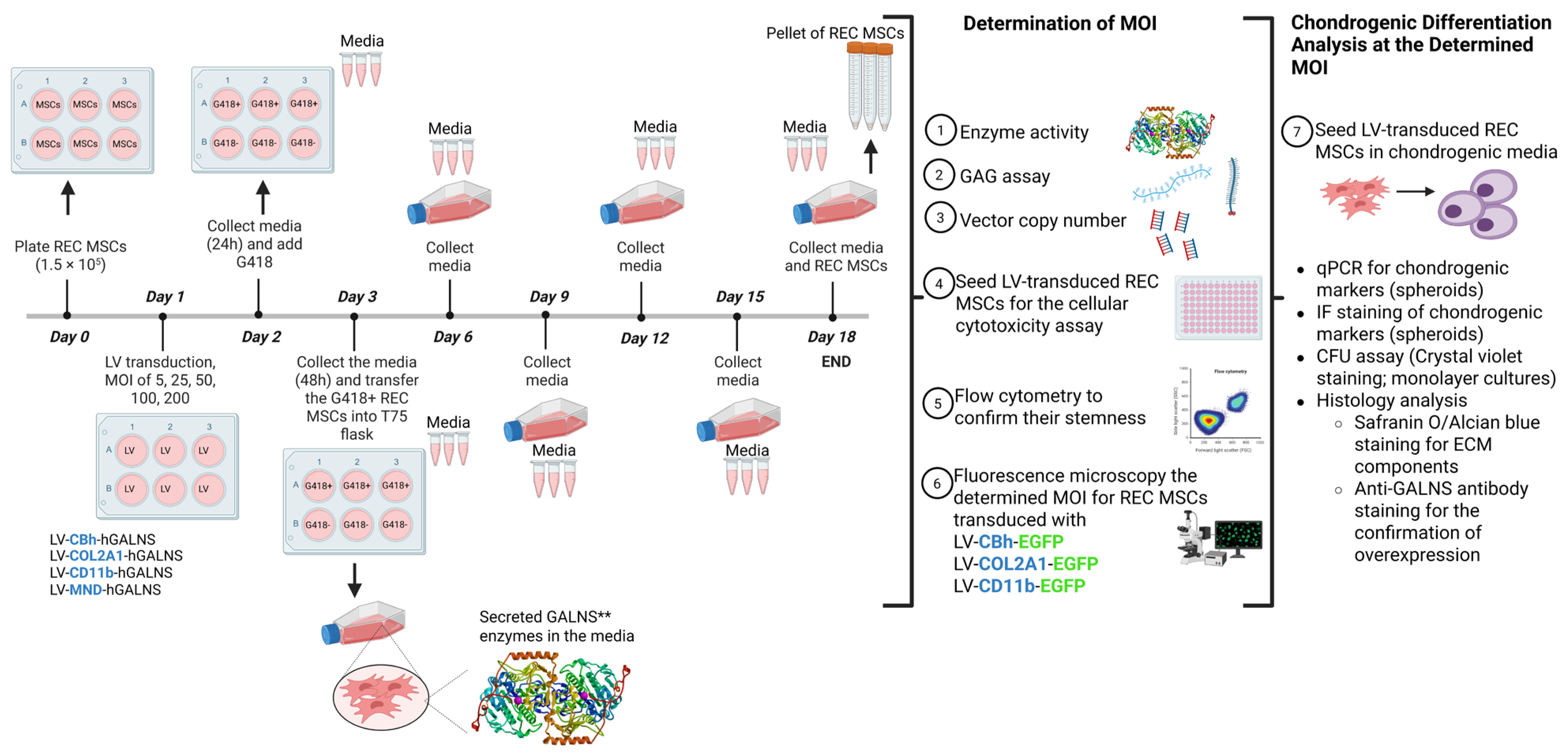
LVs were designed with the native human GALNS (hGALNS) driven by ubiquitous CBh, collagen-targeting COL2A1, myeloid-specific CD11b, and MND promoters. We used LV-CBh-hGALNS, LV-COL2A1-hGALNS, LV-CD11b-hGALNS, and LV-MND-hGALNS to transduce REC-MSCs. Moreover, we designed LVs with the CBh, CD11b, and COL2A1 promoters to drive the enhanced green fluorescent protein (EGFP) as a control (Figure 1). All lentiviral constructs contained a neomycin-resistance cassette, enabling Geneticin (G418) selection of transduced cells following transduction [18].
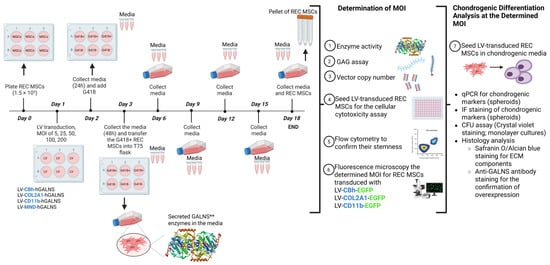
Figure 1.
Workflow for LV engineering of REC-MSCs. ** GALNS dimeric structure was adapted from Rivera-Colón et al. [19].
2.3. Culture and Transduction of REC-MSCs and mHSCs
REC-MSCs derived from clinical-grade bone marrow were supplied by PuREC Co., Ltd. (Izumo, Japan) [15,16]. REC-MSCs were isolated as a single clone from the CD90-high/CD271-high population within bone marrow mononuclear cells obtained from a healthy male donor. REC-MSCs were cultured in DMEM low-glucose medium (Gibco #11885092, Waltham, MA, USA) supplemented with 20% fetal bovine serum (FBS), penicillin-streptomycin, HEPES (Gibco #15630080), and basic fibroblast growth factor (bFGF; Biolegend #792504, San Diego, CA, USA) in a 5% CO2 atmosphere at 37 °C until reaching 80% confluence.
Mouse bone marrow (BM) cells (BMCs) were extracted from the femur and tibia of mice aged 8 to 12 weeks, including both wild-type and Galns–/– male and female specimens. We isolated Lin-HSCs using the EasySepTM mouse hematopoietic progenitor cell isolation kit (Stem Cell Technologies #18000, Vancouver, BC, Canada). The culture medium comprised Ham’s F12 nutrient mix (Gibco #11765-054) supplemented with HEPES, penicillin-streptomycin-glutamine (PSG; Gibco #10378-016), insulin-transferrin-selenium-ethanolamine (ITS-X; Gibco #51500-056), polyvinyl alcohol (PVA; Sigma-Aldrich #P8136, Burlington, MA, USA), thrombopoietin (TPO; PeproTech #31514, Cranbury, NJ, USA), and stem cell factor (SCF; PeproTech #25003, Cranbury, NJ, USA), as delineated in previous studies. Lin-HSCs were seeded at a concentration of 1 × 10⁶ cells per well in standard 6-well plates and incubated at 37 °C in a 5% CO2 atmosphere for subsequent experimental procedures.
REC-MSCs at 90% confluency were harvested, and 3 × 10⁵ cells per well of each cell line were seeded into 6-well plates (day 0). After 24 h of incubation, the 6-well plates were transduced with LVs at varying multiplicities of infection (MOIs: 5, 25, 50, 100, and 200) (day 1). After 48 h of incubation, the media were replaced, and Geneticin was added to select transduced colonies (day 3). The next day (day 4), the antibiotic-containing media were replaced with fresh media. Starting on day 4, the media were collected into separate tubes every three days and stored at −80 °C. On day 8, half of the cells were collected and stored at −80 °C, while the other half were transferred into 10 cm diameter plates for 30 days, during which media were collected every 3 days. On day 18, all cells and media were collected and stored (Figure 1).
For in vitro experiments, all cell lines and primary cultures were included if they met predefined quality criteria (viability > 90%, absence of contamination, and consistent morphology). No additional inclusion or exclusion criteria were established a priori. No wells, plates, or data points were excluded from analysis. All experiments were performed using a minimum of three independent biological replicates, and the exact n for each experimental group is reported in the figure legends. Experimental groups were assigned based on predefined treatment conditions. To minimise potential confounders, all groups were processed in parallel under identical culture conditions, and treatment/measurement order was kept consistent across replicates.
2.4. LV Vector Copy Number Quantification
Viral copy number (VCN) quantification was performed in REC-MSCs 20 days post-transduction. DNA was extracted from the transduced REC-MSC samples with the Qiagen Gentra Puregene Tissue Kit and digested with Proteinase K at 55 °C. After RNase treatment, DNA was resuspended in Tris-EDTA buffer, and its concentration was measured. Quantification of VCN was performed by ddPCR (ThermoFisher QuantStudio Absolute Q) as a single-plex (LV and Tfrc on 2 separate chips) using primers (Integrated DNA Technologies, Coralville, IA, USA) specific to the LV vector psi (Ψ) gene, resulting in an 82 bp PCR fragment. QuantStudio™ Absolute Q™ MAP16 Plate Kit and Master Mix (ThermoFisher#A53301, Waltham, MA, USA) were used to prepare the reaction. The quantification was performed using the following formula: (LV cps/µL* DNA dilution)/(Tfrc cps/µL/2).
2.5. Flow Cytometry
To confirm the stemness of REC-MSCs and mHSCs after LV transduction, we performed flow cytometry on single-cell suspensions of REC-MSCs and KO-mHSCs, prepared at a density of approximately 105 cells in 1X PBS. The cell suspensions were analyzed using Novocyte Quanteon flow cytometry (Agilent Technologies, Santa Clara, CA, USA).
For LV-REC-MSCs, we harvested and stained them with anti-human MSC antibodies. The following antibodies and isotypes were used (Biolegend, San Diego, CA, USA): Brilliant violet 421 anti-human CD34 (#343410, Biolegend), FITC anti-human CD45 Antibody (#368508, Biolegend), PE anti-human CD73-AD2 (#344004, Biolegend), PE/Cyanine 7 anti-human CD90 (Thy1; #328124, Biolegend), APC anti-human CD105 (#323208, Biolegend), Brilliant Violet 421™ Mouse IgG2a κ Isotype (#400259, Biolegend), FITC Mouse IgG1 κ Isotype (#400110, Biolegend), PE Mouse IgG1 κ Isotype (#400112, Biolegend), PE/Cyanine7 Mouse IgG1 κ Isotype (#400126, Biolegend), APC Mouse IgG1 κ Isotype (#400122, Biolegend) antibodies.
For LV-mHSCs, the cultured mouse MPS IVA Lin-HSCs were stained with anti-mouse HSC antibodies against c-Kit, Sca-1, and SLAM. The following selected antibodies and isotypes were used: APC anti-mouse c-Kit (CD117; #105811, Biolegend), FITC anti-mouse Sca-1 (Ly-6A/E; #122506, Biolegend), Pacific Blue anti-mouse SLAM (CD150; #115924), APC Rat IgG2b κ (#400612, Biolegend), FITC Rat IgG2a κ (#400506, Biolegend), and analyzed through flow cytometry to confirm the purity.
To determine LV-GFP expression intensity in REC-MSCs, we performed additional flow cytometry on live cells and compared them with untreated controls.
2.6. Chondrogenic Differentiation and Spheroid Formation
We employed two distinct methods: establishing monolayer and spheroid cultures from REC-MSCs to verify their differentiation.
For monolayer cultures, REC-MSCs were seeded at a density of 5000 cells per well, and chondrogenic media were subsequently added.
For spheroid cultures [20,21], 100 μL of autoclaved 1% agarose in PBS was transferred into 96-well flat-bottomed plates. Subsequently, 100 μL of 5000 REC-MSCs, resuspended in chondrogenic differentiation medium, were seeded into agarose-treated 96-well plates, centrifuged at 100 g for 90 s, and incubated for 72 h to facilitate spheroid formation. The culture media were replenished twice weekly. Spheroids were maintained for up to 21 days, after which they were fixed using 4% paraformaldehyde on day 21 and subsequently embedded in paraffin for further histological analysis. Alcian blue (pH 2.5) combined with Sirius red staining was performed to evaluate the proteoglycan content of spheroids derived from chondrogenic cells.
2.7. Colony Formation Assay
LV-REC-MSCs were seeded at 500 cells per well in 6-well plates and cultured in complete medium. On day 10, they were fixed for 30 min at room temperature with 3.7% formaldehyde (Sigma-Aldrich, Saint-Louis, MO, USA) in phosphate-buffered saline (PBS). Crystal violet stain (0.5% in methanol) was applied for 5 min, followed by washing with distilled water. Colony formation was documented [21].
The colony-forming unit (CFU) assay for triplicate cultures of LV-mHSCs was shown in our previously published manuscript [17].
2.8. Safranin O and Alcian Blue Staining
To ascertain whether LV transduction influenced the extracellular matrix expression of REC-MSCs, we conducted Safranin O and Alcian Blue (2.5%) staining on all chondrogenic spheroid cells derived from LV-transduced MSCs and compared the results with those obtained from untransduced REC-MSCs.
2.9. Cell Survival Analysis
To assess the effect of escalating MOI levels on cellular viability, REC-MSCs were transduced with LVs at MOIs of 5, 25, 50, 100, and 200. Subsequently, a WST-1 assay was conducted following the manufacturer’s instructions.
2.10. qPCR for Chondrogenic Markers
The expression of chondrogenic markers in chondrogenic cells post-transduction was confirmed through quantitative PCR analysis targeting Sox-9, Aggrecan, and collagen type II markers.
2.11. Confocal Microscopy
To verify the expression of GFP in REC-MSCs under LVs at the determined multiplicity of infection (MOI) of 5, confocal microscopy was performed on REC-MSCs transduced with LVs containing different promoters (CBh, COL2A1, and CD11b). The results were compared with untreated controls by assessing GFP expression at 48 h and 8 days post-transduction, utilizing a Zeiss Axio Observer inverted microscope (Carl Zeiss Microscopy GmbH, Jena, Germany). Corrected total cell fluorescence (CTCF) data were quantified using ImageJ (NIH) software (version1.54p), employing the formula: CTCF = Integrated density of the cell (raw fluorescence) − (Area of the selected cell x Mean fluorescence of the background).
2.12. Immunohistochemistry
To confirm GALNS overexpression in each cell, we performed immunohistochemistry (IHC) with a custom-made monoclonal anti-GALNS antibody (Creative Biolabs, Shirley, NY, USA). Cells harvested on day 15 were fixed in 10% formalin and sectioned at a thickness of 5 µm for IHC. The distribution and intensity patterns of GALNS were assessed immunohistochemically to determine any correlation with therapeutic effects, as previously described [17].
2.13. Co-Culture Recovery Assay (mHSCs + REC-MSCs)
To investigate whether REC-MSCs influence the recovery of LV-engineered mHSCs, two approaches were employed: (1) REC-MSCs and LV-mHSCs were co-cultured in a non-contact setting (preventing direct cell contact), and (2) REC-MSCs and LV-mHSCs were co-cultured in a standard setting (REC-MSCs and mHSCs within the same medium, enabling direct contact). We also prepared regular mHSC cultures for comparison. The duration of the experiment was limited to 8 days to account for potential transplantation scenarios. Subsequently, all cells were harvested, counted, and stained with stem cell markers (Sca-1, c-Kit, and CD150 (SLAM)) to confirm stemness via flow cytometry. All data were also compared with fresh cultures of untransduced mHSCs. Lin-Galns-/- mHSCs were seeded at a density of 2 × 10⁶ cells per well and incubated at 37 °C with 5% CO2.
2.14. GALNS Enzyme Activity Assay
GALNS enzyme activity was measured in cell homogenates, supernatants, and media using a 4-methylumbelliferone assay, as described [17].
2.15. GAG Quantification
We verified the decrease in accumulated GAGs employing LC–MS/MS, as previously delineated [17].
2.16. Statistical Analysis
Quantitative variables following a normal distribution were summarized as mean ± standard error (SEM). Conversely, data that did not meet the assumption of normality were presented as medians with 95% confidence intervals. Normality was evaluated using the Shapiro–Wilk and Kolmogorov–Smirnov tests. For comparisons involving more than two groups, one-way ANOVA with Tukey’s post hoc test was employed for normally distributed data, whereas the Kruskal–Wallis test with Dunn’s multiple comparison test was used for non-parametric data. Two-way ANOVA was utilized for analyses involving two independent variables. A p-value of ≤0.05 was deemed statistically significant. All experiments were performed in three independent experimental replicates, each containing technical replicates. All statistical analyses were conducted using GraphPad Prism 10.
3. Results
3.1. GALNS Enzyme Activity in REC-MSCs Driven by CD11b and COL2A1 Promoters
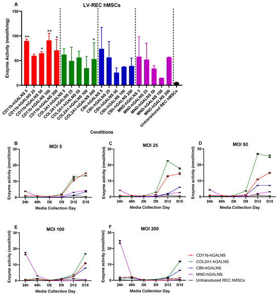
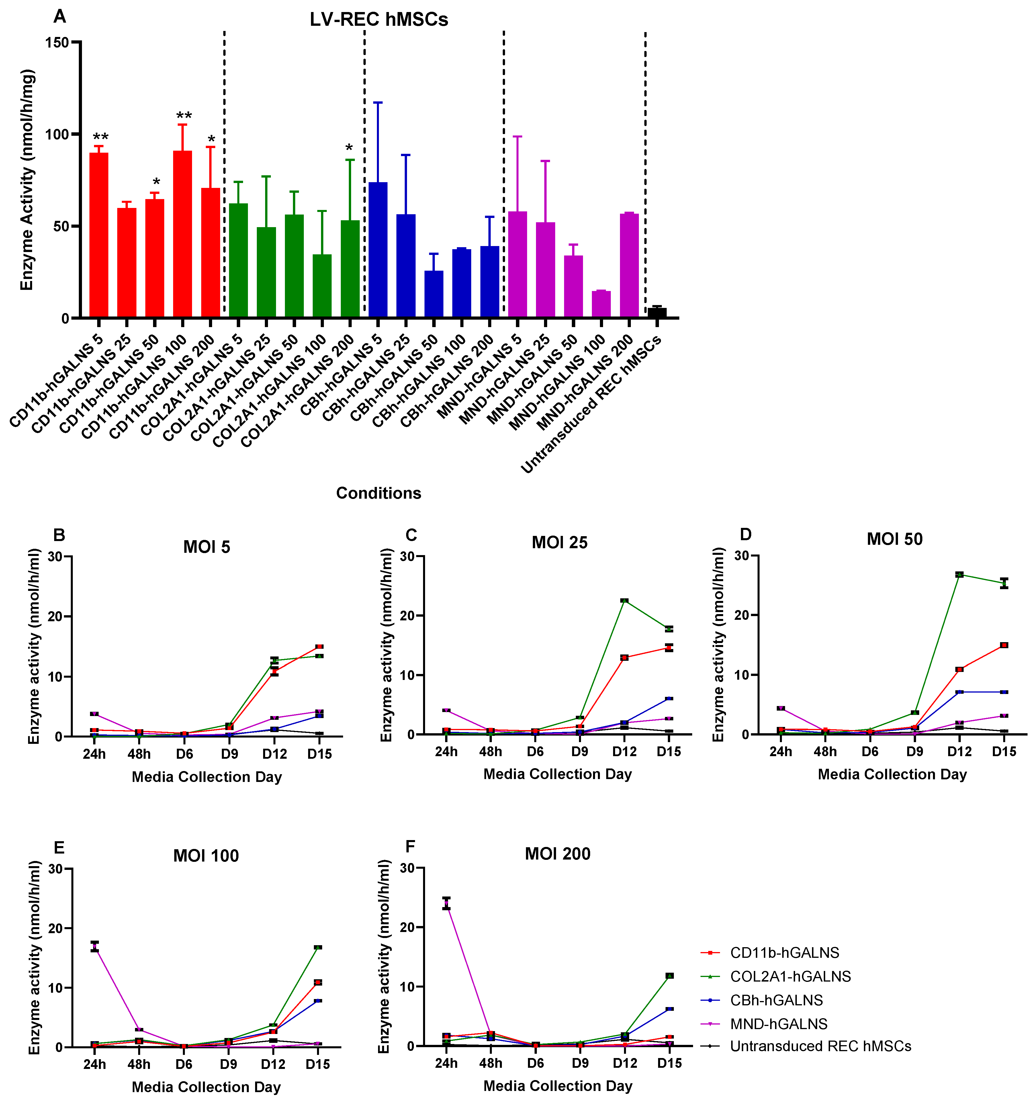
We engineered healthy REC-MSCs utilizing self-inactivating LVs (SIN-LVs) harboring the GALNS transgene under the control of the CBh, CD11b, COL2A1, and MND promoters across multiplicities of infection (MOIs) of 5, 25, 50, 100, and 200 to identify the optimal combination of promoter and MOI. The baseline physiological activity of GALNS in untransduced REC-MSCs was 5.5 ± 1.01 nmol/h/mg. Among all conditions tested, an MOI of 5 demonstrated the most advantageous balance between high expression levels and a low viral load, resulting in stable, robust enzyme activity. At this MOI, intracellular levels of GALNS increased to 16.3-, 11.3-, 13.4-, and 10.5-fold with the CD11b, COL2A1, CBh, and MND promoters, respectively, with the CD11b promoter producing the highest and most sustained activity across all MOIs (Figure 2A). Secreted GALNS activity also increased in all LV-treated groups over a period of 15 days, with the CD11b and COL2A1 promoters inducing the strongest extracellular secretion, reaching 14.9 ± 1.0 and 13.4 ± 0.09 nmol/h/mL at MOI 5 (Figure 2B–F). Collectively, these findings demonstrate that LV-engineering significantly enhances both intracellular and secreted GALNS levels in REC-MSCs, with the CD11b and COL2A1 promoters emerging as the most efficient for sustained GALNS delivery.
Figure 2.
Overexpression of GALNS enzymes in REC-MSCs transduced with lentiviral (LV) vectors carrying the CBh, CD11b, COL2A1, and MND promoters. (A) Intracellular GALNS activity (n = 3) and (B–F) Secreted GALNS activity (n = 2) in REC-MSCs were quantified following LV transduction and compared with those of untransduced controls (24 and 48 h, Day 6, 9, 12, and 15). Red bars: LV-CD11b-GALNS; Green bars: LV-COL2A1-GALNS; Blue bars: LV-CBh-GALNS; Purple bars: LV-MND-GALNS. Groups: CD11b-GALNS 5: LV-CD11b-GALNS transduction in REC-MSCs at MOI 5; CD11b-GALNS 25: LV-CD11b-GALNS at MOI 25; CD11b-GALNS 50: LV-CD11b-GALNS at MOI 50; CD11b-GALNS 100: LV-CD11b-GALNS at MOI 100; CD11b-GALNS 200: LV-CD11b-GALNS at MOI 200; COL2A1-GALNS 5: LV-COL2A1-GALNS at MOI 5; COL2A1-GALNS 25: LV-COL2A1-GALNS at MOI 25; COL2A1-GALNS 50: LV-COL2A1-GALNS at MOI 50; COL2A1-GALNS 100: LV-COL2A1-GALNS at MOI 100; COL2A1-GALNS 200: LV-COL2A1-GALNS at MOI 200; CBh-GALNS 5: LV-CBh-GALNS at MOI 5; CBh-GALNS 25: LV-CBh-GALNS at MOI 25; CBh-GALNS 50: LV-CBh-GALNS at MOI 50; CBh-GALNS 100: LV-CBh-GALNS at MOI 100; CBh-GALNS 200: LV-CBh-GALNS at MOI 200; Untransduced REC-MSCs: Control). Statistical significance was evaluated for the intracellular GALNS enzyme activity using one-way ANOVA with Tukey’s multiple comparison test (*: p < 0.05, **: p < 0.005, n = 3).
3.2. Promoter- and MOI-Dependent Modulation of KS Clearance in LV-Engineered REC-MSCs
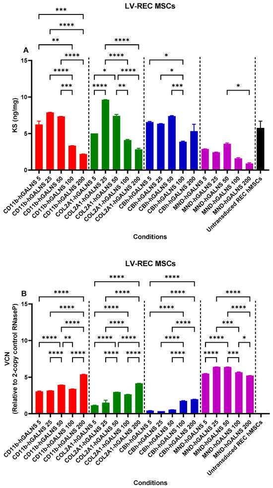
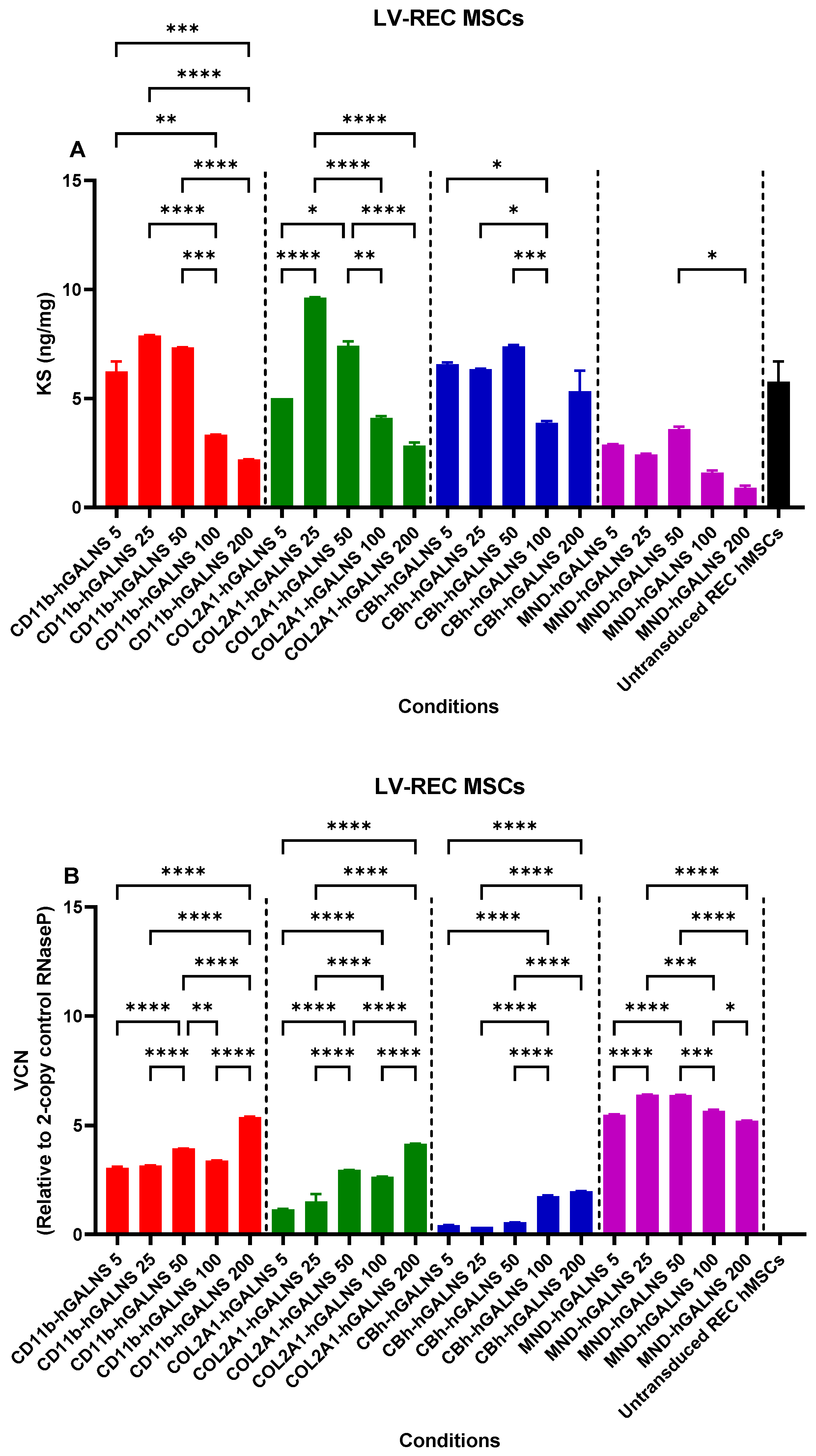
REC-MSCs were evaluated for their endogenous KS content and for changes in KS biology following LV transduction across different MOIs. GAGs were isolated and quantified by LC-MS/MS, establishing a baseline KS level of 5.8 ± 0.9 ng/mg. At the optimized MOI of 5, COL2A1-GALNS–transduced REC-MSCs showed a 0.9-fold KS level relative to control, representing a measurable and statistically significant reduction compared with untransduced cells. This indicates that even low-dose GALNS expression is sufficient to initiate KS clearance when driven by a promoter capable of supporting functional enzyme delivery. However, KS reduction did not strictly parallel the GALNS activity: some higher-MOI conditions, such as COL2A1 at 25, showed relatively elevated KS (9.6 ± 0.03 ng/mg), whereas others, including the selected MND or CBh conditions at higher MOIs, exhibited lower KS levels (Figure 3A). Overall, these data suggest that while LV GALNS expression can decrease KS accumulation, the relationship between enzyme activity and KS clearance is non-linear and promoter- and MOI-dependent, implying that factors beyond total GALNS activity—such as cellular stress, differentiation state, or saturation of lysosomal processing—may modulate KS storage under different engineering conditions. Although significant KS reduction was more pronounced at higher MOIs for selected promoters, we selected MOI 5 for subsequent studies as a low-exposure condition that produced increased GALNS activity with comparatively limited vector integration while preserving cell viability and differentiation, indicating a favorable balance between efficacy and biosafety.
Figure 3.
Intracellular keratan sulfate (KS) levels and lentiviral (LV) vector copy numbers in REC-MSCs following LV transduction. (A) Quantification of intracellular KS in REC-MSCs before and after LV transduction across increasing multiplicity of infection (MOIs), measured by LC–MS/MS. (B) LV VCN per cell in the corresponding transduced groups. Red bars: LV-CD11b-GALNS; Green bars: LV-COL2A1-GALNS; Blue bars: LV-CBh-GALNS; Purple bars: LV-MND-GALNS. Groups: CD11b-GALNS 5: LV-CD11b-GALNS transduction in REC-MSCs at MOI 5; CD11b-GALNS 25: LV-CD11b-GALNS at MOI 25; CD11b-GALNS 50: LV-CD11b-GALNS at MOI 50; CD11b-GALNS 100: LV-CD11b-GALNS at MOI 100; CD11b-GALNS 200: LV-CD11b-GALNS at MOI 200; COL2A1-GALNS 5: LV-COL2A1-GALNS at MOI 5; COL2A1-GALNS 25: LV-COL2A1-GALNS at MOI 25; COL2A1-GALNS 50: LV-COL2A1-GALNS at MOI 50; COL2A1-GALNS 100: LV-COL2A1-GALNS at MOI 100; COL2A1-GALNS 200: LV-COL2A1-GALNS at MOI 200; CBh-GALNS 5: LV-CBh-GALNS at MOI 5; CBh-GALNS 25: LV-CBh-GALNS at MOI 25; CBh-GALNS 50: LV-CBh-GALNS at MOI 50; CBh-GALNS 100: LV-CBh-GALNS at MOI 100; CBh-GALNS 200: LV-CBh-GALNS at MOI 200; Untransduced REC-MSCs: Control. Statistical comparisons were performed between transduced groups using one-way ANOVA with Tukey’s multiple comparison test (*: p < 0.05, **: p < 0.005, ***: p < 0.001, ****: p < 0.0001).
3.3. Promoter- and MOI-Dependent LV Vector Copy Numbers in Engineered REC-MSCs
To confirm the viral load in REC-MSCs at varying MOIs, we quantified the LV-specific psi sequence by ddPCR. VCNs increased proportionally with MOI and were comparable across promoters, ranging from 0.34 to 6.4 copies per cell across all conditions. Both CD11b and MND promoters exceeded approximately 3 copies per cell at all MOIs, reflecting high transduction efficiency. Notably, the MND promoter consistently exhibited the highest VCNs across all MOIs (5.5 ± 0.03, 6.4 ± 0.005, 6.4 ± 0.005, 5.7 ± 0.04, and 5.2 ± 0.005 copies per cell), indicating greater vector uptake or integration efficiency than the other promoters. In contrast, the COL2A1 and CBh promoters maintained lower observed VCNs (approximately 1.1–4.2 and 0.34–2.0 copies per cell, respectively), supporting their suitability for applications requiring minimal integration burden (Figure 3B). Focusing on the optimized MOI of 5, VCNs remained low across promoters, reinforcing MOI 5 as a condition that achieves robust GALNS expression while maintaining a minimal, reduced integration burden relative to other tested constructs.
3.4. Viability of LV-Transduced REC-MSCs Across Promoters and MOIs
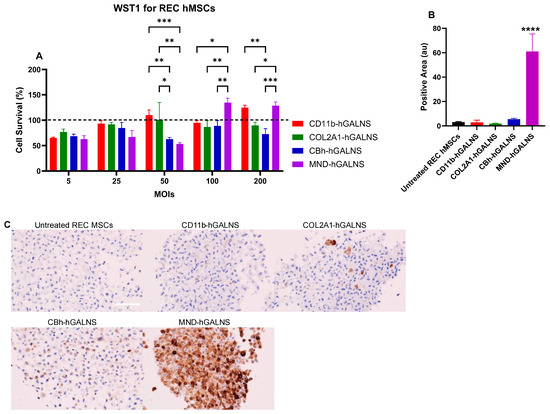
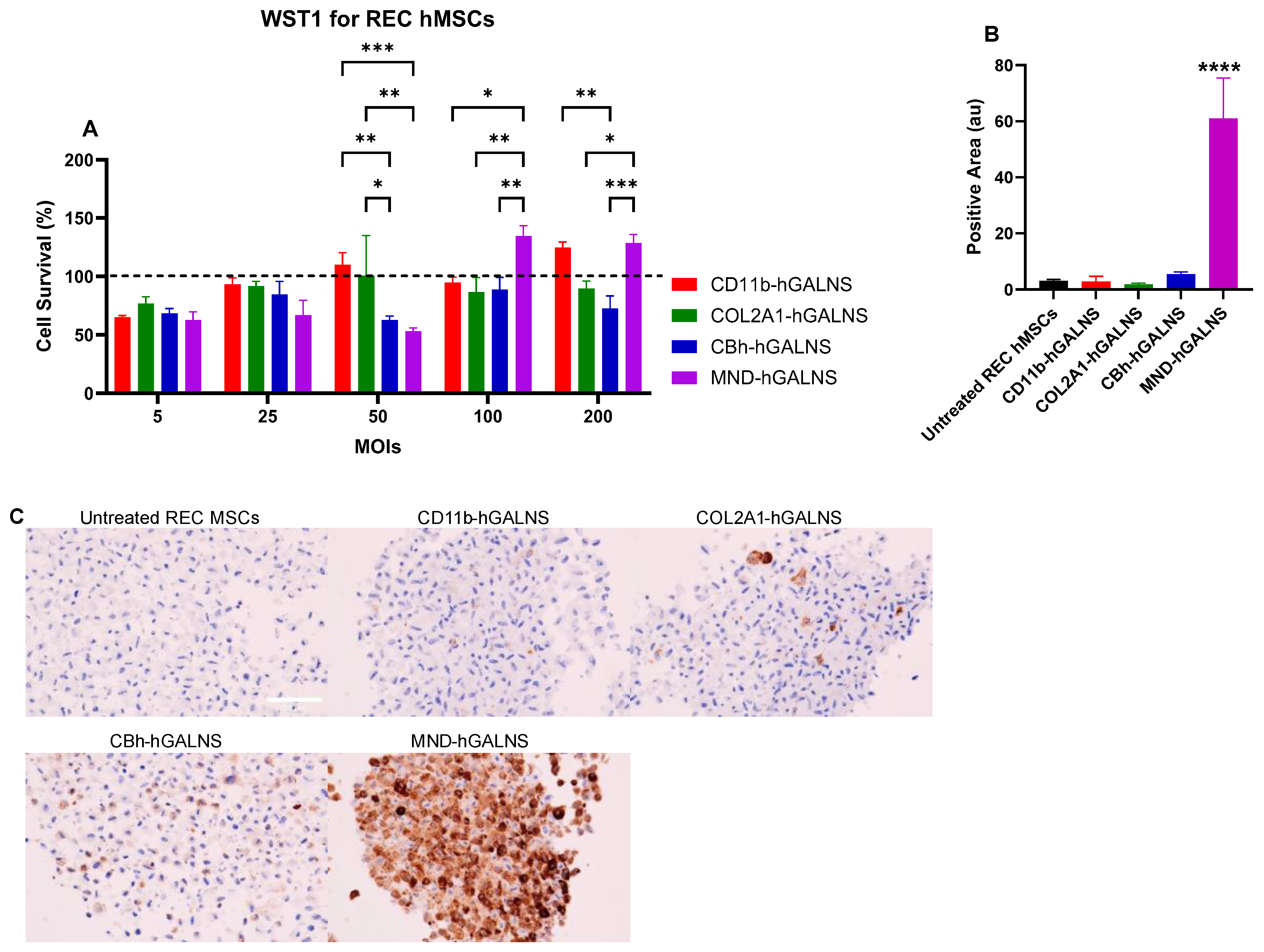
To confirm the viability of transduced REC-MSCs across MOIs, we conducted a WST-1 assay. Cell survival remained at or above the 100% baseline for all promoters and MOIs, indicating no cytotoxicity from LV transduction, even at the highest viral doses tested. Although we observed statistically significant differences between promoters at specific MOIs, these increases are likely to reflect enhanced metabolic activity rather than true over-proliferation. Additionally, viability did not consistently decrease as MOI increased. Overall, the data demonstrate that LV-mediated GALNS expression under the promoters CD11b, COL2A1, CBh, or MND does not compromise the metabolic activity of REC-MSCs. These findings support MOI 5 as an effective and well-tolerated condition for subsequent experiments (Figure 4A).
Figure 4.
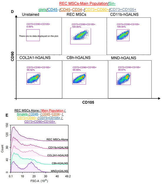
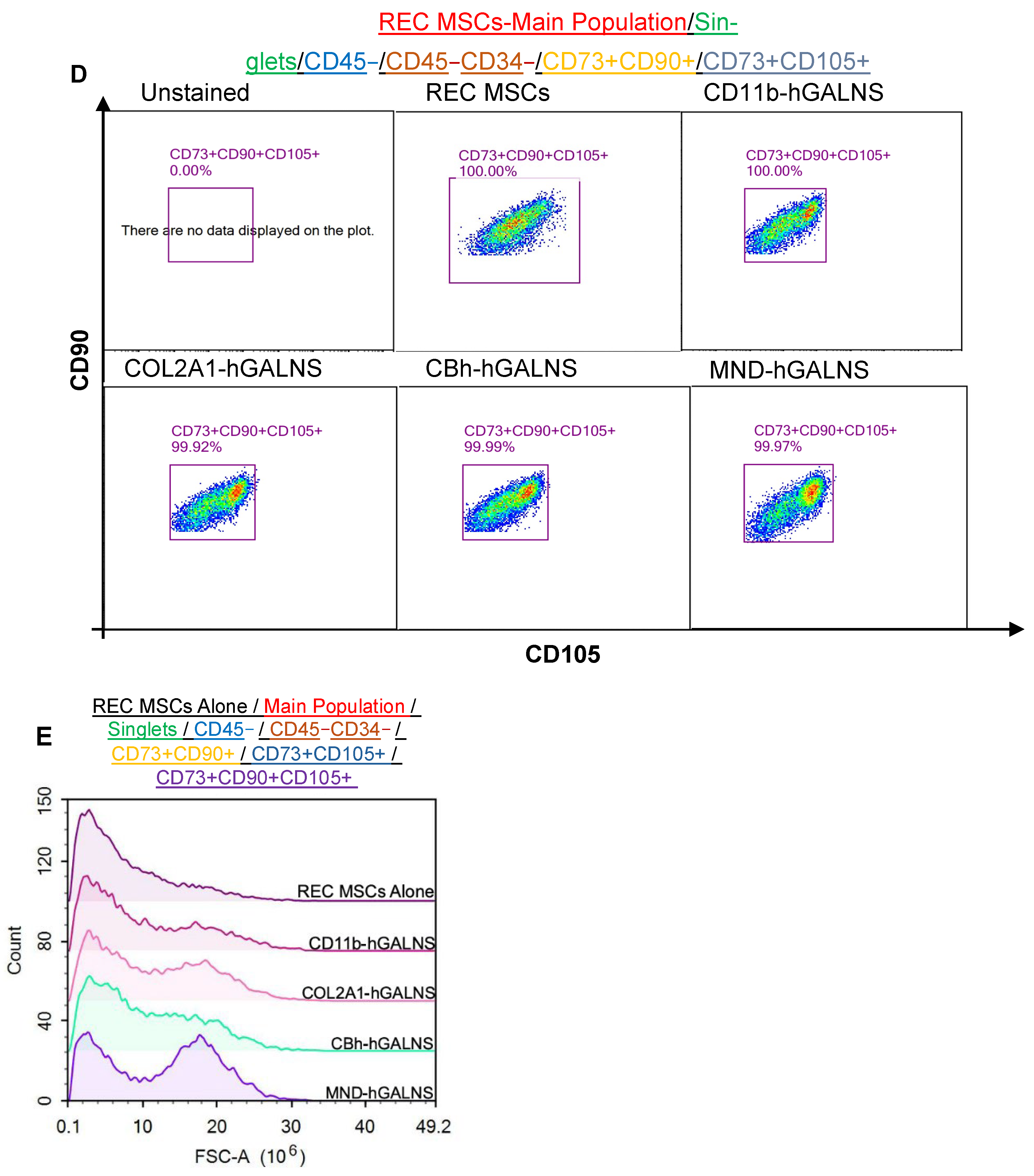
Viability, GALNS protein expression, and phenotypic characterization of REC-MSCs following lentiviral (LV) vector transduction. (A) Survival analysis based on the WST-1 (cell viability) assay of REC-MSCs transduced with LV vectors (CD11b-, COL2A1-, CBh-, and MND-GALNS) at increasing MOIs (5, 25, 50, 100, and 200). (B,C) Immunohistochemical detection of GALNS protein in transduced REC-MSCs and quantification of staining intensity (arbitrary units). (D,E) Confirmation of MSC identity in LV-transduced REC-MSCs (MSC-specific stem cell markers CD73, CD90, CD105 staining with antibodies for CD34 − CD45 − CD73 + CD90 + CD105 + MSCs). Statistical comparisons among transduced groups and between transduced and untransduced controls were conducted using one-way ANOVA with Tukey’s multiple comparison test (*: p < 0.05, **: p < 0.005, ***: p < 0.001, ****: p < 0.0001).
3.5. Immunohistochemical Assessment of GALNS Expression Across LV Promoters in REC-MSCs
IHC staining for GALNS confirmed promoter-dependent differences in transgene expression. Among all constructs, the MND promoter drove the highest GALNS protein expression, consistent with its elevated VCNs and strong transcriptional activity. In contrast, CD11b, COL2A1, and CBh promoters produced comparable GALNS staining intensities, with no significant differences detected between them (Figure 4B,C). These findings validate that all promoters successfully mediate GALNS overexpression in REC-MSCs, while MND uniquely achieves the strongest protein-level expression.
3.6. Validation of REC-MSC Identity Following Lentiviral Transduction
Flow cytometric analysis confirmed that lentiviral transduction did not alter the characteristic MSC immunophenotype of REC-MSCs. Across all promoter constructs (CD11b-hGALNS, COL2A1-hGALNS, CBh-hGALNS, and MND-hGALNS), >99% of cells remained uniformly positive for the MSC markers CD73, CD90, and CD105, comparable to unmodified REC-MSCs. The gated CD73+CD90+CD105+ population was preserved with no detectable loss of marker expression, indicating that LV delivery of hGALNS does not disrupt REC-MSC identity. Consistent with this, forward scatter area (FSC-A) profiles showed overlapping size/complexity distributions between transduced and untransduced cells, further supporting that LV transduction maintains normal MSC morphology and population integrity (Figure 4D,E).
3.7. Early CBh-Driven and Late COL2A1-Enhanced EGFP Expression in REC-MSCs
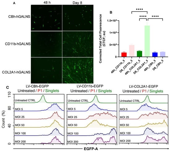
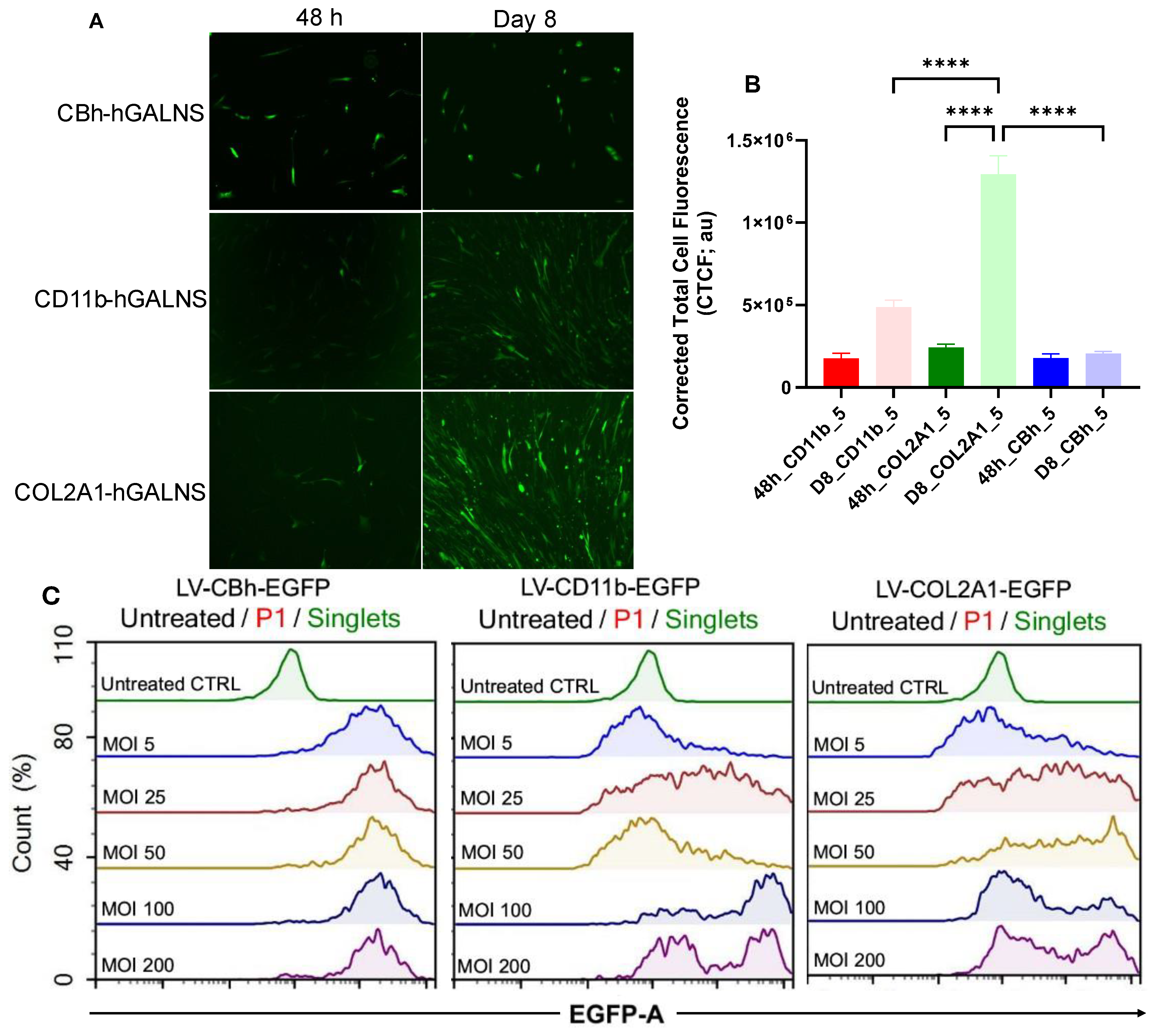
REC-MSCs transduced at a multiplicity of infection (MOI) of 5 with promoters CBh, COL2A1, or CD11b driving EGFP were evaluated at 48 h and on day 8 to assess promoter-dependent expression dynamics. At 48 h, the CBh promoter yielded the highest EGFP intensity, indicating strong early transcriptional activity, whereas the CD11b and COL2A1 promoters exhibited only modest fluorescence at this time point. By day 8, however, COL2A1 demonstrated the highest and most sustained EGFP expression, surpassing both CBh and CD11b, which remained stable (Figure 5A,B). These findings illustrate distinct time-dependent expression patterns, with CBh inducing robust early expression and COL2A1 supporting greater long-term stability.
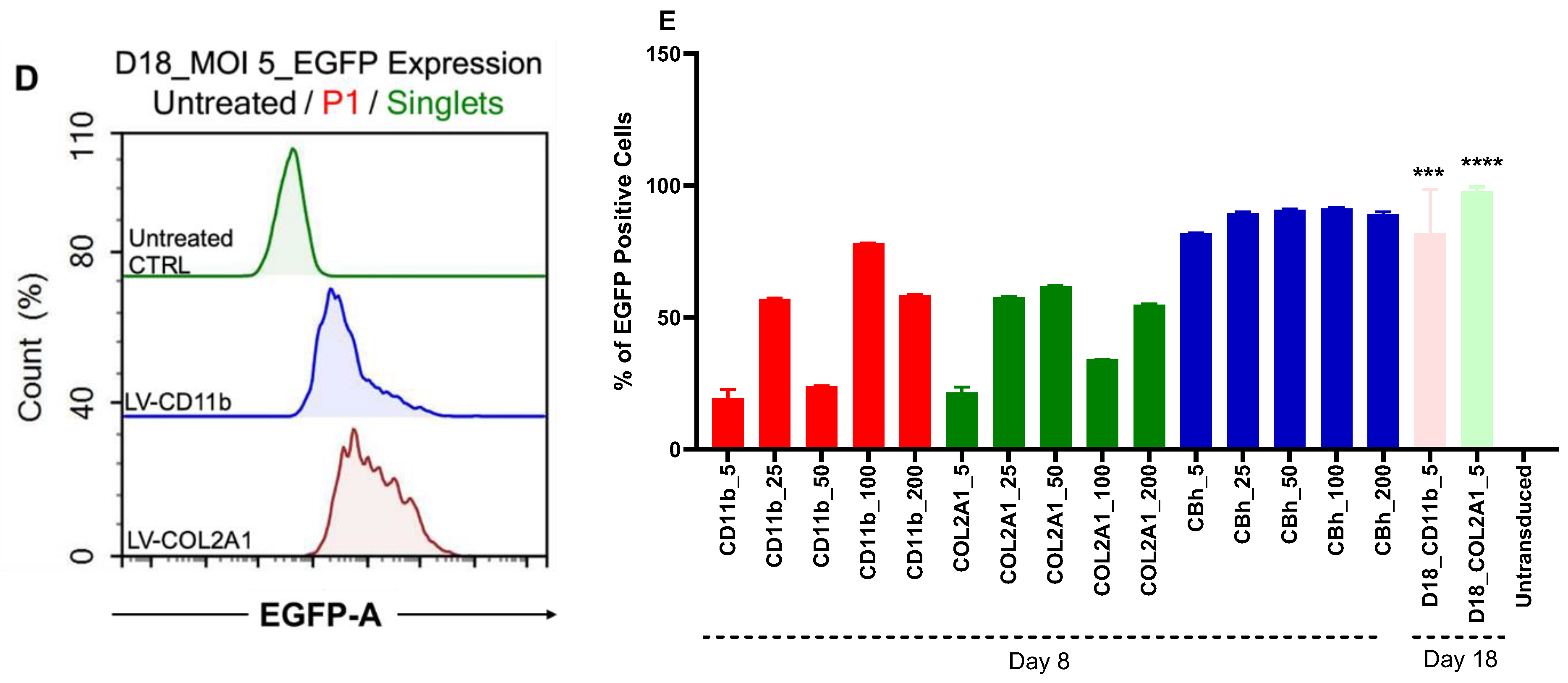
Figure 5.
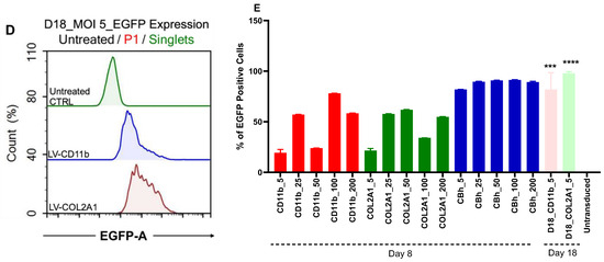
Early and long-term enhanced green fluorescent protein (EGFP) expression driven by CBh, CD11b, and COL2A1 promoters in REC-MSCs (MOI 5; CTRL: Control; untransduced: REC-MSC alone). (A) Immunofluorescence imaging at 48 h and Day 8 (D8) demonstrates promoter (CBh, CD11b, and COL2A1)-dependent variations in EGFP expression (48 h_CD11b_5: 48 h post-transduction with LV-CD11b-EGFP at MOI 5; D8_CD11b_5: 8 days post-transduction with LV-CD11b-EGFP at MOI 5; 48 h_COL2A1_5: 48 h post-transduction with LV-COL2A1-EGFP at MOI 5; D8_COL2A1_5: 8 days post-transduction with LV-COL2A-EGFP at MOI 5; 48 h_CBh_5: 48 h post-transduction with LV-CBh-EGFP at MOI 5; D8_CBh_5: 8 days post-transduction with LV-CBh-EGFP at MOI 5). (B) Quantification of EGFP fluorescence intensity derived from immunofluorescence images. (C) Flow cytometric analysis conducted on Day 8. (D) Confirmation of EGFP expression on Day 18 (D18) at an MOI of 5. (E) Alterations in the percentage of EGFP positivity in REC-MSCs transduced at different MOIs and with different promoters (Groups: CD11b_5: LV-CD11b-EGFP transduction in REC-MSCs at MOI 5; CD11b_25: LV-CD11b-EGFP at MOI 25; CD11b_50: LV-CD11b-EGFP at MOI 50; CD11b_100: LV-CD11b-EGFP at MOI 100; CD11b_200: LV-CD11b-EGFP at MOI 200; COL2A1_5: LV-COL2A1-EGFP at MOI 5; COL2A1_25: LV-COL2A1-EGFP at MOI 25; COL2A1_50: LV-COL2A1-EGFP at MOI 50; COL2A1_100: LV-COL2A1-EGFP at MOI 100; COL2A1_200: LV-COL2A1-EGFP at MOI 200; CBh_5: LV-CBh-EGFP at MOI 5; CBh_25: LV-CBh-EGFP at MOI 25; CBh_50: LV-CBh-EGFP at MOI 50; CBh_100: LV-CBh-EGFP at MOI 100; CBh_200: LV-CBh-EGFP at MOI 200). (***: p < 0.001, ****: p < 0.0001).
Flow cytometry performed on Days 8 and 18 further highlighted promoter-dependent differences in long-term stability. All CBh-transduced REC-MSCs were non-viable at this time point and could not be analyzed. The loss of viability may be related to the strong early transgene expression driven by the CBh promoter, which could impose a higher metabolic burden on REC-MSCs and compromise long-term cell survival. In contrast, CD11b- and COL2A1-transduced cells remained viable and were successfully analyzed, with COL2A1 maintaining the highest EGFP expression; the intensity of EGFP increased by 2.8-, 5.4-, and 1.2-fold for CD11b, COL2A1, and CBh promoters, respectively (Figure 5C–E). These findings demonstrate that while CBh supports robust early expression, COL2A1 provides the most stable long-term expression and cell viability under MOI 5 conditions.
3.8. Colony Morphology and Chondrogenic Stability of LV-Transduced REC-MSCs
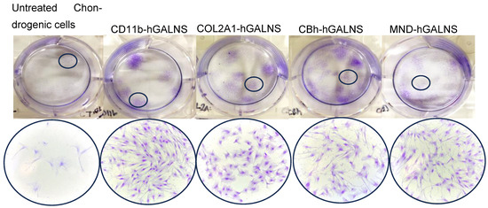
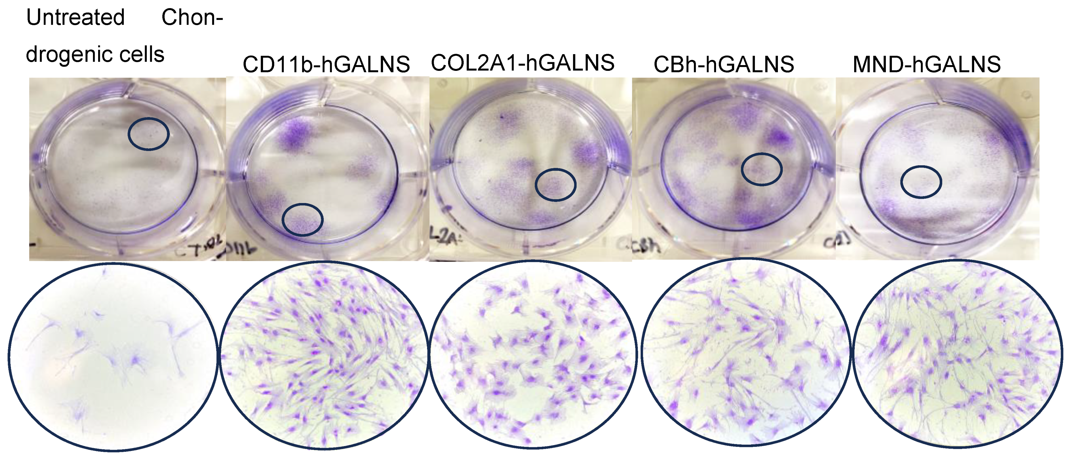
Crystal violet staining of monolayer chondrogenic cultures derived from LV-transduced REC-MSCs (MOI 5) demonstrated that all promoter groups, as well as untransduced controls, retained colony-forming capacity with qualitative differences in colony morphology (Figure 6). This indicates that LV transduction did not compromise the cells’ fundamental ability to form colonies. However, qualitative evaluation of colony morphology revealed distinct promoter-dependent variations in cellular stability. COL2A1-GALNS–transduced cells formed uniformly shaped, structurally stable colonies without signs of saturation, elongation, or morphological distortion, thus maintaining chondrogenic identity under this optimized transduction condition. Conversely, colonies derived from CD11b, CBh, MND, and untransduced cells frequently contained elongated, spindle-like, or stretched cells, suggesting greater heterogeneity and diminished structural integrity. These findings identify COL2A1 as the promoter that most effectively preserves chondrogenic morphology at MOI 5, consistent with its tissue-specific regulatory profile.
Figure 6.
Colony formation of chondrogenic cells derived from LV-transduced REC-MSCs at MOI 5. Crystal violet staining demonstrates that all promoter groups and untransduced controls generated adherent monolayer colonies (n = 3). Small black circles in each represent the selected colonies. Large circles represent a colony inside the selected region at 20× magnification.
3.9. Preservation of GALNS Activity and Lineage Stability During Early Chondrogenesis of Engineered REC-MSCs
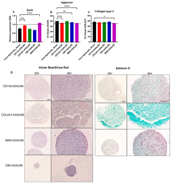
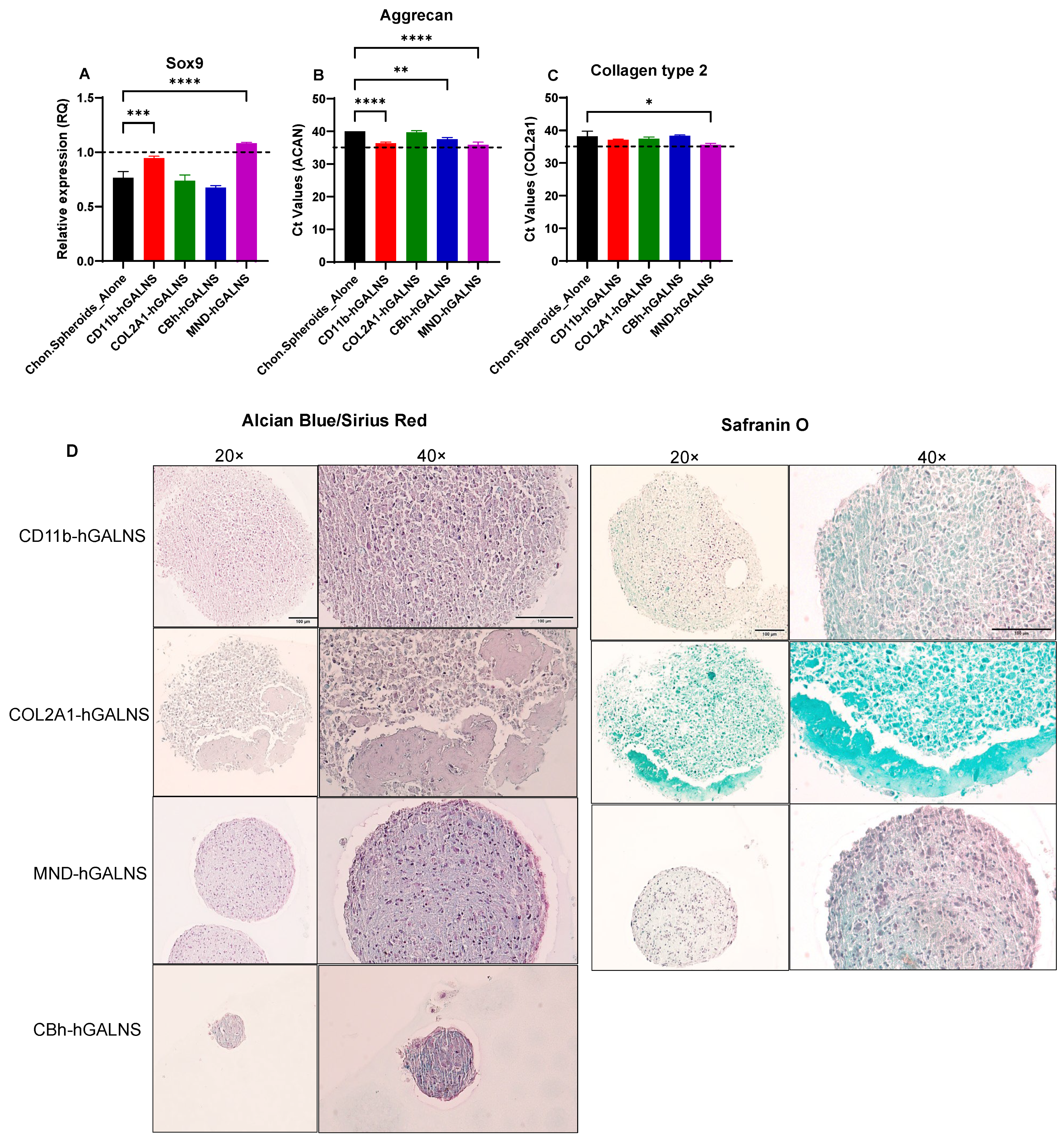
Chondrogenic markers Sox9, aggrecan, and collagen type II were assessed by RT-qPCR in spheroids derived from LV-transduced REC-MSCs to determine whether lentiviral engineering altered their chondrogenic differentiation potential. In REC-MSC spheroids, RT-qPCR showed increased Sox9 expression with only modest induction of aggrecan and collagen type II, consistent with early chondrogenic commitment but limited progression toward a mature, matrix-producing chondrocyte phenotype under the current differentiation conditions. Sox9 expression was highly similar across promoter groups, with only minor variation around the untransduced control, indicating that LV engineering and promoter choice did not substantially alter early chondrogenic transcriptional activation (Figure 7A–C).
Figure 7.
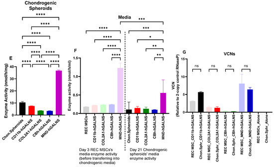
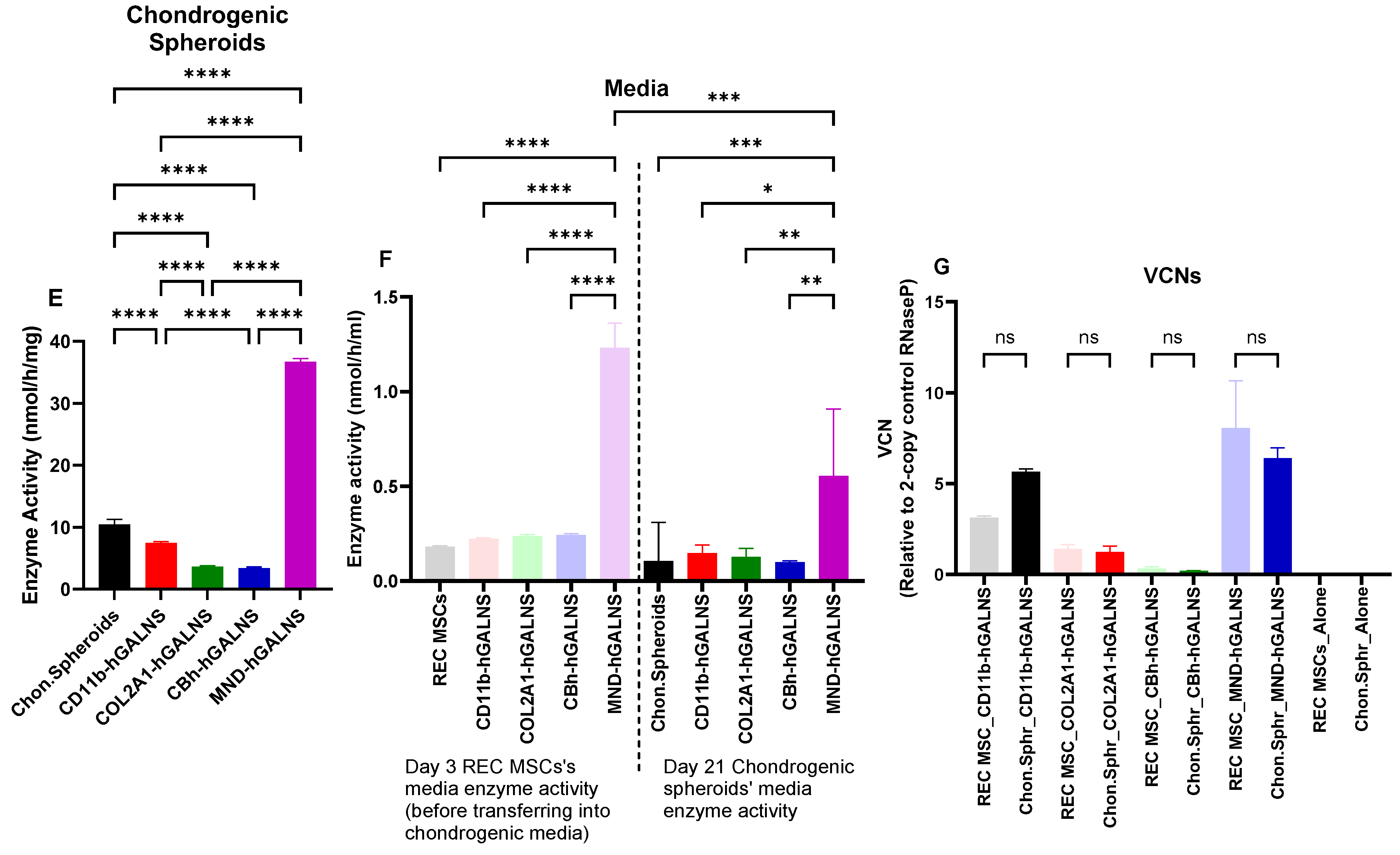
Integrated assessment of chondrogenic differentiation, extracellular matrix deposition, transcriptional activation, GALNS persistence, and viral integration stability in LV-transduced REC-MSC spheroids (n = 3; Chon. Spheroids: Chondrogenic spheroids; CBh, CD11b, COL2A1, and MND promoters driving GALNS expression in REC-MSCs under lentiviral (LV) vectors). (A–C) Real-time polymerase chain reaction (RT-qPCR) analysis of Sox9, Acan, and Col2a1 expression in REC-MSC chondrogenic spheroids. Sox9 mRNA expression in chondrogenic spheroids normalized to ACTB and Chondrogenic Spheroids_Reference Sample (dotted line; relative quantification (RQ) = 1.0). Due to very low expression of Acan and Col2a1 (Cycle threshold (Ct) values approaching or exceeding 35), ΔCt/ΔΔCt normalization and RQ calculations were not performed. Raw Ct values are shown to accurately represent detection levels. A dashed line at Ct = 35 marks the approximate limit of reliable quantification. Bars represent mean ± SEM; significance was determined by one-way ANOVA with a post hoc test. Only comparisons were shown between chondrogenic spheroids alone and LV-treated groups. (D) Safranin O and Alcian Blue staining to assess extracellular matrix deposition (20× and 40× magnification). (E,F) Intracellular and secreted GALNS enzyme activity measured in differentiated spheroids. (G) Lentiviral VCN in REC-MSCs and chondrogenic spheroids. (*: p < 0.05, **: p < 0.005, ***: p < 0.001, ****: p < 0.0001).
The Safranin O and Alcian Blue staining of chondrogenic spheroids demonstrated detectable yet modest extracellular matrix deposition. The spheroids predominantly showed bluish Alcian Blue staining, reflecting a glycosaminoglycan-rich matrix. Safranin O staining was present but not intensely pronounced, indicating comparatively lower levels of proteoglycan accumulation in comparison to mature cartilage (Figure 7D). Overall, the staining confirms extracellular matrix production within the spheroids; however, the extent of matrix deposition appears relatively limited at this developmental stage.
Chondrogenic cells derived from LV-transduced REC-MSCs were harvested and analyzed for GALNS enzyme activity. Differentiated chondrogenic cells maintained robust GALNS activity, indicating that transgene expression persists throughout the chondrogenic transition (Figure 7E,F). To evaluate the stability of lentiviral integration following differentiation, we quantified LV vector copy numbers in REC-MSCs and in chondrogenic spheroids derived from LV-transduced cells (Figure 7G). VCN showed no significant change, demonstrating that lentiviral integration remains stable throughout chondrogenic differentiation. Overall, these findings confirm that LV-mediated GALNS expression is maintained during chondrogenesis without loss of vector integrity.
3.10. Recovery Potential of LV-Transduced mHSCs After Co-Culture with REC-MSCs
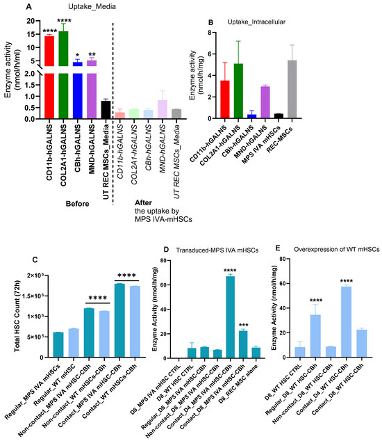
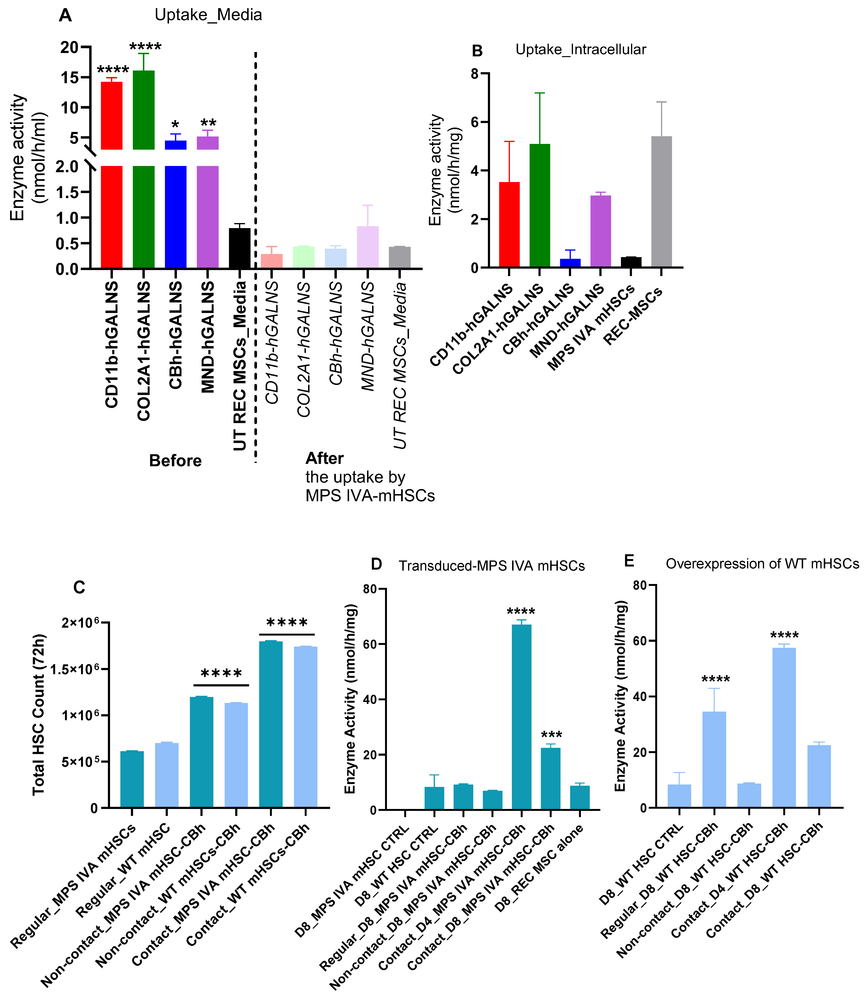
To determine whether GALNS secreted from LV-engineered REC-MSCs could be taken up by MPS IVA mHSCs, conditioned media collected from REC-MSCs transduced with different promoter constructs were transferred to individual MPS IVA mHSC cultures. Before transfer, GALNS enzyme activity was elevated in the media of LV-transduced REC-MSCs, demonstrating promoter-dependent secretion of functional enzyme. After incubation with MPS IVA mHSCs, intracellular GALNS activity increased in the recipient mHSCs, indicating effective uptake of extracellular GALNS from the transferred media. Consistent with this uptake, GALNS activity in the transferred media was markedly reduced after exposure to MPS IVA mHSCs, supporting enzyme internalization by the deficient hematopoietic cells. These findings demonstrate secretion of functional GALNS by LV-engineered REC-MSCs and its uptake by MPS IVA mHSCs. However, because downstream correction of disease-associated substrate accumulation was not assessed, these results should be interpreted as evidence of enzyme transfer and cellular uptake rather than definitive demonstration of cross-correction (Figure 8A,B).
Figure 8.
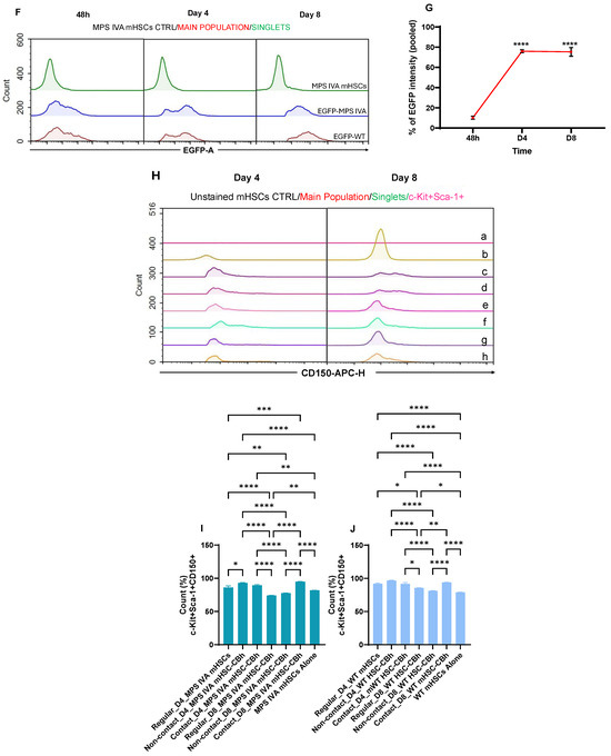
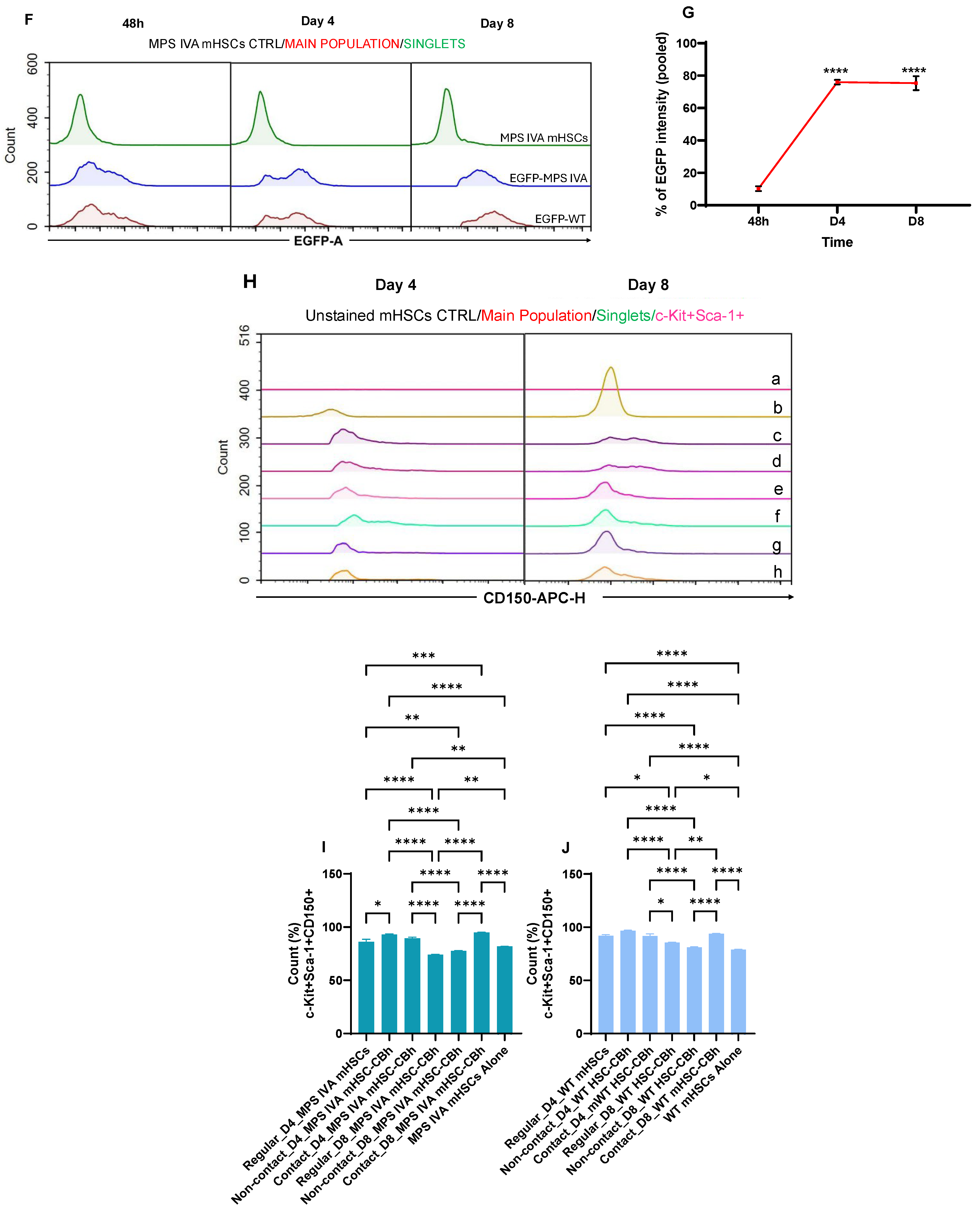
GALNS enzyme uptake, functional recovery, and overexpression capacity in MSC–HSC co-culture and engineering conditions (D4: Day 4; D8: Day 8; CTRL: Control; WT: Wild-type; mHSC: mouse HSCs; n = 3). (A) Extracellular GALNS enzyme activity in media from REC-MSCs transduced with CD11b, COL2A1, CBh, and MND promoters before and after uptake by MPS IVA mHSCs. Comparisons were made as before vs. after uptake for each promoter (e.g., LV-CBh before vs. LV-CBh after). (B) Intracellular GALNS enzyme activity in MPS IVA mHSCs after the uptake of secreted enzyme from LV-engineered REC MSCs. All groups were compared; no significant differences were found. (C–E) Regular, non-contact, and contact culture conditions were used. Regular cultures: no REC-MSCs were involved. Non-contact cultures: HSC medium was conditioned for 48 h in the REC-MSC cultures and then transferred into LV-CBh-hGALNS-transduced HSC cultures. Contact cultures (Co-cultures): LV-CBh-hGALNS-transduced mHSCs were transferred into REC-MSC cultures; REC-MSCs and mHSCs were in direct physical contact. (C) Total counts of LV-transduced MPS IVA and WT mHSCs at 72 h post-transduction in regular, non-contact, and contact co-cultures. Comparisons were made as follows: regular vs. non-contact, regular vs. contact, and non-contact vs. contact cultures. (D) GALNS enzyme activity in MPS IVA mHSCs transduced with LV-CBh-hGALNS, compared with non-transduced MPS IVA and wild-type mHSC controls. Also, we compared GALNS enzyme activity from contact cultures on days 4 and 8. We compared day 8 MPS IVA mHSCs (untreated control) with each group separately. (E) GALNS enzyme activity in WT mHSCs overexpressing GALNS via LV-CBh-hGALNS. Comparisons were made between day 8 WT mHSCs (untransduced control) and each group separately. (F) EGFP intensity in LV-CBh-EGFP-mHSC culture conditions at 48 h, day 4, and day 8. (G) Changes in the percentage of EGFP intensity in the pooled mHSCs, including MPS IVA and WT mHSCs. Following LV-CBh-EGFP transduction, both MPS IVA and WT mHSCs showed similar EGFP intensities. Therefore, the results were pooled. Two-way ANOVA comparisons were made as follows: 48 h vs. D4 and 48 h vs. D8. (H–J) Stem cell marker (c-Kit+Sca-1+CD150+) expression in LV-CBh-hGALNS-transduced MPS IVA and WT mHSCs in regular, non-contact, and contact co-cultures at 4 and 8 days post-transduction. (H) Groups at days 4 and 8 were labeled as follows: Days 4 and 8: (a) Unstained mHSC control; (b) CD150 isotype control; (c) LV-CBh-hGALNS-transduced MPS IVA mHSCs in contact culture; (d) LV-CBh-hGALNS-transduced WT mHSCs in contact culture; (e) LV-CBh-hGALNS-transduced MPS IVA mHSCs in non-contact culture; (f) LV-CBh-hGALNS-transduced WT mHSCs in non-contact culture; (g) LV-CBh-hGALNS-transduced MPS IVA mHSCs in regular culture; (h) LV-CBh-hGALNS-transduced WT mHSCs in regular culture. (*: p < 0.05, **: p < 0.005, ***: p < 0.001, ****: p < 0.0001).
To further assess the recovery and proliferative potential of LV-CBh-hGALNS-transduced mHSCs, we established three culture conditions: regular mHSC cultures without REC-MSC support, non-contact cultures using REC-MSC-conditioned medium transferred to mHSCs, and direct contact co-cultures in which mHSCs were cultured together with REC-MSCs. Both MPS IVA and wild-type (WT) mHSCs were evaluated under the same conditions to compare cell recovery, GALNS enzyme restoration in MPS IVA mHSCs, and GALNS overexpression capacity in WT mHSCs. Direct contact co-culture with REC-MSCs significantly increased the number of LV-transduced MPS IVA and WT mHSCs within 72 h, followed by the non-contact condition, compared with regular mHSC cultures (Figure 8C). In LV-CBh-hGALNS-transduced MPS IVA mHSCs, contact cultures showed significantly elevated GALNS enzyme activity on days 4 and 8 compared with the other culture conditions, indicating enhanced functional recovery of enzyme-deficient mHSCs (Figure 8D). Similarly, LV-transduced WT mHSCs displayed increased GALNS overexpression in contact cultures, with a significant elevation on day 4 compared with the other groups (Figure 8E). Together, these findings demonstrate that direct interaction between mHSCs and REC-MSCs provides a supportive microenvironment that improves mHSC recovery and enhances transgene expression.
To confirm transgene expression kinetics in LV-engineered mHSCs, MPS IVA and WT mHSCs were transduced with LV-CBh-EGFP and evaluated at 48 h, day 4, and day 8 after transduction. Flow cytometric analysis demonstrated an EGFP-positive trend over time, indicating sustained expression of the CBh-driven reporter construct during the culture period (Figure 8F). MPS IVA and WT mHSCs showed comparable EGFP expression profiles across all time points, suggesting that disease status did not substantially alter LV-CBh-EGFP expression dynamics under these conditions. Therefore, data from both MPS IVA and WT mHSCs were pooled to calculate the overall percentage of EGFP intensity over time. EGFP intensity was relatively low at 48 h, peaked on day 4, and then stabilized by day 8, indicating that LV-CBh-mediated reporter expression reached its highest level during the early post-transduction period and was maintained thereafter (Figure 8G). These results support the efficiency and stability of LV-CBh-mediated transgene expression in mHSCs and further validate the lentiviral engineering strategy used in the co-culture recovery assays.
We next evaluated whether LV-CBh-hGALNS-transduced mHSCs retained hematopoietic stem cell marker expression after culture under regular, non-contact, and direct contact REC-MSC co-culture conditions. Stem cell marker expression was assessed on days 4 and 8 by flow cytometry using c-Kit, Sca-1, and CD150, with unstained and CD150 isotype controls included for comparison (Figure 8H). Across all tested conditions, LV-transduced MPS IVA and WT mHSCs showed positive expression of c-Kit, Sca-1, and CD150 compared with the control populations, indicating preservation of the HSC immunophenotype during the culture period. To more clearly define the effect of genotype and culture condition, the percentage of c-Kit+Sca-1+CD150+ cells was analyzed separately for MPS IVA and WT mHSCs (Figure 8I,J). In both groups, direct contact co-culture with REC-MSCs resulted in a significantly higher c-Kit+Sca-1+CD150+ population compared with regular and non-contact cultures, demonstrating that physical interaction with REC-MSCs supports maintenance and recovery of the stem-like mHSC population after LV transduction.
4. Discussion
The present study establishes lentiviral engineering of rapidly expanding MSC clones as a technically feasible strategy to enhance GALNS delivery for MPS IVA while preserving cellular properties relevant to skeletal repair. The central challenge in MPS IVA is not simply systemic enzyme deficiency, but inadequate enzyme availability within cartilage and skeletal tissues, where avascularity, extracellular matrix density, and limited cellular turnover restrict the efficacy of conventional enzyme replacement [1,5,17,22,23] and hematopoietic stem cell-based approaches [2,3,4]. In this context, REC-MSCs offer a distinct therapeutic rationale: they combine the manufacturing advantages of a purified, clonally stable MSC population with the biological relevance of a cell type capable of chondrogenic commitment and stromal support [24,25]. By demonstrating that REC-MSCs can be engineered to express and secrete functional GALNS without losing viability, MSC identity, or early chondrogenic potential, this study provides an important foundation for developing cell-based strategies directed toward the skeletal compartment of MPS IVA.
A major finding of this work is that promoter selection substantially influences the balance between GALNS expression, secretion, vector integration, and lineage suitability. The promoters evaluated in this study were selected based on our previous work assessing LV-mediated GALNS expression across multiple disease-relevant and metabolically active cell types, including MPS IVA fibroblasts, chondrocytes, and HSCs, where they exhibited distinct expression profiles and functional capacities [17,18]. Building on that prior promoter validation, this study extends the evaluation to REC-MSCs, a purified MSC population directly relevant to skeletal and chondrogenic applications. In this REC-MSC context, CD11b and COL2A1 promoters produced robust GALNS activity, particularly at the optimized MOI of 5, supporting their utility for enzyme production and extracellular delivery. However, the promoter effects were not uniform across all biological readouts. MND was associated with strong GALNS protein expression and higher VCN, whereas COL2A1 maintained comparatively favorable integration profiles and showed advantages in preserving chondrogenic morphology and longer-term reporter expression. These observations argue against a single universal promoter choice and instead support a context-dependent promoter selection strategy. For skeletal applications, promoter activity must be evaluated not only by peak enzyme output but also by integration burden, durability of expression, compatibility with differentiation, and preservation of tissue-relevant cell behavior. In this respect, MOI 5 emerged as a rational engineering condition because it achieved substantial GALNS overexpression while minimizing unnecessary viral load and avoiding detectable impairment of REC-MSC survival or phenotype. Notably, while the CBh promoter induced high early transgene expression in REC-MSCs, it was associated with reduced cell viability upon extended culture in that specific lineage. Nevertheless, CBh was chosen for the HSC co-culture model as a robust benchmark promoter capable of driving high-level expression in hematopoietic progenitor cells, allowing sensitive evaluation of transgene kinetics, intracellular uptake, and paracrine support, as previously used [17]. For future translational applications targeting skeletal repair, tissue-specific or milder ubiquitous promoters such as COL2A1 or CD11b offer a superior biosafety profile for REC-MSCs, whereas CBh remains valuable for short-term ex vivo HSC manipulation.
Interpreting KS changes in REC-MSCs requires distinguishing them from the disease context of MPS IVA cells. Because the REC-MSCs used in this study were derived from a healthy donor, the baseline KS detected (5.8 ± 0.9 ng/mg) reflects normal physiological turnover rather than pathological lysosomal accumulation. Therefore, measuring KS levels post-transduction aimed to confirm whether exogenous GALNS expression alters endogenous GAG homeostasis without disrupting physiological cell functions. The non-linear relationship observed between GALNS activity and KS levels across MOIs underscores that substrate dynamics in healthy MSCs are tightly constrained by normal metabolic regulation. Substrate clearance as a direct therapeutic endpoint should thus be validated in primary MPS IVA patient-derived MSCs or animal models in future studies.
The chondrogenic differentiation data further support the relevance of REC-MSCs for MPS IVA skeletal applications while also defining the limits of the current in vitro system. LV-transduced REC-MSCs formed spheroid structures, retained Sox-9 expression, produced detectable extracellular matrix, and maintained GALNS activity after chondrogenic differentiation. These findings indicate that lentiviral engineering did not abrogate early chondrogenic commitment or transgene persistence during lineage transition. At the same time, Aggrecan and COL2A1 expression remained modest, and Safranin O/Alcian Blue staining suggested limited matrix maturation. Therefore, the data best support preserved early chondrogenic potential rather than complete formation of mature cartilage-like tissue. This distinction is important for translational interpretation. In MPS IVA, an engineered REC-MSC platform may not need to fully replace cartilage to be therapeutically meaningful; sustained local enzyme production by cells with chondrogenic and stromal properties may be sufficient to modify the skeletal microenvironment, particularly if combined with hematopoietic or systemic enzyme sources. Nevertheless, future optimization of differentiation conditions, extracellular matrix maturation, and in vivo skeletal localization will be necessary to determine the full regenerative and corrective potential of engineered REC-MSCs.
The REC-MSC–HSC co-culture experiments provide an additional mechanistic dimension to the study. REC-MSC-derived GALNS was taken up by MPS IVA mHSCs, as shown by increased intracellular GALNS activity in recipient HSCs and a parallel reduction in enzyme activity in transferred media. This supports a cross-correction mechanism in which enzyme secreted by engineered REC-MSCs remains biologically available to deficient hematopoietic cells. More importantly, direct contact between REC-MSCs and LV-transduced mHSCs enhanced cell recovery, increased GALNS activity, and preserved the c-Kit+Sca-1+CD150+ stem-like population more effectively than conditioned media and regular cultures alone. These findings suggest that REC-MSCs act not merely as passive enzyme-producing cells but also as active niche-supporting elements. The superiority of direct contact culture implies that membrane-bound signals, extracellular matrix interactions, adhesion-dependent survival pathways, or localized paracrine gradients may contribute to recovery of engineered mHSCs after lentiviral transduction [26,27,28,29,30,31,32]. This is particularly relevant because ex vivo gene modification can compromise HSC fitness, and strategies that improve post-transduction recovery without sacrificing stemness are central to successful transplantation. Although this co-culture system utilized human REC-MSCs and mouse mHSCs, direct contact superiorly preserved the c-Kit+Sca-1+CD150+ HSC population better than non-contact conditions. This demonstrates that key stromal support factors—such as CXCL12, stem cell factor (SCF), and extracellular matrix interactions—exhibit functional cross-species conservation capable of maintaining murine hematopoietic stemness ex vivo. These findings reinforce the utility of REC-MSCs as a versatile feeder microenvironment for gene-modified HSC recovery prior to transplantation.
Together, these results support a dual-function model for engineered REC-MSCs in MPS IVA: first, as a source of secreted GALNS capable of mediating enzyme uptake and cross-correction, and second, as a stromal support platform that enhances the recovery and maintenance of engineered HSCs (Table 1). This model is conceptually important because a single therapeutic mechanism is unlikely to correct skeletal disease in MPS IVA. A combined REC-MSC/HSC approach may provide complementary benefits: engineered HSCs could provide systemic, long-term enzyme delivery, whereas REC-MSCs could provide localized skeletal and stromal support while improving early recovery of transplanted or co-administered HSCs. The findings therefore extend beyond demonstrating lentiviral transduction efficiency and propose a biologically integrated strategy that addresses enzyme supply, skeletal lineage relevance, and hematopoietic niche support together.
Table 1.
Summary of previous therapeutic approaches and key findings relevant to MPS IVA and the rationale for the current REC-MSC/HSC strategy. ERT: Enzyme replacement therapy; HSCT: hematopoietic stem cell transplantation; LV-HSC Gene Therapy: lentiviral vector-mediated hematopoietic stem cell gene therapy; LV-Mediated GALNS Expression: lentiviral vector-mediated GALNS gene transfer and expression; MSC-Supported CRISPR-Cas9 HSC Editing: mesenchymal stromal cell-supported CRISPR-Cas9 gene editing of hematopoietic stem cells; REC-MSC + LV-HSC: rapidly expanding clone-derived mesenchymal stromal cells combined with lentiviral vector-transduced hematopoietic stem cells.
5. Study Limitations
Several limitations should be acknowledged. This study was conducted in vitro and used human REC-MSCs together with mouse HSCs; therefore, the extent to which these interactions translate to an in vivo transplantation setting remains to be determined. The culture duration was limited, and this work did not evaluate long-term persistence, biodistribution, engraftment, immunologic compatibility, skeletal homing, or the safety of engineered REC-MSCs. In addition, although vector copy number and viability data support the feasibility of the approach, comprehensive insertion-site analysis and long-term tumorigenicity assessment will be required before clinical translation. Finally, the chondrogenic assays demonstrated early lineage commitment and matrix deposition but did not establish full cartilage maturation or functional correction of growth plate pathology.
6. Conclusions
This study provides a mechanistic framework for using LV-engineered REC-MSCs as a translational platform for MPS IVA. The data show that REC-MSCs can be engineered to produce functional GALNS while maintaining MSC identity, early chondrogenic potential, and vector stability, and that they can enhance recovery and preserve stem-like marker expression in LV-transduced mHSCs through direct co-culture. These findings support further investigation of combined engineered REC-MSC and HSC strategies as a rational approach to overcoming the skeletal limitations of current therapies for MPS IVA. Future studies will evaluate the long-term safety, engraftment, biodistribution, and therapeutic efficacy of this approach in relevant animal models, while also assessing its translational potential as a platform technology for MPS IVA and other lysosomal storage disorders characterized by limited therapeutic access to skeletal and connective tissues.
7. Patents
The data have been filed as a patent. We have filed a patent as follows:
Inventors: Shunji Tomatsu, Wilmington, DE (US); Betul Celik, Wilmington, DE (US); Yumi Iyoku, Izumo, Shimane (Japan); Takashi Suyama, Izumo, Shimane (Japan). Title: Co-Transplantation of Hematopoietic and Rapidly Expanding Mesenchymal Stem Cells Ameliorates Bone Pathology in MPS IVA. U.S. Provisional Application No.: 63/966,383. Date of Filing: 23 January 2026.
Author Contributions
Conceptualization: B.C. and S.T.; Validation, formal analysis, investigation, data curation: B.C.; Study design and administration: B.C.; REC-MSC clone selection and transportation: T.S. and Y.M.; Mouse maintenance and mHSC isolation: B.C.; Enzyme assay: B.C.; GAG isolation and analysis: B.C. and S.K.; Survival analysis, histological evaluations, and all chondrogenic work: B.C.; Statistical analysis: B.C.; Resources: B.C. and S.T.; Writing—Original draft preparation: B.C., Review and editing: B.C., S.T., S.K. and Y.M., Visualization: B.C., Supervision: S.T., Project administration: B.C. and S.T., Funding acquisition: B.C. and S.T. All authors have read and agreed to the published version of the manuscript.
Funding
This research was funded by grants from the National MPS Society (ID 4892), the Austrian MPS Society, A Cure for Robert, Inc., Angelo R. Cali & Mary V. Cali Family Foundation, Inc., The Bennett Foundation, and Nemours Funds. B.C. was supported by the Turkish Ministry of National Education (YLSY2017–Doctoral Scholarship). S.T. was supported by an Institutional Development Award from the Eunice Kennedy Shriver National Institute of Child Health and Human Development of the National Institutes of Health (NICHD) (1R01HD102545–01A1).
Institutional Review Board Statement
The study was conducted in accordance with the Institu-tional Animal Care and Use Committee (IACUC) of Nemours Children’s Health, Delaware Valley (protocol code RSP21–12482–001–“Ex vivo gene therapy with lentiviral vectors“; Latest approval date: 10 February 2025).
Informed Consent Statement
Human REC-MSCs used in this study were obtained from PuREC Co., Ltd. (Izumo, Japan), which confirmed that donor recruitment, cell collection, and processing were conducted under institutional ethical approval and with written informed consent from all donors. The materials were provided to the authors under a commercial Material Transfer Agreement, and all cells were supplied in a fully de-identified form. According to the institutional policy, no additional IBC approval was obtained for the use of commercially sourced, de-identified human cells.
Data Availability Statement
This investigation did not produce novel reagents. REC-MSCs may be acquired through collaboration with Shunji Tomatsu or purchased from PuREC Co., Ltd. The original contributions outlined in this study are included within the article. For additional inquiries, please contact the corresponding author.
Acknowledgments
REC-MSCs were provided by PuREC Co., Ltd. We would like to thank Andrés Felipe Leal for his assistance with EGFP fluorescence imaging and for his valuable recommendations on the qPCR kit used for chondrogenic marker analysis. During the preparation of this manuscript/study, the author(s) used Microsoft Copilot (version 149.0.4022.66, released on 11 June 2026) for the purposes of language refinements. The authors have reviewed and edited the output and take full responsibility for the content of this publication.
Conflicts of Interest
Authors Takashi Suyama and Yumi Matsuzaki were employed by PuREC Company. The remaining authors declare that the research was conducted in the absence of any commercial or financial relationships that could be construed as a potential conflict of interest. The authors have filed a patent application under Provisional Application No. 63/966,383, with the filing date of 23 January 2026.
Abbreviations
The following abbreviations are used in this manuscript:
| MSC | Mesenchymal stem cells |
| REC | Rapidly expanding clones |
| HSC | Hematopoietic stem cells |
| MPS | Mucopolysaccharidosis |
| GALNS | N-acetylgalactosamine-6-sulfate sulfatase |
| WT-HSC | Wild-type hematopoietic stem cells |
| GAG | Glycosaminoglycan |
| KS | Keratan sulfate |
| DS | Dermatan sulfate |
| HS | Heparan sulfate |
| HA | Hyaluronic acid |
| C6S | Chondroitin-6-sulfate |
| ERT | Enzyme replacement therapy |
| AAV | Adeno-associated virus |
| LV | Lentiviral vector |
| BM | Bone marrow |
| CD | Cluster differentiation |
| mHSCs | Mouse hematopoietic stem cells |
| DMEM | Dulbecco’s modified Eagle medium |
| FBS | Fetal bovine serum |
| bFGF | Basic fibroblast growth factor |
| PSG | Penicillin-streptomycin-glutamine |
| ITSX | Insulin-transferrin-selenium-ethanolamine |
| PVA | Polyvinyl alcohol |
| TPO | Thrombopoietin |
| SCF | Stem cell factor |
| Lin- | Lineage negative cells |
| BMCs | Bone marrow cells |
| WBCs | White blood cells |
| AC | Articular cartilage |
| M | Meniscus |
| KO | Knockout |
References
- Wang, J.; Luan, Z.; Jiang, H.; Fang, J.; Qin, M.; Lee, V.; Chen, J. Allogeneic Hematopoietic Stem Cell Transplantation in Thirty-Four Pediatric Cases of Mucopolysaccharidosis-A Ten-Year Report from the China Children Transplant Group. Biol. Blood Marrow Transplant. 2016, 22, 2104–2108. [Google Scholar] [CrossRef] [Scilit]
- Dede, O.; Thacker, M.M.; Rogers, K.J.; Oto, M.; Belthur, M.V.; Baratela, W.; Mackenzie, W.G. Upper Cervical Fusion in Children with Morquio Syndrome. J. Bone Jt. Surg. 2013, 95, 1228–1234. [Google Scholar] [CrossRef] [Scilit]
- Mohammadian, M.; Abasi, E.; Zare, M.; Akbarzadeh, A. Mesenchymal Stem Cell-Based Gene Therapy: A Promising Therapeutic Strategy. Artif. Cells Nanomed. Biotechnol. 2016, 44, 1206–1211. [Google Scholar] [CrossRef] [Scilit]
- Wang, L.T.; Ting, C.H.; Yen, M.L.; Liu, K.J.; Sytwu, H.K.; Wu, K.K.; Yen, B.L. Human Mesenchymal Stem Cells (MSCs) for Treatment towards Immune- and Inflammation-Mediated Diseases: Review of Current Clinical Trials. J. Biomed. Sci. 2016, 23, 76. [Google Scholar] [CrossRef] [Scilit]
- Hughes, D.; Giugliani, R.; Guffon, N.; Jones, S.A.; Mengel, K.E.; Parini, R.; Matousek, R.; Hawley, S.M.; Quartel, A. Clinical Outcomes in a Subpopulation of Adults with Morquio A Syndrome: Results from a Long-Term Extension Study of Elosulfase Alfa. Orphanet J. Rare Dis. 2017, 12, 98. [Google Scholar] [CrossRef] [Scilit]
- Guo, Y.; Yu, Y.; Hu, S.; Chen, Y.; Shen, Z. The Therapeutic Potential of Mesenchymal Stem Cells for Cardiovascular Diseases. Cell Death Dis. 2020, 11, 349. [Google Scholar] [CrossRef] [Scilit]
- White, I.A.; Sanina, C.; Balkan, W.; Hare, J.M. Mesenchymal Stem Cells in Cardiology. Methods Mol. Biol. 2016, 1416, 55–87. [Google Scholar] [CrossRef] [Scilit]
- Chang, D.; Fan, T.; Gao, S.; Jin, Y.; Zhang, M.; Ono, M. Application of Mesenchymal Stem Cell Sheet to Treatment of Ischemic Heart Disease. Stem Cell Res. Ther. 2021, 12, 384. [Google Scholar] [CrossRef] [Scilit]
- Wu, F.; She, Z.; Li, C.; Mao, J.; Luo, S.; Chen, X.; Tian, J.; Wen, C. Therapeutic Potential of MSCs and MSC-Derived Extracellular Vesicles in Immune Thrombocytopenia. Stem Cell Res. Ther. 2023, 14, 79. [Google Scholar] [CrossRef] [Scilit]
- Corradi, G.; Baldazzi, C.; Očadlíková, D.; Marconi, G.; Parisi, S.; Testoni, N.; Finelli, C.; Cavo, M.; Curti, A.; Ciciarello, M. Mesenchymal Stromal Cells from Myelodysplastic and Acute Myeloid Leukemia Patients Display In Vitro Reduced Proliferative Potential and Similar Capacity to Support Leukemia Cell Survival. Stem Cell Res. Ther. 2018, 9, 271. [Google Scholar] [CrossRef] [Scilit]
- Plakhova, N.; Panagopoulos, V.; Vandyke, K.; Zannettino, A.C.W.; Mrozik, K.M. Mesenchymal Stromal Cell Senescence in Haematological Malignancies. Cancer Metastasis Rev. 2023, 42, 277–296. [Google Scholar] [CrossRef] [Scilit]
- Chen, J.; Luo, L.; Tian, R.; Yu, C. A Review and Update for Registered Clinical Studies of Stem Cells for Non-Tumorous and Non-Hematological Diseases. Regen. Ther. 2021, 18, 355–362. [Google Scholar] [CrossRef] [Scilit]
- Wang, L.; Huang, S.; Li, S.; Li, M.; Shi, J.; Bai, W.; Wang, Q.; Zheng, L.; Liu, Y. Efficacy and Safety of Umbilical Cord Mesenchymal Stem Cell Therapy for Rheumatoid Arthritis Patients: A Prospective Phase I/II Study. Drug Des. Devel. Ther. 2019, 13, 4331–4340. [Google Scholar] [CrossRef] [Scilit]
- Ha, C.-W.; Park, Y.-B.; Kim, S.H.; Lee, H.-J. Intra-Articular Mesenchymal Stem Cells in Osteoarthritis of the Knee: A Systematic Review of Clinical Outcomes and Evidence of Cartilage Repair. Arthroscopy 2019, 35, 277–288.e2. [Google Scholar] [CrossRef] [Scilit]
- Yang, J.; Liu, L.; Oda, Y.; Wada, K.; Ago, M.; Matsuda, S.; Hattori, M.; Goto, T.; Kawashima, Y.; Matsuzaki, Y.; et al. Highly-Purified Rapidly Expanding Clones, RECs, Are Superior for Functional-Mitochondrial Transfer. Stem Cell Res. Ther. 2023, 14, 40. [Google Scholar] [CrossRef] [Scilit]
- Mabuchi, Y.; Morikawa, S.; Harada, S.; Niibe, K.; Suzuki, S.; Renault-Mihara, F.; Houlihan, D.D.; Akazawa, C.; Okano, H.; Matsuzaki, Y. LNGFR+THY-1+VCAM-1hi+ Cells Reveal Functionally Distinct Subpopulations in Mesenchymal Stem Cells. Stem Cell Rep. 2013, 1, 152–165. [Google Scholar] [CrossRef] [Scilit]
- Celik, B.; Rintz, E.; Sansanwal, N.; Khan, S.; Bigger, B.; Tomatsu, S. Lentiviral Vector-Mediated Ex Vivo Hematopoietic Stem Cell Gene Therapy for Mucopolysaccharidosis IVA Murine Model. Hum. Gene Ther. 2024, 35, 917–937. [Google Scholar] [CrossRef] [Scilit]
- Celik, B.; Leal, A.F.; Khan, S.; Tomatsu, S. Assessment of Different Promoters in Lentiviral Vectors for Expression of the N-Acetyl-Galactosamine-6-Sulfate Sulfatase Gene. J. Hum. Genet. 2025, 9, 463–473. [Google Scholar] [CrossRef] [Scilit]
- Rivera-Colón, Y.; Schutsky, E.K.; Kita, A.Z.; Garman, S.C. The Structure of Human GALNS Reveals the Molecular Basis for Mucopolysaccharidosis IV A. J. Mol. Biol. 2012, 423, 736–751. [Google Scholar] [CrossRef] [Scilit]
- Kyffin, J.A.; Sharma, P.; Leedale, J.; Colley, H.E.; Murdoch, C.; Harding, A.L.; Mistry, P.; Webb, S.D. Characterisation of a Functional Rat Hepatocyte Spheroid Model. Toxicology 2019, 55, 160–172. [Google Scholar] [CrossRef] [Scilit]
- Mélou, C.; Pellen-Mussi, P.; Novello, S.; Brézulier, D.; Novella, A.; Tricot, S.; Bellaud, P.; Chauvel-Lebret, D. Spheroid Culture System, a Promising Method for Chondrogenic Differentiation of Dental Mesenchymal Stem Cells. Biomedicines 2023, 11, 1314. [Google Scholar] [CrossRef] [Scilit]
- Long, B.; Tompkins, T.; Decker, C.; Jesaitis, L.; Khan, S.; Slasor, P.; Harmatz, P.; O’Neill, C.A.; Schweighardt, B. Long-Term Immunogenicity of Elosulfase Alfa in the Treatment of Morquio A Syndrome: Results From MOR-005, a Phase III Extension Study. Clin. Ther. 2017, 39, 118–129.e3. [Google Scholar] [CrossRef] [Scilit]
- Melton, A.C.; Soon, R.K.; Tompkins, T.; Long, B.; Schweighardt, B.; Qi, Y.; Vitelli, C.; Bagri, A.; Decker, C.; O’Neill, C.A.; et al. Antibodies That Neutralize Cellular Uptake of Elosulfase Alfa Are Not Associated with Reduced Efficacy or Pharmacodynamic Effect in Individuals with Morquio A Syndrome. J. Immunol. Methods 2017, 440, 41–51. [Google Scholar] [CrossRef] [Scilit]
- Xia, Z.; Locklin, R.M.; Triffitt, J.T. Fates and Osteogenic Differentiation Potential of Human Mesenchymal Stem Cells in Immunocompromised Mice. Eur. J. Cell Biol. 2008, 87, 353–364. [Google Scholar] [CrossRef] [Scilit]
- Taketani, T.; Oyama, C.; Mihara, A.; Tanabe, Y.; Abe, M.; Hirade, T.; Yamamoto, S.; Bo, R.; Kanai, R.; Tadenuma, T.; et al. Ex Vivo Expanded Allogeneic Mesenchymal Stem Cells with Bone Marrow Transplantation Improved Osteogenesis in Infants with Severe Hypophosphatasia. Cell Transplant. 2015, 24, 1931–1943. [Google Scholar] [CrossRef] [Scilit]
- Wagner, W.; Roderburg, C.; Wein, F.; Diehlmann, A.; Frankhauser, M.; Schubert, R.; Eckstein, V.; Ho, A.D. Molecular and Secretory Profiles of Human Mesenchymal Stromal Cells and Their Abilities to Maintain Primitive Hematopoietic Progenitors. Stem Cells 2007, 25, 2638–2647. [Google Scholar] [CrossRef] [Scilit]
- Liang, H.; Ao, Y.; Li, W.; Liang, K.; Tang, B.; Li, J.; Wang, J.; Zhu, X.; Du, Y. Injectable Bone Marrow Microniches by Co-Culture of HSPCs with MSCs in 3D Microscaffolds Promote Hematopoietic Reconstitution from Acute Lethal Radiation. Bioact. Mater. 2023, 22, 453–465. [Google Scholar] [CrossRef] [Scilit]
- Li, J.; Li, X.; Sun, W.; Zhang, J.; Yan, Q.; Wu, J.; Jin, J.; Lu, R.; Miao, D. Specific Overexpression of SIRT1 in Mesenchymal Stem Cells Rescues Hematopoiesis Niche in BMI1 Knockout Mice through Promoting CXCL12 Expression. Int. J. Biol. Sci. 2022, 18, 2091–2103. [Google Scholar] [CrossRef] [Scilit]
- Omatsu, Y.; Seike, M.; Sugiyama, T.; Kume, T.; Nagasawa, T. Foxc1 Is a Critical Regulator of Haematopoietic Stem/Progenitor Cell Niche Formation. Nature 2014, 508, 536–540. [Google Scholar] [CrossRef] [Scilit]
- Li, Z.Y.; Wang, C.Q.; Lu, G.; Pan, X.Y.; Xu, K.L. Effects of Bone Marrow Mesenchymal Stem Cells on Hematopoietic Recovery and Acute Graft-Versus-Host Disease in Murine Allogeneic Umbilical Cord Blood Transplantation Model. Cell Biochem. Biophys. 2014, 70, 115–122. [Google Scholar] [CrossRef] [Scilit]
- Zhao, Y.; He, J. Research Progress on How Mesenchymal Stem Cells Regulate Hematopoietic Stem Cell Behavior. Mol. Biol. Rep. 2026, 53, 226. [Google Scholar] [CrossRef] [Scilit]
- Crippa, S.; Santi, L.; Berti, M.; De Ponti, G.; Bernardo, M.E. Role of Ex Vivo Expanded Mesenchymal Stromal Cells in Determining Hematopoietic Stem Cell Transplantation Outcome. Front. Cell Dev. Biol. 2021, 9, 663316. [Google Scholar] [CrossRef] [Scilit]
- Crippa, S.; Conti, A.; Vavassori, V.; Ferrari, S.; Beretta, S.; Rivis, S.; Bosotti, R.; Scala, S.; Pirroni, S.; Jofra-Hernandez, R.; et al. Mesenchymal Stromal Cells Improve the Transplantation Outcome of CRISPR-Cas9 Gene-Edited Human HSPCs. Mol. Ther. 2023, 31, 230–248, Erratum in Mol. Ther. 2022, 30, 3333. [Google Scholar] [CrossRef] [Scilit]

Disclaimer/Publisher’s Note: The statements, opinions and data contained in all publications are solely those of the individual author(s) and contributor(s) and not of MDPI and/or the editor(s). MDPI and/or the editor(s) disclaim responsibility for any injury to people or property resulting from any ideas, methods, instructions or products referred to in the content. |
© 2026 by the authors. Licensee MDPI, Basel, Switzerland. This article is an open access article distributed under the terms and conditions of the Creative Commons Attribution (CC BY) license.