


Abstract
FLASH radiotherapy (FLASH-rt) is an emerging cancer treatment approach that has shown promising normal-tissue-sparing effects in preclinical studies. This is particularly relevant for whole-brain irradiation, in which conventional radiotherapy (CONV-rt) can produce long-term cognitive deficits. This narrative review summarizes preclinical evidence on the cognitive-sparing effects of FLASH-rt following whole-brain irradiation and examines the neurobiological mechanisms that may underlie these effects. Recent studies indicate that FLASH-rt preserves declarative memory in rodents following both single-dose and fractionated irradiation. Compared with CONV-rt, FLASH-rt is associated with reduced oxidative stress, attenuated neuroinflammation, and preservation of neurogenesis and synaptic plasticity. Together, these findings suggest that FLASH-rt’s cognitive-sparing effects may result from coordinated attenuation of multiple pathways involved in radiation-induced brain injury. Understanding how these mechanisms interact may clarify FLASH-rt’s potential to reduce radiation-induced cognitive dysfunction while maintaining effective tumor control.
1. Introduction
FLASH radiotherapy (FLASH-rt) is an emerging technology that has the potential to represent a major advance in clinical radiotherapy and radiation oncology. FLASH-rt uses ultra-high-dose rate (UHDR) irradiation, typically defined as a mean dose rate of ≥40 Gy/s [1,2]. Preclinical studies have demonstrated that FLASH-rt can enhance normal-tissue protection across multiple species, including mice, pigs, cats, and zebrafish, and in several organs, including the brain, lung, gastrointestinal tract, skin, and hematopoietic system. Importantly, FLASH-rt has also demonstrated antitumor efficacy comparable to that of CONV-rt when administered at equivalent doses. This differential response between normal and tumor tissues, observed in vivo, is broadly referred to as the “FLASH effect” [3,4,5,6]. The pioneering study by Favaudon et al. [7] demonstrated that FLASH-rt could effectively control lung tumors in an animal model while reducing the incidence and severity of both early and late normal-tissue complications. Since then, numerous studies have reported antitumor efficacy of FLASH-rt across a wide range of preclinical models, including ovarian cancer metastases [8], head and neck cancer [9], mammary carcinoma [10], sarcoma [11], osteosarcoma [12], and lung tumors [13]. Brain tumors, particularly glioblastoma, have also become an important model for investigating the FLASH effect [14,15,16], given the aggressive nature and rapid progression of this malignancy, which is among the most common primary malignant brain tumors in adults [17]. CONV-rt remains a standard treatment for several types of cancer. However, its use can be associated with acute and long-term toxicities that vary according to the irradiated anatomical site, radiation dose, treatment volume, and tissues exposed [18]. For example, patients receiving radiotherapy for lung cancer may develop fibrosis, dysphagia, chronic cough, dyspnea, and pneumonitis [19]. Similarly, radiotherapy for pelvic, abdominal, or thoracic malignancies may result in lymphedema, an increased risk of major coronary events, radiation colitis, radiation mucositis, and pelvic radiation disease [20,21]. Among the most concerning late effects of cranial irradiation is neurocognitive decline, particularly following CONV-rt [22,23]. Given growing preclinical evidence that FLASH-rt may better protect normal brain tissue than CONV-rt, this review examines evidence on FLASH-rt effects on cognitive function following whole-brain irradiation. In addition, we discuss neurobiological mechanisms that may contribute to the cognitive-sparing effects of FLASH-rt, with particular emphasis on pathways involved in oxidative stress, neuroinflammation, neurogenesis, and synaptic plasticity. This article is a narrative review of cognitive outcomes and potential neuroprotective mechanisms associated with FLASH-rt. We identified relevant publications through a structured search of PubMed, ScienceDirect, Web of Science, and Scopus for English-language studies published from database inception to June 2026. Search terms included “FLASH radiotherapy”, “ultra-high dose rate”, “cranial irradiation”, “whole-brain irradiation”, “single dose”, “fractionation schedule”, “hippocampus”, “cognition”, “memory”, “spatial memory”, “recognition memory”, “neuroprotection”, “neuroinflammation”, “neurogenesis”, “oxidative stress”, “synaptic plasticity” combined as appropriate. We considered studies potentially eligible if they addressed cognitive outcomes after brain irradiation and mechanisms that may underlie cognitive preservation, including oxidative stress, neuroinflammation, neurogenesis, and synaptic plasticity. As a narrative review, the article critically synthesizes selected evidence rather than providing a systematic or exhaustive assessment of the literature.
2. FLASH-rt Preserves Declarative Memory in Rodents
Declarative memory refers to the ability to encode, store, and retrieve facts, events, and experiences. It is conventionally divided into semantic and episodic memory. Increasing evidence indicates that the neural substrates underlying these processes are supported by coordinated activity across several brain regions, including the hippocampus, parahippocampal region (including the presubiculum, parasubiculum, and entorhinal cortex), basal ganglia, and frontal cortex [24,25,26]. In mice, the novel object recognition (NOR) test is widely used to assess recognition memory. It is considered an established behavioral paradigm for investigating episodic-like declarative memory and its associated mechanisms [27].
2.1. Single Doses
In 2017, Montay-Gruel et al. first reported that whole-brain irradiation delivered at UHDR reduced radiation-induced toxicity in normal brain tissue. They evaluated recognition memory two months after irradiation using the NOR test. Mice irradiated at FLASH average dose rates (100 and 500 Gy/s) showed no significant differences in recognition ratio compared with non-irradiated controls, suggesting preservation of recognition memory [28]. Consistent findings were subsequently reported in tumor-free mice, demonstrating that FLASH-rt did not induce detectable memory deficits in the NOR test at 1 and 2 months following a single 10 Gy dose [16,29]. Further evidence of cognitive preservation comes from the object-in-updated-location (OUL) task. Juvenile mice treated with FLASH-rt successfully updated their object-location memory over time, whereas CONV-rt-treated animals showed impaired performance 4 months after whole-brain irradiation with a single 8 Gy dose [30]. Cognitive sparing after FLASH-rt has also been reported at high single doses. A single 25 Gy dose of proton FLASH-rt prevented the memory impairment observed following CONV-rt [31]. Similarly, Simmons et al. [32] reported in mice preserved novel object location (NOL) and NOR performance 10 weeks after 30 Gy electron FLASH-rt, whereas CONV-rt significantly impaired both outcomes relative to unirradiated controls. Interestingly, when mice bearing orthotopic glioblastoma were exposed to 25 Gy irradiating only the tumor-bearing hemisphere, no significant impairment relative to controls was detected following either CONV-rt or FLASH-rt [14]. Contradictory evidence has also been reported in free tumor animals. Williams et al. [33] reported that a single 18 Gy whole-brain proton dose delivered at FLASH-dose rate did not reduce cognitive toxicity in adult Sprague–Dawley rats compared with CONV-rt. These contrasting findings indicate that total dose, beam type, or a single dose rate threshold cannot predict cognitive sparing. In particular, studies with similar average rates may differ in dose per pulse, pulse spacing, or total delivery time. The lack of cognitive protection after proton irradiation at reported dose rates of 60 or 95 Gy/s [33] suggests that exceeding 40 Gy/s is not sufficient. Nevertheless, delivery differences remain possible explanations rather than established causes of discrepant outcomes, because species, age, tumor status, irradiation volume, and behavioral endpoints also vary across studies.
2.2. Fractionation Schedules
Fractionation is particularly relevant to treating brain malignancies because it aims to optimize intracranial tumor control while limiting radiation-induced injury to surrounding healthy brain tissue. Accordingly, several preclinical studies have investigated whether the cognitive-sparing effects of FLASH-rt are maintained when radiation is delivered using fractionated schedules. Recently, juvenile male and female mice were subjected to whole-brain irradiation using a fractionation scheme of 2 × 10 Gy delivered with either FLASH-rt or CONV-rt. Consistent with single-dose irradiation findings, the long-term behavioral performance of FLASH-rt-treated mice was statistically indistinguishable from non-irradiated controls in both the OUL and NOR tests. In contrast, both male and female mice receiving CONV-rt exhibited marked cognitive deficits 4 months after irradiation, including impaired discrimination of novel objects and locations [34]. Although modified hypofractionation schedules, such as 3 × 10 Gy, have also been evaluated in tumor-free mice, the overall findings have been consistent, supporting a protective effect of FLASH-rt on declarative memory in rodents [35]. Importantly, the cognitive-sparing effects of hypofractionated FLASH-rt have also been investigated in tumor-bearing models. In mice bearing orthotopic H454 glioblastoma, CONV-rt delivered as 2 × 7 Gy reduced the recognition ratio compared with non-irradiated controls. In contrast, mice treated with FLASH-rt exhibited preserved performance in the NOR test, with recognition ratios even exceeding those of controls [14]. Taken together, these findings suggest that both single-dose and fractionated FLASH-rt can preserve recognition memory in preclinical models (Table 1). However, the studies summarized in Table 1 differ substantially in species, age, sex, tumor status, sample size, irradiation parameters, behavioral tasks, and follow-up intervals, limiting direct comparisons. Although multiple studies report favorable findings, independent replication across laboratories using matched irradiation protocols and behavioral endpoints remains necessary.
Table 1.
Preclinical evidence of memory preservation following FLASH-rt. The term *sparing* refers to the protection observed upon direct comparison between FLASH-rt and CONV-rt at the behavioral assessment time point. Conversely, *no sparing* denotes persistent impairment without any detectable benefit from FLASH-rt.
3. Underlying Mechanisms of Cognitive Preservation After FLASH-rt
Memory relies on neuronal activity within distributed neural circuits; however, the hippocampus and its afferent pathways are considered essential for encoding and retrieving episodic-like declarative memories [36]. Because the hippocampus is particularly vulnerable to radiation-induced injury, changes in its cellular and molecular homeostasis may relate to radiation-associated cognitive dysfunction. Accordingly, this section examines selected hippocampal mechanisms that have been explored in preclinical models following whole-brain FLASH-rt.
3.1. Oxidative Stress
After cranial irradiation, brain tissue undergoes a complex cascade of biochemical and metabolic alterations that disrupt cellular energy homeostasis and promote a pronounced state of oxidative stress. This condition is characterized by excessive generation and accumulation of reactive oxygen species (ROS) and impaired endogenous antioxidant defenses [37,38]. The resulting pro-oxidant environment leaves neural cells highly vulnerable to oxidative damage. Lipid peroxidation is a major consequence of radiation-induced oxidative stress in the brain; it involves the oxidative degradation of polyunsaturated fatty acids in cellular membranes, producing reactive aldehydes and other toxic byproducts. Experimental studies have demonstrated significant increases in oxidative markers such as malondialdehyde (MDA) and 4-hydroxynonenal (4-HNE) in the dentate gyrus from 24 h to 30 days after CONV whole-brain irradiation with single doses of 6 or 10 Gy [39,40,41]. In parallel, CONV-rt disrupts antioxidant defenses. For example, glutathione peroxidase (GPX), a key enzyme that detoxifies hydrogen peroxide and lipid hydroperoxides, is downregulated in the mouse hippocampus after irradiation [41]. Similarly, Xu et al. [42] reported that a single 15 Gy dose significantly reduced the activity of catalase and superoxide dismutase (SOD) in rat brain tissue following CONV-rt. Direct in vivo evidence on the brain’s oxidant–antioxidant balance after FLASH-rt remains limited. An experimental manipulation of oxygen availability showed that exposing animals to a hyperoxic gas mixture (95% O2, 5% CO2) during FLASH-rt abolished the observed cognitive protection, resulting in deficits comparable to those following CONV-rt [29]. This finding supports oxygen dependence of cognitive sparing but does not identify the responsible biochemical process. In particular, increased mitochondrial electron leakage and consequent ROS generation remain as hypothetical explanations because they have not been established in brain tissue.
The oxygen-depletion hypothesis proposes that FLASH-rt consumes oxygen faster than vascular replenishment, inducing transient hypoxia and reducing oxygen-mediated fixation of radiation damage. However, Grilj et al. [43] measured oxygen depletion directly in vivo using oximetry, but the magnitude did not support depletion-induced radioresistance as the explanation for FLASH tissue sparing under the tested conditions. Oxygen loss increased with dose rate up to approximately 20 Gy/s and subsequently plateaued, highlighting competition between oxygen consumption and replenishment. A recent compartmental model extends this interpretation by proposing that the temporal interplay between irradiation, oxygen perfusion, and physiological consumption can influence lipid peroxidation. The simulations suggest that FLASH irradiation limits oxygen replenishment during exposure, whereas prolonged CONV irradiation permits continued oxygen supply to sustain oxidative reactions [44]. Thus, oxygen dynamics may contribute to dose rate-dependent oxidative injury. This remains a model-based explanation, rather than an experimentally established mechanism of cognitive preservation.
Direct measurements of hippocampal oxidative biomarkers provide additional evidence of a differential biological response. Following 20 Gy irradiation, FLASH-rt was associated with lower MDA levels and greater preservation of SOD and GPX activity than CONV-rt [45]. These findings support reduced oxidative injury in the irradiated hippocampus and suggest that this process could be associated with cognitive preservation. Complementary studies in cell-free systems have reported lower hydrogen peroxide yields after UHDR electron or proton irradiation of water [29,46,47] and greater amino-acid oxidation after CONV than FLASH irradiation [48]. These radiochemical observations support the plausibility of dose rate-dependent differences in oxidative reactions, but cannot be directly equated with ROS production or mitochondrial responses in living brain tissue. Overall, redox regulation remains a candidate contributor to cognitive sparing rather than an established mediator.
3.2. Neuroinflammation
Depending on radiation dose and the time elapsed after exposure, whole-brain irradiation can induce persistent neuroinflammation characterized by the upregulation of inflammatory cytokines, including tumor necrosis factor-α (TNF-α), interleukin-1β (IL-1β), interleukin-6 (IL-6), and monocyte chemoattractant protein-1 (MCP-1), transcription factors such as nuclear factor-κB (NF-κB) and cell markers including glial fibrillary acidic protein (GFAP) and ionized calcium-binding adapter molecule 1 (Iba1) [37,49]. CONV-rt quickly induces DNA damage and oxidative stress by generating ROS. These events can activate NF-κB, a central transcriptional regulator of the inflammatory response. Once translocated to the nucleus, NF-κB promotes the expression of multiple proinflammatory genes, including TNF-α, IL-6, and IL-1β, thereby sustaining and amplifying neuroinflammation [50,51]. The mitogen-activated protein kinase (MAPK) family is another key pathway in regulating inflammatory responses in the brain [52]. CONV-rt indirectly activates MAPK signaling by triggering radiation-induced oxidative stress, which upregulates NF-κB and ultimately increases inflammatory mediator expression [49,53]. Thus, the interaction between oxidative stress, NF-κB, and MAPK signaling provides a mechanistic framework through which radiation can initiate and sustain chronic neuroinflammation. However, MAPK signaling has not yet been evaluated after FLASH-rt, and its potential contribution to the reduced neuroinflammatory response associated with this irradiation modality remains unexplored.
Consistent with previous reports, CONV-rt induces astrogliosis and microglial activation in the hippocampus and striatum, as evidenced by increased GFAP, Iba1, and CD68 expression. In contrast, FLASH-rt did not induce detectable astrogliosis and significantly attenuated microglial activation for up to 6 months following a single dose of 8 or 10 Gy [29,30,34]. These findings suggest that FLASH-rt can substantially limit the persistent glial response associated with CONV irradiation. In addition, Simmons et al. [32] evaluated a broad panel of proinflammatory cytokines in the hippocampus 10 weeks after whole-brain irradiation with a single 30 Gy dose. FLASH-rt resulted in lower levels of IL-1β and IL-6 compared with CONV-rt. Another study observed similar neuroprotective effects in male and female mice exposed to hypofractionated whole-brain irradiation (3 × 10 Gy). In this setting, FLASH-rt attenuated prolonged hippocampal neuroinflammation by reducing the expression of CD68, Iba1, and Toll-like receptor 4 (TLR4), an upstream mediator that can trigger proinflammatory signaling [35]. More recently, FLASH-rt was also shown to significantly reduce GFAP expression compared with CONV-rt in the dentate gyrus two months after whole-brain irradiation [45].
Collectively, these studies directly demonstrate differences in glial markers and inflammatory mediators between FLASH-rt and CONV-rt under the examined irradiation conditions. These outcomes are consistent with attenuation of radiation-associated neuroinflammation. However, changes in GFAP, Iba1, CD68, cytokines, or TLR4 expression do not independently establish the cellular processes responsible for cognitive sparing. Their occurrence alongside preserved cognition supports an association, whereas a causal contribution requires interventions that selectively modify the relevant inflammatory response and determine whether cognitive protection is altered.
3.3. Neurogenesis
Neurogenesis occurs in specialized neurogenic regions, including the lateral subventricular zone and the dentate gyrus of the hippocampus [54]. Although the persistence and functional relevance of adult hippocampal neurogenesis remain subjects of ongoing scientific debate, evidence supports the continuation of neurogenesis in the hippocampus throughout adulthood [55]. Growing preclinical evidence indicates that extrinsic factors that suppress hippocampal neurogenesis can impair cognitive function, leading to deficits in spatial, contextual, and recognition memory [56,57]. Since its implementation in the treatment of brain tumors, CONV-rt has been recognized as a major adverse factor affecting hippocampal function and abolishing neurogenesis [58]. At the cellular level, CONV-rt induces multiple forms of DNA damage, including single- and double-strand breaks, base and sugar modifications, and DNA–protein crosslinks, which can occur in clustered configurations known as clustered DNA damage [59,60]. This is particularly relevant in the hippocampus, where neural stem and progenitor cells are highly proliferative to sustain neurogenesis and may therefore be especially vulnerable to radiation-induced DNA damage. CONV-rt can activate pro-apoptotic pathways, deplete the neural stem and progenitor cell pool, and ultimately reduce the capacity to generate new neurons [61]. Radiation-induced depletion of neurogenesis is also associated with disruption of the neurogenic niche. CONV-rt can induce vascular injury, alter the extracellular matrix, reduce neurotrophic factor availability, and promote microglial and astrocytic activation, contributing to a sustained neuroinflammatory environment within the hippocampus [62,63,64]. Collectively, these alterations create a hostile cellular and molecular microenvironment that further compromises the survival, proliferation, and differentiation of neural stem and progenitor cells, thereby disrupting hippocampal neurogenesis.
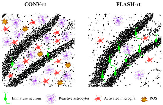
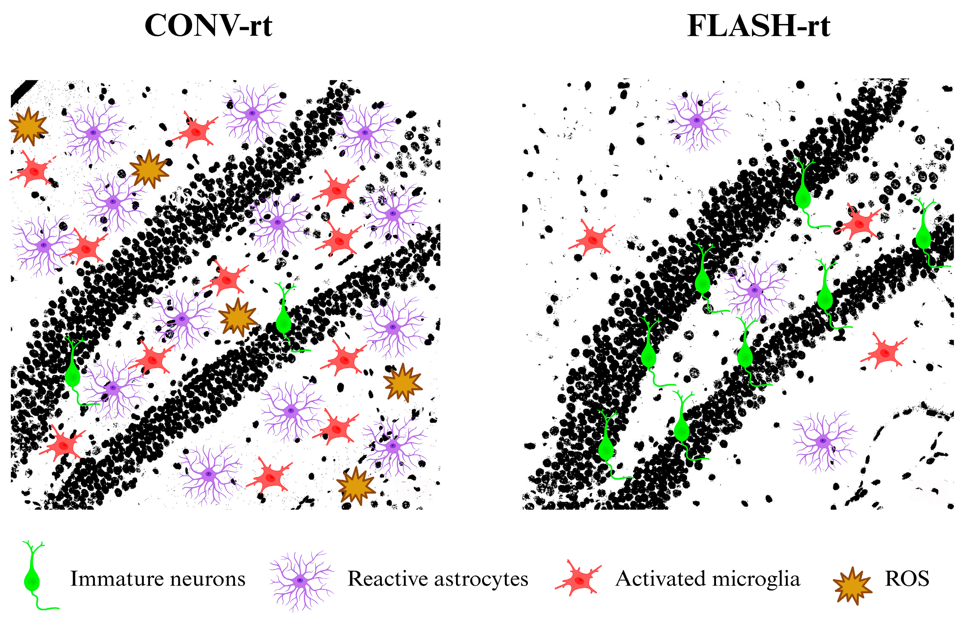
Several studies have examined whether cognitive preservation after FLASH-rt is accompanied by preservation of hippocampal cell proliferation and neuronal lineage markers. Montay-Gruel and colleagues reported more BrdU-positive cell clusters after 10 Gy whole-brain irradiation delivered with electron or X-ray FLASH-rt than after CONV-rt [28,65]. BrdU incorporation identifies DNA synthesis associated with proliferation; without neuronal phenotyping, therefore, it does not establish that the labeled cells differentiate into neurons. Other studies in juvenile mice reported greater abundance of doublecortin (DCX)-positive immature neurons and BrdU+/NeuN+ cells after FLASH-rt than after CONV-rt at assessed time points ranging from 3 weeks to 4 months [30,66]. These markers provide evidence supporting preservation of immature neuronal populations and the generation or survival of cells expressing a mature neuronal marker, respectively. However, neither marker establishes functional integration into hippocampal circuits. Moreover, the parallel preservation of these cellular outcomes and cognitive performance suggests that neurogenesis may be associated with cognitive protection (Figure 1). However, selective manipulation of adult hippocampal neurogenesis, combined with behavioral assessment, is needed to test this possibility.
Figure 1.
Composite illustration of cellular and oxidative responses associated with CONV-rt and FLASH-rt in the hippocampal dentate gyrus. The black-and-white backgrounds are photomicrographs with the nuclei stained with DAPI. We converted the original photomicrographs to black and white and superimposed schematic elements to illustrate immature neurons (green), reactive astrocytes (purple), activated microglia (red), and reactive oxygen species (ROS; ochre symbols). These overlays conceptually summarize reports of reduced immature-neuron preservation, greater glial reactivity, and increased oxidative stress following CONV-rt, with relative attenuation of these responses following FLASH-rt. The backgrounds provide anatomical context; the composite panels are not a quantitative histological comparison. Schematic elements are not drawn to scale.
3.4. Synaptic Plasticity
Synaptic plasticity plays a central role in learning and memory by enabling the brain to transform transient patterns of neuronal activity into long-lasting changes in synaptic strength. Although researchers have described several forms of synaptic plasticity in the hippocampus, long-term potentiation (LTP) is among the most extensively studied [67]. Glutamatergic neurotransmission and NMDA receptor activation initiate LTP, allowing a transient influx of Ca2+ into the postsynaptic neuron. The resulting increase in intracellular Ca2+ activates Ca2+/calmodulin-dependent protein kinase II (CaMKII), promoting its translocation and accumulation at the synapse [68]. CaMKII also interacts with NMDA receptors and phosphorylates GluA1 subunits, thereby enhancing AMPA receptor function and synaptic transmission [69,70]. Collectively, these molecular events increase synaptic strength and provide a fundamental cellular mechanism underlying activity-dependent plasticity.
Given LTP’s importance for hippocampal information processing, researchers have proposed its disruption as a potential mechanism contributing to radiation-induced cognitive dysfunction. Zhang and collaborators [71] demonstrated that CONV-rt with a single 10 Gy dose inhibited LTP in both adult and juvenile rats as early as 3 days after irradiation. Notably, LTP recovered in adult animals by 8 weeks, whereas juvenile rats exhibited a persistent reduction in LTP amplitude for up to 5 months following CONV-rt. Similar deficits have been reported following higher radiation doses, including single doses of up to 30 Gy. In addition, CONV-rt has been shown to reduce the intrinsic excitability of hippocampal pyramidal neurons, as reflected by decreased spike generation, and to impair LTP at CA3–CA1 synapses [72].
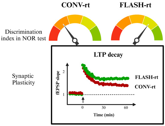
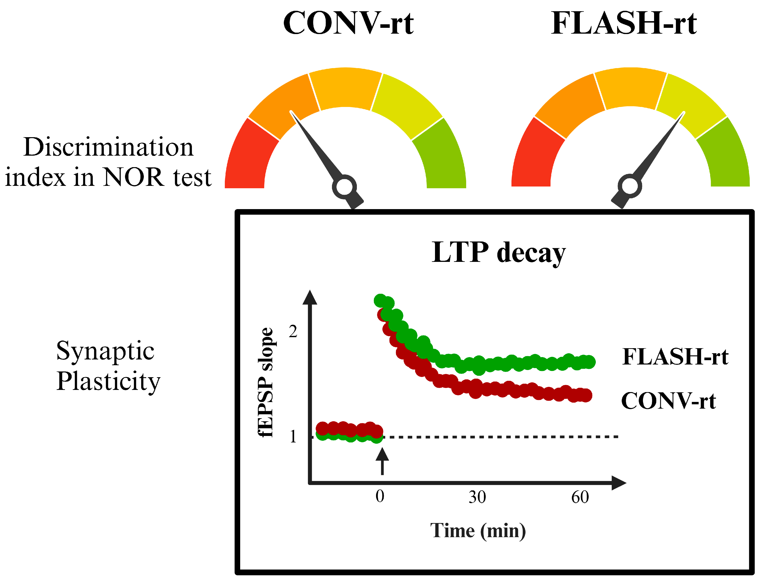
In contrast, accumulating evidence indicates that FLASH-rt can preserve synaptic plasticity following cranial irradiation. Charles Limoli’s group was the first to demonstrate that, in hypofractionated models (2 × 10 Gy or 3 × 10 Gy), FLASH-rt prevents the reduction in LTP observed six months after CONV-rt. Specifically, FLASH-rt maintained potentiation of evoked field excitatory postsynaptic potentials (fEPSPs) at levels comparable to those observed in non-irradiated control mice [34,35]. The same group subsequently evaluated a standard-of-care fractionation regimen (10 × 3 Gy) delivered at either FLASH or CONV dose rates and assessed synaptic plasticity four months later. LTP was preserved following FLASH-rt but not CONV-rt when burst stimulation was applied either to the Schaffer collaterals in the hippocampus or to layer IV of the prelimbic/infralimbic regions of the medial prefrontal cortex [73]. Importantly, FLASH-rt’s synaptic-sparing effects are not restricted to fractionated irradiation. After a single 10 Gy whole-brain dose, FLASH-rt similarly preserved synaptic plasticity, further supporting the reproducibility of this effect across radiation schedules [66]. Electrophysiological preservation following FLASH-rt is accompanied by structural and molecular evidence. FLASH-rt preserves hippocampal synaptic bouton and vesicle integrity while maintaining the proper juxtaposition of pre- and postsynaptic proteins involved in synaptic transmission and plasticity [34,35]. Moreover, spatial transcriptomic analyses identified gene expression patterns consistent with sustained AMPA receptor signaling in hippocampal neurons following FLASH-rt [74]. This finding supports a molecular association, suggesting increased receptor activity that may contribute to cognitive protection. Likewise, FLASH-induced preserved LTP provides direct functional evidence of synaptic sparing, while the structural and transcriptomic findings offer complementary support (Figure 2).
Figure 2.
Schematic illustration of recognition-memory performance and hippocampal synaptic plasticity following CONV-rt or FLASH-rt. The upper gauges illustrate relatively lower versus higher discrimination indices in the NOR test following CONV-rt and FLASH-rt, respectively. Needle positions and colored segments are qualitative and not numerically calibrated. The lower panel schematically depicts the time course of hippocampal LTP, expressed as fEPSP slope relative to the pre-induction baseline. The arrow at time zero indicates LTP induction, and the dashed horizontal line represents baseline synaptic responses. The red and green curves illustrate reduced maintenance of potentiation after CONV-rt and relatively preserved potentiation after FLASH-rt, respectively. All gauges, curves, and plotted points are illustrative; they are not experimental recordings, individual observations, group means, or reproductions of a published dataset. Numerical axis values indicate a conceptual time course and response scale rather than measured effect magnitudes. The illustration summarizes findings discussed in the text. NOR, novel object recognition; LTP, long-term potentiation; fEPSP, field excitatory postsynaptic potential.
4. Concluding Remarks
The evidence reviewed here indicates that FLASH-rt can preserve declarative memory following both single-dose and fractionated whole-brain irradiation. Importantly, protective effects have also been reported for spatial working memory [45], suggesting that beneficial effects of FLASH-rt may encompass multiple cognitive domains. However, we have to emphasize that the reviewed evidence is predominantly preclinical and does not establish cognitive preservation in patients.
The available findings support several candidate explanations for cognitive preservation, but their evidentiary strength differs. Differences in DNA damage outcomes between FLASH and CONV irradiation have been reported in ex vivo preparations and plasmid-DNA systems [75,76]. These observations do not establish reduced DNA damage in the irradiated brain or demonstrate that such a reduction initiates cognitive protection. Direct brain measurements instead document differences in oxidative biomarkers, glial and inflammatory responses, proliferation and neuronal lineage markers, and synaptic structure and function under specific experimental conditions. These findings identify biological correlates and candidate contributors to cognitive sparing, but do not establish a causal sequence linking them. The hyperoxia experiment provides interventional evidence that cognitive protection depends on oxygen conditions in the tested model [29], without identifying the responsible biochemical mechanism. Thus, reduced oxidative injury and inflammation, preserved neurogenesis-related outcomes, and maintained synaptic plasticity may interact, but their individual causal roles remain to be determined. Despite these encouraging findings, important questions remain about the biological and radiochemical mechanisms underlying the FLASH effect.
Furthermore, we need to determine the extent to which these mechanisms depend on age, sex, and tumor status. The reproducibility of cognitive sparing also depends on average and intrapulse dose rates, dose per pulse, pulse structure, total delivery time, and fractionation, because no single dose rate threshold reliably predicts protection across experimental settings. Future studies should also combine cognitive assessment with temporally resolved biological measurements and selective pathway inhibition, genetic manipulation, or rescue experiments to determine whether candidate processes are necessary or sufficient for cognitive protection.
Clinical translation presents additional challenges. Small-animal irradiation geometry differs from the larger, heterogeneous treatment volumes and delivery requirements encountered in patients. Moreover, high single doses and hypofractionated animal schedules do not necessarily predict responses to clinical fractionation. Scaling treatment must preserve adequate target coverage and the relevant temporal delivery characteristics. Clinical assessment must also extend beyond rodent behavioral analogs to validated measures of learning, memory, executive function, processing speed, and patient-reported functioning. Future FLASH-rt studies should be considered alongside existing cognitive preservation strategies. In appropriate patients receiving whole-brain radiotherapy for brain metastases, hippocampal avoidance combined with memantine provides an evidence-based approach. In the randomized phase III NRG CC001 trial, adding hippocampal avoidance to whole-brain radiotherapy plus memantine reduced the risk of cognitive failure compared with whole-brain radiotherapy plus memantine alone [77]. This provides a relevant clinical benchmark: future FLASH trials should evaluate meaningful cognitive benefit against an appropriate contemporary comparator while maintaining intracranial disease control. Overall, FLASH-rt remains a promising preclinical approach to reducing radiation-associated cognitive injury. Its clinical value requires prospective confirmation, with attention to delivery parameters, fractionation, patient characteristics, and long-term cognitive outcomes.
Author Contributions
Conceptualization, writing—original draft preparation, methodology: J.F.-P.; Writing—review and editing: M.H.-C. and E.G.-G. All authors have read and agreed to the published version of the manuscript.
Funding
This research was funded by Secretaría de Ciencia, Humanidades, Tecnología e Innovación (SECIHTI), grant number CBF-2025-I-3231 to M.H-C.
Institutional Review Board Statement
Not applicable.
Informed Consent Statement
Not applicable.
Data Availability Statement
No new data were created or analyzed in this study. Data sharing is not applicable to this article.
Acknowledgments
During preparation of this manuscript, the authors used OpenAI GPT-5.6 to improve the readability, clarity, and grammar of the manuscript. The authors have reviewed and edited the output and take full responsibility for the content of this publication.
Conflicts of Interest
The authors declare no conflicts of interest.
References
- Symonds, P.; Jones, G.D.D. FLASH Radiotherapy: The Next Technological Advance in Radiation Therapy? Clin. Oncol. 2019, 31, 405–406. [Google Scholar] [CrossRef] [Scilit] [PubMed]
- Farr, J.; Grilj, V.; Malka, V.; Sudharsan, S.; Schippers, M. Ultra-High Dose Rate Radiation Production and Delivery Systems Intended for FLASH. Med. Phys. 2022, 49, 4875–4911. [Google Scholar] [CrossRef] [Scilit] [PubMed]
- Vozenin, M.C.; Hendry, J.H.; Limoli, C.L. Biological Benefits of Ultra-High Dose Rate FLASH Radiotherapy: Sleeping Beauty Awoken. Clin. Oncol. 2019, 31, 407–415. [Google Scholar] [CrossRef] [Scilit] [PubMed]
- Kacem, H.; Almeida, A.; Cherbuin, N.; Vozenin, M.C. Understanding the FLASH Effect to Unravel the Potential of Ultra-High Dose Rate Irradiation. Int. J. Radiat. Biol. 2022, 98, 506–516. [Google Scholar] [CrossRef] [Scilit] [PubMed]
- Friedl, A.A.; Prise, K.M.; Butterworth, K.T.; Montay-Gruel, P.; Favaudon, V. Radiobiology of the FLASH Effect. Med. Phys. 2022, 49, 1993–2013. [Google Scholar] [CrossRef] [Scilit] [PubMed]
- McGarrigle, J.M.; Long, K.R.; Prezado, Y. The FLASH Effect—An Evaluation of Preclinical Studies of Ultra-High Dose Rate Radiotherapy. Front. Oncol. 2024, 14, 1340190. [Google Scholar] [CrossRef] [Scilit] [PubMed]
- Favaudon, V.; Caplier, L.; Monceau, V.; Pouzoulet, F.; Sayarath, M.; Fouillade, C.; Poupon, M.F.; Brito, I.; Hupé, P.; Bourhis, J.; et al. Ultrahigh Dose-Rate FLASH Irradiation Increases the Differential Response between Normal and Tumor Tissue in Mice. Sci. Transl. Med. 2014, 6, 245ra93. [Google Scholar] [CrossRef] [Scilit] [PubMed]
- Levy, K.; Natarajan, S.; Wang, J.; Chow, S.; Eggold, J.T.; Loo, P.E.; Manjappa, R.; Melemenidis, S.; Lartey, F.M.; Schüler, E.; et al. Abdominal FLASH Irradiation Reduces Radiation-Induced Gastrointestinal Toxicity for the Treatment of Ovarian Cancer in Mice. Sci. Rep. 2020, 10, 21600. [Google Scholar] [CrossRef] [Scilit] [PubMed]
- Chowdhury, P.; Velalopoulou, A.; Verginadis, I.I.; Morcos, G.; Loo, P.E.; Kim, M.M.; Motlagh, S.A.O.; Shoniyozov, K.; Diffenderfer, E.S.; Ocampo, E.A.; et al. Proton FLASH Radiotherapy Ameliorates Radiation-Induced Salivary Gland Dysfunction and Oral Mucositis and Increases Survival in a Mouse Model of Head and Neck Cancer. Mol. Cancer Ther. 2024, 23, 877–889. [Google Scholar] [CrossRef] [Scilit] [PubMed]
- Sørensen, B.S.; Sitarz, M.K.; Ankjærgaard, C.; Johansen, J.G.; Andersen, C.E.; Kanouta, E.; Grau, C.; Poulsen, P. Pencil Beam Scanning Proton FLASH Maintains Tumor Control While Normal Tissue Damage Is Reduced in a Mouse Model. Radiother. Oncol. 2022, 175, 178–184. [Google Scholar] [CrossRef] [Scilit] [PubMed]
- Velalopoulou, A.; Karagounis, I.V.; Cramer, G.M.; Kim, M.M.; Skoufos, G.; Goia, D.; Hagan, S.; Verginadis, I.I.; Shoniyozov, K.; Chiango, J.; et al. FLASH Proton Radiotherapy Spares Normal Epithelial and Mesenchymal Tissues While Preserving Sarcoma Response. Cancer Res. 2021, 81, 4808–4821. [Google Scholar] [CrossRef] [Scilit] [PubMed]
- Tinganelli, W.; Weber, U.; Puspitasari, A.; Simoniello, P.; Abdollahi, A.; Oppermann, J.; Schuy, C.; Horst, F.; Helm, A.; Fournier, C.; et al. FLASH with Carbon Ions: Tumor Control, Normal Tissue Sparing, and Distal Metastasis in a Mouse Osteosarcoma Model. Radiother. Oncol. 2022, 175, 185–190. [Google Scholar] [CrossRef] [Scilit] [PubMed]
- Dai, Y.; Liang, R.; Wang, J.; Zhang, J.; Wu, D.; Zhao, R.; Liu, Z.; Chen, F. Fractionated FLASH Radiation in Xenografted Lung Tumors Induced FLASH Effect at a Split Dose of 2 Gy. Int. J. Radiat. Biol. 2023, 99, 1542–1549. [Google Scholar] [CrossRef] [Scilit] [PubMed]
- Montay-Gruel, P.; Acharya, M.M.; Gonçalves Jorge, P.; Petit, B.; Petridis, I.G.; Fuchs, P.; Leavitt, R.; Petersson, K.; Gondré, M.; Ollivier, J.; et al. Hypofractionated FLASH-RT as an Effective Treatment against Glioblastoma That Reduces Neurocognitive Side Effects in Mice. Clin. Cancer Res. 2021, 27, 775–784. [Google Scholar] [CrossRef] [Scilit] [PubMed]
- Liljedahl, E.; Konradsson, E.; Linderfalk, K.; Gustafsson, E.; Petersson, K.; Ceberg, C.; Redebrandt, H.N. Comparable Survival in Rats with Intracranial Glioblastoma Irradiated with Single-Fraction Conventional Radiotherapy or FLASH Radiotherapy. Front. Oncol. 2024, 13, 1309174. [Google Scholar] [CrossRef] [Scilit] [PubMed]
- Almeida, A.; Togno, M.; Ballesteros-Zebadua, P.; Franco-Perez, J.; Geyer, R.; Schaefer, R.; Petit, B.; Grilj, V.; Meer, D.; Safai, S.; et al. Dosimetric and Biologic Intercomparison between Electron and Proton FLASH Beams. Radiother. Oncol. 2024, 190, 109953. [Google Scholar] [CrossRef] [Scilit] [PubMed]
- Schaff, L.R.; Mellinghoff, I.K. Glioblastoma and Other Primary Brain Malignancies in Adults: A Review. JAMA 2023, 329, 574–587. [Google Scholar] [CrossRef] [Scilit] [PubMed]
- Verginadis, I.I.; Citrin, D.E.; Ky, B.; Feigenberg, S.J.; Georgakilas, A.G.; E Hill-Kayser, C.; Koumenis, C.; Maity, A.; Bradley, J.D.; Lin, A. Radiotherapy Toxicities: Mechanisms, Management, and Future Directions. Lancet 2025, 405, 338–352. [Google Scholar] [CrossRef] [Scilit] [PubMed]
- Gegechkori, N.; Haines, L.; Lin, J.J. Long-Term and Latent Side Effects of Specific Cancer Types. Med. Clin. N. Am. 2017, 101, 1053–1073. [Google Scholar] [CrossRef] [Scilit] [PubMed]
- O’Reilly, M.; Mellotte, G.; Ryan, B.; O’Connor, A. Gastrointestinal Side Effects of Cancer Treatments. Ther. Adv. Chronic Dis. 2020, 11, 2040622320970354. [Google Scholar] [CrossRef] [Scilit] [PubMed]
- Tommasi, C.; Balsano, R.; Corianò, M.; Pellegrino, B.; Saba, G.; Bardanzellu, F.; Denaro, N.; Ramundo, M.; Toma, I.; Fusaro, A.; et al. Long-Term Effects of Breast Cancer Therapy and Care: Calm after the Storm? J. Clin. Med. 2022, 11, 7239. [Google Scholar] [CrossRef] [Scilit] [PubMed]
- Pazzaglia, S.; Briganti, G.; Mancuso, M.; Saran, A. Neurocognitive Decline Following Radiotherapy: Mechanisms and Therapeutic Implications. Cancers 2020, 12, 146. [Google Scholar] [CrossRef] [Scilit] [PubMed]
- Shamsesfandabadi, P.; Patel, A.; Liang, Y.; Shepard, M.J.; Wegner, R.E. Radiation-Induced Cognitive Decline: Challenges and Solutions. Cancer Manag. Res. 2024, 16, 1043–1052. [Google Scholar] [CrossRef] [Scilit] [PubMed]
- Eichenbaum, H. A Cortical-Hippocampal System for Declarative Memory. Nat. Rev. Neurosci. 2000, 1, 41–50. [Google Scholar] [CrossRef] [Scilit] [PubMed]
- Rolls, E.T. The Storage and Recall of Memories in the Hippocampo-Cortical System. Cell Tissue Res. 2018, 373, 577–604. [Google Scholar] [CrossRef] [Scilit] [PubMed]
- Ferbinteanu, J. The Hippocampus and Dorsolateral Striatum Integrate Distinct Types of Memories through Time and Space, Respectively. J. Neurosci. 2020, 40, 9055–9065. [Google Scholar] [CrossRef] [Scilit] [PubMed]
- Antunes, M.; Biala, G. The Novel Object Recognition Memory: Neurobiology, Test Procedure, and Its Modifications. Cogn. Process. 2012, 13, 93–110. [Google Scholar] [CrossRef] [Scilit] [PubMed]
- Montay-Gruel, P.; Petersson, K.; Jaccard, M.; Boivin, G.; Germond, J.F.; Petit, B.; Doenlen, R.; Favaudon, V.; Bochud, F.; Bailat, C.; et al. Irradiation in a Flash: Unique Sparing of Memory in Mice after Whole Brain Irradiation with Dose Rates above 100 Gy/s. Radiother. Oncol. 2017, 124, 365–369. [Google Scholar] [CrossRef] [Scilit] [PubMed]
- Montay-Gruel, P.; Acharya, M.M.; Petersson, K.; Alikhani, L.; Yakkala, C.; Allen, B.D.; Ollivier, J.; Petit, B.; Jorge, P.G.; Syage, A.R.; et al. Long-Term Neurocognitive Benefits of FLASH Radiotherapy Driven by Reduced Reactive Oxygen Species. Proc. Natl. Acad. Sci. USA 2019, 116, 10943–10951. [Google Scholar] [CrossRef] [Scilit] [PubMed]
- Alaghband, Y.; Cheeks, S.N.; Allen, B.D.; Montay-Gruel, P.; Doan, N.L.; Petit, B.; Jorge, P.G.; Giedzinski, E.; Acharya, M.M.; Vozenin, M.C.; et al. Neuroprotection of Radiosensitive Juvenile Mice by Ultra-High Dose Rate FLASH Irradiation. Cancers 2020, 12, 1671. [Google Scholar] [CrossRef] [Scilit] [PubMed]
- Iturri, L.; Bertho, A.; Lamirault, C.; Juchaux, M.; Gilbert, C.; Espenon, J.; Sebrie, C.; Jourdain, L.; Pouzoulet, F.; Verrelle, P.; et al. Proton FLASH Radiation Therapy and Immune Infiltration: Evaluation in an Orthotopic Glioma Rat Model. Int. J. Radiat. Oncol. Biol. Phys. 2023, 116, 655–665. [Google Scholar] [CrossRef] [Scilit] [PubMed]
- Simmons, D.A.; Lartey, F.M.; Schüler, E.; Rafat, M.; King, G.; Kim, A.; Ko, R.; Semaan, S.; Gonzalez, S.; Jenkins, M.; et al. Reduced Cognitive Deficits after FLASH Irradiation of Whole Mouse Brain Are Associated with Less Hippocampal Dendritic Spine Loss and Neuroinflammation. Radiother. Oncol. 2019, 139, 4–10. [Google Scholar] [CrossRef] [Scilit] [PubMed]
- Williams, M.T.; Regan, S.L.; Fritz, A.L.; Gollaway, B.M.; Mascia, A.E.; Vatner, R.E.; Perentesis, J.P.; Vorhees, C.V. Effects of whole brain proton irradiation at conventional or ultra-high dose rate (FLASH), in adult male Sprague Dawley rats. Sci. Rep. 2025, 15, 10602. [Google Scholar] [CrossRef] [Scilit] [PubMed]
- Allen, B.D.; Alaghband, Y.; Kramár, E.A.; Ru, N.; Petit, B.; Grilj, V.; Petronek, M.S.; Pulliam, C.F.; Kim, R.Y.; Doan, N.L.; et al. Elucidating the neurological mechanism of the FLASH effect in juvenile mice exposed to hypofractionated radiotherapy. Neuro Oncol. 2023, 25, 927–939. [Google Scholar] [CrossRef] [Scilit] [PubMed]
- Alaghband, Y.; Allen, B.D.; Kramár, E.A.; Zhang, R.; Drayson, O.G.G.; Ru, N.; Petit, B.; Almeida, A.; Doan, N.L.; Wood, M.A.; et al. Uncovering the Protective Neurologic Mechanisms of Hypofractionated FLASH Radiotherapy. Cancer Res. Commun. 2023, 3, 725–737. [Google Scholar] [CrossRef] [Scilit] [PubMed]
- Danieli, K.; Guyon, A.; Bethus, I. Episodic Memory Formation: A Review of Complex Hippocampus Input Pathways. Prog. Neuro-Psychopharmacol. Biol. Psychiatry 2023, 126, 110757. [Google Scholar] [CrossRef] [Scilit] [PubMed]
- Ballesteros-Zebadúa, P.; Chavarria, A.; Celis, M.A.; Paz, C.; Franco-Pérez, J. Radiation-Induced Neuroinflammation and Radiation Somnolence Syndrome. CNS Neurol. Disord. Drug Targets 2012, 11, 937–949. [Google Scholar] [CrossRef] [Scilit] [PubMed]
- Li, X.; Ding, Z. Cognitive Dysfunction Induced by Cranial Radiotherapy: Mechanisms and Therapeutic Methods. Brain Res. Bull. 2024, 218, 111106. [Google Scholar] [CrossRef] [Scilit] [PubMed]
- Manda, K.; Ueno, M.; Anzai, K. Cranial Irradiation-Induced Inhibition of Neurogenesis in Hippocampal Dentate Gyrus of Adult Mice: Attenuation by Melatonin Pretreatment. J. Pineal Res. 2009, 46, 71–78. [Google Scholar] [CrossRef] [Scilit] [PubMed]
- Gulbahar, O.; Aricioglu, A.; Akmansu, M.; Turkozer, Z. Effects of Radiation on Protein Oxidation and Lipid Peroxidation in the Brain Tissue. Transplant. Proc. 2009, 41, 4394–4396. [Google Scholar] [CrossRef] [Scilit] [PubMed]
- Shi, W.; Wang, J.; Li, Z.; Xu, S.; Wang, J.; Zhang, L.; Yang, H. Reprimo (RPRM) Mediates Neuronal Ferroptosis via CREB-Nrf2/SCD1 Pathways in Radiation-Induced Brain Injury. Free Radic. Biol. Med. 2024, 213, 343–358. [Google Scholar] [CrossRef] [Scilit] [PubMed]
- Xu, J.; Alameri, A.A.; Zabibah, R.S.; Gabr, G.A.; Ramírez-Coronel, A.A.; Bagheri, H.; Abedi-Firouzjah, R. Protective Potentials of Alpha-Lipoic Acid against Ionizing Radiation-Induced Brain Damage in Rats. Oxid. Med. Cell. Longev. 2023, 2023, 4999306. [Google Scholar] [CrossRef] [Scilit] [PubMed]
- Grilj, V.; Leavitt, R.J.; El Khatib, M.; Paisley, R.; Franco-Perez, J.; Petit, B.; Ballesteros-Zebadua, P.; Vozenin, M.C. In vivo measurements of change in tissue oxygen level during irradiation reveal novel dose rate dependence. Radiother. Oncol. 2024, 201, 110539. [Google Scholar] [CrossRef] [Scilit] [PubMed]
- Ballesteros-Zebadua, P.; Jansen, J.; Grilj, V.; Franco-Perez, J.; Vozenin, M.-C.; Abolfath, R. FLASH radiotherapy is faster than a heartbeat: A compartmental model to illustrate the interplay between tissue oxygen perfusion and ultra-high dose rate effects. bioRxiv 2026, 2026.03.12.711443. [Google Scholar] [CrossRef] [Scilit]
- He, R.; Xie, S.; Du, L.; Li, W.; Wang, J.; Liu, X.; Wu, D.; Yang, Y.; Qu, B.; Cheng, G.; et al. The Impacts of FLASH Radiation Therapy and Conventional Radiation Therapy on the Cognitive Abilities of Mice. Adv. Radiat. Oncol. 2025, 11, 101950. [Google Scholar] [CrossRef] [Scilit] [PubMed]
- Blain, G.; Vandenborre, J.; Villoing, D.; Fiegel, V.; Fois, G.R.; Haddad, F.; Koumeir, C.; Maigne, L.; Métivier, V.; Poirier, F.; et al. Proton Irradiations at Ultra-High Dose Rate vs. Conventional Dose Rate: Strong Impact on Hydrogen Peroxide Yield. Radiat. Res. 2022, 198, 318–324. [Google Scholar] [CrossRef] [Scilit] [PubMed]
- Kacem, H.; Psoroulas, S.; Boivin, G.; Folkerts, M.; Grilj, V.; Lomax, T.; Martinotti, A.; Meer, D.; Ollivier, J.; Petit, B.; et al. Comparing Radiolytic Production of H2O2 and Development of Zebrafish Embryos after Ultra-High Dose Rate Exposure with Electron and Transmission Proton Beams. Radiother. Oncol. 2022, 175, 197–202. [Google Scholar] [CrossRef] [Scilit] [PubMed]
- Gupta, S.; Inman, J.L.; Chant, J.; Obst-Huebl, L.; Nakamura, K.; Costello, S.M.; Marqusee, S.; Mao, J.H.; Kunz, L.; Paisley, R.; et al. A Novel Platform for Evaluating Dose Rate Effects on Oxidative Damage to Peptides: Toward a High-Throughput Method to Characterize the Mechanisms Underlying the FLASH Effect. Radiat. Res. 2023, 200, 523–530. [Google Scholar] [CrossRef] [Scilit] [PubMed]
- Wang, Y.; Tian, J.; Liu, D.; Li, T.; Mao, Y.; Zhu, C. Microglia in Radiation-Induced Brain Injury: Cellular and Molecular Mechanisms and Therapeutic Potential. CNS Neurosci. Ther. 2024, 30, e14794. [Google Scholar] [CrossRef] [Scilit] [PubMed]
- Hellweg, C.E. The Nuclear Factor κB Pathway: A Link to the Immune System in the Radiation Response. Cancer Lett. 2015, 368, 275–289. [Google Scholar] [CrossRef] [Scilit] [PubMed]
- Beamish, C.A.; Zawaski, J.A.; Inoue, T.; Sarkar, P.; Grosshans, D.R.; Sabek, O.M.; Gaber, M.W. NF-κB Blockade by NEMO Binding Domain Peptide Ameliorates Inflammation and Neurobehavioral Sequelae after Cranial Radiation Therapy in Juvenile Mice. Int. J. Radiat. Oncol. Biol. Phys. 2021, 109, 1508–1520. [Google Scholar] [CrossRef] [Scilit] [PubMed]
- Bachstetter, A.D.; Van Eldik, L.J. The p38 MAP Kinase Family as Regulators of Proinflammatory Cytokine Production in Degenerative Diseases of the CNS. Aging Dis. 2010, 1, 199–211. [Google Scholar] [PubMed]
- González-Johnson, L.; Fariña, A.; Farías, G.; Zomosa, G.; Pinilla-González, V.; Rojas-Solé, C. Exploring Neuroprotection against Radiation-Induced Brain Injury: A Review of Key Compounds. NeuroSci 2024, 5, 462–484. [Google Scholar] [CrossRef] [Scilit] [PubMed]
- Sánchez-Gomar, I.; Geribaldi-Doldán, N.; Santos-Rosendo, C.; Sanguino-Caneva, C.; Carrillo-Chapman, C.; Fiorillo-Moreno, O.; Villareal Camacho, J.L.; Quiroz, E.N.; Verástegui, C. Exploring the Intricacies of Neurogenic Niches: Unraveling the Anatomy and Neural Microenvironments. Biomolecules 2024, 14, 335. [Google Scholar] [CrossRef] [Scilit] [PubMed]
- Elliott, T.; Liu, K.Y.; Hazan, J.; Wilson, J.; Vallipuram, H.; Jones, K.; Mahmood, J.; Gitlin-Leigh, G.; Howard, R. Hippocampal Neurogenesis in Adult Primates: A Systematic Review. Mol. Psychiatry 2025, 30, 1195–1206. [Google Scholar] [CrossRef] [Scilit] [PubMed]
- Xie, W.S.; Shehzadi, K.; Ma, H.L.; Liang, J.H. A Potential Strategy for Treatment of Neurodegenerative Disorders by Regulation of Adult Hippocampal Neurogenesis in Human Brain. Curr. Med. Chem. 2022, 29, 5315–5347. [Google Scholar] [CrossRef] [Scilit] [PubMed]
- Li, Y.D.; Luo, Y.J.; Song, J. Optimizing Memory Performance and Emotional States: Multi-Level Enhancement of Adult Hippocampal Neurogenesis. Curr. Opin. Neurobiol. 2023, 79, 102693. [Google Scholar] [CrossRef] [Scilit] [PubMed]
- Leskinen, S.; Alsalek, S.; Wernicke, A.G. Effects of Radiotherapy on the Hippocampus and Hippocampal Neurogenesis: A Systematic Review of Preclinical Studies. Strahlenther. Onkol. 2025, 201, 383–397. [Google Scholar] [CrossRef] [Scilit] [PubMed]
- Huang, R.X.; Zhou, P.K. DNA Damage Response Signaling Pathways and Targets for Radiotherapy Sensitization in Cancer. Signal Transduct. Target. Ther. 2020, 5, 60. [Google Scholar] [CrossRef] [Scilit] [PubMed]
- Nakano, T.; Akamatsu, K.; Tsuda, M.; Tujimoto, A.; Hirayama, R.; Hiromoto, T.; Tamada, T.; Ide, H.; Shikazono, N. Formation of Clustered DNA Damage In Vivo upon Irradiation with Ionizing Radiation: Visualization and Analysis with Atomic Force Microscopy. Proc. Natl. Acad. Sci. USA 2022, 119, e2119132119. [Google Scholar] [CrossRef] [Scilit] [PubMed]
- Michaelidesová, A.; Konířová, J.; Bartůněk, P.; Zíková, M. Effects of Radiation Therapy on Neural Stem Cells. Genes 2019, 10, 640. [Google Scholar] [CrossRef] [Scilit] [PubMed]
- Ji, S.; Tian, Y.; Lu, Y.; Sun, R.; Ji, J.; Zhang, L.; Duan, S. Irradiation-Induced Hippocampal Neurogenesis Impairment Is Associated with Epigenetic Regulation of BDNF Gene Transcription. Brain Res. 2014, 1577, 77–88. [Google Scholar] [CrossRef] [Scilit] [PubMed]
- Schmal, Z.; Hammer, B.; Müller, A.; Rübe, C.E. Fractionated Low-Dose Radiation Induces Long-Lasting Inflammatory Responses in the Hippocampal Stem Cell Niche. Int. J. Radiat. Oncol. Biol. Phys. 2021, 111, 1262–1275. [Google Scholar] [CrossRef] [Scilit] [PubMed]
- Ma, L.; Ye, Z.; Zhang, Y.; Shi, W.; Wang, J.; Yang, H. Irradiated Microvascular Endothelial Cells May Induce Bystander Effects in Neural Stem Cells Leading to Neurogenesis Inhibition. J. Radiat. Res. 2022, 63, 192–201. [Google Scholar] [CrossRef] [Scilit] [PubMed]
- Montay-Gruel, P.; Bouchet, A.; Jaccard, M.; Patin, D.; Serduc, R.; Aim, W.; Petersson, K.; Petit, B.; Bailat, C.; Bourhis, J.; et al. X-rays Can Trigger the FLASH Effect: Ultra-High Dose-Rate Synchrotron Light Source Prevents Normal Brain Injury after Whole Brain Irradiation in Mice. Radiother. Oncol. 2018, 129, 582–588. [Google Scholar] [CrossRef] [Scilit] [PubMed]
- Drayson, O.G.G.; Melemenidis, S.; Katila, N.; Viswanathan, V.; Kramár, E.A.; Zhang, R.; Kim, R.; Ru, N.; Petit, B.; Dutt, S.; et al. A Multi-Institutional Study to Investigate the Sparing Effect after Whole Brain Electron FLASH in Mice: Reproducibility and Temporal Evolution of Functional, Electrophysiological, and Neurogenic Endpoints. Radiother. Oncol. 2024, 201, 110534. [Google Scholar] [CrossRef] [Scilit] [PubMed]
- Magee, J.C.; Grienberger, C. Synaptic Plasticity Forms and Functions. Annu. Rev. Neurosci. 2020, 43, 95–117. [Google Scholar] [CrossRef] [Scilit] [PubMed]
- Tao, W.; Lee, J.; Chen, X.; Díaz-Alonso, J.; Zhou, J.; Pleasure, S.; Nicoll, R.A. Synaptic Memory Requires CaMKII. eLife 2021, 10, e60360. [Google Scholar] [CrossRef] [Scilit] [PubMed]
- Incontro, S.; Díaz-Alonso, J.; Iafrati, J.; Vieira, M.; Asensio, C.S.; Sohal, V.S.; Roche, K.W.; Bender, K.J.; Nicoll, R.A. The CaMKII/NMDA Receptor Complex Controls Hippocampal Synaptic Transmission by Kinase-Dependent and Independent Mechanisms. Nat. Commun. 2018, 9, 2069. [Google Scholar] [CrossRef] [Scilit] [PubMed]
- Hayashi, Y. Molecular Mechanism of Hippocampal Long-Term Potentiation—Towards Multiscale Understanding of Learning and Memory. Neurosci. Res. 2022, 175, 3–15. [Google Scholar] [CrossRef] [Scilit] [PubMed]
- Zhang, D.; Zhou, W.; Lam, T.T.; Weng, C.; Bronk, L.; Ma, D.; Wang, Q.; Duman, J.G.; Dougherty, P.M.; Grosshans, D.R. Radiation Induces Age-Dependent Deficits in Cortical Synaptic Plasticity. Neuro Oncol. 2018, 20, 1207–1214. [Google Scholar] [CrossRef] [Scilit] [PubMed]
- Wu, M.Y.; Zou, W.J.; Yu, P.; Yang, Y.; Li, S.J.; Liu, Q.; Xie, J.; Chen, S.Q.; Lin, W.J.; Tang, Y. Cranial Irradiation Impairs Intrinsic Excitability and Synaptic Plasticity of Hippocampal CA1 Pyramidal Neurons with Implications for Cognitive Function. Neural Regen. Res. 2022, 17, 2253–2259. [Google Scholar] [CrossRef] [Scilit] [PubMed]
- Limoli, C.L.; Kramár, E.A.; Almeida, A.; Petit, B.; Grilj, V.; Baulch, J.E.; Ballesteros-Zebadua, P.; Loo, B.W., Jr.; Wood, M.A.; Vozenin, M.C. The Sparing Effect of FLASH-RT on Synaptic Plasticity Is Maintained in Mice with Standard Fractionation. Radiother. Oncol. 2023, 186, 109767. [Google Scholar] [CrossRef] [Scilit] [PubMed]
- Kunz, L.V.; Almeida, A.; Knol, M.; Petit, B.; Kramár, E.A.; Wood, M.A.; Limoli, C.L.; Vozenin, M.-C. Sustained AMPAR Expression in CA3 Neurons May Mediate Neuroprotection Following FLASH-RT. Cell Rep. 2026, 45, 117670. [Google Scholar] [CrossRef] [Scilit] [PubMed]
- Cooper, C.R.; Jones, D.; Jones, G.D.; Petersson, K. FLASH Irradiation Induces Lower Levels of DNA Damage Ex Vivo, an Effect Modulated by Oxygen Tension, Dose, and Dose Rate. Br. J. Radiol. 2022, 95, 20211150. [Google Scholar] [CrossRef] [Scilit] [PubMed]
- Perstin, A.; Poirier, Y.; Sawant, A.; Tambasco, M. Quantifying the DNA-Damaging Effects of FLASH Irradiation with Plasmid DNA. Int. J. Radiat. Oncol. Biol. Phys. 2022, 113, 437–447. [Google Scholar] [CrossRef] [Scilit] [PubMed]
- Brown, P.D.; Gondi, V.; Pugh, S.; Tome, W.A.; Wefel, J.S.; Armstrong, T.S.; Bovi, J.A.; Robinson, C.; Konski, A.; Khuntia, D.; et al. Hippocampal Avoidance During Whole-Brain Radiotherapy Plus Memantine for Patients With Brain Metastases: Phase III Trial NRG Oncology CC001. J. Clin. Oncol. 2020, 38, 1019–1029. [Google Scholar] [CrossRef] [Scilit] [PubMed]

Disclaimer/Publisher’s Note: The statements, opinions and data contained in all publications are solely those of the individual author(s) and contributor(s) and not of MDPI and/or the editor(s). MDPI and/or the editor(s) disclaim responsibility for any injury to people or property resulting from any ideas, methods, instructions or products referred to in the content. |
© 2026 by the authors. Licensee MDPI, Basel, Switzerland. This article is an open access article distributed under the terms and conditions of the Creative Commons Attribution (CC BY) license.