Immunohistochemical Study of the Tumor Immune Microenvironment in p16-Positive and p16-Negative Oral Squamous Cell Carcinoma and Its Prognostic Implications

 , , , , and
Abstract
1. Introduction
2. Materials and Methods
2.1. Patient Selection and Inclusion Criteria
2.2. Ethical Considerations
2.3. Laboratory Technique
2.4. Objectives
2.5. Statistical Analysis
3. Results
3.1. Clinical and Demographic Data
3.2. Morphological Features
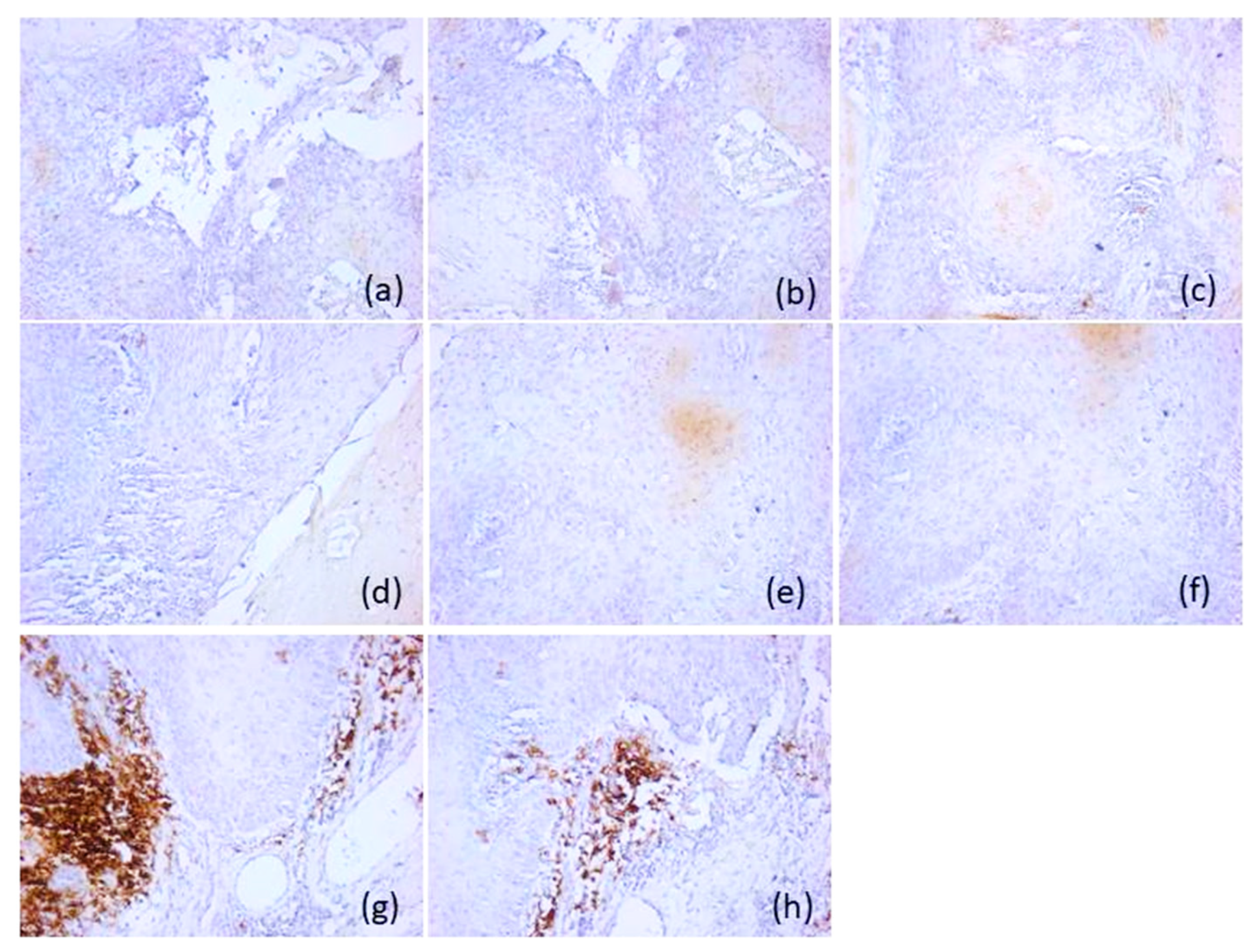
3.3. Immunohistochemical Features
3.3.1. Immunohistochemical Features of p16-Negative Tumors
3.3.2. Immunohistochemical Features of p16-Positive Tumors
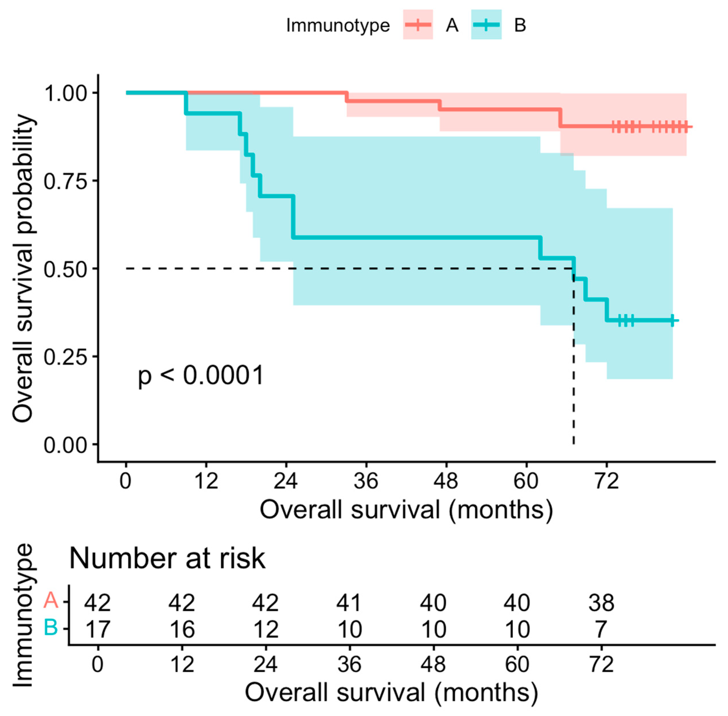
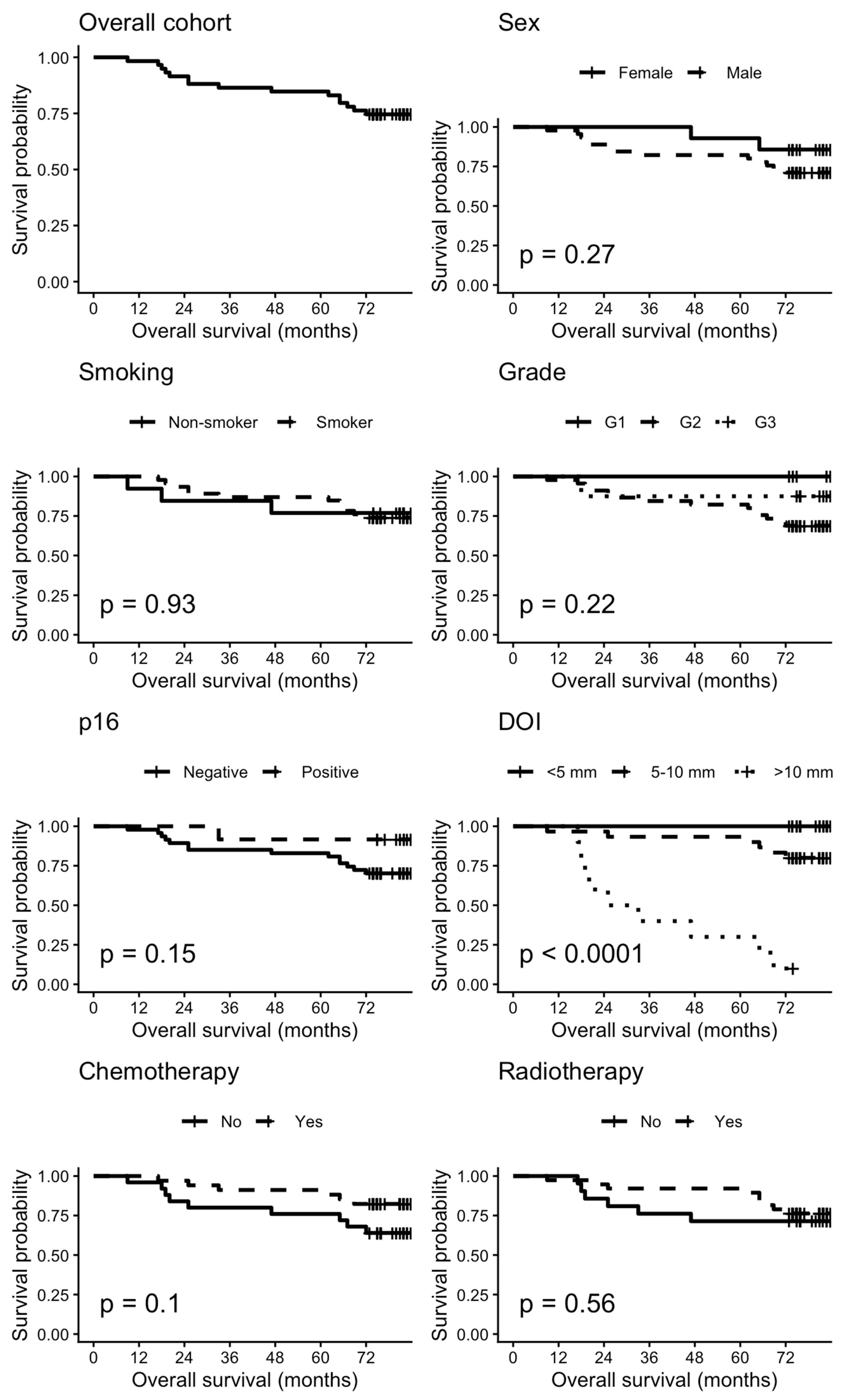
3.4. Survival Analysis
4. Discussion
5. Conclusions
Author Contributions
Funding
Institutional Review Board Statement
Informed Consent Statement
Data Availability Statement
Conflicts of Interest
Abbreviations
| OSCC | Oral Squamous Cell Carcinoma |
| NK | Natural Killer |
| TME | Tumor Microenvironment |
| TIME | Tumor Immune Microenvironment |
| HPV | Human Papillomavirus |
| DOI | Depth of Invasion |
| WPOI-5 | Worst Pattern of Invasion |
| G1 | Well Differentiated |
| G2 | Moderately Differentiated |
| G3 | Poorly Differentiated |
| TNM | Tumor, Node, Metastasis |
| p16 | Inhibitor of Cyclin-Dependent Kinase 4a |
| CD3 | Cluster of Differentiation 3 |
| CD4 | Cluster of Differentiation 4 |
| CD8 | Cluster of Differentiation 8 |
| CD20 | Cluster of Differentiation 20 |
| RTU | Ready-to-use |
| CD117 | Cluster of Differentiation 117 |
| CD1a | Cluster of Differentiation 1a |
| CD68 | Cluster of Differentiation 68 |
| CD56 | Cluster of Differentiation 56 |
| IQR | Interquartile range |
| OS | Overall Survival |
| HRs | Hazard Ratios |
| CIs | Confidence Intervals |
| T | Pathologic T stage |
| N | Pathologic N stage |
| Pn | Perineural Invasion |
| VL | Lymphovascular Invasion |
| WPI | Worst Pattern of Invasion |
| TAMs | Tumor-Associated Macrophages |
References
- Aoun, G.; El–Toum, S.; Chammas, C.; Nabbout, F. Advanced Case of Squamous Cell Carcinoma of the Posterior–Lateral Border of the Tongue Invading the Floor of the Mouth. Med. Arch. 2022, 76, 305–307. [Google Scholar]
- Leemans, C.R.; Snijders, P.J.F.; Brakenhoff, R.H. The molecular landscape of head and neck cancer. Nat. Rev. Cancer 2018, 18, 269–282. [Google Scholar] [CrossRef] [Scilit]
- Binnewies, M.; Roberts, E.W.; Kersten, K.; Chan, V.; Fearon, D.F.; Merad, M.; Coussens, L.M.; Gabrilovich, D.I.; Ostrand–Rosenberg, S.; Hedrick, C.C.; et al. Understanding the tumor immune microenvironment (TIME) for effective therapy. Nat. Med. 2018, 24, 541–550. [Google Scholar] [CrossRef] [Scilit] [PubMed]
- Caponio, V.C.A.; Zhurakivska, K.; Lo Muzio, L.; Troiano, G.; Cirillo, N. The Immune Cells in the Development of Oral Squamous Cell Carcinoma. Cancers 2023, 15, 3779. [Google Scholar] [CrossRef] [Scilit] [PubMed]
- Kouketsu, A.; Sato, I.; Oikawa, M.; Shimizu, Y.; Saito, H.; Tashiro, K.; Yamashita, Y.; Takahashi, T.; Kumamoto, H. Regulatory T cells and M2–polarized tumour–associated macrophages are associated with the oncogenesis and progression of oral squamous cell carcinoma. Int. J. Oral Maxillofac. Surg. 2019, 48, 1279–1288. [Google Scholar] [CrossRef] [Scilit]
- Fridman, W.H.; Pagès, F.; Sautès–Fridman, C.; Galon, J. The immune contexture in human tumours: Impact on clinical outcome. Nat. Rev. Cancer 2012, 12, 298–306. [Google Scholar] [CrossRef] [Scilit]
- Simoni, Y.; Becht, E.; Fehlings, M.; Loh, C.Y.; Koo, S.L.; Teng, K.W.W.; Yeong, J.P.S.; Nahar, R.; Zhang, T.; Kared, H.; et al. Bystander CD8+ T cells are abundant and phenotypically distinct in human tumour infiltrates. Nature 2018, 557, 575–579. [Google Scholar] [CrossRef] [Scilit] [PubMed]
- Freeman, P.; Mielgo, A. Cancer–Associated Fibroblast Mediated Inhibition of CD8+ Cytotoxic T Cell Accumulation in Tumours: Mechanisms and Therapeutic Opportunities. Cancers 2020, 12, 2687. [Google Scholar] [CrossRef] [Scilit]
- Gillison, M.L.; Koch, W.M.; Capone, R.B.; Spafford, M.; Westra, W.H.; Wu, L.; Zahurak, M.L.; Daniel, R.W.; Viglione, M.; Symer, D.E.; et al. Evidence for a causal association between human papillomavirus and a subset of head and neck cancers. J. Natl. Cancer Inst. 2000, 92, 709–720. [Google Scholar] [CrossRef] [Scilit]
- Ang, K.K.; Harris, J.; Wheeler, R.; Weber, R.; Rosenthal, D.I.; Nguyen–Tân, P.F.; Westra, W.H.; Chung, C.H.; Jordan, R.C.; Lu, C.; et al. Human papillomavirus and survival of patients with oropharyngeal cancer. N. Engl. J. Med. 2010, 363, 24–35. [Google Scholar] [CrossRef] [Scilit]
- Kather, J.N.; Suarez–Carmona, M.; Charoentong, P.; Weis, C.A.; Hirsch, D.; Bankhead, P.; Horning, M.; Ferber, D.; Kel, I.; Herpel, E.; et al. Topography of cancer–associated immune cells in human solid tumors. Elife 2018, 7, e36967. [Google Scholar] [CrossRef] [Scilit]
- Brierly, G.; Celentano, A.; Breik, O.; Moslemivayeghan, E.; Patini, R.; McCullough, M.; Yap, T. Tumour Necrosis Factor Alpha (TNF–α) and Oral Squamous Cell Carcinoma. Cancers 2023, 15, 1841. [Google Scholar] [CrossRef] [Scilit]
- Trandafir, C.M.; Closca, R.M.; Poenaru, M.; Sarau, O.S.; Sarau, C.A.; Rakitovan, M.; Baderca, F.; Sima, L.V. Morphological and Immunohistochemical Aspects with Prognostic Implications and Therapeutic Targets of Primary Sinonasal Mucosal Melanoma: A Retrospective Study. Cancers 2024, 16, 2863. [Google Scholar] [CrossRef] [Scilit]
- Breaza, G.M.; Closca, R.M.; Cindrea, A.C.; Hut, F.E.; Cretu, O.; Sima, L.V.; Rakitovan, M.; Zara, F. Immunohistochemical Evaluation of the Tumor Immune Microenvironment in Pancreatic Ductal Adenocarcinoma. Diagnostics 2025, 15, 646. [Google Scholar] [CrossRef] [Scilit]
- Rakitovan, M.; Nicoara, A.; Closca, R.M.; Ceausu, R.A.; Dumitru, C.S.; Barb, A.C.; Novacescu, D.; Armega Anghelescu, A.; Cindrea, A.C.; Zara, F. The Solitary Fibrous Tumor, the Chameleon of the Head and Neck Region—Clinical, Epidemiological, and Histopathological Aspects. Diagnostics 2025, 15, 2721. [Google Scholar] [CrossRef] [Scilit]
- Cabral, L.G.d.S.; Martins, I.M.; Paulo, E.P.d.A.; Pomini, K.T.; Poyet, J.–L.; Maria, D.A. Molecular Mechanisms in the Carcinogenesis of Oral Squamous Cell Carcinoma: A Literature Review. Biomolecules 2025, 15, 621. [Google Scholar] [CrossRef] [Scilit] [PubMed]
- Hut, A.–R.; Boia, E.R.; Para, D.; Iovanescu, G.; Horhat, D.; Mikša, L.; Chiriac, M.; Galant, R.; Motofelea, A.C.; Balica, N.C. Laryngeal Cancer in the Modern Era: Evolving Trends in Diagnosis, Treatment, and Survival Outcomes. J. Clin. Med. 2025, 14, 3367. [Google Scholar] [CrossRef] [Scilit] [PubMed]
- Boia, E.R.; Huț, A.R.; Roi, A.; Luca, R.E.; Munteanu, I.R.; Roi, C.I.; Riviș, M.; Boia, S.; Duse, A.O.; Vulcănescu, D.D.; et al. Associated Bacterial Coinfections in COVID–19–Positive Patients. Medicina 2023, 59, 1858. [Google Scholar] [CrossRef] [Scilit]
- Tsai, C.–C.; Hsu, Y.–C.; Chu, T.–Y.; Hsu, P.–C.; Kuo, C.–Y. Immune Evasion in Head and Neck Squamous Cell Carcinoma: Roles of Cancer–Associated Fibroblasts, Immune Checkpoints, and TP53 Mutations in the Tumor Microenvironment. Cancers 2025, 17, 2590. [Google Scholar] [CrossRef] [Scilit] [PubMed]
- Sutera, S.; Furchì, O.A.; Pentenero, M. Investigating Tumor–Infiltrating Lymphocytes in the Microenvironment of Oral Squamous Cell Carcinoma (OSCC) and Oral Potentially Malignant Disorders (OPMDs): Can They Shift Our Perspective? A Scoping Review. J. Clin. Med. 2025, 14, 606. [Google Scholar] [CrossRef] [Scilit]
- Morais, H.G.d.F.; Costa, C.F.d.; de Souto Medeiros, M.R.; Araújo, B.d.A.; Morais, E.F.d.; Coletta, R.D.; Freitas, R.d.A. Prognostic Value of the Immunohistochemical Detection of Cellular Components of the Tumor Microenvironment in Oral Squamous Cell Carcinoma: A Systematic Review. Curr. Issues Mol. Biol. 2025, 47, 544. [Google Scholar] [CrossRef] [Scilit]
- Missale, F.; Bugatti, M.; Mattavelli, D.; Lonardi, S.; Lombardi, D.; Nicolai, P.; Piazza, C.; Battocchio, S.; Bozzola, A.M.; Calza, S.; et al. Metavariables Resuming Host Immune Features and Nodal Involvement Are Associated with Oncological Outcomes in Oral Cavity Squamous Cell Carcinoma. Cells 2021, 10, 2203. [Google Scholar] [CrossRef] [Scilit]
- Maleki Vareki, S. High and low mutational burden tumors versus immunologically hot and cold tumors and response to immune checkpoint inhibitors. J. Immunother. Cancer 2018, 6, 157. [Google Scholar] [CrossRef] [Scilit]
- Galon, J.; Bruni, D. Approaches to treat immune hot, altered and cold tumours with combination immunotherapies. Nat. Rev. Drug Discov. 2019, 18, 197–218. [Google Scholar] [CrossRef] [Scilit] [PubMed]
- Li, B.; Cui, Y.; Nambiar, D.K.; Sunwoo, J.B.; Li, R. The immune subtypes and landscape of squamous cell carcinoma. Clin. Cancer Res. 2019, 25, 3528–3537. [Google Scholar] [CrossRef] [Scilit]
- Chen, D.S.; Mellman, I. Oncology meets immunology: The cancer–immunity cycle. Immunity 2013, 39, 1–10. [Google Scholar] [CrossRef] [Scilit]
- Ali, A.; Molska, G.R.; Yeo, H.; Esfandiari, N.; Jeong, W.; Huang, M.; Magalhaes, M. Immune Microenvironment in Oral Potentially Malignant Disorders and Oral Cancer: A Narrative Review. Int. J. Mol. Sci. 2025, 26, 6650. [Google Scholar] [CrossRef] [Scilit] [PubMed]
- Lequerica–Fernández, P.; Suárez–Canto, J.; Rodriguez–Santamarta, T.; Rodrigo, J.P.; Suárez–Sánchez, F.J.; Blanco–Lorenzo, V.; Domínguez–Iglesias, F.; García–Pedrero, J.M.; de Vicente, J.C. Prognostic Relevance of CD4+, CD8+ and FOXP3+ TILs in Oral Squamous Cell Carcinoma and Correlations with PD–L1 and Cancer Stem Cell Markers. Biomedicines 2021, 9, 653. [Google Scholar] [CrossRef] [Scilit]
- Suárez–Sánchez, F.J.; Lequerica–Fernández, P.; Rodrigo, J.P.; Hermida–Prado, F.; Suárez–Canto, J.; Rodríguez–Santamarta, T.; Domínguez–Iglesias, F.; García–Pedrero, J.M.; de Vicente, J.C. Tumor–Infiltrating CD20+ B Lymphocytes: Significance and Prognostic Implications in Oral Cancer Microenvironment. Cancers 2021, 13, 395. [Google Scholar] [CrossRef] [Scilit]
- Nauta, I.H.; Nijenhuis, D.N.L.M.; Ganzevles, S.H.; Raaff, P.I.; Kloosterman, J.; Bloemena, E.; Brakenhoff, R.H.; Leemans, C.R.; van de Ven, R. Richness for Tumor–Infiltrating B–Cells in the Oral Cancer Tumor Microenvironment Is a Prognostic Factor in Early–Stage Disease and Improves Outcome in Advanced–Stage Disease. Cancers 2025, 17, 113. [Google Scholar] [CrossRef] [Scilit] [PubMed]
- Da Silva, L.C.; Fonseca, F.P.; de Almeida, O.P.; de Almeida Mariz, B.A.L.; Lopes, M.A.; Radhakrish–nan, R.; Sharma, M.; Kowalski, L.P.; Vargas, P.A. CD1a+ and CD207+ cells are reduced in oral sub–mucous fibrosis and oral squamous cell carcinoma. Med. Oral Patol. Oral Cir. Bucal 2020, 25, e49–e55. [Google Scholar] [CrossRef] [Scilit]
- Dadi, S.; Chhangawala, S.; Whitlock, B.M.; Franklin, R.A.; Luo, C.T.; Oh, S.A.; Toure, A.; Pritykin, Y.; Huse, M.; Leslie, C.S.; et al. Cancer Immunosurveillance by Tissue–Resident Innate Lymphoid Cells and Innate–like T Cells. Cell 2016, 164, 365–377. [Google Scholar] [CrossRef] [Scilit]
- Zhang, Y.; Xie, J.; Wu, H.; Huang, J.; Zheng, D.; Wang, S.; Jia, X.; He, Z.; Gong, Y.; Ju, L.; et al. NK cell based immunotherapy against oral squamous cell carcinoma. Front. Immunol. 2024, 15, 1440764. [Google Scholar] [CrossRef] [Scilit]
- Zitvogel, L. Dendritic and natural killer cells cooperate in the control/switch of innate immunity. J. Exp. Med. 2002, 195, F9–F14. [Google Scholar] [CrossRef] [Scilit] [PubMed]
- Suárez–Sánchez, F.J.; Lequerica–Fernández, P.; Suárez–Canto, J.; Rodrigo, J.P.; Rodriguez–Santamarta, T.; Domínguez–Iglesias, F.; García–Pedrero, J.M.; de Vicente, J.C. Macrophages in Oral Carcinomas: Relationship with Cancer Stem Cell Markers and PD–L1 Expression. Cancers 2020, 12, 1764. [Google Scholar] [CrossRef] [Scilit] [PubMed]
- Wachter, E.; Fox, L.H.; Lu, Z.; Jones, A.D.; Casto, N.D.; Waltz, S.E. RON Receptor Signaling and the Tumor Microenvironment. Genes 2025, 16, 437. [Google Scholar] [CrossRef] [Scilit]
- Li, C.; Dong, X.; Li, B. Tumor microenvironment in oral squamous cell carcinoma. Front. Immunol. 2024, 15, 1485174. [Google Scholar] [CrossRef] [Scilit]
- Ribatti, D.; Tamma, R.; Vacca, A. Mast Cells and Angiogenesis in Human Plasma Cell Malignancies. Int. J. Mol. Sci. 2019, 20, 481. [Google Scholar] [CrossRef] [Scilit]
- Kawczak, P.; Feszak, I.J.; Bączek, T. Comparative Mechanistic Insights and Therapeutic Potential of Pembrolizumab, Durvalumab, and Ipilimumab as Immune Checkpoint Inhibitors in the Targeted Management of Oral and Head and Neck Squamous Cell Carcinoma. Cancers 2025, 17, 2805. [Google Scholar] [CrossRef] [Scilit]
- Xue, N.; Wang, Y.; Wang, Z.; Zeng, X.; Wang, J.; Zhang, X. Trends in immunotherapy for oral squamous cell carcinoma. Cell. Oncol. 2025, 48, 1159–1179. [Google Scholar] [CrossRef] [Scilit]
- Pawar, J.S.; Salam, M.A.; Dipto, M.S.U.; Al–Amin, M.Y.; Salam, M.T.; Sengupta, S.; Kumari, S.; Gujjari, L.; Yadagiri, G. Cancer–Associated Fibroblasts: Immunosuppressive Crosstalk with Tumor–Infiltrating Immune Cells and Implications for Therapeutic Resistance. Cancers 2025, 17, 2484. [Google Scholar] [CrossRef] [Scilit] [PubMed]
- Harper, J.; Sainson, R.C. Regulation of the anti–tumour immune response by cancer–associated fibroblasts. Semin. Cancer Biol. 2014, 25, 69–77. [Google Scholar] [CrossRef] [Scilit] [PubMed]
- Balica, N.C.; Poenaru, M.; Preda, M.A.; Boia, E.R.; Burlacu, O.N.; Horhat, I.D.; Mogoantă, C.A.; Vlăescu, A.N.; Baderca, F.; Jifcu, E.M.; et al. Primary tonsillar tuberculosis—Case report. Rom. J. Morphol. Embryol. 2019, 60, 267–271. [Google Scholar] [PubMed]
- Segneanu, A.–E.; Vlase, G.; Vlase, T.; Ciocalteu, M.–V.; Bejenaru, C.; Buema, G.; Bejenaru, L.E.; Boia, E.R.; Dumitru, A.; Boia, S. Romanian Wild–Growing Chelidonium majus—An Emerging Approach to a Potential Antimicrobial Engineering Carrier System Based on AuNPs: In Vitro Investigation and Evaluation. Plants 2024, 13, 734. [Google Scholar] [CrossRef] [Scilit]





| Antibody | Substrate | Dilution | Clone | Cell Type |
|---|---|---|---|---|
| 1 p16 | Monoclonal, mouse | 1:100 | CS1 | Malignant squamous cells |
| 2 CD3 | Monoclonal, mouse | 1:500 | LN10 | T lymphocyte |
| 3 CD4 | Monoclonal, mouse | 1:100 | 4B12 | T helper lymphocyte |
| 4 CD8 | Monoclonal, mouse | 1:500 | 4B11 | T cytotoxic lymphocyte |
| 5 CD20 | Monoclonal, mouse | 6 RTU | L26 | B Lymphocyte |
| 7 CD117 | Monoclonal, rabbit | 1:200 | EP10 | Mast cell |
| 8 CD1a | Monoclonal, mouse | RTU | MTB1 | Dendritic cell |
| 9 CD68 | Monoclonal, mouse | 1:100 | 514H12 | Macrophage |
| 10 CD56 | Monoclonal, rabbit | RTU | MRQ-42 | Natural killer lymphocyte |
| Variable | All Patients (N = 59) | Immunotype A (N = 42) | Immunotype B (N = 17) | p-Value |
|---|---|---|---|---|
| Age # | 62 (55.5–69.5) | 62 (56–68.5) | 61 (54–73) | 0.602 |
| Sex (male) | 45 (76%) | 31 (74%) | 14 (82%) | 0.753 |
| Smoker | 46 (78%) | 31 (74%) | 14 (82%) | 0.921 |
| Localization | 0.055 | |||
| Oral commissure | 1 (1.7%) | – | 1 (5.9%) | |
| Mandibular alveolar ridge | 1 (1.7%) | – | 1 (5.9%) | |
| Anterior two-thirds of the tongue | 1 (1.7%) | – | 1 (5.9%) | |
| Dorsal surface of the tongue | 13 (22%) | 10 (24%) | 3 (18%) | |
| Ventral surface of the tongue | 6 (10%) | 3 (7.1%) | 3 (18%) | |
| Buccal mucosa | 1 (1.7%) | – | 1 (5.9%) | |
| Lower labial mucosa | 11 (19%) | 8 (19%) | 3 (18%) | |
| Upper labial mucosa | 2 (3.4%) | 1 (2.4%) | 1 (5.9%) | |
| Lateral border of the tongue | 9 (15%) | 9 (21%) | – | |
| Hard palate | 1 (1.7%) | 1 (2.4%) | – | |
| Anterior floor of the mouth | 10 (17%) | 7 (17%) | 3 (18%) | |
| Palatoglossal arch | 3 (5.1%) | 3 (7.1%) | – | |
| Ulcerated tumor | 27 (46%) | 18 (43%) | 9 (53%) | 0.632 |
| Necrotic tumor | 21 (36%) | 14 (33%) | 7 (41%) | 0.645 |
| Chemotherapy | 34 (58%) | 27 (64%) | 7 (41%) | 0.152 |
| Radiotherapy | 38 (64%) | 27 (64%) | 7 (41%) | 0.913 |
| Recurrence | 13 (22%) | 7 (17%) | 6 (35%) | 0.298 |
| Deceased | 15 (25%) | 4 (9.5%) | 11 (65%) | <0.001 |
| Variable | All Patients (N = 59) | Immunotype A (N = 42) | Immunotype B (N = 17) | p-Value |
|---|---|---|---|---|
| Histological type | 0.321 | |||
| Conventional | 40 (68%) | 29 (69.1%) | 11 (64.8%) | |
| Basaloid | 9 (15%) | 8 (19%) | 1 (5.9%) | |
| Acantholytic | 5 (8.5%) | 3 (7.1%) | 2 (11.7%) | |
| Papillary | 3 (5.1%) | 1 (2.4%) | 2 (11.7%) | |
| Spindle cell | 2 (3.4%) | 1 (2.4%) | 1 (5.9%) | |
| Histologic grade | 0.913 | |||
| G1 | 6 (10%) | 5 (12%) | 1 (5.9%) | |
| G2 | 45 (76%) | 31 (74%) | 14 (82.4%) | |
| G3 | 8 (14%) | 6 (14%) | 2 (11.7%) | |
| Keratinized | 20 (34%) | 15 (36%) | 5 (29%) | 0.851 |
| Pathologic T stage | <0.001 ** | |||
| T1 | 22 (37.3%) | 21 (50%) | 1 (5.9%) | |
| T2 | 25 (42%) | 16 (38%) | 9 (52.9%) | |
| T3 | 11 (19%) | 5 (12%) | 6 (35.3%) | |
| T4a | 1 (1.7%) | 0 | 1 (5.9%) | |
| Pathologic N stage | 0.027 * | |||
| N0 | 28 (47%) | 23 (54.3%) | 5 (29%) | |
| N1 | 6 (10%) | 3 (7.1%) | 3 (18%) | |
| N2a | 3 (5.1%) | 2 (4.8%) | 1 (5.9%) | |
| N2b | 5 (8.5%) | 1 (2.4%) | 4 (23.2%) | |
| N2c | 1 (1.7%) | 1 (2.4%) | – | |
| N3b | 1 (1.7%) | – | 1 (5.9%) | |
| Nx | 15 (25%) | 12 (29%) | 3 (18%) | |
| Perineural invasion | 0.991 | |||
| Pn0 | 35 (59%) | 25 (60%) | 10 (59%) | |
| Pn1 | 24 (41%) | 17 (40%) | 7 (41%) | |
| Lymphovascular invasion | ||||
| VL0 | 39 (66%) | 30 (71%) | 9 (53%) | |
| VL1 | 20 (34%) | 12 (29%) | 8 (47%) | |
| Depth of invasion | <0.001 ** | |||
| <5mm | 19 (32%) | 19 (45.2%) | – | |
| 5–10 mm | 30 (51%) | 19 (45.2%) | 11 (65%) | |
| >10 mm | 10 (17%) | 4 (9.6%) | 6 (35%) | |
| 1 WPI—present | 22 (37%) | 11 (26%) | 11 (65%) | 0.008 * |
Disclaimer/Publisher’s Note: The statements, opinions and data contained in all publications are solely those of the individual author(s) and contributor(s) and not of MDPI and/or the editor(s). MDPI and/or the editor(s) disclaim responsibility for any injury to people or property resulting from any ideas, methods, instructions or products referred to in the content. |
© 2026 by the authors. Licensee MDPI, Basel, Switzerland. This article is an open access article distributed under the terms and conditions of the Creative Commons Attribution (CC BY) license.
Share and Cite
Barcan, I.-D.; Stoia-Djeska, T.-S.; Rakitovan, M.; Zara, F.; Closca, R.M.; Cindrea, A.C.; Banta, A.-M.; Militaru, A.G.; Urechescu, H.; Horhat, I.D. Immunohistochemical Study of the Tumor Immune Microenvironment in p16-Positive and p16-Negative Oral Squamous Cell Carcinoma and Its Prognostic Implications. Diagnostics 2026, 16, 1283. https://doi.org/10.3390/diagnostics16091283
Barcan I-D, Stoia-Djeska T-S, Rakitovan M, Zara F, Closca RM, Cindrea AC, Banta A-M, Militaru AG, Urechescu H, Horhat ID. Immunohistochemical Study of the Tumor Immune Microenvironment in p16-Positive and p16-Negative Oral Squamous Cell Carcinoma and Its Prognostic Implications. Diagnostics. 2026; 16(9):1283. https://doi.org/10.3390/diagnostics16091283
Chicago/Turabian StyleBarcan, Ingrid-Denisa, Tudor-Stelian Stoia-Djeska, Marina Rakitovan, Flavia Zara, Raluca Maria Closca, Alexandru Cristian Cindrea, Andreea-Mihaela Banta, Anda Gabriela Militaru, Horatiu Urechescu, and Ioana Delia Horhat. 2026. "Immunohistochemical Study of the Tumor Immune Microenvironment in p16-Positive and p16-Negative Oral Squamous Cell Carcinoma and Its Prognostic Implications" Diagnostics 16, no. 9: 1283. https://doi.org/10.3390/diagnostics16091283
APA StyleBarcan, I.-D., Stoia-Djeska, T.-S., Rakitovan, M., Zara, F., Closca, R. M., Cindrea, A. C., Banta, A.-M., Militaru, A. G., Urechescu, H., & Horhat, I. D. (2026). Immunohistochemical Study of the Tumor Immune Microenvironment in p16-Positive and p16-Negative Oral Squamous Cell Carcinoma and Its Prognostic Implications. Diagnostics, 16(9), 1283. https://doi.org/10.3390/diagnostics16091283