


Abstract
Background: Sex estimation represents a pivotal element of forensic anthropological investigation, conventionally dependent on highly dimorphic skeletal components such as the pelvis and skull. The purpose of the current study was to systematically evaluate the diagnostic accuracy of sternal measurements for sex estimation and to identify methodological- or population-based moderators that influence classification performance. Methods: A systematic review and meta-analysis were conducted in accordance with PRISMA 2020 guidelines. R programming software was used to perform statistical meta-analysis. Pooled sensitivity, specificity, diagnostic odds ratio (DOR), likelihood ratios (LR±), and overall accuracy were calculated using random-effects meta-analysis. Subgroup analyses and meta-regression were performed based on population origin, study design, statistical approach, and measurement protocol. Results: Forty-one studies comprising 293 predictive models were included. The overall pooled sensitivity and specificity were 80.9% (95% CI: 79.7–82.1) and 74.0% (95% CI: 72.4–75.5), respectively, with a mean accuracy of 77.3%. Subgroup analysis revealed that studies involving African populations and imaging-based methods achieved the highest accuracy. Machine learning- and ROC-based methods outperformed traditional discriminant analysis. Combined sternal measurements (manubrium and body) yielded the most robust diagnostic performance (accuracy: 87.3%). Significant heterogeneity (I2 > 85%) was observed. Conclusions: Sternal morphometry exhibits a moderate to high degree of diagnostic accuracy in sex estimation and possesses significant forensic importance, especially in situations where more sexually dimorphic features are inaccessible. Nonetheless, variations across populations, the absence of standardized protocols, and methodological heterogeneity constrain its universal applicability.
1. Introduction
Sex estimation in forensic anthropology is irreplaceable, as it corresponds to the first step in constructing the biological profile of unidentified human remains. Accurate biological sex estimation (male or female) follows the estimation of age, stature, and ancestry [1]. Typically, the pelvis is considered the most sexually dimorphic human bone, with accuracy exceeding 95% [2], while the femur and the skull are reliable alternatives [3]. This is not always the case, as in several contexts (mass disasters, criminal cases, or archeological excavations) the pelvis or skull might be missing or fragmented [4,5,6].
Therefore, researchers over the years have investigated alternatives to several landmarks and measurements. Machine learning classifiers of long bones have produced accuracies of 90–92% [7]. Other cranial measurements have been proposed, such as the foramen magnum [8], the sella region [9], orbit [10] and the pterion [11]. These anatomical structures have been traditionally considered with substantial levels of sexual dimorphism, ranging from moderate to high diagnostic accuracy. Nevertheless, dental morphometrics such as permanent canines have also been evaluated [12].
Amid the wide variability in the sex estimation literature, the sternum has been proposed as a reliable and promising element. Anatomically, it is composed of the manubrium, sternal body, and xiphoid process. Other measurable parameters are the sternal index (manubrium divided by the sternal body) and the sternal area [13]. The sternum is often well-preserved due to its compact structure and central location in the thorax. However, in the current literature, a vast number of anatomical measurements and their combinations have been used in forensic studies [13].
Based on these details of the literature, a systematic review and meta-analysis about the diagnostic accuracy of sternal measurements was missing. Specifically, several sternal measurements and statistical analyses are currently being used. Therefore, the purpose of the current study was to conduct such a meta-analysis, combined with meta-regression, on the geographical distribution, the statistical analyses used across the studies, and the measurements used for sex estimation. Ultimately, the identification of the most accurate measurements and the corresponding statistical analysis will maximize the diagnostic accuracy of the sex identification based on the sternum.
2. Materials and Methods
2.1. Methodology
The current systematic review was performed in accordance with the protocol proposed by the Preferred Reporting Items for Systematic Reviews (PRISMA) 2020 guidelines [14]. According to the guidelines, the protocol was registered in the PROSPERO database with the number CRD420251147325. Moreover, risk of bias assessment for the studies was performed according to the “Checklist for Diagnostic Test Accuracy Studies” proposed by the Joanna Briggs Institute (JBI). According to this tool, the studies were classified as “high risk of bias”, “moderate risk of bias”, and “low risk of bias” based on the percentage of “Yes” answers on the scoring process. Specifically, “low risk” was considered when “Yes” exceeded 70%, “moderate risk” when “Yes” ranged between 50 and 69%, and “high Risk” when “Yes” was lower than 50%. Studies categorized as “High Risk” were subsequently excluded from the quantitative meta-analysis to ensure data integrity [15].
2.2. Research Question
The present study was designed to address the following research question: What is the diagnostic accuracy of sternal measurements for sex estimation in the existing literature? To this end, we included studies that applied predictive models to evaluate sex classification based on various sternal morphometric parameters. Only studies reporting explicit accuracy outcomes for biological sex determination were considered eligible. Studies were excluded if the biological sex of the skeletal remains was unknown, predictive modeling was not employed, or the study design involved non-human samples, case reports, or review articles.
2.3. Literature Search
Two independent reviewers (IP, DG) performed the literature search and data extraction. Potential disagreements were resolved in discussion with the senior authors (G.T., S.P., and M.P.). Additionally, we have calculated and included the inter-rater reliability (Cohen’s Kappa) to further validate the selection process. The electronic databases PubMed/MEDLINE, Scopus, Web of Science, and Google Scholar were used to search the primary literature through May 2025. The following keywords were used in several combinations and searches: “sternum”, “sternal”, “measurements”, “morphometry”, “sex estimation”, and “sex determination”. There were no date or language restrictions for this process. A secondary literature analysis was conducted by hand-searching the reference lists of all included articles. The eligible studies were gathered, and their data were extracted into Microsoft Excel sheets prior to statistical analysis.
2.4. Statistical Meta-Analysis
Statistical analysis was conducted using the open-source R programming language (RStudio version 4.3.2) with the “meta”, “metafor”, and “mada” packages, and the Python programming language (Version 3.12.3) with “matplotlib” for the schematic representation, by a single researcher (GT). The diagnostic accuracy used for sex estimation requires sensitivity and specificity measurements. Sensitivity is the percentage of individuals correctly classified as positive, while specificity is the percentage of individuals correctly classified as negative. For the current study, sensitivity was defined as correctly assigned male, and specificity as correctly assigned female.
For each included study, we constructed 2 × 2 contingency tables of true positives (TP, males correctly classified), true negatives (TN, females correctly classified), false positives (FP, females misclassified as male), and false negatives (FN, males misclassified as female). From these, we calculated the following:
- Sensitivity = TP/(TP + FN).
- Specificity = TN/(TN + FP).
- Diagnostic odds ratio (DOR) = (TP × TN)/(FP × FN).
All accuracy parameters were logit-transformed (for sensitivity and specificity) or log-transformed (for DOR) prior to pooling. Pooled estimates were calculated using random-effects meta-analysis (DerSimonian–Laird method), which accounts for both within- and between-study variance. Back-transformation yielded summary estimates of sensitivity, specificity, and DOR, with 95% confidence intervals (CIs). To provide an overall measure of diagnostic performance, we constructed a summary receiver operating characteristic (sROC) curve using the Moses–Littenberg method.
In addition to these measurements, we also calculated:
- Overall accuracy = (TP + TN)/(TP + TN + FP + FN), representing the proportion of correctly classified individuals.
- Positive likelihood ratio (LR+) = Sensitivity/(1—Specificity), indicating how much more likely a positive test result is to occur in males compared to females.
- Negative likelihood ratio (LR–) = (1—Sensitivity)/Specificity, indicating how much less likely a negative test result is to occur in males compared to females.
Interpretation of LRs followed pre-established thresholds: LR+ > 10 or LR– < 0.1, indicating a large and often conclusive change from pre-test to post-test probability. LR+ 5–10 or LR– 0.1–0.2, corresponds to moderate change. LR+ 2–5 or LR– 0.2–0.5 a small but sometimes important change. LR+ 1–2 or LR– 0.5–1.0 is negligible impact [12].
Cochran’s Q statistics were used to evaluate the presence of heterogeneity across studies, and the Higgins I2 statistic was used to quantify heterogeneity. Cochran’s Q p-value < 0.10 was considered significant. Higgins I2 values between 0 and 40% were regarded as not necessary, 30–60% as moderate heterogeneity, 50–90% as substantial heterogeneity, and 75–100% as considerable heterogeneity.
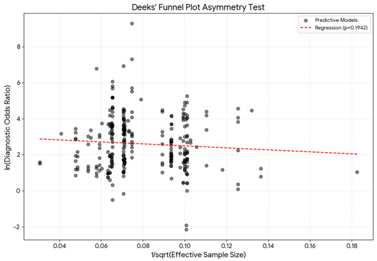
Lastly, to assess publication bias, the Deeks’ funnel plot asymmetry test was used, regressing the log diagnostic odds ratio against the inverse of the square root of the effective sample size. A p-value of less than 0.05 was considered significant across the current meta-analysis.
3. Results
3.1. Selection Process
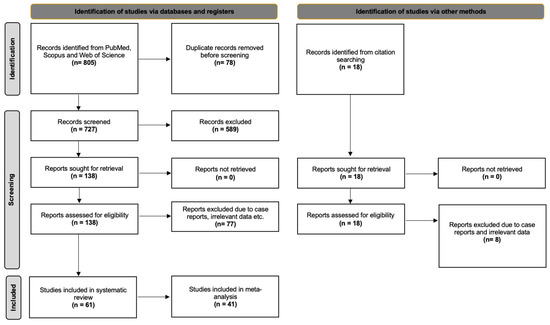
The databases retrieved 805 results that were exported to Mendeley version 2.10.9 (Elsevier, London). After excluding irrelevant and duplicate papers, 138 studies were retrieved for full-text screening. Finally, 51 studies were considered eligible for our initial meta-analysis questions. Furthermore, 10 studies were identified from references and a hands-on search. Hence, 61 studies were included in the current systematic review. However, during the risk of bias assessment, 20 studies were considered “High” risk of bias and subsequently were excluded from the statistical meta-analysis. Cohen’s Kappa for inter-rater variability was 0.937, indicating excellent agreement. In accordance with the PRISMA 2020 guidelines, Figure 1 presents the flow diagram of the selection process.
Figure 1.
PRISMA 2020 flow chart of literature search analysis.
3.2. Studies Characteristics
The main characteristics of the included studies are presented in Table 1 and Supplementary Materials. A total of 41 studies, comprising 293 predictive models, were eligible for quantitative synthesis. In terms of population origin, 181 models were derived from Asian cohorts, 60 from African cohorts, 42 from European cohorts, and 10 from American cohorts. According to the study design, 182 models were based on imaging techniques, 59 on osteological measurements, and 52 on cadaveric investigations. With respect to the analytical approach, discriminant analysis was the most frequently applied method (n = 172). Regarding the variables used for sex estimation, combined measurement models that incorporated the manubrium, sternal body, and sternebrae were the most common (n = 84).
Table 1.
Characteristics of the included studies.
3.3. Main Meta-Analysis Outcomes
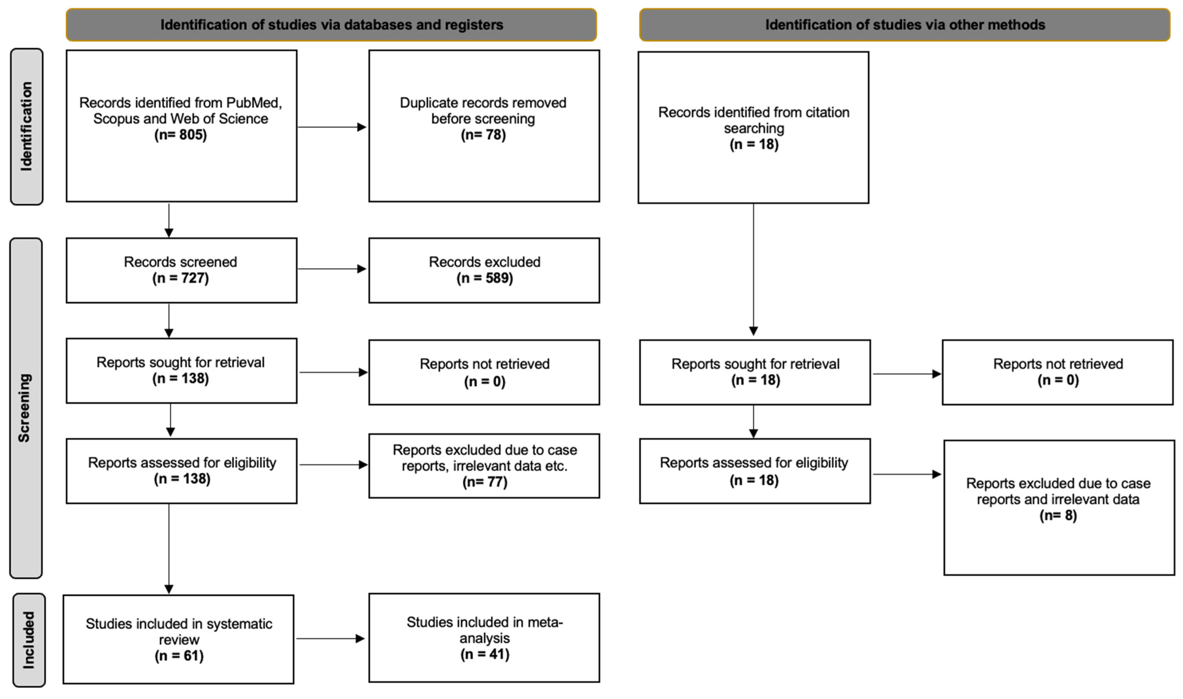
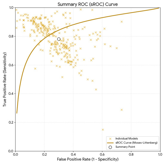
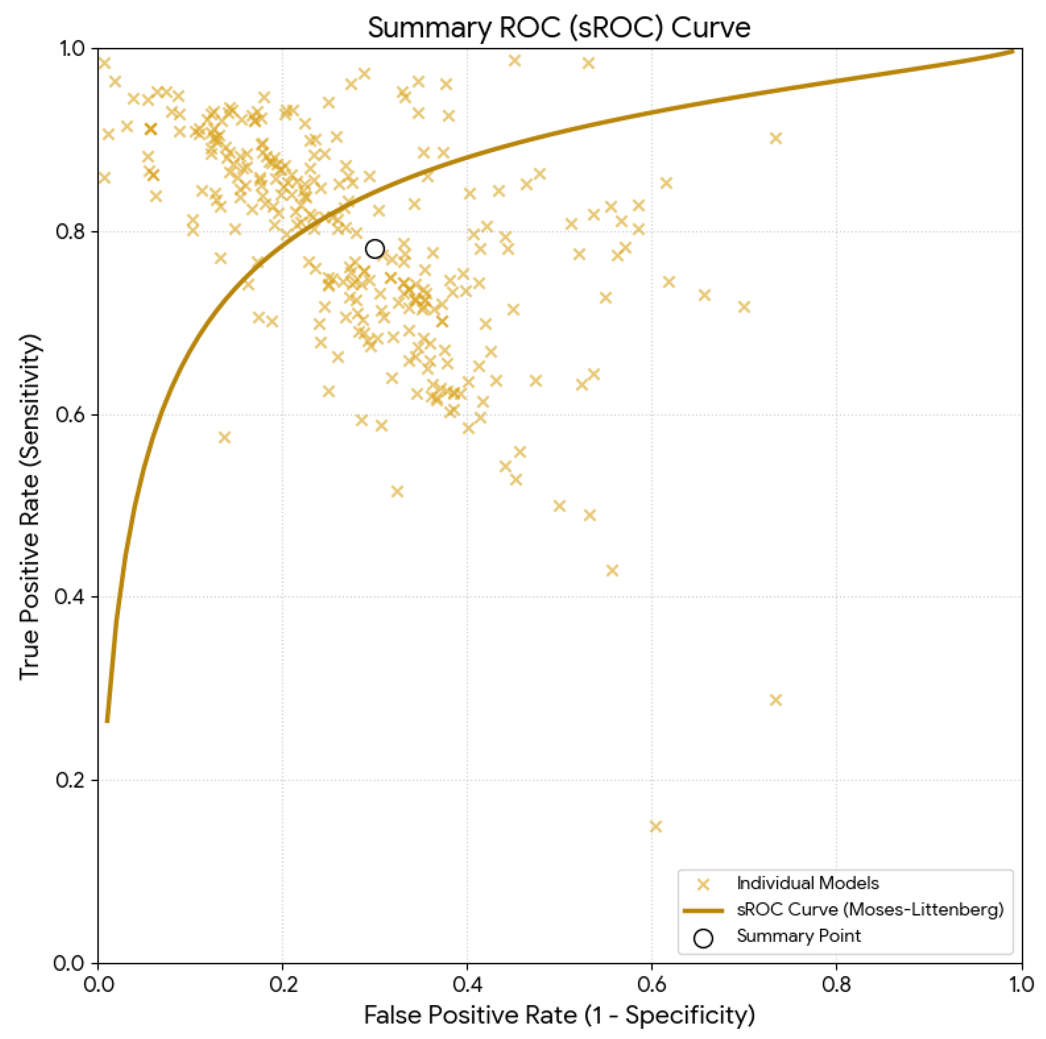
The analysis of 293 predictive models from 41 studies calculated a pooled sensitivity of 80.94% (95% CI: 79.7–82.1) and a pooled specificity of 73.96% (95% CI: 72.4–75.5). Both values depicted substantial heterogeneity (I2 = 85.51% and I2 = 87.40%, respectively). The mean accuracy was estimated as 77.3% (95% CI: 76.1–78.5), and it is illustrated in Figure 2 with its sROC curve. The LR+ was 3.01 (95% CI: 2.83–3.20) and the LR- was 0.27 (95% CI: 0.25–0.29), indicating moderate changes in the pre-test-to-post-test probability. Finally, the DOR was calculated as 12.88 (95% CI: 11.23–14.78).
Figure 2.
Summary receiver operating characteristic (sROC) curve using the Moses–Littenberg method for the overall estimation.
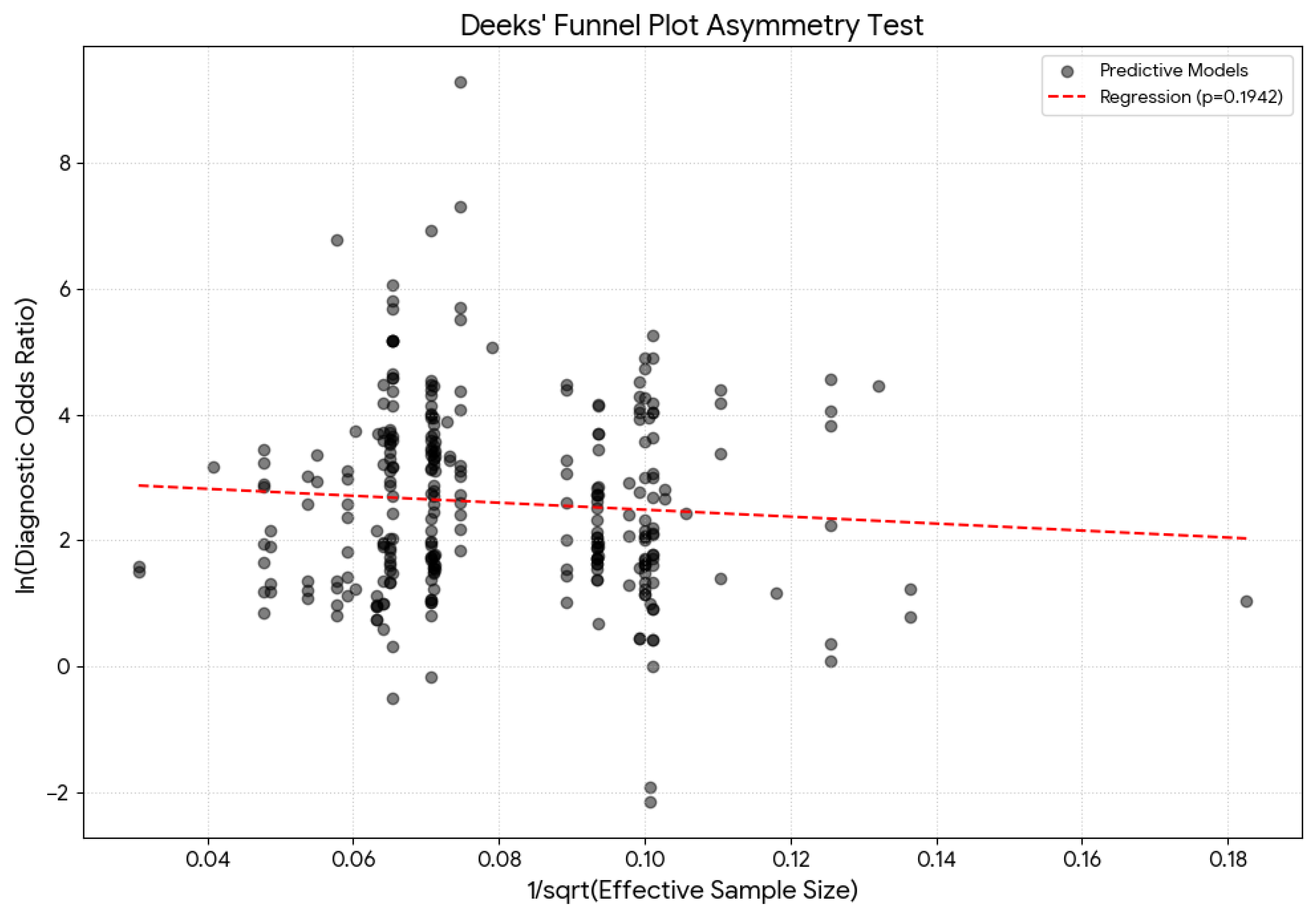
The respective Deeks’ funnel plot is presented in Figure 3. The Deeks’ test yielded a p-value of 0.1942, indicating a lack of significant asymmetry in the funnel plot.
Figure 3.
Deeks’ funnel plot test for the overall estimation asymmetry with the associated regression test.
3.4. Subgroup Analysis
Studies were subgrouped based on nationality (continent), type of study (osteological, imaging, or cadaveric), type of test (discriminant, logistic, ROC, area under the curve, limiting point, or unique algorithm), and sternal measurements used (manubrium, body, xiphoid, sternal area, sternal index, or combinations) (Table 2).
Table 2.
Detailed subgroup analysis of the sensitivity, specificity, DOR, LR+/− and mean accuracy. Sensitivity, specificity, and mean accuracy are presented in frequencies (%), while DOR and LR+/− are presented in their absolute values.
3.4.1. Subgroup Analysis: Nationality
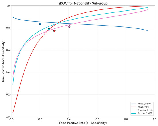
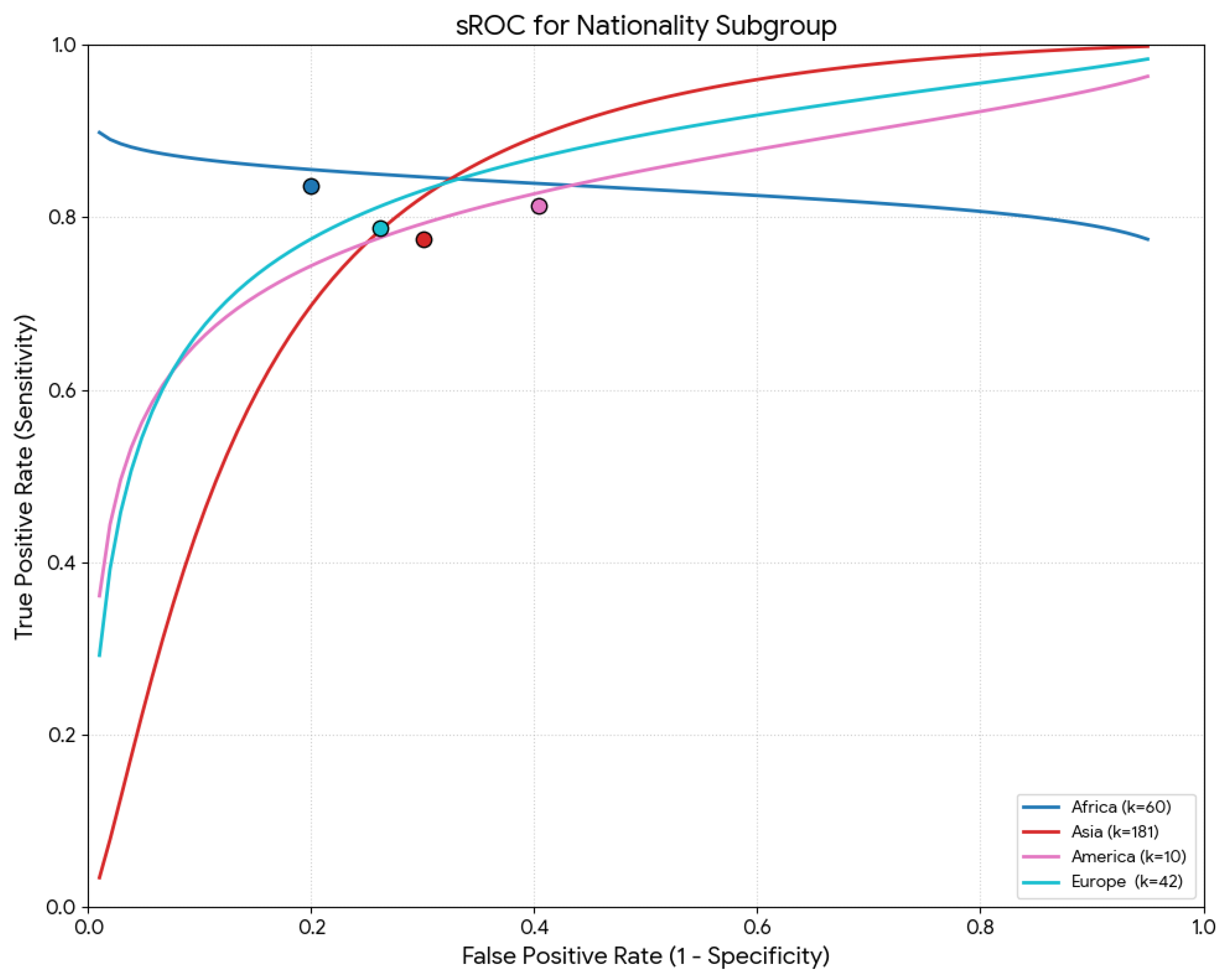
Table 2 summarizes all parameters (sensitivity, specificity, DOR, mean accuracy, and LR+/−) by continent of origin. Regarding the meta-regression results, the global test comparison retrieved no statistical significance for the sensitivity (p = 0.472). However, nationality was a statistically significant moderator of specificity and mean accuracy (p < 0.001). Specifically, studies with the African population depicted the highest pooled specificity (82.4%) and accuracy (84.2%), while studies from the Asian population had the lowest pooled specificity (59.5%) and accuracy (72.1%) (Figure 4).
Figure 4.
Summary receiver operating characteristic (sROC) curve using the Moses–Littenberg method for the nationality subgroup.
3.4.2. Subgroup Analysis: Type of Study
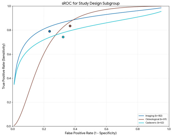
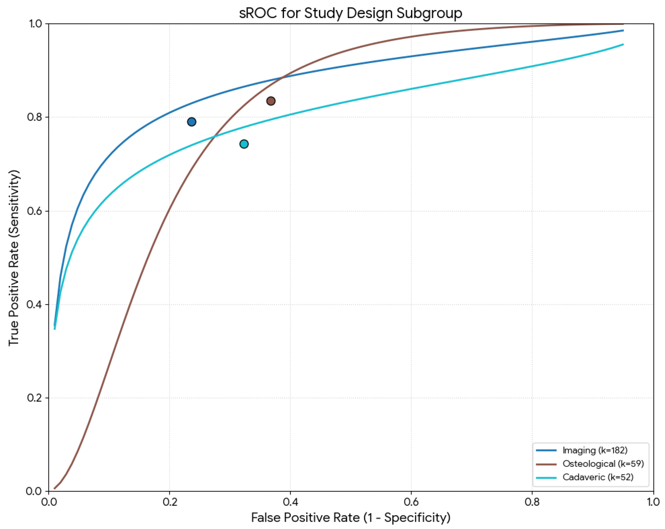
Table 2 summarizes the parameters (sensitivity, specificity, DOR, mean accuracy, and LR+/−) by study type. Regarding the meta-regression results, the global test comparison found no statistical significance for sensitivity or mean accuracy (p = 0.154 and p = 0.276, respectively). However, the type of study was a statistically significant moderator for the specificity (p = 0.003). Specifically, studies based on imaging techniques had the highest pooled specificity (77.8%), and the osteological studies had the lowest (64.3%) (Figure 5).
Figure 5.
Summary receiver operating characteristic (sROC) curve using the Moses–Littenberg method for the study design.
3.4.3. Subgroup Analysis: Type of Statistical Analysis
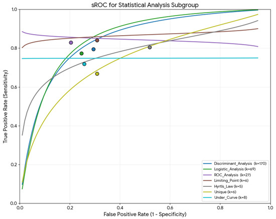
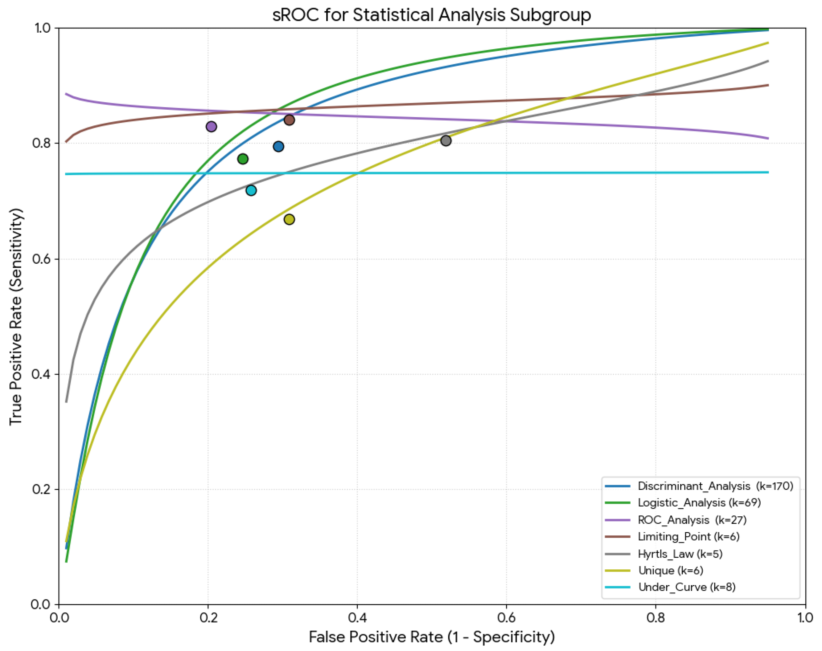
Table 2 summarizes all parameters (sensitivity, specificity, DOR, mean accuracy, and LR+/−) by statistical analysis type. Regarding the meta-regression results, all global test comparisons were statistically significant (p < 0.001). Studies using the rule “136” had the highest sensitivity (95.6%), studies with ROC analysis had the highest specificity (80.9%) and the highest mean accuracy (83.6%) (Figure 6).
Figure 6.
Summary receiver operating characteristic (sROC) curve using the Moses–Littenberg method for the statistical analysis used for the sex estimation.
3.4.4. Subgroup Analysis: Sternal Measurements
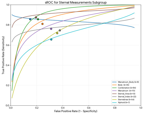
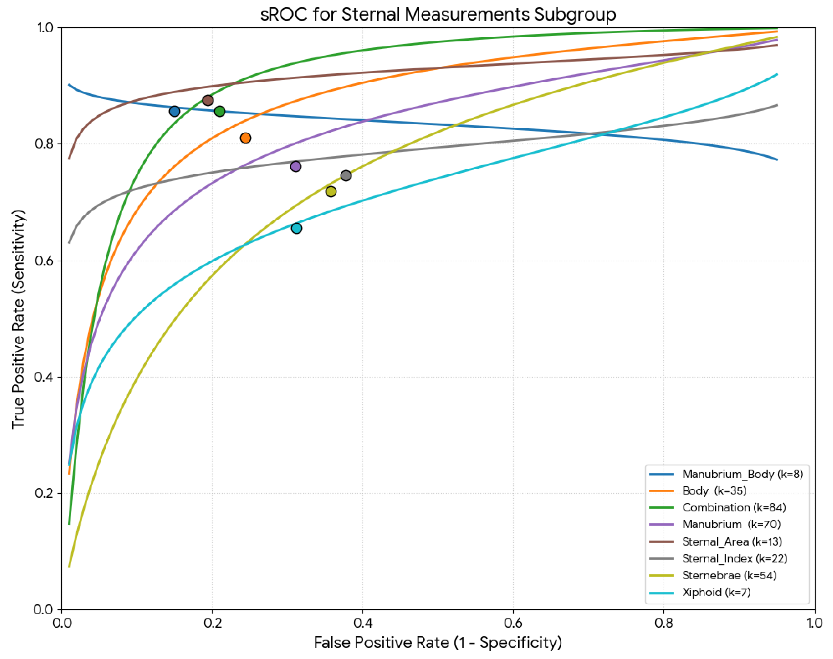
Table 2 summarizes all the parameters (sensitivity, specificity, DOR, mean accuracy, and LR+/−) based on sternal measurements. Regarding the meta-regression results, all global test comparisons were statistically significant (p < 0.001). Studies based on sternal area measurements had the highest pooled sensitivity (88.9%), studies using combined manubrium and sternal body measurements had the highest pooled specificity (86.8%) and accuracy (87.3%) (Figure 7).
Figure 7.
Summary receiver operating characteristic (sROC) curve using the Moses–Littenberg method for the measurements used for the sex estimation.
4. Discussion
The present systematic review and meta-analysis evaluated the diagnostic accuracy of sternal measurements for sex estimation. Analysis of 293 predictive models from 41 studies revealed a pooled sensitivity of 80.9% and a specificity of 74.0%, with an overall accuracy of 77.3%. While sternal morphometry exhibits a theoretical capacity for moderate to high diagnostic accuracy, its practical application is currently circumscribed by significant methodological- and population-based heterogeneity. Our findings suggest that high accuracy (up to 87.3%) is achievable primarily through the adoption of standardized, combined measurement protocols and advanced algorithmic analyses, rather than through generalized or historical linear rules.
4.1. Subgroup Interpretations
Subgroup analyses in the present review revealed significant moderators. Studies from African populations achieved the highest pooled specificity and accuracy, whereas those from Asian populations reported the lowest values. These differences may reflect population-specific variation in skeletal dimorphism and reinforce the importance of localized standards. The sternum’s development, including the fusion of its component parts, is highly sensitive to these localized factors, reinforcing the necessity for population-specific forensic standards rather than universal models.
The study type was identified as a statistically significant moderator for specificity, with imaging-based analyses achieving 77.8% compared to 64.3% for traditional osteological approaches. This variability likely stems from the superior resolution and three-dimensional reconstruction capabilities of CT, which enable more precise landmark identification than manual measurements on dry bone. Virtual anthropology techniques reduce the physical handling of the remains [1,8].
The shift toward advanced statistical modeling appears to reduce operator-dependent variability and improve diagnostic utility. Machine learning- and ROC-based analyses yielded a mean accuracy of 83.6%, higher than that of classical discriminant functions at 77.1%. Traditional historical protocols, such as Hyrtl’s Law, demonstrated a notably low accuracy of 59.6%. Advanced classifiers are better equipped to handle the non-linear biological variance. Thus, machine learning methods and ROC-based analyses provided higher accuracy than classical discriminant functions, consistent with recent evidence highlighting the superiority of advanced classifiers in forensic applications [3,7].
Heterogeneity is also driven by the lack of standardized measurement protocols across the global literature. Our findings indicate that multidimensional approaches, specifically combined indices of the manubrium and sternal body, capture dimorphism more effectively than single linear dimensions, achieving a robust accuracy of 87.3%. Conversely, univariate measurements such as XP (65.1%) and SI (70.4%) showed significantly lower reliability. This suggests that isolated bone segments may not adequately reflect the overall dimorphism.
4.2. Comparison with Other Skeletal Elements
The pelvis is consistently regarded as the gold standard in sex estimation, achieving accuracies above 95% in both traditional assessments and modern metric or morphometric approaches [1,2]. Geometric morphometric analyses of the pubis reach accuracies exceeding 95%, even when applied to fragmented remains [2]. Deep learning algorithms applied to CT reconstructions of coxal bones have recently achieved near-perfect accuracy, ranging from 97.9% to 99.8% [1]. Long bones represent another reliable alternative, with machine learning models yielding 90–92% accuracy [3].
By contrast, cranial and dental structures generally display lower levels of dimorphism. For instance, the foramen magnum provides moderate diagnostic utility, with accuracy varying between 70 and 90% depending on measurement type [8]. Similarly, the sella turcica demonstrates statistically significant but modest dimorphism [9], while orbital metrics analyzed with machine learning achieve only moderate classification performance (accuracy ~68%) [10]. Teeth, particularly permanent canines, are highly durable but yield accuracies of approximately 77–83% [12].
4.3. Forensic Implications
The forensic applicability of sternal measurements lies primarily in their preservation, accessibility, and versatility across different contexts. The sternum is among the most frequently recovered thoracic elements, with preservation rates reported at nearly 60% in skeletal collections [18]. It offers a valuable alternative when highly dimorphic elements such as the pelvis or long bones are missing, fragmented, or otherwise unavailable.
However, important caveats remain, as observed in the current results. First, population specificity is crucial: expression of sexual dimorphism in sternal traits varies substantially across groups, as demonstrated by the current meta-analysis, reinforcing that generalized models are not suitable for universal forensic application. Second, while traditional rules such as Hyrtl’s Law or “the 136 rule” are often cited, their reliability has been inconsistent across studies. Instead, multivariate and machine learning approaches consistently outperform univariate or rule-based methods. Finally, even with high accuracies in controlled studies, heterogeneity and publication bias observed in the present meta-analysis suggest variability in real-world forensic performance.
In practice, sternal morphometry should, therefore, be viewed as a secondary but highly valuable option. When the pelvis or long bones are absent, the sternum can provide robust sex estimates, especially if combined with other thoracic or cranial indicators. Advances in imaging and machine learning will likely increase its utility, enabling forensic anthropologists to leverage clinical CT repositories for model training and validation. In this sense, the sternum not only complements traditional approaches but also expands the forensic toolkit in cases of incomplete or compromised remains.
4.4. Strengths and Limitations
This meta-analysis represents the most comprehensive synthesis to date of the diagnostic accuracy of sternal measurements for sex estimation. By pooling results from 41 studies and 293 predictive models, it provides robust estimates of sensitivity, specificity, and diagnostic odds ratios across diverse populations and methodological frameworks. Subgroup analyses also allowed exploration of moderators, including study type, statistical model, measurement protocol, and geographic origin, thereby clarifying sources of heterogeneity and providing valuable insights for forensic practice.
Nevertheless, several limitations must be acknowledged. First, substantial heterogeneity was observed across studies (I2 > 85% for sensitivity and specificity), indicating variability in study design, population background, and measurement protocols. Although subgroup analyses identified some moderators (such as imaging versus osteological studies and statistical methods), residual heterogeneity remained unexplained. This may be explained by the use of inconsistent anatomical landmarks and the lack of standardized imaging planes (in CT-based studies), which introduce significant technical noise, complicating direct comparisons and potentially inflating pooled estimates. Moreover, operator-dependent variability remains a concern, especially in manual osteometric and cadaveric studies where inter-observer reliability is not always reported. Even in virtual anthropology, technical variations, such as differences in CT slice thickness, can affect the precision of sternal measurements. Second, measurement protocols were not standardized across studies; some relied on traditional rules (such as Hyrtl’s Law), others on linear dimensions or indices, and more recent work employed CT-derived morphometrics and machine learning. This diversity complicates direct comparisons and may inflate pooled estimates. While machine learning- and ROC-based analyses demonstrated superior performance over traditional methods, many of these models carry an inherent risk of overfitting. These limitations underscore the urgent need for standardized measurement protocols and larger, multicenter datasets to validate sternal morphometry across diverse populations
5. Conclusions
The current systematic review with meta-analysis yielded a pooled sensitivity of 80.94%, a pooled specificity of 73.96%, and an accuracy of 77.3%. According to meta-regression analysis, these pooled estimates were significantly influenced by nationality, study type (imaging or osteological), statistical analysis used for estimation (traditional methods or machine learning), and the measurements used (manubrium, body, others, or combination). This meta-analysis provides important pooled evidence, supporting the forensic value of sternal morphometry. From a practical perspective, sternal morphometry exhibits moderate to high diagnostic utility and is of significant forensic importance, especially when more sexually dimorphic skeletal features, such as the pelvis, femur, or skull, are inaccessible or fragmented. However, sternum-based sex estimation must be strictly viewed as a secondary or complementary diagnostic method. Its universal applicability is constrained by substantial interpopulation variations and a current lack of standardized global measurement protocols. Although advanced statistical approaches and imaging techniques, particularly the combined measurement of the manubrium and body, yielded the most robust accuracy (87.3%), these findings are subject to the methodological limitations of the primary literature.
Supplementary Materials
The following are available online at https://www.mdpi.com/article/10.3390/diagnostics16091255/s1, Supplementary Materials: PRISMA 2020 Checklist.
Author Contributions
Conceptualization, G.T., S.P., and M.P.; methodology, G.T., D.G., and I.P.; software, G.T.; validation, E.B., L.M., and S.P.; investigation, G.T., D.G., and I.P.; data curation, E.B., L.M., and S.P.; writing—original draft preparation, G.T., D.G., and M.P.; writing—review and editing, I.P., E.B., L.M., and S.P.; supervision, S.P. and M.P. All authors have read and agreed to the published version of the manuscript.
Funding
This research received no external funding.
Institutional Review Board Statement
Not applicable.
Informed Consent Statement
Not applicable.
Data Availability Statement
Please contact the authors for data requests.
Conflicts of Interest
The authors declare no conflicts of interest.
References
- Epain, M.; Valette, S.; Zou, K.; Faisan, S.; Heitz, F.; Croisille, P.; Fracasso, T.; Fanton, L. Sex Estimation from Coxal Bones Using Deep Learning in a Population Balanced by Sex and Age. Int. J. Leg. Med. 2024, 138, 2617–2623. [Google Scholar] [CrossRef] [Scilit]
- Baca, K.; Bridge, B.; Snow, M. Three-Dimensional Geometric Morphometric Sex Determination of the Whole and Modeled Fragmentary Human Pubic Bone. PLoS ONE 2022, 17, e0265754. [Google Scholar] [CrossRef] [Scilit]
- Knecht, S.; Nogueira, L.; Servant, M.; Santos, F.; Alunni, V.; Bernardi, C.; Quatrehomme, G. Sex Estimation from the Greater Sciatic Notch: A Comparison of Classical Statistical Models and Machine Learning Algorithms. Int. J. Leg. Med. 2021, 135, 2603–2613. [Google Scholar] [CrossRef] [Scilit]
- Ubelaker, D.H.; DeGaglia, C.M. Population Variation in Skeletal Sexual Dimorphism. Forensic Sci. Int. 2017, 278, 407.e1–407.e7. [Google Scholar] [CrossRef] [Scilit]
- Brůžek, J.; Santos, F.; Dutailly, B.; Murail, P.; Cunha, E. Validation and Reliability of the Sex Estimation of the Human Os Coxae Using Freely Available DSP2 Software for Bioarchaeology and Forensic Anthropology. Am. J. Phys. Anthropol. 2017, 164, 440–449. [Google Scholar] [CrossRef] [Scilit]
- Bruzek, J. A Method for Visual Determination of Sex, Using the Human Hip Bone. Am. J. Phys. Anthropol. 2002, 117, 157–168. [Google Scholar] [CrossRef]
- Knecht, S.; Santos, F.; Ardagna, Y.; Alunni, V.; Adalian, P.; Nogueira, L. Sex Estimation from Long Bones: A Machine Learning Approach. Int. J. Leg. Med. 2023, 137, 1887–1895. [Google Scholar] [CrossRef] [Scilit] [PubMed]
- Fernandes, A.L.V.C.; Shetti, A.; Lagali-Jirge, V.; Keluskar, V. Accuracy of Sex Estimation by Morphometric Evaluation of Foramen Magnum Using Computed Tomography—A Systematic Review and Meta-Analysis. Forensic Sci. Med. Pathol. 2023, 20, 268–279. [Google Scholar] [CrossRef] [Scilit] [PubMed]
- Ribeiro, E.C.; de Mendonça, D.S.; de Barros Silva, P.G.; Kurita, L.M.; de Aguiar, A.S.W.; Tuji, F.M.; Neves, F.S.; Carvalho, F.S.R.; Costa, F.W.G. Potential Role of the Sella Turcica X-Ray Imaging Aspects for Sex Estimation in the Field of Forensic Anthropology: A Systematic Review and Meta-Analysis. Forensic Sci. Res. 2024, 9, owad046. [Google Scholar] [CrossRef] [Scilit] [PubMed]
- Triantafyllou, G.; Botis, G.G.; Piagkou, M.; Papanastasiou, K.; Tsakotos, G.; Paschopoulos, I.; Matsopoulos, G.K.; Papadodima, S. Sex Estimation Through Orbital Measurements: A Machine Learning Approach for Forensic Science. Diagnostics 2024, 14, 2773. [Google Scholar] [CrossRef] [Scilit]
- Uabundit, N.; Chaiyamoon, A.; Iamsaard, S.; Yurasakpong, L.; Nantasenamat, C.; Suwannakhan, A.; Phunchago, N. Classification and Morphometric Features of Pterion in Thai Population with Potential Sex Prediction. Medicina 2021, 57, 1282. [Google Scholar] [CrossRef] [Scilit]
- Tanga, C.; López-Lázaro, S.; Soto-Álvarez, C.; Viciano, J. Reliability of Predictive Models Based on the Mesiodistal and Buccolingual Diameters of Permanent Canine Teeth for Sex Estimation in Forensic Contexts: A Systematic Review and Meta-Analysis. Forensic Sci. Int. 2024, 361, 112143. [Google Scholar] [CrossRef] [Scilit]
- Vatzia, K.; Fanariotis, M.; Bugajski, M.; Fezoulidis, I.V.; Piagkou, M.; Vlychou, M.; Triantafyllou, G.; Vezakis, I.; Botis, G.; Papadodima, S.; et al. Assessing Sternal Dimensions for Sex Classification: Insights from a Greek Computed Tomography-Based Study. Diagnostics 2025, 15, 1649. [Google Scholar] [CrossRef] [Scilit]
- Page, M.J.; McKenzie, J.E.; Bossuyt, P.M.; Boutron, I.; Hoffmann, T.C.; Mulrow, C.D.; Shamseer, L.; Tetzlaff, J.M.; Akl, E.A.; Brennan, S.E.; et al. The PRISMA 2020 Statement: An Updated Guideline for Reporting Systematic Reviews. BMJ 2021, 372, n71. [Google Scholar] [CrossRef] [Scilit] [PubMed]
- Campbell, J. Chapter 9: Diagnostic Test Accuracy Systematic Reviews. In JBI Manual for Evidence Synthesis; JBI: Adelaide, Australia, 2020. [Google Scholar]
- Abdel Aal, K.; Yossef, H. Determination of Sex from Sternal Dimensions in Upper Egypt Population by Using Multislice Computed Axial Tomography. Ain Shams J. Forensic Med. Clin. Toxicol. 2014, 23, 103–114. [Google Scholar] [CrossRef] [Scilit]
- Ahmed, A.A.; Alshammari, F.O.; Alrafiaah, A.S.; Almohaisani, A.A.; Al-Mohrej, O.A.; Alkubaidan, F.O. Estimation of Sex in a Contemporary Saudi Population Based on Sternal Measurements Using Multidetector Computed Tomography. HOMO 2017, 68, 411–421. [Google Scholar] [CrossRef] [Scilit]
- Ahmed, A.A.; Koko, A.O.; Elsayed, M.M. Multi-Detector Computed Tomography for the Estimation of Sex from Sternal Medullary Cavity Measurements in Sudanese Individuals. Forensic Sci. Int. Rep. 2021, 4, 100244. [Google Scholar] [CrossRef] [Scilit]
- Ali, M.I.M.; Mosallam, W.; Mostafa, E.M.; Aly, S.M.; Ali, N.M. Sternum as an Indicator for Sex and Age Estimation Using Multidetector Computed Tomography in an Egyptian Population. Forensic Imaging 2021, 26, 200457. [Google Scholar] [CrossRef] [Scilit]
- Adhvaryu, A.V.; Adhvaryu, M.A.; Rathod, S.P.; Chauhan, P.R.; Joshi, H.G. A Study of Sexual Dimorphism in Human Sterna. Int. J. Med. Res. Health Sci. 2013, 2, 577. [Google Scholar] [CrossRef] [Scilit]
- Aragão, J.A.; Matos, Í.Q.; Carvalho, H.D.G.; Aragão, I.C.S.; Aragão, F.M.S.; Feitosa, V.L.C.; Reis, F.P. Sexual Dimorphism Based on the Sternum Bone Morphometry in Human Fetuses. 2022. Available online: https://www.researchsquare.com/article/rs-2076534/v1 (accessed on 21 September 2022).
- Atal, D.K.; Murari, A.; Naik, S.K. Gender Differentiation from Sternal Width. J. Indian Acad. Forensic Med. 2008, 30, 198–201. [Google Scholar] [CrossRef] [Scilit]
- Banyeh, M.; Abdulai, A.-R.; Annan, E.K.; Mensah, E.K.; Nkansah, C.; Nathan, J.A.; Twum, M.B.; Achumboro, P.A. Assessing the Reliability and Accuracy of Sex Estimation Models Utilizing Sternal Morphometry Derived from Computed Tomography in the Ghanaian Population. Forensic Sci. Int. Rep. 2024, 9, 100368. [Google Scholar] [CrossRef] [Scilit]
- Baraw, R.; Barwa, J.; Pradhan, M.; Khanna, S.K. Width of Sternum at Suprasternal Notch and at the Level of Fourth Rib for Determination of Sex in the Population of Delhi: A New & Reliable Tool for Sex Prediction. J. Indian Acad. Forensic Med. 2017, 39, 83. [Google Scholar] [CrossRef] [Scilit]
- Bedalov, A.; Bašić, Ž.; Marelja, I.; Dolić, K.; Bukarica, K.; Missoni, S.; Šlaus, M.; Primorac, D.; Andjelinović, Š.; Kružić, I. Sex Estimation of the Sternum by Automatic Image Processing of Multi-Slice Computed Tomography Images in a Croatian Population Sample: A Retrospective Study. Croat. Med. J. 2019, 60, 237–245. [Google Scholar] [CrossRef] [Scilit]
- Bongiovanni, R.; Spradley, M.K. Estimating Sex of the Human Skeleton Based on Metrics of the Sternum. Forensic Sci. Int. 2012, 219, 290.e1–290.e7. [Google Scholar] [CrossRef] [Scilit]
- Chandrakanth, H.V.; Kanchan, T.; Krishan, K. Osteometric Analysis for Sexing of Modern Sternum—An Autopsy Study from South India. Leg. Med. 2014, 16, 350–356. [Google Scholar] [CrossRef] [Scilit] [PubMed]
- Chowdhuri, S.; Roy, P.; Dey, A.; Das, S.; Ghosh, R. A Study for Determination of Sex by Multidetector Computed Tomography of Sternum Using Discriminant Function and Logistic Regression. Arab J. Forensic Sci. Forensic Med. 2019, 1, 1444–1453. [Google Scholar] [CrossRef] [Scilit]
- Darade, B.; Sabale, P.; Mohite, S. An Autopsy Based Study of Sexual Dimorphism of Adult Human Sterna. Indian J. Forensic Med. Toxicol. 2020, 14, 117. [Google Scholar] [CrossRef] [Scilit]
- Darwish, R.T.; Abdel-Aziz, M.H.; El Nekiedy, A.-A.M.; Sobh, Z.K. Sex Determination from Chest Measurements in a Sample of Egyptian Adults Using Multislice Computed Tomography. J. Forensic Leg. Med. 2017, 52, 154–158. [Google Scholar] [CrossRef] [Scilit]
- Dhkar, W. Radiological Age & Sex Determination from Sternum. Int. J. Sci. Res. 2014, 3, 1–3. [Google Scholar]
- Dorado-Fernández, E.; Cáceres-Monllor, D.A.; Carrillo-Rodríguez, M.F.; Botella-López, M.; Murillo-González, J. The Sternum as a Sex Discriminator in a Contemporary Spanish Population. Rom. J. Leg. Med. 2021, 29, 96–104. [Google Scholar] [CrossRef] [Scilit]
- Ekizoglu, O.; Hocaoglu, E.; Inci, E.; Bilgili, M.G.; Solmaz, D.; Erdil, I.; Can, I.O. Sex Estimation from Sternal Measurements Using Multidetector Computed Tomography. Medicine 2014, 93, e240. [Google Scholar] [CrossRef] [Scilit] [PubMed]
- Elmansy, A.; Ammar, A.M.; Elhawary, A.E.; Khalifa, H.K. Sex Identification Using Sternal Measurements by Multi-Slice Computed Tomography in a Sample of the Egyptian Population. Egypt. J. Forensic Sci. Appl. Toxicol. 2024, 24, 1–15. [Google Scholar] [CrossRef] [Scilit]
- Elsayed, R.M.; Soliman, S.A.; Said, A.M.; Allam, S.A. Determination of Gender from Sternal Measurements Using Computed Tomography Imaging in a Sample of Sohag Governorate Population. Zagazig J. Forensic Med. Toxicol. 2022, 20, 90–110. [Google Scholar] [CrossRef] [Scilit]
- Franklin, D.; Flavel, A.; Kuliukas, A.; Cardini, A.; Marks, M.K.; Oxnard, C.; O’Higgins, P. Estimation of Sex from Sternal Measurements in a Western Australian Population. Forensic Sci. Int. 2012, 217, 230.e1–230.e5. [Google Scholar] [CrossRef] [Scilit]
- García-Parra, P.; Pérez Fernández, Á.; Djorojevic, M.; Botella, M.; Alemán, I. Sexual Dimorphism of Human Sternum in a Contemporary Spanish Population. Forensic Sci. Int. 2014, 244, 313.e1–313.e9. [Google Scholar] [CrossRef] [Scilit] [PubMed]
- Ghorbanlou, M.; Moradi, F.; Asgari, H.R. Morphometric Study of Sternum by Computed Tomography in an Iranian Population: A Method to Discriminate between Male and Female. Forensic Imaging 2022, 28, 200501. [Google Scholar] [CrossRef] [Scilit]
- Gupta, M.; Kumar, A.; Khanna, S. Determination of Sex from Sternal Bone in Central Delhi Population. J. Indian Acad. Forensic Med. 2014, 36, 234–237. [Google Scholar] [CrossRef] [Scilit]
- Hunnargi, S.A.; Menezes, R.G.; Kanchan, T.; Lobo, S.W.; Binu, V.S.; Uysal, S.; Kumar, H.R.S.; Baral, P.; Herekar, N.G.; Garg, R.K. Sexual Dimorphism of the Human Sternum in a Maharashtrian Population of India: A Morphometric Analysis. Leg. Med. 2008, 10, 6–10. [Google Scholar] [CrossRef] [Scilit]
- Hunnargi, S.A.; Menezes, R.G.; Kanchan, T.; Lobo, S.W.; Uysal, S.; Herekar, N.G.; Krishan, K.; Garg, R.K. Sternal Index: Is It a Reliable Indicator of Sex in the Maharashtrian Population of India? J. Forensic Leg. Med. 2009, 16, 56–58. [Google Scholar] [CrossRef] [Scilit]
- Iqbal, M. Sex Determination from Measurements of the Sternum Using Multislice Computed Tomography of the Chest in Pashtun Population of Swat. Pak Euro J. Med. Life Sci. 2021, 4, 200–205. [Google Scholar]
- Jaiswal, R. Sternum as an Index for Determination of Sex. Int. J. Anat. Res. 2019, 7, 7070–7079. [Google Scholar] [CrossRef] [Scilit]
- Jit, I.; Jhingan, V.; Kulkarni, M. Sexing the Human Sternum. Am. J. Phys. Anthropol. 1980, 53, 217–224. [Google Scholar] [CrossRef] [Scilit]
- Kalbouneh, H.; Mubarak, N.; Daradkeh, S.; Ashour, O.; Alkhatib, A.M.; Suboh, L.; Nofal, A.; Mahafzah, W.; Alsalem, M. Estimation of Sex Based on Metrics of the Sternum in a Contemporary Jordanian Population. Medicine 2021, 100, e28169. [Google Scholar] [CrossRef] [Scilit]
- Karki, R.K.; Singh, P.K.; Karki, S.; Shah, D.K. Sex- Related Morphometric Difference in Sternal Index and Dimensions amongst Nepalese Adults. J. Gandaki Med. Coll. Nepal 2020, 13, 104–110. [Google Scholar] [CrossRef] [Scilit]
- Koşar, M.İ.; Uğuz Gençer, C.; Tetiker, H.; Yeniçeri, İ.Ö.; Çullu, N. Sex and Stature Estimation Based on Multidetector Computed Tomography Imaging Measurements of the Sternum in Turkish Population. Forensic Imaging 2022, 28, 200495. [Google Scholar] [CrossRef] [Scilit]
- Macaluso, P.J. The Efficacy of Sternal Measurements for Sex Estimation in South African Blacks. Forensic Sci. Int. 2010, 202, 111.e1–111.e7. [Google Scholar] [CrossRef] [Scilit]
- Macaluso, P.J.; Lucena, J. Estimation of Sex from Sternal Dimensions Derived from Chest Plate Radiographs in Contemporary Spaniards. Int. J. Leg. Med. 2014, 128, 389–395. [Google Scholar] [CrossRef] [Scilit]
- Manoharan, C.; Jeyasingh, T.; Dhanalakshmi, V.; Thangam, D. Is Human Sternum a Tool for Determination of Sex? Indian J. Forensic Community Med. 2016, 3, 60. [Google Scholar] [CrossRef] [Scilit]
- McCormick, W.F.; Stewart, J.H.; Langford, L.A. Sex Determination from Chest Plate Roentgenograms. Am. J. Phys. Anthropol. 1985, 68, 173–195. [Google Scholar] [CrossRef] [Scilit] [PubMed]
- Mukhopadhyay, P.P. Determination of Sex from Adult Sternum by Discriminant Function Analysis on Autopsy Sample of Indian Bengali Population: A New Approach. J. Indian Acad. Forensic Med. 2010, 32, 321–324. [Google Scholar] [CrossRef] [Scilit]
- Nasab, S.H.M. Sex Determination and Length and Stature Estimation from The Sternum Bone. J. Pioneer. Med. Sci. 2024, 13, 13–19. [Google Scholar] [CrossRef] [Scilit]
- Partido Navadijo, M.; Muñoz Molina, J.F.; Navarro Merino, F.; Alemán Aguilera, I. Sex Estimation by CT Image Analysis of the Rib Cage in a Mediterranean Population. Int. J. Leg. Med. 2025, 139, 419–427. [Google Scholar] [CrossRef] [Scilit]
- Oner, Z.; Turan, M.K.; Oner, S.; Secgin, Y.; Sahin, B. Sex Estimation Using Sternum Part Lengths by Means of Artificial Neural Networks. Forensic Sci. Int. 2019, 301, 6–11. [Google Scholar] [CrossRef] [Scilit] [PubMed]
- Peleg, S.; Pelleg Kallevag, R.; Dar, G.; Steinberg, N.; Masharawi, Y.; May, H. New Methods for Sex Estimation Using Sternum and Rib Morphology. Int. J. Leg. Med. 2020, 134, 1519–1530. [Google Scholar] [CrossRef] [Scilit] [PubMed]
- Puttabanthi, S. Sexing of Unknown Adult Human Sterna by Metrical Analysis. Int. J. Biol. Med. Res. 2012, 3, 1516–1519. [Google Scholar]
- Uysal Ramadan, S.; Türkmen, N.; Dolgun, N.A.; Gökharman, D.; Menezes, R.G.; Kacar, M.; Koşar, U. Sex Determination from Measurements of the Sternum and Fourth Rib Using Multislice Computed Tomography of the Chest. Forensic Sci. Int. 2010, 197, 120.e1–120.e5. [Google Scholar] [CrossRef] [Scilit]
- VSharma, S.; Atam, V.; Kumar, N.; Sharma, N.; Atam, I. Study of Gender Determination in Living Adult Male and Female Subjects by Evaluating the Size of Sternum about North Indian Population. IP Indian J. Anat. Surg. Head Neck Brain 2020, 4, 118–122. [Google Scholar] [CrossRef] [Scilit]
- Singh, J.; Pathak, R.K. Sex and Age Related Non-Metric Variation of the Human Sternum in a Northwest Indian Postmortem Sample: A Pilot Study. Forensic Sci. Int. 2013, 228, 181.e1–181.e12. [Google Scholar] [CrossRef] [Scilit]
- Soltani, S.; Aghabiklooei, A.; Ameri, M.; Memarian, A.; Nikanzad, A. Gender Identification Based on the Parameters of the Sternum Bone in the Cadavers. Int. J. Med. Toxicol. Forensic Med. 2020, 10, 26740. [Google Scholar] [CrossRef] [Scilit]
- Sravan, J.S.; Arora, A.; Yadav, J.; Vidua, R.K.; Gayathri, B.H. Determination of Sex from Morphometry of Sternum Using Multidetector Computed Tomography in Central Indian Population. Int. J. Appl. Basic Med. Res. 2025, 15, 32–37. [Google Scholar] [CrossRef] [Scilit]
- Sweilum, O.A.-H.; Galal, H.K.A.; Habib, R.M. Determination of Sex from the Sternum and Fourth Rib Measurements (A Cross-Sectional Study (Using Thoracic Computed Tomography (CT) Images. Egypt. J. Forensic Sci. Appl. Toxicol. 2017, 17, 27–38. [Google Scholar] [CrossRef] [Scilit]
- Toneva, D. Reliability of the Sternal Index as a Sex Indicator in Medieval Skeletal Remains from Northeastern Bulgaria. J. BioSci. Biotech. 2014, 149–152. [Google Scholar]
- Torimitsu, S.; Makino, Y.; Saitoh, H.; Sakuma, A.; Ishii, N.; Inokuchi, G.; Motomura, A.; Chiba, F.; Hoshioka, Y.; Iwase, H. Estimation of Sex in Japanese Cadavers Based on Sternal Measurements Using Multidetector Computed Tomography. Leg. Med. 2015, 17, 226–231. [Google Scholar] [CrossRef] [Scilit] [PubMed]
- Das, S.; Ruengdit, S.; Singsuwan, P.; Mahakkanukrauh, P. Sex Determination from Different Sternal Measurements: A Study in a Thai Population. J. Anat. Soc. India 2015, 64, 155–161. [Google Scholar] [CrossRef] [Scilit]
- Verna, E.; Piercecchi-Marti, M.; Chaumoitre, K.; Bartoli, C.; Leonetti, G.; Adalian, P. Discrete Traits of the Sternum and Ribs: A Useful Contribution to Identification in Forensic Anthropology and Medicine. J. Forensic Sci. 2013, 58, 571–577. [Google Scholar] [CrossRef] [Scilit]
- Yonguc, G.N.; Kurtulus, A.; Bayazit, O.; Adiguzel, E.; Unal, I.; Demir, S.; Acar, K. Estimation of Stature and Sex from Sternal Lengths: An Autopsy Study. Anat. Sci. Int. 2015, 90, 89–96. [Google Scholar] [CrossRef] [Scilit]
- Zhan, M.; Fan, F.; Qiu, L.; Peng, Z.; Zhang, K.; Deng, Z. Estimation of Stature and Sex from Sacrum and Coccyx Measurements by Multidetector Computed Tomography in Chinese. Leg. Med. 2018, 34, 21–26. [Google Scholar] [CrossRef] [Scilit]
- Zhang, K.; Luo, Y.; Chen, X.; Deng, Z. Sexual Dimorphism of Sternum Using Computed Tomography—Volume Rendering Technique Images of Western Chinese. Aust. J. Forensic Sci. 2016, 48, 297–304. [Google Scholar] [CrossRef] [Scilit]
- Zhang, K.; Yang, M.; Cui, J.; Zhan, M.; Qiu, L.; Deng, Z. Stature Estimation from the Combination of Clavicle, Scapula, and Sternum from 3D-VRT Images in Chinese Population. Anthropol. Anz. 2019, 76, 69–78. [Google Scholar] [CrossRef] [Scilit]

Disclaimer/Publisher’s Note: The statements, opinions and data contained in all publications are solely those of the individual author(s) and contributor(s) and not of MDPI and/or the editor(s). MDPI and/or the editor(s) disclaim responsibility for any injury to people or property resulting from any ideas, methods, instructions or products referred to in the content. |
© 2026 by the authors. Licensee MDPI, Basel, Switzerland. This article is an open access article distributed under the terms and conditions of the Creative Commons Attribution (CC BY) license.