Abstract
Background: The accurate prediction of response to neoadjuvant therapy (NAT) is crucial for optimizing breast cancer management. Conventional breast Dynamic Contrast-Enhanced Magnetic Resonance Imaging (DCE-MRI) radiomics typically relies on single post-contrast phases and may not fully capture temporal enhancement patterns related to tumor heterogeneity. This study evaluated a machine learning model based on time-dependent radiomic features extracted from pre-treatment DCE-MRI for predicting NAT response in breast cancer patients. Methods: Breast DCE-MRI examinations of women scheduled for NAT, acquired on 1.5 T scanners from three different vendors, were retrospectively collected from two centers. Tumors were automatically segmented on the third post-contrast DCE image using a 3D nnUNet model trained on 30 lesions. All DCE phases were registered to the reference image, and radiomic features were extracted from a consistent tumor region of interest across all phases. Time-dependent radiomic features were computed using linear regression modeling of feature evolution over time. A random forest classifier integrating static and time-dependent radiomic features was developed to predict pathological complete response (pCR), partial response (pPR), and non-response (pNR). Model performance was evaluated using internal validation (Center 1) and an independent external test cohort (Center 2). Results: A total of 212 patients were included (173 from Center 1 and 39 from Center 2), comprising 103 pCR, 103 pPR and 6 pNR cases. Among 759 extracted features, 30 showed significant differences across response groups. Several time-dependent texture features related to intratumoral heterogeneity were significantly associated with pNR. The model achieved AUC values of 0.80, 0.81, and 0.95 in the internal validation cohort and 0.75, 0.74, and 0.86 in the external test cohort for predicting pCR, pPR, and pNR, respectively. Conclusions: Time-dependent radiomic features derived from pre-treatment breast DCE-MRI enable the accurate prediction of response to NAT, with particularly strong performance in identifying non-responders. This approach may support imaging-based risk stratification and contribute to more personalized treatment.
Keywords:
neo-adjuvant therapy; breast cancer; MRI; radiomics; machine learning; response prediction 1. Background
Neoadjuvant therapy (NAT) has become an integral part of breast cancer management [1]. Initially used for locally advanced or inoperable tumors to enable breast-conserving surgery, NAT is now routinely administered to patients with lymph node-positive disease and to those with biologically aggressive early-stage tumors, including high-grade, HER2-positive, and hormone receptor-negative subtypes [1,2,3,4].
Several clinical trials have demonstrated that achieving a pathological complete response (pCR) following NAT is associated with improved disease-free survival and overall survival [5,6,7,8]. However, despite substantial response rates—reaching up to 55% in hormone receptor-positive/HER2-positive tumors and up to 90% in hormone receptor-negative/HER2-positive tumors [9,10]—a large proportion of patients fail to achieve pCR [11,12].
The accurate prediction of response to NAT is essential to optimize treatment strategies, as it may allow early risk stratification, reduce unnecessary toxicity and support timely treatment adaptation. In particular, the early identification of non-responders remains a major unmet clinical need, as these patients derive limited benefit from standard NAT regimens and may require alternative therapeutic approaches [11,12,13].
International guidelines recommend breast Dynamic Contrast-Enhanced Magnetic Resonance Imaging (DCE-MRI) before and after NAT to assess treatment response, owing to its high sensitivity and ability to capture tumor vascularity and functional characteristics [14,15].
In clinical practice, response to NAT is mainly evaluated through visual assessments of tumor size, volume, and enhancement patterns on MRI, with categorization according to the RECIST v1.1 criteria [16]. In selected cases, diffusion-weighted imaging (DWI) and quantitative apparent diffusion coefficient (ADC) measurements have been shown to improve response assessment [17,18,19]. However, these approaches are limited by subjectivity, inter-reader variability, and an incomplete characterization of tumor biology.
Despite the widespread use of imaging for response assessment during NAT, its translation into response-guided treatment remains limited. A recent systematic review by Lafci et al. [20] showed that, although imaging is employed in more than 80% of NAT breast cancer trials, treatment modification based on imaging response occurs in only a small fraction of cases, largely due to the lack of standardized imaging protocols and actionable response criteria.
In recent years, MRI-based radiomics has emerged as a promising imaging biomarker for predicting treatment response by extracting quantitative features that reflect tumor heterogeneity and microstructural organization [13,21,22,23,24]. Several studies have demonstrated the potential of radiomics and machine learning (ML) approaches applied to pre-treatment MRI to predict pathological complete response [13,25,26,27,28,29]. Recent evidence further suggests that texture-based radiomic descriptors of intratumoral heterogeneity may provide complementary prognostic information when integrated with conventional radiological assessment, improving patient stratification beyond radiological or radiomic features alone [30].
However, most existing models rely on static radiomic features extracted from a single post-contrast phase, potentially overlooking clinically relevant temporal information embedded in DCE-MRI [31].
DCE-MRI inherently provides time-resolved information on contrast uptake and washout, reflecting tumor perfusion, vascular permeability, and microenvironmental heterogeneity [4,14,32,33,34,35]. These dynamic properties are closely related to treatment sensitivity and resistance, suggesting that time-dependent radiomic imaging biomarkers may offer additional predictive value beyond static features. Nevertheless, the integration of temporal radiomics into ML-based prediction models remains limited, and external validation across independent institutions is often lacking, raising concerns about generalizability and clinical translation [12,32].
Together, these findings highlight the need for integrated, quantitative and reproducible imaging approaches capable of translating imaging information into clinically meaningful patient stratification.
To address these limitations, we conducted a retrospective multicenter study including breast cancer patients undergoing NAT at two independent institutions. Using pre-treatment DCE-MRI, we developed and validated a ML-based prediction model integrating static and time-dependent radiomic features to stratify patients according to pCR, pathological partial response (pPR) and pathological non-response (pNR). Data from one center were used for model development and internal validation, while data from the second center served as an independent external test cohort enabling an objective assessment of model robustness and generalizability.
2. Methods
2.1. Study Design and Population
This retrospective study was approved by the institutional review board (protocol number: 59/24) and conducted at two centers in Milan (Center 1) and in Bergamo (Center 2). Study procedures adhered to the CLEAR (CheckList for EvaluAtion of Radiomics) reporting guidelines (Supplementary Material S1) [36]. The requirement for informed consent was waived due to the retrospective nature of the study. All patient data were handled in compliance with the principles of good scientific practice.
Consecutive women with newly diagnosed breast cancer who were candidates for NAT and underwent breast DCE-MRI at Humanitas Research Hospital or Humanitas Gavazzeni between January 2019 to May 2023 were retrospectively evaluated.
Women were included if pre- and post-treatment MRI and surgical pathology results were available. Exclusion criteria included incomplete examinations or poor image quality that precluded adequate lesion segmentation.
NAT regimens were administered according to standard protocol outlined in the National Comprehensive Cancer Network (NCCN) guidelines for breast cancer [15] based on the histological subtype. Before starting NAT, all lesions were marked with a non-magnetic clip to enable the localization of the original tumor site, particularly in patients achieving pCR.
For each patient, the following variables were recorded: age, histologic subtype, tumor grade, receptor status, proliferation rate (Ki67), tumor size on pre- and post-NAT DCE-MRI, T and N stage on pre-and post-NAT DCE-MRI, and NAT regimens.
2.2. Magnetic Resonance Imaging Acquisition
Patients underwent breast DCE-MRI at two time points: before NAT (within two weeks prior to treatment initiation) and after NAT (within two weeks after the last treatment cycle).
All DCE-MRI examinations were performed with patients in the prone position using 1.5 T scanners from three different vendors: Magnetom Avanto (Siemens Healthcare, Erlangen, Germany), Signa HDx (GE Healthcare, Little Chalfont, UK), and Achieva Tx (Philips Medical Systems, Best, the Netherlands). Scanners were equipped with dedicated bilateral breast coils (16-channel for Siemens; 8-channel for GE and Philips systems).
For each patient, a standardized multiparametric protocol was employed, including an axial T2-weighted STIR sequence, an axial DWI echo-planar sequence (b values of 50 and 800 s/mm2), and an axial T1-weighted 3D gradient-echo sequence. The T1-weighted dynamic sequence was acquired once before and six times after intravenous administration of 0.1 mmol/kg of Gadobutrol (Gadovist®, Bayer, Berlin, Germany) using a power injector at a flow rate of 2 mL/s. Post-processed subtraction images, multiplanar reconstruction (MPRs), and maximum intensity projection (MIP) images were subsequently generated.
2.3. Molecular Subtype Classification
Hormone receptor (HR) status was assessed by immunohistochemistry (IHC), considering estrogen receptor (ER) and progesterone receptor (PR) positivity when immunoreactivity was present in more than 1% of tumor cells. HER2 status was also evaluated by IHC, using a standardized scoring system ranging from 0 to 3+. Tumors scoring 0 or 1+ were classified as HER2-negative, whereas tumors scoring 3+ or showing a 2+ score with confirmed gene amplification on fluorescence in situ hybridization (FISH) were classified as HER2-positive (HER2+). Patients with ER and/or PR positivity and HER2-negative status were categorized as HR-positive, while those with HER2 overexpression were categorized as HER2-positive, regardless of hormone receptor expression. Luminal B HER2+ cancers were included in the HER2-positive group based on the biological relevance of HER2 overexpression, which strongly influences prognosis and therapeutic strategy.
Patients who were negative for both HR and HER2 were classified as in triple-negative. Therapeutic regimens were assigned according to the molecular subtype classification.
2.4. Reference Standard and Definition of Pathological Response
At the end of NAT, all patients underwent surgery, and the histopathological assessment of the surgical specimen served as the reference standard. Pathological response was evaluated by a breast pathologist with 15 years of experience using the Pinder classification [37,38] (Table 1).
Table 1.
Pinder classification system describing the histopathological grading of tumor response after neoadjuvant therapy.
For the purpose of this study, patients with Pinder 1i (absence of invasive cancer without residual ductal carcinoma in situ) and Pinder 1ii (absence of invasive cancer with the presence of ductal carcinoma in situ) were classified as pCR.
Patients with Pinder 2i, 2ii, or 2iii were classified as pPR and those with Pinder 3 were classified as pNR.
2.5. Image Analysis
2.5.1. Tumor Segmentation
Subtracted images acquired at tumor peak enhancement (third DCE-T1-weighted sequence) from the pre-treatment DCE-MRI, approximately two minutes after contrast administration, were used for volumetric segmentation, as tumors are best visualized at this time point.
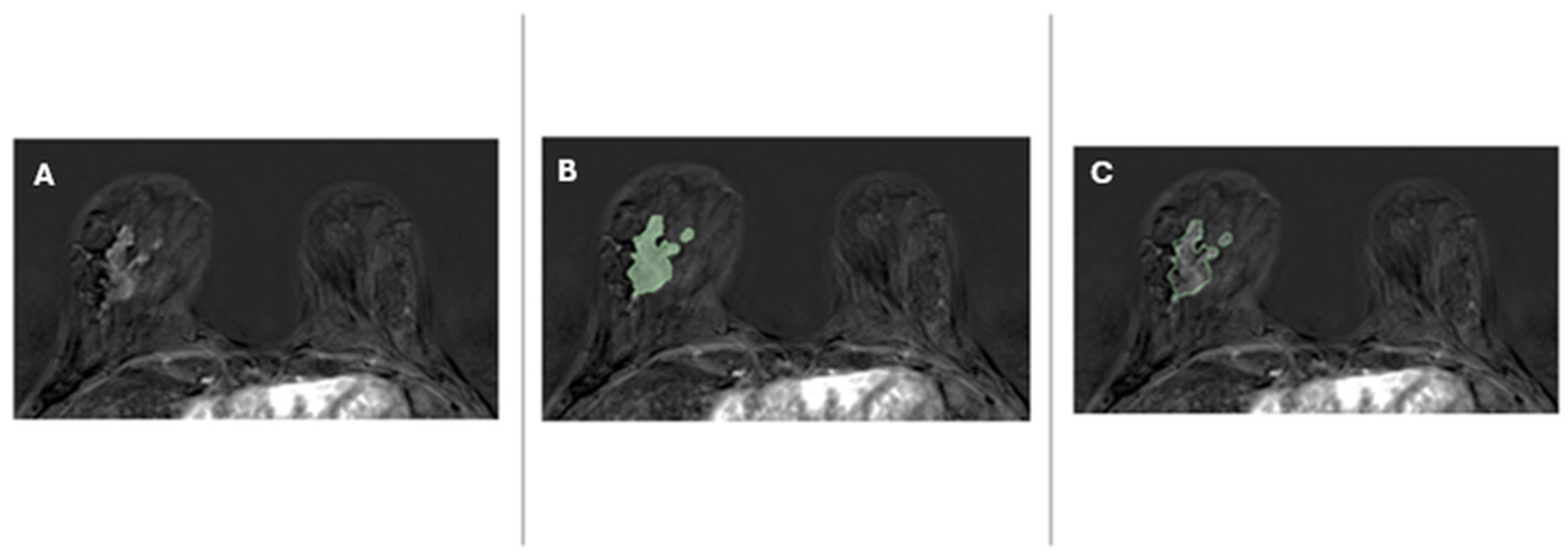
All histologically confirmed invasive tumors were manually segmented using open-source research software (3D Slicer, version 4.10.2) (Figure 1). Radiology reports were available during segmentation, and image windowing was adjusted to optimize lesion visualization. Axillary lymph nodes were considered a non-measurable disease and were excluded from the segmentation.
Figure 1.
A 52-year-old woman with invasive ductal carcinoma (G2, luminal B HER2+) of the right breast. The image shows an example of segmented malignant breast lesion candidate for NAT. To enhance the graphical representation, a cropped version of a 512 × 512 pixel subtracted dynamic contrast-enhanced MRI (DCE-MRI) is displayed. All images maintain the same zoom level. The MRI reveals a mass measuring 45 mm in diameter. The lesion exhibits strong post-contrast enhancement with a high wash-in rate and a type II enhancement curve. (A) Original axial post-contrast fat-saturated T1-weighted subtracted image from post-NAT breast MRI; (B) overlaid segmentation on the original image, highlighting the lesion; (C) contour of the segmented region on the original image, outlining the lesion’s borders.
A convolutional neural network (CNN) based on the nnU-Net framework was trained on 30 manually segmented cases (training set) by a radiology resident with 2 years of experience in breast imaging, under the supervision of a senior breast radiologist with more than 10 years of experience. Model training parameters are reported in Supplementary Material S2. After training, the CNN was applied to automatically segment the entire dataset.
Segmentation accuracy was evaluated using the DICE similarity coefficient (DSC) between manual and automated segmentations. In the external cohort, all DCE-MRI images were segmented by a single radiologist and compared with the CNN-generated volumes. The mean DSC achieved was 0.90 (range: 0.80–0.99) in the internal cohort and 0.94 (range: 0.61–0.99) in the external cohort, indicating an excellent agreement between manual and automated segmentation.
2.5.2. Radiomic Feature Extraction
Radiomic features were computed from all six sequences of DCE-T1-weighted sequences. The tumor segmentation obtained from the CNN model was superimposed onto all the DCE-T1w sequences using an affine registration algorithm (SimpleITK library). Each DCE-MRI volume was normalized for signal intensity using N4 bias-field correction (SimpleITK library). Radiomic features were then extracted from each volume of interest (VOI) with the Pyradiomics library. Complete extraction parameters and settings are reported in Supplementary Material S3.
To characterize the temporal evolution of radiomic features across the dynamic phases, a linear regression model was fitted to each feature for every patient using the least-squares method (SciPy linregress function, default settings without regularization or weighting). The order of DCE acquisitions was treated as the time variable (independent variable), and the corresponding feature values across phases served as the dependent variable. This modeling approach yielded two parameters for each feature: the slope, representing the rate of change in the feature over time, and the intercept, representing its estimated baseline value before contrast enhancement.
These parameters were considered time-dependent features and used as independent predictors for model training. For each original radiomic feature, two derived variables (slope and intercept) were generated. No further feature combination or dimensionality reduction was applied.
2.6. Machine Learning Model
A ML model was developed to predict pCR, pPR and pNR. The dataset from Center 1 was randomly divided into a training set (75%) and a test set (25%), while the dataset from Center 2 served as an external validation cohort.
In addition to the primary analysis, an exploratory subgroup analysis was performed by further stratifying patients according to refined Pinder categories (Pinder 1i, 1ii, and ≥2) to investigate whether time-dependent radiomic features differed between true complete responders and cases with residual in situ or invasive disease. Detailed methods are reported in Supplementary Material S4.
Radiomic features were standardized using z-score normalization, computed from the Center 1 training set and applied to both the Center 1 test and Center 2 validation set. Feature selection was performed using recursive feature elimination (RFE) with 5-fold cross-validation, optimizing the area under the receiver operating characteristic curve (AUC) in a one-vs-rest configuration.
The most discriminative features identified by RFE were used to train a random forest classifier. Model hyperparameters were optimized via nested cross-validation using a Bayesian Tree-Parzen Estimator (B-TPE) implemented in Optuna (v4.4.0). Model performance was then evaluated on the test sets in terms of accuracy, sensitivity, specificity, and macro-averaged AUC.
Additionally, the final ML model was compared with a reference model trained using clinical variables only, including parameters and tumor extent on DCE-MRI. Eventually, the prediction of both radiomics-based ML model and clinical-based ML were combined together using late-fusion strategy to assess the combined effect on target prediction.
2.7. Statistical Analysis
Continuous variables were assessed for normality using the Shapiro–Wilk test. Differences among the three pathological response groups (pCR, pPR, pNR) were evaluated using one-way analysis of variance (ANOVA) for normally distributed variables and the Kruskal–Wallis test for non-normally distributed variables.
For the evaluation of the ML models, classification performance was quantified using accuracy, sensitivity, specificity, and macro-averaged area under the receiver operating characteristic curve (AUC). Receiver operating characteristic (ROC) curves were generated using a one-vs-rest approach to account for the multiclass nature of the classification problem.
Comparisons of model performance, including AUC values, were performed using the DeLong test. All statistical tests were two-sided, and statistical significance was defined as a p-value of < 0.05. Statistical analyses and machine learning experiments were performed in Python (version 3.9) using the SciPy and scikit-learn libraries.
All analyses were conducted in accordance with the current recommendations for statistical reporting in radiomics research.
3. Results
3.1. Patients
During the study period, 242 women candidates for NAT who underwent breast MRI at two hospitals—Center 1 (Humanitas Research Hospital) and Center 2 (Humanitas Gavazzeni)—were identified. A total of 173 patients from Center 1 and 39 patients from Center 2 were included in the analysis. Thirty patients were excluded: 23 due to poor image quality (15 related to motion artifacts, 5 to susceptibility artifacts, and 3 to inadequate fat suppression) and 7 due to missing post-treatment examination.
For Center 1, the median age was 51 years [interquartile range (IQR): 46–59] and the median tumor size before NAT was 3.0 cm (IQR: 2.2–4.5 cm). Among the 173 patients, 83 (48%) achieved pCR, 86 (50%) were classified as pPR, and 4 (2%) as pNR. Molecular subtype distribution was 16 HR-positive, 99 HER2-positive, and 58 triple-negative.
For Center 2, the median age was 50 years (IQR: 43–56), and the median tumor size before NAT was 4.6 cm (IQR: 2.7–5.5 cm). Among the 39 patients, 20 (51%) achieved pCR, 17 (44%) were classified as pPR, and 2 (5%) as pNR.
Molecular subtype distribution was 12 HR-positive, 16 HER2-positive, and 11 triple-negative. Type of response according to molecular histotypes is summarized in Supplementary Material S5.
Patients and tumor characteristics of both centers are summarized in Table 2.
Table 2.
Patients and tumor characteristics.
HR-positive/HER2-negative patients primarily received anthracycline- and/or taxane-based chemotherapy [e.g., AC (Doxorubicin + Cyclophosphamide) ± taxane]. HER2-positive patients were treated with chemotherapy combined with HER2-targeted agents, including trastuzumab alone or in combination with pertuzumab. Regimens included AC + taxane + trastuzumab, taxane + trastuzumab ± pertuzumab, and, in a minority of cases, carboplatin-based combinations.
Triple-negative breast cancer patients received anthracycline- and taxane-based chemotherapy, with carboplatin added in selected cases. A subgroup also received immunotherapy with pembrolizumab combined with chemotherapy.
Overall, 14 of 242 patients (5.8%) discontinued therapy prematurely (11 in Center 1 and 3 in Center 2) due to adverse events such as hypersensitivity reactions, neutropenia, or atrial fibrillation.
Treatment regimens are summarized in Supplementary Material S6.
3.2. Radiomic Feature Analysis
A total of 759 radiomic features were extracted and analyzed from the tumor volumes of interest (VOIs) for each patient. After computing the slope and intercept for each feature across the DCE-MRI sequences, the total number of derived features increased to 3920.
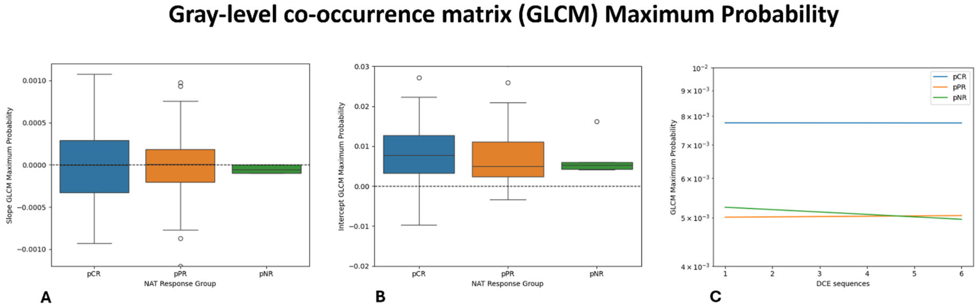
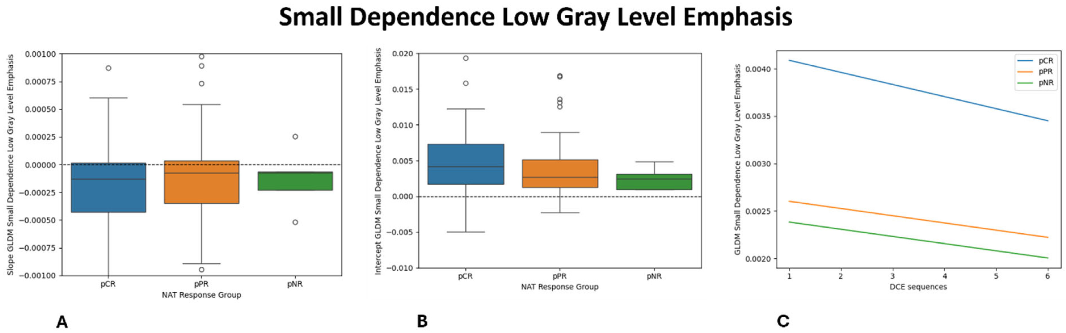
Univariate analysis identified 30 features that differ significantly among the three response groups (pCR, pPR, and pNR). Among these, the gray-level co-occurrence matrix (GLCM) Maximum Probability (p = 0.009) and the intercept of Small-Dependence Low-Gray-Level Emphasis (SDLGLE) (p = 0.010) showed the strongest associations with pNR. The distribution of these features across response groups are shown in Figure 2 and Figure 3.
Figure 2.
Boxplot and Lineplot showing the dynamic changes in the GLCM Maximum Probability: (A): slope; (B): intercept; and (C): average linear regression model of each NAT response group.
Figure 3.
Boxplot and Lineplot showing the dynamic changes in the GLDM Small-Dependence Low-Gray-Level Emphasis: (A): slope (B): intercept (C): and average linear regression model of each NAT response group.
Regarding feature selection for the ML model, the recursive feature elimination with cross-validation (RFECV) procedure selected eight features, which were subsequently used for model training.
3.3. Machine Learning Model Performance
The final radiomics models achieved an overall accuracy of 67.4% (31/42), a macro-averaged sensitivity of 56.5%, and a specificity of 78.9% in the internal validation cohort.
In the external validation cohort, the model reached an accuracy of 72.5% (28/39), a macro-averaged sensitivity of 65.6%, and a specificity of 83.1%.
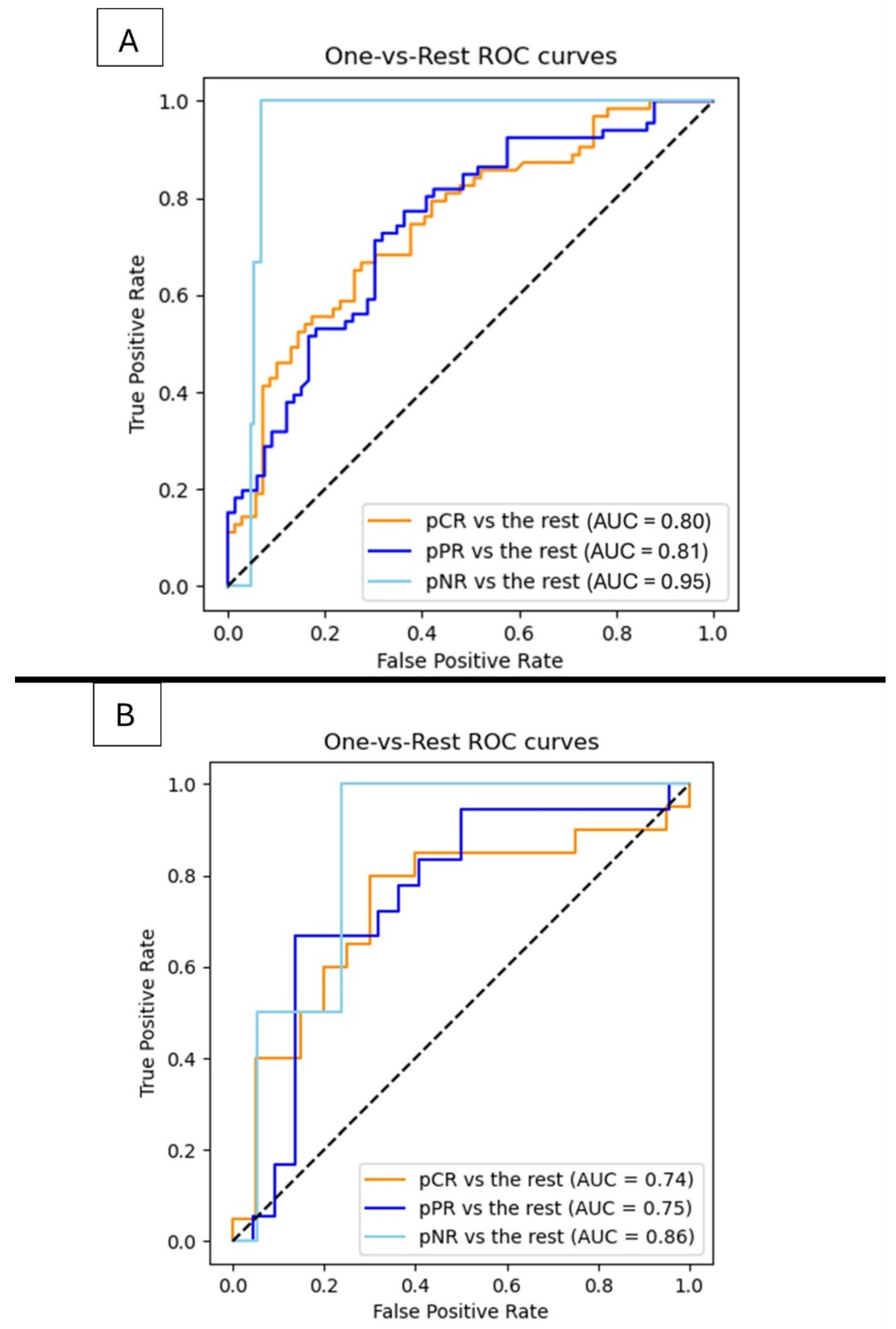
Regarding the ROC analysis, the model achieved an AUC of 0.80 for pCR vs. the rest, 0.81 for pPR vs. the rest, and 0.95 for pNR vs. the rest in the internal validation cohort (Figure 4). The corresponding AUCs in the external cohort were 0.74, 0.75, and 0.86, respectively.
Figure 4.
AUROC (one vs. rest) evaluated in Center 1 ((A); internal validation) and Center 2 ((B); external validation).
Confusion matrices for both cohorts are provided in Supplementary Material S7.
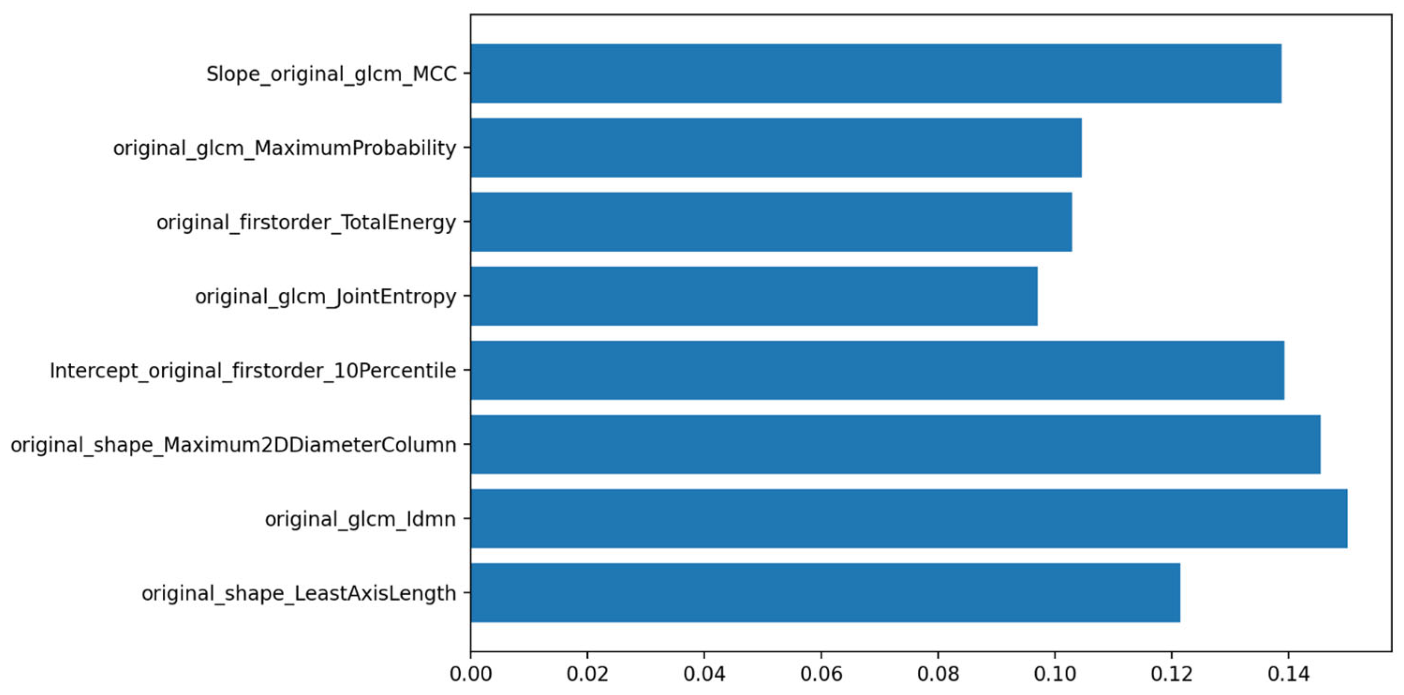
Feature importance analysis identified GLCM Maximum Probability and the intercept of Small-Dependence Low-Gray-Level Emphasis (SDLGLE) as the most discriminative variables (Figure 5), consistent with the univariate statistical results.
Figure 5.
Features importance in prediction of pCR class, pPR class and pNR class.
For comparison, a clinical-only model—including histopathological and MRI-based tumor extension parameters—achieved similar AUCs of 0.80, 0.81, and 0.95 in the internal validation cohort and 0.75, 0.74, and 0.86 in the external cohort.
The late-fusion ML achieved lower performances and similar AUCs of 0.78, 0.75, and 0.85 in the internal validation cohort and 0.72, 0.70, and 0.84 in the external cohort.
The exploratory subgroup analysis results are summarized in Supplementary Material S4.
4. Discussion
In this study, we developed and validated an ML model based on time-dependent radiomic features extracted from pre-treatment DCE-MRI to predict response to NAT in breast cancer patients. This model successfully predicted patients’ pathological responses to NAT, with the highest performance observed in non-responders, the subgroup for whom early treatment adaptation and avoidance of unnecessary toxicity are most clinically relevant. On the external validation cohort, although performance slightly declined, the model retained good discriminative ability, supporting the robustness and potential transferability of the model, underscoring the potential of generalizing radiomics models across institutions due to differences in MRI acquisition protocols, scanners, and patient populations.
Although pCR is a key therapeutic goal in breast cancer owing to its association with improved disease-free and overall survival and its potential role in surgical de-escalation, it is achieved in only a minority of patients [8,39].
Evidence from recent clinical trials, such as TRAIN-3 [35], has demonstrated that a subset of patients can achieve pCR after only a few treatment cycles and that MRI may facilitate the early identification of responders, supporting treatment de-escalation strategies. Conversely, patients with resistant disease require early risk stratification to allow the timely modification of ineffective therapies and to avoid unnecessary delays in definitive surgery. In this context, the ability to accurately predict non-response before treatment initiation could have substantial clinical impact.
Currently, the histopathological evaluation of the surgical specimen remains the reference standard for assessing response to NAT [40]. However, this approach is inherently invasive and provides outcome information only after treatment completion.
Although imaging is widely used during NAT, its role in guiding treatment adaptation remains limited. As reported by a recent systematic review [20], imaging-guided treatment modification is implemented in only a small fraction of trials, mainly due to the lack of standardized protocols and quantitative, actionable imaging biomarkers.
Several strategies have been explored to predict response to NAT. Clinicopathological factors—such as molecular subtype, tumor grade, Ki-67, and baseline tumor burden—are known to influence treatment sensitivity but offer only static and indirect information. Imaging-based approaches have largely relied on morphological assessments with conventional MRI, including RECIST-based changes in tumor size and volume, which often occur late in the treatment course [4]. Functional imaging techniques, including DWI, DCE-MRI pharmacokinetic parameters, and PET/CT metabolic response, have also been investigated, with variable results [41].
These limitations highlight the need for non-invasive imaging biomarkers capable of enabling earlier and more informative response assessments capable of predicting response earlier and enabling the real-time monitoring of therapeutic effects. In this setting, DCE-MRI is particularly well suited for this purpose, as it allows the longitudinal assessment of tumor vascularity and perfusion, supporting treatment personalization based on imaging-derived functional information. More recently, radiomics and ML-based methods applied to MRI, DWI, or PET imaging have shown promising performance for response prediction [25].
Radiomics further extends the value of DCE-MRI enabling the quantitative characterization of intratumoral heterogeneity, capturing information beyond conventional visual interpretation.
Several studies have reported promising results in predicting pCR using radiomic features extracted from pre-treatment DCE-MRI [21,25,28,30,42,43,44,45,46,47].
However, many of these investigations have focused on binary classification (pCR vs. non-pCR), which may oversimplify the biological spectrum of treatment response. In contrast, our study extends prior work by incorporating time-dependent radiomic features derived from all dynamic phases of pre-treatment DCE-MRI, enabling a multiclass stratification of treatment response and providing an externally validated framework for the early identification of non-responders. This refined stratification is clinically meaningful, as it better reflects real-world decision-making scenarios and supports tailored strategies such as treatment escalation, early switching, or de-escalation.
In the primary analysis, our cohort exhibited an overall pCR rate of 50%, which is higher than the 40% reported in the ACOSOG Z1071 trial [48]. This difference may be explained by the high prevalence of HER2+ and triple-negative subtypes in our population, both of which are known to achieve higher response rates to NAT compared with luminal tumors [28,42,49]. When compared with the prior literature, our model achieved performance comparable or superior to previously published ML-based radiomics models [25,28,42]. Moreover, our findings are consistent with those reported by Xiong et al. [42] and Zhuang et al. [45], who also demonstrated the potential of time-dependent radiomic features for response prediction.
Among the 759 extracted radiomic features, 30 showed significant differences across response groups. In particular, GLCM Maximum Probability and the intercept of the Small-Dependence Low-Gray-Level Emphasis were strongly associated with pNR, suggesting that resistant tumors exhibit a more disorganized internal architecture. GLCM Maximum Probability reflects the uniformity of gray-level distribution within the tumor with lower values indicating a greater intratumoral heterogeneity—a feature consistently associated with poor treatment response. Conversely, higher values denote more homogeneous tissue organization, which was more frequently observed in tumors achieving pCR. The Small-Dependence Low-Gray-Level Emphasis, which captures fine textural variations in low-intensity regions related to structural disorganization, reached statistical significance primarily through its intercept parameter, suggesting baseline differences across response groups rather than dynamic temporal changes.
Overall, patients achieving pCR exhibited lower heterogeneity-related feature values, consistent with a more homogeneous and treatment-sensitive tumor composition, whereas non-responders displayed a higher baseline heterogeneity and a disorganized internal architecture—features characteristic of resistant phenotypes. Partial responders demonstrated intermediate values, supporting the concept of a biological continuum of tumor response rather than a dichotomous outcome.
Our supplementary exploratory subgroup analysis based on refined pathological response categories further supported this continuum, showing that cases with residual in situ disease (pCRis) exhibited enhancement dynamics intermediate between a true complete response and a partial response or non-response. Although hypothesis-generating, these findings suggest that residual in situ disease may retain microstructural and vascular characteristics distinct from both complete eradication and persistent invasive disease, providing additional biological insights into response heterogeneity.
These observations align with previous reports [4,50], indicating that a true complete response is associated with the early normalization of enhancement patterns, flattening of time–intensity curves, and disappearance of delayed washout, whereas a partial response or non-response corresponds to persistent enhancement irregularities and complex signal behavior. The integration of dynamic textural markers reinforces the hypothesis that temporal radiomics can detect subtle yet biologically meaningful differences in microvascular remodeling and tissue composition induced by therapy, contributing to a more physiologically grounded interpretation of treatment response. In such a perspective, tumor heterogeneity provides a biological explanation for the variable therapeutic responses observed among patients with similar clinical and molecular profiles. Supporting our findings, the recent studies [30,51] demonstrated that models integrating quantitative radiomic features of intratumoral heterogeneity with conventional radiological assessment and clinicopathological variables significantly improves prognostic stratification and prediction of NAT response, compared with models based on radiological or radiomic features alone, supporting the biological relevance of tumor heterogeneity as a key determinant of therapeutic sensitivity. Such approaches may support safer and more effective personalization of therapy, as well as tailored surgical and follow-up strategies, with the potential to improve patient outcomes [52,53].
A key strength of our study lies in the comprehensive modeling of tumor vascularization achieved by incorporating all dynamic phases of DCE-MRI rather than restricting analysis to a single post-contrast image. By summarizing the temporal evolution of radiomic features through slope and intercept parameters, our approach captures both baseline vascular characteristics and their dynamic changes throughout contrast uptake. This contrasts with prior studies that primarily analyzed a single enhancement phase, typically assuming it best represented tumor heterogeneity [54,55,56]. Our multiphase strategy provides a more comprehensive representation of tumor physiology and may explain the improved performance observed in identifying non-responders.
This study has several limitations. First, as with any retrospective study, selection bias is an inherent issue. Second, the data were collected over an extended period where DCE-MRI protocols evolved. Although these optimizations were intended to improve image quality, they may have introduced variations that could affect the consistency of the radiomic features extracted across different time points. Third, the sample size—particularly for the non-responder subgroup—was relatively small, which may limit statistical power. Additionally, in our analyses, we specifically chose to use the third DCE-T1w images, as breast tumors were most clearly visible in this sequence. However, the modeling of the dynamics of radiomic feature was conducted using a linear regression model. While this approach is computationally efficient and simplifies the analysis of trends, it may not fully capture the complex, non-linear nature of tumor dynamics.
Future studies should aim to validate these findings in larger, prospective and multi-institutional cohorts using standardized imaging protocols. The application of advanced modeling techniques, including non-linear temporal fitting, delta-radiomics, and deep learning-based approaches, may further enhance the prediction of non-response and improve generalizability across imaging platforms.
Despite these limitations, our results support the feasibility and clinical relevance of time-dependent radiomics as a promising non-invasive imaging biomarker for identifying non-responders to neoadjuvant therapy. Incorporating temporal radiomics into the clinical workflow may facilitate earlier treatment adaptation, reduce unnecessary toxicity, and contribute to AI-guided, patient-tailored therapeutic strategies.
5. Conclusions
Time-dependent radiomic features from DCE-MRI can effectively predict response to NAT in breast cancer patients, with the highest accuracy in identifying non-responders, the subgroup for whom early treatment adaptation is most crucial. By capturing subtle temporal enhancement dynamics, temporal radiomics provide unique biological insights beyond static features, enabling a more precise, non-invasive assessment of therapeutic resistance. These findings highlight the potential of dynamic MRI-based AI models to support personalized, response-guided treatment strategies.
Supplementary Materials
The following supporting information can be downloaded at: https://www.mdpi.com/article/10.3390/diagnostics16040611/s1, Supplementary Material S1: Clear checklist; Supplementary Material S2: Parameters and setting for training the CNN for tumor segmentation using the third Breast DCE-MRI.; Supplementary Material S3: Parameters and settings for Radiomics features extraction.; Supplementary Material S4: Exploratory Subgroup Analysis; Supplementary Material S5: Type of response according to molecular Histotypes; Supplementary Material S6: Type of therapy; Supplementary Material S7: Confusion Matrix of Internal and External Validation Cohorts.
Author Contributions
Conceptualization, G.V., R.L. and D.B.; methodology, G.V. and R.L.; software, R.L.; validation, G.V., R.L. and D.B.; formal analysis, R.L.; investigation, G.V., P.N., G.P., E.S., F.F., C.M. and R.M.T.; resources, D.B.; data curation, G.V. and D.B.; writing—original draft preparation, G.V.; writing—review and editing, R.L. and D.B.; visualization, G.V.; supervision, G.V. and D.B.; project administration, D.B.; funding acquisition, D.B. All authors have read and agreed to the published version of the manuscript.
Funding
The publication fee for this work was covered by the Italian Ministry of Health’s “Ricerca Corrente” funding to the IRCCS Humanitas Research Hospital.
Institutional Review Board Statement
This study was conducted in accordance with the Declaration of Helsinki and approved by the Institutional Review Board (Comitato Etico Territoriale Lombardia 5) (protocol number: 59/24, approval date: 17 December 2024).
Informed Consent Statement
Patient consent was waived due to the retrospective nature of the study.
Data Availability Statement
The data supporting the findings of this study are stored in an institutional database in the Excel format. Due to privacy and ethical restrictions, the data are not publicly available but may be made available from the corresponding author upon reasonable request.
Conflicts of Interest
The authors declare no conflicts of interest.
References
- Makhoul, I.; Kiwan, E. Neoadjuvant Systemic Treatment of Breast Cancer. J. Surg. Oncol. 2011, 103, 348–357. [Google Scholar] [CrossRef]
- Kaufmann, M.; Hortobagyi, G.N.; Goldhirsch, A.; Scholl, S.; Makris, A.; Valagussa, P.; Blohmer, J.-U.; Eiermann, W.; Jackesz, R.; Jonat, W.; et al. Recommendations From an International Expert Panel on the Use of Neoadjuvant (Primary) Systemic Treatment of Operable Breast Cancer: An Update. J. Clin. Oncol. 2006, 24, 1940–1949. [Google Scholar] [CrossRef]
- Asaoka, M.; Gandhi, S.; Ishikawa, T.; Takabe, K. Neoadjuvant Chemotherapy for Breast Cancer: Past, Present, and Future. Breast Cancer Basic Clin. Res. 2020, 14, 1–8. [Google Scholar] [CrossRef]
- Reig, B.; Lewin, A.A.; Du, L.; Heacock, L.; Toth, H.K.; Heller, S.L.; Gao, Y.; Moy, L. Breast MRI for Evaluation of Response to Neoadjuvant Therapy. RadioGraphics 2021, 41, 665–679. [Google Scholar] [CrossRef]
- Von Minckwitz, G.; Untch, M.; Blohmer, J.-U.; Costa, S.D.; Eidtmann, H.; Fasching, P.A.; Gerber, B.; Eiermann, W.; Hilfrich, J.; Huober, J.; et al. Definition and Impact of Pathologic Complete Response on Prognosis After Neoadjuvant Chemotherapy in Various Intrinsic Breast Cancer Subtypes. J. Clin. Oncol. 2012, 30, 1796–1804. [Google Scholar] [CrossRef] [PubMed]
- Yoshioka, T.; Hosoda, M.; Yamamoto, M.; Taguchi, K.; Hatanaka, K.C.; Takakuwa, E.; Hatanaka, Y.; Matsuno, Y.; Yamashita, H. Prognostic Significance of Pathologic Complete Response and Ki67 Expression after Neoadjuvant Chemotherapy in Breast Cancer. Breast Cancer 2015, 22, 185–191. [Google Scholar] [CrossRef] [PubMed]
- Broglio, K.R.; Quintana, M.; Foster, M.; Olinger, M.; McGlothlin, A.; Berry, S.M.; Boileau, J.-F.; Brezden-Masley, C.; Chia, S.; Dent, S.; et al. Association of Pathologic Complete Response to Neoadjuvant Therapy in HER2-Positive Breast Cancer with Long-Term Outcomes: A Meta-Analysis. JAMA Oncol. 2016, 2, 751–760. [Google Scholar] [CrossRef] [PubMed]
- Gentile, D.; Sagona, A.; De Carlo, C.; Fernandes, B.; Barbieri, E.; Di Maria Grimaldi, S.; Jacobs, F.; Vatteroni, G.; Scardina, L.; Biondi, E.; et al. Pathologic Response and Residual Tumor Cellularity after Neo-Adjuvant Chemotherapy Predict Prognosis in Breast Cancer Patients. Breast 2023, 69, 323–329. [Google Scholar] [CrossRef]
- Van Ramshorst, M.S.; Van Der Voort, A.; Van Werkhoven, E.D.; Mandjes, I.A.; Kemper, I.; Dezentjé, V.O.; Oving, I.M.; Honkoop, A.H.; Tick, L.W.; Van De Wouw, A.J.; et al. Neoadjuvant Chemotherapy with or without Anthracyclines in the Presence of Dual HER2 Blockade for HER2-Positive Breast Cancer (TRAIN-2): A Multicentre, Open-Label, Randomised, Phase 3 Trial. Lancet Oncol. 2018, 19, 1630–1640. [Google Scholar] [CrossRef]
- Nitz, U.A.; Gluz, O.; Christgen, M.; Grischke, E.-M.; Augustin, D.; Kuemmel, S.; Braun, M.; Potenberg, J.; Kohls, A.; Krauss, K.; et al. De-Escalation Strategies in HER2-Positive Early Breast Cancer (EBC): Final Analysis of the WSG-ADAPT HER2+/HR− Phase II Trial: Efficacy, Safety, and Predictive Markers for 12 Weeks of Neoadjuvant Dual Blockade with Trastuzumab and Pertuzumab ± Weekly Paclitaxel. Ann. Oncol. 2017, 28, 2768–2772. [Google Scholar] [CrossRef]
- Valero, V.; Buzdar, A.U.; McNeese, M.; Singletary, E.; Hortobagyi, G.N. Primary Chemotherapy in the Treatment of Breast Cancer: The University of Texas M. D. Anderson Cancer Center Experience. Clin. Breast Cancer 2002, 3, S63–S68. [Google Scholar] [CrossRef] [PubMed]
- Vasudevan, D.; Jayalakshmy, P.S.; Kumar, S.; Mathew, S. Assessment of Pathological Response of Breast Carcinoma in Modified Radical Mastectomy Specimens after Neoadjuvant Chemotherapy. Int. J. Breast Cancer 2015, 2015, 536145. [Google Scholar] [CrossRef] [PubMed]
- Lo Gullo, R.; Marcus, E.; Huayanay, J.; Eskreis-Winkler, S.; Thakur, S.; Teuwen, J.; Pinker, K. Artificial Intelligence-Enhanced Breast MRI: Applications in Breast Cancer Primary Treatment Response Assessment and Prediction. Investig. Radiol. 2024, 59, 230–242. [Google Scholar] [CrossRef] [PubMed]
- Marinovich, M.L.; Houssami, N.; Macaskill, P.; Sardanelli, F.; Irwig, L.; Mamounas, E.P.; Von Minckwitz, G.; Brennan, M.E.; Ciatto, S. Meta-Analysis of Magnetic Resonance Imaging in Detecting Residual Breast Cancer After Neoadjuvant Therapy. J. Natl. Cancer Inst. 2013, 105, 321–333. [Google Scholar] [CrossRef]
- Gradishar, W.J.; Moran, M.S.; Abraham, J.; Aft, R.; Agnese, D.; Allison, K.H.; Anderson, B.; Burstein, H.J.; Chew, H.; Dang, C.; et al. Breast Cancer, Version 3.2022, NCCN Clinical Practice Guidelines in Oncology. J. Natl. Compr. Cancer Netw. 2022, 20, 691–722. [Google Scholar] [CrossRef]
- Eisenhauer, E.A.; Therasse, P.; Bogaerts, J.; Schwartz, L.H.; Sargent, D.; Ford, R.; Dancey, J.; Arbuck, S.; Gwyther, S.; Mooney, M.; et al. New Response Evaluation Criteria in Solid Tumours: Revised RECIST Guideline (Version 1.1). Eur. J. Cancer 2009, 45, 228–247. [Google Scholar] [CrossRef]
- Pereira, N.P.; Curi, C.; Osório, C.A.B.T.; Marques, E.F.; Makdissi, F.B.; Pinker, K.; Bitencourt, A.G.V. Diffusion-Weighted Magnetic Resonance Imaging of Patients with Breast Cancer Following Neoadjuvant Chemotherapy Provides Early Prediction of Pathological Response—A Prospective Study. Sci. Rep. 2019, 9, 16372. [Google Scholar] [CrossRef]
- Partridge, S.C.; Zhang, Z.; Newitt, D.C.; Gibbs, J.E.; Chenevert, T.L.; Rosen, M.A.; Bolan, P.J.; Marques, H.S.; Romanoff, J.; Cimino, L.; et al. Diffusion-Weighted MRI Findings Predict Pathologic Response in Neoadjuvant Treatment of Breast Cancer: The ACRIN 6698 Multicenter Trial. Radiology 2018, 289, 618–627. [Google Scholar] [CrossRef]
- Santamaría, G.; Bargalló, X.; Ganau, S.; Alonso, I.; Muñoz, M.; Mollà, M.; Fernández, P.; Prat, A. Multiparametric MR Imaging to Assess Response Following Neoadjuvant Systemic Treatment in Various Breast Cancer Subtypes: Comparison between Different Definitions of Pathologic Complete Response. Eur. J. Radiol. 2019, 117, 132–139. [Google Scholar] [CrossRef]
- Lafci, O.; Resch, D.; Santonocito, A.; Clauser, P.; Helbich, T.; Baltzer, P.A.T. Role of Imaging Based Response Assesment for Adapting Neoadjuvant Systemic Therapy for Breast Cancer: A Systematic Review. Eur. J. Radiol. 2025, 187, 112105. [Google Scholar] [CrossRef]
- Pesapane, F.; Rotili, A.; Botta, F.; Raimondi, S.; Bianchini, L.; Corso, F.; Ferrari, F.; Penco, S.; Nicosia, L.; Bozzini, A.; et al. Radiomics of MRI for the Prediction of the Pathological Response to Neoadjuvant Chemotherapy in Breast Cancer Patients: A Single Referral Centre Analysis. Cancers 2021, 13, 4271. [Google Scholar] [CrossRef]
- Reig, B.; Heacock, L.; Geras, K.J.; Moy, L. Machine Learning in Breast MRI. J. Magn. Reson. Imaging 2020, 52, 998–1018. [Google Scholar] [CrossRef] [PubMed]
- Gillies, R.J.; Kinahan, P.E.; Hricak, H. Radiomics: Images Are More than Pictures, They Are Data. Radiology 2016, 278, 563–577. [Google Scholar] [CrossRef] [PubMed]
- Gaudio, M.; Vatteroni, G.; De Sanctis, R.; Gerosa, R.; Benvenuti, C.; Canzian, J.; Jacobs, F.; Saltalamacchia, G.; Rizzo, G.; Pedrazzoli, P.; et al. Incorporating Radiomic MRI Models for Presurgical Response Assessment in Patients with Early Breast Cancer Undergoing Neoadjuvant Systemic Therapy: Collaborative Insights from Breast Oncologists and Radiologists. Crit. Rev. Oncol./Hematol. 2025, 210, 104681. [Google Scholar] [CrossRef] [PubMed]
- Pesapane, F.; Agazzi, G.M.; Rotili, A.; Ferrari, F.; Cardillo, A.; Penco, S.; Dominelli, V.; D’Ecclesiis, O.; Vignati, S.; Raimondi, S.; et al. Prediction of the Pathological Response to Neoadjuvant Chemotherapy in Breast Cancer Patients with MRI-Radiomics: A Systematic Review and Meta-Analysis. Curr. Probl. Cancer 2022, 46, 100883. [Google Scholar] [CrossRef]
- Liang, X.; Yu, X.; Gao, T. Machine Learning with Magnetic Resonance Imaging for Prediction of Response to Neoadjuvant Chemotherapy in Breast Cancer: A Systematic Review and Meta-Analysis. Eur. J. Radiol. 2022, 150, 110247. [Google Scholar] [CrossRef]
- Codari, M.; Schiaffino, S.; Sardanelli, F.; Trimboli, R.M. Artificial Intelligence for Breast MRI in 2008–2018: A Systematic Mapping Review. Am. J. Roentgenol. 2019, 212, 280–292. [Google Scholar] [CrossRef]
- Braman, N.M.; Etesami, M.; Prasanna, P.; Dubchuk, C.; Gilmore, H.; Tiwari, P.; Plecha, D.; Madabhushi, A. Intratumoral and Peritumoral Radiomics for the Pretreatment Prediction of Pathological Complete Response to Neoadjuvant Chemotherapy Based on Breast DCE-MRI. Breast Cancer Res. 2017, 19, 57. [Google Scholar] [CrossRef]
- Aghaei, F.; Tan, M.; Hollingsworth, A.B.; Qian, W.; Liu, H.; Zheng, B. Computer-aided Breast MR Image Feature Analysis for Prediction of Tumor Response to Chemotherapy. Med. Phys. 2015, 42, 6520–6528. [Google Scholar] [CrossRef]
- Trombadori, C.; Boccia, E.; Tran, E.H.; Franco, A.; Orlandi, A.; Franceschini, G.; Carbognin, L.; Di Leone, A.; Masiello, V.; Marazzi, F.; et al. Role of Radiomics in Predicting Early Disease Recurrence in Locally Advanced Breast Cancer Patients: Integration of Radiomic Features and RECIST Criteria. Radiol. Med. 2025, 130, 753–765. [Google Scholar] [CrossRef]
- Khan, N.; Adam, R.; Huang, P.; Maldjian, T.; Duong, T.Q. Deep Learning Prediction of Pathologic Complete Response in Breast Cancer Using MRI and Other Clinical Data: A Systematic Review. Tomography 2022, 8, 2784–2795. [Google Scholar] [CrossRef] [PubMed]
- Tsukada, H.; Tsukada, J.; Schrading, S.; Strobel, K.; Okamoto, T.; Kuhl, C.K. Accuracy of Multi-Parametric Breast MR Imaging for Predicting Pathological Complete Response of Operable Breast Cancer Prior to Neoadjuvant Systemic Therapy. J. Magn. Reson. Imaging 2019, 62, 242–248. [Google Scholar] [CrossRef] [PubMed]
- Herrero Vicent, C.; Tudela, X.; Moreno Ruiz, P.; Pedralva, V.; Jiménez Pastor, A.; Ahicart, D.; Rubio Novella, S.; Meneu, I.; Montes Albuixech, Á.; Santamaria, M.Á.; et al. Machine Learning Models and Multiparametric Magnetic Resonance Imaging for the Prediction of Pathologic Response to Neoadjuvant Chemotherapy in Breast Cancer. Cancers 2022, 14, 3508. [Google Scholar] [CrossRef] [PubMed]
- Jansen, S.A.; Shimauchi, A.; Zak, L.; Fan, X.; Karczmar, G.S.; Newstead, G.M. The Diverse Pathology and Kinetics of Mass, Nonmass, and Focus Enhancement on MR Imaging of the Breast. J. Magn. Reson. Imaging 2011, 33, 1382–1389. [Google Scholar] [CrossRef]
- Van Der Voort, A.; Louis, F.M.; Van Ramshorst, M.S.; Kessels, R.; Mandjes, I.A.; Kemper, I.; Agterof, M.J.; Van Der Steeg, W.A.; Heijns, J.B.; Van Bekkum, M.L.; et al. MRI-Guided Optimisation of Neoadjuvant Chemotherapy Duration in Stage II–III HER2-Positive Breast Cancer (TRAIN-3): A Multicentre, Single-Arm, Phase 2 Study. Lancet Oncol. 2024, 25, 603–613. [Google Scholar] [CrossRef]
- Kocak, B.; Baessler, B.; Bakas, S.; Cuocolo, R.; Fedorov, A.; Maier-Hein, L.; Mercaldo, N.; Müller, H.; Orlhac, F.; Pinto Dos Santos, D.; et al. CheckList for EvaluAtion of Radiomics Research (CLEAR): A Step-by-Step Reporting Guideline for Authors and Reviewers Endorsed by ESR and EuSoMII. Insights Imaging 2023, 14, 75. [Google Scholar] [CrossRef]
- Goorts, B.; Dreuning, K.M.A.; Houwers, J.B.; Kooreman, L.F.S.; Boerma, E.-J.G.; Mann, R.M.; Lobbes, M.B.I.; Smidt, M.L. MRI-Based Response Patterns during Neoadjuvant Chemotherapy Can Predict Pathological (Complete) Response in Patients with Breast Cancer. Breast Cancer Res. 2018, 20, 34. [Google Scholar] [CrossRef]
- Pinder, S.E.; Provenzano, E.; Earl, H.; Ellis, I.O. Laboratory Handling and Histology Reporting of Breast Specimens from Patients Who Have Received Neoadjuvant Chemotherapy. Histopathology 2007, 50, 409–417. [Google Scholar] [CrossRef]
- Cortazar, P.; Zhang, L.; Untch, M.; Mehta, K.; Costantino, J.P.; Wolmark, N.; Bonnefoi, H.; Cameron, D.; Gianni, L.; Valagussa, P.; et al. Pathological Complete Response and Long-Term Clinical Benefit in Breast Cancer: The CTNeoBC Pooled Analysis. Lancet 2014, 384, 164–172. [Google Scholar] [CrossRef]
- Korde, L.A.; Somerfield, M.R.; Carey, L.A.; Crews, J.R.; Denduluri, N.; Hwang, E.S.; Khan, S.A.; Loibl, S.; Morris, E.A.; Perez, A.; et al. Neoadjuvant Chemotherapy, Endocrine Therapy, and Targeted Therapy for Breast Cancer: ASCO Guideline. J. Clin. Oncol. 2021, 39, 1485–1505. [Google Scholar] [CrossRef]
- Gerosa, R.; Gelardi, F.; Tiberio, P.; Jacobs, F.; Benvenuti, C.; Gaudio, M.; Canzian, J.; Tinterri, B.; Zambelli, A.; Santoro, A.; et al. [18F]FDG PET/CT as a Biomarker for Response Evaluation in Neoadjuvant Treatment of Early Breast Cancer: Could It Become a Game-Changer in the Scenario of the Emerging (de)-Escalation Strategies? Radiol. Med. 2025. Epub ahead of printing. [Google Scholar] [CrossRef]
- Liu, Z.; Li, Z.; Qu, J.; Zhang, R.; Zhou, X.; Li, L.; Sun, K.; Tang, Z.; Jiang, H.; Li, H.; et al. Radiomics of Multiparametric MRI for Pretreatment Prediction of Pathologic Complete Response to Neoadjuvant Chemotherapy in Breast Cancer: A Multicenter Study. Clin. Cancer Res. 2019, 25, 3538–3547. [Google Scholar] [CrossRef] [PubMed]
- Xiong, Q.; Zhou, X.; Liu, Z.; Lei, C.; Yang, C.; Yang, M.; Zhang, L.; Zhu, T.; Zhuang, X.; Liang, C.; et al. Multiparametric MRI-Based Radiomics Analysis for Prediction of Breast Cancers Insensitive to Neoadjuvant Chemotherapy. Clin. Transl. Oncol. 2020, 22, 50–59. [Google Scholar] [CrossRef] [PubMed]
- Cain, E.H.; Saha, A.; Harowicz, M.R.; Marks, J.R.; Marcom, P.K.; Mazurowski, M.A. Multivariate Machine Learning Models for Prediction of Pathologic Response to Neoadjuvant Therapy in Breast Cancer Using MRI Features: A Study Using an Independent Validation Set. Breast Cancer Res. Treat. 2019, 173, 455–463. [Google Scholar] [CrossRef] [PubMed]
- Zhuang, X.; Chen, C.; Liu, Z.; Zhang, L.; Zhou, X.; Cheng, M.; Ji, F.; Zhu, T.; Lei, C.; Zhang, J.; et al. Multiparametric MRI-Based Radiomics Analysis for the Prediction of Breast Tumor Regression Patterns after Neoadjuvant Chemotherapy. Transl. Oncol. 2020, 13, 100831. [Google Scholar] [CrossRef]
- Choudhery, S.; Gomez-Cardona, D.; Favazza, C.P.; Hoskin, T.L.; Haddad, T.C.; Goetz, M.P.; Boughey, J.C. MRI Radiomics for Assessment of Molecular Subtype, Pathological Complete Response, and Residual Cancer Burden in Breast Cancer Patients Treated with Neoadjuvant Chemotherapy. Acad. Radiol. 2022, 29, S145–S154. [Google Scholar] [CrossRef]
- Zhang, Y.; Cai, J.; Cui, C.; Qi, S.; Zhao, D. Predicting Breast Cancer Response to Neoadjuvant Therapy by Integrating Radiomic and Deep-Learning Features from Early-and-Peak Phases of DCE-MRI. BMC Cancer 2025, 25, 1747. [Google Scholar] [CrossRef]
- Boughey, J.C. Sentinel Lymph Node Surgery After Neoadjuvant Chemotherapy in Patients with Node-Positive Breast Cancer: The ACOSOG Z1071 (Alliance) Clinical Trial. JAMA 2013, 310, 1455–1461. [Google Scholar] [CrossRef]
- Vila, J.; Mittendorf, E.A.; Farante, G.; Bassett, R.L.; Veronesi, P.; Galimberti, V.; Peradze, N.; Stauder, M.C.; Chavez-MacGregor, M.; Litton, J.F.; et al. Nomograms for Predicting Axillary Response to Neoadjuvant Chemotherapy in Clinically Node-Positive Patients with Breast Cancer. Ann. Surg. Oncol. 2016, 23, 3501–3509. [Google Scholar] [CrossRef]
- Chamming’s, F.; Ueno, Y.; Aldis, A.; Seidler, M.; Mesurolle, B.; Omeroglu, A.; Gallix, B.; Reinhold, C. Enhancement of Breast Cancer on Pre-Treatment Dynamic Contrast-Enhanced MRI Using Computer-Aided Detection Is Associated with Response to Neo-Adjuvant Chemotherapy. Diagn. Interv. Imaging 2018, 99, 773–781. [Google Scholar] [CrossRef]
- Shi, Z.; Huang, X.; Cheng, Z.; Xu, Z.; Lin, H.; Liu, C.; Chen, X.; Liu, C.; Liang, C.; Lu, C.; et al. MRI-Based Quantification of Intratumoral Heterogeneity for Predicting Treatment Response to Neoadjuvant Chemotherapy in Breast Cancer. Radiology 2023, 308, e222830. [Google Scholar] [CrossRef]
- Zardavas, D.; Irrthum, A.; Swanton, C.; Piccart, M. Clinical Management of Breast Cancer Heterogeneity. Nat. Rev. Clin. Oncol. 2015, 12, 381–394. [Google Scholar] [CrossRef]
- Lüönd, F.; Tiede, S.; Christofori, G. Breast Cancer as an Example of Tumour Heterogeneity and Tumour Cell Plasticity during Malignant Progression. Br. J. Cancer 2021, 125, 164–175. [Google Scholar] [CrossRef]
- Zhang, Y.; Chan, S.; Park, V.Y.; Chang, K.-T.; Mehta, S.; Kim, M.J.; Combs, F.J.; Chang, P.; Chow, D.; Parajuli, R.; et al. Automatic Detection and Segmentation of Breast Cancer on MRI Using Mask R-CNN Trained on Non–Fat-Sat Images and Tested on Fat-Sat Images. Acad. Radiol. 2022, 29, S135–S144. [Google Scholar] [CrossRef]
- Militello, C.; Rundo, L.; Dimarco, M.; Orlando, A.; Woitek, R.; D’Angelo, I.; Russo, G.; Bartolotta, T.V. 3D DCE-MRI Radiomic Analysis for Malignant Lesion Prediction in Breast Cancer Patients. Acad. Radiol. 2022, 29, 830–840. [Google Scholar] [CrossRef]
- Teruel, J.R.; Heldahl, M.G.; Goa, P.E.; Pickles, M.; Lundgren, S.; Bathen, T.F.; Gibbs, P. Dynamic Contrast-enhanced MRI Texture Analysis for Pretreatment Prediction of Clinical and Pathological Response to Neoadjuvant Chemotherapy in Patients with Locally Advanced Breast Cancer. NMR Biomed. 2014, 27, 887–896. [Google Scholar] [CrossRef]
Disclaimer/Publisher’s Note: The statements, opinions and data contained in all publications are solely those of the individual author(s) and contributor(s) and not of MDPI and/or the editor(s). MDPI and/or the editor(s) disclaim responsibility for any injury to people or property resulting from any ideas, methods, instructions or products referred to in the content. |
© 2026 by the authors. Licensee MDPI, Basel, Switzerland. This article is an open access article distributed under the terms and conditions of the Creative Commons Attribution (CC BY) license.