


Abstract
Background and Clinical Significance: Mesenteric lymphatic malformations (LMs) are rare congenital anomalies that may involve extensive visceral structures and present significant therapeutic challenges. We report an unusual case of congenital mesenteric LM complicated by recurrent intra-abdominal disease and severe hypertriglyceridemia during infancy. Case Presentation: A female infant with an antenatally diagnosed mesenteric LM underwent surgical excision on the second day of life, with histopathology confirming cystic lymphangioma. Despite surgery and treatment with sirolimus and propranolol, further intra-abdominal cystic lesions were identified. At 10 months, intestinal obstruction required repeat laparotomy, revealing milky peritoneal fluid suggestive of chylous leakage. During a January 2026 admission at 14 months of age, following a recent viral infection (varicella/influenza A), severe hypertriglyceridemia (990 mg/dL) was documented, and sirolimus was withheld following recognition of dyslipidemia because of its known lipid-related adverse effects. Temporary bowel rest, intravenous hydration, a short insulin infusion, and low-fat MCT-based nutritional management were followed by declining triglycerides—391 mg/dL on day 9 and 192 mg/dL on day 15. Conclusions: This case highlights the challenges of managing extensive mesenteric LM and sirolimus-associated hypertriglyceridemia, potentially accompanied by additional precipitating factors. The response cannot be attributed to a single intervention; careful longitudinal metabolic and nutritional monitoring is essential.
1. Introduction
Mesenteric lymphatic malformations (formerly “mesenteric lymphangiomas”) are very rare congenital anomalies of the lymphatic system [1]. Accounting for less than 1% of all lymphangiomas, these malformations typically occur in children [2]. Most lymphatic malformations (LM) present in the head or neck region, with abdominal involvement (e.g., mesentery, omentum) being much less common [3]. From an epidemiological perspective, mesenteric lymphatic malformations typically manifest in early childhood, with a mean age at presentation of approximately two years, and show a marked male predominance, with a reported male-to-female ratio of 3:1 [3]. The estimated incidence is approximately 1 case per 20,000 to 250,000 pediatric hospital admissions [4]. Many congenital lymphatic malformations are detected incidentally or on imaging performed for unrelated reasons [5]; prenatal diagnosis using fetal ultrasound—and in selected cases supplemented by fetal MRI—has been increasingly reported [6].
Clinically, mesenteric LMs may be asymptomatic or present with nonspecific abdominal symptoms [7]. The most frequent clinical manifestations of mesenteric lymphatic malformations are abdominal pain, reported in approximately 70% of cases, and the presence of a palpable mass [1]. Acute complications may occur when the cystic lesion leads to bowel obstruction [8], volvulus [9], infection, or hemorrhage [10]. Given the nonspecific nature of clinical symptoms, the diagnosis of mesenteric lymphatic malformations generally depends on imaging studies. Abdominal ultrasound is the first-line modality, frequently demonstrating a multiloculated cystic lesion [3]. Computed tomography or magnetic resonance imaging may provide additional anatomical detail and delineate the full extent of the lesion, while definitive diagnosis is established through histopathological examination following surgical resection [11].
Herein, we present a rare case of a congenital lymphatic malformation with atypical mesenteric localization and complex metabolic manifestations, including severe hypertriglyceridemia, which illustrates the challenges of neonatal surgical management and medical therapy, and emphasizes the need for a multidisciplinary approach.
2. Case Report
We report a female infant evaluated at 14 months of age with a history of a mesenteric lymphatic malformation surgically treated during the neonatal period.
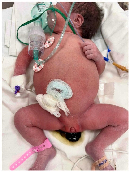
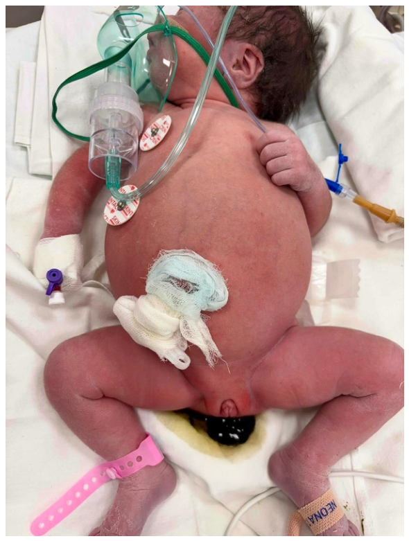
Regarding the personal history, the patient is the sixth-born child, delivered vaginally at 38 weeks’ gestation, with a birth weight of 3750 g and Apgar scores of 7 at both 1 and 5 min. The pregnancy was partially monitored, and a mesenteric lymphatic tumor was diagnosed antenatally by fetal magnetic resonance imaging at 31 weeks’ gestation. Postnatal examination confirmed a palpable abdominal distension (Figure 1).
Figure 1.
Significant abdominal distension at 24 h of life despite timely passage of meconium.
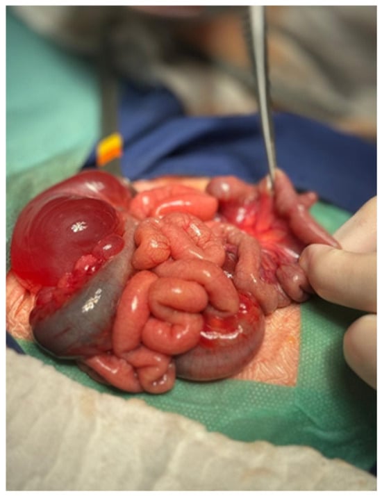
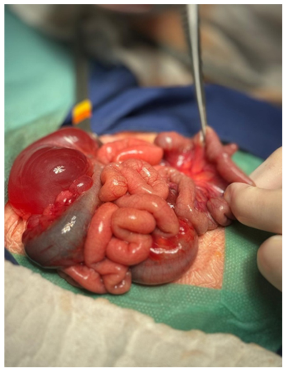
On the second day of life (8 November 2024), the patient underwent surgery under general anesthesia. A midline supra- and infra-umbilical laparotomy revealed a large volume of peritoneal fluid originating from a mesenteric lymphatic cyst involving the entire colonic frame, which was evacuated. Complete macroscopic excision of the lymphatic malformation was achieved, with no visible residual disease or apparent involvement of the bowel wall or major mesenteric vessels. Meticulous hemostasis and thorough peritoneal exploration were performed, during which a Meckel’s diverticulum was also identified (Figure 2). The procedure was completed with peritoneal lavage and drainage. Histopathological examination revealed a multiloculated cystic lesion composed of spaces separated by thin fibrovascular septa. Vascular congestion, lymphoid aggregates, and focal clusters of foamy histiocytes were also identified. The cystic spaces were lined by flattened endothelial cells without cytological atypia and contained serous fluid. These morphological findings confirmed the diagnosis of cystic mesenteric lymphangioma. Histopathological examination also confirmed negative resection margins. Nevertheless, given the extensive nature of the malformation, the possibility of microscopic lymphatic abnormalities beyond the resected tissue cannot be entirely excluded.
Figure 2.
Intraoperative view showing the extensive mesenteric lymphatic cystic malformation and adjacent intestinal loops. A Meckel’s diverticulum was also identified during surgical exploration.
Propranolol (0.3 mg/kg/day divided every 8 h) and sirolimus (0.8 mg/m2 per dose, twice daily) were started immediately after neonatal surgery because of the diffuse, microcystic nature of the mesenteric lymphatic malformation and the risk of persistent disease despite macroscopic excision; subsequent doses were adjusted during surveillance.
On the fourth day of life, bilateral peripheral vascular spasms of the lower limbs with Raynaud-like manifestations were noted. Pentoxifylline and massage of the cyanotic extremities were followed by clinical improvement. Because this event occurred during the early postoperative period and in proximity to initiation of systemic therapy, a possible drug relationship cannot be definitively excluded.
Follow-up ultrasonography identified intraperitoneal encapsulated collections, supporting residual or recurrent intra-abdominal lymphatic disease. Propranolol and sirolimus doses were adjusted according to weight, and sirolimus levels were periodically monitored and consistently remained within the therapeutic target range of 10–15 ng/mL throughout treatment. No documented hypoglycemia or bronchospasm was reported; nevertheless, the early vasospastic event limits any categorical assertion of complete propranolol safety. Similarly, initially, no sirolimus-related adverse events were noted, such as hypokalemia, hypophosphatemia, hypercholesterolemia, hyperglycemia, hypertriglyceridemia, gastrointestinal adverse effects, edema, or fever. Serial imaging assessments showed that the mesenteric lymphatic lesions remained stable in size and extent during systemic treatment, without evidence of significant progression.
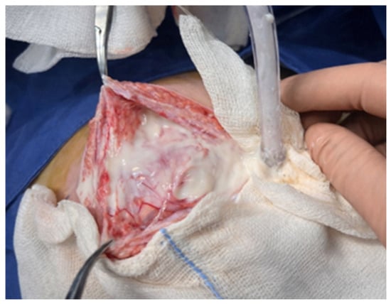
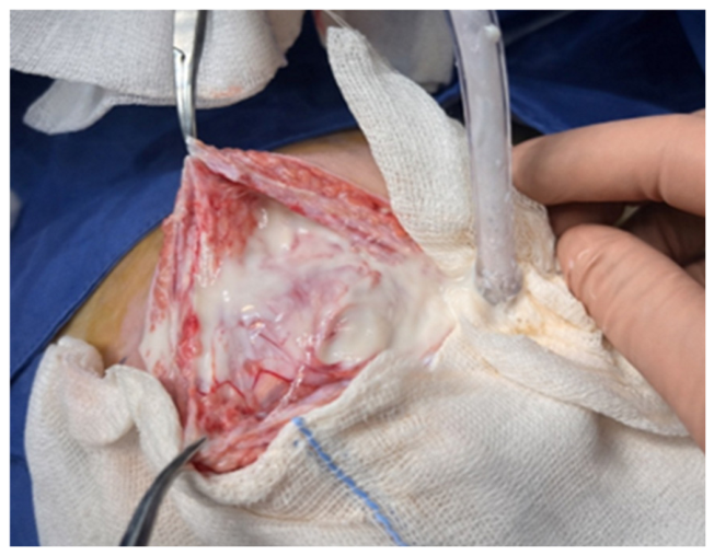
At 10 months of age (August 2025), adhesive intestinal obstruction prompted repeat midline laparotomy. Milky-appearing peritoneal fluid was observed and evacuated (Figure 3), raising clinical suspicion of chylous leakage; peritoneal triglycerides, chylomicrons, and differential cell counts were not available to biochemically confirm its chylous nature. Adhesiolysis and excision of five mesenteric and peritoneal cystic formations were performed, together with evaluation of intestinal patency, peritoneal lavage, closure of the right deep inguinal ring and prophylactic appendectomy.
Figure 3.
Intraoperative view showing milky-appearing peritoneal fluid observed during repeat laparotomy at 10 months of age, raising clinical suspicion of chylous leakage.
Postoperatively, the patient received broad-spectrum antibiotics and subcutaneous octreotide (1 µg/kg every 8 h) to reduce lymphatic output. The peritoneal drain was removed after five days, with subsequent clinical improvement. The patient was subsequently discharged in good general condition, with instructions to continue propranolol and sirolimus.
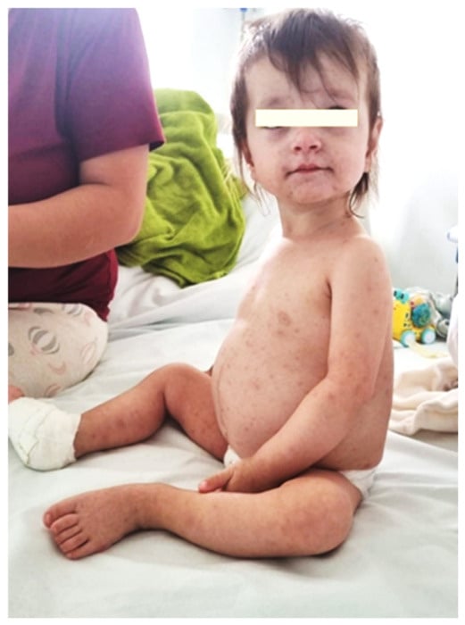
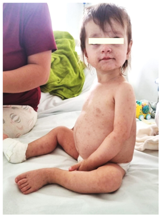
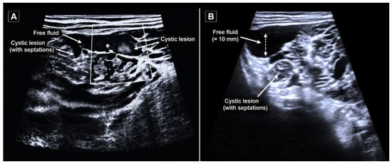
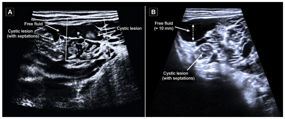
At 14 months of age, the patient was hospitalized for varicella and influenza A, which subsequently improved with supportive care. Two weeks later, she was electively admitted for reassessment of her lymphatic disease, when abdominal distension and dyslipidemia were documented. Admission weight was 9.5 kg (weight-for-age Z-score, +0.10), length 76 cm (length-for-age Z-score, −0.14), and BMI 16.4 kg/m2 (BMI-for-age Z-score, +0.23; 59th percentile). Examination showed a soft, distended abdomen and healed laparotomy scars (Figure 4). Ultrasonography revealed free intraperitoneal fluid measuring 8.5 mm in the right flank and 10 mm subhepatically, together with multiple confluent septated mesenteric cystic structures (Figure 5).
Figure 4.
Distended abdomen with uniform protrusion. Multiple hyperpigmented and crusted lesions at different stages of healing, consistent with post-varicella changes.
Figure 5.
Abdominal ultrasonography showing septated mesenteric cystic structures and free intraperitoneal fluid. (A) Multiple mesenteric cystic structures, some containing internal septations, associated with free intraperitoneal fluid. (B) Septated mesenteric cystic lesion accompanied by an adjacent collection of free intraperitoneal fluid measuring approximately 10 mm in depth. Arrows indicate the cystic lesions and free fluid collections.
On 19 January 2026, the first documented markedly abnormal lipid profile showed serum triglycerides of 990 mg/dL (reference <150 mg/dL), total cholesterol 237 mg/dL, HDL cholesterol 19 mg/dL, LDL cholesterol 105.8 mg/dL, and VLDL cholesterol 191 mg/dL. Blood samples were collected in the fasting state, and serum lipid parameters were analyzed using a direct enzymatic colorimetric method. Sirolimus was discontinued upon recognition of hypertriglyceridemia, in view of its known potential to induce dyslipidemia. The recent varicella and influenza A infections were relevant concomitant clinical considerations. No increased triglyceride concentrations had been identified before January 2026. The patient was clinically asymptomatic, with no abdominal pain, vomiting, or biochemical evidence of acute pancreatitis (serum lipase 32 U/L).
A targeted evaluation was undertaken to investigate potential primary and secondary causes of severe hypertriglyceridemia. The patient had no known family history of dyslipidemia, and previous clinical assessments had not identified elevated triglyceride concentrations. Given the severity of hypertriglyceridemia, a comprehensive clinical and laboratory assessment was undertaken to identify potential underlying causes and contributing factors, including nutritional, endocrine, hepatic, renal, inflammatory, infectious, and medication-related conditions. The main findings of this etiological evaluation are summarized below:
- Nutritional and metabolic factors: The patient had an age-appropriate nutritional status and no clinical evidence of obesity or obesity-related insulin resistance. Although nutritional factors could not be completely excluded, excessive dietary fat intake was not identified as an established cause of the lipid abnormalities.
- Endocrine disorders: Fasting blood glucose levels were within normal limits, with no biochemical evidence of diabetes mellitus. Thyroid function was normal, with TSH and free T4 concentrations within the age-appropriate reference ranges, thereby excluding overt hypothyroidism as a potential secondary cause of hypertriglyceridemia.
- Hepatic and renal disorders: Liver enzymes and renal function parameters were within the reference ranges, with no biochemical evidence of significant hepatic or renal dysfunction. Urinary protein was absent on repeat urinalysis, making nephrotic syndrome an unlikely explanation.
- Inflammatory and infectious conditions: The patient had recently experienced varicella and influenza A infection, which may have contributed to transient metabolic disturbances. However, inflammatory markers were not significantly elevated during the metabolic assessment (CRP 4.5 mg/L), suggesting the absence of a marked ongoing systemic inflammatory response.
- Medication-related factors: Particular attention was given to sirolimus exposure, as dyslipidemia, including hypertriglyceridemia, is a recognized adverse effect of mTOR inhibition. Sirolimus was discontinued following the identification of hypertriglyceridemia, and serum triglyceride concentrations subsequently decreased under combined metabolic and nutritional management.
Taken together, these findings favored a possible sirolimus-associated lipid disturbance, potentially influenced by recent infectious illness and other individual metabolic factors. Nevertheless, an underlying genetic predisposition or an additional disorder of triglyceride metabolism could not be definitively excluded, as comprehensive genetic and specialized lipoprotein investigations were not performed.
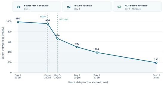
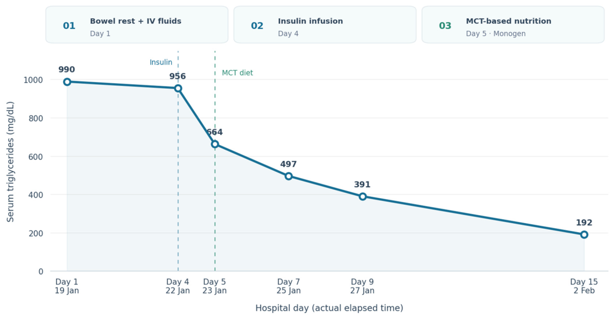
At the next documented lipid assessment, on 22 January 2026 (day 4, with 19 January defined as day 1), triglycerides remained markedly elevated at 956 mg/dL following initial supportive management. Given persistent severe hypertriglyceridemia and concern regarding pancreatitis risk, an individualized short intravenous insulin intervention was undertaken. Rapid-acting insulin (insulin lispro) was infused at 0.03 U/kg/h for approximately 8 h with 5% dextrose; capillary blood glucose was checked every 30 min and the dextrose rate adjusted to maintain glycemia between 70 and 200 mg/dL. Only one insulin infusion was administered. It was discontinued after an initial triglyceride reduction, to avoid prolonging exposure to potential complications, particularly hypoglycemia. During combined management, serum triglycerides declined to 664 mg/dL on day 5 (23 January 2026) and to 497 mg/dL on day 7 (25 January 2026).Continued strict dietary management with a very-low-fat regimen based on medium-chain triglyceride supplementation resulted in a further progressive decrease in serum triglyceride levels to 391 mg/dL on day 9 (27 January 2026) and to 192 mg/dL on day 15 (2 February 2026) (Figure 6). The patient tolerated the low-fat diet and the gradual introduction of the MCT-based formula (Monogen), with no reported hypoglycemic episodes or gastrointestinal adverse effects. Clinical evolution was favorable, with progressive abdominal decompression and adequate oral intake. An individualized nutritional regimen was established considering the patient’s age, body weight, and severe hypertriglyceridemia. A daily energy target of approximately 900–1000 kcal was used for dietary planning, with the aim of maintaining adequate nutritional intake and supporting normal growth. A very-low-long-chain-fat diet supplemented with MCTs was introduced. The nutritional strategy prioritized adequate energy and protein provision while restricting long-chain triglycerides (LCTs). The individualized nutritional plan provided approximately 950 kcal/day, with a targeted macronutrient distribution of 120–135 g of carbohydrates, 25–30 g of protein, and 30–40 g of total fat, predominantly supplied as medium-chain triglycerides (MCTs), while ensuring a controlled intake of essential long-chain fatty acids. This nutritional strategy aimed to maintain adequate energy and protein provision, support normal growth, and minimize intestinal chylomicron formation through restriction of dietary long-chain triglycerides (LCTs). An MCT-enriched formula (Monogen®, Nutricia, Fulda, Germany) was progressively introduced at 60–90 mL three times daily (180–270 mL/day), subsequently increased to 90–120 mL three times daily (270–360 mL/day), according to digestive tolerance. The patient was advised to eliminate all conventional vegetable oils from her diet and to use exclusively medium-chain triglyceride (MCT) oil, employing 100% MCT oil without thermal processing and 77% MCT oil for cooking when necessary. Animal fats, including butter and sour cream, were to be avoided, and fried foods were strictly contraindicated, with alternative cooking methods such as baking, steaming, or boiling recommended. Processed meats and commercially prepared products were excluded, with preference for homemade legume-based spreads prepared using MCT oil. Meat-based soups were to be replaced with vegetable-based soups. Whole eggs were contraindicated; however, egg whites could be incorporated into homemade preparations such as vegetable puddings, pancakes, muffins, or white omelets. Industrially processed foods and refined sweets were to be strictly avoided. The dietary plan allowed unrestricted intake of fruits, except for avocado, as well as vegetables, whole grains including oats, rice, barley, corn, wheat, bulgur, rye, and millet, pseudocereals such as quinoa and buckwheat, and legumes including lentils, chickpeas, dried peas, and beans. Low-fat rice-based plant beverages fortified with calcium were permitted. Lean meat and lean fish were recommended, along with low-fat dairy products containing 0.2–2% fat and milk with 0.1–1.5% fat content. Low-fat cereal-based snack products, such as corn or rice cakes and selected baked goods, could be included provided their lipid content was verified.
Figure 6.
The evolution of the lipid profile.
In July 2026, approximately five months after discharge, the patient was readmitted with recurrent intestinal obstruction requiring further surgical intervention. At readmission, serum triglyceride levels were 187 mg/dL. Following surgery, sirolimus therapy was reinitiated after an individualized risk–benefit assessment, considering the recurrent abdominal complications and the previous episode of severe hypertriglyceridemia. Continued clinical, imaging, and metabolic monitoring was recommended to evaluate disease progression and treatment tolerability.
To provide a clearer overview of the clinical course, the key diagnostic findings, surgical interventions, and therapeutic milestones are summarized chronologically in Table 1; serial lipid results and contemporaneous management are given in Table 2.
Table 1.
Timeline of the patient’s clinical course.
Table 2.
Chronological overview of lipid parameters and therapeutic management.
3. Discussion
3.1. Antenatal Diagnosis and Extensive Neonatal Presentation
Mesenteric lymphatic malformations represent a rare and heterogeneous subset of congenital lymphatic anomalies, with clinical behavior ranging from incidental, asymptomatic findings to extensive and potentially life-threatening neonatal disease [12]. In a review encompassing 69 pediatric cases of mesenteric lymphangioma, abdominal pain was the most frequently reported presenting symptom, occurring in over 70% of patients, followed by abdominal distension, vomiting, constipation, and fever. A palpable abdominal mass was the most common clinical sign, while acute abdominal presentations—most frequently related to volvulus or intestinal obstruction—occurred in nearly half of the reported cases [2]. These findings emphasize that mesenteric lymphatic malformations frequently present through nonspecific gastrointestinal manifestations rather than through a pathognomonic clinical syndrome.
Our patient differs from the majority of children described in these series because the disease manifested during the neonatal period and was characterized by extensive intra-abdominal involvement. This early presentation is clinically relevant because neonatal disease may represent a particularly severe phenotype, in which the lesion is sufficiently extensive to interfere with bowel function, mesenteric anatomy, or lymphatic drainage from the earliest stages of life [13]. Recent reviews similarly emphasize that complex lymphatic malformations involving multiple anatomical compartments or visceral structures may produce chylous effusions and systemic complications, distinguishing them from localized macrocystic lesions that can often be managed with a more limited intervention [14].
In this context, antenatal recognition is more than a diagnostic curiosity. Prenatal identification of an intra-abdominal cystic lesion may allow clinicians to anticipate the possibility of a congenital lymphatic anomaly and to organize postnatal imaging, pediatric surgical assessment, neonatal nutritional support, and, when appropriate, early multidisciplinary discussion regarding systemic therapy. Antenatal diagnosis may therefore influence not only diagnostic accuracy but also the timing and safety of subsequent interventions. Earlier recognition may be particularly important when there is evidence of progressive lesion enlargement, bowel displacement, abdominal distension, or associated fetal ascites [15]. In selected severe cases, antenatal recognition may also create an opportunity to consider fetal or perinatal intervention when the lesion threatens fetal well-being or the safety of delivery. Reported prenatal strategies have included ultrasound-guided cyst aspiration for temporary decompression, intrauterine sclerotherapy, and, in highly selected cases, ex utero intrapartum treatment (EXIT) procedures [16]. More recently, maternal pharmacological approaches targeting the mTOR pathway have been proposed as an emerging strategy for severe fetal lymphatic malformations, although available evidence remains limited and these interventions should currently be considered investigational and restricted to highly specialized multidisciplinary settings [17].
Although antenatal diagnosis of lymphatic malformations is well recognized at other anatomical sites, particularly cervicofacial and thoracic disease, intra-abdominal mesenteric involvement is considerably less frequently described [18]. Neonatal reports have demonstrated that lymphatic disorders associated with chylous effusions may nevertheless be detectable antenatally and may require intervention immediately after birth [19]. This supports the concept that antenatal imaging can provide the first indication of a systemic or extensive lymphatic disorder rather than merely identifying an isolated cystic mass.
Owing to their nonspecific clinical and imaging characteristics, lymphatic malformations—particularly those arising in atypical locations such as the mesentery, retroperitoneum, inguinal canal, or scrotum—may be mistaken for more common congenital or acquired conditions, including hydrocele, inguinal hernia, hematoma, or other cystic abdominal lesions, with definitive diagnosis sometimes established only after histopathological examination [20]. Early presentation in the neonatal period, as observed in our case, is uncommon and usually reflects extensive disease burden.
3.2. Surgical Management of Congenital Lymphatic Malformation and the Risk of Recurrence
Complete excision is an important treatment option for localized mesenteric lymphatic malformations, but infiltrative or extensive lesions may be difficult to remove without injury to mesenteric vessels or bowels [21]. Historically, surgery was considered the treatment of choice; however, more recent literature has emphasized the limitations of surgery in extensive or infiltrative disease, particularly when lesions involve multiple tissue planes or are closely associated with bowel and major mesenteric structures. In such circumstances, complete macroscopic excision may be technically difficult, and aggressive resection may itself result in substantial morbidity. In our case, although complete excision was achieved initially, the subsequent development of recurrent cystic lesions and adhesive bowel obstruction illustrates the well-recognized propensity of lymphatic malformations for recurrence, even after apparently adequate surgical management [22]. This course should not necessarily be interpreted simply as “recurrence after adequate surgery.” An alternative and, in our view, more biologically relevant interpretation is that the patient had multifocal or diffuse lymphatic disease that extended beyond the dominant cysts identified at the time of the initial operation. This distinction is important because lymphatic malformations are developmental abnormalities of the lymphatic system rather than conventional neoplasms [23]. In extensive disease, abnormal lymphatic channels may permeate the mesentery and surrounding tissues, making complete eradication difficult even when the dominant macrocystic components can be removed [24].
Our experience also compares with the larger pediatric literature. In a multicenter retrospective series of 201 children with mesenteric and omental lymphatic malformations, complete cyst excision was achieved in most patients and postoperative complications were relatively uncommon; nevertheless, residual cysts or recurrence occurred in approximately 5.6% during follow-up [25]. While this percentage suggests that durable surgical control is achievable in the majority of patients, it also demonstrates that recurrence is not negligible. The difference between these generally favorable outcomes and the prolonged course observed in our patient may relate to the particularly extensive and diffuse phenotype, with repeated bowel and mesenteric involvement.
More recent treatment algorithms increasingly distinguish localized macrocystic disease from complex or diffuse lymphatic malformations [26]. The latter are more likely to require a combination of surgery, sclerotherapy, nutritional interventions, and systemic therapy [27]. Thus, the clinical course in our patient supports a shift from viewing surgery as a definitive endpoint toward considering it one component of a longitudinal treatment strategy. In diffuse mesenteric disease, surgery may be most effective when directed toward immediate mechanical complications or removal of dominant symptomatic lesions, while systemic and nutritional approaches may be necessary to control the underlying lymphatic abnormality. The need for two surgical interventions in our patient therefore provides an important clinical message: recurrence should prompt reassessment of disease extent and biology rather than simply repetition of the same surgical strategy. Persistent or recurrent cysts, especially when accompanied by chylous leakage or visceral involvement, may indicate a diffuse lymphatic phenotype in which additional surgery alone is unlikely to provide durable control.
3.3. Systemic Therapy for Congenital Lymphatic Malformation: Therapeutic Response, Limitations, and Metabolic Complications
The increasing use of systemic pharmacological therapy has substantially changed the therapeutic landscape of complex lymphatic malformations. Historically, extensive lesions were often approached surgically despite the technical limitations and morbidity associated with incomplete resection.
Current evidence supports the use of sirolimus as an adjunct to surgery or as primary systemic therapy in selected patients with extensive, recurrent, or anatomically unresectable lymphatic malformations [28]. Sirolimus, an mTOR inhibitor, has demonstrated efficacy in reducing lesion size and controlling lymphatic leakage through effects on lymphatic endothelial proliferation and lymphangiogenesis [29]. Its efficacy has been demonstrated across several pediatric cohorts. A systematic review of 105 children with head and neck lymphatic malformations reported clinical response in more than 91% of patients, although treatment duration and target blood concentrations varied considerably between studies [30]. A more recent systematic review including a broader pediatric LM population similarly identified sirolimus as one of the most consistently effective systemic treatments, while emphasizing substantial heterogeneity between studies [28]. These findings support the role of sirolimus in complex disease but also indicate that treatment response is not uniform.
However, disease phenotype and anatomical distribution may influence therapeutic response. In one pediatric series, all localized lymphatic malformations responded to sirolimus, whereas treatment failure occurred in patients with diffuse disease and visceral involvement [31]. Although the small sample size and severe baseline organ involvement limit the generalizability of this observation, it is relevant to the present case, in which residual mesenteric cysts persisted despite systemic therapy. These findings suggest that anatomical distribution and visceral involvement may be important considerations when assessing the likelihood and extent of response to sirolimus.
Amodeo et al. described four neonates with giant lymphatic malformations refractory to surgical management who demonstrated marked clinical improvement following oral sirolimus therapy, with significant lesion regression and limited adverse effects [32]. Our patient’s initial response was broadly consistent with these observations and supports the potential role of sirolimus in the postoperative control of extensive neonatal lymphatic disease. However, the persistence of residual cystic lesions and the subsequent metabolic complication highlight an important limitation: therapeutic response should not be assessed solely by reduction in lesion size, but also by control of lymphatic leakage, mechanical complications, nutritional status, and treatment-related toxicity.
Propranolol has also been investigated in lymphatic anomalies, although the evidence remains considerably more limited than that for sirolimus. Wu et al. reported an initial case series describing the use of propranolol in lymphatic anomalies, suggesting potential therapeutic activity through modulation of β-adrenergic signaling pathways [33]. Cîrstoveanu et al. subsequently reported four neonates with mixed vascular anomalies, including one case of congenital lymphatic malformation, treated with combined sirolimus and propranolol [34]. The authors observed lesion reduction in all patients without life-threatening toxicity; however, hypertriglyceridemia was reported in each case. This observation is particularly relevant to the present patient and supports the importance of metabolic monitoring during systemic therapy in neonates.
Nevertheless, the precise contribution of propranolol to the response of isolated lymphatic malformations remains uncertain. Unlike sirolimus, for which the PI3K/AKT/mTOR pathway has a well-established role in lymphatic endothelial proliferation and lymphatic malformation biology, evidence supporting propranolol in isolated LM remains comparatively limited [33]. Therefore, although combination therapy may be considered in selected patients, its efficacy should be interpreted cautiously and in the context of the overall vascular anomaly phenotype.
In the present case, combined therapy was used in the context of extensive lymphatic disease, but objective evidence of lesion response attributable to either drug is insufficient. Dyslipidemia is a recognized adverse effect of sirolimus [35]. Sirolimus was withheld after hypertriglyceridemia was recognized, given the established risk of sirolimus-associated dyslipidemia; recent varicella and influenza A were additional clinical considerations. Severe hypertriglyceridemia has also been reported in infants receiving sirolimus, emphasizing the need for careful lipid monitoring in this particularly vulnerable population [36].
Pediatric dyslipidemia management generally requires individualized assessment of the underlying disorder, age, severity of the lipid abnormality, nutritional status, and potential risks and benefits of pharmacological treatment [37]. In our case, treatment combined temporary bowel rest, hydration, intravenous insulin with glucose monitoring and subsequent very-low-fat, MCT-based dietary advice. The observed improvement in serial triglyceride levels is encouraging, but the interventions overlapped and cannot be compared independently. A single approximately eight-hour insulin infusion was stopped once an initial triglyceride decrease had been observed, also to minimize hypoglycemia risk. Insulin may increase lipoprotein lipase-mediated triglyceride clearance [38], yet evidence for its routine use in asymptomatic infants without pancreatitis is limited; the choice in this case should be interpreted as an individualized clinical decision rather than a validated protocol. No evidence is available that insulin specifically prevented pancreatitis. The surgical management of mesenteric and omental lymphatic malformations also remains important, particularly when lesions are localized and amenable to complete excision. In a large multicenter retrospective series of 201 children with mesenteric and omental lymphatic malformations, complete cyst excision was achieved in most patients and postoperative complications were uncommon; however, recurrence or residual cysts were reported in approximately 5.6% of patients during follow-up, indicating that re-intervention may occasionally be required despite initially adequate treatment [25]. Thus, although complete excision can be curative for appropriately selected localized lesions, extensive or anatomically complex disease may require a combination of surgical, interventional, and systemic approaches.
In recent years, systemic therapy has increasingly been incorporated into the management of extensive or recurrent lymphatic malformations. Sirolimus has been widely reported in the treatment of lymphatic anomalies and may be particularly useful when surgical resection is incomplete or not feasible [39]. Through its effects on the mTOR pathway and lymphatic endothelial proliferation, sirolimus may reduce the size and activity of lymphatic lesions [32]. Amodeo et al. described four neonates with giant cystic lymphatic malformations refractory to surgical management demonstrating marked clinical improvement following oral sirolimus therapy, with significant lesion regression and limited adverse effects [32]. In this patient, sirolimus therapy was initiated postoperatively with gradual dose escalation and was associated with initial disease control.
Overall, the present case illustrates the therapeutic complexity of extensive congenital lymphatic malformation. Systemic therapy may provide meaningful disease control when complete surgical eradication is not feasible, but treatment response may vary according to lesion distribution and visceral involvement, and clinically important metabolic toxicity may limit continued therapy [40]. The decision to continue, reduce, or discontinue sirolimus should therefore be individualized according to clinical response, disease severity, triglyceride levels, nutritional status, and available alternative treatment options.
In the present case, multidisciplinary management involved not only pediatric surgery, interventional radiology, and vascular anomaly expertise, but also neonatology, nutrition, and metabolic assessment when severe lipid abnormalities emerged. Such an integrated approach may be particularly important in infants with extensive lymphatic disease, in whom the benefits of systemic therapy must be balanced against nutritional vulnerability and treatment-related metabolic complications.
3.4. Severe Hypertriglyceridemia: A Multifactorial Metabolic Complication
The development of severe hypertriglyceridemia represented the most challenging metabolic complication in our patient. Sirolimus is a recognized cause of dyslipidemia; additional factors, including recent infection and changes in intake, may have con-tributed, although their relative effects cannot be determined [36,41].
Hypertriglyceridemia is one of the most consistently reported metabolic adverse effects of systemic sirolimus therapy. In a systematic review of pediatric lymphatic malformations, hyperlipidemia was among the characteristic adverse events observed during treatment [28]. Also, oral sirolimus is associated with dyslipidemia and hypertriglyceridemia across pediatric vascular anomaly cohorts [35]. In a recent pediatric series, dyslipidemia represented the most common adverse event, with hypertriglyceridemia documented in a substantial proportion of treated patients [31]. These observations support sirolimus exposure as a potential contributor to the dyslipidemia observed in our patient.
The lipid phenotype was more severe than the relatively mild laboratory dyslipidemia commonly described in many pediatric sirolimus cohorts. The series reported by Cîrstoveanu et al. described hypertriglyceridemia in all four neonates receiving combined sirolimus and propranolol therapy [34], whereas severe hypertriglyceridemia has also been reported in an infant receiving sirolimus [36]. Although sirolimus was discontinued following recognition of severe hypertriglyceridemia, the available clinical data do not allow its precise contribution to the lipid disturbance to be determined. Recent viral infections and other metabolic factors may have contributed; however, their individual roles remain uncertain.
The underlying lymphatic disease raises additional physiological questions. Intraoperative milky-appearing fluid suggested, but did not biochemically confirm, chylous leakage. Lymphatic lipid loss, reduced intestinal absorption of long-chain fats, and impaired clearance of circulating triglyceride-rich lipoproteins are different processes. Neither lymphatic leakage nor intestinal lymphangiectasia was demonstrated as the direct cause of high plasma triglycerides in this child. Extensive visceral disease may coexist with metabolic abnormalities, but mechanisms involving lipoprotein lipase, hepatic VLDL production, or endothelial dysfunction were not directly investigated [42,43].
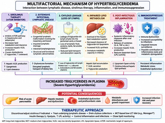
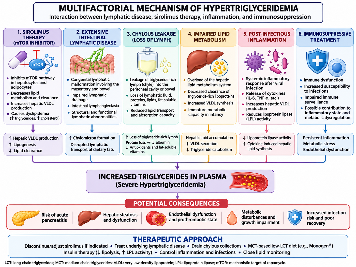
Intercurrent infection is a further plausible trigger: inflammation can modify lipid synthesis and clearance [44]. In our patient, the temporal association with viral infection provides a plausible additional trigger. Thus, the metabolic deterioration may have resulted from a “multiple-hit” mechanism involving sirolimus exposure, diffuse lymphatic dysfunction, chylous leakage, infection-associated inflammation, and nutritional/metabolic adaptation. Figure 7 therefore illustrates conceptual, literature-informed possibilities rather than a demonstrated causal pathway in this patient. This distinction preserves the educational value of a multifactorial model while avoiding unsupported conclusions.
Figure 7.
Conceptual model of potential contributors to hypertriglyceridemia. Observed: sirolimus exposure, recent viral infection, marked circulating triglyceride elevation, extensive lymphatic malformation, and previously milky-appearing peritoneal fluid. Unverified hypotheses: intestinal lymphangiectasia, persistent chylous leakage, reduced lipoprotein lipase activity, increased hepatic VLDL output, protein loss, and endothelial dysfunction.
3.5. Nutritional and Pharmacological Solutions to Hypertriglyceridemia
The management of severe hypertriglyceridemia during infancy remains challenging because evidence-based treatment protocols are limited and therapeutic recommendations are frequently extrapolated from older pediatric or adult populations.
Insulin was introduced as an acute metabolic intervention to enhance triglyceride clearance. Insulin stimulates lipoprotein lipase (LPL) activity, thereby promoting the hydrolysis of triglycerides contained in circulating chylomicrons and very-low-density lipoproteins and facilitating their subsequent clearance [45]. This mechanism provides a physiological rationale for the use of intravenous insulin in selected patients with severe or rapidly progressive hypertriglyceridemia. However, insulin infusion is not currently recommended as a routine treatment for severe hypertriglyceridemia in infants, and pediatric-specific evidence remains limited. Available data are derived predominantly from case reports, small observational studies, and extrapolation from other clinical settings, particularly diabetes-associated hypertriglyceridemia and hypertriglyceridemia-associated pancreatitis [46]. Nevertheless, in selected patients with extreme triglyceride elevations, particularly when nutritional measures alone are insufficient, insulin may be considered as an individualized short-term intervention, with careful monitoring of blood glucose and electrolytes. In our patient, the marked decline in triglyceride concentrations following insulin infusion supports its potential usefulness as an acute metabolic rescue strategy, although this response should not be interpreted as evidence that insulin is superior to dietary intervention alone.
Milky-appearing peritoneal fluid was observed during surgery and may indicate lymphatic leakage [42], but biochemical testing of the fluid was unavailable. No fluid studies established that this process persisted during the January metabolic episode. It would therefore be inappropriate to infer a demonstrated causal link between that surgical observation and circulating hypertriglyceridemia several months later.
Chylous leakage has been described in association with complex lymphatic malformations and is generally considered a marker of more extensive lymphatic dysfunction [42]. In contrast to localized macrocystic lesions, which may remain clinically silent or produce primarily mass-effect symptoms, diffuse lymphatic abnormalities may interfere with normal lymph transport and lead to chylous ascites, chylothorax, protein loss, lymphopenia, and nutritional deficiencies [47]. The presence of milky peritoneal fluid in our patient therefore provides additional evidence that the underlying disease was not limited to discrete cystic structures but involved the functional lymphatic circulation.
The nutritional management of such patients is based on the physiology of intestinal lymphatic transport. Long-chain triglycerides are incorporated into chylomicrons and transported predominantly through intestinal lymphatics, whereas medium-chain triglycerides are absorbed more directly into the portal circulation [48]. Consequently, replacing long-chain fat with MCTs can reduce intestinal lymphatic flow and may facilitate resolution of chylous leakage [49]. This principle has been used for decades in the management of congenital chylous disorders. For example, a comparative report in infants with congenital chyloperitoneum demonstrated a reduction in lymphatic drainage with MCT-based nutrition, although total parenteral nutrition was able to reduce flow further [50]. More recent neonatal reports have similarly described successful resolution of chylous effusions following introduction of MCT-enriched formulas [51].
An important feature of our case is that nutritional therapy subsequently became relevant not only to lymphatic leakage but also to severe hypertriglyceridemia. This creates a clinically complex therapeutic balance. On the one hand, adequate energy and fat intake are essential for growth and development; on the other, excessive delivery of long-chain triglycerides may increase lymphatic flow, while systemic metabolic disturbances may require temporary restriction of lipid exposure. The nutritional strategy must therefore be individualized according to both the lymphatic and metabolic phenotype.
Taken together, the intraoperative appearance, diffuse mesenteric disease, and clinical rationale for MCT-based nutrition suggest a possible role of lymphatic dysfunction in treatment planning; they do not establish that lymphatic leakage caused the subsequent plasma triglyceride elevation.
3.6. Clinical Implications and Future Perspectives
The present case illustrates the complex and dynamic nature of extensive mesenteric lymphatic malformation in infancy. Distinguishing features include antenatal recognition, extensive neonatal disease, subsequent multifocal cystic findings, clinically suspected chylous leakage at surgery, systemic drug exposure, and severe hypertriglyceridemia.
The most clinically relevant implication is that severe hypertriglyceridemia should be interpreted in the context of the underlying lymphatic phenotype. Severe hypertriglyceridemia in a patient receiving sirolimus may initially suggest drug toxicity, but persistent abnormalities after discontinuation should prompt consideration of additional mechanisms, including chylous leakage, altered intestinal lymphatic lipid transport, systemic inflammation, and nutritional factors. This distinction is particularly important because discontinuation of sirolimus may improve drug-related dyslipidemia but may not immediately correct a metabolic disorder driven by persistent lymphatic dysfunction.
Nutritional management should be integrated into the treatment pathway for extensive lymphatic malformations. MCT-based nutrition is physiologically attractive when lymphatic leakage is suspected, but adequate energy, essential fatty acids, and fat-soluble vitamins must be ensured through repeated dietetic assessment. In this child, improvement occurred during combined care and cannot be assigned exclusively to the MCT formula.
The presented case provides a useful framework for clinical practice. In neonates with extensive lymphatic malformations, management should extend beyond anatomical control of the lesion and include systematic surveillance of lymphatic leakage, nutritional status, triglycerides, liver function, infection, and drug toxicity. This may be particularly important during sirolimus therapy, given the established association between treatment and dyslipidemia.
Future studies should focus on identifying clinical and molecular predictors of response to systemic therapy, particularly in diffuse visceral lymphatic disease. The increasing recognition of abnormalities within the PI3K/AKT/mTOR pathway may also allow more precise molecular stratification and targeted treatment in selected patients [23,41]. In addition, prospective studies are needed to establish standardized approaches to nutritional management of chylous leakage and to determine whether early MCT-based therapy can prevent some of the metabolic consequences associated with persistent lymphatic dysfunction.
Overall, this case highlights extensive mesenteric lymphatic disease with substantial clinical and metabolic management needs, without establishing a systemic lymphatic developmental disorder. Long-term individualized follow-up and careful monitoring of drug toxicity and lipid status remain necessary.
4. Conclusions
The present case demonstrates the complexity of congenital mesenteric lymphatic malformation with recurrent or persistent intra-abdominal cystic disease and severe hypertriglyceridemia during infancy. The lipid abnormality was temporally associated with prior sirolimus exposure and may have had additional triggers, but causation remains uncertain. Combined supportive, pharmacological, and nutritional management was followed by marked lipid improvement.
Milky peritoneal fluid raised suspicion of chylous leakage at the earlier operation, without biochemical confirmation or proof of persistence at the later metabolic admission. The case supports coordinated surgical and metabolic care rather than any single established mechanism linking lymphatic disease to plasma triglyceride elevation.
Follow-up lipid testing, growth and dietary surveillance, and reassessment of lymphatic disease and treatment-related adverse effects are essential.
Author Contributions
Conceptualization, E.-L.S.; E.Ț.; L.M.T. and G.P.; methodology, G.P., P.P., A.I. and L.M.T.; software, A.I. and P.P.; validation, J.B. and S.Ș.; formal analysis, L.I.B.; investigation, L.M.T., E.-L.S., L.I.B., E.Ț., G.P., J.B. and S.Ș.; data curation, E.-L.S. and G.P.; writing—original draft preparation, L.M.T., E.-L.S., L.I.B., E.Ț., G.P., A.I., P.P. and J.B.; writing—review and editing, E.Ț., S.Ș., G.P. and E.-L.S.; visualization, L.M.T.; supervision, E.Ț. All authors have read and agreed to the published version of the manuscript.
Funding
This research received no external funding.
Institutional Review Board Statement
The study was conducted in accordance with the Declaration of Helsinki and approved by the Ethics Committee of Sfânta Maria Emergency Hospital for Children, Iasi, Romania (No. 11918/8 April 2026).
Informed Consent Statement
Written informed consent has been obtained from the patient’s legal guardian for the publication of this case report, including the clinical information and accompanying clinical photographs and images.
Data Availability Statement
The original contributions presented in this study are included in the article. Further inquiries can be directed to the corresponding author.
Conflicts of Interest
The authors declare no conflicts of interest.
References
- Sun, J.; Yang, C. A Retrospective Study about Lymphangioma of the Mesentery: An Unusual Cause of Bowel Volvulus. Medicine 2025, 104, e46864. [Google Scholar] [CrossRef] [Scilit]
- Li, J.; Luo, L.; Liu, Y.; Li, W.; Wei, X. Mesenteric Lymphangioma in Childhood: A Case Report and Narrative Literature Review. Front. Oncol. 2025, 15, 1541445. [Google Scholar] [CrossRef] [Scilit]
- Lal, A.; Gupta, P.; Singhal, M.; Sinha, S.K.; Lal, S.; Rana, S.; Khandelwal, N. Abdominal Lymphatic Malformation: Spectrum of Imaging Findings. Indian J. Radiol. Imaging 2016, 26, 423–428. [Google Scholar] [CrossRef] [Scilit]
- Gunadi; Kashogi, G.; Prasetya, D.; Fauzi, A.R.; Daryanto, E.; Dwihantoro, A. Pediatric Patients with Mesenteric Cystic Lymphangioma. Int. J. Surg. Case Rep. 2019, 64, 89–93. [Google Scholar] [CrossRef] [Scilit]
- Francavilla, M.L.; White, C.L.; Oliveri, B.; Lee, E.Y.; Restrepo, R. Intraabdominal Lymphatic Malformations: Pearls and Pitfalls of Diagnosis and Differential Diagnoses in Pediatric Patients. AJR Am. J. Roentgenol. 2017, 208, 637–649. [Google Scholar] [CrossRef] [Scilit]
- Nzelu, D.; Panayotidis, I.; Smith, G.D.; Pandya, P. Fetal Cystic Lymphatic Malformations. J. Ultrasound Med. 2024, 43, 2327–2337. [Google Scholar] [CrossRef] [Scilit]
- Merino-Mateo, L.; Morante Valverde, R.; Redondo Sedano, J.V.; Benavent Gordo, M.I.; Gómez Fraile, A. Mesenteric Lymphatic Malformation: A Rare Cause of an Acute Abdomen. An. Pediatr. (Engl. Ed.) 2020, 92, 49–51. [Google Scholar] [CrossRef] [Scilit]
- Stimac, G.; Seifarth, F.G.; Drews, J.D.; Bacaj, P.; Wehrli, L.A. Mesenteric Cystic Lymphangioma Causing Small Bowel Obstruction in a Neonate. Cureus 2025, 17, e91130. [Google Scholar] [CrossRef] [Scilit]
- Boubekri, N.; Akammar, A.; Ouazzani, H.; Chaouche, I.; El Bouardi, N.; Haloua, M.; Alami, B.; Lamrani Alaoui, M.Y.; Maaroufi, M.; Boubbou, M. An Unusual Cause of Ileal Volvulus, a Giant Mesenteric Cystic Lymphangioma. Radiol. Case Rep. 2025, 20, 5281–5285. [Google Scholar] [CrossRef] [Scilit]
- Llapur, E.C.; Alvarez, J.M.L.; Quevedo, O.P.; Lemaur, M.V.; Nesic, S.P.; Urgellés, X.G. Cystic Lymphangioma as a Cause of Massive Abdominal Hemorrhage. Case Rep. Crit. Care 2011, 2011, 492564. [Google Scholar] [CrossRef] [Scilit]
- Gonzalez-Moneo, M.J.; Corisco, M.; Olivera, R.; Salgado, P. Clinical Insights into Abdominal Cystic Lesions: A Spotlight on Differential Diagnoses and Prognosis through a Case Report. J. Med. Ultrasound 2025, 33, 165–167. [Google Scholar] [CrossRef] [Scilit]
- Kim, S.-H.; Kim, H.-Y.; Lee, C.; Min, H.S.; Jung, S.-E. Clinical Features of Mesenteric Lymphatic Malformation in Children. J. Pediatr. Surg. 2016, 51, 582–587. [Google Scholar] [CrossRef] [Scilit]
- Rohilla, M.; Siwatch, S.; Malik, M.A.; Muthyala, T.; Samujh, R.; Kannan, N. Fetal Abdominal Cystic Lesion: A Diagnostic Dilemma and Prognostic Challenge-Report of Two Cases of Mesentric Lymphangioma with Review of Literature. J. Women’s Reprod. Health 2021, 2, 1–6. [Google Scholar] [CrossRef] [Scilit]
- Lee, S.; Luhar, A.; Miller, J. Lymphatic Malformations: Review of Diagnosis and Management for the Interventional Radiologist. Semin. Intervent. Radiol. 2024, 41, 389–403. [Google Scholar] [CrossRef] [Scilit]
- Liu, Y.-P.; Huang, Y.-L.; Tsai, P.-S.; Lin, D.-C.; Chen, C.-P. Prenatal Diagnosis of Abdominal Lymphatic Malformations. Taiwan J. Obstet. Gynecol. 2021, 60, 13–19. [Google Scholar] [CrossRef] [Scilit]
- De Jong, R.; Fordham, T. Ex Utero Intrapartum Treatment (EXIT) Procedure; StatPearls Publishing: Treasure Island, FL, USA, 2025. [Google Scholar]
- Reynolds, E.; Rosas, D.; Byrd, E.; Farmer, D.L. In Utero Treatment of Lymphatic Malformations: A Narrative Review of the Literature, Considerations, and Future Directions. Lymphatics 2025, 3, 46. [Google Scholar] [CrossRef] [Scilit]
- Yang, C.; Qiu, T.; Yang, M.; Zhou, J.; Gong, X.; Yang, K.; Zhang, Z.; Lan, Y.; Zhang, X.; Zhou, Z.; et al. Clinical Characteristics and Risk Factors for Acute Abdomen in Patients with Abdominal Lymphatic Malformations. J. Vasc. Surg. Venous Lymphat. Disord. 2025, 13, 101969. [Google Scholar] [CrossRef] [Scilit]
- Loo, S.; Anand, A.J.; Rajadurai, V.S.; Chandran, S. Management of Large Congenital Chylous Ascites in a Preterm Infant: Fetal and Neonatal Interventions. BMJ Case Rep. 2020, 13, e235849. [Google Scholar] [CrossRef] [Scilit]
- Wu, W.; Mo, J.; Tan, K.; Chen, X.; Xu, W.; Liu, J.; Lv, Z. Misdiagnosis of Scrotal and Retroperitoneal Lymphangioma in Children. BMC Pediatr. 2023, 23, 551. [Google Scholar] [CrossRef] [Scilit]
- Ben Khalifa, M.; Aouadi, S.; Missaoui, W.; Elguedr, W.; Maatouk, M.; Boudokhane, M. Partial Resection of a Giant Intra-Abdominal Cystic Lymphangioma with No Recurrence for 10 Years: A Case Report. Int. J. Surg. Open 2025, 63, 274–277. [Google Scholar] [CrossRef] [Scilit]
- Yan, J.; Xie, C.; Chen, Y. Surgical Treatment of Mesenteric Lymphatic Malformations in Children: An Observational Cohort Study. J. Pediatr. Surg. 2023, 58, 1762–1769. [Google Scholar] [CrossRef] [Scilit]
- Petkova, M.; Ferby, I.; Mäkinen, T. Lymphatic Malformations: Mechanistic Insights and Evolving Therapeutic Frontiers. J. Clin. Investig. 2024, 134, e172844. [Google Scholar] [CrossRef] [Scilit]
- Miceli, A.; Stewart, K.M. Lymphangioma; StatPearls Publishing: Treasure Island, FL, USA, 2025. [Google Scholar]
- Yan, J.; Fu, Y.; Liu, S.; Bai, Y.; Chen, Y. Mesenteric and Omental Lymphatic Malformations in Children: Seven-Year Surgical Experience from Two Centers in China. BMC Pediatr. 2024, 24, 360. [Google Scholar] [CrossRef] [Scilit]
- Ghaffarpour, N.; Baselga, E.; Boon, L.M.; Diociaiuti, A.; Dompmartin, A.; Dvorakova, V.; El Hachem, M.; Gasparella, P.; Haxhija, E.; Kyrklund, K.; et al. The VASCERN-VASCA Working Group Diagnostic and Management Pathways for Lymphatic Malformations. Eur. J. Med. Genet. 2022, 65, 104637. [Google Scholar] [CrossRef] [Scilit]
- Huerta, C.T.; Beres, A.L.; Englum, B.R.; Gonzalez, K.; Levene, T.; Wakeman, D.; Yousef, Y.; Gulack, B.C.; Chang, H.L.; Christison-Lagay, E.R.; et al. Management and Outcomes of Pediatric Lymphatic Malformations: A Systematic Review from the APSA Outcomes and Evidence-Based Practice Committee. J. Pediatr. Surg. 2024, 59, 161589. [Google Scholar] [CrossRef] [Scilit]
- Hollman, D.; Cheema, H.; Yu, A.C.; Mahinpey, N.; AlZahrani, F. Medical Therapies for Pediatric Lymphatic Malformations: A Systematic Review. J. Cutan. Med. Surg. 2026, 30, 259–264. [Google Scholar] [CrossRef] [Scilit]
- Ozeki, M.; Asada, R.; Saito, A.M.; Hashimoto, H.; Fujimura, T.; Kuroda, T.; Ueno, S.; Watanabe, S.; Nosaka, S.; Miyasaka, M. Efficacy and Safety of Sirolimus Treatment for Intractable Lymphatic Anomalies: A Study Protocol for an Open-Label, Single-Arm, Multicenter, Prospective Study (SILA). Regen. Ther. 2019, 10, 84–91. [Google Scholar] [CrossRef] [Scilit]
- Wiegand, S.; Dietz, A.; Wichmann, G. Efficacy of Sirolimus in Children with Lymphatic Malformations of the Head and Neck. Eur. Arch. Otorhinolaryngol. 2022, 279, 3801–3810. [Google Scholar] [CrossRef] [Scilit]
- Neirotti, A.; Barat, V.; Coppo, P.; La Selva, R.; Manicone, R.; Cotti, R.; Sensini, M.; Mussa, A.; Gatto, M.; Farri, F.; et al. Therapy with Sirolimus in Vascular Anomalies: The Experience of Two Italian Centers on 14 Pediatric Patients. Front. Pediatr. 2024, 12, 1434493. [Google Scholar] [CrossRef] [Scilit]
- Amodeo, I.; Colnaghi, M.; Raffaeli, G.; Cavallaro, G.; Ciralli, F.; Gangi, S.; Leva, E.; Pignataro, L.; Borzani, I.; Pugni, L. The Use of Sirolimus in the Treatment of Giant Cystic Lymphangioma. Medicine 2017, 96, e8871. [Google Scholar] [CrossRef] [Scilit]
- Wu, J.K.; Hooper, E.D.; Laifer-Narin, S.L.; Simpson, L.L.; Kandel, J.; Shawber, C.J. On Behalf of the Lymphatics Work Group at CUMC. Initial Experience with Propranolol Treatment of Lymphatic Anomalies: A Case Series. Pediatrics 2016, 138, e20154545. [Google Scholar] [CrossRef] [Scilit]
- Cîrstoveanu, C.; Bizubac, A.; Mustea, C.; Ștefan Istrate-Bârzan, A.; Sfrijan, D.; Marcu, V.; Iozsa, D.-A.; Spătaru, R.-I. Antiproliferative Therapy with Sirolimus and Propranolol for Congenital Vascular Anomalies in Newborns (Case Reports). Exp. Ther. Med. 2021, 22, 1097. [Google Scholar] [CrossRef] [Scilit]
- Zhang, Z.; Li, Y.; Zhang, G.; Yang, K.; Qiu, T.; Zhou, J.; Gong, X.; Ji, Y. Safety Evaluation of Oral Sirolimus in the Treatment of Childhood Diseases: A Systematic Review. Children 2022, 9, 1295. [Google Scholar] [CrossRef] [Scilit]
- Clark, H.; Schulte, R.; Haggstrom, A.N. Severe Hypertriglyceridemia Following Sirolimus Use in an Infant. Pediatr. Dermatol. 2021, 38, 1581–1582. [Google Scholar] [CrossRef] [Scilit]
- Spoiala, E.L.; Cinteza, E.; Vatasescu, R.; Vlaiculescu, M.V.; Moisa, S.M. Statins-beyond Their Use in Hypercholesterolemia: Focus on the Pediatric Population. Children 2024, 11, 117. [Google Scholar] [CrossRef] [Scilit]
- Perez, V.; Faust, A.C.; Taburyanskaya, M.; Patil, R.A.; Ortegon, A. Effectiveness of an Intravenous Insulin–Based Treatment Protocol for the Management of Hypertriglyceridemia-Associated Acute Pancreatitis. J. Pharm. Technol. 2023, 39, 55–61. [Google Scholar] [CrossRef] [Scilit]
- Geeurickx, M.; Labarque, V. A Narrative Review of the Role of Sirolimus in the Treatment of Congenital Vascular Malformations. J. Vasc. Surg. Venous Lymphat. Disord. 2021, 9, 1321–1333. [Google Scholar] [CrossRef] [Scilit]
- Butnariu, L.I.; Gorduza, E.V.; Țarcă, E.; Pânzaru, M.-C.; Popa, S.; Stoleriu, S.; Lupu, V.V.; Lupu, A.; Cojocaru, E.; Trandafir, L.M.; et al. Current Data and New Insights into the Genetic Factors of Atherogenic Dyslipidemia Associated with Metabolic Syndrome. Diagnostics 2023, 13, 2348. [Google Scholar] [CrossRef] [Scilit]
- Țarcă, E.; Cojocaru, E.; Butnariu, L.I.; Roșu, S.T.; Luca, A.C.; Susanu, S.; Lupu, V.V.; Roșu, E.V.; Popovici, P.; Țarcă, V. Sirolimus Treatment for Complex Fetal and Neonatal Vascular Anomalies: Emerging Evidence and Safety Considerations. J. Clin. Med. 2026, 15, 3739. [Google Scholar] [CrossRef] [Scilit]
- Trenor, C.C., III; Chaudry, G. Complex Lymphatic Anomalies. Semin. Pediatr. Surg. 2014, 23, 186–190. [Google Scholar] [CrossRef] [Scilit]
- Kim, M.; Hong, K.T.; Park, H.J.; Kim, B.K.; Choi, J.Y.; Kim, H.-Y.; Kang, H.J. Clinical Effectiveness and Safety of Sirolimus in Pediatric Patients with Complex Vascular Anomalies: Necessitating Personalized and Comprehensive Approaches. Front. Pediatr. 2023, 11, 1304133. [Google Scholar] [CrossRef] [Scilit]
- Feingold, K.R.; Grunfeld, C. Endotext [Internet]; updated 18 June 2025; MDText.com, Inc.: South Dartmouth, MA, USA, 2000. Available online: https://www.ncbi.nlm.nih.gov/books/NBK326741/ (accessed on 15 November 2026).
- De La Torre, A.; Hamilton, L.; Wilson, D.P. Management of Hospitalized Children with Severe Hypertriglyceridemia. In Endotext; Feingold, K.R., Adler, R.A., Ahmed, S.F., Anawalt, B., Blackman, M.R., Chrousos, G., Corpas, E., de Herder, W.W., Dhatariya, K., Dungan, K., Eds.; MDText.com, Inc.: South Dartmouth, MA, USA, 2026. Available online: https://www.ncbi.nlm.nih.gov/books/NBK553648/ (accessed on 11 November 2026).
- Spoială, E.-L.; Oltean, C.; Vasiliu, I.; Paduraru, G.; Danilă, D.-C.; Ghiga, G.; Țugui, M.; Butnariu, L.I.; Cojocaru, E.; Trandafir, L.M. Multimodal Management of Extreme Hypertriglyceridemia in a Child with Recurrent Pancreatitis: Clinical Challenges and Solutions. J. Clin. Med. 2026, 15, 636. [Google Scholar] [CrossRef] [Scilit]
- Kothandaraman, V.; Wisener, J.; Helgerson, P.W. Complex Lymphatic Anomalies: Challenging Diagnostic Considerations. Clin. Case Rep. 2024, 12, e8370. [Google Scholar] [CrossRef] [Scilit]
- Kiela, P.R.; Ghishan, F.K. Physiology of Intestinal Absorption and Secretion. Best Pract. Res. Clin. Gastroenterol. 2016, 30, 145–159. [Google Scholar] [CrossRef] [Scilit]
- Wang, K.; Xiao, J.; Li, L.; Li, X.; Yang, Y.; Liu, Z.; Jiang, J. The Application of a Medium-Chain Fatty Diet and Enteral Nutrition in Post-Operative Chylous Leakage: Analysis of 63 Patients. Front. Nutr. 2023, 10, 1128864. [Google Scholar] [CrossRef] [Scilit]
- Kroczek, R.A. Congenital Chyloperitoneum: Direct Comparison of Medium-Chain Triglyceride Treatment with Total Parenteral Nutrition. Eur. J. Pediatr. 1985, 144, 77–79. [Google Scholar] [CrossRef] [Scilit]
- Zeng, W.; Hu, Y.; Feng, J.; Luo, X. Chylous Ascites Following Repair of Total Anomalous Pulmonary Venous Connection Coexisting with a Persistent Left Superior Vena Cava in a Neonate: A Case Report. Transl. Pediatr. 2021, 10, 188–193. [Google Scholar] [CrossRef] [Scilit]

Disclaimer/Publisher’s Note: The statements, opinions and data contained in all publications are solely those of the individual author(s) and contributor(s) and not of MDPI and/or the editor(s). MDPI and/or the editor(s) disclaim responsibility for any injury to people or property resulting from any ideas, methods, instructions or products referred to in the content. |
© 2026 by the authors. Licensee MDPI, Basel, Switzerland. This article is an open access article distributed under the terms and conditions of the Creative Commons Attribution (CC BY) license.