


Abstract
Background/Objectives: Neurofibromatosis type 1 (NF1) has variable clinical expression and marked allelic heterogeneity. This study assessed the diagnostic yield of molecular testing in a regional Romanian cohort with an initial clinical diagnosis of NF1 and described the clinical features of patients with NF1 molecular findings. Methods: This retrospective study included 77 patients evaluated between 1990 and 2025. We reviewed the use and results of next-generation sequencing (NGS), multiplex ligation-dependent probe amplification (MLPA), and fluorescence in situ hybridization (FISH). Results: Of the 77 patients, 42 (54.5%) underwent molecular testing. NGS was performed in 36 patients, MLPA in 15, and FISH in 7. NGS identified pathogenic or likely pathogenic NF1 sequence variants in 30 of 36 sequenced patients (83.3%), representing 30 of 42 tested patients (71.4%). In one additional patient with a negative NGS result, MLPA detected a suspected intragenic NF1 deletion that was not independently confirmed. Including this finding, the observed yield was 31/36 (86.1%) among sequenced patients and 31/42 (73.8%) among all tested patients. FISH detected no 17q11.2 microdeletions. One patient had an NF1 variant of uncertain significance. Each of the three recurrent NF1 variants was observed within a single family. A homozygous pathogenic MSH6 variant led to the reclassification of one patient as having constitutional mismatch repair deficiency (CMMRD). Conclusions: NGS identified a pathogenic or likely pathogenic NF1 variant in 30 of the 36 sequenced patients. Because access to testing and the methods used varied across the cohort, this proportion applies to the sequenced subgroup. In one child initially diagnosed with NF1, molecular testing established a diagnosis of CMMRD.
1. Introduction
Neurofibromatosis type 1 (NF1) is a common autosomal dominant neurocutaneous disorder. Its clinical expression is highly variable. It affects many organ systems and increases the risk of benign and malignant tumors. NF1 is caused by pathogenic variants in the NF1 gene. This gene encodes neurofibromin, a tumor suppressor that negatively regulates the RAS/MAPK pathway. Loss of neurofibromin function leads to abnormal cell growth and tumor predisposition. About half of affected individuals carry a de novo pathogenic variant [1,2].
NF1 diagnosis has traditionally relied on clinical criteria. These include multiple café-au-lait macules (CALMs), axillary or inguinal freckling, cutaneous or plexiform neurofibromas, optic pathway glioma, Lisch nodules, distinctive bone lesions and an affected first-degree relative. Clinical diagnosis can be difficult in young children, in patients with incomplete features, in mosaic forms and in overlapping pigmentary disorders such as Legius syndrome. The revised diagnostic criteria published in 2021 included genetic testing. They also defined criteria to separate NF1 from Legius syndrome [2,3].
Molecular confirmation is increasingly important in NF1 care. It can support early diagnosis and clarify uncertain phenotypes. It also helps genetic counseling and the testing of relatives. However, molecular diagnosis of NF1 is complex. The NF1 gene is large, has no single mutation hotspot and shows broad allelic heterogeneity. Next-generation sequencing (NGS) is central to current testing. When deletions, duplications or larger rearrangements are suspected, NGS may need to be combined with multiplex ligation-dependent probe amplification (MLPA) or fluorescence in situ hybridization (FISH) [4,5].
Molecular results must be interpreted with care. Pathogenic and likely pathogenic variants can confirm the diagnosis. A variant of uncertain significance (VUS) should not be used alone for clinical decisions. This is stated in the standards of the American College of Medical Genetics and Genomics and the Association for Molecular Pathology (ACMG/AMP). These standards also recommend reclassifying such variants when new evidence becomes available. In NF1, clinical evaluation, family history, segregation analysis and follow-up remain essential for interpretation [6].
Genotype–phenotype correlations in NF1 are limited for most variants. Expressivity is highly variable, even within the same family. Some correlations have been described for specific variants or deletions. In routine practice, however, the molecular result alone does not predict tumor burden, neurodevelopmental problems, disease progression or treatment needs. Real-world studies that combine molecular and clinical data are therefore useful [7,8].
The treatment of NF1 has changed with the arrival of targeted therapy for plexiform neurofibromas. These tumors are a major cause of morbidity. They may cause pain, disfigurement, functional impairment and nerve compression, and they are often difficult to remove surgically. Selumetinib, a MEK inhibitor, is now an important option for selected patients with symptomatic, inoperable plexiform neurofibromas. It was first approved for children. In 2025, the FDA also approved it for adults [9].
Molecular testing can help clarify the diagnosis in patients with overlapping or incomplete clinical features. However, access to testing remains uneven [10], as we also observed in patients with other neurocutaneous disorders evaluated at our center [11]. We previously described the clinical phenotype and multisystem burden of this cohort, without molecular data [12], and 19 of our patients were included in a national multicenter study of the molecular spectrum and genotype–phenotype correlations of NF1 in Romanian patients, currently under review [13]. Here, we report the molecular findings of the whole regional cohort together with the clinical features of the molecularly characterized patients.
The aim of this study was to evaluate the molecular diagnostic yield of NGS, MLPA and FISH in a retrospective regional cohort of patients with NF1. We assessed testing coverage, the use of each method, and the type and classification of NF1-related findings. We also described familial segregation and genotype–phenotype observations. The clinical and treatment data of the whole cohort have been reported previously [12] and are only summarized here.
2. Materials and Methods
2.1. Study Design and Setting
This was a retrospective observational cohort study of patients diagnosed with NF1. Data were taken from a clinical and molecular database. The database included demographic, phenotypic, genetic, treatment and follow-up variables. We evaluated the molecular diagnostic yield of NF1 testing. We also described the clinical phenotype of the molecularly characterized patients. Patients were evaluated at the Regional Center of Medical Genetics Bihor (CRGM Bihor), Bihor County Emergency Clinical Hospital, Oradea, between 1990 and 2025. The clinical characteristics of this cohort have been described previously [12]. The present study focuses on molecular testing and its clinical correlates. The previous report did not include molecular data. Of the 42 patients with molecular testing, 19 were also included in the multicenter study by Toc et al. [13]. Of the 23 tested patients who were not included in the multicenter study, 13 had NF1 molecular findings, including one patient with a suspected intragenic deletion detected by MLPA that was not independently confirmed. The complete testing pathway of the regional cohort (NGS, MLPA and FISH), the diagnostic yield calculated for this cohort and the integration of molecular results with the clinical data are also new.
Specifically, we analyzed testing coverage, the methods used, NF1 variant classification, zygosity, and exploratory genotype–phenotype observations.
2.2. Study Population
The cohort included 77 consecutive patients with clinically documented NF1. Inclusion required a recorded NF1 diagnosis and clinical data in the database. For the molecular analysis, patients were grouped by whether molecular testing was documented. Patients without recorded NGS, MLPA or FISH testing stayed in the cohort. They were classed as not tested, not as molecularly negative. All patients were clinically evaluated and followed at CRGM Bihor. Because NGS is not available at our center, sequencing was performed in collaborating laboratories. Nineteen of our patients were sequenced with TruSight One at the Center of Genomic Medicine, Timișoara, and were also included in a multicenter Romanian study [13]. One patient with an initial clinical diagnosis of NF1 was reclassified as constitutional mismatch repair deficiency (CMMRD) after molecular testing and was kept in the cohort, because the study population was defined by the clinical diagnosis of NF1 at referral (yields without this patient are given in Section 3.2). Patients were diagnosed according to the clinical criteria in use at the time of evaluation. We interpreted the molecular findings in the context of the 2021 revised international diagnostic criteria [3]. The historical cohort was not systematically reassessed against these criteria.
Molecular testing was not performed according to a uniform protocol. All patients in the cohort were invited for molecular testing, but whether testing was performed depended mainly on access. Genetic testing was not free of charge for most patients, and free testing was available only for some of them, within the multicenter study coordinated by the Center of Genomic Medicine, Timișoara, where testing was funded through the National Plan for Rare Diseases or by the patient association “Noi Pentru Ei” [13]; the other patients were tested only if they could cover the cost. The choice of method depended on the tests available at the time and on the clinical question. FISH, available at CRGM Bihor, was used mainly to screen for 17q11.2 microdeletions. MLPA was used when a deletion was suspected, after a negative or inconclusive NGS result, or when sequencing was not available. NGS, performed in collaborating laboratories, was used as the main test. The two deceased patients in the cohort [12] did not have molecular testing.
2.3. Molecular Testing
2.3.1. Next-Generation Sequencing
DNA was extracted from peripheral blood lymphocytes by standard methods. NGS was performed in three laboratories. Most patients were tested with the TruSight One panel (Illumina Inc., San Diego, CA, USA), which covers 4813 genes. Sequencing was done on a MiSeq platform (Illumina) at the Center of Genomic Medicine, “Victor Babes” University of Medicine and Pharmacy Timisoara [14], or at the Human Genomics Laboratory, University of Medicine and Pharmacy of Craiova. The other patients were tested with a targeted neurofibromatosis panel at Blueprint Genetics (Espoo, Finland). The TruSight One analysis did not include copy-number analysis. Therefore, NF1-specific MLPA was used to detect exon-level deletions or duplications.
The Blueprint Genetics Neurofibromatosis Panel Plus Analysis includes sequence analysis and copy number variation analysis of the following genes: KIT, KITLG, LZTR1, NF1*, NF2, PTPN11, RAF1, SMARCB1 and SPRED1. This panel targets protein-coding exons, exon–intron boundaries (± 20 bps) and selected non-coding, deep intronic variants.
Sequencing followed the manufacturer’s protocols. Reads were aligned to the human reference genome (GRCh37). Single nucleotide variants (SNVs) and small insertions/deletions (indels) were called with validated bioinformatic pipelines. The analysis covered coding regions, exon–intron boundaries and flanking intronic sequences. Selected non-coding regions were evaluated when included in the assay. Variants were interpreted using ClinVar, OMIM, HGMD and population frequency databases. They were classified according to the ACMG/AMP guidelines.
2.3.2. MLPA Analysis
MLPA analysis was performed at the Center for Genomic Medicine of the “George Emil Palade” University of Medicine, Pharmacy, Sciences and Technology in Târgu Mureș and at the Human Genomics Laboratory of the University of Medicine and Pharmacy in Craiova. MLPA was performed in 15 patients. It was used after a negative or inconclusive NGS result, when a deletion was suspected, or in five patients without sequencing. Genomic DNA from peripheral blood leukocytes was analyzed with the SALSA MLPA Probemix P081 and P082 NF1 kits (MRC Holland, Amsterdam, The Netherlands), following the manufacturer’s protocol. Fragments were separated by capillary electrophoresis and analyzed with Coffalyser.Net. Results were interpreted together with the clinical and other molecular data.
2.3.3. FISH Analysis
FISH was performed at CRGM Bihor in seven patients, mainly to screen for 17q11.2 microdeletions. Commercial locus-specific probes targeting the NF1 region were used according to the manufacturer’s protocol. Hybridization signals were evaluated by fluorescence microscopy, and the results were interpreted together with the clinical and molecular findings.
2.4. Statistical Analysis
Data analysis was performed using the open-source software R version 4.6.2, Microsoft Excel for Mac version 16.115.1 (build 26111533), Microsoft Corporation, Redmond, WA, USA. Categorical variables were expressed as absolute frequencies and percentages. Continuous variables were summarized using the mean ± standard deviation or median and the interquartile range depending on the data distribution.
2.5. Ethical Considerations
The study was conducted in accordance with the Declaration of Helsinki and was approved by the Ethics Committee of the University of Oradea (approval no. 33, 30 July 2025). Written informed consent for clinical genetic testing was obtained from the tested patients or their legal representatives. The genetic testing consent form included a separate option to authorize the use of anonymized or pseudonymized data for research. For patients who did not undergo genetic testing, the study used only data already recorded during routine clinical care. No additional procedures were performed for research purposes.
2.6. Use of Generative AI
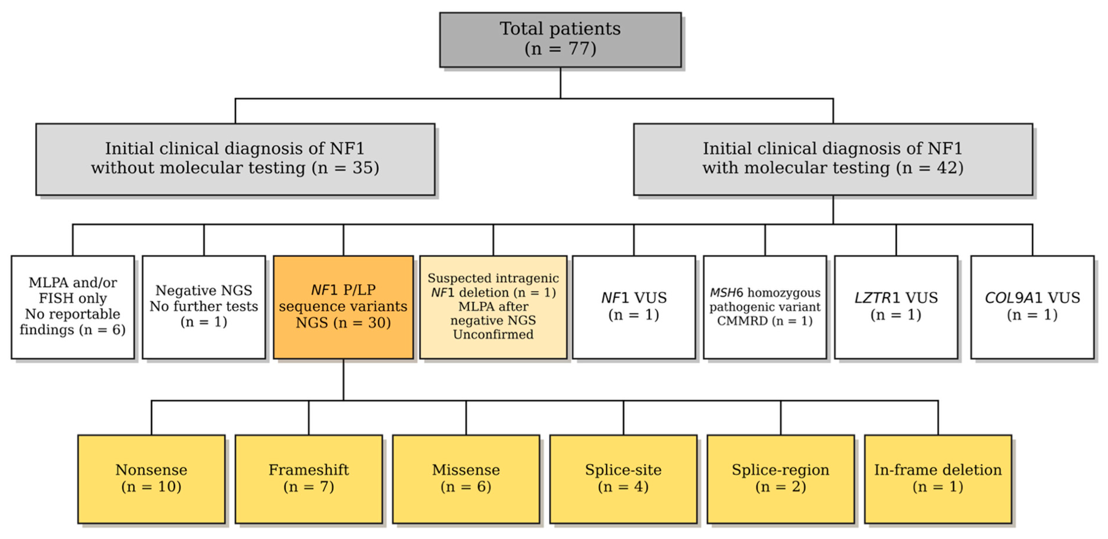
During manuscript preparation, the authors used ChatGPT (GPT-6.1, OpenAI) and Claude Sonnet 5.5 (Anthropic, San Francisco, CA, USA) to edit the language, improve the structure of selected paragraphs, check cited references, and check the arithmetic consistency of the reported results. The authors collected the data and performed the statistical analyses. For Figure 1, the authors reviewed an initial draft generated in ChatGPT and then supplied a reference layout. The final figure was generated using ChatGPT’s image generation tool, based on that layout and the study data supplied by the authors. The authors reviewed the final figure for scientific accuracy.
Figure 1.
Molecular testing workflow and results in 77 patients with an initial clinical diagnosis of neurofibromatosis type 1 (NF1). NGS identified pathogenic or likely pathogenic NF1 sequence variants in 30 patients. In one additional patient with a negative NGS result, MLPA detected a suspected intragenic NF1 deletion that was not independently confirmed. NGS, next-generation sequencing; MLPA, multiplex ligation-dependent probe amplification; FISH, fluorescence in situ hybridization; P/LP, pathogenic or likely pathogenic; VUS, variant of uncertain significance; CMMRD, constitutional mismatch repair deficiency. Grey boxes indicate the cohort and testing groups. Orange and yellow boxes highlight NF1 molecular findings and sequence variant types.
3. Results
3.1. Cohort Overview
The demographic and clinical characteristics of the 77 patients have been reported in detail previously [12] and are only summarized here. The cohort included 46 females (59.7%) and 31 males (40.3%). The median age at diagnosis was 8.0 years (IQR 2.0–26.0), and 56 patients (72.7%) were diagnosed before the age of 18. All patients had CALMs, 72 (93.5%) had axillary and/or inguinal freckling, 49 (63.6%) had neurofibromas, and 22 (28.6%) had plexiform tumors. At least one therapeutic intervention was documented in 29 patients (37.7%), most often surgery (26 patients), and selumetinib was documented in 3 patients (3.9%) [12]. The remainder of the Results focuses on molecular testing and on the phenotype of the molecularly characterized patients.
3.2. Molecular Testing Coverage and Diagnostic Workflow
All 77 patients had a clinical diagnosis of NF1. Molecular testing was performed in 42 of them (54.5%). NGS was the main method and was used in 36 patients. It was used alone in 23 patients and together with MLPA and/or FISH in 13. MLPA was performed in 15 patients and FISH in 7. Six patients were tested only by MLPA and/or FISH, without sequencing. The testing methods are shown in Table 1.
Table 1.
Distribution of genetic testing methods.
Thirty-five patients (45.5%) had no molecular testing. NGS identified P/LP NF1 sequence variants in 30 of the 42 tested patients (71.4%). One patient (2.4%) had an NF1 VUS. Two patients had a VUS in another gene (LZTR1 and COL9A1). SPRED1 is covered by both NGS tests (TruSight One and the Blueprint Genetics panel), and none of the available NGS reports listed a SPRED1 variant. One patient had a homozygous pathogenic MSH6 variant (NM_000179.2: c.3261dup, p.(Phe1088LeufsTer5)). This result established a diagnosis of constitutional mismatch repair deficiency (CMMRD) instead of NF1. One patient had a negative NGS result and no further testing. Six patients tested only by MLPA and/or FISH had normal results. We considered them incompletely tested, not molecularly negative. Three of them were relatives of patients with a known familial NF1 variant (c.574C>T or c.1246C>T), which MLPA cannot detect; targeted testing for the familial variant is still needed. Among the 36 patients who underwent NGS, 30 had a P/LP NF1 sequence variant (83.3%). Including the unconfirmed MLPA finding, the proportion was 31/36 (86.1%). Nine of the 30 patients with P/LP sequence variants belonged to three families. When each of these families was counted once, the yield was 24/30 (80.0%) based on NGS findings and 25/30 (83.3%) when the unconfirmed MLPA finding was included. Of the 32 patients with NF1 findings, 22 (68.8%) had a pathogenic sequence variant, eight (25.0%) had a likely pathogenic sequence variant, one (3.1%) had a VUS, and one (3.1%) had a suspected intragenic NF1 deletion detected by MLPA.
NGS identified P/LP NF1 sequence variants in 30 of the 36 sequenced patients (83.3%) and in 30 of the 42 patients who underwent molecular testing (71.4%). The latter group included six patients tested only by MLPA and/or FISH. MLPA detected a suspected intragenic NF1 deletion in one of the 10 patients tested by MLPA after NGS. No copy-number change was detected in the five patients who underwent MLPA without NGS. FISH detected no 17q11.2 microdeletions in the seven patients tested. Including the unconfirmed MLPA finding, the proportion among all tested patients was 31/42 (73.8%).
After exclusion of the patient reclassified as having CMMRD, the proportions based on the NGS findings were 30/35 (85.7%) among sequenced patients and 30/41 (73.2%) among all tested patients. Including the unconfirmed MLPA finding, these proportions were 31/35 (88.6%) and 31/41 (75.6%), respectively. The cohort comprised 77 patients with an initial clinical diagnosis of NF1, including 30 in whom NGS identified a P/LP NF1 sequence variant.
Among the 30 patients with P/LP NF1 sequence variants detected by NGS, the variants were nonsense (n = 10), frameshift (n = 7), missense (n = 6), canonical splice-site (n = 4), splice-region (n = 2) and in-frame deletion (n = 1). Variants were classified by their predicted effect on the coding sequence. However, two of the six missense variants, c.1466A>G, p.(Tyr489Cys) and c.5609G>A, p.(Arg1870Gln) (c.5546G>A, p.(Arg1849Gln) on NM_000267.3), are known to affect splicing. The c.1466A>G variant creates a new donor splice site, with loss of the last 62 nucleotides of legacy exon 10b [15], and c.5609G>A causes exon skipping [16]. RNA studies were not performed in our patients. All nonsense variants were pathogenic. The likely pathogenic variants were five frameshift, one missense, one splice-site and one splice-region variant. In one patient with a negative NGS result, MLPA showed reduced signals for probes targeting NF1 exons 34 and 35, one in each probemix, suggesting an intragenic deletion. The patient had café-au-lait macules, freckling, cutaneous and plexiform neurofibromas, and skeletal changes. Independent confirmation and segregation data were unavailable. We therefore report this as a suspected intragenic NF1 deletion. The diagnostic yield among sequenced patients was 30/36 (83.3%) when this finding was excluded and 31/36 (86.1%) when it was included. The only NF1 VUS was a splice-region variant (c.5812+3_5812+4insCT). In two other patients, MLPA showed a reduced signal for a single probe (exon 13 and exon 19). The laboratory recommended confirmation. In both patients, NGS found an NF1 sequence variant, so we did not consider these signals as copy-number changes. Figure 1 shows the testing results and the variant types.
3.3. Spectrum of Molecular Variants in Patients with NF1
All NF1 variants are given on the MANE Select transcript NM_001042492.3. Variants reported on NM_000267.3 were converted with VariantValidator. We found 24 distinct P/LP sequence variants. Twenty-one were found in single patients. Three variants were recurrent, each within one family. The nonsense variant c.574C>T, p.(Arg192Ter) was found in five members of one extended family. The nonsense variant c.1246C>T, p.(Arg416Ter) was found in two sisters. The missense variant c.1139T>C, p.(Leu380Pro) was found in a mother and her daughter. The frameshift variant c.952_953del was found in one patient. Her mother meets the clinical criteria for NF1 and is presumed to carry the same variant, but her test result was not yet available.
Five patients with NF1 variants had additional findings in other genes. One infant had a pathogenic LDLR variant (NM_000527.5:c.1618G>A, p.(Ala540Thr)) and documented hypercholesterolemia. The other findings were a pathogenic BEST1 variant (NM_001139443.2:c.704_706del, p.(Ile235del)), a pathogenic ENAM variant (NM_031889.3:c.1259_1260insAG, p.(Pro422ValfsTer27)), a likely pathogenic CRX variant (NM_000554.6:c.119G>A, p.(Arg40Gln)) and a likely pathogenic COL11A1 variant (NM_001854.4:c.1690C>T, p.(Arg564Ter)). These findings were also reported in the multicenter study that included part of the CRGM Bihor patients [13]. Table 2 summarizes the NF1 findings in 32 patients: 30 with pathogenic or likely pathogenic sequence variants, one with a variant of uncertain significance, and one with a suspected intragenic deletion detected by MLPA.
Table 2.
NF1 variants (NM_001042492.3) in 32 patients: cDNA and predicted protein change, zygosity, classification and variant type.
3.4. Genotype–Phenotype Correlation and Clinical Stratification
The observations below are descriptive and exploratory. Twenty-one of the 24 P/LP sequence variants were found in single patients, and the three recurrent variants were found only within families, so similarities among carriers of the same variant may reflect a shared familial background rather than a variant-specific effect. Patients also differed in age at last evaluation. Cutaneous and plexiform neurofibromas and scoliosis become more frequent with age, so their absence in younger patients does not exclude later onset.
Among the 32 patients with NF1 findings, c.574C>T was the most frequent variant. It was found in five members of one extended family. All five had CALMs, freckling and cutaneous neurofibromas. Freckling was most frequent in the axilla and groin. Neurofibromas were most frequent on the lower limbs. Four of them had plexiform neurofibromas on the scalp, chest, upper or lower limbs or abdomen. Three had scoliosis (dorsolumbar or dextro-convex lumbar).
The nonsense variant c.1246C>T was found in two patients. Both had CALMs, freckling and neurofibromas. One of them also had dorsal scoliosis. Her psychological assessment showed mild intellectual disability, affective crises and language impairment.
The missense variant c.1139T>C was found in two patients. Both had multiple millimetric CALMs over the whole body and axillary and inguinal freckling. One of them had neurofibromas on the abdomen and upper limbs.
All patients with unique variants had CALMs and freckling, except the patients with c.2991-1G>T and c.7386dup. Ophthalmologic findings were seen in 7 patients: 5 had only Lisch nodules and 2 had only optic glioma. The most frequent neurobehavioral findings were anxiety, irritability, and behavioral and language disorders. Table 3 shows the main NF1 phenotypes in the 32 patients with NF1 molecular findings.
Table 3.
Distribution of NF1 patients by phenotype and genotype.
4. Discussion
This retrospective study evaluated molecular testing coverage, the NF1 variant spectrum, and genotype–phenotype correlations in 77 patients with clinically documented NF1. There were three main findings. First, molecular testing was documented in 54.5% of the cohort. NGS was the main method, and MLPA or FISH were used as complementary methods. Second, NGS identified pathogenic or likely pathogenic NF1 sequence variants in 30 of the 36 patients who underwent sequencing (83.3%). Third, most variants were heterozygous P/LP nonsense, frameshift or missense variants.
The high yield supports the clinical value of molecular testing in NF1. The 2021 consensus criteria added molecular testing to improve diagnosis in young children and in overlapping pigmentary phenotypes, such as Legius syndrome. In our cohort, testing was not universal. When it was done, it found an NF1 finding in most patients. This suggests that testing was mainly used in patients with a high pre-test probability or complex disease [3,17,18].
NGS was the most used method, and MLPA and FISH were complementary. This reflects the technical challenges of NF1 testing. NF1 is a large gene with marked allelic heterogeneity and no common mutation hotspots. Current strategies combine sequencing with methods that detect deletions, duplications or larger rearrangements. The use of MLPA and FISH in our cohort is therefore justified, especially when sequencing alone cannot detect copy-number changes [4,5,19]. MLPA detected a suspected intragenic NF1 deletion in one patient with a negative NGS result. The deletion was not independently confirmed. FISH detected no 17q11.2 microdeletions in the seven patients tested.
Our yield should be compared with that of established NF1 testing series with caution. Pasmant et al. identified an NF1 alteration in 246 of 279 patients (88%) referred for routine diagnosis, using targeted NGS with copy-number analysis and MLPA confirmation of deletions [4], and Messiaen et al. identified about 95% of mutations in unrelated patients who met the NIH criteria, using a comprehensive approach that included RNA analysis [20]. These series tested patients uniformly, applied copy-number analysis systematically and, in the second study, used RNA-based methods that detect deep intronic and splicing defects. In our cohort, testing was selective, the assays changed over the study period, copy-number analysis was not performed in all sequenced patients, and RNA studies were not available. The diagnostic yield among sequenced patients in our cohort (30/36, 83.3%) was similar to that reported by Pasmant et al. (88%), although patient selection and testing protocols differed. Two recent studies from Romania reported molecular testing results in regional NF1 cohorts. In the Iași study, 38 of 54 patients with clinically diagnosed NF1 underwent molecular testing, most by MLPA alone. The authors conducted an exploratory genotype–phenotype analysis in 12 patients with molecularly confirmed NF1 and complete clinical records [21]. In the Craiova referral cohort, pathogenic or likely pathogenic NF1 variants were reported in 15 of 80 patients (18.75%) [22]. These differences probably reflect differences in referral criteria and in the methods used and show that the yield of NF1 testing depends strongly on which patients are tested and how.
Almost all NF1 findings were heterozygous, as expected for an autosomal dominant disorder. Each child of an affected person has a 50% risk of inheriting the variant. Penetrance is almost complete, but severity can vary widely, even within one family [23].
The NF1 variant spectrum was heterogeneous. Nonsense variants were the most frequent, followed by frameshift, missense and splice variants. We also found one intragenic deletion. This matches the known NF1 spectrum, which includes truncating, splice and missense variants and deletions. All three recurrent variants were found within single families, so their recurrence reflects familial aggregation [24]. A larger multicenter Romanian study reported a similar spectrum [13].
One patient carried the in-frame deletion c.2970_2972del, p.(Met992del). This variant has been associated with a mild phenotype, without cutaneous neurofibromas or clinically suspected plexiform neurofibromas [25]. Our patient had CALMs, freckling and skeletal changes, but no neurofibromas, Lisch nodules or optic glioma, in line with these reports. Since this is a single patient and neurofibromas may appear later in life, our data do not by themselves support a genotype–phenotype correlation.
Pathogenic or likely pathogenic variants represented 96.9% of the annotated subgroup, and a small proportion were VUS. According to ACMG/AMP standards, a VUS should not be used alone for clinical decisions. In NF1, clinical criteria, family history, segregation, imaging and the evolution of the phenotype remain essential [6].
Molecular testing also changed the diagnosis in one patient. A child with multiple café-au-lait macules and a family history of childhood lymphoma had a homozygous pathogenic MSH6 variant. This is consistent with CMMRD, a cancer predisposition syndrome that can mimic NF1. CMMRD requires a different surveillance plan. We kept this patient in the cohort because the study population was defined by the clinical diagnosis at referral, and the reclassification is itself a result of testing; excluding this patient changed the yields only slightly (Section 3.2). Similar cases were described in the multicenter Romanian cohort [13,26]. This case supports molecular testing in children with pigmentary signs only. We also found actionable secondary findings. One infant had a pathogenic LDLR variant, with direct consequences for cascade testing in the family.
In our data, plexiform neurofibromas occurred in patients with and without molecular findings. NF1 has variable expressivity, and severity can differ even among relatives with the same variant. In a retrospective cohort, genotype–phenotype analysis is also limited by incomplete testing, non-uniform imaging, variable follow-up and referral bias. Therefore, the lack of a clear pattern does not mean that the genotype is irrelevant. It shows that the molecular result alone does not capture clinical severity [7,27,28,29,30,31].
Anxiety, irritability, and behavioral and language difficulties were the most frequently recorded neurobehavioral findings in patients with NF1 molecular findings. These problems are well described in NF1 [32,33] and should be considered during follow-up, alongside the physical manifestations of the disease.
In our previous report on this cohort, plexiform tumors were more frequent in patients with progressive or unfavorable disease than in those with stationary disease (38.6% vs. 15.2%) [12]. This is in line with the known clinical importance of plexiform neurofibromas in NF1 [34]. ERN GENTURIS recommends structured surveillance of NF1-associated tumors and early detection of complications that need specialized care [35]. In our cohort, plexiform neurofibromas were a practical marker of disease complexity [36].
In the pivotal phase 2 trial in children with NF1 and inoperable plexiform neurofibromas, selumetinib was associated with durable tumor shrinkage and clinical benefit [37]. In adults, selumetinib increased the objective response rate compared with placebo in the phase 3 KOMET trial (20% vs. 5%) [38], and it is now approved for this indication by the FDA [39] and the EMA [40]. In the present cohort, only three patients received selumetinib, so no efficacy conclusions can be drawn.
In our cohort, molecular testing confirmed NF1 in most tested patients and changed the diagnosis in one child. It also provided information for genetic counseling and testing of relatives. Clinical assessment and follow-up remained necessary to evaluate disease severity and treatment needs.
Several limitations should be acknowledged. The retrospective design does not allow causal inference and depends on the completeness of the records. Molecular testing was not performed in all patients, which may cause selection bias. Access to testing depended largely on the ability of patients to cover its cost and on the availability of free testing through research collaborations, and testing practice changed over the 35-year study period. The reported yields therefore describe the tested subgroup and cannot be extrapolated to the whole cohort. Lack of testing does not mean lack of an NF1 pathogenic variant. Variant-level data were available only for a subset of patients, which limited the genotype–phenotype analysis. Most variants were found in single patients or within one family, and age at evaluation varied, so no variant-specific correlation can be inferred. Imaging was not standardized, so internal plexiform neurofibromas may have been underestimated. Patients were not systematically reassessed against the 2021 revised criteria, although almost all had more than ten CALMs and most had freckling. The suspected intragenic NF1 deletion involving exons 34–35 was not independently confirmed. Finally, this was a regional cohort, and the findings may not apply to all NF1 populations.
Overall, molecular testing in this NF1 cohort had a high diagnostic yield among tested patients. It showed marked allelic heterogeneity, with mostly heterozygous P/LP variants.
5. Conclusions
In this regional NF1 cohort, 42 of 77 patients (54.5%) underwent molecular testing. NGS identified pathogenic or likely pathogenic NF1 sequence variants in 30 of 36 sequenced patients (83.3%). The variant spectrum showed marked allelic heterogeneity. Testing also identified an NF1 mimic (CMMRD) and additional clinically relevant findings, supporting its role in genetic counseling, cascade testing and differential diagnosis. Wider and earlier access to molecular testing, integrated with long-term multidisciplinary follow-up, would improve the care of patients with NF1.
Author Contributions
Conceptualization, A.A.J. and C.M.V.; methodology, A.D.J. and C.M.V.; formal analysis, R.H. and T.C.G.; investigation, C.D.P., K.K. and O.I.; resources, N.A.; A.C.-E.; C.B.; K.K.; O.I. and M.C.; writing—original draft preparation, A.A.J.; T.C.G. and C.B.-Z.; writing—review and editing, R.H.; A.C.-E.; E.S. and C.M.J.; supervision, A.C.-E.; E.S. and C.M.J. All authors have read and agreed to the published version of the manuscript.
Funding
This research was funded by the University of Oradea from “Scientific and Research Funding”.
Institutional Review Board Statement
The study was conducted in accordance with the Declaration of Helsinki and approved by the Ethics Committee of the University of Oradea (number 33, date 30 July 2025).
Informed Consent Statement
Written informed consent for clinical genetic testing was obtained from all patients who underwent testing or from their parents or legal guardians. The consent form included a separate option to authorize the use of data for research. For patients who did not undergo genetic testing, including the two deceased patients, the study used only data previously recorded during routine clinical care. The Ethics Committee of the University of Oradea approved the retrospective study (approval no. 33, 30 July 2025). No identifiable patient data are presented.
Data Availability Statement
The data supporting the findings of this study are available from the corresponding author upon reasonable request, subject to applicable ethical and data protection requirements. The data are not publicly available due to patient privacy and confidentiality considerations.
Acknowledgments
The authors thank the patients and their families for their participation. We also thank the teams of the Center of Genomic Medicine Timișoara, the Laboratory of Human Genomics Craiova, the Laboratory of Medical Genetics Târgu Mureș and the Regional Center of Medical Genetics Bihor for their support with the molecular investigations. We used ChatGPT (OpenAI) and Claude Opus 5.5 (Anthropic) to polish the language, reorganize selected paragraphs, check references, and check the arithmetic in the reported results. We collected the study data and performed the statistical analyses ourselves. We also used ChatGPT to generate Figure 1 based on the study data and a layout we provided.
Conflicts of Interest
The authors declare no conflicts of interest.
Abbreviations
The following abbreviations are used in this manuscript:
| ACMG | American College of Medical Genetics and Genomics |
| AMP | Association for Molecular Pathology |
| CALMs | Café-au-lait macules |
| CMMRD | Constitutional mismatch repair deficiency |
| FISH | Fluorescence in situ hybridization |
| MLPA | Multiplex ligation-dependent probe amplification |
| NF1 | Neurofibromatosis type 1 |
| NGS | Next-generation sequencing |
| VUS | Variant of uncertain significance |
References
- Cimino, P.J.; Gutmann, D.H. Neurofibromatosis type 1. Handb. Clin. Neurol. 2018, 148, 799–811. [Google Scholar] [CrossRef] [Scilit] [PubMed]
- Kehrer-Sawatzki, H.; Cooper, D.N. Challenges in the diagnosis of neurofibromatosis type 1 (NF1) in young children facilitated by means of revised diagnostic criteria including genetic testing for pathogenic NF1 gene variants. Hum. Genet. 2022, 141, 177–191. [Google Scholar] [CrossRef] [Scilit] [PubMed]
- Legius, E.; Messiaen, L.; Wolkenstein, P.; Pancza, P.; Avery, R.A.; Berman, Y.; Blakeley, J.; Babovic-Vuksanovic, D.; Cunha, K.S.; Ferner, R.; et al. Revised diagnostic criteria for neurofibromatosis type 1 and Legius syndrome: An international consensus recommendation. Genet. Med. 2021, 23, 1506–1513. [Google Scholar] [CrossRef] [Scilit] [PubMed]
- Pasmant, E.; Parfait, B.; Luscan, A.; Goussard, P.; Briand-Suleau, A.; Laurendeau, I.; Fouveaut, C.; Leroy, C.; Montadert, A.; Wolkenstein, P.; et al. Neurofibromatosis type 1 molecular diagnosis: What can NGS do for you when you have a large gene with loss of function mutations? Eur. J. Hum. Genet. 2015, 23, 596–601. [Google Scholar] [CrossRef] [Scilit] [PubMed]
- Maruoka, R.; Takenouchi, T.; Torii, C.; Shimizu, A.; Misu, K.; Higasa, K.; Matsuda, F.; Ota, A.; Tanito, K.; Kuramochi, A.; et al. The use of next-generation sequencing in molecular diagnosis of neurofibromatosis type 1: A validation study. Genet. Test. Mol. Biomark. 2014, 18, 722–735. [Google Scholar] [CrossRef] [Scilit] [PubMed]
- Richards, S.; Aziz, N.; Bale, S.; Bick, D.; Das, S.; Gastier-Foster, J.; Grody, W.W.; Hegde, M.; Lyon, E.; Spector, E.; et al. Standards and guidelines for the interpretation of sequence variants: A joint consensus recommendation of the American College of Medical Genetics and Genomics and the Association for Molecular Pathology. Genet. Med. 2015, 17, 405–424. [Google Scholar] [CrossRef] [Scilit] [PubMed]
- Scala, M.; Schiavetti, I.; Madia, F.; Chelleri, C.; Piccolo, G.; Accogli, A.; Riva, A.; Salpietro, V.; Bocciardi, R.; Morcaldi, G.; et al. Genotype-Phenotype Correlations in Neurofibromatosis Type 1: A Single-Center Cohort Study. Cancers 2021, 13, 1879. [Google Scholar] [CrossRef] [Scilit] [PubMed]
- Gjorgjievska, M.; Bozhinovski, G.; Sukarova-Angelovska, E.; Kocova, M.; Kanzoska, L.M.; Plaseska-Karanfilska, D. Mutational Spectrum and Genotype-phenotype Correlations in Neurofibromatosis Type 1 Patients from North Macedonia: Identification of Ten Novel NF1 Pathogenic Variants. Balk. Med. J. 2023, 40, 252–261. [Google Scholar] [CrossRef] [Scilit] [PubMed]
- Kim, H.; Polen, J.; Kaur, G. Plexiform neurofibromas in neurofibromatosis type 1: Current and emergent therapeutic strategies. Rare Dis. Orphan Drugs J. 2026, 5, 6. [Google Scholar] [CrossRef] [Scilit]
- Liu, Y.; Zhang, X.; Jiang, C.; Yang, X.; Luo, W.; Ai, Y.; Zhang, L. Diagnosis, treatment management, and challenges in neurofibromatosis type 1: A case report with mixed features. Ann. Med. Surg. 2025, 88, 1907–1912. [Google Scholar] [CrossRef] [Scilit] [PubMed]
- Jurca, A.A.; Hodisan, R.; Jurca, A.D.; Severin, E.; Jurca, S.; Trandafir, A.; Ilias, T.; Vesa, C.; Jurca, C.M. Tuberous Sclerosis Complex: A Case Series from a Romanian Genetics Center and a Review of the Literature. J. Clin. Med. 2025, 14, 2974. [Google Scholar] [CrossRef] [Scilit] [PubMed]
- Jurca, A.A.; Ghitea, T.C.; Jurca, C.M.; Buzlea-Zaha, C.; Bembea, D.; Jurca, A.D.; Gug, M.; Severin, E.; Vesa, C.M. Clinical Phenotypic Spectrum and Multisystem Burden in Neurofibromatosis Type 1: A Retrospective Regional Cohort Study. Biomedicines 2026, 14, 1747. [Google Scholar] [CrossRef] [Scilit] [PubMed]
- Toc, T.; Andreescu, N.I.; Munteanu, C.V.; Simina, I.-E.; Stoica, F.; Jurca, A.A.; Cucu, M.G.; Burada, F.; Pavaloaia, O.-M.; Puiu, M.; et al. Molecular spectrum and genotype–phenotype correlations in Romanian patients with neurofibromatosis type 1. Sci. Rep. 2026. submitted, under review. [Google Scholar] [CrossRef] [Scilit]
- Chirita-Emandi, A.; Andreescu, N.; Zimbru, C.G.; Tutac, P.; Arghirescu, S.; Serban, M.; Puiu, M. Challenges in reporting pathogenic/potentially pathogenic variants in 94 cancer predisposing genes—In pediatric patients screened with NGS panels. Sci. Rep. 2020, 10, 223. [Google Scholar] [CrossRef] [Scilit] [PubMed]
- Messiaen, L.M.; Callens, T.; Roux, K.J.; Mortier, G.R.; De Paepe, A.; Abramowicz, M.; Pericak-Vance, M.A.; Vance, J.M.; Wallace, M.R. Exon 10b of the NF1 gene represents a mutational hotspot and harbors a recurrent missense mutation Y489C associated with aberrant splicing. Genet. Med. 1999, 1, 248–253. [Google Scholar] [CrossRef] [Scilit] [PubMed]
- ClinVar. VCV000185354, NM_001042492.3(NF1):c.5609G>A (p.Arg1870Gln). Available online: https://www.ncbi.nlm.nih.gov/clinvar/variation/185354/ (accessed on 22 October 2026).
- Douben, H.C.W.; Nellist, M.; van Unen, L.; Elfferich, P.; Kasteleijn, E.; Hoogeveen-Westerveld, M.; Louwen, J.; van Veghel-Plandsoen, M.; de Valk, W.; Saris, J.J.; et al. High-yield identification of pathogenic NF1 variants by skin fibroblast transcriptome screening after apparently normal diagnostic DNA testing. Hum. Mutat. 2022, 43, 2130–2140. [Google Scholar] [CrossRef] [Scilit] [PubMed]
- Chelleri, C.; Brolatti, N.; De Marco, P.; Ognibene, M.; Diana, M.C.; Madia, F.; Duca, M.D.; Santangelo, A.; Capra, V.; Striano, P.; et al. Novel causative variants in Legius syndrome: SPRED1 genotype spectrum expansion. Am. J. Med. Genet. A 2024, 194, e63824. [Google Scholar] [CrossRef] [Scilit] [PubMed]
- Grech, V.S.; Lotsaris, K.; Touma, T.E.; Kefala, V.; Rallis, E. The Role of Artificial Intelligence in Identifying NF1 Gene Variants and Improving Diagnosis. Genes 2025, 16, 560. [Google Scholar] [CrossRef] [Scilit] [PubMed]
- Messiaen, L.M.; Callens, T.; Mortier, G.; Beysen, D.; Vandenbroucke, I.; Van Roy, N.; Speleman, F.; De Paepe, A. Exhaustive mutation analysis of the NF1 gene allows identification of 95% of mutations and reveals a high frequency of unusual splicing defects. Hum. Mutat. 2000, 15, 541–555. [Google Scholar] [CrossRef] [Scilit]
- Butnariu, L.I.; Grigore, E.; Schreiner, T.G.; Darie, L.; Popa, S.; Grigore, I. Toward More Accurate Diagnosis in Neurofibromatosis Type 1: A Dual-Level Analysis of Clinical and Molecular Data with Exploratory Genotype–Phenotype Correlations in a Romanian Cohort. Genes 2026, 17, 843. [Google Scholar] [CrossRef] [Scilit] [PubMed]
- Gavrila, I.; Riza, A.-L.; Pleșea, R.M.; Serban, R.-C.; Cucu, M.G.; Roza, E.; Budisteanu, M.; Teleanu, R.-I.; Varut, R.M.; Gheonea, C.; et al. Neurofibromatosis Type 1: Clinical and Molecular Characterization in a Romanian Cohort of 80 Patients. Diagnostics 2026, 16, 3618. [Google Scholar] [CrossRef] [Scilit] [PubMed]
- Friedman, J.M. Neurofibromatosis 1. In GeneReviews®; Adam, M.P., Bick, S., Mirzaa, G.M., Pagon, R.A., Wallace, S.E., Amemiya, A., Bird, T.D., Ledbetter, N., Mefford, H.C., Smith, R.J.H., et al., Eds.; University of Washington: Seattle, WA, USA, 1993–2026. Available online: https://www.ncbi.nlm.nih.gov/books/NBK1109/ (accessed on 11 May 2026).
- Riva, M.; Martorana, D.; Uliana, V.; Caleffi, E.; Boschi, E.; Garavelli, L.; Ponti, G.; Sangiorgi, L.; Graziano, C.; Bigoni, S.; et al. Recurrent NF1 gene variants and their genotype/phenotype correlations in patients with neurofibromatosis type I. Genes Chromosom. Cancer 2022, 61, 10–21. [Google Scholar] [CrossRef] [Scilit] [PubMed]
- Koczkowska, M.; Callens, T.; Gomes, A.; Sharp, A.; Chen, Y.; Hicks, A.D.; Aylsworth, A.S.; Azizi, A.A.; Basel, D.G.; Bellus, G.; et al. Expanding the clinical phenotype of individuals with a 3-bp in-frame deletion of the NF1 gene (c.2970_2972del): An update of genotype–phenotype correlation. Genet. Med. 2019, 21, 867–876. [Google Scholar] [CrossRef] [Scilit] [PubMed]
- Pinti, E.; Nemeth, K.; Staub, K.; Lengyel, A.; Fekete, G.; Haltrich, I. Diagnostic difficulties and possibilities of NF1-like syndromes in childhood. BMC Pediatr. 2021, 21, 331. [Google Scholar] [CrossRef] [Scilit] [PubMed]
- Jiang, C.; McKay, R.M.; Lee, S.Y.; Romo, C.G.; Blakeley, J.O.; Haniffa, M.; Serra, E.; Steensma, M.R.; Largaespada, D.; Le, L.Q. Cutaneous Neurofibroma Heterogeneity: Factors that Influence Tumor Burden in Neurofibromatosis Type 1. J. Investig. Dermatol. 2023, 143, 1369–1377. [Google Scholar] [CrossRef] [Scilit] [PubMed]
- Napolitano, F.; Dell’Aquila, M.; Terracciano, C.; Franzese, G.; Gentile, M.T.; Piluso, G.; Santoro, C.; Colavito, D.; Patanè, A.; De Blasiis, P.; et al. Genotype-Phenotype Correlations in Neurofibromatosis Type 1: Identification of Novel and Recurrent NF1 Gene Variants and Correlations with Neurocognitive Phenotype. Genes 2022, 13, 1130. [Google Scholar] [CrossRef] [Scilit] [PubMed]
- Zhu, B.; Zheng, T.; Wang, W.; Gu, Y.; Wei, C.; Li, Q.; Wang, Z. Genotype-phenotype correlations of neurofibromatosis type 1: A cross-sectional study from a large Chinese cohort. J. Neurol. 2024, 271, 1893–1900. [Google Scholar] [CrossRef] [Scilit] [PubMed]
- Peduto, C.; Zanobio, M.; Nigro, V.; Perrotta, S.; Piluso, G.; Santoro, C. Neurofibromatosis Type 1: Pediatric Aspects and Review of Genotype–Phenotype Correlations. Cancers 2023, 15, 1217. [Google Scholar] [CrossRef] [Scilit] [PubMed]
- Futagawa, M.; Okazaki, T.; Nakata, E.; Fukano, C.; Osumi, R.; Kato, F.; Urakawa, Y.; Yamamoto, H.; Ozaki, T.; Hirasawa, A. Genotypes and phenotypes of neurofibromatosis type 1 patients in Japan: A Hereditary Tumor Cohort Study. Hum. Genome Var. 2024, 11, 42. [Google Scholar] [CrossRef] [Scilit] [PubMed]
- Abedi, A.S.; Asad, N.; Patel, H.; Abedi, M.S.; Ali, R. Occurrence of Multiple Psychiatric Comorbidities in a Child with Neurofibromatosis Type 1 (NF1): A Case Report. Cureus 2025, 17, e77745. [Google Scholar] [CrossRef] [Scilit] [PubMed]
- Brar, K.S.; Trivedi, C.; Kaur, N.; Adnan, M.; Patel, H.; Beg, U.; Qureshi, M.; Mansuri, Z.; Ibrahim, A.; Zafar, M.K. Prevalence of Psychiatric Comorbidities in Patients With Neurofibromatosis. Prim. Care Companion CNS Disord. 2023, 25, 23m03514. [Google Scholar] [CrossRef] [Scilit] [PubMed]
- Fisher, M.J.; Blakeley, J.O.; Weiss, B.D.; Dombi, E.; Ahlawat, S.; Akshintala, S.; Belzberg, A.J.; Bornhorst, M.; Bredella, M.A.; Cai, W.; et al. Management of neurofibromatosis type 1-associated plexiform neurofibromas. Neuro-Oncology 2022, 24, 1827–1844. [Google Scholar] [CrossRef] [Scilit] [PubMed]
- Carton, C.; Evans, D.G.; Blanco, I.; Friedrich, R.E.; Ferner, R.E.; Farschtschi, S.; Salvador, H.; Azizi, A.A.; Mautner, V.; Röhl, C.; et al. ERN GENTURIS tumour surveillance guidelines for individuals with neurofibromatosis type 1. EClinicalMedicine 2023, 56, 101818. [Google Scholar] [CrossRef] [Scilit] [PubMed]
- de Brons, B.; Dhaenens, B.; van Minkelen, R.; Oostenbrink, R. Identification of the Determinants of Plexiform Neurofibroma Morbidity in Pediatric and Young Adult Neurofibromatosis Type 1 Patients: A Pilot Multivariate Approach. Cancers 2025, 17, 123. [Google Scholar] [CrossRef] [Scilit] [PubMed]
- Gross, A.M.; Wolters, P.L.; Dombi, E.; Baldwin, A.; Whitcomb, P.; Fisher, M.J.; Weiss, B.; Kim, A.; Bornhorst, M.; Shah, A.C.; et al. Selumetinib in children with inoperable plexiform neurofibromas. N. Engl. J. Med. 2020, 382, 1430–1442. [Google Scholar] [CrossRef] [Scilit] [PubMed]
- Chen, A.P.; Coyne, G.O.; Wolters, P.L.; Martin, S.; Farschtschi, S.; Blanco, I.; Chen, Z.; Darrigo, L.G.; Eoli, M.; Whittle, J.R.; et al. Efficacy and safety of selumetinib in adults with neurofibromatosis type 1 and symptomatic, inoperable plexiform neurofibromas (KOMET): A multicentre, international, randomised, placebo-controlled, parallel, double-blind, phase 3 study. Lancet 2025, 405, 2217–2230. [Google Scholar] [CrossRef] [Scilit] [PubMed]
- U.S. Food and Drug Administration. FDA Approves Selumetinib for Adults with Neurofibromatosis Type 1 with Symptomatic, Inoperable Plexiform Neurofibromas. Available online: https://www.fda.gov/drugs/resources-information-approved-drugs/fda-approves-selumetinib-adults-neurofibromatosis-type-1-symptomatic-inoperable-plexiform (accessed on 22 October 2026).
- European Medicines Agency. Koselugo (Selumetinib): European Public Assessment Report. Available online: https://www.ema.europa.eu/en/medicines/human/EPAR/koselugo (accessed on 22 October 2026).

Disclaimer/Publisher’s Note: The statements, opinions and data contained in all publications are solely those of the individual author(s) and contributor(s) and not of MDPI and/or the editor(s). MDPI and/or the editor(s) disclaim responsibility for any injury to people or property resulting from any ideas, methods, instructions or products referred to in the content. |
© 2026 by the authors. Licensee MDPI, Basel, Switzerland. This article is an open access article distributed under the terms and conditions of the Creative Commons Attribution (CC BY) license.