


Abstract
Background and Clinical Significance: Intrabiliary rupture is one of the most frequent complications of hepatic hydatid disease and may lead to cholangitis, obstructive jaundice, and, rarely, anaphylaxis. Case Presentation: We report a 52-year-old man in whom a hepatic cystic lesion of probable parasitic origin was discovered incidentally in 2023; he received albendazole for three months but declined surgery. Two and a half years later he presented with abdominal pain and cholestasis, two days after an episode of near-syncope. Contrast-enhanced computed tomography showed a multivesicular, partially calcified hydatid cyst with an ill-defined wall segment and left intrahepatic bile duct dilation, suggesting rupture with biliary communication; ELISA was positive. Endoscopic retrograde cholangiopancreatography (ERCP) with sphincterotomy was performed first, followed by laparoscopic cholecystectomy, cyst evacuation with hypertonic saline inactivation, partial pericystectomy, and drainage, under albendazole therapy. Histopathology confirmed hepatic echinococcosis. A persistent low-output biliary fistula, due to communication between the cyst and the left hepatic duct, required a second ERCP with two plastic biliary stents, removed four months after surgery without incident; the evolution was favorable. Conclusions: Combined endoscopic–laparoscopic management is feasible but may require staged re-intervention, and a presyncopal episode in a patient with known hydatid disease should raise suspicion of cyst rupture.
1. Introduction
Hepatic hydatid disease, caused by the larval stage of Echinococcus granulosus, remains endemic in regions with a tradition of sheep and cattle husbandry, including rural areas of Romania [1,2,3,4]. Intrabiliary rupture is among the most common complications of hepatic hydatid cysts, with a clinical spectrum ranging from occult, asymptomatic cystobiliary communication to massive rupture complicated by cholangitis, obstructive jaundice or, exceptionally, anaphylactic shock [5,6]. Diagnosis relies on the correlation of clinical findings, laboratory data (cholestasis, eosinophilia, elevated total IgE), and imaging—ultrasound, computed tomography, and magnetic resonance cholangiopancreatography—with cyst staging performed on ultrasound according to the WHO-IWGE classification [7,8,9,10]. Contemporary management combines endoscopic retrograde cholangiopancreatography (ERCP), useful both for clearing the common bile duct and for promoting closure of the cystobiliary fistula, with surgical treatment—open or laparoscopic—of the residual cavity, under perioperative albendazole cover [8,11,12,13,14,15,16,17,18,19,20]. We report a case of hepatic hydatid disease that was first identified incidentally and treated only with a three-month course of albendazole, because the patient declined surgery, and that re-presented more than two years later as a complicated, ruptured cyst with a cystobiliary communication; the rupture appears to have been heralded by a transient presyncopal episode two days before the patient sought medical attention, and the case was managed with a staged, combined endoscopic–laparoscopic approach. This report was prepared in accordance with the CARE guidelines for case reports [21]; the completed CARE checklist is provided as Supplementary Materials.
2. Case Presentation
We report the case of a 52-year-old male patient, from a rural background, with no other significant known comorbidities, and a documented penicillin allergy.
In late 2023 (at 50 years of age), the patient underwent an abdominal CT scan for non-specific complaints, which revealed a nodular lesion in hepatic segment IVb measuring approximately 43/86/42 mm, hypodense on both native and contrast-enhanced phases, with a thickened (~6 mm), contrast-enhancing wall and minimal adjacent dilation of the intrahepatic bile ducts of segment IV, possibly secondary to ductal compression. The lesion was considered a hepatic cystic mass of probable parasitic origin, and further clinical and biological correlation was recommended. Laboratory tests at that time showed a mildly elevated ALT (59.53 U/L) with normal AST (32.41 U/L), and normal alkaline phosphatase, gamma-GT, C-reactive protein, and serum protein electrophoresis. Serum immunoglobulins showed a markedly elevated total IgE (8027 IU/mL, reference < 100 IU/mL for adults), with normal IgA, IgG, and IgM—a non-specific finding that is compatible with, but not diagnostic of, parasitic disease. The patient received albendazole for three months (400 mg twice daily) and completed the full course, but declined the surgical treatment that was recommended. He remained registered with his family physician with a diagnosis of hepatic hydatid cyst, and had no further specialist or imaging reassessment until the current, symptomatic presentation in 2026.
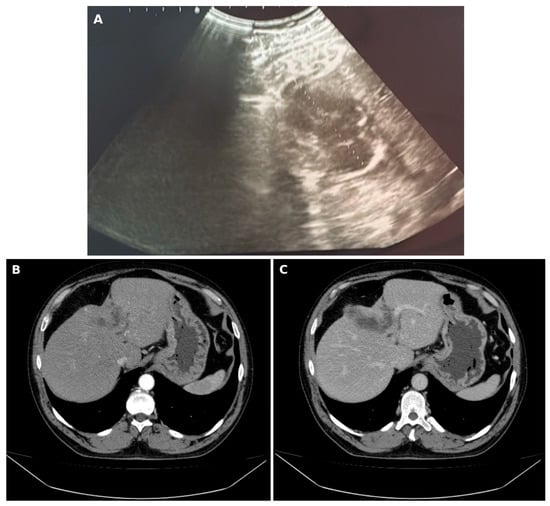
Two days prior to presenting at our emergency department in 2026, the patient experienced an episode of presyncope accompanied by hypotension and severe abdominal pain; this episode was managed by the family physician, who performed an abdominal ultrasound (Figure 1A) and recommended a surgical consultation, suspecting a cyst rupture. There was no loss of consciousness, so the episode was a presyncope rather than a syncope. A blood pressure of 70/50 mmHg was recorded during the episode, and no urticaria, pruritus, angioedema, or bronchospasm was reported. The ultrasound showed a complex cystic hepatic lesion, measuring approximately 68 mm in the plane shown, with heterogeneous content, internal echogenic septations, and a thickened, partly hyperechoic wall. He subsequently presented to the emergency department, approximately two and a half years after the initial imaging diagnosis, with epigastric and right hypochondrial pain. He was admitted for further investigation and specialized treatment.
Figure 1.
Imaging of the ruptured hepatic hydatid cyst. (A) Abdominal ultrasound showing a complex cystic hepatic lesion with internal echogenic septations and a thickened wall; (B,C) contrast-enhanced abdominal CT, axial section at the level of the left hepatic lobe, showing the hydatid cyst in the hepatic segment IVb: (B) arterial phase; (C) venous (portal) phase.
Emergency contrast-enhanced CT of the abdomen and pelvis showed a polycystic, thick-walled (up to 5.3 mm) lesion with parietal calcifications and multiple internal septations in hepatic segment IVb, extending toward segments IVa and VIII, measuring 40/88/40 mm, with an ill-defined wall segment (~11 mm); several smaller cysts were present in segments IVa and II (the largest measuring 38 mm, subcapsular). The remaining liver parenchyma showed mild steatosis and hepatomegaly. The left intrahepatic bile ducts were dilated (up to 6.4 mm), while the common bile duct was distally stenosed. The gallbladder contained sludge-like material and gallstones, and a small amount of free intraperitoneal fluid was present in the pericholecystic region. These findings were considered consistent with a hepatic hydatid cyst ruptured into the biliary tract (Figure 1B,C).
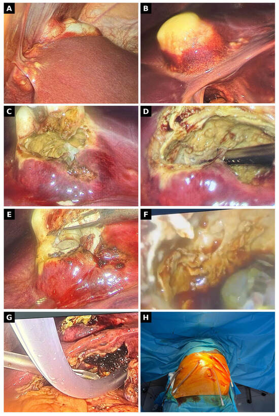
On admission, laboratory findings included a normal leukocyte count (5.41 × 10³/µL) with eosinophilia (13.5%), hepatic cytolysis (ALT 358 U/L, AST 116 U/L), marked cholestasis (GGT 849 U/L, alkaline phosphatase 324 U/L, direct bilirubin 3.39 mg/dL, total bilirubin 5.22 mg/dL), and a mild elevation of C-reactive protein (2.24 mg/dL); amylase, lipase, and coagulation studies were within normal limits. Serological testing for Echinococcus granulosus (ELISA) was positive, with elevated IgE. Preoperatively, during the same admission, the patient underwent endoscopic retrograde cholangiopancreatography (ERCP), indicated for obstructive cholestasis with jaundice, distal common bile duct narrowing on CT, and suspected intrabiliary rupture; the procedure had both a diagnostic and a therapeutic purpose. Cholangiography showed narrowing of the distal common bile duct, with no abnormalities of the left intrahepatic ducts and no contrast filling of the cyst cavity, so a cystobiliary communication could not be demonstrated at this stage. Endoscopic treatment consisted of access sphincterotomy and repeated balloon-catheter clearance maneuvers, resulting in the evacuation of biliary sludge and balloon dilation of the distal common bile duct stenosis, attributed to inflammation related to the hydatid cyst; no hydatid membranes were extracted from the main bile duct. Because of the documented penicillin allergy, the patient was tested for ceftriaxone and metronidazole, and preoperative antibiotic prophylaxis with these two agents was administered without adverse reactions. Surgery was then performed: exploratory laparoscopy, retrograde (fundus-first) laparoscopic cholecystectomy, puncture of the hepatic cyst with inactivation using hypertonic saline instillation, evacuation of the cystic content, partial pericystectomy, extensive lavage of the residual cavity and peritoneal cavity, and placement of four drains (Figure 2).
Figure 2.
Intraoperative laparoscopic images obtained during management of the ruptured hepatic hydatid cyst. (A) Overview of the liver; (B) hydatid cyst bulging at the liver surface before opening; (C) the cyst after opening, with hydatid material and membranes visible; (D) the opened cyst cavity during evacuation of its contents; (E) opening of the cyst wall with exposure of the laminated membrane; (F) daughter vesicles within the cyst cavity; (G) residual cavity after evacuation, with suction in place; (H) abdominal wall at the end of the procedure, with the four drains in place.
Bile cultured from the common bile duct was sterile (no aerobic bacterial or fungal growth at 48 h; no leukocytes or bacterial flora on microscopy), and screening for multidrug-resistant organisms was negative.
Histopathological examination confirmed the diagnosis. The gallbladder (9.2 cm in length) showed chronic cholecystitis with villous mucosal hyperplasia, Rokitansky-Aschoff sinuses, a mild lympho-plasmacytic infiltrate, and discrete parietal fibrosis. Fragments of the cyst wall showed marked pericystic sclerohyalinosis, with extensive hemorrhagic necrosis on the inner aspect, a moderate polymorphic inflammatory infiltrate, and bands of acellular, laminated, pale eosinophilic material with a concentric lamellar structure, corresponding to the laminated layer of the cyst, focally containing free and degenerating parasitic elements (protoscolices). The adjacent hepatic parenchyma was reduced in quantity, compressed, with hydropic degeneration. The final pathological diagnosis was hepatic hydatid cyst (hepatic echinococcosis).
Treatment with albendazole 400 mg every 12 h was initiated preoperatively and continued postoperatively. The postoperative course was slow but favorable, with resolution of the initial symptoms, return of physiological bowel transit, and good oral tolerance. Laboratory studies showed moderate leukocytosis with marked eosinophilia (up to 26.6% on postoperative day four), consistent with the immune response associated with parasitic disease, together with progressive improvement of the cytolysis and cholestasis (ALT from 358 to 105 U/L, GGT from 849 to 326 U/L, total bilirubin from 5.22 to 1.33 mg/dL from admission to postoperative day five). Three of the four drains were removed, the surgical wounds remained clean, and the sutures were removed.
The patient developed a low-output biliary fistula (20–100 mL/24 h), which remained low-output until the repeat ERCP, and was discharged home with one drain left in situ, in good general condition, afebrile, with a soft, non-tender abdomen, continued albendazole therapy, and a scheduled follow-up visit under joint surgical and infectious disease supervision.
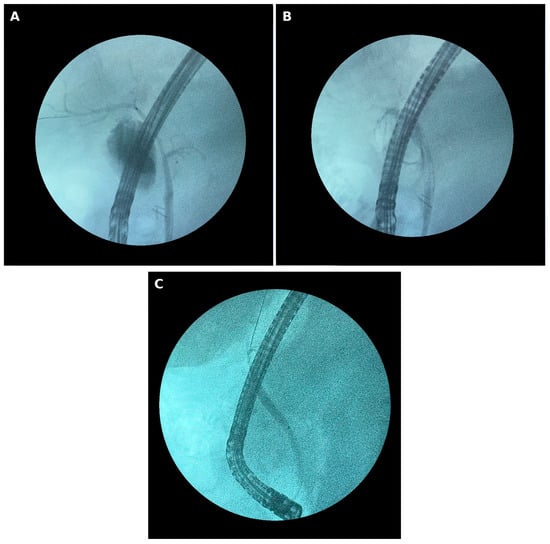
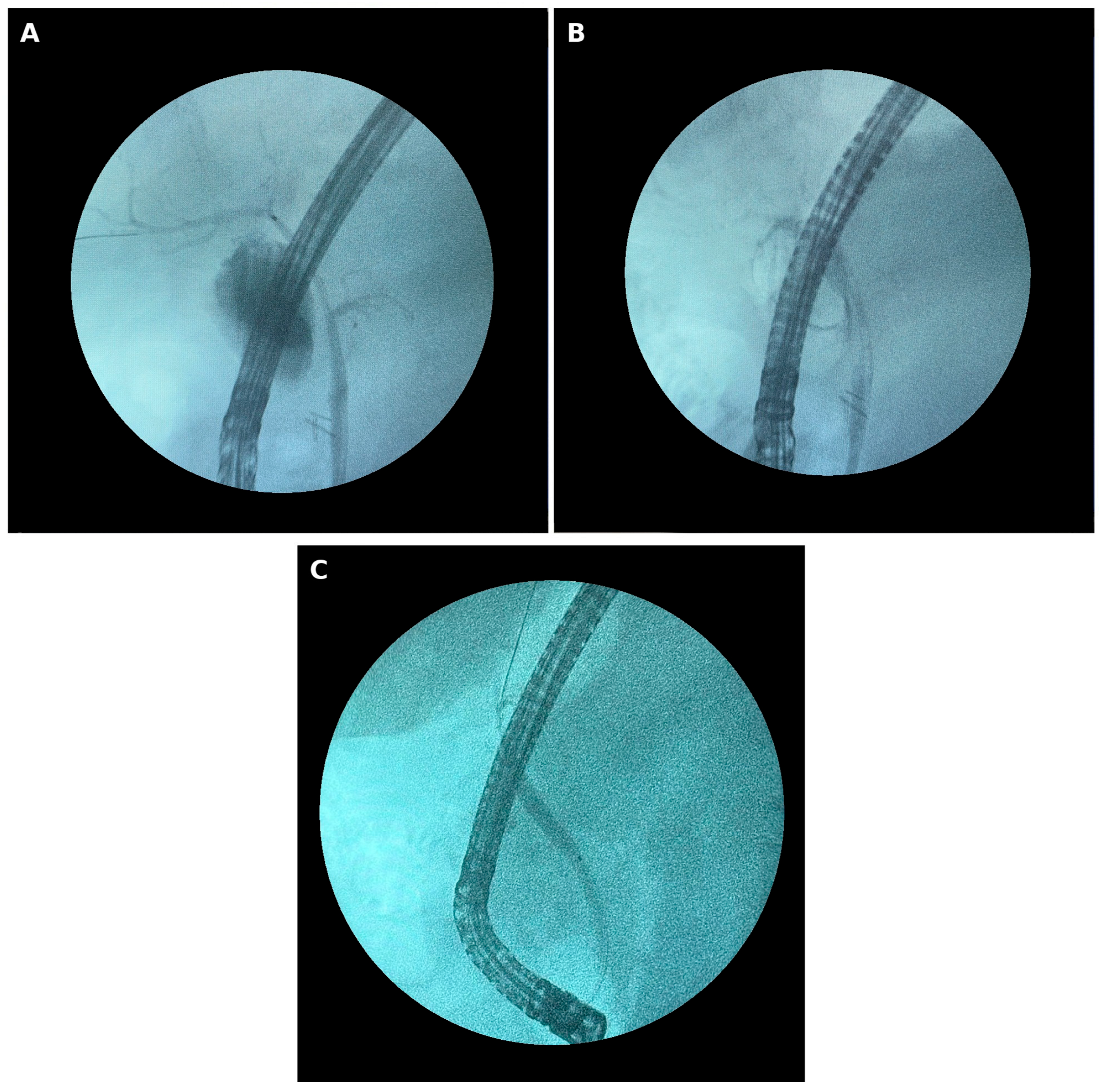
One month after discharge, because of persistence of the biliary fistula, the patient was readmitted and underwent a repeat ERCP. Endoscopic evaluation showed the expected post-sphincterotomy anatomy of the papilla, and the common bile duct was cannulated over a guidewire. Cholangiography demonstrated contrast extravasation into a subhepatic collection through a communication between the residual cyst cavity and the left hepatic duct. A plastic biliary stent (8.5 Fr/12 cm) was placed into the left hepatic duct and a second stent (8.5 Fr/9 cm) at the biliary confluence; contrast injection after stent placement confirmed filling of the collection, consistent with a persistent subhepatic biliary collection communicating with the biliary tree (Figure 3A,B). The remaining drain was removed after stent placement. Until the repeat ERCP, drain output was approximately 10–30 mL/24 h, no antibiotic therapy was given for the fistula, and on ultrasound the subhepatic collection was minimal, almost undetectable. The biliary stents were removed endoscopically four months after surgery, electively, in the absence of recurrent bile leakage or clinical signs of cholangitis, with no post-procedural incidents; the fluoroscopic control after stent removal is shown in Figure 3C, and the evolution was favorable. At the last follow-up, four months after surgery, the patient was continuing albendazole therapy and remained under clinical and imaging surveillance for recurrence.
Figure 3.
Fluoroscopic images from the follow-up ERCP. (A,B) Sequential frames during stent placement, showing the guidewire and the biliary stents across the site of the cystobiliary communication, with opacification of the subhepatic collection; (C) fluoroscopic control after stent removal.
3. Discussion
Cystic echinococcosis remains a neglected zoonotic disease with a substantial global burden, particularly in regions with traditional sheep and cattle husbandry, including parts of Eastern Europe [1,2,3,4,22]. Recent global burden analyses suggest that, although mortality and disability-adjusted life year rates have declined, the age-standardized prevalence and incidence of cystic echinococcosis are projected to remain stable through 2036, with the greatest burden in low- and middle-income regions [22]. Several recent single- and multicenter surgical series from our country confirm that hepatic hydatid disease continues to require regular surgical treatment [23,24,25,26,27]. Intrabiliary rupture is considered one of the most frequent complications of hepatic hydatid cysts, with a clinical spectrum ranging from occult, asymptomatic cystobiliary communication to massive rupture with cholangitis or obstructive jaundice [5,6,15,18]. In the present case, the cyst did not grow appreciably between 2023 and 2026 (approximately 86 mm versus 88 mm in maximal diameter); what changed was its structure, from a single, thick-walled lesion to a complicated, multivesicular cyst with an ill-defined wall segment, additional cysts in adjacent segments, and dilation of the left intrahepatic bile ducts. This illustrates that incompletely treated hydatid disease can progress in complexity without increasing in size, and underlines the importance of active surveillance, even in initially asymptomatic patients.
Cyst staging according to the WHO-IWGE ultrasound classification, originally derived from the Gharbi classification, reflects the biological activity of the parasite and helps guide management decisions [7,8,9,10]. Because the classification is based on ultrasound, a formal stage could not be assigned to the 2023 lesion, which was documented only by CT; its appearance, a single, well-defined cystic lesion with a thickened wall and no internal septation, would correspond most closely to an early, univesicular stage. The 2026 ultrasound (Figure 1A), showing a complex cystic lesion with internal echogenic septations and a thickened, partly hyperechoic wall, is consistent with a CE2–CE3b stage, and the multivesicular, partially calcified appearance on CT supports this. This apparent change in stage over approximately two and a half years, in the absence of appreciable growth, supports the interpretation of structural progression of an incompletely treated cyst. A learning point from this case is that the findings already present in 2023—a cyst of more than 8 cm with a thick, contrast-enhancing wall, adjacent bile duct dilation, and a markedly elevated total IgE—suggested biologically active disease, for which surgery was recommended. Total IgE is a non-specific marker, and a single observation cannot establish its predictive value; nevertheless, in a patient who declines surgery, such a constellation should prompt closer surveillance rather than reassurance.
The markedly elevated total IgE also deserves a more specific interpretation. An elevated total IgE is non-specific and may reflect atopy, an allergic reaction, or a parasitic infection; in a patient with a cystic liver lesion, however, it is an important clue to a parasitic etiology. In our patient, the 2023 value (8027 IU/mL, about eighty times the upper reference limit) was far higher than would be expected from his documented penicillin allergy alone and, together with the CT appearance, already pointed toward hydatid disease. The persistently elevated IgE in 2026 is consistent with continued antigenic stimulation by viable parasite, in keeping with the protoscolices identified on histopathology. Because specific antibodies may remain detectable for years after successful treatment, serial total IgE measurements may be one of the few accessible markers of parasitic activity in routine practice; a progressive decline during follow-up would support a response to treatment, whereas a renewed rise should prompt imaging for recurrence.
The interval between diagnosis and rupture also deserves comment. In 2023 the patient received a three-month course of albendazole but declined the surgical treatment that was recommended. According to the WHO-IWGE expert consensus, benzimidazoles alone are an option mainly for small, uncomplicated, univesicular cysts, whereas large cysts and multivesicular stages respond poorly and generally require surgery or percutaneous treatment, with medical therapy as an adjunct [8]. Long-term series of patients treated with benzimidazole carbamates likewise report incomplete responses and relapses in a proportion of cysts, which makes prolonged follow-up necessary [28]. In a cyst of more than 8 cm with a thick wall and adjacent bile duct dilation, a short course of albendazole was therefore unlikely to be curative, and the subsequent change in structure without a change in size is consistent with a partially treated but still active cyst. When surgery is declined, the decision should be revisited at each contact, and the patient should be offered scheduled ultrasound surveillance and clear information on the warning signs of rupture.
Diagnostic imaging plays a central role at every stage of hepatic hydatid disease, from incidental detection to the recognition of complications. Abdominal ultrasound remains the first-line modality worldwide, owing to its low cost and wide availability, and underlies the WHO-IWGE classification; however, its sensitivity for directly demonstrating a cystobiliary communication is limited. Contrast-enhanced CT, used in both examinations of our patient, offers superior delineation of wall thickness, calcification, daughter cysts, and involvement of adjacent structures, and was sufficient in this case to raise strong suspicion of intrabiliary rupture. Magnetic resonance cholangiopancreatography is generally regarded as the most sensitive non-invasive technique for directly visualizing communication between the cyst and the biliary tree, and is increasingly used, in patients with a cholestatic biochemical pattern and an equivocal CT, to plan the subsequent endoscopic or surgical strategy. In our patient, the combination of cholestatic, cytolytic, and eosinophilic laboratory abnormalities with the CT findings was considered sufficiently suggestive of intrabiliary rupture to justify proceeding directly to ERCP, which also served a therapeutic purpose. MRCP was not performed before ERCP because the CT and laboratory findings were considered sufficient to indicate ERCP, which was in any case required for biliary decompression. In retrospect, MRCP might have shown the communication with the left hepatic duct earlier, as it was demonstrated only at the second ERCP.
A particularly notable feature of this case is the episode of near-syncope and severe abdominal pain that occurred two days before admission and was assessed by the family physician. Rupture of a hepatic hydatid cyst, with sudden release of antigenic material, can precipitate severe hypersensitivity reactions, including anaphylactic shock [29]. In our patient, the retrospectively identified episode may plausibly have corresponded to the acute rupture event, without progression to overt anaphylaxis; because no laboratory data are available from that moment, this interpretation remains a hypothesis. It nevertheless suggests that clinicians evaluating patients with known hepatic hydatid disease and unexplained syncope, hypotension, or acute abdominal pain should maintain a high index of suspicion for occult cyst rupture, even in the absence of classical allergic manifestations.
Preoperative endoscopic management, through ERCP with sphincterotomy and clearance of biliary debris, has a well-documented role in the treatment of intrabiliary rupture of hepatic hydatid cysts, allowing decompression of the common bile duct, reduction in intrabiliary pressure, and promotion of cystobiliary fistula closure, at times obviating the need for surgery altogether [15,16,17,18,19,20,30]. In the case presented, preoperative ERCP allowed clearance of biliary sludge without retrieval of migrated hydatid membranes, which would be compatible with a narrow-caliber cystobiliary communication rather than frank migration of hydatid material into the common bile duct. The persistence of a subhepatic biliary collection, fed by a communication between the cyst and the left hepatic duct that was demonstrated only at the second ERCP and required biliary stenting one month after discharge, further illustrates that endoscopic management of complicated cystobiliary disease is frequently a staged process rather than a single intervention, and that close clinical and biochemical monitoring is required even after apparently successful initial treatment.
The site of the biliary communication is also relevant. The cyst occupied the central liver, in segment IV, close to the hilum, and the fistula demonstrated at the second ERCP involved a large hilar duct, in keeping with the selective dilation of the left intrahepatic ducts on CT. This proximity may explain why the leak did not close with external drainage alone. Elevated gamma-glutamyl transferase has been proposed as a preoperative marker of occult cystobiliary communication [31], although patients with jaundice or biliary dilatation were excluded from that series, and larger cyst size has been associated with postoperative bile leakage [32]. In our patient, GGT was markedly elevated (849 U/L), whereas the cyst diameter (88 mm) was moderate. In patients with such features, a communication should be actively sought during surgery, and early endoscopic treatment considered when postoperative bile drainage persists [17,18,19,20]. In our patient, sphincterotomy alone did not prevent the leak, whereas transpapillary stenting across the confluence was followed by removal of the last drain and by uneventful stent extraction four months after surgery.
From a surgical standpoint, treatment of complicated hepatic hydatid cysts remains individualized, depending on size, location, relationship to vasculobiliary structures, and the presence of complications, a principle already emphasized in early comparative surgical series [33]. Conservative techniques (partial cystectomy with drainage) are favored in patients at higher risk from extensive resections, whereas radical techniques (total cystectomy, hepatic resections) provide better control of recurrence at the cost of higher morbidity [6,13,14]. Contemporary Romanian surgical series, including a 19-year single-center analysis of 146 surgically managed hepatic hydatid cysts and a 5-year tertiary-center review of biliary complications, confirm that conservative open techniques remain the most frequently performed approach in these series, while minimally invasive options are used selectively, with postoperative complication rates depending on the type of procedure [24,25]. The laparoscopic approach, with strict adherence to protocols for operative field isolation and scolicidal inactivation with hypertonic saline, is increasingly reported as a feasible alternative to open surgery, including for complicated or multivesicular cysts, with outcomes comparable to open surgery in experienced centers [13,14,23]. In our case, combining retrograde laparoscopic cholecystectomy (indicated by the presence of gallstones and biliary sludge) with partial pericystectomy in the same operative session allowed both the biliary and the hepatic pathology to be addressed through a minimally invasive approach, avoiding laparotomy, although the subsequent biliary fistula required endoscopic re-intervention.
Low-output postoperative biliary fistula is a frequent complication after conservative surgery for hepatic hydatid cysts, particularly in the presence of a residual cystobiliary communication; most such fistulas close spontaneously with prolonged drainage and antiparasitic therapy, while therapeutic endoscopy (sphincterotomy, biliary stenting) is reserved for high-output or persistent fistulas [15,19,31]. Recurrence after surgery for hepatic hydatid disease is well documented, with rates that vary widely between series, and cyst size, multiplicity, and biliary complications are among the factors discussed as contributing to it [34]. This reinforces the rationale for prolonged postoperative surveillance in our patient, who combined several of these features—a multivesicular, ruptured cyst with documented biliary communication and a disease duration of over two years before definitive treatment. Adjuvant albendazole therapy is recommended perioperatively, both to reduce the risk of recurrence and to treat any residual viable parasitic elements [8,11,12,28]; in our patient, protoscolices were identified within the hydatid membrane on histopathology.
Finally, this case highlights the role of primary care in the pathway of patients with known hydatid disease. The family physician who assessed the presyncopal episode performed an abdominal ultrasound, suspected rupture of the known cyst, and referred the patient for surgical consultation, so that he reached the surgical service within two days. Because recurrence may become apparent years after apparently successful treatment, prolonged clinical and imaging follow-up is recommended [8,34]. The patient remains on albendazole, under joint surgical and infectious disease supervision, with periodic imaging.
Beyond individual case management, control of cystic echinococcosis ultimately depends on interrupting the dog–livestock transmission cycle through coordinated veterinary and public health measures, in line with the World Health Organization’s 2021–2030 roadmap for neglected tropical diseases [35].
This report has several limitations inherent to single-case descriptions. Genotyping of the parasite was not performed. In addition, the communication between the cyst and the left hepatic duct was demonstrated directly only at the second ERCP; at the initial presentation it was inferred from the clinical, biochemical, and imaging findings. Finally, follow-up is so far limited to four months after surgery, and longer-term imaging surveillance for recurrence is ongoing.
4. Conclusions
This case illustrates how an incidentally discovered hepatic hydatid cyst—initially asymptomatic and treated only with albendazole after the patient declined surgery—reappeared after more than two years as a complicated, ruptured cyst with cystobiliary communication; it also demonstrates the feasibility of a combined endoscopic–laparoscopic approach in this context, although this did not prevent the development of a postoperative biliary fistula. In practice, an incidentally discovered hepatic hydatid cyst requires a comprehensive diagnostic workup and structured, sustained monitoring, rather than reliance on a single imaging report. Furthermore, in a large hepatic hydatid cyst such as this one, albendazole alone was not sufficient; surgical treatment retains a central role, and a presyncopal episode in a patient with known hydatid disease should raise suspicion of cyst rupture.
Supplementary Materials
The following supporting information can be downloaded at: https://www.mdpi.com/article/10.3390/diagnostics16223841/s1, Checklist S1: CARE checklist.
Author Contributions
Conceptualization, M.-A.C. and A.M.V.; methodology, M.-A.C. and A.M.V.; validation, M.-A.C. and A.M.V.; investigation, M.-A.C., A.C., V.B., I.S., G.B. and A.M.V.; resources, M.-A.C. and A.M.V.; data curation, M.-A.C. and A.M.V.; writing—original draft preparation, M.-A.C. and A.M.V.; writing—review and editing, M.-A.C., A.C., V.B., I.S., C.B., C.D.L., G.B. and A.M.V.; visualization, M.-A.C. and A.M.V.; supervision, C.B. and C.D.L.; project administration, M.-A.C. and A.M.V. All authors have read and agreed to the published version of the manuscript.
Funding
This research received no external funding.
Institutional Review Board Statement
Ethical review and approval were waived for this study because it describes the routine diagnostic and therapeutic care of a single patient and does not constitute a research study on human subjects, in accordance with the policy of “St. Spiridon” Emergency Clinical County Hospital, Iași, regarding single case reports.
Informed Consent Statement
Written informed consent has been obtained from the patient to publish this case report and any accompanying images.
Data Availability Statement
The data presented in this study are available on request from the corresponding author due to patient confidentiality restrictions.
Conflicts of Interest
The authors declare no conflict of interest.
References
- Wen, H.; Vuitton, L.; Tuxun, T.; Li, J.; Vuitton, D.A.; Zhang, W.; McManus, D.P. Echinococcosis: Advances in the 21st Century. Clin. Microbiol. Rev. 2019, 32, e00075-18. [Google Scholar] [CrossRef] [Scilit] [PubMed]
- Eckert, J.; Deplazes, P. Biological, epidemiological, and clinical aspects of echinococcosis, a zoonosis of increasing concern. Clin. Microbiol. Rev. 2004, 17, 107–135. [Google Scholar] [CrossRef] [Scilit] [PubMed]
- Moro, P.; Schantz, P.M. Echinococcosis: A review. Int. J. Infect. Dis. 2009, 13, 125–133. [Google Scholar] [CrossRef] [Scilit] [PubMed]
- McManus, D.P.; Zhang, W.; Li, J.; Bartley, P.B. Echinococcosis. Lancet 2003, 362, 1295–1304. [Google Scholar] [CrossRef] [Scilit] [PubMed]
- Junghanss, T.; da Silva, A.M.; Horton, J.; Chiodini, P.L.; Brunetti, E. Clinical management of cystic echinococcosis: State of the art, problems, and perspectives. Am. J. Trop. Med. Hyg. 2008, 79, 301–311. [Google Scholar] [CrossRef] [Scilit]
- Nunnari, G.; Pinzone, M.R.; Gruttadauria, S.; Celesia, B.M.; Madeddu, G.; Malaguarnera, G.; Pavone, P.; Cappellani, A.; Cacopardo, B. Hepatic echinococcosis: Clinical and therapeutic aspects. World J. Gastroenterol. 2012, 18, 1448–1458. [Google Scholar] [CrossRef] [Scilit] [PubMed]
- WHO Informal Working Group on Echinococcosis. International classification of ultrasound images in cystic echinococcosis for application in clinical and field epidemiological settings. Acta Trop. 2003, 85, 253–261. [Google Scholar] [CrossRef] [Scilit] [PubMed]
- Brunetti, E.; Kern, P.; Vuitton, D.A. Writing Panel for the WHO-IWGE. Expert consensus for the diagnosis and treatment of cystic and alveolar echinococcosis in humans. Acta Trop. 2010, 114, 1–16. [Google Scholar] [PubMed]
- Gharbi, H.A.; Hassine, W.; Brauner, M.W.; Dupuch, K. Ultrasound examination of the hydatid liver. Radiology 1981, 139, 459–463. [Google Scholar] [CrossRef] [Scilit] [PubMed]
- Wang, Y.; Zhang, X.; Bartholomot, B.; Liu, B.; Luo, J.; Li, T.; Wen, X.; Zheng, H.; Zhou, H.; Wen, H.; et al. Classification, follow-up and recurrence of hepatic cystic echinococcosis using ultrasound images. Trans. R. Soc. Trop. Med. Hyg. 2003, 97, 203–211. [Google Scholar] [CrossRef] [Scilit] [PubMed]
- Bildik, N.; Cevik, A.; Altintaş, M.; Ekinci, H.; Canberk, M.; Gülmen, M. Efficacy of preoperative albendazole use according to months in hydatid cyst of the liver. J. Clin. Gastroenterol. 2007, 41, 312–316. [Google Scholar] [CrossRef] [Scilit] [PubMed]
- Manterola, C.; Mansilla, J.A.; Fonseca, F. Preoperative albendazole and scolices viability in patients with hepatic echinococcosis. World J. Surg. 2005, 29, 750–753. [Google Scholar] [CrossRef] [Scilit] [PubMed]
- Bayrak, M.; Altintaş, Y. Current approaches in the surgical treatment of liver hydatid disease: Single center experience. BMC Surg. 2019, 19, 95. [Google Scholar] [CrossRef] [Scilit] [PubMed]
- Mihetiu, A.; Bratu, D.; Neamtu, B.; Sabau, D.; Sandu, A. Therapeutic Options in Hydatid Hepatic Cyst Surgery: A Retrospective Analysis of Three Surgical Approaches. Diagnostics 2024, 14, 1399. [Google Scholar] [CrossRef] [Scilit] [PubMed]
- Habeeb, T.A.A.M.; Podda, M.; Tadic, B.; Shelat, V.G.; Tokat, Y.; Abo Alsaad, M.I.; Kalmoush, A.-E.; Nassar, M.S.; Mustafa, F.M.; Badawy, M.H.M.; et al. Biliary fistula and late recurrence of liver hydatid cyst: Role of cysto-biliary communication: A prospective multicenter study. World J. Methodol. 2023, 13, 272–286. [Google Scholar] [CrossRef] [Scilit] [PubMed]
- Muhammedoğlu, B.; Pircanoğlu, E.M.; Pişkin, E.; Torun, S.; Karadağ, M.; Topuz, S.; Köktaş, S. Treatment of Hepatic Hydatid Disease: Role of Surgery, ERCP, and Percutaneous Drainage: A Retrospective Study. Surg. Laparosc. Endosc. Percutan. Tech. 2020, 31, 313–320. [Google Scholar] [CrossRef] [Scilit] [PubMed]
- Goumas, K.; Poulou, A.; Dandakis, D.; Tyrmpas, I.; Georgouli, A.; Sgourakis, G.; Soutos, D.; Karaliotas, K. Role of endoscopic intervention in biliary complications of hepatic hydatid cyst disease. Scand. J. Gastroenterol. 2007, 42, 1113–1119. [Google Scholar] [CrossRef] [Scilit] [PubMed]
- Dolay, K.; Akbulut, S. Role of endoscopic retrograde cholangiopancreatography in the management of hepatic hydatid disease. World J. Gastroenterol. 2014, 20, 15253–15261. [Google Scholar] [CrossRef] [Scilit] [PubMed]
- Massit, H.; Seddik, H.; Basr, H.; Benkirane, A. Endoscopic treatment of biliary complications of hepatic hydatid disease. Pan Afr. Med. J. 2015, 22, 67. [Google Scholar] [PubMed]
- Gümüşoğlu, A.Y.; Kabuli, H.A.; Değerli, M.S.; Atar, B.; Köneş, O.; Kocataş, A.; Bingül, N.D.; Dolay, K. Endoscopic definitive treatment of liver hydatid cysts ruptured into bile duct. Surg. Endosc. 2024, 38, 7187–7194. [Google Scholar] [CrossRef] [Scilit] [PubMed]
- Gagnier, J.J.; Kienle, G.; Altman, D.G.; Moher, D.; Sox, H.; Riley, D.; CARE Group. The CARE guidelines: Consensus-based clinical case reporting guideline development. Glob. Adv. Health Med. 2013, 2, 38–43. [Google Scholar] [CrossRef] [Scilit] [PubMed]
- Wang, Y.; Zhang, R.; Zhang, D.; Liang, Y.; Xu, C.; Zhang, C. Global burden of cystic echinococcosis: A 32-year analysis of 204 countries with 2036 projections. BMC Public Health 2025, 25, 3213. [Google Scholar] [CrossRef] [Scilit] [PubMed]
- Moldovan, D.; Moldovan, C.A.; Ungurianu, L.; Ungureanu, D.F. A Retrospective Surgical Experience Regarding Open and Laparoscopic Procedures of the Hepatic Hydatid Cyst with an Up-to-Date Complete Review of the Literature and a Focus on Original Romanian Techniques. J. Mind Med. Sci. 2016, 3, 182–193. [Google Scholar] [CrossRef] [Scilit]
- Mancino, S.; Michelini, S.; Mancino, N.; Civita, R.; Coros, M.; Mocian, F. 19-Year Retrospective Analysis of 146 Surgically Managed Hepatic Hydatid Cysts: Complications Across Four Procedure-Based Groups. Cureus 2026, 18, e101381. [Google Scholar] [CrossRef] [Scilit] [PubMed]
- Vâlcea, S.; Dumitriu, B.C.; Beuran, M.; Feier, C.V.I. Biliary Complications After Surgery for Hydatid Disease: A Five-Year Experience in a Tertiary Care Center. Healthcare 2025, 13, 1077. [Google Scholar] [CrossRef] [Scilit] [PubMed]
- Botezatu, C.; Mastalier, B.; Patrascu, T. Hepatic hydatid cyst—Diagnose and treatment algorithm. J. Med. Life 2018, 11, 203–209, Correction in J. Med. Life 2018, 11, 394. [Google Scholar] [CrossRef] [Scilit] [PubMed]
- Hogea, M.O.; Ciomaga, B.F.; Muntean, M.M.; Muntean, A.A.; Popa, M.I.; Popa, G.L. Cystic Echinococcosis in the Early 2020s: A Review. Trop. Med. Infect. Dis. 2024, 9, 36. [Google Scholar] [CrossRef] [Scilit] [PubMed]
- Franchi, C.; Di Vico, B.; Teggi, A. Long-term evaluation of patients with hydatidosis treated with benzimidazole carbamates. Clin. Infect. Dis. 1999, 29, 304–309. [Google Scholar] [CrossRef] [Scilit] [PubMed]
- Zayati, M.; Chaouch, M.A.; Mokni, S.; Maaref, M.; Abdelwahed, M.; Daly, H.; Ladib, A.; Jebali, F.; Kawech, A. Acute rupture of a huge liver hydatid cysts in the peritoneal cavity causing an anaphylactic shock: A case report. Radiol. Case Rep. 2025, 20, 2428–2431. [Google Scholar] [CrossRef] [Scilit] [PubMed]
- Kumar, N.; Sardiwalla, I.I. A case report of definitive endoscopic management of a large right liver lobe hydatid cyst—A novel approach. Int. J. Surg. Case Rep. 2024, 120, 109825. [Google Scholar] [CrossRef] [Scilit] [PubMed]
- Atahan, K.; Küpeli, H.; Deniz, M.; Gür, S.; Çökmez, A.; Tarcan, E. Can occult cystobiliary fistulas in hepatic hydatid disease be predicted before surgery? Int. J. Med. Sci. 2011, 8, 315–320. [Google Scholar] [CrossRef] [Scilit] [PubMed]
- Kilic, M.; Yoldas, O.; Koc, M.; Keskek, M.; Karakose, N.; Ertan, T.; Gocmen, E.; Tez, M. Can biliary-cyst communication be predicted before surgery for hepatic hydatid disease: Does size matter? Am. J. Surg. 2008, 196, 732–735. [Google Scholar] [CrossRef] [Scilit] [PubMed]
- Xynos, E.; Pechlivanides, G.; Tzortzinis, A.; Papageorgiou, A.; Vassilakis, J.S. Hydatid disease of the liver: Diagnosis and surgical treatment. HPB Surg. 1991, 4, 59–67. [Google Scholar] [CrossRef] [Scilit] [PubMed]
- Kapan, M.; Kapan, S.; Goksoy, E.; Perek, S.; Kol, E. Postoperative Recurrence in Hepatic Hydatid Disease. J. Gastrointest. Surg. 2006, 10, 734–739. [Google Scholar] [CrossRef] [Scilit] [PubMed]
- World Health Organization. Ending the Neglect to Attain the Sustainable Development Goals: A Road Map for Neglected Tropical Diseases 2021–2030; WHO: Geneva, Switzerland, 2020. [Google Scholar]
Disclaimer/Publisher’s Note: The statements, opinions and data contained in all publications are solely those of the individual author(s) and contributor(s) and not of MDPI and/or the editor(s). MDPI and/or the editor(s) disclaim responsibility for any injury to people or property resulting from any ideas, methods, instructions or products referred to in the content. |
© 2026 by the authors. Licensee MDPI, Basel, Switzerland. This article is an open access article distributed under the terms and conditions of the Creative Commons Attribution (CC BY) license.