


Abstract
C-reactive protein (CRP) remains the most widely used inflammatory biomarker in clinical practice, valued for its assay standardization, low cost, and extensive validation. However, CRP is a systemic, hepatically derived marker that typically peaks 24–48 h after an inflammatory stimulus and does not capture localized, tissue-level inflammatory activity. Pentraxin 3 (PTX3), a long pentraxin produced locally by endothelial cells, macrophages, and other stromal cells, rises within 6–8 h of an inflammatory trigger and may provide complementary, tissue-specific information in selected clinical contexts. This narrative review searched PubMed, Scopus, and Web of Science for studies published between January 2014 and March 2026 using combinations of the terms “pentraxin 3,” “PTX3,” and “C-reactive protein”; 15 studies met the inclusion criteria after a structured screening process. Across the reviewed literature, PTX3 showed an earlier peak and stronger associations with disease severity and mortality than CRP in specific settings, including acute coronary syndrome, sepsis, and selected neurological and pregnancy-related conditions, although the findings were not uniform across all patient subgroups, particularly between sexes. Rather than replacing CRP, PTX3 is best understood as a complementary biomarker whose value is realized within a tiered testing strategy, in which CRP remains the first-line test and PTX3 is reserved for defined high-risk or diagnostically ambiguous scenarios. The wider clinical adoption of PTX3 will require assay standardization; defined reference intervals, including sex-specific ranges; and cost-effectiveness data before it can be incorporated into routine practice.
1. Introduction
The inflammation process is a central biological response and is integral to the development of many health issues (cardiovascular disease (CVD), infection, neurological disorders, etc.). For instance, in CVD, chronic low-level inflammation contributes to atherosclerosis, plaque instability, and thrombotic events; in contrast, in infection (including sepsis), the intensity of the inflammatory response correlates with the severity of disease and clinical outcomes; and in neurological disorders (e.g., neurodegenerative diseases), inflammation might contribute to disease progression through the activation of microglia and neuronal damage mediated by cytokines [1]. Furthermore, inflammatory assessments play a critical role in the early diagnosis, risk stratification, and monitoring of therapy for CVD, infection, and neurological disorders [2].
Among inflammatory biomarkers, C-reactive protein (CRP) has been widely used in clinical practice because of its advantageous characteristics (i.e., sensitivity, standardized assays, and low cost) [3]. CRP is a pentameric plasma protein that reflects the liver’s response to the inflammatory cytokine IL-6. Overall, CRP may be considered a reliable measure of systemic inflammation [4].
The pentraxin protein family has recently gained increased attention, especially for its use as next-generation inflammatory biomarker candidates. Long pentraxins (e.g., PTX3) are produced at the local site of inflammation by multiple cell types (e.g., endothelial cells, macrophages and dendritic cells) in response to primary inflammatory signals (e.g., TNF-alpha, IL-1beta) [5]. PTX3 demonstrates an ability to rapidly rise during the early phases of an inflammatory response and offers a more accurate representation of tissue inflammation because of its localized production compared to CRP [6].
Emerging evidence suggests that PTX3 may offer advantages over CRP as a diagnostic inflammatory biomarker in many clinical scenarios. For example, in the area of cardiovascular disease, PTX3 correlates better with vascular inflammation and endothelial dysfunction than does CRP; and within the context of infection (e.g., sepsis), PTX3 has been shown to have an earlier rise as well as a stronger association with the severity of disease and mortality than CRP [7]. Finally, PTX3’s role in neurological inflammation is increasingly being documented and supports its potential to serve as a versatile biomarker [8].
Compared with CRP, PTX3 has been described as a more precise indicator of vascular inflammation. Whereas short-acting CRP is produced mainly by the liver in response to circulating IL-6, long-acting PTX3 is produced locally by vascular and cardiac tissues in response to primary inflammatory triggers [6]. This review provides a comparative analysis of CRP and PTX3, examining their biological characteristics, clinical performances, and the practical barriers that currently limit PTX3’s routine clinical use, with the aim of clarifying whether PTX3 could complements, rather than replace, CRP testing.
2. Methodology
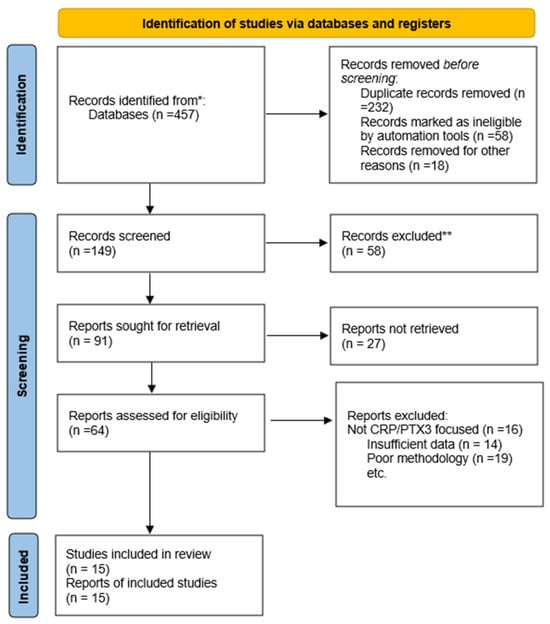
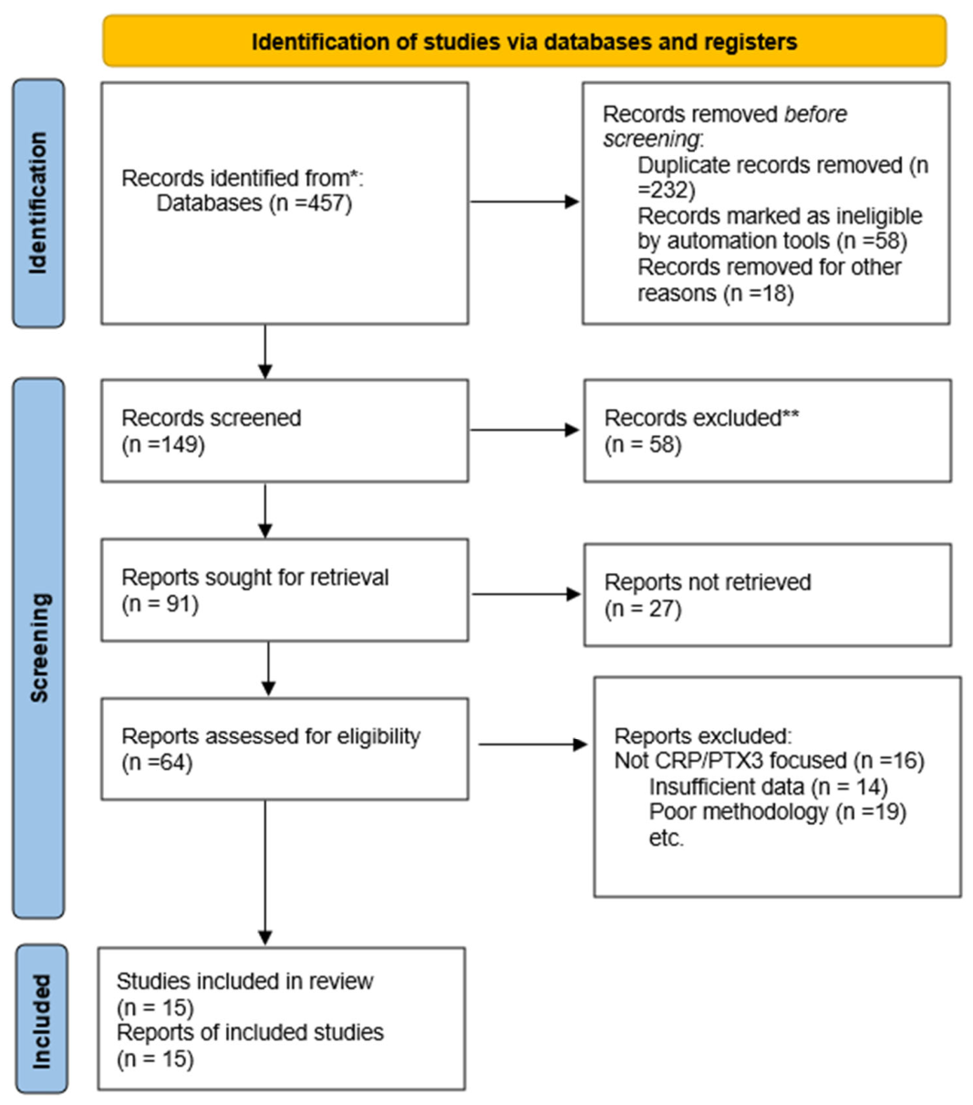
This is a narrative review; the PRISMA-style search (Figure 1) and screening process below is reported for transparency. The findings are synthesized narratively rather than pooled quantitatively, given the heterogeneity of included study designs.
Figure 1.
PRISMA flow diagram of study identification, screening, and inclusion. * Records identified from PubMed, Scopus, and Web of Science (combined total shown). ** Records were excluded manually following title/abstract screening; no automation tool was used.
Search Strategy and Study Selection
A literature search was conducted across PubMed, Scopus, and Web of Science databases using search terms including: “pentraxin 3,” “PTX3,” “C-reactive protein,” “CRP,” “pentraxins inflammatory biomarkers,” and combinations thereof. The search encompassed publications from January 2014 to March 2026, with an English language restriction.
The database search yielded 457 records. Upon elimination of duplicates and other ineligible records, 149 unique records were screened. Out of those, 58 were excluded. The full-text evaluation of 91 articles resulted in 27 articles not being retrieved. In total, 64 studies underwent evaluation for eligibility, with 15 studies being included in the final review.
The narrowing from 457 identified records to 15 included studies reflects the strict relevance filtering for direct PTX3-CRP comparisons rather than search inadequacy. The exclusions at the full-text stage were primarily due to the studies not directly comparing the two biomarkers; insufficient extractable data; or methodological limitations, such as small sample size or absence of a comparator group.
Inclusion criteria: (1) Narrative reviews; (2) investigation of pentraxins as inflammatory biomarkers; (3) comparative analysis of pentraxin family members; (4) human studies; and (5) quantitative assessment of biomarker performance. Exclusion criteria: (1) Editorials or opinion pieces; (2) studies lacking quantitative biomarker data; (3) non-English publications; and (4) case reports with insufficient sample sizes.
3. Results
Fifteen studies met the inclusion criteria and are summarized in Table 1 below. Rather than reading as an undifferentiated list, the findings cluster into four broad clinical themes that are synthesized narratively here, with the table serving as a reference companion.
Cardiovascular and cerebrovascular disease: Six of the included studies examined PTX3 in cardiovascular contexts. In stable coronary artery disease, PTX3 was significantly elevated in affected patients, and a cut-off of 0.89 ng/mL helped exclude atherosclerosis, although PTX3 did not correlate with the number of diseased vessels [9]. In acute coronary syndrome, the PLATO substudy of 5154 patients found that PTX3 peaked earlier than high-sensitivity CRP and correlated more strongly with NT-proBNP and troponin, translating into a modest—not dramatic—prognostic advantage over CRP [7]. Similar patterns were seen in carotid plaque vulnerability, where PTX3 tracked with fibrous cap discontinuity [10], and in acute minor stroke, where PTX3 was an independent predictor of unfavorable 90-day outcome [8]. Two mechanistic reports linked PTX3 to endothelial dysfunction, angiogenesis regulation, and pulmonary arterial hypertension pathophysiology, reinforcing a tissue-level rather than purely systemic role [11].
Metabolic and diabetic disease: PTX3 was consistently elevated in type 2 diabetes and its vascular complications, with a reported AUC of 0.720 for diagnosing T2DM in one prediabetic/obese cohort [12]. The antidiabetic drug class appeared to modulate PTX3 levels differently. GLP-1 receptor agonists and DPP-4 inhibitors were associated with reductions, while SGLT-2 inhibitors were paradoxically associated with increases [13], a pattern that has not yet been mechanistically explained and that complicates use of PTX3 as a simple marker of glycemic control. A mouse model further suggested that PTX3 deficiency protects against streptozotocin-induced β-cell apoptosis via endoplasmic reticulum stress pathways [14].
Infection, sepsis, and inflammatory bowel disease: In neonates, PTX3 rose up to 100-fold within 6–8 h of onset of infection and performed at least as well as procalcitonin, and better than CRP alone, for early sepsis detection. In a broader combined-marker analysis, PTX3 together with CRP, procalcitonin, and the white cell count achieved an AUC of 0.855 for sepsis detection, with 83.3% sensitivity and 80% specificity, a performance that depended on combining the markers rather than using PTX3 alone [15]. In inflammatory bowel disease, PTX3 correlated with endoscopic disease activity scores alongside IL-6 and presepsin [16].
Autoimmune, renal, and other systemic diseases: In rheumatoid arthritis and spondyloarthritis, PTX3 was higher in RA and correlated with disease activity, supporting a dual role as both an inflammatory and cardiovascular-risk marker [17]. In end-stage renal disease, PTX3 independently predicted all-cause mortality and correlated negatively with nutritional status [18]. Two studies examined CRP isoforms (native pentameric CRP and monomeric CRP) rather than PTX3 directly, finding that the two isoforms have divergent, sometimes opposing, roles in atherogenesis and autoimmune regulation [19,20]: a reminder that CRP itself is not a single, uniform biomarker and that comparisons between “CRP” and “PTX3” necessarily simplify a more complex biology [21]. Finally, in myeloproliferative neoplasms, PTX3 was elevated in JAK2V617F homozygous carriers and associated with a two-fold increase in disease risk, independent of mutation status [22].
Taken together, the included studies show a consistent direction of effect: PTX3 tends to rise earlier and track more closely with localized tissue injury than CRP, but the magnitude of benefit varies considerably by disease context, is frequently modest rather than large, and is not consistently observed across sexes. This heterogeneity is the central reason this review frames PTX3 as complementary to, rather than a replacement for, CRP.
Table 1.
Summary of included studies.
3.1. Pentraxin
The pentraxin family of proteins is part of a superfamily of proteins (i.e., that share a specific structure and function). All members of the pentraxin family share a similar domain; exist as monomers assembled into pentameric forms, which exhibit a discoid morphology; and the pentraxin family of proteins contains a highly conserved C-terminal domain of 205 amnio acids (AAs) (known as the “Pentraxin domain”) [5]. The pentraxin family also shares an eight AA conserved sequence within the pentraxin domain, known as the “Pentraxin signature” (His-x-Cys-x-Ser/Thr-Trp-x-Ser, where “x” can be any AA). According to the length of the protein, not the length of the pentraxin signature, the pentraxin family is subdivided into two subfamilies based on their relative protein size: short pentraxins and long pentraxins. The short pentraxin proteins include C-reactive protein (CRP) and serum amyloid P component (SAP); the long pentraxin proteins include neuronal pentraxin 1 (NPTX1), neuronal pentraxin 2 (NPTX2), neuronal pentraxin receptor (NPTXR), pentraxin 3 (PTX3), and pentraxin 4 (PTX4) (Table 2) [29].
Table 2.
Structural and functional comparison of pentraxin family [5,29,30].
3.2. Mechanisms of Action and Inflammatory Signaling
Pentraxins participate in directing inflammatory responses by acting through a variety of mechanisms that are complementary. The major mechanism is through binding of pentraxins to complement component C1q, thereby activating the classical pathway of complement activation [31]. The degree of C1q recruitment to PTX3 for subsequent enhancement of complement-dependent phagocytosis is significantly higher than that of C-reactive protein, although the time course of these processes is different from that of C-reactive protein [32].
PTX3 also has a complex regulatory role in macrophage phenotype and function. PTX3 decreases macrophage production of the pro-inflammatory cytokines IL-6 and TNF-α, while increasing anti-inflammatory IL-10 [33]. Furthermore, PTX3 regulates macrophage polarization markers and modulates the expression of CD86, HLA-DR, and the hemoglobin–haptoglobin receptor CD163 in a dose-dependent manner [34]. This indicates that PTX3 functions more as a modulator of inflammation than as a simple pro-inflammatory mediator. PTX3 is also involved in angiogenesis regulation through inhibition of fibroblast growth factor-2 (FGF-2) activity, contributing to its protective role in both inflammation-induced tissue damage and vascular repair [35].
3.3. Differentiating Biomarker Activities in Clinical Conditions
3.3.1. Acute Coronary Syndrome and Cardiovascular Disease
PTX3 and CRP show different biological activities and different time courses in patients with acute coronary syndrome. According to data from the Platelet Inhibition and Patient Outcomes (PLATO) study, which included 5154 patients, the two biomarkers reached peak levels at different time points relative to high-sensitivity CRP, NT-proBNP, and troponin T [7]. PTX3, measured at admission, was a significant predictor of cardiovascular mortality, myocardial infarction, and stroke, whereas hs-CRP was not statistically significant after adjustment for other assays. In patients with coronary artery disease, PTX3 performed better than hs-CRP at estimating the severity of coronary stenosis. Sex differences were consistently observed across the cardiovascular literature reviewed here, and are treated in detail in the dedicated section below. In brief, the predictive value of inflammatory biomarkers for long-term overall mortality was demonstrated in male patients, but did not reach statistical significance in female patients in the same cohorts [7].
3.3.2. Infection and Sepsis
In neonates, PTX3 has proven at least as useful as procalcitonin for diagnosis, and more specific than CRP when used as a single-marker test. When combined with CRP, procalcitonin, and the white blood cell count—the latter two being established indicators of infection and inflammation—PTX3 achieved an AUC of 0.855 for detecting sepsis, with 83.3% sensitivity and 80% specificity. In polytrauma patients, a biomarker panel of serum amyloid A (SAA), CRP, and procalcitonin outperformed any single marker for predicting subsequent sepsis; combining the three markers discriminated better between patients who did and did not develop sepsis than any marker used alone [15].
3.3.3. COVID-19 and Severe Respiratory Infection
Research during the COVID-19 pandemic identified PTX3 as a strong predictor of severe disease. A broader biomarker panel including interferon gamma-induced protein 10 and serum amyloid A was subsequently identified as offering additional predictive value for disease progression and severity [36].
3.3.4. Autoimmune and Rheumatic Diseases
Rheumatoid synovium produces PTX3 following exposure to IL-1β and TNF-α, but not IL-6 [37]. Significantly more PTX3 protein was detected in synovial fluid from patients with rheumatoid arthritis (RA) than from patients with osteoarthritis or healthy controls, and endothelial cell staining within synovial tissue was more prominent in RA. These findings indicate that PTX3 may be useful as a marker of disease activity in RA and in other autoimmune diseases characterized by localized inflammation [20].
3.3.5. Neurological and Aging-Related Diseases
Recent research has identified PTX3 as a potential biomarker of neurovascular disease. In a proteomics study of patients with intracerebral hemorrhage (ICH), 18 inflammatory proteins showed significantly different expression compared with the controls [38]. PTX3 levels were higher in the plasma and brain tissue of ICH patients than in the controls; combined with IL-6, KIM-1, and galectin-9, PTX3 showed good diagnostic performance for ICH, suggesting it could form part of a novel diagnostic biomarker panel [38].
PTX3’s relationship with cognitive impairment has also shown clear sex-based differences, discussed further below. The Cardiovascular Health Study found that each one-standard-deviation increase in PTX3 was associated with a 0.20-point-per-year decline in the Modified Mini-Mental State Examination score in women (95% CI, −0.37 to −0.03), with no corresponding association in men [38].
3.3.6. Pregnancy-Related Disorders
PTX3 is particularly useful in obstetric contexts. Both CRP and PTX3 rise significantly in the third trimester of pregnancy compared with the first and second trimesters; the mean PTX3 in the third trimester (7.70 μg/L) is roughly double that seen in the first trimester (3.33 μg/L). This late-gestation rise is atypical relative to conventional (non-pregnant) reference ranges, so pregnancy-specific reference intervals are required to avoid misclassifying normal physiologic inflammation as pathological [39].
PTX3, combined with markers of oxidative stress, adds diagnostic value in preeclampsia. PTX3, together with 8-iso-prostaglandin F2α, showed the highest diagnostic utility among the inflammatory biomarkers evaluated, with both markers contributing independent predictive power. PTX3 in amniotic fluid has shown high predictive performance (AUC 0.85–0.95) for microbial invasion of the amniotic cavity and intra-amniotic inflammation in women with preterm premature rupture of membranes, performing comparably to IL-8 for this diagnosis [38,39].
4. Discussion
4.1. Sex-Related Differences in Pentraxin Biology and Clinical Interpretation
A recurring theme across the cardiovascular, cognitive, and mortality literature reviewed above is that PTX3’s prognostic value is not sex neutral. In acute coronary syndrome cohorts, PTX3 and related inflammatory markers predicted long-term all-cause mortality in men but failed to reach statistical significance in women from the same cohorts [7]. In the Cardiovascular Health Study, elevated PTX3 was associated with faster cognitive decline in women but showed no corresponding association in men [38]. These are not isolated findings from a single study design; they recur across at least two independent cohorts and two distinct clinical endpoints (mortality and cognitive decline), which strengthens the case that the sex effect is real rather than a chance finding.
The biological basis for this divergence is not yet established, but several non-exclusive explanations have been proposed in the broader literature: sex differences in baseline innate immune activity and complement regulation; the modulatory effect of sex hormones (particularly estrogen) on endothelial inflammation and vascular pentraxin production; and differences in body composition and the metabolic–inflammatory load between men and women that may confound simple biomarker–outcome associations. None of the studies included in this review were specifically designed to test these mechanistic hypotheses, and this represents an important evidence gap [38].
From a practical standpoint, the sex-discordant findings summarized above mean that a single, sex-neutral PTX3 reference range or risk threshold is unlikely to perform equally well in men and women. Pregnancy-specific reference intervals are already required for PTX3 given the physiologic rise during gestation [39]; by extension, this review recommends that future validation studies report PTX3 performance separately by sex and, where sample size allows, derive sex-specific reference intervals and decision thresholds rather than assuming a single cut-off applies universally. Until such sex-specific data are available, clinicians should interpret PTX3 results in women, particularly for cardiovascular risk stratification, with added caution, and should not assume that the thresholds validated primarily in male-predominant cohorts (such as PLATO) generalize directly to female patients [7].
The body of evidence reviewed here supports a role for PTX3 as a complementary addition to, rather than a wholesale replacement for, CRP in inflammatory biomarker testing [3]. As a long pentraxin, PTX3 has distinct structural characteristics—local production, a rapid response time, and multiple immunomodulatory functions—that make it a potentially informative complementary marker alongside CRP for conditions such as acute coronary syndrome [40], sepsis and severe infection [15], peripheral vascular disease [36], and respiratory infections [3]. PTX3’s inflammatory properties allow it to function in two ways: promoting activation of the innate immune system, and repairing tissue damage while limiting excessive complement activation. This dual character suggests PTX3 is best used as a biomarker of site-specific tissue inflammation that adds context to, rather than substitutes for, the systemic signal captured by CRP [41]. This characteristic exemplifies the evolutionary role of PTX3 in linking the innate and adaptive immune response, and is a new way of thinking about inflammatory responses that is not reliant on CRP but that uses a context-specific assessment [41].
4.2. Tiered Testing Framework and Decision Pathways
The “tiered approach” referenced in the abstract and conclusion is defined explicitly here, rather than left as a general principle. In all scenarios, CRP is retained as the first-line, universally ordered inflammatory marker; PTX3 is added as a second-tier test only in the specific circumstances below, where it is most likely to change management.
4.3. Future Research Directions
- Assay harmonization: Establish an internationally certified reference material and harmonization program for PTX3 immunoassays (analogous to existing CRP standardization efforts), so that the cut-offs and AUCs reported across studies and platforms become directly comparable.
- Sex-specific and pregnancy-specific reference intervals: Conduct adequately powered studies to derive PTX3 reference ranges stratified by sex, menopausal status, and gestational trimester, replacing the current reliance on largely male-predominant or non-stratified cohorts.
- Prospective validation of a defined tiered algorithm: Prospectively test the tiered decision pathway proposed in Table 3 (starting with ACS and neonatal sepsis, where the evidence base is strongest) against pre-specified clinical endpoints, comparing a CRP-plus-PTX3 pathway with a CRP-only pathway.
- Cost-effectiveness analysis: Perform formal cost–utility analyses comparing PTX3-inclusive tiered testing against CRP-only testing in the specific high-risk populations identified in this review (ACS, neonatal sepsis, and preeclampsia).
- Point-of-care assay development: Support the development and clinical validation of a rapid, point-of-care PTX3 assay suitable for emergency department, labor-and-delivery, and ICU workflows, addressing the turnaround time barrier identified above.
- Genetic and mechanistic studies: Investigate PTX3 gene polymorphisms known to alter PTX3 expression, and their relationship to disease susceptibility and treatment response, alongside mechanistic studies exploring the hormonal and immune contributors to the sex differences identified in this review.
- Direct comparative studies across disease states: Conduct head-to-head prospective studies directly comparing PTX3 and CRP performance across, rather than within, disease categories, to clarify where PTX3’s added value is greatest and where it is negligible.
- Therapeutic target exploration: Given PTX3’s multiple biological functions, evaluate its potential as a therapeutic target, not only as a diagnostic marker, building on the pulmonary hypertension and macrophage-polarization mechanistic data summarized above.
Table 3.
Proposed tiered decision pathway for PTX3 testing by clinical scenario.
5. Limitations, Advantages, and Recommendations for Clinical Implementation
PTX3 shows certain advantages over CRP on several dimensions: it peaks earlier in acute inflammation, is more sensitive to localized inflammatory responses, has greater predictive value in several cardiovascular diseases, provides independent prognostic information regarding COVID-19 severity, and correlates positively with tissue-level inflammation. However, CRP retains substantial practical advantages in routine clinical monitoring, including assay standardization, low relative cost, and well-established reference ranges [42]. SAA has advantages for identifying subclinical inflammation and predicting the response to immunotherapy, which may be particularly useful in individuals with cancer and autoimmune diseases [41].
Beyond this general trade-off, several specific and interrelated barriers currently limit PTX3’s translation into routine practice:
- Assay standardization: No internationally certified reference standard or calibrator currently exists for PTX3, unlike the well-harmonized, CRP-focused standardization efforts led by international clinical chemistry bodies. The reported PTX3 concentrations and diagnostic cut-offs therefore vary across studies depending on the immunoassay platform used (ELISA versus automated chemiluminescent platforms), which complicates direct comparison of the AUCs and thresholds cited throughout this review.
- Cost and accessibility: PTX3 immunoassays are substantially more expensive per test than high-sensitivity CRP, which is inexpensive, automatable, and available in essentially all clinical laboratories. In resource-limited settings, including many centers in India, the incremental cost of routine PTX3 testing is unlikely to be justified without clearer evidence of added clinical value over CRP alone.
- Confounding factors: PTX3 levels are influenced by age, renal function, obesity, pregnancy status and gestational age, and possibly genetic polymorphisms in the PTX3 gene, none of which are yet fully accounted for in the existing reference ranges. Several included studies did not adjust for these confounders, making it difficult to isolate PTX3’s independent contribution to risk prediction.
- Study heterogeneity and small sample sizes: The 15 studies included in this review varied considerably in design (cross-sectional versus prospective cohort), population, disease definition, and PTX3 cut-off values, and several had modest sample sizes. This heterogeneity limits the ability to pool the findings into precise, generalizable effect estimates and increases the risk that the individual reported AUCs reflect cohort-specific rather than universally applicable performance.
- Predominantly cross-sectional evidence: Most of the cardiovascular and metabolic studies reviewed were cross-sectional or had a relatively short follow-up, limiting causal inference and the ability to determine whether PTX3 elevation precedes, rather than merely accompanies, disease progression.
- Sex-specific evidence gaps: As detailed above, several of the strongest associations between PTX3 and clinical outcomes were observed in male-predominant cohorts or showed a significant sex interaction, yet very few studies were adequately powered to report sex-stratified reference ranges or thresholds.
Clinical Implementation Considerations
Assay availability: PTX3 is currently measured almost exclusively by research-use or laboratory-developed ELISA and chemiluminescent immunoassays; unlike CRP, no PTX3 assay is yet CLIA waived or widely available as a rapid point-of-care test suitable for emergency department or bedside use. This restricts PTX3 testing to laboratories with the relevant immunoassay infrastructure and send-out capability, and introduces turnaround time delays that limit its usefulness in time-critical decisions, such as suspected ACS.
Regulatory status: In most jurisdictions, PTX3 assays are not approved as standalone diagnostic tests for a specific indication in the way CRP assays are; where available, they are typically marketed as research-use-only or laboratory-developed tests. This has direct implications for reimbursement, quality assurance requirements, and the evidentiary bar clinicians should apply before acting on a PTX3 result in isolation.
Cost: The published cost data specific to PTX3 assays are limited, but immunoassay-based specialty biomarkers of this kind are typically several-fold more expensive per test than an automated CRP assay. Formal cost-effectiveness or cost–utility analyses comparing a PTX3-inclusive tiered pathway against a CRP-only pathway in defined high-risk populations have not yet been published and are identified as a priority in the Future Directions Section above.
Practical implications: Taken together, these factors mean that, at present, PTX3 is realistically positioned as a second line, selectively ordered test in centers with access to the relevant assay platform, rather than a routine first-line replacement for CRP—reinforcing the tiered approach set out below.
6. Conclusions
This review positions pentraxins, and PTX3 in particular, as a complementary addition to the established inflammatory biomarker toolkit rather than a replacement for CRP. CRP’s clinical utility remains well established, owing to its standardization and cost-efficiency; PTX3, meanwhile, offers an enhanced though context-dependent ability to reflect localized tissue inflammation across a range of acute and chronic conditions, including acute coronary syndrome, sepsis, neurological and aging-related disease, pregnancy-related disorders, and COVID-19. Multiple pentraxin measurements incorporated into validated, disease-specific biomarker panels are likely to improve diagnostic and prognostic accuracy compared with a single-marker approach.
As set out in Table 3, clinical implementation of PTX3 should follow a defined tiered pathway: CRP is retained as the universal first-line inflammatory marker, and PTX3 is reserved for specific high-risk or diagnostically ambiguous scenarios, such as equivocal ACS risk stratification, very early suspected sepsis, or an inconclusive preeclampsia work-up, where it is most likely to add clinically actionable information. Further studies are needed to standardize testing methods, establish sex-specific and pregnancy-specific reference ranges, and define the true cost-effectiveness of pentraxin-based testing so that its role in precision and personalized medicine can be fully realized.
Author Contributions
Conceptualization, M.C.; methodology, M.Y. and P.K.; literature search and screening, M.Y. and P.K.; writing—original draft preparation, M.Y.; writing—review and editing, M.C. and M.Y..; supervision, M.C. All authors have read and agreed to the published version of the manuscript.
Funding
This research received no external funding.
Institutional Review Board Statement
Not applicable.
Informed Consent Statement
Not applicable.
Data Availability Statement
No new data were created or analyzed in this study. Data sharing is not applicable to this article.
Conflicts of Interest
The authors declare no conflict of interest.
References
- Chavda, V.P.; Feehan, J.; Apostolopoulos, V. Inflammation: The Cause of All Diseases. Cells 2024, 13, 1906. [Google Scholar] [CrossRef] [Scilit] [PubMed] [PubMed Central]
- Chen, L.; Deng, H.; Cui, H.; Fang, J.; Zuo, Z.; Deng, J.; Li, Y.; Wang, X.; Zhao, L. Inflammatory responses and inflammation-associated diseases in organs. Oncotarget 2017, 9, 7204–7218. [Google Scholar] [CrossRef] [Scilit] [PubMed] [PubMed Central]
- C-Reactive Protein (CRP) Test: What It Is, Purpose & Results [Internet]. Available online: https://my.clevelandclinic.org/health/diagnostics/23056-c-reactive-protein-crp-test (accessed on 8 August 2025).
- Yang, Z.; Cui, D.; Li, F.; Huang, B.; Liu, Q. Prognostic Value of Inflammatory Biochemical Markers (IL-8, PCT, CRP) for Cardiovascular Disease in Patients with Pancreatitis. J. Med. Biochem. 2025, 45, 54–62. [Google Scholar] [CrossRef] [Scilit] [PubMed]
- Pentraxin—An Overview|ScienceDirect Topics [Internet]. Available online: https://www.sciencedirect.com/topics/medicine-and-dentistry/pentraxin (accessed on 14 April 2026).
- Staubli, S.M.; Schäfer, J.; Rosenthal, R.; Zeindler, J.; Oertli, D.; Nebiker, C.A. The role of CRP and Pentraxin 3 in the prediction of systemic inflammatory response syndrome and death in acute pancreatitis. Sci. Rep. 2019, 9, 18340. [Google Scholar] [CrossRef] [Scilit] [PubMed]
- Kontny, F.; Andersen, T.; Ueland, T.; Åkerblom, A.; Lakic, T.G.; Michelsen, A.E.; Aukrust, P.; Bertilsson, M.; Becker, R.C.; Himmelmann, A.; et al. Pentraxin-3 vs C-reactive protein and other prognostic biomarkers in acute coronary syndrome: A substudy of the Platelet Inhibition and Patients Outcomes (PLATO) trial. Eur. Heart J. Acute Cardiovasc. Care 2020, 9, 313–322. [Google Scholar] [CrossRef] [Scilit] [PubMed]
- Zhang, H.; Wang, R.; Wang, Z.; Wu, W.; Zhang, N.; Zhang, L.; Hu, J.; Luo, P.; Zhang, J.; Liu, Z.; et al. Molecular insight into pentraxin-3: Update advances in innate immunity, inflammation, tissue remodeling, diseases, and drug role. Biomed. Pharmacother. 2022, 156, 113783. [Google Scholar] [CrossRef] [Scilit] [PubMed]
- Knapp, M.; Gil-Mika, M.; Sawicki, R.; Lisowska, A.; Kaminski, M.; Sobkowicz, B.; Ptaszynska, K. Pentraxin 3 as a marker of development and severity of stable coronary artery disease. Adv. Med. Sci. 2024, 69, 391–397. [Google Scholar] [CrossRef] [Scilit] [PubMed]
- Li, T.; Li, L.; Wang, Q.; Sun, Y.; Lian, J.; Yu, W. Serum PTX3 Levels Correlate with Fibrous Cap Discontinuity and Focal Inflammation in Carotid Vulnerable Plaque: A High-Resolution Magnetic Resonance Vessel Wall Imaging Study. J. Clin. Med. 2026, 15, 1452. [Google Scholar] [CrossRef] [Scilit] [PubMed]
- Liu, H.; Guan, S.; Fang, W.; Yuan, F.; Zhang, M.; Qu, X. Associations between pentraxin 3 and severity of coronary artery disease. BMJ Open 2015, 5, e007123. [Google Scholar] [CrossRef] [Scilit] [PubMed]
- Ahrițculesei, R.-V.; Boldeanu, L.; Caragea, D.C.; Vladu, I.M.; Clenciu, D.; Mitrea, A.; Ungureanu, A.M.; Văduva, C.-C.; Dijmărescu, A.L.; Popescu, A.I.S.; et al. Association Between Pentraxins and Obesity in Prediabetes and Newly Diagnosed Type 2 Diabetes Mellitus Patients. Int. J. Mol. Sci. 2025, 26, 3661. [Google Scholar] [CrossRef] [Scilit] [PubMed] [PubMed Central]
- Dobriceanu, R.-C.; Meca, A.D.; Boboc, I.K.S.; Mititelu-Tartau, L.; Naidin, M.S.; Turcu-Stiolica, A.; Bogdan, M. Pentraxin-3 as a Biomarker in Diabetes Mellitus: Insights into Inflammation, Vascular Complications, and Modulation by Antidiabetic Medications. Biomedicines 2025, 13, 891. [Google Scholar] [CrossRef] [Scilit] [PubMed]
- Kim, S.; Hwang, A.-R.; Kim, S.-H.; Lim, J.H.; Woo, C.-H. Pentraxin 3 deficiency ameliorates streptozotocin-induced pancreatic toxicity via regulating ER stress and β-cell apoptosis. Mol. Cells 2025, 48, 100168. [Google Scholar] [CrossRef] [Scilit] [PubMed]
- Zhang, Y.; Li, X.; Zhang, X.; Wang, T.; Zhang, X. Progress in the study of pentraxin-3(PTX-3) as a biomarker for sepsis. Front. Med. 2024, 11. [Google Scholar] [CrossRef] [Scilit] [PubMed]
- Ishida, N.; Tani, S.; Takebe, T.; Takahashi, K.; Asai, Y.; Tamura, S.; Matsuura, T.; Yamade, M.; Iwaizumi, M.; Hamaya, Y.; et al. Role of Sepsis Markers as Potential Biomarkers for Inflammatory Bowel Disease: A Prospective Observational Study. J. Clin. Gastroenterol. 2025, ahead of print. [Google Scholar] [CrossRef] [Scilit] [PubMed]
- Targońska-Stępniak, B.; Drelich-Zbroja, A. The Role of Pentraxin 3 in the Assessment of Cardiovascular Risk and Disease Activity in Patients with Rheumatoid Arthritis and Spondyloarthritis. J. Inflamm. Res. 2024, 17, 9905–9916. [Google Scholar] [CrossRef] [Scilit] [PubMed] [PubMed Central]
- Valente, M.J.; Rocha, S.; Coimbra, S.; Catarino, C.; Rocha-Pereira, P.; Bronze-da-Rocha, E.; Oliveira, J.G.; Madureira, J.; Fernandes, J.C.; do Sameiro-Faria, M.; et al. Long Pentraxin 3 as a Broader Biomarker for Multiple Risk Factors in End-Stage Renal Disease: Association with All-Cause Mortality. Mediat. Inflamm. 2019, 2019, 3295725. [Google Scholar] [CrossRef] [Scilit] [PubMed] [PubMed Central]
- Kirkgöz, K. C-Reactive Protein in Atherosclerosis—More than a Biomarker, but not Just a Culprit. Rev. Cardiovasc. Med. 2023, 24, 297. [Google Scholar] [CrossRef] [Scilit] [PubMed] [PubMed Central]
- Ahmed, M.; Yuan, P.; Lei, J.-G.; Tang, Z.-M.; Yang, Z.-R.; Potempa, L.A.; Wang, H.-Y.; Li, H.-Y. From Homeostasis To Inflammation To Autoimmunity: The Potential Impact of CRP. Inflammation 2025, 48, 3747–3762. [Google Scholar] [CrossRef] [Scilit] [PubMed] [PubMed Central]
- Ristagno, G.; Fumagalli, F.; Bottazzi, B.; Mantovani, A.; Olivari, D.; Novelli, D.; Latini, R. Pentraxin 3 in Cardiovascular Disease. Front. Immunol. 2019, 10, 823. [Google Scholar] [CrossRef] [Scilit] [PubMed] [PubMed Central]
- Martinez de la Torre, Y.; Fabbri, M.; Jaillon, S.; Bastone, A.; Nebuloni, M.; Vecchi, A.; Mantovani, A.; Garlanda, C. Evolution of the Pentraxin Family: The New Entry PTX4. J. Immunol. 2010, 184, 5055–5564. [Google Scholar] [CrossRef] [Scilit] [PubMed]
- Nilsen, D.; Aarsetoey, R.; Poenitz, V.; Ueland, T.; Michelsen, A.; Aukrust, P.; Brugger-Andersen, T.; Staines, H.; Grundt, H. Abstract 4141048: Inflammatory biomarkers predict long-term all-cause mortality in males but not in females. Circulation 2024, 150, A4141048. [Google Scholar] [CrossRef] [Scilit]
- Liu, P.Y.; Hu, C.Y. The interaction of bone morphogenetic protein receptor type 2 and pentraxin 3 on inflammation and fibrosis: Implications for pulmonary hypertension. Eur. Heart J. 2025, 46, ehaf784.2903. [Google Scholar] [CrossRef] [Scilit]
- Ye, X.; Wang, Z.; Lei, W.; Shen, M.; Tang, J.; Xu, X.; Yang, Y.; Zhang, H. Pentraxin 3: A promising therapeutic target for cardiovascular diseases. Ageing Res. Rev. 2024, 93, 102163. [Google Scholar] [CrossRef] [Scilit] [PubMed]
- Zhang, Y.; Hu, H.; Liu, C.; Wu, J.; Zhou, S.; Zhao, T. Serum pentraxin 3 as a biomarker for prognosis of acute minor stroke due to large artery atherosclerosis. Brain Behav. 2021, 11, e01956. [Google Scholar] [CrossRef] [Scilit] [PubMed] [PubMed Central]
- Lannergård, A.; Rosenström, F.; Normann, E.; Larsson, A. Serum pentraxin 3 concentrations in neonates. Ups. J. Med. Sci. 2014, 119, 62–64. [Google Scholar] [CrossRef] [Scilit] [PubMed]
- Bottazzi, B.; Inforzato, A.; Messa, M.; Barbagallo, M.; Magrini, E.; Garlanda, C.; Mantovani, A. The pentraxins PTX3 and SAP in innate immunity, regulation of inflammation and tissue remodelling. J. Hepatol. 2016, 64, 1416–1427. [Google Scholar] [CrossRef] [Scilit] [PubMed]
- Du Clos, T.W. Pentraxins: Structure, Function, and Role in Inflammation. Int. Sch. Res. Not. 2013, 2013, 379040. [Google Scholar] [CrossRef] [Scilit] [PubMed]
- C-Reactive Protein. In Wikipedia [Internet]. 2025. Available online: https://en.wikipedia.org/w/index.php?title=C-reactive_protein&oldid=1300833657 (accessed on 7 August 2025).
- Lu, J.; Mold, C.; Du Clos, T.W.; Sun, P.D. Pentraxins and Fc Receptor-Mediated Immune Responses. Front. Immunol. 2018, 9, 2607. [Google Scholar] [CrossRef] [Scilit] [PubMed] [PubMed Central]
- Shiraki, A.; Kotooka, N.; Komoda, H.; Hirase, T.; Oyama, J.; Node, K. Pentraxin-3 regulates the inflammatory activity of macrophages. Biochem. Biophys. Rep. 2016, 5, 290–295. [Google Scholar] [CrossRef] [Scilit] [PubMed] [PubMed Central]
- Ahmed, M.I.; Spooner, B.; Isherwood, J.; Lane, M.; Orrock, E.; Dennison, A. A Systematic Review of the Barriers to the Implementation of Artificial Intelligence in Healthcare. Cureus 2023, 15, e46454. [Google Scholar] [CrossRef] [Scilit] [PubMed] [PubMed Central]
- Garlanda, C.; Bottazzi, B.; Magrini, E.; Inforzato, A.; Mantovani, A. PTX3, a Humoral Pattern Recognition Molecule, in Innate Immunity, Tissue Repair and Cancer. Physiol. Rev. 2018, 98, 623–639. [Google Scholar] [CrossRef] [Scilit] [PubMed] [PubMed Central]
- Haroun, R.A.-H.; Osman, W.H.; Eessa, A.M. Interferon-γ-induced protein 10 (IP-10) and serum amyloid A (SAA) are excellent biomarkers for the prediction of COVID-19 progression and severity. Life Sci. 2021, 269, 119019. [Google Scholar] [CrossRef] [Scilit] [PubMed] [PubMed Central]
- Jenny, N.S.; Blumenthal, R.S.; Kronmal, R.A.; Rotter, J.I.; Siscovick, D.S.; Psaty, B.M. Associations of Pentraxin 3 with Cardiovascular Disease: The Multi-Ethnic Study of Atherosclerosis. J. Thromb. Haemost. 2014, 12, 999–1005. [Google Scholar] [CrossRef] [Scilit] [PubMed] [PubMed Central]
- Hu, Z.; Chen, S.; Zhang, E.; Wei, L.; Wang, J.; Shang, Q.; Gao, X.; Huang, Y. Novel inflammatory markers in intracerebral hemorrhage: Results from Olink proteomics analysis. FASEB J. 2025, 39, e70341. [Google Scholar] [CrossRef] [Scilit] [PubMed] [PubMed Central]
- Miller, L.M.; Jenny, N.S.; Rawlings, A.M.; Arnold, A.M.; Fitzpatrick, A.L.; Lopez, O.L.; Odden, M.C. Sex Differences in the Association Between Pentraxin 3 and Cognitive Decline: The Cardiovascular Health Study. J. Gerontol. A Biol. Sci. Med. Sci. 2020, 75, 1523–1529. [Google Scholar] [CrossRef] [Scilit] [PubMed] [PubMed Central]
- Larsson, A.; Palm, M.; Helmersson, J.; Axelsson, O. Pentraxin 3 values during normal pregnancy. Inflammation 2011, 34, 448–451. [Google Scholar] [CrossRef] [Scilit] [PubMed]
- Butt, N.; Bache-Mathiesen, L.; Ushakova, A.; Nordrehaug, J.; Jensen, S.; Munk, P.; Danchin, N.; Dubois-Rande, J.; Hansen, H.; Paganelli, F.; et al. Pentraxin 3 in primary percutaneous coronary intervention for ST elevation myocardial infarction is associated with early irreversible myocardial damage. Eur. Heart J. Acute Cardiovasc. Care 2020, 9, 302–312. [Google Scholar] [CrossRef] [Scilit] [PubMed] [PubMed Central]
- Daigo, K.; Mantovani, A.; Bottazzi, B. The yin-yang of long pentraxin PTX3 in inflammation and immunity. Immunol. Lett. 2014, 161, 38–43. [Google Scholar] [CrossRef] [Scilit] [PubMed] [PubMed Central]
- Jin, Y.; Guo, S.; Xiao, Y.; Yin, C. Assessment of the diagnostic significance of pentraxin-3 in conjunction with procalcitonin (PCT) and C-reactive protein (CRP) for neonatal sepsis. BMC Infect. Dis. 2025, 25, 401. [Google Scholar] [CrossRef] [Scilit] [PubMed] [PubMed Central]

Disclaimer/Publisher’s Note: The statements, opinions and data contained in all publications are solely those of the individual author(s) and contributor(s) and not of MDPI and/or the editor(s). MDPI and/or the editor(s) disclaim responsibility for any injury to people or property resulting from any ideas, methods, instructions or products referred to in the content. |
© 2026 by the authors. Licensee MDPI, Basel, Switzerland. This article is an open access article distributed under the terms and conditions of the Creative Commons Attribution (CC BY) license.