


Abstract
Ergonomically unfavorable postures remain a major risk factor for work-related musculoskeletal disorders (WRMSDs) among ultrasound operators. Dermatologic and facial ultrasound presents specific ergonomic challenges because it requires delicate transducer manipulation and prolonged static postures. This narrative review examines key ergonomic strategies for dermatologic and facial ultrasound, integrating findings from recent kinematic and occupational studies to promote sustainable, high-quality ultrasound practice. Important considerations include probe handling, scanning technique, patient positioning, and operator posture. Technical innovations, including portable systems, footswitches, and hands-free controls, are discussed, together with education, awareness, training, and future directions. A practical ergonomic workflow for facial ultrasound is also presented. Standardization of patient positioning, ultrasound-system layout, and operator ergonomics is essential for achieving reproducible diagnostic quality while safeguarding practitioner health. Although direct prospective clinical trials specifically addressing facial ultrasound ergonomics remain limited, translating evidence from general ultrasound and interventional practices provides a practical framework for clinical optimization.
1. Introduction
Dermatologic and facial ultrasound presents specific ergonomic challenges because it requires delicate transducer manipulation and prolonged static postures. The development of portable and wireless high-frequency (HF) probes (≥15–49 MHz) has markedly improved operator comfort and image precision. Recent quantitative motion-capture studies have highlighted ergonomically unfavorable postures as a major risk factor for work-related musculoskeletal disorders (WRMSDs) among ultrasound operators. Standardization of patient positioning, ultrasound-system layout, and operator ergonomics is essential for achieving reproducible diagnostic quality while safeguarding practitioner health. This narrative review examines key ergonomic strategies for dermatologic and facial ultrasound, integrating findings from recent kinematic and occupational studies to promote sustainable, high-quality ultrasound practice.
Ergonomics is increasingly recognized as a central consideration in ultrasound practice because of its implications for both diagnostic accuracy and operator well-being. Earlier studies and reviews reported musculoskeletal symptoms in approximately 80–95% of sonographers over the course of their careers, although estimates vary substantially according to the population studied, outcome definitions, and study design [1,2,3,4,5]. Facial ultrasound is increasingly used in dermatologic and aesthetic applications to identify abnormalities and anatomical variations before rejuvenation and plastic surgery procedures, guide percutaneous interventions, and assess postprocedural outcomes and complications [6,7]. In facial ultrasound, ergonomic challenges are further amplified by the need to image minute, superficial, and deformable structures at high frequencies. Operators must apply minimal compression, requiring delicate manipulation and stable control of the transducer. Comprehensive mapping of facial mimetic muscles, vessels, and other critical structures can be time-consuming. In this context, ergonomics is not a secondary consideration but a prerequisite for image stability and diagnostic reliability. A recent systematic review and meta-analysis of 30 studies involving 13,916 sonographers estimated an overall prevalence of musculoskeletal disorders of 75.8% (95% CI, 65.37–86.23%), with the neck (63.73%) and shoulder (60.13%) among the most frequently affected regions [8]. This pooled estimate is preferable when describing contemporary overall prevalence, whereas the higher figures of 80–95% should be interpreted as estimates from individual studies or earlier literature rather than as a universally applicable prevalence value.
Despite the rapid expansion of high-frequency and ultrasound-guided applications in aesthetic medicine, the ergonomic requirements of facial ultrasound have received relatively little specific attention [9,10]. Most of the available evidence on ergonomics derives from conventional diagnostic ultrasound, vascular access, regional anesthesia, and musculoskeletal interventions, so that the relationships among operator ergonomics, probe–tissue mechanics, image reproducibility, and procedural performance in facial ultrasound remain largely unexplored.
This narrative review therefore aims to examine the ergonomic requirements specific to dermatologic and aesthetic facial ultrasound, with particular emphasis on their potential effects on image quality, examination reproducibility, and ultrasound-guided procedures, and to translate existing evidence into practical recommendations for clinical practice.
2. Methods
A bibliographic search was performed in June 2026 in the Scopus (Elsevier, Amsterdam, The Netherlands), MEDLINE (US National Library of Medicine, Bethesda, MD, USA), and Web of Science (Thomson Reuters, Toronto, ON, Canada) databases. Combinations of the following descriptors were used according to a free-text protocol: “ergonomics in facial ultrasound”, “ultrasound ergonomics”, “facial ultrasound”, “ultrasound work-related musculoskeletal disorders”, “ergonomics and probe handling”, “ergonomics and image quality”, “facial ultrasound patient position”, “facial ultrasound operator positioning”, “facial ultrasound operator posture”, “ergonomics and ultrasound probe cables”, “ergonomics and examination table”, “ergonomics and chair”, “ergonomic and portable ultrasound systems”, “ergonomic and footswitch” “ergonomics of ultrasound-guided aesthetic procedures”, “ultrasound hands-free control”, “ultrasound ergonomics education”.
The literature search was restricted to articles in English published from the year 2000. Emphasis was focused on peer-reviewed original research, reviews, editorials, commentaries and meta-analyses explicitly reporting on ergonomics, facial ultrasound, ultrasound-guided aesthetic procedures, probe cables, operator positioning, posture, chair and examination table position, portable ultrasound systems, hands-free ultrasound and ergonomics education. Conference abstracts and case reports were excluded. Consistent with the design of a purely narrative and educational review, a formal systematic screening process was not conducted. Instead, articles were purposively selected by the authors based on their clinical relevance, scientific merit, and contribution to the technological advancement in facial ultrasound ergonomics. Consequently, formal systematic counts, flowcharts, and risk-of-bias assessments were not applicable. Results were narratively reported.
3. Occupational Disorders Among Ultrasound Operators
Sonographers and physicians who perform ultrasound examinations may develop various occupational disorders. The most frequently affected body regions include the shoulders, elbows, wrists, hands, neck, and back. Common conditions and symptoms include muscle spasms, pain, inflammation, sensory changes, tendon and ligament injuries, visual symptoms, stress, and headaches [11,12]. Although mild symptoms are common, work-related musculoskeletal disorders (WRMSDs) may, in a minority of cases, lead to severe pain, sickness absence, surgery, long-term disability, or premature termination of a career [13]. Shoulder abduction, neck flexion, and wrist deviation are major contributors to WRMSDs, underscoring the need for structured ergonomic prevention strategies [5]. Recent evidence continues to support a multifactorial model in which examination volume, awkward postures, equipment configuration, and insufficient recovery contribute to cumulative musculoskeletal loading [4,8,14,15,16].
4. Probe Handling and Ergonomic Technique
The probe should be handled with precise manual control and continuous wrist stabilization. Supporting the scanning hand—such as using the fifth finger as an anatomical reference point—helps maintain transducer stability and prevents unintended movement.
Conventional cabled probes, typically used with cart-based systems, exert mechanical torque and restrict movement. Cable weight increases forearm loading and may contribute to repetitive strain during lateral scanning. To reduce this mechanical tension, operators can pass the cable over their shoulder or utilize dedicated support arms, whereas resting cables directly on the neck should be avoided to prevent localized cervical strain and potential safety hazards (Figure 1).
Figure 1.
Comparison of conventional cabled and wireless compact linear (hockey-stick) transducers. (A) Without appropriate cable management, cable weight is transferred to the probe, potentially compromising visualization, particularly in anatomically delicate regions with thin tissue planes, such as the periorbital area. (B) Routing the cable over the operator’s shoulder reduces cable-related loading of the transducer and improves probe–patient contact. (C) The portable wireless probe eliminates cable-related interference at the probe–patient interface.
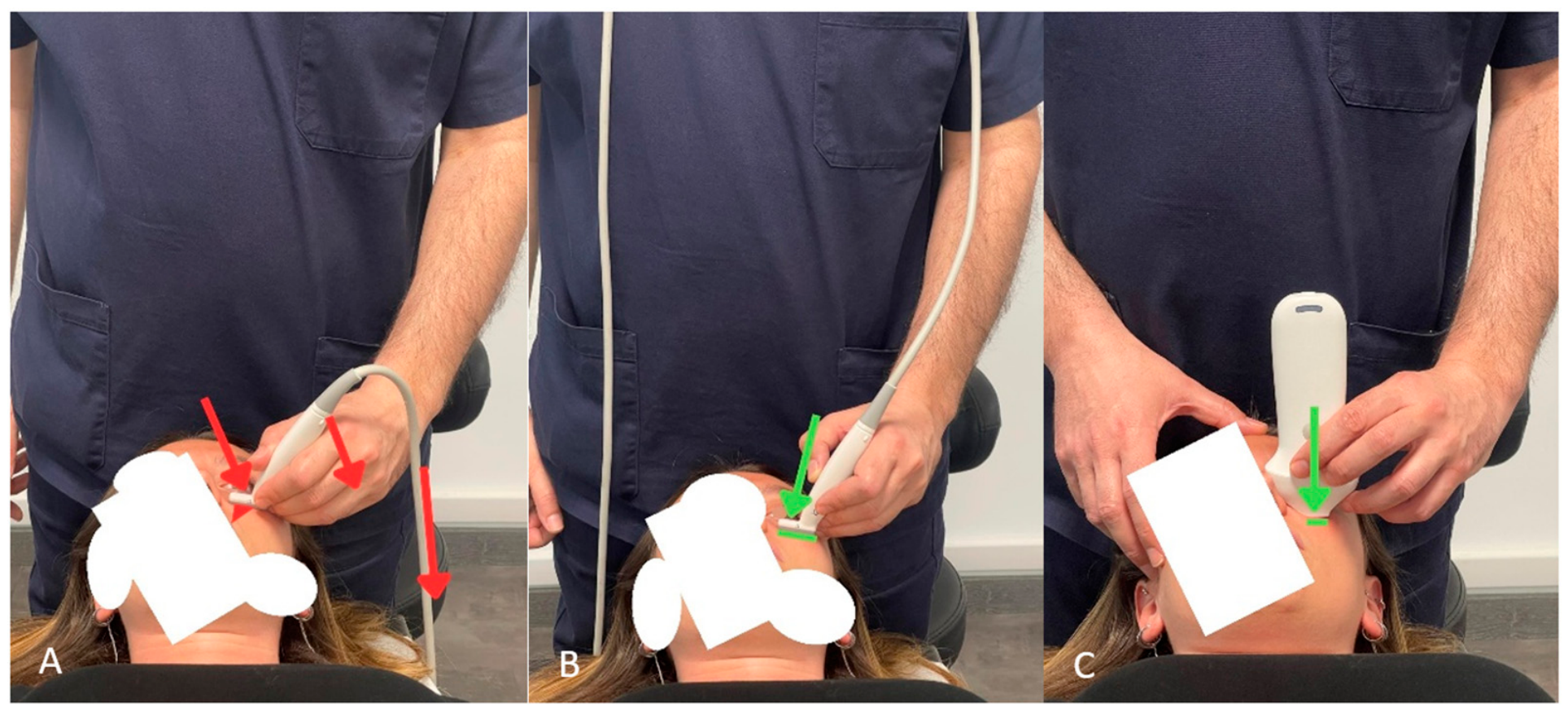
Although these design features offer plausible ergonomic advantages, direct comparative evidence that wireless probes reduce injury rates, improve procedural accuracy, or extend professional longevity remains limited. Therefore, while definitive comparative data are lacking, the reduced probe weight and the absence of cable constraints theoretically help minimize static wrist loading and cable-induced torque; however, comprehensive evaluations must also account for probe dimensions, handling characteristics, thermal behavior, and potential trade-offs in image quality and procedural requirements [14].
5. Relationship Between Ergonomics and Image Quality
This section establishes the core biomechanical framework linking operator posture, transducer forces, and tissue mechanics; these fundamental principles serve as the reference point for the specific clinical and procedural applications discussed throughout the subsequent sections.
In dermatologic and facial ultrasound, ergonomics should not be considered solely from an occupational health perspective. It is also a critical technical factor that may influence image acquisition and technical consistency. This aspect is particularly relevant when examining superficial, highly deformable structures, where even minor variations in transducer pressure, orientation, or stability can alter tissue morphology and acoustic representation on ultrasound.
5.1. Biomechanics of Transducer Pressure and Tissue Deformation
Transducer contact force deforms the tissues beneath the probe, and the force applied varies considerably among operators and clinical applications [17,18]. Unlike deeper anatomical targets, many structures examined in dermatologic and facial ultrasound are located only a few millimeters beneath the skin surface. Consequently, the mechanical interaction between the operator, transducer, coupling medium, and patient becomes an integral component of image acquisition. Contemporary reviews of high-frequency ultrasound (HFUS) emphasize its capacity for reproducible, quantitative assessment of superficial structures, underscoring the necessity of strictly controlling acquisition conditions when millimetric dimensional differences are clinically meaningful [19,20,21,22,23,24,25].
The physical and geometric effects of transducer pressure on ultrasound imaging have been quantified in the recent literature. In a systematic review, Suchoń et al. demonstrated substantial variability in the probe forces applied during ultrasound examinations and emphasized that force monitoring can significantly improve image quality and measurement reproducibility [17]. In an experimental phantom study focused on deformation correction for freehand 3D ultrasound, Dahmani et al. showed that probe indentation deforms underlying soft tissues, generating geometric distortion and reconstruction artifacts [18]. Their findings indicate that such deformations become particularly critical when precise localization or dimensional measurements are required. Although their quantitative validation involved a gelatin-based phantom in a 3D reconstruction setting, it provides experimental support for the general principle that probe-induced mechanical forces alter underlying ultrasound geometry [18].
Compression-related artifacts are especially impactful in facial anatomical mapping. Superficial fat compartments, thin mimetic muscles, facial veins, small arteries, filler deposits, and inflammatory collections can be easily displaced or flattened by excessive pressure. As a result, their apparent thickness, shape, depth, and spatial relationship with adjacent structures may be distorted during scanning. This consideration is vital during preprocedural anatomical mapping, where millimeter-scale relationships between vascular structures, muscles, and planned injection planes dictate clinical decisions and procedural safety.
5.2. Impact on Vascular Mapping and Microvascular Flow
In the context of this review, technical reproducibility primarily refers to scanning-plane consistency and intra-operator reliability. Technical reproducibility strictly depends on the correct positioning of both patient and operator, as proper ergonomic alignment directly contributes to maintaining consistent acquisition conditions.
An unstable operator posture leads to unintentional variations in transducer pressure. When the upper limb is unsupported or the shoulder remains abducted for prolonged periods, fine motor control of the hand degrades—particularly during extensive facial mapping. Similarly, excessive wrist extension or ulnar deviation reduces the operator’s ability to maintain a consistent probe orientation. These ergonomic flaws produce continuous unwanted changes in compression, angulation, and skin contact. Operator fatigue must therefore be recognized not only as an occupational hazard but also as a primary source of technical variability.
This concept is particularly critical when employing the floating technique. While a thick layer of gel minimizes direct compression of superficial structures, it also removes the mechanical stabilization normally provided by direct probe-to-skin contact [26]. The operator must keep the transducer at a constant distance from the skin while performing subtle translational and rotational movements. Supporting the forearm and stabilizing the hand—using the fifth finger as a reference point—enhances fine motor control and reduces unintended oscillations.
5.3. Summary Framework
The relationship between ergonomics and image quality operates as a continuous sequence of interrelated physical factors:
Operator Posture → Upper-Limb Stability → Probe Pressure & Orientation →
Tissue Mechanics → Image Accuracy & Reproducibility
Tissue Mechanics → Image Accuracy & Reproducibility
While this conceptual framework is strongly supported by biomechanical principles and evidence from general ultrasound literature, direct prospective clinical trials in facial ultrasound remain limited. Establishing ergonomic standardization thus serves as an essential counterpart to technical standardization, safeguarding operator health while optimizing diagnostic precision.
6. Standardization of Patient Positioning
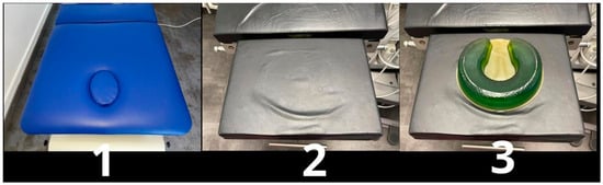
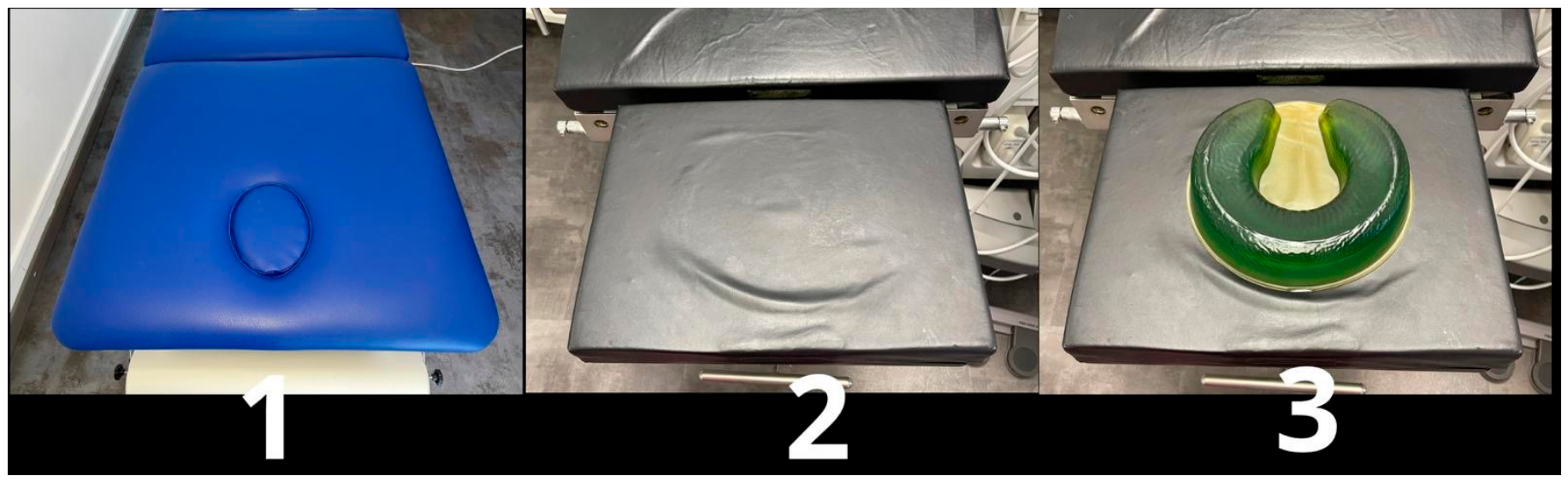
For both dermatologic and facial applications, the supine position is recommended to improve standardization and reproducibility. It helps maintain consistent spatial relationships between superficial and deeper structures, even in patients with marked elastosis, and provides a more standardized relationship between the heart and facial vasculature, potentially improving the reproducibility of Doppler and SMI microvascular assessments [9,10]. Small changes in head inclination can alter venous filling and anatomical landmarks; neutral alignment is therefore preferable. During periorbital or vascular imaging, soft cervical support and relaxed shoulder positioning help minimize motion artifacts. Examination of the neck and submandibular skin may require cervical hyperextension, with the head lowered and a pillow placed beneath the upper back. Headrest design also affects ergonomics. A wide headrest provides space for supporting the operator’s forearm and, during prolonged facial examinations, allows the use of an arm cushion to reduce wrist twisting. A narrower headrest facilitates access to the lower third of the face but provides less space for operator support. If the headrest lacks a dedicated opening for head stabilization, a contoured gel cushion can provide appropriate cervical support (Figure 2).
Figure 2.
Three types of headrests. (1) A wide headrest provides adequate support for the operator’s arms. (2) A narrow headrest facilitates access to the lower third of the patient’s face. (3) A cushion improves head stability and patient comfort.
Recent dermatologic-ultrasound literature likewise emphasizes standardized techniques and reproducible acquisition protocols as prerequisites for reliable anatomical and quantitative assessments [19,20,21,22,23,27].
7. Operator Positioning and Posture
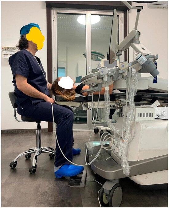
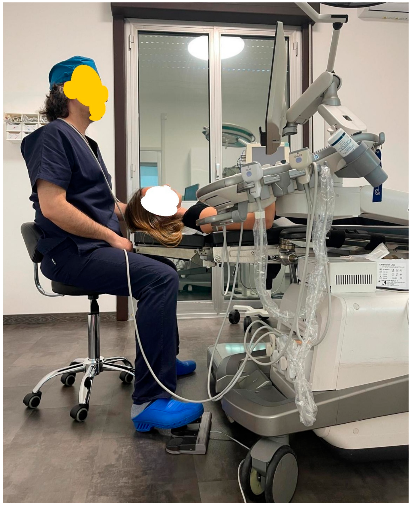
The operator may choose to stand or sit and may be positioned either in front of the patient or behind the patient’s head (Figure 3).
Figure 3.
Standardized supine positioning for facial ultrasound. The ultrasound system is placed beside the examination table, with the operator positioned at the head of the table. Other operators may prefer to work from in front of the patient.
While specific spatial setups are frequently presented as preferred configurations, it is crucial to emphasize that an absolute, universally optimal positioning does not exist. Ideal ergonomic posture depends heavily on individual factors, including operator anthropometry, handedness, the specific facial region under evaluation, ultrasound system characteristics, and procedural requirements. Rather than strictly adhering to a rigid setup, operators should prioritize individualized ergonomic adjustments that accommodate their personal physical proportions and clinical habits.
Each of these configurations has advantages and disadvantages. Sitting may provide greater stability for the elbow and forearm but can encourage an unfavorable trunk posture. Positioning the operator behind the patient’s head, with the ultrasound system on the right for right-handed operators, creates a balanced arrangement that minimizes trunk rotation. This configuration, described in ergonomic risk assessments of sonographers, reduces repetitive asymmetric loading and facilitates visual attention to both the face and the monitor. However, during facial mapping, this position may make it more difficult to visualize and interpret the orientation of superficial facial layers and structures.
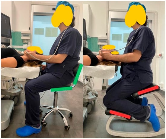
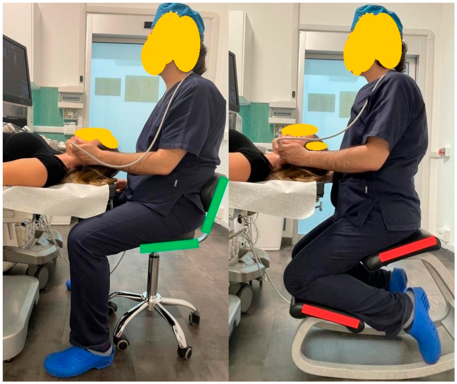
To prevent work-related musculoskeletal disorders (WRMSDs), the operator must systematically maintain a neutral posture. This requires an upright trunk with proper lumbar support, elbows kept close to the torso and flexed at approximately 90°, and the wrist aligned with the probe axis without ulnar deviation (Figure 4). According to Weimer et al., sustained shoulder abduction beyond 30°, trunk flexion or rotation, and wrist extension greater than 20° are among the postures most strongly associated with higher Rapid Upper Limb Assessment (RULA) risk scores [5]. Ergonomic awareness training and targeted postural alignment during examinations are essential to help reduce these high-risk angles.
Figure 4.
Appropriate operator posture and hand alignment, based on the relationship between chair height and examination-table height.
Although these quantitative thresholds (e.g., shoulder abduction > 30° and wrist extension > 20°) provide valuable general guidance, their strict maintenance can be challenging during delicate facial scans where micro-manipulation and fine motor control are paramount. Consequently, they should be interpreted as ergonomic reference limits rather than rigid operational constraints across all facial sub-regions.
8. Ultrasound System Management and Adjustment of Tables and Seating
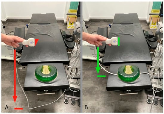
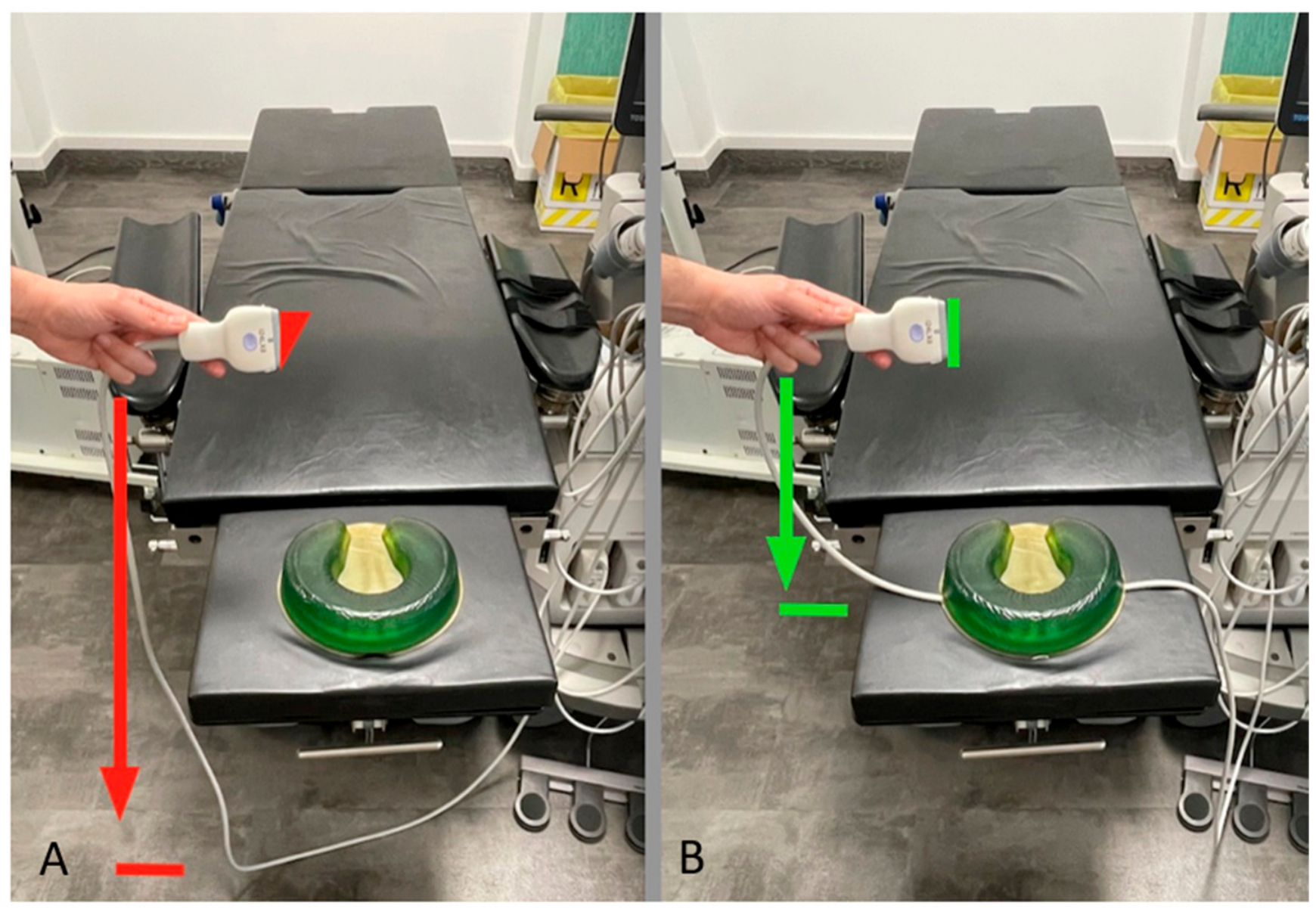
The shape and footprint of the transducer can either hinder or facilitate facial examination [28]. When a cabled probe is required, cable tension can be managed by lowering the examination table and allowing the cable to rest loosely over the operator’s shoulder or neck, thereby distributing its weight without restricting movement. If the cable is too short to be placed over the operator’s neck or shoulder, the patient’s headrest cushion can be utilized to support the cable and reduce the mechanical load transmitted directly to the transducer. Height-adjustable examination tables and operator chairs remain essential ergonomic hardware components (Figure 5).
Figure 5.
Probe-cable length as an ergonomic consideration. (A) A cable that is too short to pass over the operator’s neck can exert a downward force on the transducer. (B) Placing the cable beneath the patient’s headrest cushion reduces the load transmitted to the probe.
To achieve individualized adjustments efficiently during daily clinical practice, simple calibration tools can be implemented. For instance, a quick and practical method to adapt couch height to individual anthropometry involves using a small magnet tied to a string with a weighted bead or ball at the end. Because each operator requires a specific string length based on their height and arm span, the magnet can be fixed to the frame of the height-adjustable examination table, allowing the practitioner to rapidly set the couch to their ideal customized height before starting the procedure.
Regarding seating options, forward-sloping seats (10–15°) reduce hip flexion and promote lumbar lordosis, supporting a more neutral spinal posture compared with a flat seat [29]. However, because such seats may increase lower-limb muscle strain or cause the body to slide forward unless adequate friction is provided, they require careful selection, particularly if footswitch controls are used [30].
9. Ergonomic Innovations and Portable Systems
Recent handheld devices incorporating wireless high-frequency (HF) probes (20–24 MHz) represent important advances in ergonomics by simplifying workspace organization and providing high-resolution imaging of epidermal and subdermal structures [31,32]. Weimer et al. emphasized that ergonomic workload increases with total examination duration; accordingly, lightweight, cable-free systems may help reduce cumulative strain [5]. However, evaluating portable and handheld ultrasound requires balancing these ergonomic benefits against several technical trade-offs, including device dimensions, thermal behavior, and imaging performance. Many current wireless or handheld devices remain bulky and may cover the injection site, preventing simultaneous injection and direct observation of the treated area—a limitation that is particularly relevant in aesthetic procedures where visual monitoring during injection is recommended. To address this, fingerheld probes have recently been introduced to allow simultaneous injection and real-time imaging [33,34].
Nevertheless, portability should not automatically be equated with superior ergonomics or image performance. In a 2024 comparison of six handheld systems, experts identified image quality, ease of use, portability, probe size, and battery life among the most desirable characteristics, but no single device provided all the desired features [35]. Furthermore, handheld systems often exhibit lower image quality or reduced Doppler capabilities compared with cart-based equipment, alongside potential challenges related to device heat generation and battery duration. These findings indicate that device suitability must be assessed critically according to the specific dermatologic or facial application rather than solely on whether the system is portable or cart-based [36].
10. Ergonomics of Ultrasound-Guided Aesthetic Procedures
Ultrasound-guided aesthetic procedures represent a direct extension of the ergonomic requirements established in diagnostic ultrasound, while introducing unique visual, spatial, and procedural constraints. In conventional diagnostic scanning, the operator can generally dedicate one hand to the transducer and use the other to adjust machine settings, reposition the patient, or stabilize the probe. In contrast, interventional procedures require the simultaneous management of the transducer and the injection device (needle or cannula), continuous visualization of the target anatomy, real-time control of the trajectory, and careful organization of the sterile procedural field. The operator must simultaneously track needle advancement on the monitor, observe the external treatment area, and remain attentive to tissue responses and patient comfort. This procedural complexity is increasingly relevant in aesthetic medicine, where recent reviews and practical guidance describe both pre-injection mapping and real-time ultrasound-guided injection as complementary strategies [26,27,37,38,39,40,41,42,43].
Together, these demands create a complex visuomotor and visuospatial task. The AIUM curriculum identifies both components as fundamental psychomotor skills in ultrasound-guided procedures: visuomotor skills involve hand–eye coordination during transducer manipulation, whereas visuospatial skills require the operator to reconstruct three-dimensional anatomical relationships from two-dimensional ultrasound images [44]. Needle guidance additionally requires coordination of transducer position, ultrasound beam, needle trajectory, and target anatomy. Adhikary et al. specifically highlighted needle-beam alignment and real-time needle tracking as essential skills and noted that the operator must coordinate movements occurring in three dimensions while observing a two-dimensional ultrasound image [45]. Structured simulation-based training can help address these requirements: Tokumine et al. describe competency-based training focused on needle and probe manipulation, including probe adjustment during needle advancement, together with assessment of needle-tip visualization and hand-eye coordination [46].
Ultrasound-guided facial injection adds further geometric constraints to this already complex visuomotor task. Many facial targets are extremely superficial, so an in-plane approach may require a very shallow needle-to-skin angle and the needle must remain precisely aligned with the narrow ultrasound scan plane. Shiao and Kuo showed that ultrasound-guided targeting depends critically on both needle–beam alignment and correct control of the insertion angle, noting that needle angles below 45° are common in clinical practice [47]. These requirements are amplified by the limited working surface of the face: a conventional linear transducer may occupy a substantial proportion of the procedural field, whereas a small-footprint hockey-stick probe improves access but offers a smaller area of contact and stabilization, thereby demanding even finer coordination between probe and needle. Needle caliber adds to the technical challenge because facial aesthetic procedures may involve devices ranging from approximately 25-gauge needles for filler placement to 34-gauge needles for very superficial botulinum toxin injections [48,49]. Even 29- to 31-gauge insulin needles can be visualized under high-frequency ultrasound guidance under optimal experimental conditions. However, clinical discussions must explicitly distinguish technical feasibility from consistent real-time clinical performance. While ultra-high-frequency transducers possess the technical capability to resolve fine-gauge needles, maintaining continuous, reliable visualization throughout dynamic facial injections remains heavily constrained by tissue elasticity, needle deflection, shallow insertion angles, and minor operator movements. Consequently, technical feasibility under controlled settings should not be equated with uniform clinical reliability during everyday interventional practice [37].
Maintaining a strict aseptic field is paramount during ultrasound-guided procedures, and ergonomic maneuvers must be fully compatible with infection prevention protocols. In a single-operator setup, sterile preparation and probe handling can be efficiently accomplished without compromising asepticity. The operator can utilize a dedicated Mayo stand covered with a sterile drape placed immediately adjacent to the patient’s head. The transducer—fitted with a sterile sheath—is prepared and parked directly on this sterile Mayo stand. To maintain sterility while working unassisted, the operator can employ a One-Hand Pull-Back Technique to cover the probe with its sterile sheath and gel. This configuration provides a clean, stable rest for the transducer during procedural transitions, allowing a single operator to safely perform real-time guidance while strictly adhering to aseptic standards.
The procedural execution of ultrasound-guided injections can be structured around different organizational models: single-operator vs. two-operator (team-based) setups. Current international consensus and expert recommendations in aesthetic ultrasound do not explicitly mandate a two-operator approach for all interventional settings. Performing ultrasound-guided procedures as a single operator is not inherently unsafe in every circumstance; safety fundamentally depends on the intrinsic complexity of the intervention, operator competence, manual dexterity, strict adherence to aseptic technique, target accessibility, and the ability to maintain continuous ultrasound visualization.
However, in high-risk anatomical zones or technically complex procedures, a two-operator setup—where one clinician maintains optimal ultrasound visualization while the other performs the injection—can significantly improve procedural control, maintain sterile field integrity, and optimize the management of potential acute complications. Therefore, the choice of organizational model should be dictated by comprehensive risk assessment and procedural complexity rather than ergonomic convenience alone.
The operator’s visual attention repeatedly shifts between the ultrasound monitor and the procedural field, creating a specific ergonomic and cognitive challenge during ultrasound-guided interventions. Liao et al. described this split-attention condition during ultrasound-guided central venous catheter placement, in which operators must shift their visual focus between the ultrasound screen and the patient while coordinating hand movements [50]. In their randomized crossover trial, displaying the ultrasound image through an augmented-reality head-mounted display reduced the time required for several critical procedural steps and minimized neck strain compared with conventional ultrasound visualization. Cognitive workload, assessed using the NASA-TLX, generally tended to be lower with augmented-reality visualization, although statistically significant differences were limited to selected dimensions and trainee subgroups [50]. Although augmented-reality systems are not yet routinely used, these findings support a broader ergonomic principle: reducing the spatial separation between the ultrasound image and the procedural field could reduce unnecessary head movements and potentially decrease the cognitive burden associated with split visual attention.
This issue may be particularly relevant in aesthetic medicine. Direct observation of the treatment area remains important because changes in skin color, swelling, bleeding, patient responses, and the distribution of injected material may provide clinically relevant information that cannot be obtained from the ultrasound image alone. The ergonomics of the procedure should therefore facilitate rapid visual transitions between the screen and the patient without requiring repeated rotation or flexion of the neck and trunk. High-frequency Doppler ultrasound is increasingly used to define treatment planes and vascular anatomy before and during filler injection, particularly in anatomically high-risk facial regions [37,38,39,40,41,42,43]. The literature on facial injections provides direct support for this spatial organization: Desyatnikova describes ergonomic alignment of the operator, patient, and monitor before temple injection, followed by real-time visualization of the cannula and filler deposition [26].
Monitor positioning is therefore an important component of procedural ergonomics. Ideally, the ultrasound display, treatment area, and operator’s line of sight should be arranged to minimize extensive head movements. In a pilot ergonomic task analysis of ultrasound-guided femoral nerve blocks, Ajmal et al. identified the simultaneous demands of probe manipulation, needle advancement, and ultrasound-screen visualization as a major ergonomic challenge [51]. Excessive head, neck, or back rotation and marked thoracolumbar flexion were frequently observed, particularly among less experienced operators; however, given the small sample size, the authors regarded the apparent association between experience and better ergonomic performance as exploratory rather than definitive [51]. Expert recommendations for ultrasound-guided injections similarly emphasize workstation organization and appropriate positioning of the physician, patient, and ultrasound machine [52].
The height of the examination table should allow stabilization of the scanning arm while permitting unrestricted manipulation of the needle or cannula with the contralateral hand. During ultrasound-guided injection, stability of both hands is particularly important because movement of either the transducer or needle changes their spatial relationship. Expert-group recommendations for ultrasound-guided chemodenervation emphasize preparing the workspace in advance and optimization of physician, patient, and ultrasound-machine positioning before injection [52]. Because these recommendations are based on expert consensus rather than comparative trials, they should be interpreted as practical guidance rather than evidence of a demonstrated effect on clinical outcomes.
Probe geometry takes on particular importance during ultrasound-guided procedures. Conventional linear probes may provide excellent image quality but occupy a relatively large portion of the procedural field. Some palmar wireless probes eliminate cable-related constraints and provide greater flexibility in workstation organization, but their footprint may partially cover the injection area and limit direct visualization or access. This is particularly relevant in small anatomical regions of the face, because the available surface for simultaneous probe placement and needle entry is limited. Recent practical guidance covering the upper, middle, and lower thirds of the face also illustrates how transducer footprint, access route, vascular mapping, and the choice between “scan before injecting” and “scan while injecting” interact during procedural planning [38,39,40].
Fingerheld and other miniaturized transducers represent a promising ergonomic development because their reduced footprint may permit simultaneous injection and real-time imaging [33,34]. They can potentially reduce spatial interference between the transducer and the hand performing the procedure. However, miniaturization alone does not ensure optimal procedural ergonomics. Image resolution, stability, field of view, Doppler capabilities, heat generation, battery duration, and the ability to maintain the desired scanning plane must also be considered. A smaller transducer is advantageous only when it provides sufficient diagnostic information for the intended procedure.
Cable management is particularly important during ultrasound-guided intervention. During diagnostic scanning, cable weight primarily affects the scanning hand. During an injection procedure, however, an improperly positioned cable may also interfere with the needle, cannula, syringe, sterile field, or operator’s contralateral arm. Wireless probes eliminate this interaction, whereas conventional systems require careful cable positioning before the procedure begins. Cable position should therefore be considered part of procedural planning rather than adjusted after needle insertion.
Ergonomic optimization of ultrasound-guided aesthetic interventions ultimately requires treating the operator, patient, ultrasound system, transducer, monitor, and injection device as an integrated procedural environment. The objective is not simply to obtain a comfortable posture, but to create a configuration in which both hands can perform precise, independent tasks—whether managed by a single skilled operator or split across a two-person team based on risk assessment—while visual attention shifts efficiently between the ultrasound image and the patient. Available evidence demonstrates that ultrasound-guided procedures impose specific visuomotor, visuospatial, and ergonomic demands [44,45,46,50,51,52]. However, these relationships have been investigated predominantly in vascular access, regional anesthesia, and ultrasound-guided chemo-denervation rather than facial aesthetic ultrasound. Applying these findings to facial procedures should therefore be regarded as a physiologically and ergonomically plausible extrapolation that requires specific validation. This procedural perspective should increasingly become part of training in ultrasound-guided aesthetic medicine.
11. Footswitch and Hands-Free Control
Hands-free controls may be particularly valuable during ultrasound-guided procedures. When one hand holds the transducer and the other controls the injection device, operating the conventional console becomes difficult without interrupting the procedure. A single or multiple foot pedal, whether wired or wireless, can enable primary acquisition functions such as freezing or saving images while minimizing the need to reach for console controls. When evaluating this technology, the applicability of the cited foot-pedal literature to ultrasound image acquisition controls requires clear contextualization. Foundational studies on foot-activated devices (such as Hama et al. [53]) focused on power injectors for contrast-enhanced ultrasound rather than native console software navigation. Extrapolating these findings to diagnostic and interventional image capture is nevertheless ergonomically justified: transferring basic input commands from the hands to the feet preserves upper-limb stability, maintains uninterrupted needle tracking, and reduces overall visual switching during delicate facial interventions. Voice commands and automated image recording may offer additional solutions to improve workflow and maintain continuous procedural focus.
12. Education, Awareness, and Training
New operators should understand the benefits that ultrasound can offer in dermatology and aesthetic assessment of the face. They should also recognize that ultrasound should be used when clinically indicated, in accordance with international guidelines and the operator’s experience. Unnecessary ultrasound imaging increases costs, examination time, and operator workload without providing additional benefit. Although ergonomic principles are included in several sonography curricula, their implementation remains limited [54]. The findings of Weimer et al. support the integration of ergonomic assessment and training using kinematic feedback systems (e.g., inertial sensors combined with RULA scoring) into educational programs [5]. Active ergonomic education may promote early awareness of postural loading, self-correction, and workstation-adjustment skills, potentially helping prevent WRMSDs among early-career operators (Table 1). Such structured training could become a routine component of ultrasound certification, consistent with AIUM and EFSUMB standards emphasizing operator well-being as part of quality assurance [1,55]. Participatory ergonomics has been shown to facilitate the identification of occupation- and site-specific WRMSD risks [56]. Recent systematic reviews and participatory-ergonomics studies support combining education with workstation modifications and organizational measures rather than relying solely on postural awareness [4,14].
Table 1.
Ergonomic factors in facial ultrasound and their potential impact on examination quality.
A comprehensive training framework in facial and dermatologic ultrasound must explicitly distinguish between general ergonomic awareness and technical ultrasound competency. While ergonomic education focuses on operator posture, workstation configuration, and the prevention of work-related musculoskeletal disorders (WRMSDs), technical ultrasound competency requires rigorous, structured acquisition of specific psychomotor and diagnostic skills. Objective assessment models should systematically evaluate core technical domains.
12.1. Probe Handling and Pressure Control
Trainees must master the transition between standard contact and the “floating technique,” learning to apply the minimal necessary pressure to avoid tissue deformation, geometric alterations, and compression of compliant superficial structures such as facial vessels.
12.2. Image Optimization
Competency programs should assess the operator’s ability to adjust frequency, gain, focus, and presets efficiently to maximize resolution in superficial layers without compromising frame rate or diagnostic depth.
12.3. Anatomical Identification
Trainees must demonstrate precise recognition of multi-layered facial structures, including mimetic muscles, fat compartments, fascial planes, and high-risk vascular networks.
12.4. Visuomotor and Visuospatial Coordination
Particularly during ultrasound-guided aesthetic interventions, operators must develop advanced hand-eye coordination to manage simultaneous transducer and needle movements, aligning the needle trajectory with the narrow ultrasound scan plane while continuously interpreting two-dimensional representations of three-dimensional anatomical targets.
13. A Practical Ergonomic Workflow for Facial Ultrasound
To minimize cumulative biomechanical load and promote scanning consistency, ergonomic principles should be structured into a systematic clinical workflow. The following steps outline the practical implementation of workstation configuration, patient setup, and procedural organization across all phases of the examination (Table 2). These suggestions are presented as practical and ergonomically plausible guidance rather than validated standards.
Table 2.
Practical ergonomic workflow for facial ultrasound and ultrasound-guided procedures.
13.1. Before Scanning
Workstation preparation must occur before transducer contact. Adjust the chair and examination table height according to individual operator anthropometry—utilizing practical calibration tools such as personalized magnet-and-string height indicators—and place the monitor directly within the natural line of sight to avoid repeated trunk and neck rotation. Cable management or wireless options should be arranged prior to starting the procedure [13].
13.2. Patient Positioning
Standardized supine patient positioning with neutral head alignment is recommended to ensure reproducible spatial anatomy and consistent vascular access. Headrest selection should balance patient stabilization with adequate arm support for the operator (Table 2).
13.3. Probe Handling
Maintain a neutral wrist alignment and a relaxed grip throughout scanning. The forearm and elbow should be supported whenever anatomy permits, using the fifth finger as an anatomical anchor to stabilize fine transducer movements without exerting excessive downward pressure [17,18].
13.4. Image Acquisition
Image optimization should be performed without disrupting operator posture. Standardized presets reduce unnecessary console manipulation. During vascular assessment, maintain minimal and consistent probe pressure to preserve microvascular flow signals [26].
13.5. Ultrasound-Guided Procedures
Prior to puncture, verify visual alignment between the monitor, procedural field, and line of sight. Equipment parking, sterile draping, probe sheath placement, and cable routing must be established beforehand to ensure both asepsis and ergonomic stability. Selecting an appropriate probe footprint ensures proper balance between procedural access and plane stability, while footswitches enable continuous hands-free operation [26,27,37,38,39,40,41,42,43].
13.6. Prolonged Examinations and Repeated Scanning
To mitigate cumulative static loading during extended facial mapping sessions, operators should introduce brief muscle recovery periods, alternate posture when feasible, and adapt workstation setup dynamically when transitioning between diagnostic and interventional modes (Table 2).
14. Future Directions and Technological Perspectives
Facial examinations performed using dedicated ultrasound presets and detailed, standardized protocols may be completed more efficiently, thereby improving examination ergonomics [6,7,57,58,59]. To advance the field further, future developments and technological innovations can be systematically categorized into currently available solutions and emerging or experimental approaches. Prospective research should systematically evaluate biomechanical differences between cart-based and wireless systems, linear and angular comfort thresholds during fine movements, and the cost-effectiveness and accessibility of these ergonomic innovations.
14.1. Currently Available Solutions
Several tools and system features are already accessible in contemporary clinical practice to support workstation organization and workflow efficiency.
14.1.1. Ergonomic Hardware Adjustments
Height-adjustable examination tables, articulated monitor arms, and specialized operator seating (such as forward-sloping seats) allow for immediate anthropometric customization.
14.1.2. Consolidated Portable and Wireless Systems
Handheld and wireless high-frequency probes are commercially available, helping to eliminate cable traction and torque during examinations.
14.1.3. Standardized Footswitches
Wired or wireless foot pedals are currently implemented on many ultrasound platforms to freeze or save images without interrupting manual probe control.
14.2. Emerging and Experimental Solutions
Other technological and methodological approaches are currently in developmental, evaluative, or preliminary research phases.
14.2.1. Advanced Artificial Intelligence (AI) and Automation
Greater integration of AI software and automated functions into ultrasound systems aims to shorten examination times and reduce operator workload [60,61,62,63].
14.2.2. Real-Time Motion-Tracking and Postural Feedback
Emerging kinematic tracking systems—such as inertial sensors combined with automated Rapid Upper Limb Assessment (RULA) integration—are being explored to provide continuous feedback on operator posture during scanning.
14.2.3. AI-Based Image Stabilization
Experimental image stabilization tools seek to mitigate artifacts caused by unintentional pressure variations and physiological tremor.
14.2.4. Augmented Reality (AR) and Alternative Displays
Head-mounted AR displays and advanced visualization prototypes are being investigated to reduce neck strain and bridge the spatial gap between the screen and the procedural field.
14.2.5. Miniaturized Transducers (e.g., Fingerheld Probes)
Next-generation ultra-small or fingerheld probes designed for simultaneous injection and real-time imaging are undergoing initial clinical evaluation, though their long-term efficacy and technical trade-offs require further prospective validation.
15. Conclusions
Ergonomic optimization is essential in facial ultrasound. Modern dermatologic and facial ultrasound combines clinical precision with technological advances but continues to pose substantial ergonomic risks to operators. Appropriate workspace preparation and positioning of both the patient and the clinician are therefore necessary. Evidence from kinematic analyses indicates that prolonged static postures, excessive shoulder abduction, and wrist deviation contribute significantly to WRMSDs among ultrasound professionals. While direct prospective clinical trials specifically confirming the impact of ergonomics on diagnostic reproducibility remain limited, integrating comprehensive ergonomic measures—grounded in established biomechanical principles—helps safeguard operator health while providing a stable foundation for high-quality ultrasound practice in dermatology and facial medicine.
Rather than proposing universally optimal setups, ergonomic protocols in facial ultrasound must emphasize individualized adjustments. Practical tools for rapid anthropometric adaptation—combined with height-adjustable furniture and flexible device handling—help maintain an ergonomic operator posture regardless of physical differences, handedness, or clinical setting.
Current handheld devices differ substantially in image quality, probe geometry, Doppler availability, and other imaging capabilities. Some provide color-flow Doppler and acceptable imaging of superficial structures, but no single handheld system currently combines all desirable features. Device suitability should therefore be assessed according to the intended dermatologic or aesthetic application rather than solely on whether the system is handheld or cart-based.
Furthermore, the ergonomic principles discussed in this review may extend beyond facial ultrasonography to other diagnostic and therapeutic procedures requiring precision, sustained visual attention, and coordinated hand movements. These include ultrasound-guided interventions, injectable aesthetic treatments, and energy-based procedures. Although the specific ergonomic requirements differ across techniques, operator positioning, workspace organization, instrument handling, and visual alignment represent shared determinants of procedural efficiency and potentially of technical consistency and safety. Further studies are needed to establish the extent to which these principles translate into measurable clinical benefits.
Author Contributions
Conceptualization, A.M.S.; methodology, A.M.S., O.C., X.W., C.T. and A.C.; writing—original draft preparation, A.M.S., O.C., X.W., C.T. and A.C.; writing—review and editing, A.M.S., O.C., X.W., C.T. and A.C. All authors have read and agreed to the published version of the manuscript.
Funding
This research received no external funding.
Institutional Review Board Statement
Not applicable.
Informed Consent Statement
Written informed consent was obtained from the patient and the doctor for publication of this article and these pictures.
Data Availability Statement
No new data were created or analyzed in this study. Data sharing is not applicable to this article.
Conflicts of Interest
The authors declare no conflicts of interest.
Abbreviations
The following abbreviations are used in this manuscript:
| AI | Artificial intelligence |
| HF | High-frequency |
| RULA | Rapid upper limb assessment |
| WRMSD | Work-related musculoskeletal disorders |
References
- AIUM Practice Principles for Work-Related Musculoskeletal Disorder. J. Ultrasound Med. 2023, 42, 1139–1157. [Google Scholar] [CrossRef] [Scilit] [PubMed]
- Claes, F.; Berger, J.; Stassijns, G. Arm and neck pain in ultrasonographers. Hum. Factors 2015, 57, 238–245. [Google Scholar] [CrossRef] [Scilit] [PubMed]
- Feng, Q.; Liu, S.; Yang, L.; Xie, M.; Zhang, Q. The Prevalence of and Risk Factors Associated with Musculoskeletal Disorders among Sonographers in Central China: A Cross-Sectional Study. PLoS ONE 2016, 11, e0163903. [Google Scholar] [CrossRef] [Scilit] [PubMed]
- AlMubarek, N.A.; Al-Otaibi, S.T.; Herzallah, H.K. Musculoskeletal disorders among sonographers in secondary care hospitals in the city of Al-Ahsa, Saudi Arabia. WORK 2022, 71, 1105–1111. [Google Scholar] [CrossRef] [Scilit] [PubMed]
- Weimer, J.M.; Dumancic, B.; Weinmann-Menke, J.; Rombusch, J.; Ernst, B.; Krambeck, A.; Golbach, R.; Topal, E.; Maurer-Grubinger, C.; Ille, C.; et al. A new approach for the implementation of ergonomics in sonography to prevent work-related musculoskeletal disorders (ErgoSon). J. Occup. Med. Toxicol. 2025, 20, 11. [Google Scholar] [CrossRef] [Scilit] [PubMed]
- Chammas, M.C.; Sigrist, R.; Alfageme, F.; Gonzalez, C.; Cavallieri, F.; Desyatnikova, S.; Crisan, M.; Velthuis, P.; Schelke, L.; Catalano, O.; et al. WFUMB Position Paper: Consensus on Best Practice in Aesthetic Dermatologic Ultrasound. Ultrasound Med. Biol. 2025, 51, 2173–2193. [Google Scholar] [CrossRef] [Scilit] [PubMed]
- Velthuis, P.; Alfageme, F.; Cartier, H.; Cassuto, D.; Catalano, O.; Cavallieri, F.; Desyatnikova, S.; Diaz, C.G.; Kim, J.; Mandava, A.; et al. The use of ultrasound imaging in aesthetic injectables: A modified Delphi consensus. J. Plast. Reconstr. Aesthetic Surg. 2025, 104, 33–37. [Google Scholar] [CrossRef] [Scilit] [PubMed]
- Zangiabadi, Z.; Makki, F.; Marzban, H.; Salehinejad, F.; Sahebi, A.; Tahernejad, S. Musculoskeletal disorders among sonographers: A systematic review and meta-analysis. BMC Health Serv. Res. 2024, 24, 1233. [Google Scholar] [CrossRef] [Scilit] [PubMed]
- Catalano, O.; Wortsman, X. Dermatology Ultrasound. Imaging Technique, Tips and Tricks, High-Resolution Anatomy. Ultrasound Q. 2020, 36, 321–327. [Google Scholar] [CrossRef] [Scilit] [PubMed]
- Gonzalez, C.; Wortsman, X. How to Start on Dermatologic Ultrasound: Basic Anatomical Concepts, Guidelines, Technical Considerations, and Best Tips. Semin. Ultrasound CT MRI 2024, 45, 180–191. [Google Scholar] [CrossRef] [Scilit] [PubMed]
- Baker, J.P.; Coffin, C.T. The Importance of an Ergonomic Workstation to Practicing Sonographers. J. Ultrasound Med. 2013, 32, 1363–1375. [Google Scholar] [CrossRef] [Scilit] [PubMed]
- A Soria-Bastida, M.; Hernández-Díaz, C.; de León, A.O.-G.; Coronado-Zarco, R. Ergonomics to perform thoracic limb musculoskeletal ultrasound. Acta Ortop. Mex. 2025, 39, 93–98. [Google Scholar] [PubMed]
- Harrison, G.; Harris, A. Work-related musculoskeletal disorders in ultrasound: Can you reduce risk? Ultrasound 2015, 23, 224–230. [Google Scholar] [CrossRef] [Scilit] [PubMed]
- Sweeney, K.; Mackey, M.; Spurway, J.; Clarke, J.; Ginn, K. The effectiveness of ergonomics interventions in reducing upper limb work-related musculoskeletal pain and dysfunction in sonographers, surgeons and dentists: A systematic review. Ergonomics 2020, 64, 1–38. [Google Scholar] [CrossRef] [Scilit] [PubMed]
- Fowlkes, J.B.; Kripfgans, O.D.; Bagley, J. Safety, Ergonomics and Patient Centered Point-of-Care Ultrasound. Med. Clin. North Am. 2025, 109, 325–341. [Google Scholar] [CrossRef] [Scilit] [PubMed]
- Kong, Y.-K.; Cho, M.-U.; Park, C.-W.; Kim, S.-Y.; Kim, M.-J.; Moon, J.; Lim, S.; Oh, B.-M.; Han, B.; Choi, J.; et al. Quantification of physical stress experienced by obstetrics and gynecology sonographers: A comparative study of two ultrasound devices. Appl. Ergon. 2022, 100, 103665. [Google Scholar] [CrossRef] [Scilit] [PubMed]
- Suchoń, S.; Burkacki, M.; Chrzan, M.; Winder, M. What Ranges of Probe Pressure Are Applied During Ultrasound Examinations? A Systematic Review. Sensors 2025, 25, 3415. [Google Scholar] [CrossRef] [Scilit] [PubMed]
- Dahmani, J.; Petit, Y.; Laporte, C. Quantitative validation of two model-based methods for the correction of probe pressure deformation in ultrasound. Int. J. Comput. Assist. Radiol. Surg. 2024, 19, 309–320. [Google Scholar] [CrossRef] [Scilit] [PubMed]
- Granieri, G.; Oranges, T.; Morganti, R.; Janowska, A.; Romanelli, M.; Manni, E.; Dini, V. Ultra-high frequency ultrasound detection of the dermo-epidermal junction: Its potential role in dermatology. Exp. Dermatol. 2022, 31, 1863–1871. [Google Scholar] [CrossRef] [Scilit] [PubMed]
- Vergilio, M.M.; e Silva, S.A.M.; Jales, R.M.; Leonardi, G.R. High-frequency ultrasound as a scientific tool for skin imaging analysis. Exp. Dermatol. 2021, 30, 897–910. [Google Scholar] [CrossRef] [Scilit] [PubMed]
- Levy, J.; Barrett, D.L.; Harris, N.; Jeong, J.J.; Yang, X.; Chen, S.C. High-frequency ultrasound in clinical dermatology: A review. Ultrasound J. 2021, 13, 24. [Google Scholar] [CrossRef] [Scilit] [PubMed]
- Cisoń, H.; Białynicki-Birula, R. High-frequency ultrasonography: One of the modern imaging diagnostic methods in dermatology. Authors’ own experience and review. Postepy Dermatol. Allergol. 2024, 41, 306–313. [Google Scholar] [CrossRef] [Scilit] [PubMed]
- Wortsman, X. Top Advances in Dermatologic Ultrasound. J. Ultrasound Med. 2022, 42, 521–545. [Google Scholar] [CrossRef] [Scilit] [PubMed]
- Chai, K.; Zhu, R.; Luo, F.; Shi, Y.; Liu, M.; Xiao, Y.; Xiao, R. Updated Role of High-frequency Ultrasound in Assessing Dermatological Manifestations in Autoimmune Skin Diseases. Acta Derm.-Venereol. 2022, 102, adv00765. [Google Scholar] [CrossRef] [Scilit] [PubMed]
- Sellyn, G.E.; Lopez, A.A.; Ghosh, S.; Topf, M.C.; Chen, H.; Tkaczyk, E.; Powers, J.G. High-frequency ultrasound accuracy in preoperative cutaneous melanoma assessment: A meta-analysis. J. Eur. Acad. Dermatol. Venereol. 2025, 39, 86–96. [Google Scholar] [CrossRef] [Scilit] [PubMed]
- Desyatnikova, S. Ultrasound-Guided Temple Filler Injection. Facial Plast. Surg. Aesthetic Med. 2022, 24, 501–503. [Google Scholar] [CrossRef] [Scilit] [PubMed]
- Cavallieri, F.A.; Balassiano, L.K.d.A.; Munhoz, G.; Tembra, M.F.; Wortsman, X. Ultrasound in Aesthetics: Filler and Non-Filler Applications. Semin. Ultrasound CT MRI 2024, 45, 251–263. [Google Scholar] [CrossRef] [Scilit] [PubMed]
- Wortsman, X. Key Points to Select a Device for Dermatologic Ultrasound. J. Ultrasound Med. 2023, 42, 1367–1369. [Google Scholar] [CrossRef] [Scilit] [PubMed]
- Graf, M.; Guggenbühl, U.; Krueger, H. Investigations on the effects of seat shape and slope on posture, comfort and back muscle activity. Int. J. Ind. Ergon. 1993, 12, 91–103. [Google Scholar] [CrossRef] [Scilit]
- Rasmussen, J.; Tørholm, S.; de Zee, M. Computational analysis of the influence of seat pan inclination and friction on muscle activity and spinal joint forces. Int. J. Ind. Ergon. 2009, 39, 52–57. [Google Scholar] [CrossRef] [Scilit]
- Noh, Y.; Housden, R.J.; Gomez, A.; Knight, C.; Garcia, F.; Liu, H.; Razavi, R.; Rhode, K.; Althoefer, K. An ergonomic handheld ultrasound probe providing contact forces and pose information. Annu Int. Conf. IEEE Eng. Med. Biol. Soc. 2015, 2015, 5773–5776. [Google Scholar] [CrossRef] [Scilit] [PubMed]
- Naas, A.; Kober, J.; Trittler, T.; Nauber, R.; Dorausch, E.M.G.; Kampfrath, N.; Brinkmann, F.; Arsova, M.; Husman, J.; Langanke, R.; et al. Evaluation of image quality in five different handheld ultrasound devices and analysis of various impact factors. Ultraschall Med. 2026. [Google Scholar] [CrossRef] [Scilit] [PubMed]
- Ascher, B.; Wan, J.; Bohórquez, J.M.C.; Yi, K.-H. Next-Generation Aesthetics: Fingerheld Ultrasound for Simultaneous Injection and Real-Time Imaging. Aesthetic Plast. Surg. 2025, 49, 6807–6809. [Google Scholar] [CrossRef] [Scilit] [PubMed]
- Wan, J.; Ascher, B.; Schelke, L.; Wong, I.K.J.; Velthuis, P.J.; Lee, W.; Kim, H.; Yi, K. The SIBUS-IN Fingerheld Ultrasound Machine. J. Cosmet. Dermatol. 2025, 24, e70313. [Google Scholar] [CrossRef] [Scilit] [PubMed]
- Perez-Sanchez, A.; Johnson, G.; Pucks, N.; Soni, R.N.; Lund, T.J.S.; Andrade, A.J.; Le, M.-P.T.; Solis-McCarthy, J.; Wong, T.; Ashraf, A.; et al. Comparison of 6 handheld ultrasound devices by point-of-care ultrasound experts: A cross-sectional study. Ultrasound J. 2024, 16, 45. [Google Scholar] [CrossRef] [Scilit] [PubMed]
- Hsieh, A.; Baker, M.B.; Phalen, J.M.; Mejias-Garcia, J.; Hsieh, A.; Hsieh, A.; Canelli, R. Handheld Point-of-Care Ultrasound: Safety Considerations for Creating Guidelines. J. Intensive Care Med. 2022, 37, 1146–1151. [Google Scholar] [CrossRef] [Scilit] [PubMed]
- Vasconcelos-Berg, R.; Izidoro, J.F.; Wenz, F.; Müller, A.; A Navarini, A.; Sigrist, R.M.S. Doppler Ultrasound–Guided Filler Injections: Useful Tips to Integrate Ultrasound in Daily Practice. Aesthetic Surg. J. 2023, 43, 773–783. [Google Scholar] [CrossRef] [Scilit] [PubMed]
- Sigrist, R.; Desyatnikova, S.; Chammas, M.C.; Vasconcelos-Berg, R. Best Practices for the Use of High-Frequency Ultrasound to Guide Aesthetic Filler Injections—Part 1: Upper Third of the Face. Diagnostics 2024, 14, 1718. [Google Scholar] [CrossRef] [Scilit] [PubMed]
- Vasconcelos-Berg, R.; Desyatnikova, S.; Bonavia, P.; Chammas, M.C.; Navarini, A.; Sigrist, R. Best Practices for the Use of High-Frequency Ultrasound to Guide Aesthetic Filler Injections—Part 2: Middle Third of the Face, Nose, and Tear Troughs. Diagnostics 2024, 14, 2544. [Google Scholar] [CrossRef] [Scilit] [PubMed]
- Vasconcelos-Berg, R.; Desyatnikova, S.; Bonavia, P.; Navarini, A.; Chammas, M.C.; Sigrist, R. Best Practices for the Use of High-Frequency Ultrasound to Guide Esthetic Filler Injections—Part 3: Lower Third of the Face. Diagnostics 2025, 15, 921. [Google Scholar] [CrossRef] [Scilit] [PubMed]
- Gonzalez, C.; Garzon, H.; Bravo, C.; Kroumpouzos, G. Ultrasound Imaging of High-Risk Facial Areas for Injectables: A Practical Guide for Common Scenarios. Aesthetic Plast. Surg. 2026, 50, 5881–5891. [Google Scholar] [CrossRef] [Scilit] [PubMed]
- Gerardi, D.; Burdo, P.; Botticelli, G.; Torge, D.; Angiolani, F.; Rinaldi, F.; Varvara, G. The emerging role of ultrasound in facial aesthetic procedures. J. Stomatol. Oral Maxillofac. Surg. 2026, 127, 102714. [Google Scholar] [CrossRef] [Scilit] [PubMed]
- Arrigoni, F.; Belletti, S.; Caiazzo, C.; Cavallini, M.; Cordovana, A.; De Santis, A.; Lodigiani, A.F.; Lazzari, R.; Mariotti, M.; Merenda, V.; et al. Ultrasound imaging in aesthetic medicine: Safety and precision in injectable procedures. J. Ultrasound 2026, 29, 289–297. [Google Scholar] [CrossRef] [Scilit] [PubMed]
- Curriculum for the Performance of Ultrasound-Guided Procedures. J. Ultrasound Med. 2019, 38, 1951–1969. [Google Scholar] [CrossRef] [Scilit] [PubMed]
- Adhikary, S.D.; Hadzic, A.; McQuillan, P.M. Simulator for teaching hand–eye coordination during ultrasound-guided regional anaesthesia. Br. J. Anaesth. 2013, 111, 844–845. [Google Scholar] [CrossRef] [Scilit] [PubMed]
- Tokumine, J.; Yorozu, T.; Moriyama, K.; Suzuki, T.; Okada, C. Outcome-based simulation training for ultrasound-guided central venous catheter placement: Clinical impact on preventing mechanical complications. BMC Med. Educ. 2025, 25, 131. [Google Scholar] [CrossRef] [Scilit] [PubMed]
- Shiao, E.C.; Kuo, P.-L. Two-Dimensional Laser-Align Device for Ultrasound-Guided Injection. J. Clin. Med. 2019, 8, 1048. [Google Scholar] [CrossRef] [Scilit] [PubMed]
- Shekarriz, P.; Shojaee, P. Introducing an esthetic treatment of malocclusion: Ultrasound-assisted augmentation of premaxilla with injectable hyaluronic acid filler. Clin. Case Rep. 2023, 11, e7100. [Google Scholar] [CrossRef] [Scilit] [PubMed]
- Zhuang, J.; Jiang, L.; Zhou, Y.; Wang, C.; Chen, Y.; Su, X.; Wei, Q.; Zhang, Z.; Hu, J. Botulinum Toxin Injection Technique for Reducing the Masseter Size and Enhancing the Jawline. Aesthetic Surg. J. 2024, 44, NP567–NP573. [Google Scholar] [CrossRef] [Scilit] [PubMed]
- Liao, S.-C.; Shao, S.-C.; Gao, S.-Y.; Lai, E.C.-C. Augmented reality visualization for ultrasound-guided interventions: A pilot randomized crossover trial to assess trainee performance and cognitive load. BMC Med. Educ. 2024, 24, 1058. [Google Scholar] [CrossRef] [Scilit] [PubMed]
- Ajmal, M.; Power, S.; Smith, T.; Shorten, G.D. Ergonomic task analysis of ultrasound-guided femoral nerve block: A pilot study. J. Clin. Anesth. 2011, 23, 35–41. [Google Scholar] [CrossRef] [Scilit] [PubMed]
- Lagnau, P.; Lo, A.; Sandarage, R.; Alter, K.; Picelli, A.; Wissel, J.; Verduzco-Gutierrez, M.; Suputtitada, A.; Munin, M.C.; Carda, S.; et al. Ergonomic Recommendations in Ultrasound-Guided Botulinum Neurotoxin Chemodenervation for Spasticity: An International Expert Group Opinion. Toxins 2021, 13, 249. [Google Scholar] [CrossRef] [Scilit] [PubMed]
- Hama, Y.; Kosuda, S. Foot pedal-activated injector for contrast-enhanced sonography. J. Clin. Ultrasound 2005, 33, 457–459. [Google Scholar] [CrossRef] [Scilit] [PubMed]
- Wortsman, X.; Alfageme, F.; Roustan, G.; Arias-Santiago, S.; Martorell, A.; Catalano, O.; di Santolo, M.S.; Zarchi, K.; Bouer, M.; Gonzalez, C.; et al. Proposal for an Assessment Training Program in Dermatologic Ultrasound by the DERMUS Group. J. Ultrasound Med. 2016, 35, 2305–2309. [Google Scholar] [CrossRef] [Scilit] [PubMed]
- Wüstner, M.; Radzina, M.; Calliada, F.; Cantisani, V.; Havre, R.F.; Jenderka, K.-V.; Kabaalioğlu, A.; Kocian, M.; Kollmann, C.; Künzel, J.; et al. Professional Standards in Medical Ultrasound – EFSUMB Position Paper (Long Version) – General Aspects. Ultraschall Med. 2022, 43, e36–e48. [Google Scholar] [CrossRef] [Scilit] [PubMed]
- Sweeney, K.; Ginn, K.; Spurway, J.; Clarke, J.; Mackey, M. Does participatory ergonomics reduce musculoskeletal pain in sonographers? A mixed methods study. Ultrasound 2022, 30, 105–116. [Google Scholar] [CrossRef] [Scilit] [PubMed]
- Alfageme, F.; Wortsman, X.; Catalano, O.; Roustan, G.; Crisan, M.; Crisan, D.; Gaitini, D.E.; Cerezo, E.; Badea, R. European Federation of Societies for Ultrasound in Medicine and Biology (EFSUMB) Position Statement on Dermatologic Ultrasound. Ultraschall Med. 2021, 42, 39–47. [Google Scholar] [CrossRef] [Scilit] [PubMed]
- Wortsman, X.; Alfageme, F.; Roustan, G.; Arias-Santiago, S.; Martorell, A.; Catalano, O.; di Santolo, M.S.; Zarchi, K.; Bouer, M.; Gonzalez, C.; et al. Guidelines for Performing Dermatologic Ultrasound Examinations by the DERMUS Group. J. Ultrasound Med. 2016, 35, 577–580. [Google Scholar] [CrossRef] [Scilit] [PubMed]
- Wortsman, X. The Tremendous Importance and Impact of Guidelines and Standardized Protocols in Dermatologic Ultrasound. J. Ultrasound Med. 2026, 45, 679–681. [Google Scholar] [CrossRef] [Scilit] [PubMed]
- Czajkowska, J.; Polańska, A.; Slian, A.; Dańczak-Pazdrowska, A. The usefulness of automated high frequency ultrasound image analysis in atopic dermatitis staging. Sci. Rep. 2025, 15, 163. [Google Scholar] [CrossRef] [Scilit] [PubMed]
- Haykal, D.; Cartier, H.; Yi, K.; Wortsman, X. The Transformative Potential of AI in Ultrasound for Facial Aesthetics. J. Cosmet. Dermatol. 2025, 24, e16691. [Google Scholar] [CrossRef] [Scilit] [PubMed]
- Marosán-Vilimszky, P.; Szalai, K.; Horváth, A.; Csabai, D.; Füzesi, K.; Csány, G.; Gyöngy, M. Automated Skin Lesion Classification on Ultrasound Images. Diagnostics 2021, 11, 1207. [Google Scholar] [CrossRef] [Scilit] [PubMed]
- Wortsman, X.; Lozano, M.; Rodriguez, F.J.; Valderrama, Y.; Ortiz-Orellana, G.; Zattar, L.; de Cabo, F.; Ducati, E.; Sigrist, R.; Fontan, C.; et al. Artificial Intelligence Deep Learning Ultrasound Discrimination of Cosmetic Fillers: A Multicenter Study. J. Ultrasound Med. 2026, 45, 429–436. [Google Scholar] [CrossRef] [Scilit] [PubMed]

Disclaimer/Publisher’s Note: The statements, opinions and data contained in all publications are solely those of the individual author(s) and contributor(s) and not of MDPI and/or the editor(s). MDPI and/or the editor(s) disclaim responsibility for any injury to people or property resulting from any ideas, methods, instructions or products referred to in the content. |
© 2026 by the authors. Licensee MDPI, Basel, Switzerland. This article is an open access article distributed under the terms and conditions of the Creative Commons Attribution (CC BY) license.