


Abstract
Objectives: Vestibular migraine with positional vertigo (VMwPV) can mimic benign paroxysmal positional vertigo (BPPV), leading to diagnostic challenges. This study aimed to compare clinical and nystagmus-related features between VMwPV and BPPV patients and to establish a differential diagnostic model using machine learning. Methods: 101 VMwPV and 219 BPPV patients were enrolled. The nystagmus-related features included spontaneous nystagmus (SN) and parameters during positional testing: latency, pure vertical or vertical-component positional nystagmus (PN), PN lasting <1 min, and the maximum slow phase velocity (SPVmax) of PN. Vertigo/dizziness during positional testing was also included. Using multiple machine learning algorithms, diagnostic models were constructed, and the optimal model was selected. The Shapley Additive Explanations (SHAP) analysis was applied to rank the feature importance. Results: (1) Compared with BPPV, VMwPV patients were more likely to have SN and pure vertical/vertical-component PN but less likely to have latency, PN lasting <1 min, and provoked vertigo/dizziness; the SPVmax of PN was lower in VMwPV than in BPPV. (2) No statistically significant differences in area under the curve (AUC) were observed among the five models. Logistic Regression was selected as the optimal model based on its superior interpretability. (3) Vertigo/dizziness during positional testing and PN lasting <1 min were ranked as the top two most important features based on SHAP analysis. These findings should be interpreted with caution due to group selection and incorporation bias. Conclusions: Apart from the subjective provoked vertigo/dizziness, other objective variables such as PN lasting <1 min may have potential value in differentiating VMwPV and BPPV, which require multicenter studies and prospective external validation.
1. Introduction
Vestibular migraine (VM) is a common cause of vertigo/dizziness, characterized by recurrent episodes of vestibular symptoms accompanied by migraine features. The vestibular manifestations vary widely among individuals, and a single patient may experience more than one type of vertigo over the disease course, including spontaneous vertigo, positional vertigo, visually induced vertigo, and head motion-induced vertigo/dizziness [1,2]. Benign paroxysmal positional vertigo (BPPV) is characterized by paroxysms of vertigo and nystagmus triggered by head position changes relative to gravity. The mechanisms of BPPV include the canalithiasis theory and the cupulolithiasis theory [3]. VM and BPPV are common causes of positional vertigo [1,4]. Beh et al. found that approximately one-quarter of VM patients manifested with positional vertigo during the ictal phase [5]. These BPPV-like symptoms in vestibular migraine with positional vertigo (VMwPV) pose significant challenges to clinical differential diagnosis [6,7,8,9,10].
The mechanisms underlying positional vertigo and positional nystagmus (PN) in VMwPV patients remain unclear. A migraine episode could sensitize the vestibular nuclei to the extent that suppression from the cerebellar nodulus and uvula (NU) is no longer adequate to maintain stability, particularly when NU activity is modulated by changing head orientation, resulting in ictal episodes of vestibular dysfunction that are induced by head tilts (e.g., positional vertigo/nystagmus) [11]. The PN in VM patients is significantly correlated with lactate peaks in the occipital lobe, suggesting that occipital metabolic changes induced by cortical spreading depression may be one of the underlying pathophysiological mechanisms [12]. Migraine-related trigemino-vascular system activation may cause fluid extravasation in the inner ear, thereby altering the specific gravity between the endolymph and the cupula, producing PN and positional vertigo similar to those of a light/heavy cupula [13,14,15].
PN in VMwPV often lacks typical diagnostic features and may present with either peripheral and/or central patterns [16,17,18]. Although previous studies on this topic have involved relatively small cohorts [19,20], several key observations have emerged. El-Badry et al. summarized the characteristics of PN in VMwPV: pure vertical; neither latency nor fatigability; persistent and markedly suppressed by visual fixation; and could be cured by anti-migraine medications [21]. Several studies have already focused on the differences in PN between VM and BPPV. Young et al. found that, compared with the paroxysmal nystagmus of BPPV, VM patients exhibited persistent PN with significantly slower decay [22]. Similarly, by analyzing the temporal pattern of the slow-phase velocity (SPV) time profiles in 60 patients with positional vertigo and horizontal nystagmus during Roll test, Lechner et al. found that unlike the crescendo-decrescendo pattern and interaural asymmetry in horizontal semicircular canal canalolithiasis (HSC-BPPV-can), the temporal profile of SPV in most VMwPV patients was flat and symmetrical with either ear down [23].
Previous studies on distinguishing VMwPV from BPPV have primarily focused on individual or a limited number of nystagmus parameters using conventional statistical approaches, which may neglect the interplay among multiple features. Machine learning enables the simultaneous integration of diverse parameters and has shown utility in detecting patterns within large, unstructured, and complex datasets. Until now, no study has applied machine learning to compare the nystagmus features between VMwPV and BPPV patients. In the present study, we used multiple machine learning methods to compare VMwPV and BPPV in terms of vertigo/dizziness in positional testing and multiple objective parameters of nystagmus. Our findings may offer clinicians a practical basis for the differential diagnosis of positional vertigo and guide future research into the pathogenesis of VMwPV.
2. Participants and Methods
2.1. Participants
This study enrolled patients with positional vertigo who attended the Department of Otorhinolaryngology–Head and Neck Surgery at Union Hospital, Tongji Medical College, Huazhong University of Science and Technology, between May 2025 and June 2026 and were diagnosed as VM/probable VM or BPPV according to the Bárány Society criteria [1,24]. VMwPV was defined as a diagnosis of VM/probable VM in patients whose chief complaint was positional vertigo. VMwPV patients who experienced vertigo/dizziness during positional testing were defined as ictal-phase VMwPV patients, while those who did not experience vertigo/dizziness were defined as interictal VMwPV patients. All patients were independently evaluated by two experienced vestibular specialists. In diagnostically ambiguous cases, the two specialists discussed the case until a consensus was reached. Exclusion criteria were: (1) subjects without nystagmus during the Dix-hallpike/Roll Test; (2) final diagnosis other than BPPV or VM/probable VM; (3) secondary BPPV; (4) controversial syndromes as defined by the Bárány Society criteria [24]: multiple-canal BPPV, canalolithiasis of the anterior canal BPPV, cupulolithiasis of the posterior canal BPPV; and (5) cases with comorbid VM and BPPV.
A brain MRI or neurology consultation was recommended if patients presented with clinical features suggestive of a central vestibular disorder—such as vertigo attacks accompanied by disturbances of consciousness, visual symptoms (e.g., diplopia, visual field defects, or transient monocular visual loss), ataxia, limb numbness and weakness, hoarseness, dysphagia, or other focal neurological symptoms—or if they exhibited central oculomotor signs on videonystagmography (VNG) testing, including gaze-evoked nystagmus, saccadic dysmetria, saccadic pursuit, asymmetric optokinetic nystagmus, or pure vertical nystagmus. All patients discontinued medications known to affect nystagmus (including sedatives, vestibular suppressants, etc.), caffeine, and alcohol at least 48 h before VNG testing.
This study was conducted in compliance with the tenets of the Declaration of Helsinki. Informed consent was obtained from every participant. The project was approved by the ethical committee of Tongji Medical College of Huazhong University of Science and Technology (Institutional Review Board approval [2025] No. 0619).
2.2. VNG
All participants in the present study underwent vestibular tests by using an infrared videonystagmography system (Visual Eyes VNG, Micromedical Technologies, Chatham, IL, USA) in accordance with a standard test protocol, including the spontaneous nystagmus (SN) test, gaze test, saccades, smooth pursuit, optokinetic test, Dix-hallpike Test and Roll Test [3].
2.3. Data Collection
The clinical variables included gender, age, history of migraine, photophobia, and phonophobia. The variables for machine learning collected during vestibular tests are summarized in Table 1. SPV was automatically measured by the VNG software. When both horizontal and vertical components were present, each component was measured separately, and the larger value was taken as the maximal SPV (SPVmax) of PN. Pure vertical PN was defined as nystagmus with a vertical component only, without horizontal or torsional components, while vertical-component PN was defined as nystagmus with both a vertical and a horizontal component, but without the typical torsional component. Vertical nystagmus with a torsional component is highly typical in PSC-BPPV but is extremely rare in VMwPV. Including it as an independent variable could readily lead to overfitting and distortion of feature importance assessment in the machine learning models. Therefore, with regard to the direction of nystagmus, we did not include the presence of vertical nystagmus with a torsional component as a separate variable in our model. For pure vertical or vertical-component PN, the SPVmax of the vertical component alone was calculated and used for further analysis. The SPVmax of SN was defined as the maximal SPV of either the vertical or horizontal component. The differential diagnosis between SN and pseudo-spontaneous nystagmus (PSN) is achieved according to the study of Asprella-Libonati et al. [25].
Table 1.
The definition of characteristics for machine learning.
All patients underwent a predefined VNG protocol. The examiners who performed and recorded the VNG findings were blinded to the final diagnosis. The investigators who retrospectively extracted the six model variables from standardized VNG reports and clinical records were blinded to the diagnostic adjudication. The clinicians who established the final diagnosis were blinded to the specific combination of variables subsequently entered into the machine-learning models.
2.4. Statistical Analysis
All categorical variables were encoded as binary variables, while SPVmax and age were retained as continuous variables. A two-stage analytical strategy was adopted in this study.
Stage 1: Univariate intergroup comparisons. To comprehensively describe the differences between the VMwPV and BPPV groups, univariate analyses were performed on all 11 independent variables. Continuous variables were expressed as mean ± standard deviation or median (interquartile range, IQR). Categorical variables and grade variables were expressed as frequency (percentage). A Shapiro–Wilk test was used to test the normality of data. Normally distributed continuous variables were compared using the independent-samples t test, whereas non-normally distributed continuous variables were compared using the Mann–Whitney U test. Categorical variables were compared using the chi-square test or Fisher’s exact test for two independent samples. Statistical significance was set at p < 0.05.
Stage 2: Machine learning analysis. To focus on the discriminative value of variables during vestibular tests for differentiating VMwPV from BPPV, only characteristics in Table 1 were included in machine learning. To further evaluate whether the model’s discriminative ability depends on a specific BPPV phenotype, we performed sensitivity analyses comparing VMwPV with PSC-BPPV and VMwPV with HSC-BPPV (canalolithiasis and cupulolithiasis), using the same repeated stratified 5-fold cross-validation (10 repeats) and Logistic Regression.
2.5. Multi-Model Development and Hyperparameter Settings
Model implementation and training were conducted using R4.2.3. Various machine learning models were used, including Logistic regression, Lasso Regression [26], Random forest [27], XGBoost [28], and Support Vector Machine [29]. The rationale for the selection of these models is covered in Table 2. Hyperparameter tuning was performed within a nested cross-validation framework. The outer loop consisted of repeated stratified 5-fold cross-validation with 10 repeats (50 outer validation folds) for performance evaluation. Within each outer training set, an independent inner 5-fold cross-validation was performed to select the optimal hyperparameters. The outer validation folds were never used for hyperparameter selection. For each model, a predefined tuning grid was evaluated: Logistic Regression: No hyperparameters were tuned. Lasso Regression: alpha = 1, and lambda was searched over 20 values on a logarithmic scale from 1 × 10⁻⁴ to 1 × 10⁻¹. Random Forest: mtry was tuned using tuneLength = 5 (candidate values: 2, 3, 4, 5, 6). XGBoost: tuneLength = 5, searching over nrounds, max_depth, eta, gamma, colsample_bytree, min_child_weight, and subsample. Support Vector Machine: A full 6 × 6 grid was used, with sigma ranging from 10−3 to 100 (6 values) and C ranging from 2−2 to 24 (6 values). The optimal hyperparameter combination for each model was defined as the one that yielded the highest mean AUC across the inner cross-validation folds. The optimal hyperparameters selected for the final models were as follows: Lasso Regression, lambda = 0.0336; Random Forest, mtry = 2; XGBoost, nrounds = 50, max_depth = 1, eta = 0.4, gamma = 0, colsample_bytree = 0.6, min_child_weight = 1, subsample = 0.5; Support Vector Machine, sigma = 0.001, C = 6.964. To further address the non-independence of the resampling folds, all 95% confidence intervals for the performance metrics were calculated using a subject-level Bootstrap approach (2000 resamples). The tuning curves for all models are shown in Supplementary Figure S1.
Table 2.
Rationale for the selection of machine learning models.
2.6. Model Evaluation and Interpretability
Pairwise comparisons of area under the curve (AUC) between models were performed using DeLong’s test for paired receiver operating characteristic (ROC) curves. To account for multiple comparisons, Benjamini–Hochberg false discovery rate (FDR) correction was applied. A two-sided FDR-adjusted p-value < 0.05 was considered statistically significant. In addition, accuracy, sensitivity, specificity, positive predictive value (PPV), negative predictive value (NPV), and F1-score were also analyzed [30]. Calibration was assessed with the Brier score and calibration curves. Clinical usefulness was assessed with decision curve analysis across threshold probabilities of 0–0.99. Accuracy, sensitivity, specificity, PPV, NPV, and F1-score were all calculated using a fixed probability threshold of 0.5. That is, a patient was classified as VMwPV if the predicted probability was greater than 0.5 and as BPPV otherwise. To enhance model interpretability, SHAP was applied to the five final models trained on the full dataset after repeated stratified 5-fold cross-validation with nested hyperparameter tuning. SHAP values were computed using the fastshap package (nsim = 50), and beeswarm plots were generated to show feature contribution direction and importance ranking.
3. Results
3.1. Clinical Features of VMwPV and BPPV
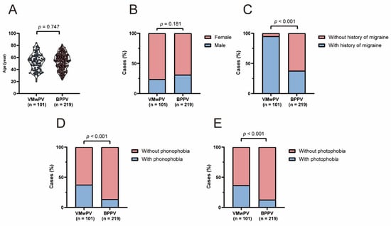
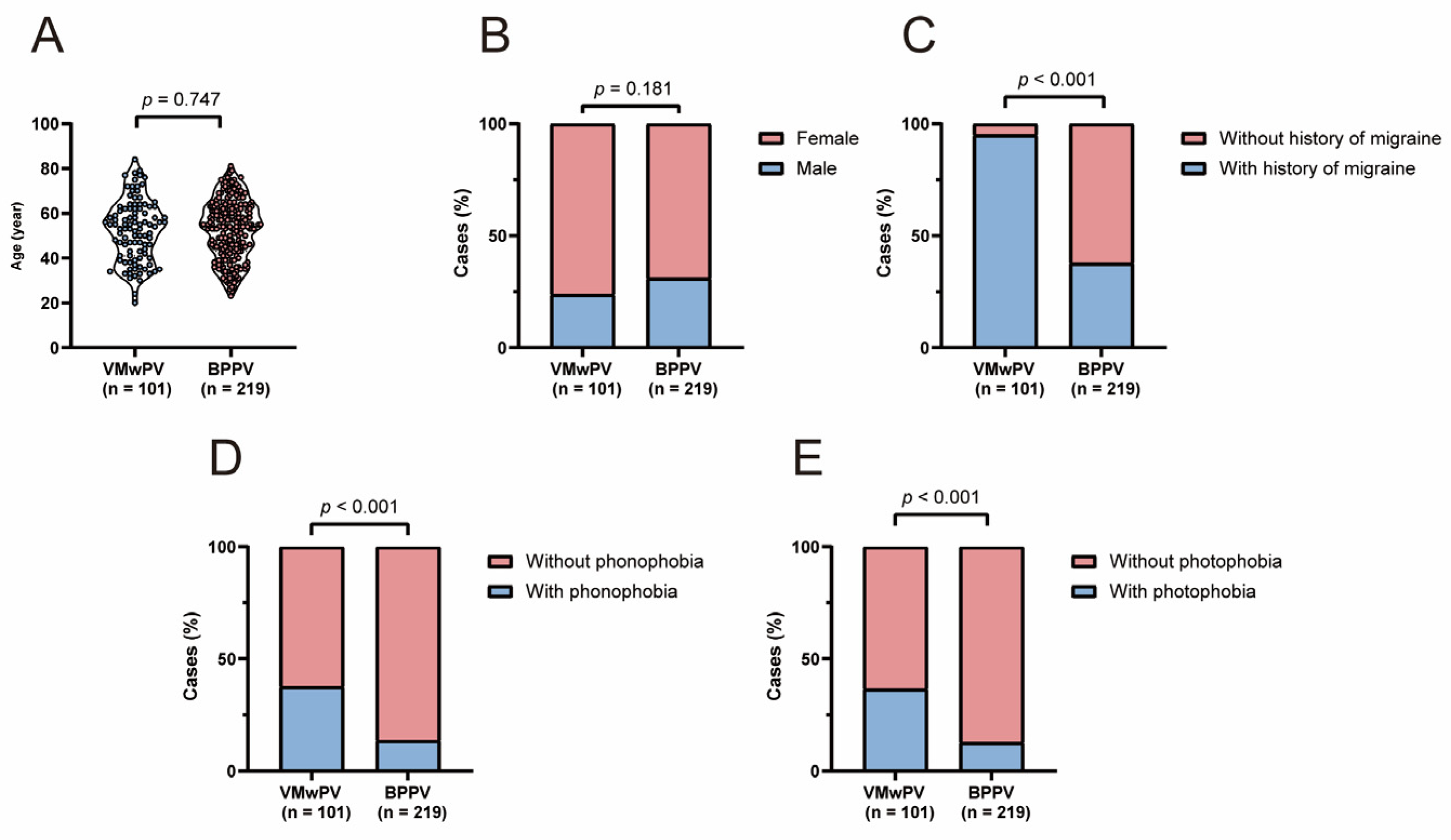
In the present study, we enrolled 101 VMwPV patients, including 34 VM cases and 67 probable VM cases. Additionally, 219 BPPV patients were included, comprising 159 patients with posterior semicircular canal BPPV (PSC-BPPV), 50 patients with HSC-BPPV-can and 10 patients with horizontal semicircular canal cupulolithiasis (HSC-BPPV-cup). The male/female ratio in VMwPV group and BPPV group were 24/77 and 68/151, with no significant difference (χ2 = 1.792, p = 0.181). The age in VMwPV group and BPPV group was 54(41, 62) and 54(43, 63), and no significant difference was found (Z = −0.323, p = 0.747). The VMwPV group had a significantly higher proportion of migraine history (95% vs. 37.9%), photophobia (36.6% vs. 12.8%), and phonophobia (37.6% vs. 13.7%) compared with the BPPV group (all p < 0.001) (Figure 1).
Figure 1.
Comparison of the clinical features between VMwPV group and BPPV group in (A) Age, (B) Gender, the prevalence of (C) migraine history, (D) phonophobia and (E) photophobia. VMwPV, vestibular migraine with positional vertigo; BPPV, benign paroxysmal positional vertigo.
3.2. Comparison of the Presence of Vertigo in Positional Testing and Features of Nystagmus Between VMwPV and BPPV
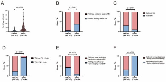
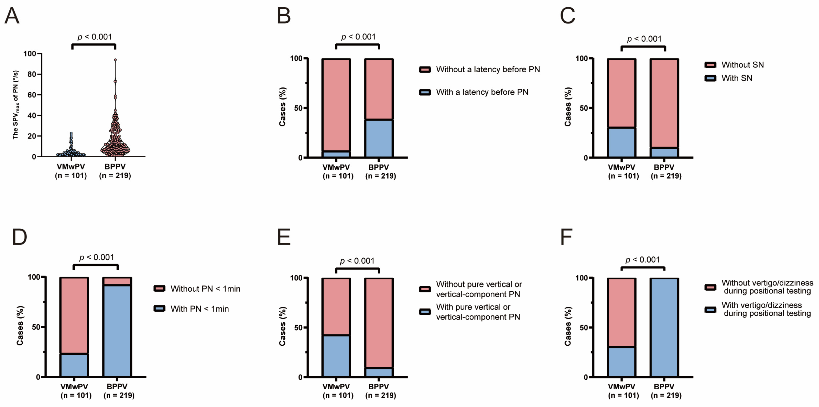
As shown in Figure 2, the median SPVmax of PN were 3 (2, 5) °/s in the VMwPV group and 11(6,23) °/s in the BPPV group. The former was significantly lower than the latter (Z = −9.440, p < 0.001). The prevalence of SN in the VMwPV group (30.7%) was significantly higher than in the BPPV group (10.5%) (χ2 = 20.088, p < 0.001). The latency was observed in 7 (6.9%) VMwPV patients and 85(38.8%) BPPV patients, which was significantly lower in the VMwPV group than in the BPPV group (χ2 = 34.299, p < 0.001). PN lasting < 1 min was present in 24(23.8%) VMwPV patients and 202(92.2%) BPPV patients. The prevalence of this phenomenon in the VMwPV group was significantly lower than in the BPPV group (χ2 = 156.223, p < 0.001). As for pure vertical or vertical-component PN during positional testing, it was observed in 43(42.6%) VMwPV patients and 21(9.6%) BPPV patients and was more frequent in the VMwPV group than in the BPPV group (χ2 = 47.004, p < 0.001). Positional vertigo/dizziness was provoked in 31(30.7%) VMwPV patients and all BPPV patients, which was less frequent in the VMwPV group than in the BPPV group (χ2 = 194.281, p < 0.001).
Figure 2.
Comparison of the nystagmus features between VMwPV group and BPPV group in (A) the SPVmax of PN, the prevalence of (B) a latency before PN, (C) spontaneous nystagmus, (D) PN lasting <1 min, (E) pure vertical or vertical-component PN, and (F) vertigo/dizziness during positional testing. VMwPV, vestibular migraine with positional vertigo; BPPV, benign paroxysmal positional vertigo; PN, positional nystagmus; SN, spontaneous nystagmus; SPVmax, maximum slow phase velocity.
Among the 31 patients with VMwPV who exhibited SN, the directions of SN were diverse: 4 cases showed upbeat nystagmus, 4 showed downbeat nystagmus, 4 showed nystagmus with a vertical-component, and 19 showed horizontal nystagmus. The directions of SN in all 23 BPPV patients were horizontal, comprising 13 cases of PSC-BPPV, 7 cases of HSC-BPPV-can, and 3 cases of HSC-BPPV-cup. As for HSC-BPPV-can, PSN was seen in 5 cases of HSC-BPPV-can, while true SN was seen in 2 cases of HSC-BPPV-can. As for the HSC-BPPV-cup, PSN was seen in 2 cases of HSC-BPPV-cup, while true SN was seen in 1 case of HSC-BPPV-cup. The SPVmax of SN in the VMwPV group and the BPPV group were 2(1,2) °/s and 2(1,2) °/s, respectively. And no significant difference was found (Z = −0.197, p = 0.843). The intensity of pure vertical or vertical-component PN in VMwPV group and BPPV group were 3(2,5) °/s and 3(2, 4) °/s, without significant difference (Z = −0.689, p = 0.491).
3.3. Machine Learning Models
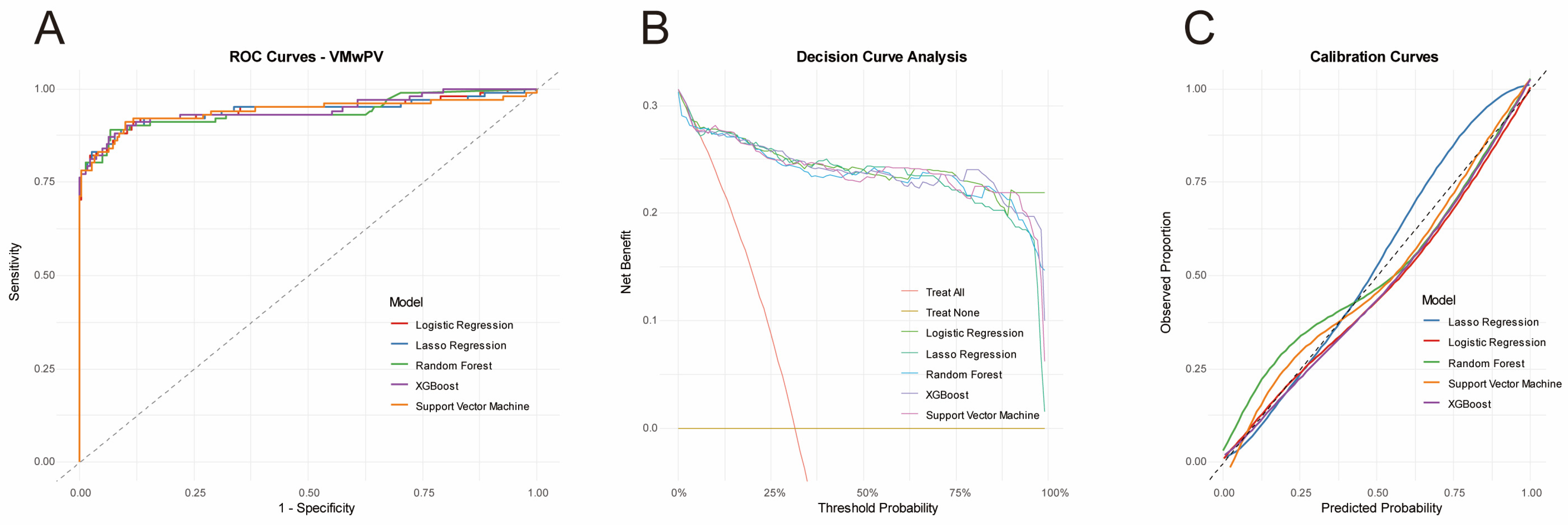
As shown in Table 3, the mean (95%CI) of AUC in Logistic regression, Lasso Regression, Random Forest, XGBoost, and Support Vector Machine were 0.9426 (0.9053–0.9742), 0.9405 (0.9009–0.9741), 0.9371 (0.8996–0.9690), 0.9427 (0.9047–0.9745), and 0.9392 (0.9001–0.9716), respectively. Because no statistically significant differences in AUC were observed among the five models (DeLong test, all FDR-adjusted p > 0.05), we selected Logistic Regression as the representative model for detailed interpretation. This choice was based on its superior interpretability, as the regression coefficients can be directly translated into odds ratios, and its ease of clinical deployment.
Table 3.
Performance of machine learning models.
Model calibration was assessed using the Brier score and calibration plots. Brier scores were 0.0592 for Logistic Regression, 0.0605 for Lasso Regression, 0.0640 for Random Forest, 0.0618 for XGBoost, and 0.0612 for Support Vector Machine, all below 0.1, indicating excellent calibration across all five models. Calibration curves showed that the predicted probabilities of all models were close to the ideal diagonal line (y = x) across the range of 0 to 1, indicating good agreement between predicted and observed probabilities (Figure 3C). Decision curve analysis showed that all models provided higher net benefit than the “treat all” and “treat none” strategies, suggesting meaningful clinical utility (Figure 3B).
Figure 3.
The performance of all models. The ROC curve of Logistic regression, Lasso Regression, Random Forest XGBoost and Support Vector Machine (A), the decision curve analysis (B), and the calibration curves (C) of all models.
As shown in Supplementary Material S1, the model showed good discriminative performance in both comparing VMwPV with PSC-BPPV and VMwPV with HSC-BPPV. The AUC of sensitivity analyses comparing VMwPV with PSC-BPPV was 0.9365 (0.8918–0.9717), and the AUC of sensitivity analyses comparing VMwPV with HSC-BPPV was 0.9554 (0.9231–0.9811). The performance was comparable across subgroups, suggesting that the model’s discriminative ability was not driven by a single BPPV subtype.
3.4. The Feature Importance
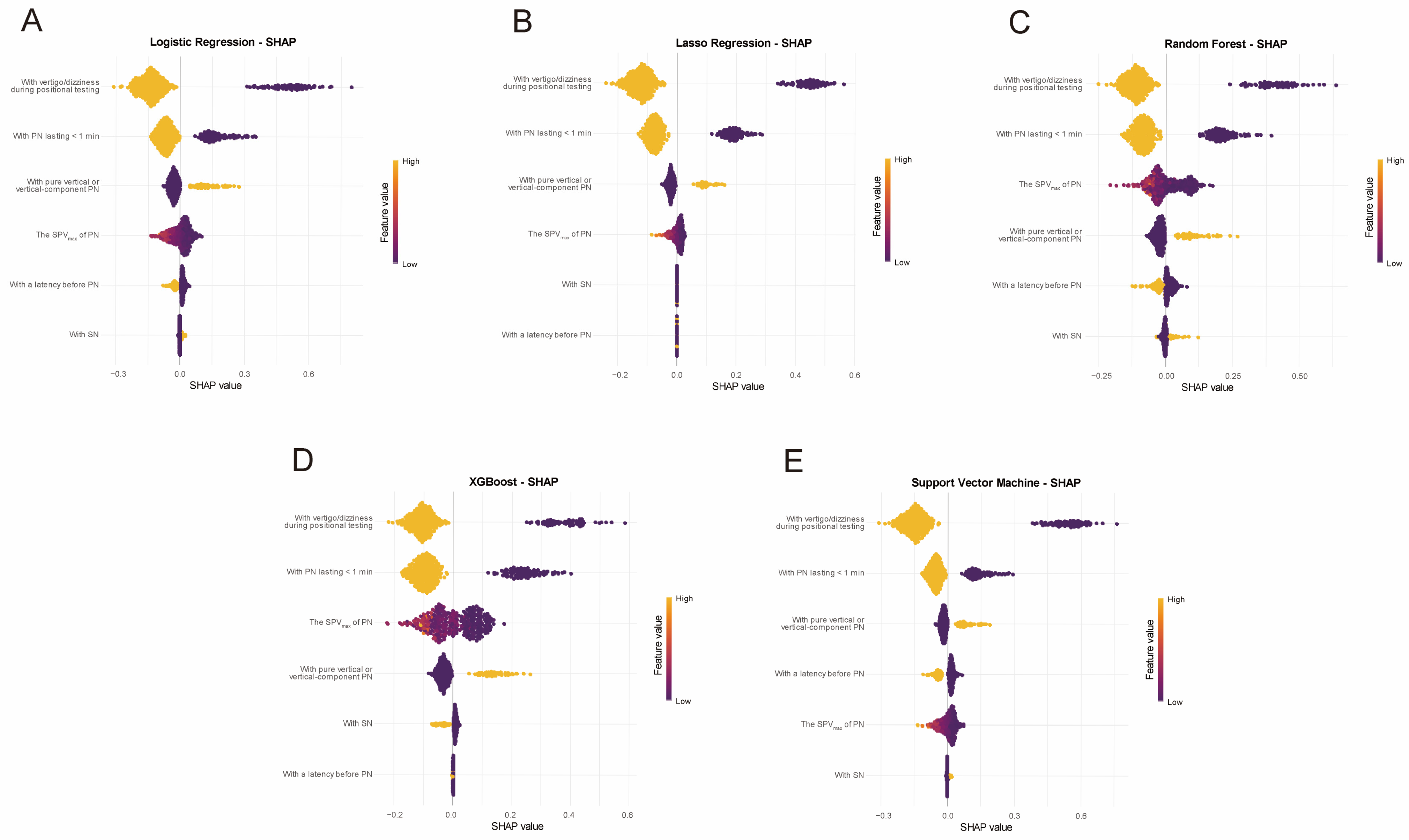
SHAP analyses were performed for all five models, and the results are shown in Figure 4. The SHAP-based feature importance rankings were largely consistent across models, with vertigo/dizziness during positional testing and PN lasting <1 min consistently ranking as the most influential predictors.
Figure 4.
SHAP feature importance beeswarm plots of five machine learning models for distinguishing VMwPV from BPPV: (A) Logistic Regression, (B) Lasso Regression, (C) Random Forest, (D) XGBoost, and (E) Support Vector Machine. Color from purple to yellow indicates the corresponding feature value from low to high (e.g., higher SPVmax is shown in yellow); x-axis (SHAP value) represents the direction and magnitude of each feature’s contribution to the model’s prediction of “VMwPV” (positive values favor the diagnosis of VMwPV, and negative values favor BPPV). y-axis lists features in descending order of mean absolute SHAP values, with the top-ranked features being the most influential predictors across models. VMwPV, vestibular migraine with positional vertigo; BPPV, benign paroxysmal positional vertigo; PN, positional nystagmus; SN, spontaneous nystagmus; SPVmax, maximum slow phase velocity; SHAP, Shapley Additive Explanations.
3.5. Sensitivity Analyses
Since provoked vertigo/dizziness was a necessary requirement for BPPV diagnosis, we further removed “vertigo/dizziness during positional testing” and reran all analyses using the same repeated stratified 5-fold cross-validation (10 repeats) framework (Supplementary Material S2). The mean AUC decreased from 0.9426 to 0.9350 for Logistic Regression, from 0.9405 to 0.9326 for Lasso Regression, from 0.9371 to 0.9235 for Random Forest, from 0.9427 to 0.9313 for XGBoost, and from 0.9392 to 0.9225 for Support Vector Machine. After removing “vertigo/dizziness during positional testing”, the DeLong comparison showed that Logistic Regression and Lasso Regression had significantly higher AUC than Support Vector Machine (FDR-adjusted p = 0.02 and 0.03, respectively), whereas no other pairwise comparisons reached statistical significance. In the SHAP analysis, PN lasting <1 min remained the leading predictor, indicating that the models learned independent nystagmus features beyond “vertigo/dizziness during positional testing”.
In addition, we further constructed model analysis using three features that are independent of the Bárány Society BPPV diagnostic criteria: SN, SPVmax of PN, and pure vertical/vertical-component PN. We repeated the same nested cross-validation framework (repeated stratified 5-fold CV, 10 repeats) to evaluate its performance. Although the sensitivity has slightly decreased, the AUCs of these models still demonstrated substantial and statistically significant discriminative ability (AUC > 0.83) (Supplementary Material S3). The DeLong comparison showed that Support Vector Machine had a significantly lower AUC compared to Logistic Regression, Lasso Regression, and XGBoost (all FDR-adjusted p < 0.05). As for the SHAP analysis, the feature importance ranking among the three objective variables was largely consistent across models, with SPVmax of PN and pure vertical or vertical-component PN ranking as the top two predictors.
3.6. Subgroup Analyses Within the VMwPV Cohort
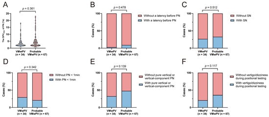
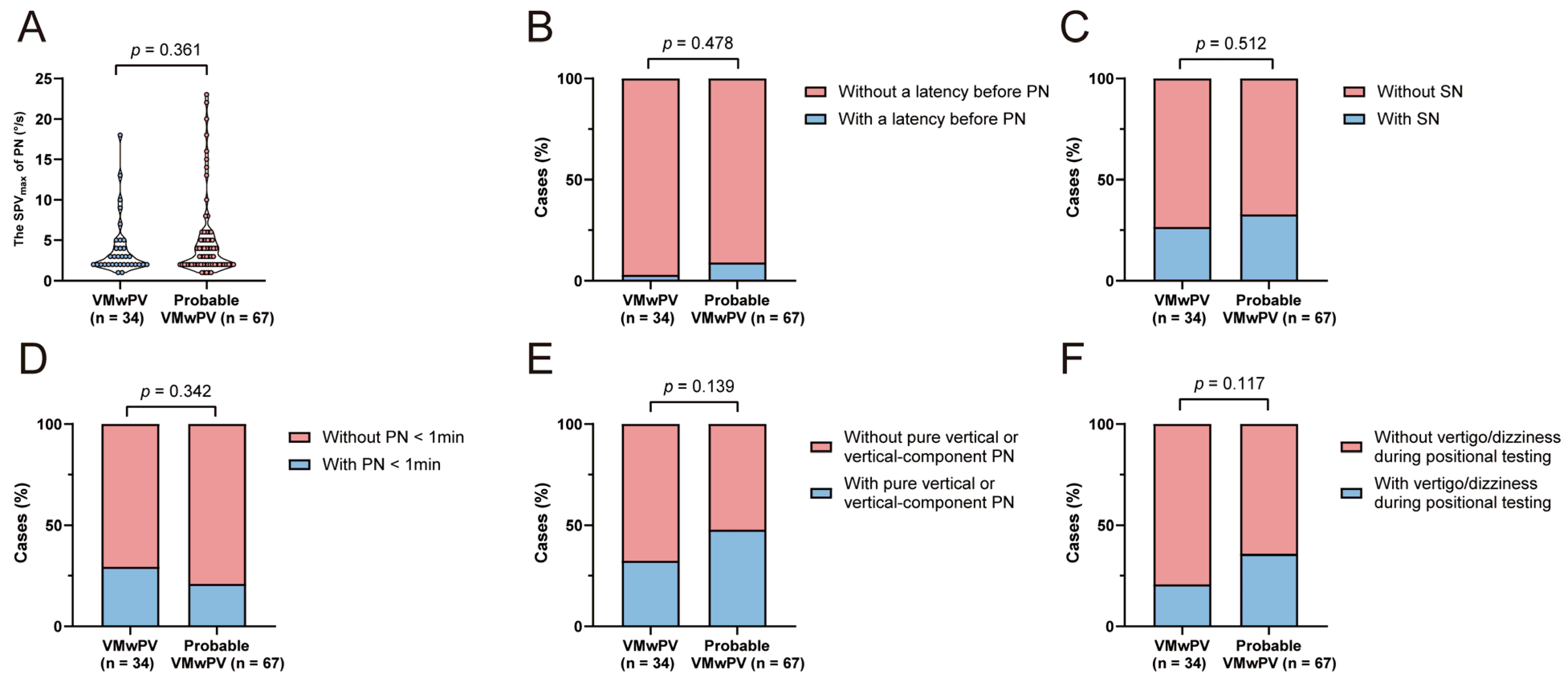
We further analyzed the nystagmus characteristic between VMwPV and probable VMwPV. The SPVmax of PN in VMwPV and probable VMwPV were 2.5(2, 4.25) °/s and 3(2, 5) °/s (Z = −0.913, p = 0.361). The latency was observed in 1(2.9%) VMwPV cases and 6(9.0%) probable VMwPV cases (χ2 = 1.265, p = 0.478). The prevalence of SN was observed in 9(26.5%) VMwPV cases and 22(32.8%) cases (χ2 = 0.430, p = 0.512). PN lasting <1 min was present in 10(29.4%) VMwPV patients and 14(20.9%) probable VMwPV patients (χ2 = 0.903, p = 0.342). Pure vertical or vertical-component PN was seen in 11(32.4%) VMwPV cases and 32(47.8%) probable VMwPV (χ2 = 2.190, p = 0.139). Vertigo/dizziness was provoked in 7(20.6%) VMwPV cases and 24(35.8%) probable VMwPV cases (χ2 = 2.460, p = 0.117). No significant difference was observed in all these nystagmus features between VMwPV and probable VMwPV (Figure 5). These results support the homogeneity of the two subgroups with respect to the variables used in the models.
Figure 5.
Comparison between VMwPV and probable VMwPV patients in (A) the SPVmax of PN, the prevalence of (B) a latency before PN, (C) SN, (D) PN < 1 min, (E) pure vertical or vertical-component PN, and (F) vertigo/dizziness during positional testing. VMwPV, vestibular migraine with positional vertigo; PN, positional nystagmus; SN, spontaneous nystagmus; SPVmax, maximum slow phase velocity.
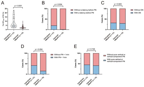
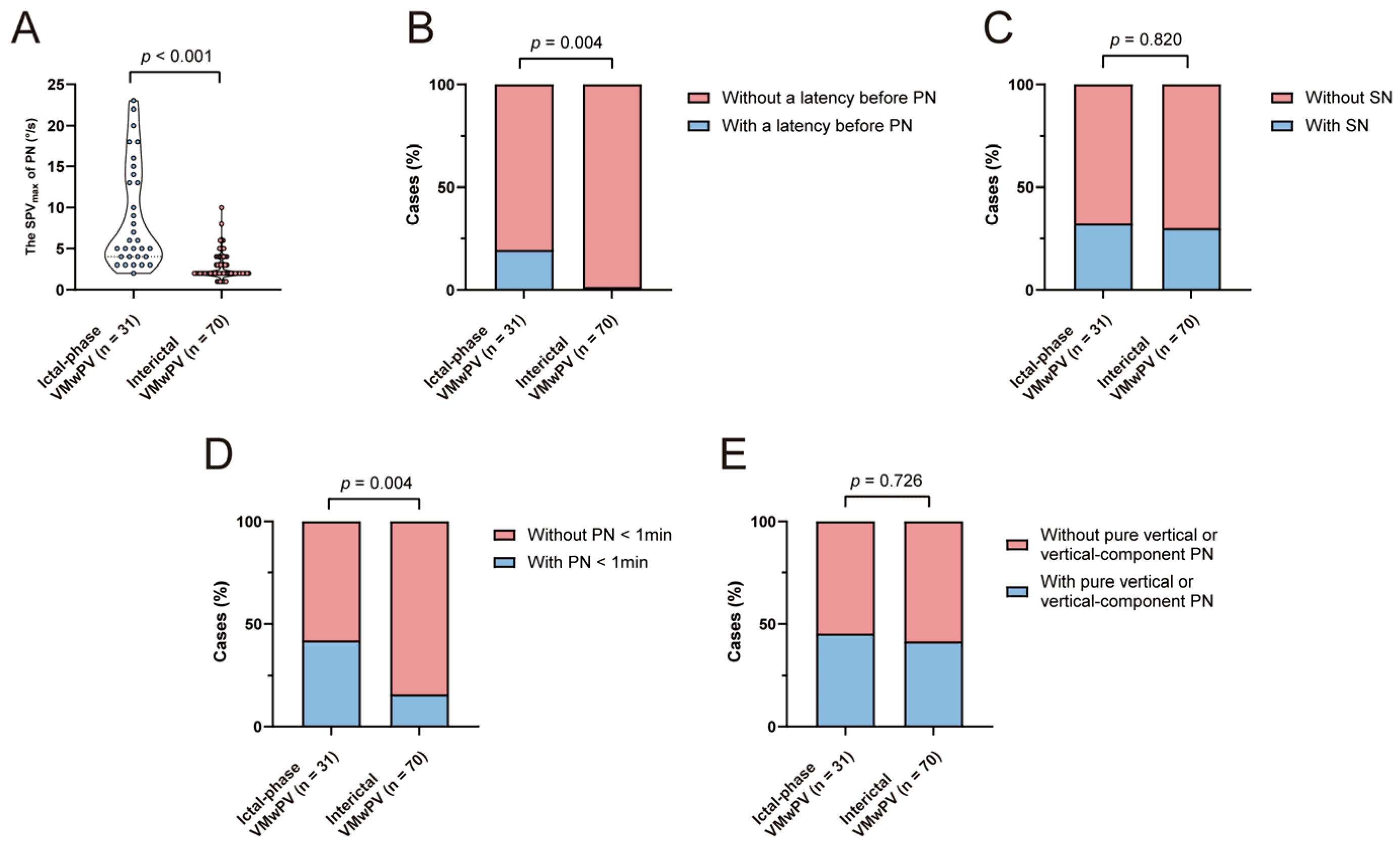
In addition, nystagmus characteristics were compared between ictal (those with vertigo/dizziness during positional testing) and interictal phases (those without vertigo/dizziness during positional testing). The SPVmax of PN in ictal and interictal phases was 6(4,14) and 2(2,3.25) °/s (Z = −6.365, p < 0.001). The latency was observed in 6(19.4%) ictal-phase VMwPV and 1(1.4%) interictal VMwPV cases (χ2 = 10.704, p = 0.004). The prevalence of SN was observed in 10(32.3%) ictal-phase VMwPV and 21(30.0%) interictal VMwPV cases (χ2 = 0.051, p = 0.820). PN lasting <1 min was present in 13(41.9%) ictal-phase VMwPV patients and 11(15.7%) interictal VMwPV cases (χ2 = 8.154, p = 0.004). Pure vertical or vertical-component PN was seen in 14(45.2%) ictal-phase VMwPV patients and 29(41.4%) interictal VMwPV cases (χ2 = 0.122, p = 0.726). Compared with those in the ictal phase, cases in interictal phases showed weaker intensity of PN, lower prevalence of latency, and lower prevalence of PN <1 min. No significant difference was observed in the prevalence of SN and pure vertical or vertical-component PN (Figure 6).
Figure 6.
Comparison of the nystagmus characteristics between VMwPV patients of ictal and interictal phases in (A) the SPVmax of PN, the prevalence of (B) a latency before PN, (C) SN, (D) PN < 1 min, and (E) pure vertical or vertical-component PN. VMwPV, vestibular migraine with positional vertigo; PN, positional nystagmus; SN, spontaneous nystagmus; SPVmax, maximum slow phase velocity.
4. Discussion
To analyze the subjective symptom (vertigo or dizziness) in positional testing and multiple objective nystagmus features in VMwPV and BPPV patients, five machine learning approaches were employed to establish differential diagnostic models. Because no statistically significant differences in AUC were observed among the five models, we selected Logistic Regression as the optimal model based on its superior interpretability. SHAP analysis was further applied to rank the above features, enabling visualization of the importance of each feature in differentiating VMwPV from BPPV. In the optimal model, the top two most important features were the presence of provoked vertigo/dizziness and PN lasting <1 min.
Previous studies typically examined individual or a limited number of nystagmus parameters in VM patients using conventional statistical approaches. Although such analyses are useful for identifying associations between individual features and disease groups, they may not fully characterize the joint information contained in multidimensional nystagmus features. In the present study, multiple machine learning models were employed to integrate both a subjective feature and a range of objective nystagmus features from patients with VMwPV and BPPV. This approach enables simultaneous modeling of multiple features and can accommodate potentially nonlinear relationships and interactions. Furthermore, SHAP analysis was used to interpret the models by quantifying the contribution of each feature to model output, allowing the information learned by the models to be examined at the feature level.
4.1. The Clinical Characteristics Between VMwPV and BPPV
Our results showed no significant differences in gender or age between patients with VMwPV and BPPV. In the study by Mok et al., VM and BPPV patients showed no significant difference in gender distribution, but VM patients were younger than those with BPPV [7]. This discrepancy may be attributed to the prolonged course and repetitive bouts of VMwPV, as disease duration and attack frequency may influence the age of consultation.
In contrast to the similarities in demographic characteristics, migraine history, phonophobia, and photophobia were more frequently observed in patients with VMwPV. It is consistent with the study by Chen et al. [8], which demonstrated that headache in patients with positional vertigo had a strong predictive value for VMwPV. In contrast, Mok et al. reported no significant differences between VM and BPPV patients in prevalence of migraine history, phonophobia, and photophobia [7]. One possible explanation is the cohort composition, as Mok et al. included VM patients with both spontaneous and positional vertigo, whereas our cohort was restricted to VMwPV patients. These findings suggest that VM patients with different vestibular symptom patterns (spontaneous versus positional vertigo) may have distinct accompanying symptom profiles, and future studies should further perform subgroup analyses based on vestibular phenotypes (e.g., spontaneous or provoked vertigo).
4.2. The Presence of Vertigo/Dizziness During Positional Testing in VMwPV and BPPV
SHAP analysis identified the presence of provoked vertigo/dizziness as the most important feature for differentiating VMwPV from BPPV. An important finding underlying this contribution was the apparent dissociation between PN and subjective vertigo/dizziness in VMwPV. In the present study, fewer VMwPV patients (30.7%) experienced vertigo/dizziness during positional testing than BPPV patients (100%). Previous studies found that 11–55.4% of VM patients exhibited interictal PN without vertigo [31,32]. These findings, together with our results, indicate that VMwPV should be considered in case of PN without associated vertigo/dizziness during positional testing.
4.3. The Nystagmus Features in VMwPV and BPPV
4.3.1. The Duration of PN in VMwPV and BPPV
SHAP analysis identified PN lasting <1 min as the second most important feature for differentiating VMwPV from BPPV. Previous studies reported persistent PN in VM patients [15,22,33]. Persistent horizontal nystagmus during Roll test was also reported in VMwPV patients [23]. Our results are generally consistent with the above findings, suggesting that the duration of PN may serve as an important differential diagnostic indicator when distinguishing VMwPV from BPPV, particularly canalolithiasis.
4.3.2. The SPVmax of PN in VMwPV and BPPV
In this study, compared with BPPV patients, VMwPV patients showed lower SPVmax of PN. Previous studies have consistently described relatively low-velocity PN in VM patients. In Lechner et al.’s study, the SPV during Roll test in HSC-BPPV-can was higher than that in VMwPV patients in both ear-down positions [23]. Polensek et al. retrospectively reviewed the nystagmus features of 26 VM patients and found that 89% of patients had low-velocity PN (2–7°/s) [34]. Our results are generally consistent with the above studies and showed that the intensity of PN carries relevant information for distinguishing VMwPV from BPPV. Moreover, nystagmus intensity in VM may not represent a stable trait. Previous studies have suggested that the intensity of nystagmus in VM patients was higher in the ictal phase than in the interictal phase [31], indicating varying nystagmus features depending on disease activity in VMwPV.
4.3.3. The Pure Vertical or Vertical-Component PN in VMwPV and BPPV
In this study, compared with BPPV patients, VMwPV patients (42.6%) had a higher proportion of pure vertical or vertical-component PN. This figure was significantly higher than that reported by Polensek et al. (19%) [34]. This discrepancy may be attributed to the inclusion of both pure vertical and vertical-component nystagmus in the present study, whereas in Polensek et al.’s study, only pure vertical nystagmus was analyzed. El-Badry et al. reported that all 13 VMwPV patients exhibited vertical PN [21]. The inconsistency between their results and ours may be due to differences in sample size.
In the present study, 9.6% of BPPV patients also exhibited low-intensity pure vertical or vertical-component PN, with an intensity of 3 (2, 4) °/s. Previous studies have reported PN in healthy subjects [35,36,37,38]. The intensity of the vertical nystagmus in our BPPV patients fell well below the upper limit of 7°/s for vertical nystagmus in healthy subjects [39], suggesting that it may be a non-specific finding similar to the low-velocity PN occasionally observed in healthy individuals, rather than a distinct pathological mechanism. Its mechanism remains uncertain and may involve gravity effects on the otolith organs or subclinical vestibular abnormalities [37].
4.3.4. The Latency of PN in VMwPV and BPPV
Latency may be a characteristic distinguishing the PN patterns of VMwPV and BPPV. Our study found that, compared with BPPV patients (38.8%), VMwPV patients had a lower incidence of latency before PN (6.9%), which is consistent with that reported by El-Badry et al., i.e., absence of latency in all VMwPV patients [21]. Polensek et al. found that 20(76%) VM patients had no latency [34]. The incidence of latency in Polensek et al.’ study was higher than our result, which may be due to the larger sample size and different inclusion criteria in our study. In the study by Yu et al., using the multiaxial repositioning chair, 187/285 (65.6%) patients with PSC-BPPV had latency [40], which is higher than the 39.9% of BPPV patients with latency in our study (using manual maneuvers). The latency preceding PN in BPPV could be influenced by the location of the detached otoconia. It has been reported that PSC-BPPV typically has a latency of one or few seconds, whereas HSC-BPPV (especially cupulolithiasis) often has a brief latency or no latency [24]. Furthermore, this difference may also be attributed to variations in positional testing methods, which may result in different accelerations during testing [41,42].
4.3.5. The SN in VMwPV and BPPV
SN was another nystagmus characteristic that differed between VMwPV and BPPV in the present study. In the present study, a higher proportion of VMwPV patients presented with SN compared with BPPV patients. To date, no study has compared the prevalence of SN between VMwPV and BPPV. In the study by Waissbluth et al., 13/60 (21.7%) VM patients exhibited SN [43], which was similar to that in the present study (30.7%). Young et al. found a difference in SN occurrence during the ictal phase (71.3%) and the interictal phase (14.9%) in patients with VM [31]. The proportion of VMwPV patients with SN in the present study fell within the range reported above.
Various directions of SN exist in our VMwPV cohort, including pure vertical SN, vertical-component SN, and horizontal SN. Previous studies also reported the various directions of SN in VM [34,44,45]. Young et al. observed the SN in 44/67 (65.7%) VM patients, of whom there were horizontal and vertical SN [22]. Furthermore, a previous study suggested that the direction of SN may change in the same VM patient [31]. The precise mechanisms underlying variable direction of SN in VM patients remain unclear, and both peripheral and central vestibular dysfunction may be involved. Decompensated peripheral vestibular damage and damage to the ventral tegmental tract or the flocculus may cause horizontal SN and vertical SN, respectively [46,47]. However, whether this mechanism can account for horizontal and vertical SN in VMwPV patients remains to be verified.
The directions of SN in all BPPV patients were horizontal in the present study. Previous studies have found the presence of PSN in patients with HSC-BPPV-can and HSC-BPPV-cup [25,48,49,50,51,52,53]. SN has also been reported in approximately 26% of patients with PSC-BPPV [54].
4.4. Strengthens and Limitations
Compared with previous studies on nystagmus characteristics in VMwPV, the present study has several advantages. First, multiple machine learning algorithms with different learning mechanisms were compared, and SHAP analysis was further applied to improve model interpretability by quantifying the contribution of individual features to model predictions. Second, the presence of vertigo in positional testing and multiple objective parameters of nystagmus are readily obtainable, making our models more operationally feasible. In addition, our study also benefits from a relatively large sample size (101 VMwPV patients), and the models were established based on real-world clinical data rather than database-derived cohorts.
This study also has several limitations. First, this was a retrospective cross-sectional study and did not include long-term follow-up data from patients with positional vertigo. Second, since only a minority of VMwPV patients underwent additional vestibular function tests (e.g., video head impulse test and caloric test) and brain MRI, these data were not analyzed in the present study. Third, VM are highly heterogeneous in nature, with substantial fluctuations in clinical and functional profiles among subjects even within a single patient during the ictal and interictal phases, which may affect nystagmus features. Fourth, the interval between the most recent vertigo/dizziness attack and the VNG examination was not systematically documented for VMwPV patients. Moreover, since some controversial types of BPPV were excluded in the present study, our findings should be interpreted only within the BPPV phenotypes actually studied (PSC-BPPV and HSC-BPPV). Finally, this was a single-center exploratory study, which may limit the generalizability of our results. Future multicenter studies with prospective external validation in patients with undifferentiated positional vertigo are needed to optimize and validate the models.
5. Conclusions
This retrospective study employed machine learning methods to integrate objective nystagmus features and provoked vertigo/dizziness during positional testing in patients diagnosed with VMwPV or BPPV (PSC-BPPV and LSC-BPPV). While provoked vertigo/dizziness and PN lasting <1 min were identified as the top features, the high importance of provoked vertigo likely reflects incorporation bias, as it is a core diagnostic criterion for BPPV. Objective features such as PN lasting <1 min may have potential reference value. Multicenter studies and prospective external validation in patients with undifferentiated positional vertigo are needed.
Supplementary Materials
The following supporting information can be downloaded at: https://www.mdpi.com/article/10.3390/diagnostics16213699/s1, Supplementary Figure S1: The tuning curves for all models; Supplementary Material S1: Logistic regression model for distinguishing VMwPV from PSC-BPPV and from HSC-BPPV; Supplementary Material S2: Model performance after removing vertigo/dizziness during positional testing; Supplementary Material S3: Model performance using three features (SN, SPVmax of PN, and pure vertical or vertical-component PN).
Author Contributions
Conceptualization, B.L. and Y.L.; methodology, K.X. and L.W. (Linlin Wang); software, K.X. and L.W. (Linlin Wang); validation, X.S. (Xingqian Shen) and X.C.; formal analysis, K.X.; investigation, H.W.; resources, B.L.; data curation, Z.X., X.S. (Xuanyu Shi) and L.W. (Lu Wang); writing—original draft preparation, K.X. and L.W. (Linlin Wang); writing—review and editing, B.L. and Y.L.; visualization, K.X. and L.W. (Linlin Wang); supervision, H.X.; project administration, H.X.; funding acquisition, H.X., B.L. and Y.L. All authors have read and agreed to the published version of the manuscript.
Funding
This research was funded by the National Key Research and Development Program of China (Nos. 2023YFC2508000, 2023YFC2508001 and 2023YFC2508400), the National Natural Science Foundation of China (No. 82101231), and the Natural Science Foundation of Hubei Province, China (No. 2021CFB547).
Institutional Review Board Statement
The study was conducted in accordance with the Declaration of Helsinki, and approved by the Ethical Committee of Union Hospital, Tongji Medical College, Huazhong University of Science and Technology, Wuhan, China (Ethical Committee Approval No. 0619), approval date 26 June 2025.
Informed Consent Statement
Informed consent was obtained from all subjects involved in the study.
Data Availability Statement
The data supporting the findings of this study are available from the corresponding author on reasonable request.
Conflicts of Interest
The authors declare no conflicts of interest.
Abbreviations
The following abbreviations are used in this manuscript:
| AUC | Area under the curve |
| BPPV | Benign paroxysmal positional vertigo |
| FDR | False discovery rate |
| HSC-BPPV-can | Horizontal semicircular canal canalolithiasis |
| HSC-BPPV-cup | Horizontal semicircular canal cupulolithiasis |
| MD | Ménière’s disease |
| NPV | Negative predictive value |
| NU | Nodulus and uvula |
| PN | Positional nystagmus |
| PPV | Positive predictive value |
| PSC-BPPV | Posterior semicircular canal benign paroxysmal positional vertigo |
| PSN | Pseudo-spontaneous nystagmus |
| ROC | Receiver operating characteristic |
| SHAP | Shapley Additive Explanations |
| SN | Spontaneous nystagmus |
| SPV | Slow-phase velocity |
| SPVmax | Maximum slow phase velocity |
| VM | Vestibular migraine |
| VMwPV | Vestibular migraine with positional vertigo |
References
- Lempert, T.; Olesen, J.; Furman, J.; Waterston, J.; Seemungal, B.; Carey, J.; Bisdorff, A.; Versino, M.; Evers, S.; Kheradmand, A.; et al. Vestibular migraine: Diagnostic criteria1. J. Vestib. Res. 2022, 32, 1–6. [Google Scholar] [CrossRef] [Scilit] [PubMed]
- Bisdorff, A.; Von Brevern, M.; Lempert, T.; Newman-Toker, D.E. Classification of vestibular symptoms: Towards an international classification of vestibular disorders. J. Vestib. Res. 2009, 19, 1–13. [Google Scholar] [CrossRef] [Scilit] [PubMed]
- Bhattacharyya, N.; Gubbels, S.P.; Schwartz, S.R.; Edlow, J.A.; El-Kashlan, H.; Fife, T.; Holmberg, J.M.; Mahoney, K.; Hollingsworth, D.B.; Roberts, R.; et al. Clinical Practice Guideline: Benign Paroxysmal Positional Vertigo (Update). Otolaryngol. Head Neck Surg. 2017, 156, S1–S47. [Google Scholar] [CrossRef] [Scilit] [PubMed]
- Sohn, J.H. Recent Advances in the Understanding of Vestibular Migraine. Behav. Neurol. 2016, 2016, 1801845. [Google Scholar] [CrossRef] [Scilit] [PubMed]
- Beh, S.C.; Masrour, S.; Smith, S.V.; Friedman, D.I. The Spectrum of Vestibular Migraine: Clinical Features, Triggers, and Examination Findings. Headache 2019, 59, 727–740. [Google Scholar] [CrossRef] [Scilit] [PubMed]
- Mahrous, M.M. Vestibular migraine and benign paroxysmal positional vertigo, close presentation dilemma. Acta Otolaryngol. 2020, 140, 741–744. [Google Scholar] [CrossRef] [Scilit] [PubMed]
- Mok, B.; Welgampola, M.S.; Rosengren, S.M. Vestibular migraine as a mimic of benign paroxysmal positioning vertigo and Meniere’s disease. J. Vestib. Res. 2025, 35, 30–38. [Google Scholar] [CrossRef] [Scilit] [PubMed]
- Chen, C.C.; Wang, C.Y.; Chen, P.Y.; Chen, M.C.; Lee, T.Y.; Lee, H.H. Clinical Spectrum of Positional Vertigo in an Outpatient Setting. Ear Hear. 2022, 43, 1256–1261. [Google Scholar] [CrossRef] [Scilit] [PubMed]
- Li, M.; Xu, X.; Qi, W.; Liang, Y.; Huang, Y.; Huang, H. Vestibular migraine: The chameleon in vestibular disease. Neurol. Sci. 2021, 42, 1719–1731. [Google Scholar] [CrossRef] [Scilit] [PubMed]
- Yu, J.; Yu, Q.; Guan, B.; Lu, Y.; Chen, C.; Yu, S. Pseudo-Benign Paroxysmal Positional Vertigo: A Retrospective Study and Case Report. Front. Neurol. 2020, 11, 187. [Google Scholar] [CrossRef] [Scilit] [PubMed]
- King, S.; Priesol, A.J.; Davidi, S.E.; Merfeld, D.M.; Ehtemam, F.; Lewis, R.F. Self-motion perception is sensitized in vestibular migraine: Pathophysiologic and clinical implications. Sci. Rep. 2019, 9, 14323. [Google Scholar] [CrossRef] [Scilit] [PubMed]
- ElSherif, M.; Reda, M.I.; Saadallah, H.; Mourad, M. Eye movements and imaging in vestibular migraine. Acta Otorrinolaringol. Esp. (Engl. Ed.) 2020, 71, 3–8. [Google Scholar] [CrossRef] [Scilit] [PubMed]
- Espinosa-Sanchez, J.M.; Lopez-Escamez, J.A. New insights into pathophysiology of vestibular migraine. Front. Neurol. 2015, 6, 12. [Google Scholar] [CrossRef] [Scilit] [PubMed]
- Koo, J.W.; Balaban, C.D. Serotonin-induced plasma extravasation in the murine inner ear: Possible mechanism of migraine-associated inner ear dysfunction. Cephalalgia 2006, 26, 1310–1319. [Google Scholar] [CrossRef] [Scilit] [PubMed]
- Chang, T.P.; Hong, Y.C.; Kuo, Y.H.; Schubert, M.C. Persistent geotropic or apogeotropic nystagmus: Recurrence and association with vestibular migraine. J. Neurol. 2025, 272, 184. [Google Scholar] [CrossRef] [Scilit] [PubMed]
- Zhai, Q.; Chen, Q.; Zhang, N.; Li, H.; Yu, Q.; Pan, Y. Exploring vestibulocerebellum-vestibular nuclei-spinal trigeminal nucleus causals communication and TRPV2 ion channel in a mouse model of vestibular migraine. J. Headache Pain 2025, 26, 47. [Google Scholar] [CrossRef] [Scilit] [PubMed]
- Noseda, R. Cerebro-Cerebellar Networks in Migraine Symptoms and Headache. Front. Pain Res. 2022, 3, 940923. [Google Scholar] [CrossRef] [Scilit] [PubMed]
- Harno, H.; Hirvonen, T.; Kaunisto, M.A.; Aalto, H.; Levo, H.; Isotalo, E.; Kallela, M.; Kaprio, J.; Palotie, A.; Wessman, M.; et al. Subclinical vestibulocerebellar dysfunction in migraine with and without aura. Neurology 2003, 61, 1748–1752. [Google Scholar] [CrossRef] [Scilit] [PubMed]
- Koohi, N.; Male, A.J.; Kaski, D. Acute positional vertigo in the emergency department-peripheral vs. central positional nystagmus. Front. Neurol. 2023, 14, 1266778. [Google Scholar] [CrossRef] [Scilit] [PubMed]
- Yetiser, S.; Ince, D. Direction-fixed positional nystagmus following head-roll testing: How is it related with a vestibular pathology? J. Otol. 2021, 16, 123–127. [Google Scholar] [CrossRef] [Scilit] [PubMed]
- El-Badry, M.M.; Samy, H.; Kabel, A.M.; Rafat, F.M.; Sanyelbhaa, H. Clinical criteria of positional vertical nystagmus in vestibular migraine. Acta Otolaryngol. 2017, 137, 720–722. [Google Scholar] [CrossRef] [Scilit] [PubMed]
- Young, A.S.; Lechner, C.; Bradshaw, A.P.; MacDougall, H.G.; Black, D.A.; Halmagyi, G.M.; Welgampola, M.S. Capturing acute vertigo: A vestibular event monitor. Neurology 2019, 92, e2743–e2753. [Google Scholar] [CrossRef] [Scilit] [PubMed]
- Lechner, C.; Taylor, R.L.; Todd, C.; Macdougall, H.; Yavor, R.; Halmagyi, G.M.; Welgampola, M.S. Causes and characteristics of horizontal positional nystagmus. J. Neurol. 2014, 261, 1009–1017. [Google Scholar] [CrossRef] [Scilit] [PubMed]
- von Brevern, M.; Bertholon, P.; Brandt, T.; Fife, T.; Imai, T.; Nuti, D.; Newman-Toker, D. Benign paroxysmal positional vertigo: Diagnostic criteria. J. Vestib. Res. 2015, 25, 105–117. [Google Scholar] [CrossRef] [Scilit] [PubMed]
- Asprella-Libonati, G. Pseudo-spontaneous nystagmus: A new sign to diagnose the affected side in lateral semicircular canal benign paroxysmal positional vertigo. Acta Otorhinolaryngol. Ital. 2008, 28, 73–78. [Google Scholar] [PubMed]
- Tibshirani, R. The lasso method for variable selection in the Cox model. Stat. Med. 1997, 16, 385–395. [Google Scholar] [CrossRef] [Scilit]
- Yang, N.; Chaturvedi, K.; Reid, N.; Dyball, A.C.; Argaet, E.C.; Bradshaw, A.P.; Wang, C.; Rafi, A.; Rosengren, S.M.; Halmagyi, G.M.; et al. The need for speed: Using nystagmus velocity profiles and machine learning models to separate canalithiasis BPV from its mimics. J. Neurol. 2025, 272, 761. [Google Scholar] [CrossRef] [Scilit] [PubMed]
- Ahn, J.M.; Kim, J.; Kim, K. Ensemble Machine Learning of Gradient Boosting (XGBoost, LightGBM, CatBoost) and Attention-Based CNN-LSTM for Harmful Algal Blooms Forecasting. Toxins 2023, 15, 608. [Google Scholar] [CrossRef] [Scilit] [PubMed]
- Pruneski, J.A.; Pareek, A. Support Vector Machines: Techniques and Applications. Hand Clin. 2026, 42, 19–25. [Google Scholar] [CrossRef] [Scilit] [PubMed]
- Hicks, S.A.; Strümke, I.; Thambawita, V.; Hammou, M.; Riegler, M.A.; Halvorsen, P.; Parasa, S. On evaluation metrics for medical applications of artificial intelligence. Sci. Rep. 2022, 12, 5979. [Google Scholar] [CrossRef] [Scilit] [PubMed]
- Young, A.S.; Nham, B.; Bradshaw, A.P.; Calic, Z.; Pogson, J.M.; D’Souza, M.; Halmagyi, G.M.; Welgampola, M.S. Clinical, oculographic, and vestibular test characteristics of vestibular migraine. Cephalalgia 2021, 41, 1039–1052. [Google Scholar] [CrossRef] [Scilit] [PubMed]
- Dieterich, M.; Brandt, T. Episodic vertigo related to migraine (90 cases): Vestibular migraine? J. Neurol. 1999, 246, 883–892. [Google Scholar] [CrossRef] [Scilit] [PubMed]
- Li, Y.; Wang, Y.; Chen, M.; Jiang, R.; Ju, Y. Eye Movement Abnormalities During Different Periods in Patients with Vestibular Migraine. J. Pain Res. 2023, 16, 3583–3590. [Google Scholar] [CrossRef] [Scilit] [PubMed]
- Polensek, S.H.; Tusa, R.J. Nystagmus during attacks of vestibular migraine: An aid in diagnosis. Audiol. Neurootol. 2010, 15, 241–246. [Google Scholar] [CrossRef] [Scilit] [PubMed]
- Nelson, M.D.; Mann, L.; Nicholson, C.; Lehman, M. Static Positional Nystagmus in the Healthy Vestibular System. J. Am. Acad. Audiol. 2019, 30, 883–895. [Google Scholar] [CrossRef] [Scilit] [PubMed]
- Martens, C.; Goplen, F.K.; Nordfalk, K.F.; Aasen, T.; Nordahl, S.H. Prevalence and Characteristics of Positional Nystagmus in Normal Subjects. Otolaryngol. Head Neck Surg. 2016, 154, 861–867. [Google Scholar] [CrossRef] [Scilit] [PubMed]
- Young, A.S.; Rosengren, S.M.; D’Souza, M.; Bradshaw, A.P.; Welgampola, M.S. Nystagmus characteristics of healthy controls. J. Vestib. Res. 2020, 30, 345–352. [Google Scholar] [CrossRef] [Scilit] [PubMed]
- Rasmussen, M.B.; Sørensen, R.; Hougaard, D.D. Positional nystagmus is observed in the vast majority of healthy individuals. Eur. Arch. Otorhinolaryngol. 2024, 281, 3499–3507. [Google Scholar] [CrossRef] [Scilit] [PubMed]
- Jeffery, H.; Hopkins, M.; Anderson, R.; Patel, V.; Rogers, J. The interpretation of static positional nystagmus in a balance clinic. Int. J. Audiol. 2017, 56, 958–966. [Google Scholar] [CrossRef] [Scilit] [PubMed]
- Yu, F.; Zhao, J.; Yu, J.; Yang, J.; Luo, X.; Wu, P.; Li, W. Quantitative Analysis of the Nystagmus of Posterior Canal Benign Paroxysmal Positional Vertigo and Its Association with Prognosis. Ear Hear. 2026, 47, 1026–1034. [Google Scholar] [CrossRef] [Scilit] [PubMed]
- Baloh, R.W.; Jacobson, K.; Honrubia, V. Horizontal semicircular canal variant of benign positional vertigo. Neurology 1993, 43, 2542–2549. [Google Scholar] [CrossRef] [Scilit] [PubMed]
- Anurin, I.; Ziemska-Gorczyca, M.; Pavlovschi, D.; Kantor, I.; Dżaman, K. The Impact of the Angular Head Movement’s Velocity during Diagnostic Maneuvers on Proper Benign Positional Paroxysmal Vertigo Diagnosis and Therapy. Diagnostics 2023, 13, 665. [Google Scholar] [CrossRef] [Scilit] [PubMed]
- Waissbluth, S.; Sepúlveda, V.; Leung, J.S.; Oyarzún, J. Vestibular and Oculomotor Findings in Vestibular Migraine Patients. Audiol. Res. 2023, 13, 615–626. [Google Scholar] [CrossRef] [Scilit] [PubMed]
- Zhang, S.; Lang, Y.; Wang, W.; Wu, Y.; Yan, S.; Zhang, T.; Li, D.; Liu, S.; Hao, Y.; Yang, X.; et al. Analysis of etiology and clinical features of spontaneous downbeat nystagmus: A retrospective study. Front. Neurol. 2024, 15, 1326879. [Google Scholar] [CrossRef] [Scilit] [PubMed]
- Ling, X.; Wu, Y.X.; Feng, Y.F.; Zhao, T.T.; Zhao, G.P.; Kim, J.S.; Yang, X.; Wang, Z.X. Spontaneous nystagmus with an upbeat component: Central or peripheral vestibular disorders? Front. Neurol. 2023, 14, 1106084. [Google Scholar] [CrossRef] [Scilit] [PubMed]
- Pierrot-Deseilligny, C.; Milea, D. Vertical nystagmus: Clinical facts and hypotheses. Brain 2005, 128, 1237–1246. [Google Scholar] [CrossRef] [Scilit] [PubMed]
- Zhang, A.J.; Yu, L.Q.; Zhou, L.; Cong, X.Z.; Liu, Q.H.; Li, W.; Zhang, A.Y. Presence of Spontaneous Nystagmus, Benign Paroxysmal Positional Vertigo, and Tumarkin Fall in Patients With Primary Headache and Their Responses to Caloric and Video Head Impulse Tests. J. Clin. Med. Res. 2024, 16, 63–74. [Google Scholar] [CrossRef] [Scilit] [PubMed]
- Lee, S.U.; Kim, H.J.; Kim, J.S. Pseudo-spontaneous and head-shaking nystagmus in horizontal canal benign paroxysmal positional vertigo. Otol. Neurotol. 2014, 35, 495–500. [Google Scholar] [CrossRef] [Scilit] [PubMed]
- Im, D.H.; Yang, Y.S.; Choi, H.; Choi, S.; Shin, J.E.; Kim, C.H. Pseudo-spontaneous nystagmus in horizontal semicircular canal canalolithiasis. Medicine 2017, 96, e7849. [Google Scholar] [CrossRef] [Scilit] [PubMed]
- Lee, H.J.; Kim, Y.H.; Hong, S.K.; Kim, H.J. Pseudo-spontaneous nystagmus in lateral semicircular canal benign paroxysmal positional vertigo. Clin. Exp. Otorhinolaryngol. 2012, 5, 201–206. [Google Scholar] [CrossRef] [Scilit] [PubMed]
- Lee, H.J.; Ahn, S.K.; Yim, C.D.; Kim, D.H.; Hur, D.G. Pseudo-spontaneous nystagmus in lateral semicircular canal benign paroxysmal positional vertigo: Correlation with bow and lean test in a pitch plane. PLoS ONE 2020, 15, e0242580. [Google Scholar] [CrossRef] [Scilit] [PubMed]
- Yetiser, S.; Ince, D. Pseudo-spontaneous nystagmus in patients with lateral canal benign paroxysmal positional vertigo. Acta Otolaryngol. 2022, 142, 43–47. [Google Scholar] [CrossRef] [Scilit] [PubMed]
- Faralli, M.; Di Giovanni, A.; Ciacca, G.; Zambonini, G.; De Bernardo, E.; Marcelli, V. Pseudo-spontaneous nystagmus increases Bow and Lean Test accuracy. Acta Otorhinolaryngol. Ital. 2021, 41, 270–276. [Google Scholar] [CrossRef] [Scilit] [PubMed]
- Kim, C.H.; Lee, D.H.; Lee, J.; Shin, J.E.; Park, J.Y. Spontaneous Nystagmus in Patients With Posterior Semicircular Canal Benign Paroxysmal Positional Vertigo. Otolaryngol. Head Neck Surg. 2023, 168, 1170–1177. [Google Scholar] [CrossRef] [Scilit] [PubMed]

Disclaimer/Publisher’s Note: The statements, opinions and data contained in all publications are solely those of the individual author(s) and contributor(s) and not of MDPI and/or the editor(s). MDPI and/or the editor(s) disclaim responsibility for any injury to people or property resulting from any ideas, methods, instructions or products referred to in the content. |
© 2026 by the authors. Licensee MDPI, Basel, Switzerland. This article is an open access article distributed under the terms and conditions of the Creative Commons Attribution (CC BY) license.