


Abstract
Kidney-protective therapies for diabetes mellitus have evolved rapidly in recent years, with glucagon-like peptide-1 receptor agonists (GLP-1RAs) offering renal protection. While some recent clinical trials have shown kidney benefits of these agents that extend well beyond improvements in blood glucose levels and reductions in albuminuria, much is still unknown about the precise molecular mechanisms. In this narrative review, we critically summarize the clinical and mechanistic evidence for GLP-1RA kidney protection, focusing on renal receptor localization, changes in glomerular endothelial cells, inflammatory signaling, mitochondrial dysfunction, and extracellular matrix turnover. We highlight recent results from the FLOW trial, which showed clinically significant kidney benefits with semaglutide, and from the REMODEL trial, which combines renal imaging with single-nucleus RNA sequencing and spatial transcriptomics. Although REMODEL did not demonstrate significant improvements in its coprimary imaging endpoints, exploratory molecular analyses identified treatment-associated changes in glomerular endothelial transcriptional programs and predicted endothelial–immune cell interactions. These findings suggest that microvascular remodeling may contribute to renal protection, although direct glomerular endothelial cell GLP1 receptor expression remains to be confirmed. In experimental systems, the pathways identified also involve control of oxidative stress, inflammasome activation, regulation of mitochondrial function, podocyte health, and fibrosis-related signaling, but their causal role in human kidney disease is yet to be proven. Novel proteomic and extracellular matrix biomarkers, such as tumor necrosis factor receptors, kidney injury molecule-1, and markers of collagen turnover, may help phenotype biological treatment responses beyond classical kidney function and albuminuria measures. Finally, tissue-based molecular phenotyping, combined with circulating and urinary markers, may resolve robust molecular signatures, and separating direct receptor effects from secondary systemic effects will be critical to translating GLP-1RA-mediated kidney benefit into biomarker-based nephrology.
1. Introduction
Chronic kidney disease (CKD) is a global health problem with a high burden of cardiovascular comorbidity, early mortality, and a significant socioeconomic burden. Despite improved treatment of CKD, mainly based on renin-angiotensin system (RAS) inhibition, sodium-glucose cotransporter-2 (SGLT2) inhibition, and nonsteroidal mineralocorticoid receptor antagonists, a high residual risk of kidney failure and cardiovascular complications persists, which is due to the presence of many interacting pathological processes involved in the progression of CKD, such as glomerular hypertension, metabolic dysfunction, endothelial damage, mitochondrial malfunction, chronic inflammation, and tissue fibrosis [1,2]. In this new treatment approach, glucagon-like peptide-1 receptor agonists (GLP-1RAs) have become a promising therapeutic class. Originally designed to manage dysglycemia by stimulating glucose-dependent insulin secretion, inhibiting unnecessary glucagon secretion, delaying gastric emptying, and regulating appetite, they have shown meaningful clinical impact on glycemia, body weight, blood pressure, cardiovascular outcomes, and kidney-related endpoints. GLP-1R activation may extend beyond glycemic control and may act directly on pathways that lead to kidney damage. Nevertheless, the extent to which kidney protection is mediated by improvements in systemic metabolic risk factors, as opposed to kidney-specific cellular and molecular mechanisms, remains incompletely understood [3,4,5].
These results were usually observed for secondary or exploratory kidney end-points, often driven by the development of macroalbuminuria rather than true loss of kidney function or kidney failure. The FLOW trial was a breakthrough because it more precisely tested kidney outcomes in 3533 patients with type 2 diabetes and established CKD. Semaglutide reduced the primary composite endpoint of major kidney disease events or cardiovascular death by 24% relative to placebo. In addition, semaglutide reduced the annual rate of eGFR decline, indicating benefit beyond changes in the urine albumin-to-creatinine ratio (UACR) [4]. Nevertheless, including cardiovascular death in the primary composite endpoint and the heterogeneous biological determinants of eGFR decline underscore the need to distinguish clinical efficacy from mechanistic interpretation.
Evidence from populations without diabetes further challenges the traditional view that GLP-1RAs’ kidney benefits are predominantly attributable to improved glycemic control. Although this benefit was partly driven by reductions in incident macroalbuminuria, the findings suggested that GLP-1RA-associated kidney protection may extend to broader cardiometabolic populations [3]. Consistent with this possibility, a randomized trial involving patients with overweight or obesity and non-diabetic CKD demonstrated a substantial reduction in albuminuria following semaglutide treatment [5].
Several potentially additive cellular pathways have been hypothesized to account for the cardiorenoprotective effects of GLP-1RAs, spanning from alterations in renal sodium transporter activity, changes in renal and intrarenal hemodynamic perfusion and glomerular function, via improvement of endothelial function, suppression of pro-inflammatory signals, reduction in oxidative stress, preservation of mitochondrial integrity, and suppression of profibrotic events. Experimental studies suggest roles for nuclear factor kappa-light-chain-enhancer of activated B cells (NF-κB), phosphoinositide 3-kinase/Akt, and adenosine monophosphate-activated protein kinase (AMPK) signaling pathways. These interpretations remain challenging because experimental models of kidney disease differ from human kidney disease, and because cellular localization and functional expression of GLP-1Rs in the human kidney remain unclear. This uncertainty matters when interpreting direct effects on podocytes, tubular epithelial cells, and interstitial cells, because improvements in their biological phenotype may also arise indirectly through vascular, immunological, or systemic metabolic mechanisms [6,7,8].
Another major conceptual step is the growing recognition of the glomerular endothelium as a potential pathway for GLP-1RA-mediated renal protection. Endothelial cell dysfunction may serve as a common node between local inflammatory activation and systemic metabolic dysregulation, both of which can alter glomerular permeability and, in turn, drive tissue remodeling [9]. These findings constitute a biologically plausible link between GLP-1RA treatment and changes in renal microvascular inflammation, without clarifying whether these changes are directly promoted by endothelial GLP-1Rs or whether they are secondary to systemic or intrarenal changes.
These developments also expose a fundamental limitation of conventional approaches to evaluating kidney protection. While albuminuria and eGFR are both clinical tools, neither provides the biological specificity needed to elucidate the cellular pathways underlying treatment response. Combining circulating and urinary biomarkers of endothelial dysfunction, tubular injury, inflammation, and extracellular matrix (ECM) remodeling with transcriptomic, proteomic, and metabolomic analysis may provide a more complete picture of treatment-related kidney protection. Such biomarkers require rigorous validation before they can be interpreted as pathway-specific indicators or predictors of individual therapeutic benefit [10,11,12].
This narrative review critically discusses potential pathogenic pathways involved in GLP-1 RA-associated kidney protection, focusing on kidney receptor pharmacology, glomerular endothelial architectural adaptation, and cross-talk between the microvasculature and tubular cell populations, as well as inflammatory signaling, mitochondrial dysregulation, and fibrotic tissue responses. By using the modulating effects of experimentally induced pathways as a surrogate for direct kidney-specific drug actions, we distinguish clinical effects from observational associations and hypotheses that require causal testing, and show how discoveries of novel pathways and candidate molecular markers can bridge biological mechanisms to kidney endpoints.
2. Materials and Methods
The review had three key axes: (i) clinical evidence of GLP-1RAs leading to renal protection; (ii) rigorous knowledge of cellular and molecular events explaining renal protection by GLP-1RAs; (iii) value of advanced molecular phenotyping for new insights. Relevant literature was identified through a targeted, non-systematic approach centered on PubMed/MEDLINE and supplemented by examination of reference lists from relevant publications. The literature considered extended through 10 October 2026. We weighted clinical trials more heavily when assessing treatment benefit. Human physiological responses, receptor localization, expression profile assessment of molecules within tissues, and experimental perturbation studies were used to infer a role of the receptor in question, keeping in mind the importance of apparent systemic and/or intrarenal compensations. Explaining changes in transcript levels, molecular expression, metabolite abundance, or computationally derived signaling networks alone was insufficient to establish activation and causation. Candidate biomarkers were assessed based on the specified context of use (prognostic, predictive, pharmacodynamic), and their potential clinical relevance was judged by their association with routine markers and endpoints. We prioritized albuminuria, estimated glomerular filtration rate (eGFR), sustained decline in kidney function, and kidney failure. Emphasis was placed on analytical reproducibility, biological interpretability, independent validation, and additional clinical benefit. For predictive biomarkers, we distinguished evidence of differential treatment effects by baseline biomarker status from associations with disease progression or treatment-associated biomarker changes. Where mechanistic mediation was proposed, temporal relationships, potential confounding, and the need for formal causal inference or experimental validation were considered.
Where results were inconsistent between studies, comparability was considered, and inconsistencies assessed given study design, population, duration of treatment, formulation of GLP1-RA used, definitions of outcomes, and weaknesses in method. The strength of evidence for clinical outcomes was weighted towards RCTs with sufficient power, and for mechanistic outcomes was weighted towards studies that employed methods validated for definitive receptor localization, such as immunostaining, or experimental approaches that validated receptor-specificity, like receptor-specific knock-down or activation. Incongruities were explained within the context of known clinical results versus emerging mechanistic data that require further validation. The limitations of emerging molecular technologies were also examined, including cell-type representation and sampling variability in kidney tissue studies; differences between transcript abundance and protein activity; computational inference of cell-to-cell communication; and the effects of dietary intake, body composition, renal clearance, and sample processing on circulating or urinary biomarker concentrations. Particular attention was given to the challenges of integrating molecular information across biological compartments and interpreting treatment-associated changes in the absence of direct functional measurements.
3. Clinical Evidence for Kidney Protection: What Do the Trials Actually Demonstrate?
While reductions in albuminuria were once considered the main sign of GLP-1RA-related kidney protection, newer evidence indicates that certain agents can also slow sustained loss of kidney function and reduce clinically relevant kidney events. Differences in trial populations, background treatments, kidney endpoint definitions, and statistical power make direct comparisons between studies difficult and limit how specifically their findings can be interpreted [4,13,14].
The earliest signals of kidney benefit came from cardiovascular outcome trials that showed reduced renal outcomes in people with type 2 diabetes and increased cardiovascular risk. In the LEADER trial, liraglutide reduced the risk of a prespecified composite kidney outcome by 22% relative to placebo (HR 0.78; 95% CI 0.67–0.92), mainly driven by reduced incidence of new-onset persistent macroalbuminuria, with more limited effects on advanced kidney events [15]. Similarly, in SUSTAIN-6, semaglutide reduced the incidence of new or worsening nephropathy mainly through effects on macroalbuminuria [16]. These findings were the first consistent evidence that activating the GLP-1R might modify the natural history of diabetic kidney disease, though cardiovascular outcomes trials were not designed to evaluate kidney failure as a primary endpoint. Subsequent studies strengthened the evidence while introducing important distinctions between albuminuria-based and kidney function-based outcomes. In RE-WIND, dulaglutide reduced a composite kidney outcome comprising new macroalbuminuria, sustained eGFR decline, or kidney replacement therapy (HR 0.85; 95% CI 0.77–0.93), with the largest contribution arising from reduced macroalbuminuria [17]. Additional analyses suggested a favorable effect on more restrictive kidney function outcomes, although these analyses were not equivalent to a dedicated kidney outcome trial. The AWARD-7 study used an active comparator in patients with medium-to-advanced CKD and showed greater eGFR retention with dulaglutide than with insulin glargine at similar glycemic targets [18]. While this study design supports the idea that divergent kidney function was not solely due to achieving equivalent glycated hemoglobin levels, it does not rule out the role of other metabolic, hemodynamic, or therapy-related influences. In AMPLITUDE-O, efpeglenatide reduced a broad composite kidney outcome by approximately 32%, extending the evidence to an exendin-based GLP-1RA [19].
The FLOW trial marked a decisive transition from secondary kidney outcomes to a dedicated evaluation of clinically meaningful kidney protection. This double-blind, placebo-controlled trial enrolled 3533 patients with type 2 diabetes and albuminuric CKD, who were randomized to once-weekly semaglutide 1.0 mg or placebo beyond standard care [4]. The primary composite endpoint, sustained decline in kidney function, terminal kidney failure, or death due to kidney or cardiovascular causes, was decreased by semaglutide versus placebo by 24% (HR 0.76; 95% CI 0.66–0.88) with follow-up over a median of 3.4 years, translating to an absolute risk difference of 4.5%. The average kidney eGFR slope was 1.16 mL/min/1.73 m2 more adverse in the placebo arm versus the semaglutide arm. These findings provided convincing evidence that semaglutide can attenuate clinically relevant kidney disease progression in patients with established diabetic CKD, rather than merely reducing albuminuria. Several FLOW characteristics are particularly relevant to interpreting its mechanism. First, the trial enrolled patients with substantial baseline kidney risk, thereby increasing the likelihood of observing clinically meaningful changes in kidney function over the follow-up period. Second, the benefit was observed despite established protection from RAS inhibition in most participants. Thirdly, benefits were observed across several clinically relevant outcomes, increasing confidence that semaglutide alters disease trajectory. The primary endpoint was a composite that included a hard endpoint, cardiovascular death, which is less relevant to kidney disease but fails to disentangle the relative effects of cardiorenal protection per se and the pleiotropic effects of weight loss, modulation of endothelial function and systemic inflammation, and blood pressure lowering. Semaglutide has now been shown to provide clinical renoprotection, but the pathways driving this effect are not established.
Evidence from individuals without diabetes has expanded the conceptual framework of GLP-1RA-associated kidney protection. In SELECT, which enrolled 17,604 participants with overweight or obesity and established cardiovascular disease but without diabetes, semaglutide 2.4 mg reduced a prespecified composite kidney end-point (HR 0.78; 95% CI 0.63–0.96) [3]. This composite included kidney-related death, kidney replacement therapy, sustained severe kidney function impairment, a persistent reduction in eGFR of at least 50%, or incident persistent macroalbuminuria. Although the absolute event rate was relatively low, the findings suggested that semaglutide’s potential kidney benefits extend beyond populations with hyperglycemia. However, incident macroalbuminuria accounted for a large proportion of the composite endpoint, so the findings should not be interpreted as definitive evidence of a reduction in kidney failure risk among people without diabetes. The lack of diabetes does not exclude potential metabolic pathways, given that adjustments for adiposity, blood pressure, insulin resistance, and systemic inflammation may still have mediated treatment effects.
More direct evidence in non-diabetic CKD was provided by a randomized, double-blind trial involving 101 patients with overweight or obesity and albuminuric CKD without diabetes [5]. After 24 weeks, semaglutide 2.4 mg reduced UACR by 52.1% vs. placebo (95% CI 33.4–65.5%). This anti-proteinuric effect is impressive, given that the study included patients with several causes of kidney disease, such as chronic glomerulonephritis and hypertensive CKD. However, because the trial lasted only 24 weeks, it was underpowered to assess long-term effects on kidney function, and because albuminuria was the primary endpoint, no assessment could be made of kidney failure risk. These encouraging data justify testing GLP-1RAs in non-diabetic CKD. Efficacy by cause of kidney disease has not yet been demonstrated.
The SOUL trial provided additional evidence concerning the relationship between GLP-1RA treatment and kidney function trajectories. Among 9650 participants with type 2 diabetes and established atherosclerotic cardiovascular disease and/or CKD, oral semaglutide reduced major adverse cardiovascular events, while prespecified analyses assessed kidney outcomes [20]. The five-component composite kidney endpoint occurred in 8.4% of those treated with semaglutide and 9.0% of those receiving placebo (HR 0.91; 95% CI 0.80–1.05), which was not statistically significant. The kidney-specific composite excluding cardiovascular death was not much reduced (HR 0.86; 95% CI 0.66–1.10). The annual decline in eGFR slowed by about 0.40 mL/min/1.73 m2 compared with placebo. These differences highlight the influence of baseline kidney risk and the composition of event outcomes. Notably, participants in SOUL had better baseline kidney function than those in FLOW, and cardiovascular death accounted for a larger share of the broader composite outcome. The lack of a statistically significant reduction in kidney events should not be interpreted as evidence of no biological effect. However, the smaller difference in eGFR slope also suggests caution in assuming all groups will experience the same kidney benefits.
A prespecified pooled trial population-level analysis of SELECT, FLOW, and SOUL in 2026 further confirmed semaglutide’s beneficial effects on kidney outcomes within the cardiovascular kidney metabolic continuum [13]. In 30,787 participants, semaglutide lowered the risk of a broad composite of sustained major eGFR decline, kidney failure, kidney-related death, or cardiovascular death (HR 0.84, 95% CI 0.77–0.91). This composite was driven by the narrower composite excluding cardiovascular deaths, which semaglutide also favorably affected (HR 0.80, 95% CI 0.69–0.92). This matters because the latter composite reduces potential confounding by the overall effect observed in reductions in cardiovascular mortality. However, this analysis pooled populations with different risks of progressive kidney disease, different semaglutide doses, routes of administration, and background therapies. Also, combining several trials evaluating the same drug does not necessarily imply that equivalent effects are expected for all members of the GLP-1RA class. The results provide strong evidence for semaglutide’s kidney-protective potential across selected cardiometabolic populations, while leaving important questions about biological heterogeneity and treatment-response mechanisms unresolved.
Beyond average treatment effects, recent observational evidence suggests considerable heterogeneity in GLP-1RA-associated kidney protection. Xu et al. studied 28,547 patients with type 2 diabetes who initiated either GLP-1RAs or dipeptidyl peptidase-4 inhibitors. Initiation of GLP-1RAs was associated with a lower risk of composite kidney outcomes (HR 0.84; 95% CI 0.72–0.97). However, the estimated five-year absolute risk difference was −0.72 percentage points (95% CI −1.53 to 0.13), indicating a modest absolute benefit with uncertainty around the estimate. Importantly, patients in the highest quartile of kidney failure risk, assessed using the Kidney Failure Risk Equation, experienced a substantially greater absolute risk reduction (2.76%; 95% CI 0.08–5.60%) than those at lower baseline risk. Lower baseline eGFR, faster prior decline in kidney function, higher albuminuria, and higher glycated hemoglobin concentrations were associated with larger estimated treatment benefits. These signals indicate that established clinical indicators could be used to select patients at increased likelihood of experiencing significant absolute kidney benefits from GLP-1RA use. However, the inability to establish causality is limited by the observational nature, residual confounding, and external validation of the models used to estimate treatment effect. Also, a greater absolute benefit in high-risk patients does not necessarily mean biologically distinct patient groups differ in their responsiveness to treatment [21].
Albuminuria marks kidney damage and is an important prognostic marker, but a treatment-induced reduction in urinary albumin excretion does not necessarily reflect structural restoration of the glomerular filtration barrier. It may also result from modifications in intraglomerular pressure, endothelial permeability, podocyte integrity, tubular albumin reuptake, or some combination of these factors. Similarly, the eGFR slope offers critical insight into how renal function evolves but does not necessarily distinguish functional hemodynamic adaptations from structural retention of renal tissue. Changes in body composition can alter creatinine generation, which may obscure interpretation of creatinine-based eGFR during dramatic weight loss. For this reason, cystatin C-based measures or measured GFR may be particularly valuable tools for research on weight-loss therapies [22,23,24,25].
This difference between clinical benefit and biological causality is most relevant for whether the kidney benefit tied to GLP-1RA medication was independent of other systemic mechanisms of benefit. While randomization was sufficient to document the total causal effect of randomized treatment allocation on renal and cardiovascular clinical endpoints, it does not determine the nature or number of other systemic pathways that mediated the treatment benefit. For instance, changes in body weight, systemic hyperglycemia, blood pressure, inflammatory activity, and kidney function may not be independent, and adjusting for some, or even all, post-randomization variables does not necessarily establish a causal effect. Definitive characterization of direct intrarenal effects therefore requires additional evidence from human physiology, tissue-specific molecular analyses, receptor localization studies, and carefully designed mechanistic interventions [5,7,26,27].
4. Renal GLP-1 Receptor Biology and Hemodynamic Regulation
4.1. Renal GLP-1 Receptor Localization and Cellular Specificity
GLP-1R localization at the cellular level affects the biological plausibility of a direct kidney-protective effect. The receptor is part of the G protein-coupled receptor (GPCR) class B family of receptors, which generally couple to Gs-promoted stimulation of adenylyl cyclase, with resultant increases in the intracellular levels of cyclic adenosine monophosphate (cAMP) and subsequent downstream effectors like protein kinase A (PKA) and the exchange protein directly activated by cAMP (EPAC). The accepted signaling pathway does not necessarily imply physiologic downstream actions in a particular cell type. To show that receptor activation mediates cellular action, receptor expression, ligand-dependent signaling, and, ideally, the absence of the biological effect with targeted receptor blockade or genetic deletion are required. This is most relevant to the kidney, where systemic and local tissue responses vary across cell populations [7,28,29].
The precise anatomical location of GLP-1R expression in the human kidney has been highly controversial. Early experiments used immunohistochemistry and showed GLP-1R expression in proximal tubular epithelial cells, glomerular structures, and glomerular and extraglomerular vasculature. Follow-up work to identify methodological issues revealed cross-reactivity in several commercial GLP-1R antibodies and, as a result, raised questions about the previously observed tissue staining patterns. Pyke et al. then mapped GLP-1R localization in human and nonhuman primate tissues using an extensively validated monoclonal antibody [7]. Their work showed that the receptor localizes to smooth muscle cells of the renal artery and arteriole within the preglomerular vasculature, not throughout the renal tubular epithelium or the glomerular filtration barrier. Subsequent experiments using validated antibodies and receptor knockdown models confirmed the renal vascular compartment as a reproducible site of GLP-1R expression [30,31]. The data do not support the often-reported but not consistently proven direct, receptor-dependent effects of GLP-1RAs on all kidney cell types. Many in vitro studies show beneficial effects of GLP-1RAs on podocytes, mesangial cells, and proximal tubular epithelial cells, including reductions in oxidative stress, inflammation, apoptosis, and profibrotic phenotypic responses. These studies also do not provide conclusive evidence of GLP-1R expression and effect in vivo. Cell culture conditions may alter cell phenotype and receptor expression and may produce biological effects at drug concentrations much higher than the clinical dose. In addition, the lack of proper receptor-negative controls or receptor-specific antagonists, as well as receptor knockout/knockdown experiments, makes causality in current reports difficult to interpret. Accordingly, experimental evidence of improved podocyte or tubular cell function should be distinguished from definitive evidence of direct receptor activation within these compartments [7,28,29].
Recent advances in single-cell and single-nucleus transcriptomics offer opportunities to refine the cellular localization of renal GLP-1R. However, these technologies introduce new interpretative challenges. Low-abundance GLP-1R transcripts can make detection challenging. Although a single missed detection does not imply receptor absence, technical dropout, sequencing depth, and preferential cell recovery can affect whether detection is observed. Minimal receptor transcripts do not necessarily indicate sufficient receptor protein to support physiologically relevant signaling. Spatial transcriptomics provides anatomical context but lacks the resolution or sensitivity to distinguish deeply embedded vascular, endothelial, and juxtaglomerular cell populations. Consequently, a convincing characterization of human renal GLP-1R distribution will require orthogonal approaches integrating validated receptor-binding assays, spatially resolved transcript detection, protein localization, and functional signaling measurements [7,32,33,34].
Evidence for GLP-1R expression varies markedly across renal cell types, experimental species, and detection methods. There is relatively uniform evidence of GLP-1R expression in renal vascular smooth muscle cells from completed validated receptor-localization studies, but evidence for glomerular endothelial cells, podocytes, tubular epithelial cells, mesangial cells, and renal immune cells is less clear. Treatment-associated cellular responses are not a requirement of receptor expression per se, nor do they necessarily denote receptor-dependent signaling. Table 1 summarizes the available human and animal evidence, with receptor localization-based results in one column and functional data in another, and highlights the main areas of uncertainty.
Table 1.
Evidence for cell-specific GLP-1 receptor expression in human and experimental kidneys.
The selective enrichment of GLP-1R in renal vascular smooth muscle cells is key to understanding the molecular signatures observed with treatment. The renal arterioles control the distribution of blood flow, glomerular capillary pressure, and the autoregulatory responses of the kidneys to systemic perfusion pressure. Experimental stimulation of vascular GLP-1R alters afferent arteriolar responsiveness and renal perfusion [30]. These studies suggest that the renal microcirculation may be a key anatomical site mediating the effects of GLP-1RAs on renal physiology. It should also be emphasized that vascular smooth muscle cells may influence the activity of the endothelial cells through changes in local perfusion, paracrine factors, the morphology of the adjacent ECM, and mechanical stimuli. This distinction matters when interpreting emerging human molecular evidence [9]. These findings align with an endothelial remodeling model of kidney protection, but do not prove that endothelial cells host the semaglutide receptor. Another possibility is that activating a semaglutide receptor in renal vascular smooth muscle cells, other extrarenal tissues, or another regulatory compartment induces secondary phenotypic changes in endothelial cells and immune cell recruitment. All of these pathways are hypothetically possible and not mutually exclusive. However, their relative contributions cannot be determined from transcriptomic associations alone.
Another complicating factor is species-specific differences in kidney and receptor distribution. While receptor knockout studies and renal autoregulation studies have helped elucidate GLP-1R mechanisms in rodents, differences in nephron number and structure, metabolic and vascular responsiveness, and renal injury disease models inherently limit translation to humans. GLP-1R expression and activity may also change in response to CKD-related conditions such as diabetes, obesity, inflammation, and renal injury. Such changes may impact receptor expression and responsiveness in humans, but this is not yet clearly established [7,37,38].
4.2. Tubuloglomerular Interactions and Renal Hemodynamics
Renal hemodynamic regulation is a biologically plausible but not fully elucidated mechanism by which GLP-1RAs may affect kidney disease progression. Glomerular filtration depends on the balance of renal plasma flow, glomerular capillary hydraulic pressure, oncotic forces, and the ultrafiltration coefficient, and autoregulatory mechanisms like the myogenic response and tubuloglomerular feedback co-regulate it, tending to preserve filtration pressure and rate despite changes in perfusion pressure. In diabetic kidney disease, altered proximal tubular sodium reabsorption, sodium chloride delivery to the distal tubule, and afferent arteriolar tone may influence the development of glomerular hyperfiltration and the mechanical stress burden on the filtration barrier. These factors are well established as nephropathy-related maladaptations in kidney injury pathophysiology, providing a meaningful physiological context for interpreting the renal effects of GLP-1RAs [37,39,40].
One of the most reproducible acute physiological responses to GLP-1 and GLP-1RAs is an increase in urinary sodium excretion. Experimental evidence suggests that alterations in the proximal tubule sodium-hydrogen exchanger isoform 3 (NHE3), a key player in proximal sodium-bicarbonate reabsorption, are involved. GLP-1 administration has been associated with reduced proximal tubule sodium reabsorption, along with changes in proximal tubule sodium handling, urinary pH, and fractional sodium excretion. However, the precise signaling pathway responsible for these effects remains uncertain, particularly given the limited evidence for functional GLP-1R expression in human proximal tubular epithelial cells. Proposed mechanisms include indirect regulation through renal vascular signaling, neurohumoral pathways, changes in renal perfusion, and interactions with the RAS. Therefore, the physiological demonstration of natriuresis should not be interpreted as definitive evidence of direct GLP-1R-dependent inhibition of tubular NHE3 [40,41,42].
Human mechanistic studies have provided particularly informative, although sometimes apparently conflicting, results. In a randomized, double-blind, placebo-controlled study of exenatide in patients with type 2 diabetes without renal impairment, Tonneijck et al. studied effects on urine Na+ and urine pH as indicators of proximal tubular sodium reabsorption, using inulin and para-aminohippurate clearance to quantify GFR and effective renal plasma flow, respectively [40]. Exenatide increased fractional excretion of urine Na+ and urine pH, consistent with a decreased ability of the proximal tubule to reabsorb Na+. This occurred without significant changes in measured GFR, effective renal plasma flow, filtration fraction, or estimated glomerular capillary hydrostatic pressure, showing that natriuresis with GLP-1RA can occur without measurable changes in global renal hemodynamics. Importantly, the absence of GFR reduction does not support the inference that GLP-1RAs replicate the innate hemodynamic response of SGLT2 inhibition. SGLT2 inhibitors inhibit proximal sodium-glucose co-transport, increase distal sodium chloride delivery, and can “reset” tubuloglomerular feedback, leading to an early GFR decline. GLP-1RAs could all affect proximal sodium transport. However, downstream effects on renal vascular resistance, neurohormones, and distal sodium delivery may differ. The coupling of macula densa sodium delivery with afferent arteriolar constriction is modulated by the baseline renal function, proximal tubule reabsorptive function, extracellular volume, and renal autoregulatory precision, so that natriuresis alone does not explain a similar pattern of intraglomerular pressure reduction [37,40,43,44].
The complexity of GLP-1RA-associated renal hemodynamic effects is further illustrated by investigations in individuals without diabetes and in a small physiological study involving healthy overweight men. Exenatide administration increased measured GFR and effective renal plasma flow, accompanied by reduced estimated afferent arteriolar resistance and increased estimated glomerular hydraulic pressure. Pharmacological inhibition of nitric oxide synthase attenuated several of these responses, suggesting that nitric oxide-dependent vascular mechanisms contributed to the observed hemodynamic effects. However, unlike the lack of significant changes in GFR and renal plasma flow in patients with type 2 diabetes [42], the results differ. Potential explanations for this discrepancy may include differences in renal physiology, metabolic status, vascular responses to adrenergic stimulation, and experimental procedures. As with clearance-based estimates, glomerular capillary pressure calculations rely on physiological assumptions and do not directly measure intraglomerular pressure.
Experimental investigations have also implicated GLP-1R signaling in renal vascular autoregulation. Jensen et al. showed that activating GLP-1R in renal vascular smooth muscle cells altered afferent arteriolar autoregulatory responses and increased renal blood flow in experimental preparations [30]. These observations link receptor localization to functional vascular responses. However, the consequences of altered autoregulation for long-term kidney protection remain uncertain. Increased renal perfusion may improve microvascular oxygen delivery under some conditions, but in others, attenuation of afferent arteriolar autoregulatory constriction could theoretically increase glomerular capillary pressure. The direction and clinical significance of hemodynamic responses cannot be inferred from receptor activation alone.
The renin–angiotensin–aldosterone system (RAAS) represents another potential mediator of GLP-1-associated renal responses. Human physiological studies have reported suppression of circulating angiotensin II during GLP-1 administration under certain experimental conditions, whereas other studies have found limited or inconsistent effects on systemic renin and aldosterone concentrations [38]. GLP-1RA administration can induce renin gene expression in renal vascular smooth muscle cells in mice [45]. Although these observations suggest interactions between GLP-1R signaling and intrarenal renin regulation, their physiological significance remains uncertain. In particular, changes in renin gene expression within vascular smooth muscle cells should not automatically be equated with increased systemic renin activity or activation of the circulating RAS. Instead, these effects may reflect local vascular adaptation or altered cellular differentiation within the renal arterial tree.
An important unresolved question is whether acute changes in sodium transport and renal perfusion meaningfully contribute to the long-term preservation of kidney function observed in clinical outcome trials. Acute clearance studies typically involve small populations, short exposure periods, and participants with relatively preserved kidney function, whereas the FLOW trial demonstrated clinically meaningful benefits over several years in patients with established albuminuric CKD [4]. So, the physiological effects shown with exenatide administration should not necessarily be assumed to mediate the long-term kidney benefits of semaglutide.
The cross-talk between SGLT2 inhibitors and GLP-1RA can affect proximal tubule sodium handling, but they seem to have different acute renal hemodynamic profiles. Their combined effects might operate through partially complementary mechanisms rather than through a shared additive effect. SGLT2 inhibition is well characterized in its relationship with tubular sodium-glucose transport and tubuloglomerular feedback, whereas GLP-1RA-associated effects may involve a broader range of vascular, neurohumoral, metabolic, and inflammatory responses. These conclusions require adequately powered randomized comparisons and mechanistic studies that specifically evaluate combination therapy [13,46,47].
Beyond increasing glomerular filtration, changes in regional perfusion distribution may also affect tissue oxygenation and injury. The kidney is physiologically predisposed to be sensitive to alterations in local oxygen balance because tubular sodium transport is an oxygen-consuming process that occurs at a high demand, and oxygenation is limited by its unique local vascular architecture. Because of these vulnerabilities, human studies have described GLP-1-dependent regulation of renal cortex and medullary perfusion [38], but this does not necessarily imply improved tissue oxygenation, because oxygen supply and demand can be altered simultaneously. Attenuation of renal hypoxia should be independently confirmed by direct measures of renal oxygenation, tubular workload, and tissue microcirculation.
5. Molecular Mechanisms of Kidney Protection: From Endothelial Dysfunction to Fibrosis
5.1. Glomerular Endothelial Remodeling and Microvascular Protection
The glomerular microvasculature is proposed to mediate the kidney-protective effects of GLP-1RAs. Traditionally, diabetic kidney disease has been considered a disease of podocyte injury, mesangial expansion, and glomerular basement membrane thickening. However, mounting evidence supports the concept that endothelial dysfunction is an early, potentially amplifying, component of progressive glomerular injury. Glomerular endothelial cells provide a specialized fenestrated vascular interface and regulate the filtration barrier via their glycocalyx, local inflammatory responses, and cell–cell cross-communication with glomerular podocytes, mesangial cells, and circulating immune cells. Loss of function in these cell types may increase albumin permeability, attract leukocytes, promote endothelial-to-mesenchymal signaling, and drive progressive glomerulosclerosis. This suggests that therapeutic approaches that restore endothelial homeostasis may be renoprotective, regardless of whether they normalize tubular or podocyte function [48,49,50]. The endothelial glycocalyx may be a more relevant interface of metabolic stress with glomerular hyperpermeability. The glycocalyx is a negatively charged, carbohydrate-rich layer lining the endothelium that extends the molecular barrier to dynamic alterations in vascular permeability, mechanotransduction, and helps preserve an anti-inflammatory state of the endothelium. Hyperglycemia, reactive oxygen species (ROS), altered shear stress, and inflammatory mediators can all contribute to glycocalyx degradation, facilitating albumin translocation and leukocyte adhesion. However, direct improvement of the glomerular endothelial glycocalyx has not been demonstrated as a mechanism underlying the renal effects of GLP-1RAs, even though all endothelial dysfunction determinants have improved [48,50,51].
The recently published REMODEL trial provides the most direct human tissue-based evidence supporting glomerular endothelial involvement in semaglutide-associated kidney protection [9]. In this trial, 106 individuals with type 2 diabetes and CKD were randomized 1:1 to semaglutide 1 mg or placebo once weekly for 52 weeks. They combined multiparametric renal MRI with kidney biopsy, single-nucleus RNA sequencing (snRNA-seq), and spatial transcriptomics. Semaglutide failed to meet the coprimary imaging endpoints of renal oxygenation, global perfusion, and tissue inflammation using MRI-derived metrics. This negative primary result must remain central to the study’s interpretation. However, secondary analyses showed a reduction in the renal artery resistive index and stabilization of the apparent diffusion coefficient, also indicating reduced alteration in fibrotic tissue. These image changes suggest potential benefits in renal vessel physiology and tissue remodeling, but do not specifically relate to glomerular capillary pressure or histological fibrosis. The molecular analyses revealed a particularly pronounced response in glomerular endothelial cell populations.
In the single-nucleus RNA sequencing analysis, only two glomerular endothelial clusters showed substantial transcriptional changes that may be linked to treatment. Genes differentially expressed in the treatment groups within the glomerular endothelial cell clusters were tied to metabolic stress, inflammatory signaling, vascular function, and ECM regulation, including genes related to transforming growth factor beta (TGF-β) signaling, NF-κB, phosphoinositide 3-kinase/Akt signaling, and cellular response to mechanical stress. Expression of genes tumor necrosis factor receptor superfamily member 14 (TNFRSF14), fms-related tyrosine kinase 4 (FLT4), and mitogen-activated protein kinase 8 interacting protein 3 (MAPK8IP3) was decreased with semaglutide treatment within the glomerular endothelial cell populations. These data are consistent with blunted endothelial stress-related transcriptional pathways but do not directly indicate down-regulation of those pathways at the protein level, nor do they demonstrate the functional relevance of specific gene expression changes. Another strength of REMODEL was its integration of spatial transcriptomic information. Spatial analyses suggested fewer immune cells near glomerular endothelial populations after semaglutide treatment, while computational ligand-receptor analyses identified changes in predicted endothelial–immune communication networks. Signaling interactions between platelet and endothelial cell adhesion molecule 1 (PECAM1) and vascular endothelial cadherin, as well as between integrins and ECM ligands, linked these responses. These data support the hypothesis that semaglutide alters the glomerular microenvironment to promote endothelial junctional integrity and reduce inflammatory interactions. However, computationally inferred ligand–receptor communication is a prediction based on gene expression, not direct experimental evidence of functional intercellular signaling [9,52].
Well-characterized receptor-localization studies show GLP-1R expression predominantly in renal vascular smooth muscle cells, with scant evidence of extensive functional receptor expression in glomerular endothelial cells. Thus, the endothelial transcriptional profiles in REMODEL might reflect indirect effects of altered vascular resistance, mechanical forces, systemic metabolic abnormalities, or paracrine signals from receptor-expressing cells. This interpretation offers a biologically coherent explanation for how a relatively restricted distribution of renal GLP-1R could generate broader molecular responses across the glomerular microvascular compartment [7,9,30].
The communication channel between glomerular endothelial cells and podocytes through soluble factors, ECM interaction, and mechanotransduction is well known. Higher endothelial barrier function may affect podocyte stress and glomerular permeability, even without direct GLP-1R activation on podocytes. The time course of endothelial transcriptome changes related to albuminuria reduction and renoprotection is poorly understood, and causality remains to be elucidated. We will likely need non-invasive assessments of endothelial function over time, spatial molecular proteomics, and well-characterized biomarkers of glomerular injury to distinguish whether endothelial remodeling mediates renoprotection or merely reflects a healthier kidney, as in reductions in renal inflammation [7,9]. The potential links between GFR decline, albuminuria, GLP-1RA-induced glomerular endothelial remodeling, inflammation, mitochondrial function, podocyte and tubular protection, and ECM remodeling are depicted in Figure 1.
Figure 1.
Proposed molecular mechanisms of glucagon-like peptide-1 receptor agonist (GLP-1RA)-related kidney protection. Illustration of pathways that may be interconnected in a complex network, including improvements in glomerular endothelial cells, inflammation, and mitochondrial function; podocyte and tubular cell protection; and extracellular matrix remodeling. These processes are thought to suppress albuminuria and slow CKD progression. These pathways are based on a mixture of experimental data, preliminary human molecular analysis, and mechanistic interpretation. Uncertainty remains as to whether glomerular endothelial cells, podocytes, and tubular epithelial cells all express GLP-1R, and it remains to be determined how the individual pathways influence kidney protection.
5.2. Inflammation, Oxidative Stress, and Mitochondrial Dysfunction
An imbalance in the generation and detoxification of ROS describes a chronic low-grade inflammation and oxidative stress imbalance that leads to further deterioration of the diabetic kidney, obesity, and CKD. Increased metabolic substrate generation, altered mitochondrial electron transport, increased advanced glycation end products (AGEs), engagement of inflammatory receptors, and reduced antioxidant capacity impair cell function. This contributes to glomerular barrier and tubulointerstitial injury affecting the endothelium, podocytes, tubular epithelium, resident macrophages, and interstitial fibroblasts. The potential anti-inflammatory effects of GLP-1RAs are of significant scientific interest, especially because reductions in albuminuria and maintenance of kidney function may involve biological pathways beyond changes in glomerular hemodynamics [53,54].
NF-κB is a key transcriptional mediator that couples metabolic stress to inflammatory gene expression. NF-κB activation may elicit a range of changes, including increased cytokine and chemokine production, increased adhesion molecule expression, and the recruitment of inflammatory cells. In experimental models of diabetic kidney disease, GLP-1RA treatment has often been linked to dampened NF-κB-associated inflammatory signals. This dampening may be secondary to improved glucose regulation and/or insulin resistance, body fat distribution, or oxidative stress. In addition, suppressing NF-κB target gene expression does not necessarily mean directly suppressing NF-κB transcriptional activity. The REMODEL study provides important evidence from human tissue supporting changes in inflammatory pathways, mainly in glomerular endothelial and immune cell populations, but it does not confirm a receptor-dependent molecular pathway linking semaglutide exposure to NF-κB inhibition [9].
The NLRP3 inflammasome adds another potential rigid link between metabolic derangement and renal inflammation. Inflammasome activation favors caspase-1-dependent cleavage of interleukin (IL)-1 and IL-18 and may contribute to a vicious circle of inflammatory perpetuation and pyroptotic cell death. Preclinical research indicates that GLP-1RAs may modulate inflammasome-related signaling within metabolically deranged tissues. However, it remains to be established whether inflammasome inhibition explains an improvement in human CKD, with NLRP3 gene induction, inflammasome formation, caspase-1 cleavage, and mature cytokine production representing discontinuous effects that cannot be substituted for each other. As a result, inflammasome blocking must be demonstrated through readouts of individual pathways, coupled with receptor-dependent techniques [55,56,57].
Oxidative stress commonly drives inflammation, mitochondrial damage, and renal dysfunction. Oxidative stress links inflammation, mitochondrial damage, and decreased renal function. Excess ROS production is connected to lipid peroxidation, protein oxidation, DNA damage, endothelial dysfunction, and stimulation of profibrotic signals. Nicotinamide adenine dinucleotide phosphate (NADPH) oxidases and the mitochondrial electron transport system are major candidate sources of ROS in metabolically impaired renal tissue. GLP-1RA-associated improvements in oxidative stress markers have been reported in several experimental models, but the interpretation of these findings is complicated by the close relationship between oxidative injury and systemic metabolic control. In addition, commonly used measures of oxidative stress, including malondialdehyde concentrations and total antioxidant activity, lack the cellular and pathway specificity needed to define a mechanism of kidney protection [54,58,59,60].
Recent studies have identified ferroptosis as a potentially relevant mechanism linking altered lipid metabolism, iron handling, and oxidative tubular injury. Ferroptosis is a regulated form of cell death characterized by iron-dependent accumulation of phospholipid hydroperoxides and failure of cellular antioxidant defense systems. In experimental models, semaglutide was associated with changes in β-Klotho expression, iron homeostasis, lipid peroxidation, and antioxidant capacity, along with attenuated inflammatory and fibrotic responses [61]. These observations suggest that metabolic regulation of ferroptotic susceptibility may contribute to tissue protection. However, several limitations constrain the interpretation of this mechanism. First, β-Klotho does not directly activate the GLP-1R in tubular epithelial cells. Second, while decreases in lipid peroxidation and cellular iron content are consistent with ferroptosis inhibition, they should be confirmed by functional rescue experiments and pathway-specific interventions. Third, the relative contribution of ferroptosis to kidney disease progression in humans receiving GLP-1RAs has not been determined. Accordingly, ferroptosis should currently be considered an emerging experimental hypothesis rather than an established mechanism of clinical renoprotection [61,62,63,64].
An additional potential target is mitochondrial dysfunction. Renal tubule epithelial cells, mostly proximal tubule cells, largely depend on mitochondrial oxidative metabolism to generate adenosine triphosphate (ATP) necessary for transport function. Changes in mitochondrial substrate utilization, respiratory chain activity, redox state, and organelle homeostasis might contribute to tubule damage and incomplete recovery after an insult. Preclinical data show that GLP-1RA treatment favorably modulates mitochondrial bioenergetics and apoptosis signaling under metabolic stress. For example, exenatide and exendin-4 have been associated with improved mitochondrial function and altered energy metabolism in experimental diabetic kidney injury models [65]. These findings support a potential relationship between GLP-1-associated metabolic adaptation and cellular resilience. Improved mitochondrial function cannot necessarily be interpreted as mitochondrial-directed effects. GLP-1RAs can, directly or indirectly, affect substrate supply, systemic insulin sensitivity, lipid metabolism, and cellular energy status. Mitochondrial membrane potential, ATP content, oxidative stress, and respiratory chain capacity reflect different aspects of mitochondrial function, and mechanisms should separate mitochondrial function from mitochondrial amount, substrate consumption, and cellular workload [28,54,66,67]. The interaction between mitochondrial function and quality-control mechanisms must also be considered. Although autophagy and mitophagy degrade proteins and organelles, abnormal autophagy often increases ROS and cytokine generation. AMP-activated protein kinase, mammalian target of rapamycin, and other downstream effectors in cell-protective pathways are involved in GLP-1RA. Upregulation of autophagy-effector proteins does not imply increased autophagic flux. Definitive mechanistic interpretation requires dynamic assessment of autophagosome formation and degradation, preferably combined with pathway-specific perturbation [68,69,70,71].
Another important consideration is emerging evidence that dual incretin receptor agonists may act through multiple pathways. In studies of mice treated with tirzepatide, diabetic kidney damage was also less severe, and oxidative stress and PI3K/Akt signaling were affected [72]. Because tirzepatide dual-activates the glucose-dependent insulinotropic polypeptide and GLP-1Rs, these studies do not provide evidence of GLP-1R signaling effects. Future head-to-head comparisons between selective GLP-1RAs and dual agonists may clarify receptor-specific mechanisms but must match pharmacodynamics and pharmacokinetics.
5.3. Podocyte Injury, Cellular Senescence, and Fibrotic Remodeling
Podocyte injury and fibrotic remodeling are important determinants of irreversible nephron impairment. Podocytes preserve the structural and functional integrity of the glomerular filtration barrier through their specialized actin cytoskeleton, slit diaphragm proteins, and interactions with the glomerular basement membrane. Because post-mitotic podocytes have limited replacement capacity in adults, sustained cellular stress leads to foot process effacement, detachment, reduced podocyte numbers, and secondary glomerular sclerosis. Metabolic problems, mechanical strain, mitochondrial dysfunction, inflammatory mediators, and altered endothelial–podocyte communication may all contribute to these processes [49,73,74,75].
Experimental studies have reported that GLP-1RAs attenuate several features of podocyte injury, including apoptosis, oxidative stress, cytoskeletal abnormalities, and changes in slit diaphragm-associated proteins. However, interpretation of these findings is complicated by uncertainty regarding functional GLP-1R expression in podocytes. Consequently, improved podocyte survival following GLP-1RA treatment should not automatically be interpreted as a direct receptor-mediated effect. Indirect mechanisms, such as reduced glomerular pressure, improved endothelial function, altered circulating inflammatory mediators, or improved systemic metabolic conditions, may offer alternative explanations [7,35,36].
By changing the ECM composition, paracrine signaling, local oxygen delivery, and inflammatory cell recruitment, endothelial damage can affect podocyte activity. Conversely, podocyte-generated mediators support glomerular capillary integrity and endothelial differentiation. Loss of this crosstalk may critically aggravate filtration barrier failure. The endothelial transcriptional changes identified in REMODEL suggest that semaglutide-associated podocyte protection may arise partly through restoring glomerular microvascular homeostasis rather than direct podocyte receptor activation [9]. This hypothesis is mechanistically attractive but requires experimental validation using multicellular glomerular systems and cell-specific receptor perturbation.
Cell senescence may also contribute to renal failure. Senescent cells exhibit persistent cell-cycle arrest and metabolic abnormalities, including mitochondrial dysfunction and inflammatory signaling. The senescence-associated secretory phenotype can drive local tissue inflammation, matrix protein accumulation, and cellular dysfunction. Chronic metabolic and oxidative stress can drive podocytes and tubular epithelial cells into senescence. GLP-1RA-induced improvements in mitochondrial function and inflammatory signaling could attenuate cell senescence or its development. However, evidence that GLP-1RAs directly reverse established senescence in human kidney tissue remains limited [76,77,78]. This distinction matters because decreases in individual senescence markers do not demonstrate reversal of the senescent phenotype. Markers like p16, p21, senescence-associated β-galactosidase, and inflammatory cytokines are not individually specific. Even changes in cellular stress responses may make a cell appear senescent without persisting cell-cycle arrest. A future approach might involve assessing a panel of markers together with functional assessment of the senescence-associated phenotype and cell-specific molecular phenotyping [79,80].
Fibrotic remodeling represents the end-stage in many progressive renal diseases. It is induced by sustained injury leading to activation of different fibrotic cell types (including interstitial fibroblasts) and induction of matrix-producing cell populations. As a consequence, ECM production increases, the organ’s architecture breaks down, and renal function is lost. Several pathways control fibrogenesis, with TGF-β/SMAD and mediators of inflammation, mechanical force, stretch, cell metabolism, and cell-ECM interaction being particularly relevant. Most experimental data link GLP-1RAs to downregulation of TGF-β, collagen, and fibronectin expression, as well as fibroblast activation markers. However, changes in these markers do not necessarily establish reduced net matrix accumulation or reversal of established fibrosis [81,82,83]. The REMODEL trial offers preliminary human evidence relevant to this question. Semaglutide treatment was associated with stabilization of the renal apparent diffusion coefficient and changes in transcriptional programs related to fibrotic signaling [9]. These data are consistent with reduced tissue remodeling, but diffusion-weighted MRI is sensitive to biological and technical factors, including cellularity, perfusion, and water mobility. The apparent diffusion coefficient cannot be used as a fibrosis-specific metric. Similarly, variation in TGF-β-associated transcripts does not specifically indicate collagen production and deposition, matrix breakdown, or fibroblast activity.
Another challenge is distinguishing inhibition of new ECM synthesis from enhanced degradation of the existing matrix. These processes may have different biological consequences and may not be adequately captured by conventional kidney function measurements. Circulating and urinary collagen fragments, including markers of type III collagen turnover, may provide complementary information about ECM dynamics. However, interpreting them requires considering systemic collagen metabolism, kidney clearance, and the relative contributions of the glomerular and tubulointerstitial compartments [84,85,86].
The association of glomerular injury and tubulointerstitial fibrosis should not be overlooked. Persistent albuminuria may trigger proximal tubular cellular stress and associated inflammatory signaling that can spill over, causing tubulointerstitial injury. Interventions that reduce albumin filtration by strengthening the glomerular filter could indirectly exert anti-inflammatory and antifibrotic effects on tubulointerstitial tissue. Conversely, systemic metabolic improvements may independently influence tubular substrate utilization and inflammatory activity. These possibilities suggest that apparently distinct glomerular and tubulointerstitial responses may be causally connected rather than representing separate pharmacological effects [87,88,89].
These responses still need not necessarily be initiated by de novo receptor activation on all cell types involved. Instead, relatively restricted receptor expression may initiate vascular or systemic adaptations that subsequently influence broader intrarenal signaling networks [7,9]. A critical next step will be to establish the timing and causal impact of these responses. Human longitudinal studies, renal imaging, tissue transcriptomics and spatial proteomics, and novel biomarkers of ECM turnover can investigate whether these receptor-dependent changes in endothelial cell phenotype are accompanied by podocyte stabilization and decreased progression of fibrotic scar formation. Experimental approaches that include cell-specific GLP-1R knockout, receptor-selective pharmacology, and multicellular kidney tissue models are needed to distinguish primary GLP-1R-dependent effects from secondary tissue adaptations [9,30,52,90].
The central question is no longer whether semaglutide can reduce kidney disease progression in selected patients, but which biological processes mediate this protection, whether these processes differ across CKD phenotypes, and whether their activity can be quantified using molecular biomarkers. Answering these questions will be essential to translating emerging mechanistic evidence into individualized, kidney-protective treatment strategies [4,9]. The proposed individual molecular mechanisms behind GLP-1RA kidney protection as discussed here have varying levels of evidence. While clinical studies have established kidney-protective effects, the specific roles of each molecular pathway remain under investigation. Evidence from in vivo animal and in vitro studies helps elucidate these mechanisms, but further research is needed to define receptor-mediated effects and their applicability to humans. Table 2 summarizes the evidence available from human (clinical), animal, and in vitro studies for the mechanisms summarized in this review.
Table 2.
Summary of evidence supporting proposed molecular mechanisms of GLP-1RA-mediated kidney protection.
6. Translating Molecular Mechanisms into Biomarkers
6.1. Biomarkers of Glomerular, Tubular, and Fibrotic Responses
The current markers, UACR and eGFR, will likely remain the fundamental features for predicting clinical benefit and CKD progression. Nevertheless, both will not exactly meet the energetic demand to have enough biological information on which cellular pathways have been directly shifted by the intervention. This is relevant for the renoprotective effects of GLP-1RAs, which could orchestrate systemic vascular inflammatory and fibrotic mechanisms. In this way, a translational combination of universal clinical response parameters and system-based molecular biomarkers might define the biological impact of GLP-1R activation on the kidney [1,4,9]. Candidate biomarkers for assessments of GLP-1RA-related kidney benefit and their clinical validation status are summarized in Table 3. While albuminuria is an existing clinical biomarker and circulating TNFR1/2 show prognostic relevance to CKD progression, both lack evidence as predictive biomarkers of GLP-1RA renal benefit. Many other molecular markers, including endothelial markers, collagen turnover markers, and kidney tissue transcriptional signatures, remain exploratory and lack independent validation.
Table 3.
Candidate biomarkers for evaluating GLP-1RA-associated kidney protection.
Prognostic, predictive, and pharmacodynamic biomarkers differ in important ways: prognostic biomarkers indicate the risk of disease progression regardless of treatment. Predictive biomarkers indicate the relative benefit of treatment, which varies by the treatment administered. Pharmacodynamic biomarkers change with treatment exposure and may not be causally related to outcomes, provide proof of mechanism, or offer unambiguous evidence of treatment benefit. These concepts are not synonymous or interchangeable. In particular, a biomarker that changes substantially in a patient receiving GLP-1RA treatment cannot be assumed to mediate kidney benefit or predict treatment response. Establishing these relationships requires appropriate longitudinal sampling, randomized treatment comparisons, formal interaction analyses, and ideally independent validation [98].
Albuminuria represents the most extensively investigated kidney biomarker in GLP-1RA trials. Several agents, including liraglutide, dulaglutide, and semaglutide, have consistently reduced UACR, and this reduction is an important component of the kidney benefits reported in cardiovascular outcome studies. More recently, the FLOW trial demonstrated that semaglutide also reduces clinically meaningful kidney events and attenuates eGFR decline in patients with type 2 diabetes and established CKD [4]. However, the mechanisms underlying reduced albuminuria are biologically diverse. Albumin excretion rates in urine could be caused by changes in intraglomerular pressures, endothelial cell barrier integrity, podocyte viability, and proximal tubule albumin processing. That means UACR is an established marker of kidney injury and therapy response but does not determine the mechanism of GLP-1RA-associated renoprotection. This limitation is particularly relevant to the emerging hypothesis that glomerular endothelial remodeling contributes to semaglutide-associated kidney protection. The REMODEL trial demonstrated treatment-associated changes in glomerular endothelial transcriptional programs and spatial relationships between endothelial and immune cell populations [9]. In particular, these results suggest that biomarkers of endothelial activation, vascular inflammation, and ECM interactions could offer insight beyond albuminuria. Candidate markers include angiopoietin-2, vascular adhesion molecules, and markers of endothelial glycocalyx degradation. However, these are not kidney-specific, and plasma levels may reflect overall vascular disease, inflammation, adiposity, and heart disease. Their use should be assessed in kidney-specific molecular signatures rather than taken as a direct marker of glomerular endothelial damage.
Tubular injury biomarkers may provide complementary information because glomerular and tubulointerstitial responses are closely interconnected. A key candidate is kidney injury molecule-1 (KIM-1), a transmembrane protein highly upregulated in injured proximal tubular epithelial cells. KIM-1 concentrations in the circulation and urine have been tied to kidney injury and adverse kidney outcomes in various clinical situations. Other candidate markers include neutrophil gelatinase-associated lipocalin (NGAL), liver-type fatty acid-binding protein (LFABP), and certain inflammatory mediators. Interpret them with caution, as some markers are strongly affected by acute tubular injury, systemic inflammation, or kidney clearance, and their value for monitoring chronic GLP-1RA treatment effects remains poorly defined [94,95,99,100].
Recent proteomic studies have further clarified cellular responses to GLP-1RA use. In a post hoc analysis of the AWARD-7 trial, McFarlin et al. studied the change at six months of 21 circulating proteins (measured as part of the Joslin Kidney Panel) in 249 patients with type 2 diabetes and moderate-to-severe CKD treated with dulaglutide (n = 124) or insulin glargine (n = 125) [93]. They identified 14 proteins that differed significantly between treatment arms, with concentrations increasing with insulin glargine and decreasing with dulaglutide. The most prominent differences were observed among eight members of the tumor necrosis factor receptor (TNFR) superfamily, TNFR1 and 2, known mediators of inflammatory and apoptotic signals. Additional treatment-associated differences were observed for cluster of differentiation (CD)160, WAP four-disulfide core domain 2 (WFDC2), Delta-like canonical Notch ligand 1 (DLL1), layilin (LAYN), syndecan-1 (SYND1), and EPH receptor A2 (EPHA2). Conversely, KIM-1 decreased in both arms with no between-group difference. Proteomic changes were especially significant in those with lower baseline eGFR or higher baseline albuminuria, hemoglobin A1c (HbA1c), or body mass index (BMI). These data suggest that GLP-1RA treatment may modulate circulating inflammatory and fibrotic protein signatures beyond kidney-specific biomarkers. Given the exploratory nature of this analysis, the small study population, and lack of tissue validation, these data cannot prove a causal effect on pathway blockade or elucidate how these pathways mediate long-term kidney benefits.
TNFRs are particularly interesting because circulating levels have been consistently associated with progression of diabetic kidney disease. TNFR1 and TNFR2 likely reflect inflammatory activity, injury, and disrupted receptor shedding, and they are also influenced by kidney function and general health. Variation in circulating TNFR levels associated with preserved kidney function could reflect changes in inflammatory activity, protein production or breakdown, or both. Showing that changes in circulating levels explain preserved kidney function would require time-dependent analyses that link biomarker changes to subsequent kidney outcomes [26,92,101,102].
Biomarkers of turnover are particularly attractive because they link molecular mechanisms to the structural progression of kidney disease. Renal fibrosis is characterized by a dynamic imbalance in ECM synthesis, degradation, and remodeling, not just collagen deposition. Biomarkers for different components of collagen metabolism should complement one another. Researchers have investigated serum PRO-C6, a marker associated with type VI collagen formation, and urinary C3M, a fragment generated during type III collagen degradation, in patients with diabetic CKD receiving dulaglutide [97]. In an exploratory analysis of AWARD-7 involving 330 participants, dulaglutide was associated with lower serum PRO-C6 concentrations and higher urinary C3M concentrations compared with insulin glargine. These changes were interpreted as potentially reflecting reduced collagen formation and altered extracellular matrix degradation, respectively.
Serum PRO-C6 measures collagen turnover outside the kidney, and urinary C3M may reflect multiple processes, including local matrix degradation, tubular handling, and urinary biomarker excretion. Increased collagen degradation fragments do not necessarily indicate a reduction in total fibrotic burden. The balance between matrix synthesis and degradation, together with the spatial distribution of remodeling, determines the structural consequences of these processes [84,85,103].
A more recent DC-Ren study further supports the potential pharmacodynamic relevance of urinary C3M [104]. Among 229 participants with type 2 diabetes, combined RAS inhibition and GLP-1RA treatment was associated with a more favorable longitudinal urinary C3M trajectory than RAS inhibition alone. Lower baseline urinary C3M levels were also linked to a higher risk of disease progression. The data imply that products of ECM breakdown may reveal information about tissue remodeling caused by treatment, which is not detected by albuminuria or eGFR. Although the treatment comparison is observational and the sample size is modest, the study provides no direct histological confirmation of antifibrotic effects.
6.2. Multi-Omics and Molecular Phenotyping
With improved access to high-dimensional molecular technologies, we can move beyond biomarker research to more globally characterize GLP-1RA kidney responses. Proteomic, metabolomic, transcriptomic, and spatially resolved tissue-based molecular profiling can capture multiple dimensions of tissue biology and reveal coordinated alterations in renal endothelial function, immune activation, tubular metabolism, and ECM remodeling. The relevance of these approaches hinges not only on measuring large numbers of molecular features, but on their ability to form robust, biologically interpretable, and clinically meaningful signatures [9,12,105,106].
Rather than examining only circulating markers, the research also assessed treatment response in kidney tissue. This resolution to single cells is mostly relevant to describing tissue anatomy, as alterations in small cell populations can be missed in bulk tissue measurements.
Single-nucleus RNA sequencing measures transcript abundance rather than protein expression or pathway activity, and differences in cell composition may influence apparent treatment-associated changes. Spatial transcriptomic approaches provide anatomical information but may have limited sensitivity for low-abundance transcripts and cannot independently establish functional cell-to-cell communication. Furthermore, REMODEL’s molecular analyses were based on relatively small subsets of participants with paired biopsy samples, increasing the potential influence of sampling variability and limiting statistical power [9].
The differential regulation of 14 Joslin Kidney Panel proteins following dulaglutide treatment suggests that coordinated changes in circulating inflammatory and fibrosis-associated proteins may provide pharmacodynamic information beyond conventional clinical endpoints [93]. However, it remains unclear whether these signatures predict subsequent kidney outcomes or identify patients with greater relative treatment benefit. Their clinical application will require longitudinal validation, integration with established kidney risk markers, and demonstration of incremental predictive value.
Proteomics profiling offers an alternative approach that could be more easily scaled to large clinical trials. Circulating proteins reflect multiple processes, including inflammation, vascular injury, ECM remodeling, and metabolic regulation. An exploratory proteomic analysis described in 2026 used SELECT samples and assessed more than 6300 proteins in roughly 11,000 patients taking semaglutide or placebo [96]. Over 350 proteins were linked to kidney-related outcomes, including KIM-1, TNFR1, collagen XVIII alpha-1 chain, matrix metalloproteinase-7 (MMP-7), and natriuretic peptide B. Approximately 1400 proteins exhibited treatment-related alterations, with a smaller percentage coinciding with recognized potential kidney biomarkers. Among these, angiopoietin-2, natriuretic peptide B, and collagen XVIII alpha-1 chain were both prognostic and therapy-responsive. This may be of interest because it suggests that semaglutide’s molecular responses map to networks involved in vascular dysfunction, ECM biology, and cardiovascular-renal crosstalk. However, the results are currently available only as a conference abstract and must be corroborated by a full peer-reviewed article. Also, treatment response and prognostic association do not imply predictive capability. A biomarker may change with semaglutide treatment and independently predict CKD progression without identifying patients who derive greater relative treatment benefit. Therefore, formal treatment-by-biomarker interaction analyses, external replication, and assessment of incremental predictive performance are necessary before such signatures can be considered clinically actionable [12,107,108].
Metabolomics may further clarify how systemic metabolic adaptation affects kidney protection. GLP-1RAs, by increasing satiety and reducing caloric intake, body weight, and adiposity, and by improving glucose homeostasis and possibly altering substrate utilization, may change the metabolite profile in blood and urine. Putative mechanisms may include lipid oxidation, amino acid pathways, tricarboxylic acid cycle metabolites, acylcarnitines, and oxidative stress-related molecules, but these are most likely markers of dietary intake and metabolism outside the kidney. During large weight loss, correcting nutritional intake and muscle mass, and the resulting substrate availability to body tissues, may also affect circulating metabolite levels and kidney function [109,110].
Urinary extracellular vesicles provide an alternative, but technically challenging, source of material. Vesicles shuttle proteins, lipids, and nucleic acids from nephron and urothelium cells and may therefore reflect kidney-related molecular processes without repeated tissue biopsies. Analyzing vesicles shed into the urine could then enable assessment of membrane transporter pathways, cellular stress responses, and cell-to-cell signaling pathways influenced by GLP-1RAs. However, vesicle purification techniques, normalization methods, non-vesicular contamination, and questions about cellular origin all impede reproducibility. For this reason, urinary extracellular vesicles should be viewed as an exploratory application rather than a validated technique for monitoring renal protection with GLP-1RAs [111,112,113].
Circulating biomarkers provide a system-wide view. Urinary biomarkers may reflect kidney injury and/or excretion, and kidney tissue cells and structure serve as the final reference. Their combined analysis could help distinguish systemic treatment effects from intrarenal molecular responses. For example, concordant changes in circulating inflammatory proteins, glomerular endothelial transcriptional programs, and urinary markers of ECM turnover would strengthen the biological plausibility of coordinated anti-inflammatory and antifibrotic effects. However, concordance alone would not establish causal mediation, particularly when several biomarkers are influenced by kidney clearance or common upstream metabolic changes [9,26,114].
A translational approach to biomarker-guided GLP-1RA therapy would therefore include a series of synergistic steps. First, discovery will require testing reproducible molecular signals across multiple randomized treatment pairs and, ultimately, validating them in independent studies using a shared sampling and measurement approach. Candidate biomarkers must show a relationship with the most relevant kidney outcomes, such as durable loss of eGFR and kidney failure, not changes in albuminuria alone. Finally, benefit prediction will rely on a baseline profile’s ability to modify the magnitude of treatment benefit, while pharmacodynamics will require that therapy-induced changes reliably indicate a well-defined biological process. When mechanistic mediation is proposed, longitudinal measurements and appropriate causal analytical frameworks are needed to distinguish mediators from treatment-related consequences [12,26,91,107].
Patients with comparable eGFR and albuminuria levels may vary widely in endothelial activation, inflammatory load, tubular stress, or extracellular matrix remodeling. It is not yet clear whether these molecular variations affect the magnitude of benefit from GLP-1RAs. Recent tissue-based evidence from REMODEL, exploratory proteomic findings from SELECT, and pharmacodynamic collagen biomarker studies provide a rationale for investigating this possibility [9,96,114].
Molecular phenotyping may help elucidate the biological processes underlying changes in conventional kidney biomarkers, such as albuminuria and eGFR. However, applying novel molecular biomarkers clinically requires demonstrating analytical validity, biological specificity, prognostic or predictive value, and incremental utility beyond established kidney risk markers. By associating molecular responses with randomized treatment effects and long-term kidney outcomes, upcoming investigations will reveal whether GLP-1RA-related renal protection can be concisely summarized by reproducible and specific molecular signatures and can be translated into personalized treatment plans [91,98,107,114]. An integrated translational framework linking molecular discovery, cross-compartment biomarker profiling, analytical and clinical validation, and mechanistic investigation to the potential development of individualized GLP-1RA treatment strategies is presented in Figure 2.
Figure 2.
Integrated translational approach to molecular phenotyping and biomarker-guided glucagon-like peptide-1 receptor agonist (GLP-1RA) therapy in chronic kidney disease. The scheme shows how circulating and urinary biomarker signatures, kidney tissue transcriptomics and proteomics, metabolomics, and imaging can be integrated into a molecular phenotyping setup to define treatment-associated molecular signatures. Candidate molecular signatures must be analytically validated and independently replicated. Derive and validate biomarkers that predict clinically meaningful kidney outcomes in predictive models, then use them to quantify the incremental prognostic value of predictive markers relative to traditional clinical risk markers. Causal effects along the treatment pathway must be validated using longitudinal data and experimental models. Ultimately, molecularly guided treatment selection should show incremental clinical utility over traditional clinical risk markers. This is a conceptual schematic; no biomarker-guided GLP-1RA treatment approach has been clinically validated.
7. Conclusions
The FLOW trial established that semaglutide reduces clinically meaningful kidney outcomes, while complementary evidence from SELECT, SOUL, and subsequent pooled analyses has expanded our understanding of its potential benefits across different cardiometabolic populations [3,4,13,20]. Still, we remain largely uncertain how well we can target these mechanisms. Our greater challenge now is to move from understanding that we can deliver clinical benefit to understanding the cellular mechanisms underlying kidney protection and to quantify, track, and intentionally target them.
A crucial difference to consider when making clinical inferences from these results is whether the effects observed are specific to semaglutide or can be generalized to the GLP-1RA class more broadly. While several GLP-1RAs have produced positive cardiovascular and renal outcomes, differences in molecular structure, pharmacological properties, meaning of outcomes, populations studied, and clinical trials may mean that we should not necessarily apply outcomes from these agents to all other GLP-1RAs. In particular, the renal outcomes benefit from FLOW and the exploratory molecular effects shown in REMODEL (affecting endothelial inflammation, mitochondria, and fibrosis) should be specific effects of semaglutide alone, rather than representing class effects that we can assume to be true of all GLP-1RAs.
At a more detailed level, an important question that remains to be resolved is the anatomical localization and functional significance of renal GLP-1R expression. Despite support from these receptor-localization studies, definitive evidence of broad receptor expression in podocytes, tubular epithelial cells, and glomerular endothelial cells remains limited [7,30,31]. In particular, changes in inflammatory signaling, mitochondrial biology, or ECM metabolism after a GLP-1RA do not necessarily imply direct action of a circulating or locally expressed receptor in the affected cells. Future studies will need to develop valid methods to localize receptor expression, including cell-specific receptor genetic perturbations, receptor-targeted drugs, and measurements of cell-specific GLP-1R downstream signaling, possibly using human kidney organoid and multicellular glomerular models. However, the limits of these models should be acknowledged.
A second challenge is establishing the causal relationship between renal vascular responses and structural tissue protection. Acute physiological studies have demonstrated GLP-1RA-associated natriuresis, but effects on glomerular filtration rate, renal plasma flow, and estimated intraglomerular pressure vary across populations and experimental conditions [38,40,45]. These observations indicate that GLP-1RAs’ long-term benefits cannot currently be explained by a uniform hemodynamic response analogous to that observed with SGLT2 inhibitors. Longitudinal studies that integrate measured kidney function, renal perfusion imaging, tubular transport assessments, and molecular biomarkers could clarify whether early physiological changes predict subsequent structural preservation.
Its findings of treatment-related changes in glomerular endothelial transcriptional programs and endothelial–immune cell interactions also substantiate the potential importance of microvascular remodeling. More generally, the absence of benefit on the coprimary imaging endpoints, combined with the exploratory nature of tissue-based analyses and the small cohort size, precludes firm mechanistic interpretation. Follow-up investigations should focus on replicating such tissue findings in an independent population with particular attention to the need for longitudinal tissue profiling and for experimental confirmation of potential candidate pathways within the endothelium, especially to distinguish GLP-1R-dependent effects on endothelial signaling from secondary endothelial changes induced by alterations in systemic metabolism, renal blood flow, or inflammation.
Another question remains: the causality of mechanisms such as inflammation, oxidative stress, cell senescence, fibrotic remodeling, and mitochondrial dysfunction. Preclinical studies suggest that NF-κB signaling, inflammasome activation, ferroptosis, mitochondrial housekeeping, and ECM regulation may be mechanisms by which GLP1 RA exerts benefit [8,53,61,65,71,82]. However, most of these have been evaluated only in preclinical models and may not be causally related to CKD in humans. In addition, increases in individual molecular markers may reflect reduced cellular stress rather than inhibition of a specific pathway. That means rigorous demonstrations should include pathway perturbation, rescue of organ function by pathway activation, and time-resolved molecular measures rather than relying on correlations between gene and protein expression.
Developing clinically informative biomarkers is a fourth major priority. Conventional measures such as the UACR and eGFR remain essential for evaluating kidney outcomes but provide limited insight into the biological mechanisms underlying treatment responses. Recent proteomic and extracellular matrix biomarker studies have identified potentially relevant changes in tumor necrosis factor receptors, inflammatory proteins, and collagen turnover markers during GLP-1RA treatment [93,97,104]. Nevertheless, no molecular biomarker has yet been validated to identify patients who derive greater relative kidney benefit from these therapies. Future studies must distinguish prognostic biomarkers from pharmacodynamic indicators and genuine predictors of treatment response, using randomized treatment comparisons, prespecified interaction analyses, and independent validation [98].
An additional priority is to determine whether molecular responses differ across CKD phenotypes. Most available robust evidence of hard clinical outcomes has been obtained in people with type 2 diabetes and albuminuric CKD. At the same time, the biological effects of GLP-1RAs in those with non-diabetic CKD, non-albuminuric disease, and more advanced levels of kidney impairment remain incompletely defined [3,4,5]. The degree to which kidney protection depends on baseline metabolic abnormality, obesity, endothelial activation, inflammation, or fibrosis remains unknown, as does the potential complementary benefit of GLP-1RAs with SGLT2 inhibitors and nonsteroidal mineralocorticoid receptor antagonists. Although these interventions affect at least partly distinct pathways, biological complementation does not necessarily translate into additive effects on the risk of kidney failure or other outcomes.
This uncertainty warrants a research system of RCTs with human kidney tissue, molecular phenotyping, and experimentally validated causal models. In particular, we suggest that long-term RCTs could clarify whether an initiating endothelial and inflammatory phenotype can be distinguished from subsequent insults such as albuminuria, tubular injury, ECM turnover, and further deterioration of kidney function. Once long-term RCTs are in place, this temporal information could differentiate initiating from secondary phenotypes. Spatial transcriptomics, proteomics, metabolomics, and noninvasive imaging, combined with clinical validators, could provide a more detailed understanding of the biological mix that affects treatment responses. Ultimately, the clinical usefulness of these strategies will hinge on their analytical reproducibility, biological specificity, additional predictive value, and cost-effectiveness in standard clinical settings [9,26,114].
The therapeutic use of GLP-1RAs has expanded well beyond glucose control to a novel and increasingly important role as a kidney-protective therapy in selected CKD populations. Newer evidence points towards the possibility that these benefits may be imparted through a coordinated shift in renal vascular regulation, glomerular endothelial homeostasis, inflammatory signaling, cellular metabolism, and the remodeling of the ECM. In this context of co-regulated molecular and cellular responses, recent studies in human tissue have drawn particular attention to glomerular endothelial remodeling, though its causal role in kidney protection remains to be elucidated.
Author Contributions
Conceptualization, M.M.S.; writing—original draft preparation, E.B.; writing—review and editing, M.O., R.I., A.C., C.D. and M.M.S.; visualization, M.M.S.; supervision, M.M.S. All authors have read and agreed to the published version of the manuscript.
Funding
This research received no external funding.
Institutional Review Board Statement
Not applicable, due to the retrospective nature of the study.
Informed Consent Statement
Not applicable, as the study was retrospective in nature.
Data Availability Statement
No new data were created or analyzed in this study. Data sharing is not applicable to this article.
Acknowledgments
This manuscript was edited using Grammarly to support consistency and language quality.
Conflicts of Interest
The authors declare no conflicts of interest.
Abbreviations
The following abbreviations are used in this manuscript:
| AMPK | Adenosine monophosphate-activated protein kinase |
| ANGPT2 | Angiopoietin-2 |
| ATP | Adenosine triphosphate |
| BMI | Body mass index |
| cAMP | Cyclic adenosine monophosphate |
| C3M | Matrix metalloproteinase-generated type III collagen degradation fragment |
| CD | Cluster of differentiation |
| CKD | Chronic kidney disease |
| CI | Confidence interval |
| DLL1 | Delta-like canonical Notch ligand 1 |
| DNA | Deoxyribonucleic acid |
| ECM | Extracellular matrix |
| eGFR | Estimated glomerular filtration rate |
| EPAC | Exchange protein directly activated by cAMP |
| EPHA2 | EPH receptor A2 |
| FLT4 | Fms-related receptor tyrosine kinase 4 |
| GPCR | G protein-coupled receptor |
| GFR | Glomerular filtration rate |
| GLP-1 | Glucagon-like peptide-1 |
| GLP-1R | Glucagon-like peptide-1 receptor |
| GLP-1RA | Glucagon-like peptide-1 receptor agonist |
| HbA1c | Hemoglobin A1c |
| HR | Hazard ratio |
| IL-18 | Interleukin-18 |
| KIM-1 | Kidney injury molecule-1 |
| LAYN | Layilin |
| LFABP | Liver-type fatty acid-binding protein |
| MAPK8IP3 | Mitogen-activated protein kinase 8 interacting protein 3 |
| MMP-7 | Matrix metalloproteinase-7 |
| MRI | Magnetic resonance imaging |
| NADPH | Nicotinamide adenine dinucleotide phosphate |
| NF-κB | Nuclear factor kappa B |
| NGAL | Neutrophil gelatinase-associated lipocalin |
| NHE3 | Sodium–hydrogen exchanger isoform 3 |
| NLRP3 | NOD-like receptor family pyrin domain-containing 3 |
| PECAM1 | Platelet and endothelial cell adhesion molecule 1 |
| PI3K | Phosphoinositide 3-kinase |
| PKA | Protein kinase A |
| PRO-C3 | Type III collagen formation biomarker |
| PRO-C6 | Type VI collagen formation biomarker |
| RAAS | Renin–angiotensin–aldosterone system |
| RAS | Renin–angiotensin system |
| RNA | Ribonucleic acid |
| ROS | Reactive oxygen species |
| SANRA | Scale for the Assessment of Narrative Review Articles |
| SGLT2 | Sodium–glucose cotransporter-2 |
| snRNA-seq | single-nucleus RNA sequencing |
| SYND1 | Syndecan-1 |
| TGF-β | Transforming growth factor beta |
| TNFR | Tumor necrosis factor receptor |
| TNFR1 | Tumor necrosis factor receptor 1 |
| TNFR2 | Tumor necrosis factor receptor 2 |
| TNFRSF14 | Tumor necrosis factor receptor superfamily member 14 |
| UACR | Urinary albumin-to-creatinine ratio |
| WFDC2 | WAP four-disulfide core domain 2 |
References
- Stevens, P.E.; Ahmed, S.B.; Carrero, J.J.; Foster, B.; Francis, A.; Hall, R.K.; Herrington, W.G.; Hill, G.; Inker, L.A.; Kazancıoğlu, R.; et al. KDIGO 2024 Clinical Practice Guideline for the Evaluation and Management of Chronic Kidney Disease. Kidney Int. 2024, 105, S117–S314. [Google Scholar] [CrossRef] [Scilit] [PubMed]
- De Boer, I.H.; Khunti, K.; Sadusky, T.; Tuttle, K.R.; Neumiller, J.J.; Rhee, C.M.; Rosas, S.E.; Rossing, P.; Bakris, G. Diabetes Management in Chronic Kidney Disease: A Consensus Report by the American Diabetes Association (ADA) and Kidney Disease: Improving Global Outcomes (KDIGO). Diabetes Care 2022, 45, 3075–3090. [Google Scholar] [CrossRef] [Scilit] [PubMed]
- Colhoun, H.M.; Lingvay, I.; Brown, P.M.; Deanfield, J.; Brown-Frandsen, K.; Kahn, S.E.; Plutzky, J.; Node, K.; Parkhomenko, A.; Rydén, L.; et al. Long-Term Kidney Outcomes of Semaglutide in Obesity and Cardiovascular Disease in the SELECT Trial. Nat. Med. 2024, 30, 2058–2066. [Google Scholar] [CrossRef] [Scilit] [PubMed]
- Perkovic, V.; Tuttle, K.R.; Rossing, P.; Mahaffey, K.W.; Mann, J.F.E.; Bakris, G.; Baeres, F.M.M.; Idorn, T.; Bosch-Traberg, H.; Lausvig, N.L.; et al. Effects of Semaglutide on Chronic Kidney Disease in Patients with Type 2 Diabetes. N. Engl. J. Med. 2024, 391, 109–121. [Google Scholar] [CrossRef] [Scilit] [PubMed]
- Apperloo, E.M.; Gorriz, J.L.; Soler, M.J.; Cigarrán Guldris, S.; Cruzado, J.M.; Puchades, M.J.; López-Martínez, M.; Waanders, F.; Laverman, G.D.; Van Der Aart-van Der Beek, A.; et al. Semaglutide in Patients with Overweight or Obesity and Chronic Kidney Disease without Diabetes: A Randomized Double-Blind Placebo-Controlled Clinical Trial. Nat. Med. 2025, 31, 278–285. [Google Scholar] [CrossRef] [Scilit] [PubMed]
- Chen, J.; Cooper, M.; Coughlan, M. Renoprotective Mechanisms of Glucagon-like Peptide-1 Receptor Agonists. Diabetes Metab. 2025, 51, 101641. [Google Scholar] [CrossRef] [Scilit] [PubMed]
- Pyke, C.; Heller, R.S.; Kirk, R.K.; Ørskov, C.; Reedtz-Runge, S.; Kaastrup, P.; Hvelplund, A.; Bardram, L.; Calatayud, D.; Knudsen, L.B. GLP-1 Receptor Localization in Monkey and Human Tissue: Novel Distribution Revealed With Extensively Validated Monoclonal Antibody. Endocrinology 2014, 155, 1280–1290. [Google Scholar] [CrossRef] [Scilit] [PubMed]
- Rroji, M.; Spasovski, G. Transforming Diabetes Care: The Molecular Pathways through Which GLP1-RAs Impact the Kidneys in Diabetic Kidney Disease. Biomedicines 2024, 12, 657. [Google Scholar] [CrossRef] [Scilit] [PubMed]
- Tuttle, K.R.; Bjornstad, P.; Smith, C.; Pruijm, M.; McCown, P.J.; Hodgin, J.B.; Cherney, D.Z.I.; Belmar, N.; Chacko, M.M.; Das, V.; et al. Effects of Semaglutide on Kidney Disease in Type 2 Diabetes: A Randomized Placebo-Controlled Trial. Nat. Med. 2026. [Google Scholar] [CrossRef] [Scilit] [PubMed]
- Marshall, W.R.; McDonnell, T.; Green, D.; Sinha, S.; Kalra, P.A. Beyond eGFR and Albuminuria: Biological Pathways and Multiomics in Cardiovascular–Kidney–Metabolic Disease. Kidney Int. Rep. 2026, 11, 106592. [Google Scholar] [CrossRef] [Scilit] [PubMed]
- Rinschen, M.M.; Saez-Rodriguez, J. The Tissue Proteome in the Multi-Omic Landscape of Kidney Disease. Nat. Rev. Nephrol. 2021, 17, 205–219. [Google Scholar] [CrossRef] [Scilit] [PubMed]
- Mischak, H.; Delles, C.; Vlahou, A.; Vanholder, R. Proteomic Biomarkers in Kidney Disease: Issues in Development and Implementation. Nat. Rev. Nephrol. 2015, 11, 221–232. [Google Scholar] [CrossRef] [Scilit] [PubMed]
- Mann, J.F.E.; Badve, S.V.; Baeres, F.M.M.; Belmar, N.; Brown-Frandsen, K.; Buse, J.B.; Colhoun, H.M.; Deanfield, J.E.; Engelmann, M.D.M.; Hovingh, G.K.; et al. Effect of Semaglutide on Kidney Outcomes in the SELECT, FLOW, and SOUL Trials: A Prespecified Pooled Analysis. Lancet Diabetes Endocrinol. 2026, 14, 818–830. [Google Scholar] [CrossRef] [Scilit] [PubMed]
- Badve, S.V.; Bilal, A.; Lee, M.M.Y.; Sattar, N.; Gerstein, H.C.; Ruff, C.T.; McMurray, J.J.V.; Rossing, P.; Bakris, G.; Mahaffey, K.W.; et al. Effects of GLP-1 Receptor Agonists on Kidney and Cardiovascular Disease Outcomes: A Meta-Analysis of Randomised Controlled Trials. Lancet Diabetes Endocrinol. 2025, 13, 15–28. [Google Scholar] [CrossRef] [Scilit] [PubMed]
- Mann, J.F.E.; Ørsted, D.D.; Brown-Frandsen, K.; Marso, S.P.; Poulter, N.R.; Rasmussen, S.; Tornøe, K.; Zinman, B.; Buse, J.B. Liraglutide and Renal Outcomes in Type 2 Diabetes. N. Engl. J. Med. 2017, 377, 839–848. [Google Scholar] [CrossRef] [Scilit] [PubMed]
- Marso, S.P.; Bain, S.C.; Consoli, A.; Eliaschewitz, F.G.; Jódar, E.; Leiter, L.A.; Lingvay, I.; Rosenstock, J.; Seufert, J.; Warren, M.L.; et al. Semaglutide and Cardiovascular Outcomes in Patients with Type 2 Diabetes. N. Engl. J. Med. 2016, 375, 1834–1844. [Google Scholar] [CrossRef] [Scilit] [PubMed]
- Gerstein, H.C.; Colhoun, H.M.; Dagenais, G.R.; Diaz, R.; Lakshmanan, M.; Pais, P.; Probstfield, J.; Botros, F.T.; Riddle, M.C.; Rydén, L.; et al. Dulaglutide and Renal Outcomes in Type 2 Diabetes: An Exploratory Analysis of the REWIND Randomised, Placebo-Controlled Trial. Lancet 2019, 394, 131–138. [Google Scholar] [CrossRef] [Scilit] [PubMed]
- Tuttle, K.R.; Lakshmanan, M.C.; Rayner, B.; Busch, R.S.; Zimmermann, A.G.; Woodward, D.B.; Botros, F.T. Dulaglutide versus Insulin Glargine in Patients with Type 2 Diabetes and Moderate-to-Severe Chronic Kidney Disease (AWARD-7): A Multicentre, Open-Label, Randomised Trial. Lancet Diabetes Endocrinol. 2018, 6, 605–617. [Google Scholar] [CrossRef] [Scilit] [PubMed]
- Gerstein, H.C.; Sattar, N.; Rosenstock, J.; Ramasundarahettige, C.; Pratley, R.; Lopes, R.D.; Lam, C.S.P.; Khurmi, N.S.; Heenan, L.; Del Prato, S.; et al. Cardiovascular and Renal Outcomes with Efpeglenatide in Type 2 Diabetes. N. Engl. J. Med. 2021, 385, 896–907. [Google Scholar] [CrossRef] [Scilit] [PubMed]
- Mann, J.F.E.; Marx, N.; Deanfield, J.E.; Emerson, S.S.; Inzucchi, S.E.; McGuire, D.K.; Mulvagh, S.L.; Pop-Busui, R.; Poulter, N.R.; Engelmann, M.D.M.; et al. Impact of Oral Semaglutide on Kidney Outcomes in People with Type 2 Diabetes: Results from the SOUL Randomized Trial. Diabetes Care 2026, 49, 257–265. [Google Scholar] [CrossRef] [Scilit] [PubMed]
- Xu, Y.; Maas, C.C.H.M.; Huang, T.; Wang, T.; Créon, A.; Sjölander, A.; Fu, E.L.; Carrero, J.-J. Heterogeneous Treatment Effects of GLP-1RA on Kidney Outcomes in Patients with Type 2 Diabetes. Kidney Int. 2026, 110, 964–976. [Google Scholar] [CrossRef] [Scilit] [PubMed]
- Benzing, T.; Salant, D. Insights into Glomerular Filtration and Albuminuria. N. Engl. J. Med. 2021, 384, 1437–1446. [Google Scholar] [CrossRef] [Scilit] [PubMed]
- Molitoris, B.A.; Sandoval, R.M.; Yadav, S.P.S.; Wagner, M.C. Albumin Uptake and Processing by the Proximal Tubule: Physiological, Pathological, and Therapeutic Implications. Physiol. Rev. 2022, 102, 1625–1667. [Google Scholar] [CrossRef] [Scilit] [PubMed]
- Chang, A.R.; George, J.; Levey, A.S.; Coresh, J.; Grams, M.E.; Inker, L.A. Performance of Glomerular Filtration Rate Estimating Equations Before and After Bariatric Surgery. Kidney Med. 2020, 2, 699–706.e1. [Google Scholar] [CrossRef] [Scilit] [PubMed]
- Inker, L.A.; Heerspink, H.J.L.; Tighiouart, H.; Levey, A.S.; Coresh, J.; Gansevoort, R.T.; Simon, A.L.; Ying, J.; Beck, G.J.; Wanner, C.; et al. GFR Slope as a Surrogate End Point for Kidney Disease Progression in Clinical Trials: A Meta-Analysis of Treatment Effects of Randomized Controlled Trials. J. Am. Soc. Nephrol. 2019, 30, 1735–1745. [Google Scholar] [CrossRef] [Scilit] [PubMed]
- VanderWeele, T.J. Mediation Analysis: A Practitioner’s Guide. Annu. Rev. Public Health 2016, 37, 17–32. [Google Scholar] [CrossRef] [Scilit] [PubMed]
- VanderWeele, T.J.; Tchetgen Tchetgen, E.J. Mediation Analysis with Time Varying Exposures and Mediators. J. R. Stat. Soc. Ser. B Stat. Methodol. 2017, 79, 917–938. [Google Scholar] [CrossRef] [Scilit] [PubMed]
- Drucker, D.J. Mechanisms of Action and Therapeutic Application of Glucagon-like Peptide-1. Cell Metab. 2018, 27, 740–756. [Google Scholar] [CrossRef] [Scilit] [PubMed]
- Pyke, C.; Knudsen, L.B. The Glucagon-Like Peptide-1 Receptor—Or Not? Endocrinology 2013, 154, 4–8. [Google Scholar] [CrossRef] [Scilit] [PubMed]
- Jensen, E.P.; Poulsen, S.S.; Kissow, H.; Holstein-Rathlou, N.-H.; Deacon, C.F.; Jensen, B.L.; Holst, J.J.; Sorensen, C.M. Activation of GLP-1 Receptors on Vascular Smooth Muscle Cells Reduces the Autoregulatory Response in Afferent Arterioles and Increases Renal Blood Flow. Am. J. Physiol.-Ren. Physiol. 2015, 308, F867–F877. [Google Scholar] [CrossRef] [Scilit] [PubMed]
- Bjørnholm, K.D.; Ougaard, M.E.; Skovsted, G.F.; Knudsen, L.B.; Pyke, C. Activation of the Renal GLP-1R Leads to Expression of Ren1 in the Renal Vascular Tree. Endocrino Diabet. Metab. 2021, 4, e00234. [Google Scholar] [CrossRef] [Scilit] [PubMed]
- Hicks, S.C.; Townes, F.W.; Teng, M.; Irizarry, R.A. Missing Data and Technical Variability in Single-Cell RNA-Sequencing Experiments. Biostatistics 2018, 19, 562–578. [Google Scholar] [CrossRef] [Scilit] [PubMed]
- Tian, L.; Chen, F.; Macosko, E.Z. The Expanding Vistas of Spatial Transcriptomics. Nat. Biotechnol. 2023, 41, 773–782. [Google Scholar] [CrossRef] [Scilit] [PubMed]
- Moses, L.; Pachter, L. Museum of Spatial Transcriptomics. Nat. Methods 2022, 19, 534–546. [Google Scholar] [CrossRef] [Scilit] [PubMed]
- Shi, S.; Chen, X.; Yu, W.; Ke, X.; Ma, T. Protective Effect of GLP-1 Analog Liraglutide on Podocytes in Mice with Diabetic Nephropathy. Endocr. Connect. 2023, 12, e230284. [Google Scholar] [CrossRef] [Scilit] [PubMed]
- Wang, J.; Zhou, Y.; Long, D.; Wu, Y.; Liu, F. GLP-1 Receptor Agonist, Liraglutide, Protects Podocytes from Apoptosis in Diabetic Nephropathy by Promoting White Fat Browning. Biochem. Biophys. Res. Commun. 2023, 664, 142–151. [Google Scholar] [CrossRef] [Scilit] [PubMed]
- Hviid, A.V.R.; Sørensen, C.M. Glucagon-like Peptide-1 Receptors in the Kidney: Impact on Renal Autoregulation. Am. J. Physiol.-Ren. Physiol. 2020, 318, F443–F454. [Google Scholar] [CrossRef] [Scilit] [PubMed]
- Hinrichs, G.R.; Hovind, P.; Asmar, A. The GLP-1-Mediated Gut-Kidney Cross Talk in Humans: Mechanistic Insight. Am. J. Physiol.-Cell Physiol. 2024, 326, C567–C572. [Google Scholar] [CrossRef] [Scilit] [PubMed]
- Tonneijck, L.; Muskiet, M.H.A.; Smits, M.M.; Van Bommel, E.J.; Heerspink, H.J.L.; Van Raalte, D.H.; Joles, J.A. Glomerular Hyperfiltration in Diabetes: Mechanisms, Clinical Significance, and Treatment. J. Am. Soc. Nephrol. 2017, 28, 1023–1039. [Google Scholar] [CrossRef] [Scilit] [PubMed]
- Tonneijck, L.; Smits, M.M.; Muskiet, M.H.A.; Hoekstra, T.; Kramer, M.H.H.; Danser, A.H.J.; Diamant, M.; Joles, J.A.; Van Raalte, D.H. Acute Renal Effects of the GLP-1 Receptor Agonist Exenatide in Overweight Type 2 Diabetes Patients: A Randomised, Double-Blind, Placebo-Controlled Trial. Diabetologia 2016, 59, 1412–1421. [Google Scholar] [CrossRef] [Scilit] [PubMed]
- Carraro-Lacroix, L.R.; Malnic, G.; Girardi, A.C.C. Regulation of Na+/H+ Exchanger NHE3 by Glucagon-like Peptide 1 Receptor Agonist Exendin-4 in Renal Proximal Tubule Cells. Am. J. Physiol.-Ren. Physiol. 2009, 297, F1647–F1655. [Google Scholar] [CrossRef] [Scilit] [PubMed]
- Gutzwiller, J.-P.; Tschopp, S.; Bock, A.; Zehnder, C.E.; Huber, A.R.; Kreyenbuehl, M.; Gutmann, H.; Drewe, J.; Henzen, C.; Goeke, B.; et al. Glucagon-Like Peptide 1 Induces Natriuresis in Healthy Subjects and in Insulin-Resistant Obese Men. J. Clin. Endocrinol. Metab. 2004, 89, 3055–3061. [Google Scholar] [CrossRef] [Scilit] [PubMed]
- Cherney, D.Z.I.; Perkins, B.A.; Soleymanlou, N.; Maione, M.; Lai, V.; Lee, A.; Fagan, N.M.; Woerle, H.J.; Johansen, O.E.; Broedl, U.C.; et al. Renal Hemodynamic Effect of Sodium-Glucose Cotransporter 2 Inhibition in Patients With Type 1 Diabetes Mellitus. Circulation 2014, 129, 587–597. [Google Scholar] [CrossRef] [Scilit] [PubMed]
- Van Bommel, E.J.M.; Muskiet, M.H.A.; Van Baar, M.J.B.; Tonneijck, L.; Smits, M.M.; Emanuel, A.L.; Bozovic, A.; Danser, A.H.J.; Geurts, F.; Hoorn, E.J.; et al. The Renal Hemodynamic Effects of the SGLT2 Inhibitor Dapagliflozin Are Caused by Post-Glomerular Vasodilatation Rather than Pre-Glomerular Vasoconstriction in Metformin-Treated Patients with Type 2 Diabetes in the Randomized, Double-Blind RED Trial. Kidney Int. 2020, 97, 202–212. [Google Scholar] [CrossRef] [Scilit] [PubMed]
- Muskiet, M.H.A.; Tonneijck, L.; Smits, M.M.; Kramer, M.H.H.; Diamant, M.; Joles, J.A.; Van Raalte, D.H. Acute Renal Haemodynamic Effects of Glucagon-like Peptide-1 Receptor Agonist Exenatide in Healthy Overweight Men. Diabetes Obes. Metab. 2016, 18, 178–185. [Google Scholar] [CrossRef] [Scilit] [PubMed]
- Van Raalte, D.H.; Bjornstad, P.; Cherney, D.Z.I.; De Boer, I.H.; Fioretto, P.; Gordin, D.; Persson, F.; Rosas, S.E.; Rossing, P.; Schaub, J.A.; et al. Combination Therapy for Kidney Disease in People with Diabetes Mellitus. Nat. Rev. Nephrol. 2024, 20, 433–446. [Google Scholar] [CrossRef] [Scilit] [PubMed]
- Van Ruiten, C.C.; Van Der Aart-van Der Beek, A.B.; IJzerman, R.G.; Nieuwdorp, M.; Hoogenberg, K.; Van Raalte, D.H.; Heerspink, H.J.L. Effect of Exenatide Twice Daily and Dapagliflozin, Alone and in Combination, on Markers of Kidney Function in Obese Patients with Type 2 Diabetes: A Prespecified Secondary Analysis of a Randomized Controlled Clinical Trial. Diabetes Obes. Metab. 2021, 23, 1851–1858. [Google Scholar] [CrossRef] [Scilit] [PubMed]
- Jourde-Chiche, N.; Fakhouri, F.; Dou, L.; Bellien, J.; Burtey, S.; Frimat, M.; Jarrot, P.-A.; Kaplanski, G.; Le Quintrec, M.; Pernin, V.; et al. Endothelium Structure and Function in Kidney Health and Disease. Nat. Rev. Nephrol. 2019, 15, 87–108. [Google Scholar] [CrossRef] [Scilit] [PubMed]
- Hu, S.; Hang, X.; Wei, Y.; Wang, H.; Zhang, L.; Zhao, L. Crosstalk among Podocytes, Glomerular Endothelial Cells and Mesangial Cells in Diabetic Kidney Disease: An Updated Review. Cell Commun. Signal. 2024, 22, 136. [Google Scholar] [CrossRef] [Scilit] [PubMed]
- Gamez, M.; Ramnath, R.D.; Butler, M.J.; Foster, R.R.; Satchell, S.C. The Glomerular Endothelial Glycocalyx as a Therapeutic Target in Proteinuric Kidney Disease. Nat. Rev. Nephrol. 2026, 22, 229–241. [Google Scholar] [CrossRef] [Scilit] [PubMed]
- Nieuwdorp, M.; Van Haeften, T.W.; Gouverneur, M.C.L.G.; Mooij, H.L.; Van Lieshout, M.H.P.; Levi, M.; Meijers, J.C.M.; Holleman, F.; Hoekstra, J.B.L.; Vink, H.; et al. Loss of Endothelial Glycocalyx During Acute Hyperglycemia Coincides with Endothelial Dysfunction and Coagulation Activation In Vivo. Diabetes 2006, 55, 480–486. [Google Scholar] [CrossRef] [Scilit] [PubMed]
- Jin, S.; Guerrero-Juarez, C.F.; Zhang, L.; Chang, I.; Ramos, R.; Kuan, C.-H.; Myung, P.; Plikus, M.V.; Nie, Q. Inference and Analysis of Cell-Cell Communication Using CellChat. Nat. Commun. 2021, 12, 1088. [Google Scholar] [CrossRef] [Scilit] [PubMed]
- Alicic, R.Z.; Rooney, M.T.; Tuttle, K.R. Diabetic Kidney Disease: Challenges, Progress, and Possibilities. Clin. J. Am. Soc. Nephrol. 2017, 12, 2032–2045. [Google Scholar] [CrossRef] [Scilit] [PubMed]
- Forbes, J.M.; Cooper, M.E. Mechanisms of Diabetic Complications. Physiol. Rev. 2013, 93, 137–188. [Google Scholar] [CrossRef] [Scilit] [PubMed]
- Komada, T.; Muruve, D.A. The Role of Inflammasomes in Kidney Disease. Nat. Rev. Nephrol. 2019, 15, 501–520. [Google Scholar] [CrossRef] [Scilit] [PubMed]
- Mangan, M.S.J.; Olhava, E.J.; Roush, W.R.; Seidel, H.M.; Glick, G.D.; Latz, E. Targeting the NLRP3 Inflammasome in Inflammatory Diseases. Nat. Rev. Drug Discov. 2018, 17, 588–606. [Google Scholar] [CrossRef] [Scilit] [PubMed]
- Nagar, A.; Bharadwaj, R.; Shaikh, M.O.F.; Roy, A. What Are NLRP3-ASC Specks? An Experimental Progress of 22 Years of Inflammasome Research. Front. Immunol. 2023, 14, 1188864. [Google Scholar] [CrossRef] [Scilit] [PubMed]
- Jha, J.C.; Banal, C.; Chow, B.S.M.; Cooper, M.E.; Jandeleit-Dahm, K. Diabetes and Kidney Disease: Role of Oxidative Stress. Antioxid. Redox Signal. 2016, 25, 657–684. [Google Scholar] [CrossRef] [Scilit] [PubMed]
- Østergaard, J.A.; Cooper, M.E.; Jandeleit-Dahm, K.A.M. Targeting Oxidative Stress and Anti-Oxidant Defence in Diabetic Kidney Disease. J. Nephrol. 2020, 33, 917–929. [Google Scholar] [CrossRef] [Scilit] [PubMed]
- Pellegrini, N.; Vitaglione, P.; Granato, D.; Fogliano, V. Twenty-five Years of Total Antioxidant Capacity Measurement of Foods and Biological Fluids: Merits and Limitations. J. Sci. Food Agric. 2020, 100, 5064–5078. [Google Scholar] [CrossRef] [Scilit] [PubMed]
- Tian, S.; Zhou, S.; Wu, W.; Lin, Y.; Wang, T.; Sun, H.; A-Ni-Wan, A.-S.-J.; Li, Y.; Wang, C.; Li, X.; et al. GLP-1 Receptor Agonists Alleviate Diabetic Kidney Injury via β-Klotho-Mediated Ferroptosis Inhibition. Adv. Sci. 2025, 12, e2409781. [Google Scholar] [CrossRef] [Scilit] [PubMed]
- Mishima, E.; Nakamura, T.; Doll, S.; Proneth, B.; Fedorova, M.; Pratt, D.A.; Friedmann Angeli, J.P.; Dixon, S.J.; Wahida, A.; Conrad, M. Recommendations for Robust and Reproducible Research on Ferroptosis. Nat. Rev. Mol. Cell Biol. 2025, 26, 615–630. [Google Scholar] [CrossRef] [Scilit] [PubMed]
- Bayır, H.; Dixon, S.J.; Tyurina, Y.Y.; Kellum, J.A.; Kagan, V.E. Ferroptotic Mechanisms and Therapeutic Targeting of Iron Metabolism and Lipid Peroxidation in the Kidney. Nat. Rev. Nephrol. 2023, 19, 315–336. [Google Scholar] [CrossRef] [Scilit] [PubMed]
- Wang, T.; Sun, S.; Linkermann, A.; Min, J.; Wang, F. Ferroptosis in Kidney Disease. Nat. Rev. Nephrol. 2026. [Google Scholar] [CrossRef] [Scilit] [PubMed]
- Wang, L.; Chen, Z.; Liu, X.; Wang, L.; Zhou, Y.; Huang, J.; Liu, Z.; Lin, D.; Liu, L. GLP-1 Receptor Agonist Improves Mitochondrial Energy Status and Attenuates Nephrotoxicity In Vivo and In Vitro. Metabolites 2023, 13, 1121. [Google Scholar] [CrossRef] [Scilit] [PubMed]
- Divakaruni, A.S.; Jastroch, M. A Practical Guide for the Analysis, Standardization and Interpretation of Oxygen Consumption Measurements. Nat. Metab. 2022, 4, 978–994. [Google Scholar] [CrossRef] [Scilit] [PubMed]
- Perry, S.W.; Norman, J.P.; Barbieri, J.; Brown, E.B.; Gelbard, H.A. Mitochondrial Membrane Potential Probes and the Proton Gradient: A Practical Usage Guide. BioTechniques 2011, 50, 98–115. [Google Scholar] [CrossRef] [Scilit] [PubMed]
- Klionsky, D.J.; Abdel-Aziz, A.K.; Abdelfatah, S.; Abdellatif, M.; Abdoli, A.; Abel, S.; Abeliovich, H.; Abildgaard, M.H.; Abudu, Y.P.; Acevedo-Arozena, A.; et al. Guidelines for the Use and Interpretation of Assays for Monitoring Autophagy (4th Edition)1. Autophagy 2021, 17, 1–382. [Google Scholar] [CrossRef] [Scilit] [PubMed]
- Bhatia, D.; Choi, M.E. Autophagy and Mitophagy: Physiological Implications in Kidney Inflammation and Diseases. Am. J. Physiol.-Ren. Physiol. 2023, 325, F1–F21. [Google Scholar] [CrossRef] [Scilit] [PubMed]
- Fu, Y.; Livingston, M.J.; Dong, G.; Chen, A.; Cai, J.; Dong, Z. Selective Autophagy in Kidney Health and Disease. Nat. Rev. Nephrol. 2026, 22, 559–574. [Google Scholar] [CrossRef] [Scilit] [PubMed]
- Tang, C.; Cai, J.; Yin, X.-M.; Weinberg, J.M.; Venkatachalam, M.A.; Dong, Z. Mitochondrial Quality Control in Kidney Injury and Repair. Nat. Rev. Nephrol. 2021, 17, 299–318. [Google Scholar] [CrossRef] [Scilit] [PubMed]
- Tian, Y.; Tian, R.; Juan, H.; Guo, Y.; Yan, P.; Cheng, Y.; Li, R.; Wang, B. GLP-1/GIP Dual Agonist Tirzepatide Normalizes Diabetic Nephropathy via PI3K/AKT Mediated Suppression of Oxidative Stress. Int. Immunopharmacol. 2025, 146, 113877. [Google Scholar] [CrossRef] [Scilit] [PubMed]
- Pavenstädt, H.; Kriz, W.; Kretzler, M. Cell Biology of the Glomerular Podocyte. Physiol. Rev. 2003, 83, 253–307. [Google Scholar] [CrossRef] [Scilit] [PubMed]
- Wharram, B.L.; Goyal, M.; Wiggins, J.E.; Sanden, S.K.; Hussain, S.; Filipiak, W.E.; Saunders, T.L.; Dysko, R.C.; Kohno, K.; Holzman, L.B.; et al. Podocyte Depletion Causes Glomerulosclerosis: Diphtheria Toxin–Induced Podocyte Depletion in Rats Expressing Human Diphtheria Toxin Receptor Transgene. J. Am. Soc. Nephrol. 2005, 16, 2941–2952. [Google Scholar] [CrossRef] [Scilit] [PubMed]
- Nagata, M. Podocyte Injury and Its Consequences. Kidney Int. 2016, 89, 1221–1230. [Google Scholar] [CrossRef] [Scilit] [PubMed]
- Docherty, M.-H.; O’Sullivan, E.D.; Bonventre, J.V.; Ferenbach, D.A. Cellular Senescence in the Kidney. J. Am. Soc. Nephrol. 2019, 30, 726–736. [Google Scholar] [CrossRef] [Scilit] [PubMed]
- Docherty, M.H.; Baird, D.P.; Hughes, J.; Ferenbach, D.A. Cellular Senescence and Senotherapies in the Kidney: Current Evidence and Future Directions. Front. Pharmacol. 2020, 11, 755. [Google Scholar] [CrossRef] [Scilit] [PubMed]
- Huang, W.; Hickson, L.J.; Eirin, A.; Kirkland, J.L.; Lerman, L.O. Cellular Senescence: The Good, the Bad and the Unknown. Nat. Rev. Nephrol. 2022, 18, 611–627. [Google Scholar] [CrossRef] [Scilit] [PubMed]
- Suryadevara, V.; Hudgins, A.D.; Rajesh, A.; Pappalardo, A.; Karpova, A.; Dey, A.K.; Hertzel, A.; Agudelo, A.; Rocha, A.; Soygur, B.; et al. SenNet Recommendations for Detecting Senescent Cells in Different Tissues. Nat. Rev. Mol. Cell Biol. 2024, 25, 1001–1023. [Google Scholar] [CrossRef] [Scilit] [PubMed]
- Gorgoulis, V.; Adams, P.D.; Alimonti, A.; Bennett, D.C.; Bischof, O.; Bishop, C.; Campisi, J.; Collado, M.; Evangelou, K.; Ferbeyre, G.; et al. Cellular Senescence: Defining a Path Forward. Cell 2019, 179, 813–827. [Google Scholar] [CrossRef] [Scilit] [PubMed]
- Humphreys, B.D. Mechanisms of Renal Fibrosis. Annu. Rev. Physiol. 2018, 80, 309–326. [Google Scholar] [CrossRef] [Scilit] [PubMed]
- Li, Y.-K.; Ma, D.-X.; Wang, Z.-M.; Hu, X.-F.; Li, S.-L.; Tian, H.-Z.; Wang, M.-J.; Shu, Y.-W.; Yang, J. The Glucagon-like Peptide-1 (GLP-1) Analog Liraglutide Attenuates Renal Fibrosis. Pharmacol. Res. 2018, 131, 102–111. [Google Scholar] [CrossRef] [Scilit] [PubMed]
- Meng, X.; Nikolic-Paterson, D.J.; Lan, H.Y. TGF-β: The Master Regulator of Fibrosis. Nat. Rev. Nephrol. 2016, 12, 325–338. [Google Scholar] [CrossRef] [Scilit] [PubMed]
- Sparding, N.; Neprasova, M.; Maixnerova, D.; Genovese, F.; Karsdal, M.A.; Kollar, M.; Koprivova, H.; Hruskova, Z.; Tesar, V. Unique Biomarkers of Collagen Type III Remodeling Reflect Different Information Regarding Pathological Kidney Tissue Alterations in Patients with IgA Nephropathy. Biomolecules 2023, 13, 1093. [Google Scholar] [CrossRef] [Scilit] [PubMed]
- Rasmussen, D.G.K.; Boesby, L.; Nielsen, S.H.; Tepel, M.; Birot, S.; Karsdal, M.A.; Kamper, A.-L.; Genovese, F. Collagen Turnover Profiles in Chronic Kidney Disease. Sci. Rep. 2019, 9, 16062. [Google Scholar] [CrossRef] [Scilit] [PubMed]
- Pilemann-Lyberg, S.; Rasmussen, D.G.K.; Hansen, T.W.; Tofte, N.; Winther, S.A.; Holm Nielsen, S.; Theilade, S.; Karsdal, M.A.; Genovese, F.; Rossing, P. Markers of Collagen Formation and Degradation Reflect Renal Function and Predict Adverse Outcomes in Patients with Type 1 Diabetes. Diabetes Care 2019, 42, 1760–1768. [Google Scholar] [CrossRef] [Scilit] [PubMed]
- Fogo, A.B.; Harris, R.C. Crosstalk between Glomeruli and Tubules. Nat. Rev. Nephrol. 2025, 21, 189–199. [Google Scholar] [CrossRef] [Scilit] [PubMed]
- Cortinovis, M.; Remuzzi, G.; Perico, N. Tubulointerstitial Inflammation and Fibrosis Induced by Proteinuria: Fresh Insights. Front. Pharmacol. 2026, 17, 1837046. [Google Scholar] [CrossRef] [Scilit] [PubMed]
- Kang, H.M.; Ahn, S.H.; Choi, P.; Ko, Y.-A.; Han, S.H.; Chinga, F.; Park, A.S.D.; Tao, J.; Sharma, K.; Pullman, J.; et al. Defective Fatty Acid Oxidation in Renal Tubular Epithelial Cells Has a Key Role in Kidney Fibrosis Development. Nat. Med. 2015, 21, 37–46. [Google Scholar] [CrossRef] [Scilit] [PubMed]
- Jain, S.; Eadon, M.T. Spatial Transcriptomics in Health and Disease. Nat. Rev. Nephrol. 2024, 20, 659–671. [Google Scholar] [CrossRef] [Scilit] [PubMed]
- Levey, A.S.; Gansevoort, R.T.; Coresh, J.; Inker, L.A.; Heerspink, H.L.; Grams, M.E.; Greene, T.; Tighiouart, H.; Matsushita, K.; Ballew, S.H.; et al. Change in Albuminuria and GFR as End Points for Clinical Trials in Early Stages of CKD: A Scientific Workshop Sponsored by the National Kidney Foundation in Collaboration with the US Food and Drug Administration and European Medicines Agency. Am. J. Kidney Dis. 2020, 75, 84–104. [Google Scholar] [CrossRef] [Scilit] [PubMed]
- Niewczas, M.A.; Gohda, T.; Skupien, J.; Smiles, A.M.; Walker, W.H.; Rosetti, F.; Cullere, X.; Eckfeldt, J.H.; Doria, A.; Mayadas, T.N.; et al. Circulating TNF Receptors 1 and 2 Predict ESRD in Type 2 Diabetes. J. Am. Soc. Nephrol. 2012, 23, 507–515. [Google Scholar] [CrossRef] [Scilit] [PubMed]
- McFarlin, B.E.; Tye, S.C.; Satake, E.; Md Dom, Z.I.; Kechter, A.; Wilson, J.M.; Krolewski, A.S.; Duffin, K.L. Dulaglutide Effect on Proteins Associated with CKD Progression. Kidney Int. Rep. 2026, 11, 103789. [Google Scholar] [CrossRef] [Scilit] [PubMed]
- Waikar, S.S.; Sabbisetti, V.; Ärnlöv, J.; Carlsson, A.C.; Coresh, J.; Feldman, H.I.; Foster, M.C.; Fufaa, G.D.; Helmersson-Karlqvist, J.; Hsu, C.; et al. Relationship of Proximal Tubular Injury to Chronic Kidney Disease as Assessed by Urinary Kidney Injury Molecule-1 in Five Cohort Studies. Nephrol. Dial. Transplant. 2016, 31, 1460–1470. [Google Scholar] [CrossRef] [Scilit] [PubMed]
- Hsu, C.; Xie, D.; Waikar, S.S.; Bonventre, J.V.; Zhang, X.; Sabbisetti, V.; Mifflin, T.E.; Coresh, J.; Diamantidis, C.J.; He, J.; et al. Urine Biomarkers of Tubular Injury Do Not Improve on the Clinical Model Predicting Chronic Kidney Disease Progression. Kidney Int. 2017, 91, 196–203. [Google Scholar] [CrossRef] [Scilit] [PubMed]
- Bjornstad, P.; Colhoun, H.M.; Tuttle, K.; Kahn, S.; Refsgaard, J.C.; Hessellund, K.B.; Sulek, K.; Rathor, N.; Wistisen, T.N.; Palluzzi, F.; et al. 1728-P: Large-Scale Proteomic Discovery of Prognostic and Treatment-Responsive CKD Biomarkers in SELECT. Diabetes 2026, 75, 1728-P. [Google Scholar] [CrossRef] [Scilit]
- Tuttle, K.R.; Wilson, J.M.; Lin, Y.; Qian, H.-R.; Genovese, F.; Karsdal, M.A.; Duffin, K.L.; Botros, F.T. Indicators of Kidney Fibrosis in Patients with Type 2 Diabetes and Chronic Kidney Disease Treated with Dulaglutide. Am. J. Nephrol. 2023, 54, 74–82. [Google Scholar] [CrossRef] [Scilit] [PubMed]
- FDA-NIH Biomarker Working Group. BEST (Biomarkers, EndpointS, and Other Tools) Resource; Food and Drug Administration (US): Silver Spring, MD, USA, 2016. [Google Scholar]
- McDonnell, T.; Söderberg, M.; Taal, M.W.; Vuilleumier, N.; Kalra, P.A.; NURTuRE-CKD Academic Steering Group. Plasma and Urinary KIM-1 in Chronic Kidney Disease: Prognostic Value, Associations with Albuminuria, and Implications for Kidney Failure and Mortality. Am. J. Nephrol. 2025, 1–14. [Google Scholar] [CrossRef] [Scilit] [PubMed]
- Zhang, W.R.; Parikh, C.R. Biomarkers of Acute and Chronic Kidney Disease. Annu. Rev. Physiol. 2019, 81, 309–333. [Google Scholar] [CrossRef] [Scilit] [PubMed]
- Gohda, T.; Niewczas, M.A.; Ficociello, L.H.; Walker, W.H.; Skupien, J.; Rosetti, F.; Cullere, X.; Johnson, A.C.; Crabtree, G.; Smiles, A.M.; et al. Circulating TNF Receptors 1 and 2 Predict Stage 3 CKD in Type 1 Diabetes. J. Am. Soc. Nephrol. 2012, 23, 516–524. [Google Scholar] [CrossRef] [Scilit] [PubMed]
- Ye, X.; Luo, T.; Wang, K.; Wang, Y.; Yang, S.; Li, Q.; Hu, J. Circulating TNF Receptors 1 and 2 Predict Progression of Diabetic Kidney Disease: A Meta-analysis. Diabetes Metab. Res. 2019, 35, e3195. [Google Scholar] [CrossRef] [Scilit] [PubMed]
- Hewitson, T.D.; Smith, E.R.; Samuel, C.S. Qualitative and Quantitative Analysis of Fibrosis in the Kidney. Nephrology 2014, 19, 721–726. [Google Scholar] [CrossRef] [Scilit] [PubMed]
- Møller, A.L.; Thöni, S.; Keller, F.; Sharifli, S.; Genovese, F.; Karsdal, M.; Rasmussen, D.G.K.; Mayer, G. Combination Therapy of RAS Inhibition and Glucagon-like Peptide-1 Receptor Agonist Improves a Marker of Kidney Fibrosis in People with Type 2 Diabetes. Am. J. Physiol.-Cell Physiol. 2026, 331, C825–C833. [Google Scholar] [CrossRef] [Scilit] [PubMed]
- Gong, X.; Zeng, X.; Fu, P. Multi-Omics in Kidney Disease Research: Technological Advances, Biomarker Discovery and Clinical Translation. Expert Rev. Mol. Med. 2026, 28, e43. [Google Scholar] [CrossRef] [Scilit] [PubMed]
- Lake, B.B.; Menon, R.; Winfree, S.; Hu, Q.; Melo Ferreira, R.; Kalhor, K.; Barwinska, D.; Otto, E.A.; Ferkowicz, M.; Diep, D.; et al. An Atlas of Healthy and Injured Cell States and Niches in the Human Kidney. Nature 2023, 619, 585–594. [Google Scholar] [CrossRef] [Scilit] [PubMed]
- Ballman, K.V. Biomarker: Predictive or Prognostic? J. Clin. Oncol. 2015, 33, 3968–3971. [Google Scholar] [CrossRef] [Scilit] [PubMed]
- Riley, R.D.; Snell, K.I.E.; Archer, L.; Ensor, J.; Debray, T.P.A.; Van Calster, B.; Van Smeden, M.; Collins, G.S. Evaluation of Clinical Prediction Models (Part 3): Calculating the Sample Size Required for an External Validation Study. BMJ 2024, 384, e074821. [Google Scholar] [CrossRef] [Scilit] [PubMed]
- Hocher, B.; Adamski, J. Metabolomics for Clinical Use and Research in Chronic Kidney Disease. Nat. Rev. Nephrol. 2017, 13, 269–284. [Google Scholar] [CrossRef] [Scilit] [PubMed]
- Kalim, S.; Rhee, E.P. An Overview of Renal Metabolomics. Kidney Int. 2017, 91, 61–69. [Google Scholar] [CrossRef] [Scilit] [PubMed]
- Welsh, J.A.; Goberdhan, D.C.I.; O’Driscoll, L.; Buzas, E.I.; Blenkiron, C.; Bussolati, B.; Cai, H.; Di Vizio, D.; Driedonks, T.A.P.; Erdbrügger, U.; et al. Minimal Information for Studies of Extracellular Vesicles (MISEV2023): From Basic to Advanced Approaches. J. Extracell. Vesicle 2024, 13, e12404. [Google Scholar] [CrossRef] [Scilit] [PubMed]
- Grange, C.; Bussolati, B. Extracellular Vesicles in Kidney Disease. Nat. Rev. Nephrol. 2022, 18, 499–513. [Google Scholar] [CrossRef] [Scilit] [PubMed]
- Erdbrügger, U.; Blijdorp, C.J.; Bijnsdorp, I.V.; Borràs, F.E.; Burger, D.; Bussolati, B.; Byrd, J.B.; Clayton, A.; Dear, J.W.; Falcón-Pérez, J.M.; et al. Urinary Extracellular Vesicles: A Position Paper by the Urine Task Force of the International Society for Extracellular Vesicles. J. Extracell. Vesicle 2021, 10, e12093. [Google Scholar] [CrossRef] [Scilit] [PubMed]
- Eddy, S.; Mariani, L.H.; Kretzler, M. Integrated Multi-Omics Approaches to Improve Classification of Chronic Kidney Disease. Nat. Rev. Nephrol. 2020, 16, 657–668. [Google Scholar] [CrossRef] [Scilit] [PubMed]
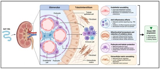
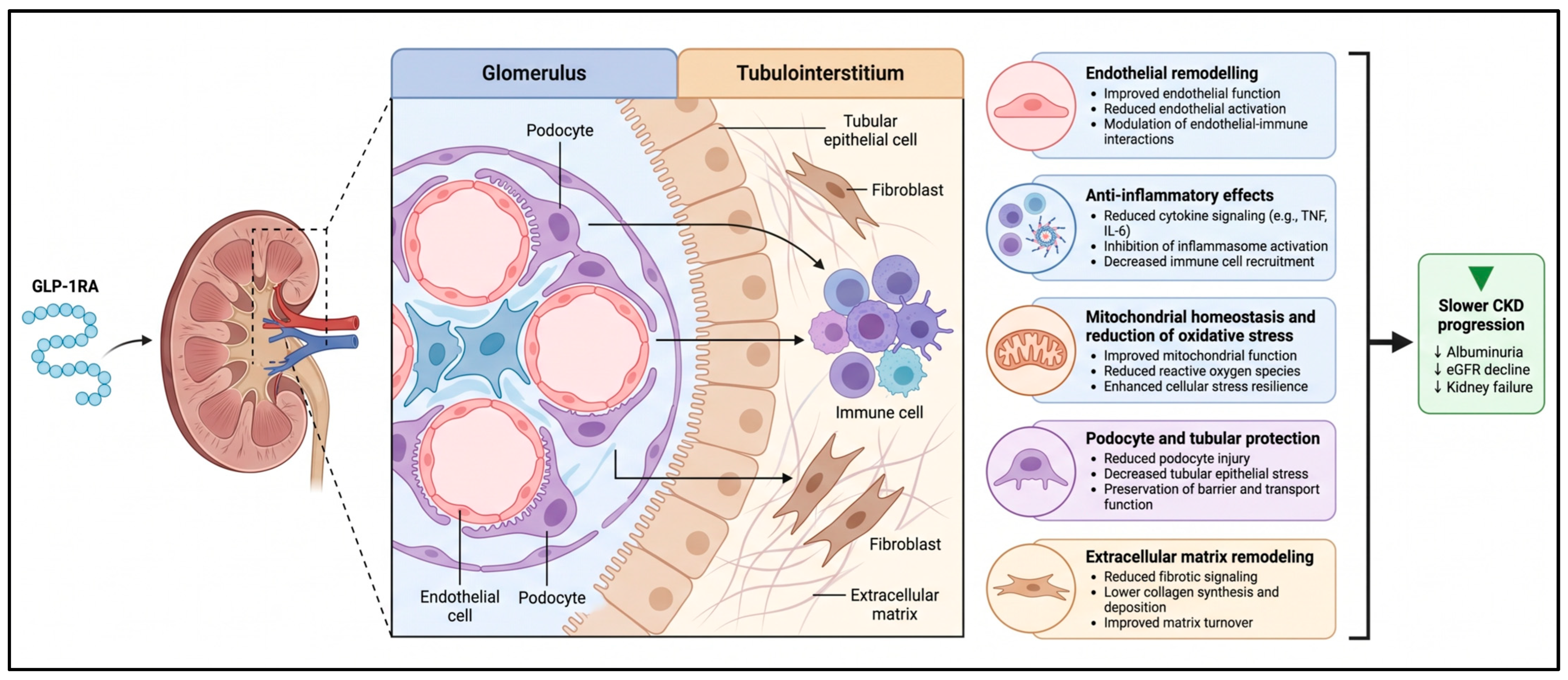
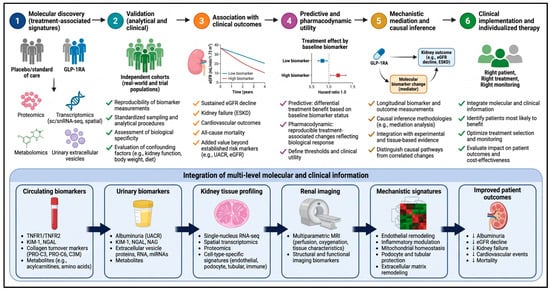
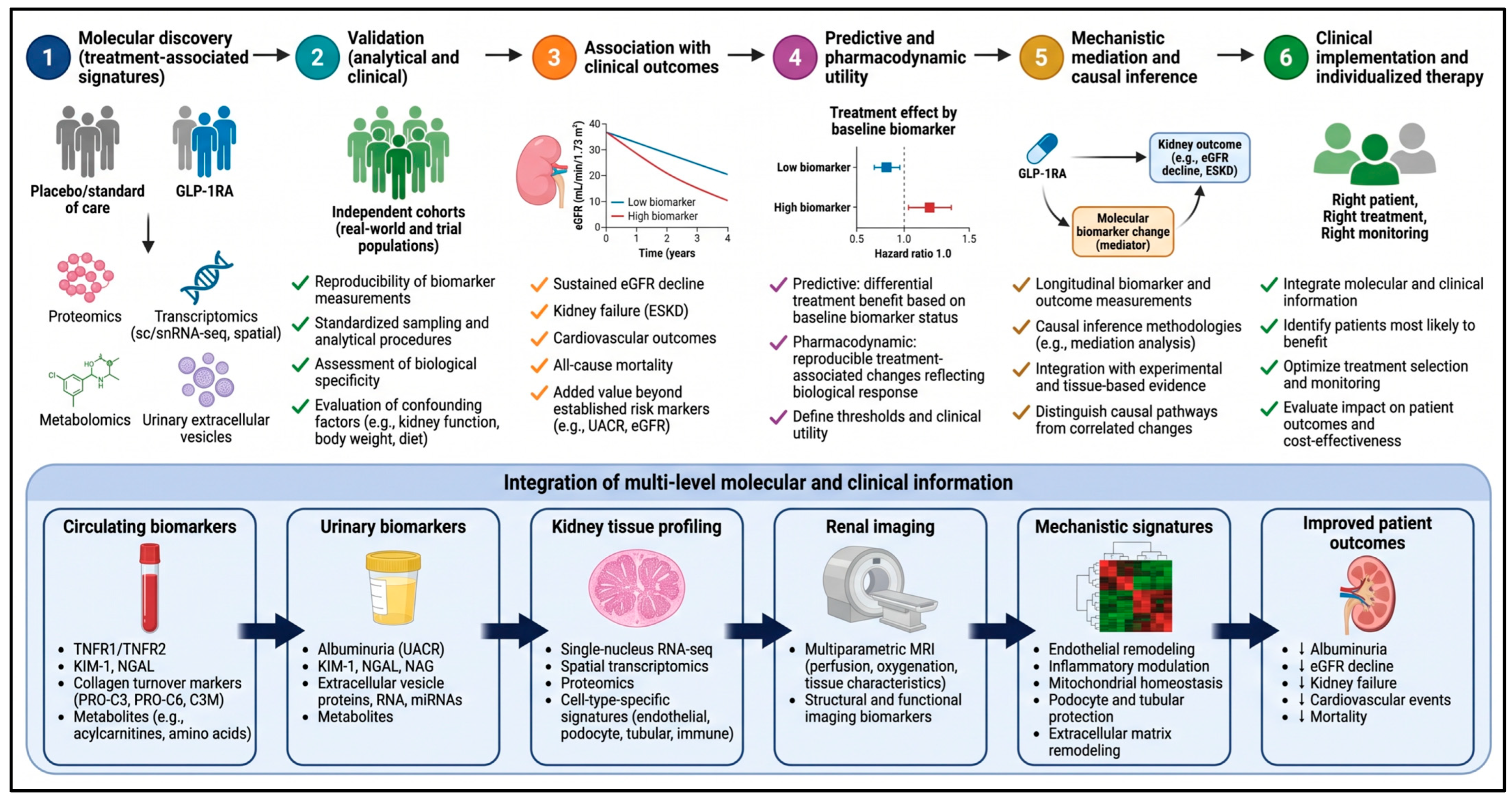

Disclaimer/Publisher’s Note: The statements, opinions and data contained in all publications are solely those of the individual author(s) and contributor(s) and not of MDPI and/or the editor(s). MDPI and/or the editor(s) disclaim responsibility for any injury to people or property resulting from any ideas, methods, instructions or products referred to in the content. |
© 2026 by the authors. Licensee MDPI, Basel, Switzerland. This article is an open access article distributed under the terms and conditions of the Creative Commons Attribution (CC BY) license.