


Abstract
Background/Objectives: Neuroradiological features of neuroinvasive Usutu virus (USUV) infection are poorly understood, as current knowledge is largely limited to case reports. This study aimed to describe brain MRI patterns, and assess disease severity and vital and functional outcomes in patients with neuroinvasive USUV infection. Methods: This multicentric retrospective observational cohort study analyzed adult patients of both sexes, aged ≥18 years, in the continental region of Croatia who developed symptoms of neuroinvasive disease during the arbovirus transmission season between 2017 and 2025. Patients with serological and/or molecular confirmation of USUV infection were included in further analyses. Only patients who underwent a brain MRI during hospitalization were included in the study. Clinical data and blood/CSF laboratory parameters were also evaluated. The observed outcomes were the severity of clinical symptoms, vital status at hospital discharge, and functional status at hospital discharge. Results: Of 214 arbovirus-positive cases, 8 patients had confirmed neuroinvasive USUV infection, and 4 had a brain MRI. Three patients were male. One patient had a normal MRI, and three patients had abnormal MRIs. All patients with abnormal MRI demonstrated meningeal contrast enhancement (a finding not previously reported), uni- or bilateral hippocampal involvement suggestive of limbic encephalitis, and basal ganglia involvement. All patients required ICU admission, experienced prolonged hospitalization, and were discharged with moderate to severe disability. Conclusions: This study provides important insights into MRI patterns in patients with neuroinvasive USUV infection and addresses the current knowledge gap regarding the neuroimaging of this underdiagnosed disease.
1. Introduction
Usutu virus (USUV) is an enveloped, single-stranded RNA arthropod-borne virus belonging to the family Orthoflaviviridae, genus Flavivirus, Japanese encephalitis serocomplex [1]. The natural cycle of USUV resembles that of West Nile virus (WNV), with birds as the main amplifying reservoir host and mosquitoes of the genus Culex as the primary vectors. Due to short-term and low-level viremia, humans represent incidental or dead-end hosts [1,2,3].
Human USUV infections were first described in the Central African Republic and Burkina Faso in patients presenting with fever, rash, and jaundice in 1981 and 2004, respectively [1,4]. The first cases of human neuroinvasive USUV infection in Europe were reported in two immunocompromised patients in Italy in 2009 [5,6]. Between 2012 and 2021, a total of 105 autochthonous USUV infections were detected across several European countries, including Italy [7], Austria [8], Croatia [2,9], the Czech Republic [10], France [11], Germany [12], Hungary [13], and the Netherlands [14]. The majority of cases occurred in Italy (56) and Austria (27). The number of reported human USUV infections nearly doubled from 2017 to 2021, reaching 70 cases. This increase was largely attributable to a significant rise in cases in 2018, which coincided with a surge in human WNV infections [7].
In humans, the incubation period for USUV is typically 3 to 12 days following a mosquito bite. Detecting human infections is difficult because the disease is usually asymptomatic. When symptomatic, the typical presentation includes fever, sometimes accompanied by a maculopapular rash, whereas a febrile icteric illness occurs less frequently [15]. Sporadically, the infection may evolve into a neuroinvasive form, primarily in immunocompromised patients [2], the elderly, or those with underlying comorbidities, but cases in otherwise healthy adults have also been reported [16].
USUV has been increasingly recognized as a cause of neuroinvasive disease in humans, although such cases are underreported. Patients with neuroinvasive diseases typically present with features indistinguishable from other viral meningoencephalitides caused by flaviviruses, particularly WNV [17]. Although clinical reports of USUV infection indicate involvement of brain regions similar to those affected by WNV, the clinical manifestations are generally less severe and less frequent, and do not appear to progress to the same extent as in many cases of WNV-associated neuroinvasive disease [17]. Movement disorders and cerebellar signs, including tremor, dysmetria, and ataxia, as well as cranial nerve palsies, have been documented [11,18].
In the neuroradiological workup of patients with encephalitis, magnetic resonance imaging (MRI) is preferred over computed tomography because of its superior sensitivity and specificity [19]. The neuroradiological features of neuroinvasive USUV infection remain poorly characterized, as current knowledge is limited to a small number of case reports and case series. Consequently, the spectrum of brain MRI abnormalities associated with neuroinvasive USUV infection has not yet been clearly defined.
Croatia has reported several neuroinvasive human cases, often during WNV outbreaks (2013: 3 cases; 2018: 3 cases; 2024: 5 cases) [2,3,9,20].
The aim of this study was twofold: (a) to characterize brain MRI findings among patients with neuroinvasive USUV infection in our cohort and (b) to systematically review the published literature to summarize the current reported neuroradiological manifestations of neuroinvasive USUV infection.
2. Materials and Methods
2.1. Patients
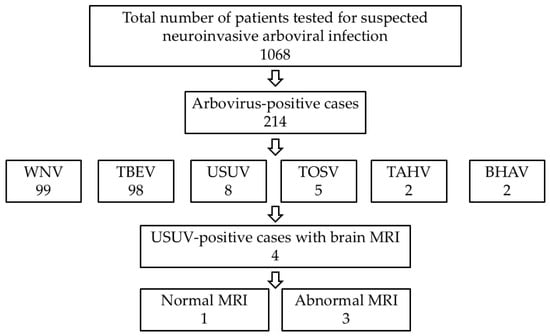
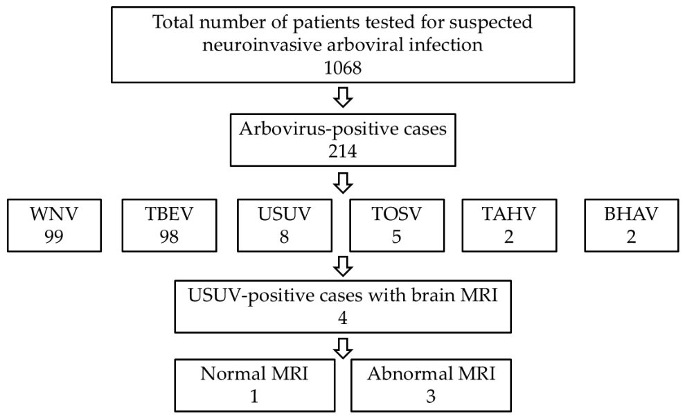
This multicenter retrospective observational cohort study was conducted in accordance with the Strengthening the Reporting of Observational Studies in Epidemiology (STROBE) guidelines [21]. The study population was derived from a nationwide multicenter cohort established as part of the CRONEUROARBO, FLAVIR, and CROIMUNOLAB research projects on arboviral infections in Croatia. Adult patients of both sexes (≥18 years of age) presenting with symptoms of neuroinvasive disease during the arbovirus transmission seasons between January 2017 and December 2025 were enrolled in the study. For the present analysis, patients with serologically (virus neutralization test, VNT) and/or molecularly (reverse transcription quantitative polymerase chain reaction, RT-qPCR) confirmed neuroinvasive USUV infection were identified from the parent cohort. Only patients who underwent a brain MRI during hospitalization were included in the neuroradiological analysis. Details of the patient selection process are shown in Figure 1.
Figure 1.
Study flow chart of patients included in the study (WNV—West Nile virus; TBEV—tick-borne encephalitis virus; USUV—Usutu virus; TOSV—Toscana virus; TAHV—Tahyna orthobunyavirus; BHAV—Bhanja bandavirus).
2.2. Virological and Laboratory Testing
USUV diagnosis was confirmed according to the ECDC criteria for WNV, which included: (a) virus isolation; (b) viral RNA in blood and/or cerebrospinal fluid (CSF); (c) specific IgM antibody response in CSF; and/or (d) high IgM titer and IgG antibody detection confirmed by a virus neutralization test (VNT) [22]. Since recent studies have highlighted the utility of urine RT-qPCR for confirming WNV neuroinvasive infections, owing to its higher diagnostic sensitivity than CSF-based molecular testing, urine was included in the diagnostic evaluation for its noninvasive collection and the potential for prolonged, higher viral shedding [23,24]. In all patients, USUV infection was confirmed by detection of USUV RNA in CSF samples and by serology [24]. Clinical data and blood/CSF laboratory parameters were also evaluated.
2.3. Magnetic Resonance Imaging
MRI examinations were performed on 3T scanners (MAGNETOM Vida and MAGNETOM Lumina, Siemens Healthineers, Erlangen, Germany) according to standardized protocols including: T2-weighted (T2W) images (axial and coronal planes), fluid-attenuated inversion recovery (FLAIR, axial plane), and T1-weighted (T1W) images before (sagittal plane) and after gadolinium-based contrast agent administration (axial, coronal and sagittal planes), susceptibility-weighted images (SWI, axial plane), and diffusion-weighted images (DWI) with apparent diffusion coefficient (ADC) maps (axial plane). MRI examinations were independently reviewed by two radiologists: first by a general radiologist with less than 5 years of experience, and then by a neuroradiologist with more than 10 years of experience, with no prior knowledge of the original reports. Discrepancies between radiologists were resolved by consensus.
2.4. Outcome Evaluation
The observed outcomes were: (1) the severity of clinical symptoms, quantified as mild (0–8 points), moderate (9–22 points), or severe (≥23 points) according to Bogović et al. [25] (Table S1), (2) vital status at hospital discharge (survival or death), and (3) functional status at hospital discharge, quantified using the modified Rankin scale (mRS): 0—no symptoms at all, 1—no significant disability despite symptoms, 2—slight disability, 3—moderate disability, 4—moderately severe disability, 5—severe disability, 6—death. The severity of clinical symptoms and vital/functional status at hospital discharge were evaluated and scored by an infectious disease specialist with more than 10 years of experience in neuroinfections, who had no prior knowledge of the MRI reports.
2.5. Literature Review of Published Usutu Virus Cases
A literature review of PubMed/MEDLINE was conducted up to 2 September 2026 by two researchers independently (T.F. and A.T.B.) to identify confirmed human cases of neuroinvasive USUV infection with brain MRI findings. The search strategy consisted of the following keywords: (Usutu virus [Mesh] OR Usutu virus OR USUV) AND (brain magnetic resonance imaging OR brain MRI OR magnetic resonance imaging OR neuroimaging). No restrictions were applied regarding language or publication year. Eligible studies included original articles, case reports, and case series reporting confirmed neuroinvasive USUV infection with available brain MRI findings. Animal and in vitro studies, and studies without MRI data were excluded. Full texts were assessed, and reference lists of eligible studies were screened for additional reports. Disagreements were resolved by consensus. The database search identified 2 articles, and reference screening identified 4 additional eligible studies.
3. Results
3.1. Brain MRI Findings in Patients with Human Neuroinvasive Usutu Virus Infections
Of the 214 arbovirus-positive cases, 8 patients had neuroinvasive USUV infection (3 in 2018 and 5 in 2024), of whom 4 had a brain MRI (all in 2024). All patients were from the northern continental region of the country. Sociodemographic, clinical, and laboratory features of patients are presented in Table 1, and virology results are presented in Table 2.
Table 1.
Sociodemographic, clinical, and laboratory features of patients with neuroinvasive USUV infection.
Table 2.
Virology results of patients with neuroinvasive USUV infection.
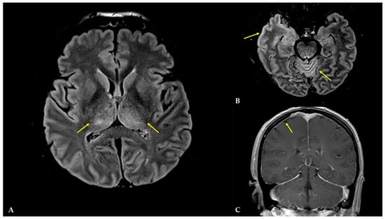
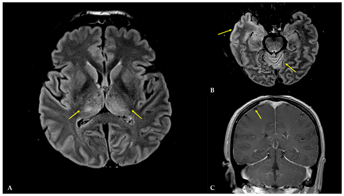
Patient 1 had a normal brain MRI, and three patients (Patients 2–4) had abnormal brain MRI findings. All patients with abnormal brain MRI demonstrated meningeal contrast enhancement. Patient 2 demonstrated high signal intensity in both thalami, the entire right temporal lobe (including hippocampus), and cerebellum on T2W and FLAIR images (Figure 2).
Figure 2.
MRI images of patient 2. (A) Axial plane, FLAIR: asymmetric high signal intensities in both thalami (arrows); (B) axial plane, FLAIR: high signal intensities in the entire right temporal lobe (including hippocampus) and cerebellum (arrows); (C) coronal plane, T1WCE: meningeal contrast enhancement (arrows).
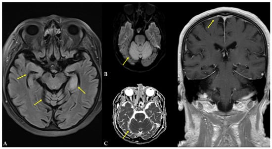
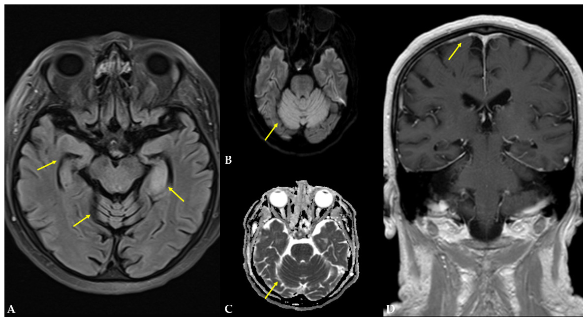
Patient 3 demonstrated asymmetric high signal intensity in both hippocampi and symmetric high signal intensity in the cerebellar hemispheres on T2W and FLAIR images, with restricted diffusion on DWI/ADC (Figure 3).
Figure 3.
MRI images of patient 3. (A) Axial plane, FLAIR: asymmetric high signal intensities in both hippocampi and symmetric in cerebellar hemispheres (arrows); (B,C) axial plane, DWI/ADC: restricted diffusion in cerebellum; (D) coronal plane, T1WCE: meningeal contrast enhancement (arrows).
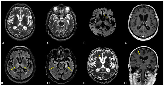
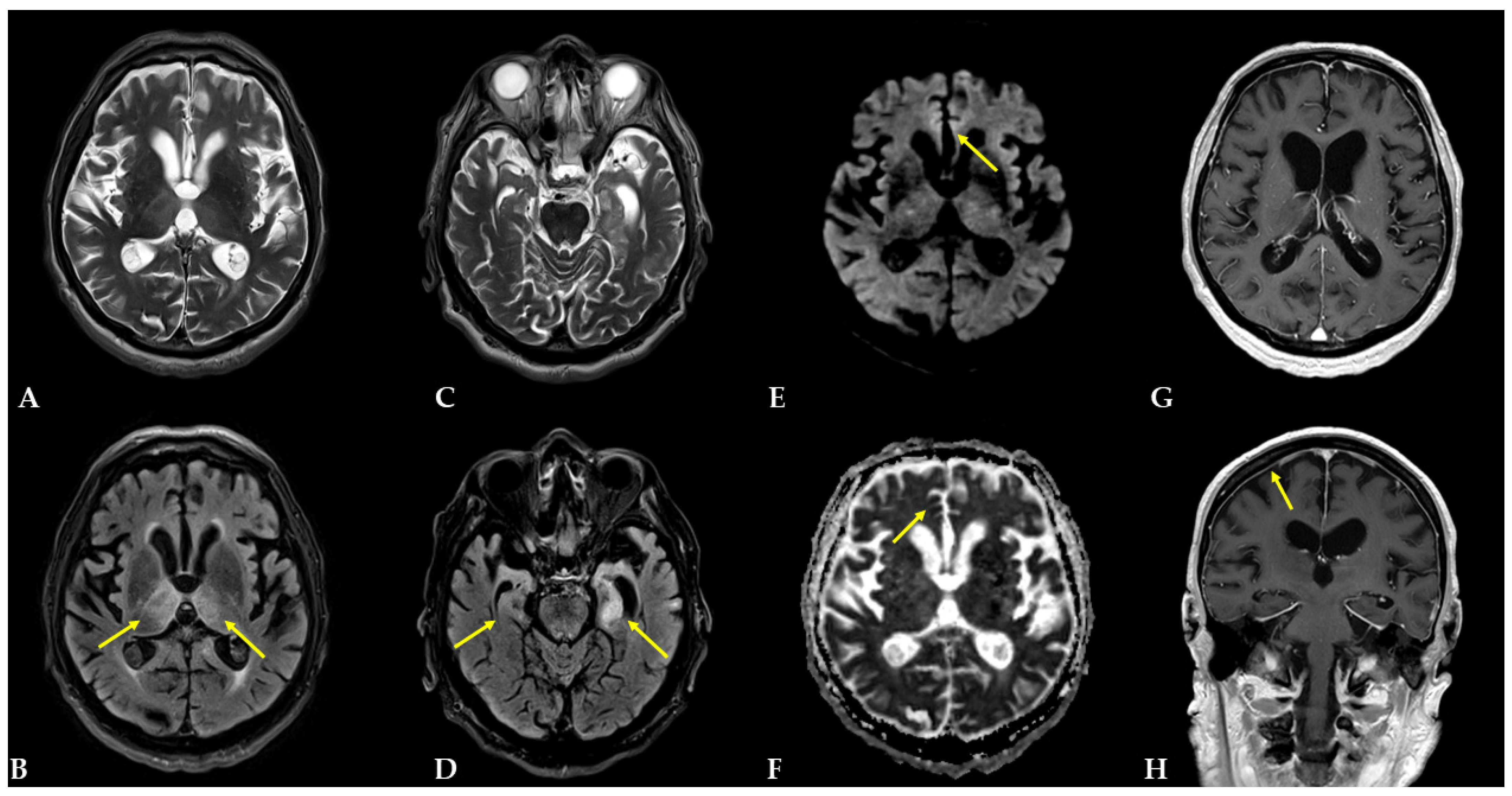
Patient 4 demonstrated an asymmetric high signal intensity in both hippocampi and thalami on T2W and FLAIR images, and restricted diffusion on DWI/ADC. Only on DWI/ADC were abnormal signal intensities found in the cortex of the frontal lobe (Figure 4). A summary of the abnormal brain MRI findings in USUV patients is presented in Table S2.
Figure 4.
MRI images of patient 4 annotated with arrows. (A,B) Axial plane, T2W and FLAIR: asymmetric high signal intensities in both thalami; (C,D) axial plane, T2W and FLAIR: asymmetric high signal intensities in both hippocampi; (E,F) axial plane, only in DWI/ADC: abnormal signal intensities in the cortex of the frontal lobe; (G,H) axial and coronal plane, T1WCE: meningeal contrast enhancement.
3.2. Literature Review of Brain MRI Findings in Neuroinvasive Usutu Virus Infection
Six studies reported patients with neuroinvasive USUV disease and brain MRI, all from Europe [5,9,11,18,26,27]. The characteristics of patients with MRI findings are presented in Table 3.
Table 3.
Studies reporting patients with human neuroinvasive USUV infection and with brain MRI features.
4. Discussion
In this study, we described four patients with confirmed acute neuroinvasive USUV infection who underwent brain MRI, three of whom demonstrated abnormal imaging findings. The patients included both sexes and ranged from middle to older age. One patient was immunocompromised, although all had underlying comorbidities. Among patients with abnormal MRI findings, basal ganglia and hippocampal involvement, as well as meningeal contrast enhancement, were consistently observed. According to the disease severity scale proposed by Bogović et al. [25], the patient with a normal brain MRI had moderately severe disease, whereas all patients with abnormal MRI findings had severe disease. All four patients required ICU admission and experienced prolonged hospitalization. Despite survival in all cases, neurological recovery at discharge remained incomplete, and all patients were discharged with moderate to severe disability.
Our findings expand the limited body of literature describing brain MRI abnormalities in neuroinvasive USUV infection. Like the patient reported by Santini et al. [9], our patient with a normal brain MRI experienced moderate to severe disability despite the absence of detectable neuroradiological abnormalities. In contrast, other previously reported patients with normal brain MRI findings recovered without significant neurological sequelae [11,26]. Notably, all patients with abnormal MRI findings in our cohort demonstrated meningeal contrast enhancement, a feature not previously described in published cases of neuroinvasive USUV infection. Furthermore, all patients with abnormal MRI findings showed uni- or bilateral hippocampal involvement. This observation is particularly interesting, given the report by Graninger et al. [18], which described signal abnormalities in the hippocampus associated with severe neurological disability. Other reported MRI abnormalities have involved the substantia nigra, subcortical regions [5], and the thalami, cerebral peduncles, medulla oblongata, and pons [27], highlighting the heterogeneous neuroradiological presentation of neuroinvasive USUV infection. Taken together, the available evidence suggests that limbic system involvement, particularly of the hippocampus, may represent a recurrent imaging feature in a subset of patients with neuroinvasive USUV infection. However, given the very limited number of reported cases, the clinical significance of this finding remains uncertain and warrants further investigation.
When interpreting brain MRI in patients with suspected encephalitis, particularly limbic encephalitis, the main differential diagnosis includes viral and autoimmune causes [28,29]. The primary viral pathogen affecting the temporal lobes, including the limbic system, is herpes simplex virus (HSV), particularly HSV-1 [19]. Unilateral temporal lobe abnormalities, involvement of the insular cortex, and sparing of the basal ganglia are imaging features that may suggest HSV encephalitis. In contrast, bilateral and symmetric MRI abnormalities, highly confined to the medial temporal lobes, even in the absence of the autoantibodies associated with autoimmune encephalitis, may suggest autoimmune limbic encephalitis [29]. In this study, we found that USUV may also affect the limbic system and, in some cases, the basal ganglia, underscoring the importance of ruling out potential neuroinvasive infection in patients with suspected autoimmune limbic encephalitis before initiating treatment, as the therapeutic approach is fundamentally different. The limbic system involvement, post-contrast meningeal enhancement, and possible basal ganglia involvement may suggest USUV encephalitis. Given the limited characterization of the neuroradiological manifestations of human USUV infection, MRI of the spinal cord and, where clinically indicated, the entire neuroaxis may be considered in patients presenting with severe disease. Such imaging may provide valuable insights into the neuroradiological spectrum of this rare infection and contribute to a better understanding of its underlying neurological involvement.
The main limitations of this study include its retrospective design and small sample size, which prevented formal statistical analysis and limited the generalizability of the findings. The small sample size may reflect both the predominantly asymptomatic course and the underreporting of the disease, as well as the historically limited nationwide availability of MRI, although access to MRI has improved in recent years. Another limitation is the inability to assess lesion evolution over time, given the lack of follow-up MRI examinations. Nevertheless, this study also has several strengths. To the best of our knowledge, this is the largest series of patients with neuroinvasive USUV infection to include standardized clinical assessment and brain MRI evaluation, enabling a systematic analysis of neuroradiological findings in relation to disease severity and functional outcomes. Therefore, this study provides important insights into MRI findings in such patients and significantly addresses the current knowledge gap regarding the neuroimaging of this rare condition.
5. Conclusions
Patients with neuroinvasive USUV infection and abnormal brain MRI all exhibited meningeal contrast enhancement, a finding not reported in previous studies, uni- or bilateral hippocampal involvement, suggestive of limbic encephalitis, and, in some cases, basal ganglia involvement. These observations warrant further neuroradiological investigations in this emerging infectious disease. In patients presenting with encephalitis and imaging findings of limbic system involvement, neuroinvasive USUV infection should be considered in the differential diagnosis, particularly during the arbovirus transmission season. Our results are comparable to those of Graniger et al. [18], suggesting a potential MRI pattern in patients with neuroinvasive USUV infection. It is a clinically significant recognition because patients with neuroinvasive USUV infection may present with a severe disease course, while its management differs substantially from that of autoimmune limbic encephalitis. Brain MRI in patients with neuroinvasive USUV infection may provide clinicians with additional information that could affect decision-making and the disease course. Spinal MRI may also be considered in these patients, as it could help further refine the differential diagnosis and facilitate earlier diagnostic clarification. However, this needs to be verified in studies with a larger number of participants.
Supplementary Materials
The following supporting information can be downloaded at: https://www.mdpi.com/article/10.3390/diagnostics16203269/s1: Table S1: Evaluation of disease severity in patients with neuroinvasive USUV infection according to Bogović et al. [25]; Table S2: Brain MRI findings in patients with neuroinvasive USUV infection.
Author Contributions
Conceptualization, T.F. and T.V.-Č.; methodology, T.F., T.V.-Č., A.T.B., V.S. (Vladimir Stevanović) and M.S.; formal analysis, T.F.; investigation, T.F., V.S. (Vladimir Savić), M.B., V.S. (Vladimir Stevanović), D.S., T.P.-H., F.K., K.V., T.J.-N., R.Š., L.B. and D.H.; writing—original draft preparation, T.F.; writing—review and editing, T.V.-Č., V.S. (Vladimir Savić), A.T.B., M.B., V.S. (Vladimir Stevanović), D.S., K.V. and M.S.; visualization, T.P.-H., F.K., T.J.-N., R.Š. and D.H.; supervision, T.V.-Č., A.T.B., L.B. and M.S. All authors have read and agreed to the published version of the manuscript.
Funding
This research was funded by the Croatian Science Foundation project “Prevalence and Molecular Epidemiology of Emerging and Re-Emerging Neuroinvasive Arboviral Infections in Croatia” (CRONEUROARBO, grant number 2016-06-7456 (to T.V.-Č.)); the European Union Next Generation EU project supported by the Ministry of Science and Education of the Republic of Croatia (grant number NPOO1); the Croatian Veterinary Institute (“Epidemiology of Avian Flaviviruses in the “One Health” Context in Croatia”; FLAVIR (to V.S., Vladimir Savić); and the School of Medicine, University of Zagreb “Immunopathogenesis of Neuroinvasive Zoonoses in Croatia: Clinical-Laboratory Correlation” (CROIMUNOLAB, to T.V.-Č, grant number NPOO 10106-25-2649).
Institutional Review Board Statement
The study was conducted in accordance with the Declaration of Helsinki and approved by the Ethics Committees of the Croatian Institute of Public Health 548 (protocol code 053-01/25-06/02, approved on 18 June 2025), University Hospital for Infectious Diseases “Dr. Fran Mihaljević” (protocol code 01-2341-2-2025, approved on 29 October 2025) and School of Medicine, University of Zagreb (protocol code 641-01/26-02/04, approved on 19 March 2026).
Informed Consent Statement
Written informed consent has been obtained from the patients included in the study.
Data Availability Statement
Data is contained within the article, and further inquiries can be directed to the corresponding author.
Acknowledgments
The authors thank Marko Kutleša for his valuable clinical contribution to the care of patients included in this study. The authors thank Ljiljana Milašinčić, Ljiljana Antolašić, and Marta Batur for providing technical assistance.
Conflicts of Interest
The authors declare no conflicts of interest.
References
- Clé, M.; Barthelemy, J.; Desmetz, C.; Foulongne, V.; Lapeyre, L.; Bolloré, K.; Tuaillon, E.; Erkilic, N.; Kalatzis, V.; Lecollinet, S.; et al. Study of Usutu virus neuropathogenicity in mice and human cellular models. PLoS Neglected Trop. Dis. 2020, 14, e0008223. [Google Scholar] [CrossRef] [Scilit] [PubMed]
- Vilibic-Cavlek, T.; Savić, V.; Sabadi, D.; Peric, L.; Barbic, L.; Klobucar, A.; Miklausic, B.; Tabain, I.; Santini, M.; Vucelja, M.; et al. Prevalence and molecular epidemiology of West Nile and Usutu virus infections in Croatia in the ‘One Health’ context, 2018. Transbound. Emerg. Dis. 2019, 66, 1946–1957. [Google Scholar] [CrossRef] [Scilit] [PubMed]
- Vilibic-Cavlek, T.; Kaic, B.; Barbic, L.; Pem-Novosel, I.; Slavic-Vrzic, V.; Lesnikar, V.; Kurecic-Filipovic, S.; Babic-Erceg, A.; Listes, E.; Stevanovic, V.; et al. First evidence of simultaneous occurrence of West Nile virus and Usutu virus neuroinvasive disease in humans in Croatia during the 2013 outbreak. Infection 2014, 42, 689–695. [Google Scholar] [CrossRef] [Scilit]
- Nikolay, B.; Weidmann, M.; Dupressoir, A.; Faye, O.; Boye, C.S.; Diallo, M.; Sall, A.A. Development of a Usutu Virus Specific Real-Time Reverse Transcription PCR Assay Based on Sequenced Strains from Africa and Europe. J. Virol. Methods 2014, 197, 51–54. [Google Scholar] [CrossRef] [Scilit]
- Pecorari, M.; Longo, G.; Gennari, W.; Grottola, A.; Sabbatini, A.; Tagliazucchi, S.; Savini, G.; Monaco, F.; Simone, M.; Lelli, R.; et al. First human case of Usutu virus neuroinvasive infection, Italy, August-September 2009. Eurosurveillance 2009, 14, 19446. [Google Scholar] [CrossRef] [Scilit]
- Cavrini, F.; Gaibani, P.; Longo, G.; Pierro, A.M.; Rossini, G.; Bonilauri, P.; Gerunda, G.E.; Di Benedetto, F.; Pasetto, A.; Girardis, M.; et al. Usutu virus infection in a patient who underwent orthotropic liver transplantation, Italy, August-September 2009. Eurosurveillance 2009, 14, 19448. [Google Scholar] [CrossRef] [Scilit]
- Angeloni, G.; Bertola, M.; Lazzaro, E.; Morini, M.; Masi, G.; Sinigaglia, A.; Trevisan, M.; Gossner, C.M.; Haussig, J.M.; Bakonyi, T.; et al. Epidemiology, surveillance and diagnosis of Usutu virus infection in the EU/EEA, 2012 to 2021. Eurosurveillance 2023, 28, 2200929, Correction in Eurosurveillance 2023, 28, 230824c. https://doi.org/10.2807/1560-7917.ES.2023.28.34.230824c. [Google Scholar] [CrossRef] [Scilit] [PubMed]
- Aberle, S.W.; Kolodziejek, J.; Jungbauer, C.; Stiasny, K.; Aberle, J.H.; Zoufaly, A.; Hourfar, M.K.; Weidner, L.; Nowotny, N. Increase in human West Nile and Usutu virus infections, Austria, 2018. Eurosurveillance 2018, 23, 1800545. [Google Scholar] [CrossRef] [Scilit] [PubMed]
- Santini, M.; Vilibic-Cavlek, T.; Barsic, B.; Barbic, L.; Savic, V.; Stevanovic, V.; Listes, E.; Di Gennaro, A.; Savini, G. First cases of human Usutu virus neuroinvasive infection in Croatia, August-September 2013: Clinical and laboratory features. J. NeuroVirol. 2015, 21, 92–97. [Google Scholar] [CrossRef] [Scilit] [PubMed]
- Zelená, H.; Kleinerová, J.; Šikutová, S.; Straková, P.; Kocourková, H.; Stebel, R.; Husa, P.; Husa, P., Jr.; Tesařová, E.; Lejdarová, H.; et al. First Autochthonous West Nile Lineage 2 and Usutu Virus Infections in Humans, July to October 2018, Czech Republic. Pathogens 2021, 10, 651. [Google Scholar] [CrossRef] [Scilit] [PubMed]
- Simonin, Y.; Sillam, O.; Carles, M.J.; Gutierrez, S.; Gil, P.; Constant, O.; Martin, M.F.; Girard, G.; Van de Perre, P.; Salinas, S.; et al. Human Usutu Virus Infection with Atypical Neurologic Presentation, Montpellier, France, 2016. Emerg. Infect. Dis. 2018, 24, 875–878. [Google Scholar] [CrossRef] [Scilit] [PubMed]
- Cadar, D.; Maier, P.; Müller, S.; Kress, J.; Chudy, M.; Bialonski, A.; Schlaphof, A.; Jansen, S.; Jöst, H.; Tannich, E.; et al. Blood donor screening for West Nile virus (WNV) revealed acute Usutu virus (USUV) infection, Germany, September 2016. Eurosurveillance 2017, 22, 30501. [Google Scholar] [CrossRef] [Scilit] [PubMed]
- Nagy, A.; Mezei, E.; Nagy, O.; Bakonyi, T.; Csonka, N.; Kaposi, M.; Koroknai, A.; Szomor, K.; Rigó, Z.; Molnár, Z.; et al. Extraordinary increase in West Nile virus cases and first confirmed human Usutu virus infection in Hungary, 2018. Eurosurveillance 2019, 24, 1900038. [Google Scholar] [CrossRef] [Scilit] [PubMed]
- Zaaijer, H.L.; Slot, E.; Molier, M.; Reusken, C.B.E.M.; Koppelman, M.H.G.M. Usutu virus infection in Dutch blood donors. Transfusion 2019, 59, 2931–2937. [Google Scholar] [CrossRef] [Scilit] [PubMed]
- Cadar, D.; Simonin, Y. Human Usutu Virus Infections in Europe: A New Risk on Horizon? Viruses 2022, 15, 77. [Google Scholar] [CrossRef] [Scilit]
- Trillocco, G.; Giuliano, G.; Alessandri, G.; Trezzi, M.; Pippi, F.; Tumbarello, M.; Terrosi, C.; Cucini, S.; Cusi, M.G. Diagnostic challenges in Flaviviridae infections: First human case of Usutu virus aseptic meningitis in an immunocompetent subject in Italy. Int. J. Infect. Dis. 2026, 167, 108667. [Google Scholar] [CrossRef] [Scilit] [PubMed]
- Marshall, E.M.; Barzon, L.; Koopmans, M.; Rockx, B. Extrapolation and comparison of West Nile virus- and Usutu virus-associated neurological diseases in humans: Linking pathology to clinical symptoms. Clin. Microbiol. Rev. 2025, 38, e0023224. [Google Scholar] [CrossRef] [Scilit] [PubMed]
- Graninger, M.; Hubmer, S.; Riederer, F.; Kettner, S.; Hauk, M.; Auf, T.; Aberle, J.H.; Stiasny, K.; Aberle, S.W.; Camp, J.V. The First Case of Usutu Virus Neuroinvasive Disease in Austria, 2021. Open Forum Infect. Dis. 2022, 9, ofac255. [Google Scholar] [CrossRef] [Scilit] [PubMed]
- Venkatesan, A.; Jagdish, B. Imaging in Encephalitis. Semin. Neurol. 2019, 39, 312–321. [Google Scholar] [CrossRef] [Scilit] [PubMed]
- Vilibić-Čavlek, T.; Barbić, L.; Klobučar, A.; Vucelja, M.; Bogdanić, M.; Sabadi, D.; Kutleša, M.; Gjurašin, B.; Stevanović, V.; Curman Posavec, M.; et al. Re-Emergence of Usutu Virus and Spreading of West Nile Virus Neuroinvasive Infections During the 2024 Transmission Season in Croatia. Viruses 2025, 17, 846. [Google Scholar] [CrossRef] [Scilit] [PubMed]
- von Elm, E.; Altman, D.G.; Egger, M.; Pocock, S.J.; Gotzsche, P.C.; Vandenbroucke, J.P.; Initiative, S. The Strengthening the Reporting of Observational Studies in Epidemiology (STROBE) statement: Guidelines for reporting observational studies. Lancet 2007, 370, 1453–1457. [Google Scholar] [CrossRef] [Scilit] [PubMed]
- European Center for Disease Control and Prevention. EU Case Definitions. Available online: https://www.ecdc.europa.eu/en/all-topics/eu-case-definitions (accessed on 8 June 2026).
- Cvjetković, I.H.; Radovanov, J.; Kovačević, G.; Turkulov, V.; Patić, A. Diagnostic value of urine qRT-PCR for the diagnosis of West Nile virus neuroinvasive disease. Diagn. Microbiol. Infect. Dis. 2023, 107, 115920. [Google Scholar] [CrossRef] [Scilit] [PubMed]
- Vilibic-Cavlek, T.; Bogdanic, M.; Savic, V.; Hruskar, Z.; Barbic, L.; Stevanovic, V.; Antolasic, L.; Milasincic, L.; Sabadi, D.; Miletic, G.; et al. Diagnosis of West Nile virus infections: Evaluation of different laboratory methods. World J. Virol. 2024, 13, 95986. [Google Scholar] [CrossRef] [Scilit] [PubMed]
- Bogovic, P.; Logar, M.; Avsic-Zupanc, T.; Strle, F.; Lotric-Furlan, S. Quantitative evaluation of the severity of acute illness in adult patients with tick-borne encephalitis. BioMed Res. Int. 2014, 2014, 841027. [Google Scholar] [CrossRef] [Scilit] [PubMed]
- Pacenti, M.; Sinigaglia, A.; Martello, T.; De Rui, M.E.; Franchin, E.; Pagni, S.; Peta, E.; Riccetti, S.; Milani, A.; Montarsi, F.; et al. Clinical and virological findings in patients with Usutu virus infection, northern Italy, 2018. Eurosurveillance 2019, 24, 1900180. [Google Scholar] [CrossRef] [Scilit] [PubMed]
- Szabó, B.G.; Nagy, A.; Nagy, O.; Koroknai, A.; Csonka, N.; Korózs, D.; Jeszenszky, K.; Hardi, A.; Deézsi-Magyar, N.; Sztikler, J.; et al. First documented case of a fatal autochthonous Usutu virus infection in an immunocompromised patient in Hungary: A clinical-virological report and implications from the literature. Virol. J. 2025, 22, 261. [Google Scholar] [CrossRef] [Scilit] [PubMed]
- Jayaraman, K.; Rangasami, R.; Chandrasekharan, A. Magnetic Resonance Imaging Findings in Viral Encephalitis: A Pictorial Essay. J. Neurosci. Rural Pract. 2018, 9, 556–560. [Google Scholar] [CrossRef] [Scilit] [PubMed]
- Budhram, A.; Leung, A.; Nicolle, M.W.; Burneo, J.G. Diagnosing autoimmune limbic encephalitis. Can. Med. Assoc. J. 2019, 191, E529–E534. [Google Scholar] [CrossRef] [Scilit] [PubMed]

Disclaimer/Publisher’s Note: The statements, opinions and data contained in all publications are solely those of the individual author(s) and contributor(s) and not of MDPI and/or the editor(s). MDPI and/or the editor(s) disclaim responsibility for any injury to people or property resulting from any ideas, methods, instructions or products referred to in the content. |
© 2026 by the authors. Licensee MDPI, Basel, Switzerland. This article is an open access article distributed under the terms and conditions of the Creative Commons Attribution (CC BY) license.