


Abstract
Background/Objectives: Dilated cardiomyopathy is a heterogeneous disorder with a substantial genetic contribution from a variety of pathogenic variants. Hereditary isolated DCM is often caused by variants in genes encoding sarcomere proteins, as well as proteins involved in desmosomes or other cardiac cell functions. Identifying genetic causes improves our understanding of DCM pathophysiology, facilitates prognostic assessment, and enables more personalized disease management. Methods: We retrospectively analyzed genetic data from adult patients with a clinical diagnosis of isolated DCM evaluated at a Lithuanian tertiary university hospital between 2019 and 2024. All patients were tested with a next-generation sequencing cardiovascular gene panel. Results: We gathered 169 patients and initially reached a 16.0% (n = 27) genetic testing diagnostic yield. We performed all genetic variant reanalyses with the most current classification guidelines, and we found an additional eight positive cases. Our final diagnostic yield was 20.7% (n = 35). TTN was the most frequently affected gene (n = 30), whereas variants in BAG3 (n = 2), DSP (n = 1), LMNA (n = 1), and FLNC (n = 1) were rare. In total, 15 variants were novel—not described in the literature or databases. We did not observe significant clinical differences between patients with pathogenic variants and those without pathogenic variants. We expected a different clinical course with variants in genes like BAG3 or LMNA, but there were only a few cases. Conclusions: Genetic testing remains an important tool for confirming complex DCM cases and allows earlier disease management for relatives at risk.
1. Introduction
Dilated cardiomyopathy is a major public health burden as it is one of the leading indications for heart transplantation and a major cause of heart failure [1]. The defining feature of DCM is left ventricular dilatation with reduced function in the absence of hypertension, valvular, congenital, or ischemic heart disease [2,3]. DCM has both genetic and non-genetic causes, and this heterogeneity makes it difficult to determine its true prevalence. Non-genetic DCM may result from drug use, exposure to toxins, abuse of alcohol, viral and bacterial infections, or autoimmune disorders, may be related to other conditions such as pregnancy, or may be the combined effect of all mentioned factors [1]. In addition to monogenic causes, syndromic forms such as muscular dystrophies, mitochondrial disorders, and metabolic diseases should also be considered.
Reported prevalence ranges from approximately 1 in 250 to 1 in 2700 individuals [4,5], which possibly reflects various causes of DCM. A UK Biobank analysis searching only isolated monogenic DCM found a prevalence of about 1 in 220 [6]. Interestingly, pathogenic TTN variants have been identified in approximately 1 in 250 adults without known heart failure [7]. Not all DCM cases may be recognized clinically, partly due to age-dependent penetrance.
Establishing a diagnosis of DCM often requires comprehensive evaluation to identify the underlying cause and enable targeted treatment [8]. This includes many laboratory tests like checking calcium, ferritin, complete blood count, liver, thyroid, renal function, and troponin. Depending on suspicion and age of onset, additional tests can include secondary metabolic screening such as carnitine profile, free fatty acids, and antibody testing. Confirmation of ventricular dilatation and systolic dysfunction requires imaging studies such as echocardiography and cardiac magnetic resonance imaging. These evaluations should also be offered to first-degree family members to identify familial cases. In selected cases, suspicion of myocardial infiltration or inflammation may require endomyocardial biopsy. Finally, genetic evaluation may help confirm a hereditary etiology.
Isolated hereditary DCM is most commonly caused by variants in sarcomere-encoding genes, particularly TTN. Rare variants in at least 12 genes have been shown to be enriched in DCM and are considered potentially causative [9]. Haploinsufficiency of sarcomere genes leads to the reduced expression of proteins such as titin, myosin, actin, and troponin [10]. Also, protein deficiency can increase metabolic demand, which further facilitates eccentric heart remodeling [11]. The involvement of other genes is more complex; for example, lamin deficiency causes nuclear envelope distortion, especially in contractile cardiomyocytes. Lamin is coded by LMNA, and cardiomyopathy associated with this gene presents with a more severe phenotype, with higher incidence of malignant arrhythmias and a need for heart transplantation [12]. Variants in DSP, which encodes desmoplakin, can also cause a distinct phenotype with high arrhythmic burden, variable left ventricle ejection fraction, and left ventricle scarring [13]. Loss of desmoplakin disrupts intercellular adhesion, which allows easier fibrosis formation. BAG3 is one of the more recently established genes associated with DCM [14]. Its protein has complex functions, including the stabilization of myofibril structure under stress [15]. Its phenotype presents great variability but fewer arrhythmia events are noted. These findings illustrate the diversity of genetic mechanisms and their associated phenotypic variability.
The first genetic cause of DCM was identified in 1998 by MT Olson et al. [16]. At that time, missense variants in the ACTC1 gene were identified as the causative factor. Later studies based on this discovery tried to find more cases but were largely unsuccessful [17,18]. Now this gene is more commonly associated with hypertrophic cardiomyopathies and very rarely is it the cause of DCM [19]. The identification of causative variants may help confirm diagnosis in ambiguous cases or adjust patient care as some genetic variants lead to more pronounced conduction damage. In this study, we report our experience with genetic testing in patients with DCM at a tertiary university hospital.
2. Materials and Methods
2.1. Patient Selection
A retrospective study was performed between 2019 and 2024 at the Department of Genetics and Molecular Medicine, Hospital of the Lithuanian University of Health Sciences Kauno Klinikos. Ethical approval was granted by the Kaunas Regional Biomedical Research Ethics Committee (No. BE-2-63). Demographic, clinical, and genetic data were collected from electronic health records. Patients referred for genetic evaluation because of a confirmed or suspected diagnosis of dilated cardiomyopathy were included in the study. Clinical diagnosis was confirmed through echocardiography or cardiac MRI. We used the European Society of Cardiology (ESC) definition of dilated cardiomyopathy, which is characterized by a left ventricular (LV) end-diastolic diameter greater than 58 mm in males and greater than 52 mm in females, along with an LV end-diastolic volume index of 75 mL/m2 in males and 62 mL/m2 in females as measured by echocardiography. Left ventricular global systolic dysfunction is identified when the left ventricular ejection fraction (LVEF) is less than 50% [2]. All patients received genetic testing. We included only isolated non-syndromic cases (for example, patients with a primary skeletal muscle disorder which causes secondary DCM are excluded). We also excluded asymptomatic individuals who underwent genetic evaluation solely because of an affected family member had been identified.
Inclusion criteria:
- Age ≥ 18 years at the time of genetic consultation;
- Availability of cardiac imaging, including echocardiography and/or cardiac magnetic resonance imaging (MRI);
- Diagnosis or clinical suspicion of dilated cardiomyopathy established by a multidisciplinary team consisting of a clinical geneticist and cardiologists;
- Underwent genetic testing.
2.2. Cardiac Investigation
The cohort consisted of patients with a long-standing diagnosis of DCM as well as those who were newly diagnosed. Data collected included symptoms, age at onset, laboratory and instrumental findings, complications such as atrial or ventricular fibrillation, and treatments including cardioverter or pacemaker implantation, as well as mortality data. As not all patients had MRI scans performed, the ejection fraction was evaluated using the first available data from health records, regardless of whether it was measured by MRI or ultrasound. Other cardiac parameters were compared based on the imaging method used (MRI or ultrasound). MRI scans were conducted on a 3T magnetic resonance imaging scanner with an 18-channel cardiac coil (MAGNETOM Skyra, Siemens Healthcare, Erlangen, Germany). Images were obtained during expiratory breath-holding with ECG gating. In cases where MRI was not performed or no data were available, parameters from heart ultrasound were utilized. Two-dimensional (2D) transthoracic echocardiography (TTE) at rest was performed by an experienced echocardiographer using a diagnostic ultrasound system (EPIQ 7, Philips Ultrasound, Inc., Bothell, WA, USA). All measurements were taken in accordance with established guidelines. A family history was considered positive if first-degree relatives experienced sudden cardiac death before the age of 60 or if there were known cases of DCM within the family.
2.3. Genetic Investigation
Patients’ peripheral blood samples were collected with EDTA tubes. Genomic DNA extraction was performed via automatic workflow with QIA symphony (QIAGEN, Hilden, Germany) with the reagent kit DNA midi Kit, or with QIcube (QIAGEN, Hilden, Germany) with the reagent QIAmp DNA blood Mini Kit. Purity and concentration were assessed using Qubit (Thermo Fisher Scientific, Waltham, MA, USA) and dsDNA HS Assay kit reagents. Next-generation sequencing was performed with the Sistemas Genómicos (Valencia, Spain) Cardio-GeneSGKit library preparation kit and the Illumina NextSeq 550 system (San Diego, CA, USA). The Cardio-GeneSGKit library gene list was updated during the study period, but genes important/established for the DCM were not compromised. Detailed lists are in the Supplementary Materials (S1 list—older panel; S2 list—newest panel). All laboratory procedures were conducted in accordance with the manufacturers’ instructions. Sequencing achieved an average depth of 200 times per nucleotide. The genetic test was repeated when it did not achieve the targeted depth. Most target genes achieved complete coverage. Variant analysis and reporting were restricted to coding exons and flanking intronic regions extending 20 bp from exon–intron boundaries.
Sequencing reads were aligned against human reference genome version GRCh38. Read alignment was performed using the Burrows–Wheeler algorithm (BWA). After read mapping, low-quality reads and sequences flagged as PCR duplicates were removed. Variant calling was performed using the Genome Analysis Toolkit (GATK). Low-quality variants (coverage < 10×, allele balance < 20%, Phred < 20) were removed. We selected only those genes for analysis that showed evidence of causing DCM by PanelApp: TNNT2, ANKRD1, DSP, NEXN, DSG2, ACTC1, MYBPC3, RBM20, DES, SCN5A, LDB3, VCL, FLNC, JUP, TCAP, FKRP, MYH6, TNNI3, PKP2, EMD, TBX5, BAG3, SGCD, TMEM43, CSRP3, FKTN, ACTN2, TNNI3K, TTN, MYPN, GATA6, TPM1, LAMP2, CRYAB, PLN, TNNC1, DMD, MYH7, TBX20, DSC2, NKX2-5, ANK2, RYR2, PRDM16, and LMNA [20]. Variants were initially classified according to the 2015 ACMG/AMP [21] and relevant ClinGen recommendations. Given the retrospective nature of the study and the evolution of variant interpretation criteria over time, all identified variants were re-evaluated using the most recent gene-specific ClinGen recommendations [22]. Variants of uncertain significance (VUS) were excluded from the present analysis; however, clinically actionable variants were communicated to patients as part of routine clinical care.
2.4. Statistical Analysis
Cases were coded and research data were stored in a Microsoft Office Excel spreadsheet. Statistical analysis was performed using nonparametric tests like Chi square, the Mann–Whitney U test, and Spearman, as data was not normally distributed (confirmed with the Kolmogorov–Smirnov test). Statistical calculations were performed using IBM SPSS 26.0 software.
3. Results
Data from 169 patients with dilated cardiomyopathy phenotype were collected, of whom 69.8% were males and 30.2% females. The mean age at genetic consultation was 51.54 ± 12.26 years, with males tested at a slightly older age than females (51.80 ± 12.02 vs. 50.97 ± 12.90; p < 0.05). Pathogenic (P) or likely pathogenic (LP) variants in one of the DCM-causing genes were identified in 20.7% of cases (n = 35) (Table 1). The detection rate was similar between males and females (21.2% vs. 19.6%; p = 0.054). Patients carrying pathogenic variants were slightly younger at the time of testing (49.63 ± 11.93 vs. 52.05 ± 12.34) and more frequently had affected first-degree family members (27.8% vs. 19.2%; p = 0.266).
Table 1.
Patients’ statistics.
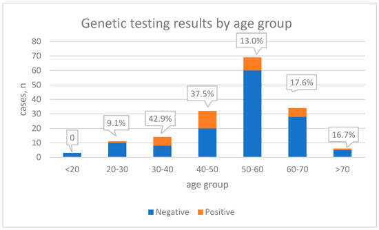
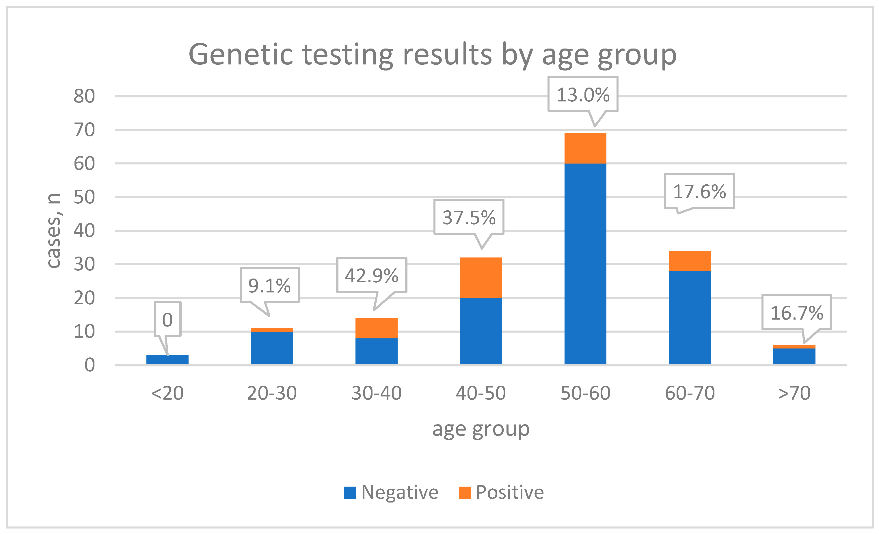
Patients were divided into two equal groups based on age: younger and older than 54 years. Comparing clinical parameters or genetic testing results, we found no differences. When stratified by decade, the highest proportion of pathogenic or likely pathogenic (P/LP) variants was observed in patients aged 30–40 and 40–50 years (42.9% and 37.5%, respectively) (Figure 1). Patients younger than 54 years with a positive family history had a significantly higher probability of carrying a causative variant than all other patients (40% vs. 18.1%; p = 0.023; Chi square). These patients also had higher LVEF (36.86 ± 12.47 vs. 30.20 ± 11.78; p = 0.027; Mann–Whitney).
Figure 1.
Cohort presented by age group and percentage of positive genetic testing.
Patients with and without pathogenic variants had similar ejection fraction (32.09 ± 12.99 vs. 30.70 ± 11.80; p = 0.595). After at least one year of follow-up, ejection fraction improved similarly in both groups (n = 130; 36.12 ± 11.67 vs. 34.94 ± 12.21; p = 0.57). When comparing other clinical parameters, such as left ventricle end-diastolic volume/diameter, arrhythmias, and complications, we found no differences. Clinical features are summarized in Table 2.
Table 2.
Clinical and echocardiographic data.
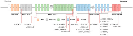
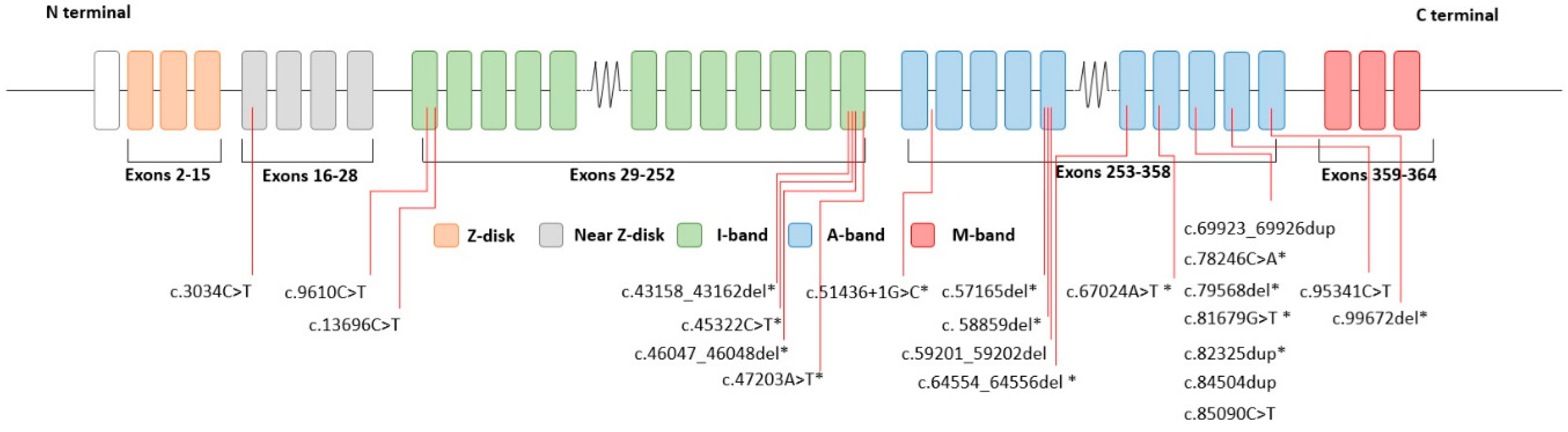
Most patients carried TTN gene pathogenic variants—30 (16 nonsense, 13 indels with frameshift, and 1 splice site). Two patients had pathogenic variants in BAG3, and single cases were identified in DSP, FLNC, and LMNA. All variants were reanalyzed according to current guidelines, resulting in 8 additional patients receiving a positive genetic diagnosis (included in the total 35 cases). Six of these variants are found in the I-band of titin, but their exon percentage spliced-in is 100%. Overall, 10 TTN-positive cases affected the I-band and 20 the A-band; however, this did not correlate with clinical phenotype. In total, 15 variants were novel - not described in the literature or clinical databases. TTN variants c.13696C>T and c.69923_69926dup were found 5 and 4 times. The recurrence of these variants suggests that they are unlikely to be de novo; however, no affected family members were reported. De novo status could not be assessed, as many patients’ parents were deceased. The TTN gene 326 exon was found to be the most commonly affected (n = 11). No statistically significant differences were observed between patients with variants in exon 326 and those with variants in other regions of TTN. TTN affected regions are marked in Figure 2. All detected variants are presented in Table 3.
Figure 2.
Schematic representation of TTN gene and P/LP variants found. Many variants are found in distal half of TTN gene. Variants marked with * are novel. Regions marked using Cardiodb data [23].
Table 3.
List of all pathogenic variants detected.
Among TTN-positive patients, those with frameshift variants showed a higher prevalence of ventricular arrhythmias (4 vs. 0 cases) and a greater need for implantable devices (6 vs. 1 cases) compared to those with nonsense variants; however, these differences did not reach statistical significance.
4. Discussion
We present our experience with genetic testing in patients with DCM at a tertiary university hospital. Our patient selection criteria were relatively broad, as individuals with suspected DCM were also included (following multidisciplinary team decision). Despite that, we achieved a 20% genetic testing diagnostic yield, which is consistent with reports from other centers [9,24,25,26]. Mazzarotto et al. showed that patients selected by specialists have a higher diagnostic yield: 17% in unselected DCM cases vs. 26% with diagnostic referral (with early-onset disease or enriched familial history) [9]. It remains unclear whether offering genetic testing to a broader population is beneficial, as many individuals carrying pathogenic sarcomere gene variants do not develop cardiomyopathy. Thus, unselected patients with DCM might not benefit from genetic testing as their disease might not be necessarily managed differently. It has been proposed that DCM can be divided into three entities (risk of DCM; asymptomatic clinical DCM; symptomatic DCM), which would explain why some patients with pathogenic variants do not develop DCM [27]. Part of this cohort was analyzed previously for cases without P/LP in order to search for additional polymorphisms which could explain phenotype variability [28]. Disease severity was associated with variant burden in genes GATAD1, LOX, RASA1, KRAS, and KRIT1, but this finding could be accidental.
Although MYH7 is described as the second most common cause of hereditary DCM, we did not identify any pathogenic variants in this gene in our cohort [29]. Previously, we described our cohort with hypertrophic cardiomyopathies, and MYH7 was the second most common causative gene [30]. DCM caused by MYH7 is described with less frequent AF and atrioventricular conduction problems than other DCMs, which may lead to reduced healthcare-seeking behavior. Such behavior may contribute to the lower detection rate of MYH7-associated DCM in our cohort. However, this inference remains speculative.
Patients with BAG3 P/LP variants are reported to have a lower arrhythmogenic risk and possibly more rapidly deteriorating left ventricle function [31]. Our case involving BAG3 c.514c>t has been previously published with unusually early manifestation at 22 years old with AF [32]. The same was found for his father—early onset with AF. Our other case was with variant c.1385T>C, which we classified as a variant of unknown significance. This variant is reported multiple times with DCM cases [33], and despite not reaching P/LP criteria, we reported it as causative for the patient. The patient presented with symptoms at 44 years old and had ventricular tachycardia episodes. These two cases confirm early onset, but arrhythmias may also occur.
A hallmark of hereditary DCM is truncating variants in the TTN gene. Despite TTN coding the biggest protein in humans and having distinct regions with different functions, clinical variability is generally not strongly associated with the location or type of TTN gene variant [34]. Similarly, we did not observe significant phenotypic differences among TTN-positive patients.
Previous analyses of clinical significance of TTN genetic variants were focused on A-band titin protein [22]. This was possibly an oversimplification of the titin pathogenicity mechanism, as variants in other locations have been described as causative [34]. The TTN I-band region is extensively spliced, but not every exon is affected equally, as it has been shown that some I band exons have a high percentage spliced-in (PSI) score in cardiac muscle cells [23,35]. We reanalyzed our genetic variants according to PSI, which resulted in the reclassification of TTN variants to P/LP for eight patients, seven of them in the I-band. The variant c.13696C>T reoccurred in five of our unrelated cases and is described in the literature from 2012 [36]. Gnomad V4.1.1 shows that the variant appears only in the European non-Finnish population (n = 10), which may suggest a potential founder effect in this population [37].
We found that the TTN 326 exon was affected the most. This may indicate that exon 326 represents an important functional domain. The Clinvar database contains 5762 records of different P/LP TTN variants (accessed in April 2026) [38], 1183 (20%) of which are located in the 326 exon which is the biggest exon of TTN. It contains 17,106 (15%) coding base pairs of the gene, while the average exon size is 314 base pairs. Counting only exons with PSI > 90%, it contains 21% base pairs of all these exons. Therefore, approximately one-fifth of the detected variants would be expected to occur in this exon. There is limited information on the evolutionary significance of such large exons. It may be speculated that larger exons could provide stability against alternative splicing.
Truncating TTN variants have been shown to be associated not only with DCM but also with atrial fibrillation and heart failure across different populations [39]. Some studies show that these variants lead to a more severe phenotype when additional environmental triggers are present [40]. Atrial fibrillation could be the first manifestation of DCM, showing a possible arrhythmogenic substrate [34]. We did not have data on whether atrial fibrillation was diagnosed before or after DCM diagnosis, but when analyzing if patients started seeking medical attention due to arrhythmia or other causes, we did not find differences in the TTN-positive group.
Limitations
This study overviews our data regarding patients with DCM. We acknowledge that not all data are equally comparable. We did not find significant differences in heart ultrasound parameters between patients with positive and negative genetic findings, which could be due to the different specialists who performed the heart ultrasound. In this retrospective study, we did not have the opportunity to have all images evaluated by the same specialist. We lack comprehensive data regarding additional disorders or conditions that could have affected our comparison.
5. Conclusions
Genetic testing is important in DCM cases with a non-genetic cause as it allows us to establish diagnoses, especially in ambiguous cases. Despite not identifying clear clinical distinction between cases with and without causative genetic variants, in rare cases, finding a genetic cause allows better disorder control as with BAG3 or LMNA variants. Furthermore, the identification of a genetic cause enables cascade testing of family members and facilitates earlier surveillance and disease management. Although our sample size is relatively small, the cohort may be broadly representative of a substantial proportion of the Lithuanian population, as LSMUL Kauno Klinikos is the country’s largest tertiary referral center. Comparable data are still lacking from centers in other countries.
Supplementary Materials
The following supporting information can be downloaded at: https://www.mdpi.com/article/10.3390/diagnostics16132115/s1, List S1: Gene panel used for the samples until May 2020; List S2: Gene panel used after May 2020.
Author Contributions
Conceptualization, R.U., E.E. and M.Š.; methodology, R.U., E.E. and M.Š.; validation, M.Š., R.J., K.M.-A., P.B. and K.A.; formal analysis, M.Š., R.J. and K.A.; investigation, M.Š., R.J. and K.A.; resources, M.Š., R.J., K.A., K.M.-A. and P.B; data curation, M.Š., R.J., K.A., K.M.-A. and P.B.; writing—original draft preparation, M.Š.; writing—R.U. and E.E.; supervision, R.U.; project administration, R.U. All authors have read and agreed to the published version of the manuscript.
Funding
This research received no external funding.
Institutional Review Board Statement
The study was approved by the Kaunas Regional Biomedical Research Ethics Committee (No. BE-2-63) on 28 November 2023.
Informed Consent Statement
As approved by our regional ethics committee, some retrospective data were allowed to be collected without written consent (as no personal data were collected). Informed consent was obtained from other subjects involved in the study.
Data Availability Statement
The original contributions presented in this study are included in the article/Supplementary Materials. Further inquiries can be directed to the corresponding author: marius.sukys@lsmu.lt.
Acknowledgments
During the preparation of this manuscript/study, the authors used the artificial intelligence tool GPT-5.3-mini for the purposes of checking grammar or improving language clarity. The authors have reviewed and edited the output and take full responsibility for the content of this publication.
Conflicts of Interest
The authors declare no conflicts of interest.
Abbreviations
The following abbreviations are used in this manuscript:
| ACMG | American College of Medical Genetics |
| AF | Atrial Fibrillation |
| AMP | Association for Molecular Pathology |
| DCM | Dilated Cardiomyopathy |
| ECG | Electrocardiography |
| LV | Left Ventricle |
| LVEDDi | Left Ventricular End-Diastolic Diameter Indexed |
| LVEDVi | Left Ventricular End-Diastolic Volume Indexed |
| LVEF | Left Ventricular Ejection Fraction |
| LVMMi | Left Ventricular Mass Indexed |
| MRI | Magnetic Resonance Imaging |
| P/LP | Pathogenic/Likely Pathogenic |
| PSI | Percentage Spliced-In |
| TTE | Transthoracic Echocardiography |
| VF | Ventricular Fibrillation |
| VT | Ventricular Tachycardia |
| VUS | Variant of Uncertain Significance |
References
- Orphanou, N.; Papatheodorou, E.; Anastasakis, A. Dilated cardiomyopathy in the era of precision medicine: Latest concepts and developments. Heart Fail. Rev. 2022, 27, 1173–1191. [Google Scholar] [CrossRef] [Scilit] [PubMed]
- Arbelo, E.; Protonotarios, A.; Gimeno, J.R.; Arbustini, E.; Barriales-Villa, R.; Basso, C.; Bezzina, C.R.; Biagini, E.; A Blom, N.; A de Boer, R.; et al. 2023 ESC Guidelines for the management of cardiomyopathies: Developed by the task force on the management of cardiomyopathies of the European Society of Cardiology (ESC). Eur. Heart J. 2023, 44, 3503–3626. [Google Scholar] [CrossRef] [Scilit] [PubMed]
- Bozkurt, B.; Colvin, M.; Cook, J.; Cooper, L.T.; Deswal, A.; Fonarow, G.C.; Francis, G.S.; Lenihan, D.; Lewis, E.F.; McNamara, D.M.; et al. Current Diagnostic and Treatment Strategies for Specific Dilated Cardiomyopathies: A Scientific Statement From the American Heart Association. Circulation 2016, 134, e579–e646. [Google Scholar] [CrossRef] [Scilit] [PubMed]
- Naso, P.; Falco, L.; Porcari, A.; Di Lenarda, A.; Lardieri, G. Epidemiology. In Dilated Cardiomyopathy; Springer International Publishing: Cham, Switzerland, 2019; pp. 11–16. [Google Scholar] [CrossRef] [Scilit] [PubMed]
- Huggins, G.S.; Kinnamon, D.D.; Haas, G.J.; Jordan, E.; Hofmeyer, M.; Kransdorf, E.; Ewald, G.A.; Morris, A.A.; Owens, A.; Lowes, B.; et al. Prevalence and Cumulative Risk of Familial Idiopathic Dilated Cardiomyopathy. JAMA 2022, 327, 454. [Google Scholar] [CrossRef] [Scilit] [PubMed]
- McGurk, K.A.; Zhang, X.; Theotokis, P.; Thomson, K.; Harper, A.; Buchan, R.J.; Mazaika, E.; Ormondroyd, E.; Wright, W.T.; Macaya, D.; et al. The penetrance of rare variants in cardiomyopathy-associated genes: A cross-sectional approach to estimating penetrance for secondary findings. Am. J. Hum. Genet. 2023, 110, 1482–1495. [Google Scholar] [CrossRef] [Scilit] [PubMed]
- Pirruccello, J.P.; Bick, A.; Chaffin, M.; Aragam, K.G.; Choi, S.H.; Lubitz, S.A.; Ho, C.Y.; Ng, K.; Philippakis, A.; Ellinor, P.T.; et al. Titin Truncating Variants in Adults Without Known Congestive Heart Failure. J. Am. Coll. Cardiol. 2020, 75, 1239–1241. [Google Scholar] [CrossRef] [Scilit] [PubMed]
- Sorella, A.; Galanti, K.; Iezzi, L.; Gallina, S.; Mohammed, S.F.; Sekhri, N.; Akhtar, M.M.; Prasad, S.K.; Chahal, C.A.A.; Ricci, F.; et al. Diagnosis and management of dilated cardiomyopathy: A systematic review of clinical practice guidelines and recommendations. Eur. Heart J. Qual. Care Clin. Outcomes 2025, 11, 206–222. [Google Scholar] [CrossRef] [Scilit] [PubMed]
- Mazzarotto, F.; Tayal, U.; Buchan, R.J.; Midwinter, W.; Wilk, A.; Whiffin, N.; Govind, R.; Mazaika, E.; de Marvao, A.; Dawes, T.J.; et al. Reevaluating the Genetic Contribution of Monogenic Dilated Cardiomyopathy. Circulation 2020, 141, 387–398. [Google Scholar] [CrossRef] [Scilit] [PubMed]
- De Paris, V.; Biondi, F.; Stolfo, D.; Merlo, M.; Sinagra, G. Pathophysiology. In Dilated Cardiomyopathy; Springer International Publishing: Cham, Switzerland, 2019; pp. 17–25. [Google Scholar] [CrossRef] [Scilit] [PubMed]
- Heymans, S.; Lakdawala, N.K.; Tschöpe, C.; Klingel, K. Dilated cardiomyopathy: Causes, mechanisms, and current and future treatment approaches. Lancet 2023, 402, 998–1011. [Google Scholar] [CrossRef] [Scilit] [PubMed]
- Wang, Z.; Wu, J.; Lv, Z.; Liang, P.; Li, Q.; Li, Y.; Guo, Y. LMNA-related cardiomyopathy: From molecular pathology to cardiac gene therapy. J. Adv. Res. 2025, 77, 443–464. [Google Scholar] [CrossRef] [Scilit] [PubMed]
- Di Lorenzo, F.; Marchionni, E.; Ferradini, V.; Latini, A.; Pezzoli, L.; Martino, A.; Romeo, F.; Iorio, A.; Bianchi, S.; Iascone, M.; et al. DSP-Related Cardiomyopathy as a Distinct Clinical Entity? Emerging Evidence from an Italian Cohort. Int. J. Mol. Sci. 2023, 24, 2490. [Google Scholar] [CrossRef] [Scilit] [PubMed]
- Norton, N.; Li, D.; Rieder, M.J.; Siegfried, J.D.; Rampersaud, E.; Züchner, S.; Mangos, S.; Gonzalez-Quintana, J.; Wang, L.; McGee, S.; et al. Genome-wide Studies of Copy Number Variation and Exome Sequencing Identify Rare Variants in BAG3 as a Cause of Dilated Cardiomyopathy. Am. J. Hum. Genet. 2011, 88, 273–282. [Google Scholar] [CrossRef] [Scilit] [PubMed]
- Qu, H.; Feldman, A.M.; Hakonarson, H. Genetics of BAG3: A Paradigm for Developing Precision Therapies for Dilated Cardiomyopathies. J. Am. Heart Assoc. 2022, 11, e027373. [Google Scholar] [CrossRef] [Scilit] [PubMed]
- Olson, T.M.; Michels, V.V.; Thibodeau, S.N.; Tai, Y.S.; Keating, M.T. Actin Mutations in Dilated Cardiomyopathy, a Heritable Form of Heart Failure. Science (1979) 1998, 280, 750–752. [Google Scholar] [CrossRef] [Scilit] [PubMed]
- Takai, E.; Akita, H.; Shiga, N.; Kanazawa, K.; Yamada, S.; Terashima, M.; Matsuda, Y.; Iwai, C.; Kawai, K.; Yokota, Y.; et al. Mutational analysis of the cardiac actin gene in familial and sporadic dilated cardiomyopathy. Am. J. Med. Genet. 1999, 86, 325–327. [Google Scholar] [CrossRef] [Scilit]
- Mayosi, B.M.; Khogali, S.S.; Zhang, B.; Watkins, H. Cardiac and skeletal actin gene mutations are not a common cause of dilated cardiomyopathy. J. Med. Genet. 1999, 36, 796–797. [Google Scholar] [CrossRef] [Scilit] [PubMed]
- Despond, E.A.; Dawson, J.F. Classifying Cardiac Actin Mutations Associated With Hypertrophic Cardiomyopathy. Front. Physiol. 2018, 9, 405. [Google Scholar] [CrossRef] [Scilit] [PubMed]
- Martin, A.R.; Williams, E.; Foulger, R.E.; Leigh, S.; Daugherty, L.C.; Niblock, O.; Leong, I.U.S.; Smith, K.R.; Gerasimenko, O.; Haraldsdottir, E.; et al. PanelApp crowdsources expert knowledge to establish consensus diagnostic gene panels. Nat. Genet. 2019, 51, 1560–1565. [Google Scholar] [CrossRef] [Scilit] [PubMed]
- Richards, S.; Aziz, N.; Bale, S.; Bick, D.; Das, S.; Gastier-Foster, J.; Grody, W.W.; Hegde, M.; Lyon, E.; Spector, E.; et al. Standards and guidelines for the interpretation of sequence variants: A joint consensus recommendation of the American College of Medical Genetics and Genomics and the Association for Molecular Pathology. Genet. Med. 2015, 17, 405–424. [Google Scholar] [CrossRef] [Scilit] [PubMed]
- Morales, A.; Kinnamon, D.D.; Jordan, E.; Platt, J.; Vatta, M.; Dorschner, M.O.; Starkey, C.A.; Mead, J.O.; Ai, T.; Burke, W.; et al. Variant Interpretation for Dilated Cardiomyopathy: Refinement of the American College of Medical Genetics and Genomics/ClinGen Guidelines for the DCM Precision Medicine Study. Circ. Genom. Precis. Med. 2020, 13, e002480. [Google Scholar] [CrossRef] [Scilit] [PubMed]
- Cardiovascular Genetics and Genomics Group. TTN Transcript & Exon Structure. Titin Variants in Dilated Cardiomyopathy [Internet]. Available online: https://www.cardiodb.org/titin/titin_transcripts.php (accessed on 6 April 2026).
- Dellefave-Castillo, L.M.; Cirino, A.L.; Callis, T.E.; Esplin, E.D.; Garcia, J.; Hatchell, K.E.; Johnson, B.; Morales, A.; Regalado, E.; Rojahn, S.; et al. Assessment of the Diagnostic Yield of Combined Cardiomyopathy and Arrhythmia Genetic Testing. JAMA Cardiol. 2022, 7, 966. [Google Scholar] [CrossRef] [Scilit] [PubMed]
- Martina, M.; Barison, A.; Giannoni, A.; Aimo, A.; Vergar, G.; Botto, N.; Emdin, M.; Passino, C. PO79 Genetic landscape and diagnostic yield in 449 dilated cardiomyopathy patients: A 27-year tertiary center experience. Eur. J. Prev. Cardiol. 2026, 33, zwag115.075. [Google Scholar] [CrossRef] [Scilit]
- Bertero, E.; Fracasso, G.; Eustachi, V.; Coviello, D.; Cecconi, M.; Giovinazzo, S.; Toma, M.; Merlo, M.; Sinagra, G.; Porto, I.; et al. Diagnostic Yield and Predictive Value on Left Ventricular Remodelling of Genetic Testing in Dilated Cardiomyopathy. ESC Heart Fail 2023, 10, 2745–2750. [Google Scholar] [CrossRef] [Scilit] [PubMed]
- Hershberger, R.E.; Zareba, K.M. Dilated Cardiomyopathy. J. Am. Coll. Cardiol. 2022, 79, 2233–2235. [Google Scholar] [CrossRef] [Scilit] [PubMed]
- Mėlinytė-Ankudavičė, K.; Šukys, M.; Kasputytė, G.; Krikštolaitis, R.; Ereminienė, E.; Galnaitienė, G.; Mizarienė, V.; Šakalytė, G.; Krilavičius, T.; Jurkevičius, R. Association of uncertain significance genetic variants with myocardial mechanics and morphometrics in patients with nonischemic dilated cardiomyopathy. BMC Cardiovasc. Disord. 2024, 24, 224. [Google Scholar] [CrossRef] [Scilit] [PubMed]
- de Frutos, F.; Ochoa, J.P.; Navarro-Peñalver, M.; Baas, A.; Bjerre, J.V.; Zorio, E.; Méndez, I.; Lorca, R.; Verdonschot, J.A.; García-Granja, P.E.; et al. Natural History of MYH7-Related Dilated Cardiomyopathy. J. Am. Coll. Cardiol. 2022, 80, 1447–1461. [Google Scholar] [CrossRef] [Scilit] [PubMed]
- Šukys, M.; Ereminienė, E.; Aleknavičienė, K.; Jonikas, R.; Tamulėnaitė-Stuokė, E.; Ažukaitė, J. Hypertrophic Cardiomyopathy Genotype–Phenotype Analysis in Lithuanian Single-Center Cohort. Int. J. Mol. Sci. 2025, 27, 221. [Google Scholar] [CrossRef] [Scilit] [PubMed]
- Shaw, R.M.; Nikolova, A.P. The Clinical Course of a Genetic Dilated Cardiomyopathy. J. Am. Coll. Cardiol. 2018, 72, 2482–2484. [Google Scholar] [CrossRef] [Scilit] [PubMed]
- Mėlinytė-Ankudavičė, K.; Šukys, M.; Plisienė, J.; Jurkevičius, R.; Ereminienė, E. Genotype-Phenotype Correlation in Familial BAG3 Mutation Dilated Cardiomyopathy. Genes 2022, 13, 363. [Google Scholar] [CrossRef] [Scilit] [PubMed]
- Nagueh, S.F.; Zoghbi, W.A. Role of Imaging in the Evaluation of Patients at Risk for Sudden Cardiac Death. JACC Cardiovasc. Imaging 2015, 8, 828–845. [Google Scholar] [CrossRef] [Scilit] [PubMed]
- Johnson, R.; Fletcher, R.A.; Peters, S.; Ohanian, M.; Soka, M.; Smolnikov, A.; E Abihider, K.; Ackerman, M.J.; Ader, F.; Akhtar, M.M.; et al. Titin-related familial dilated cardiomyopathy: Factors associated with disease onset. Eur. Heart J. 2025, 46, 5240–5257. [Google Scholar] [CrossRef] [Scilit] [PubMed]
- Vatta, M.; Regalado, E.; Parfenov, M.; Swartzlander, D.; Nagl, A.; Mannello, M.; Lewis, R.; Clemens, D.; Garcia, J.; Ellsworth, R.E.; et al. Analysis of TTN Truncating Variants in >74 000 Cases Reveals New Clinically Relevant Gene Regions. Circ. Genom. Precis. Med. 2025, 18, e004982. [Google Scholar] [CrossRef] [Scilit] [PubMed]
- Herman, D.S.; Lam, L.; Taylor, M.R.G.; Wang, L.; Teekakirikul, P.; Christodoulou, D.; Conner, L.; DePalma, S.R.; McDonough, B.; Sparks, E.; et al. Truncations of Titin Causing Dilated Cardiomyopathy. N. Engl. J. Med. 2012, 366, 619–628. [Google Scholar] [CrossRef] [Scilit] [PubMed]
- Karczewski, K.J.; Francioli, L.C.; Tiao, G.; Cummings, B.B.; Alföldi, J.; Wang, Q.; Collins, R.L.; Laricchia, K.M.; Ganna, A.; Birnbaum, D.P.; et al. The mutational constraint spectrum quantified from variation in 141,456 humans. Nature 2020, 581, 434–443. [Google Scholar] [CrossRef] [Scilit] [PubMed]
- Landrum, M.J.; Lee, J.M.; Riley, G.R.; Jang, W.; Rubinstein, W.S.; Church, D.M.; Maglott, D.R. ClinVar: Public archive of relationships among sequence variation and human phenotype. Nucleic Acids Res. 2014, 42, D980–D985. [Google Scholar] [CrossRef] [Scilit] [PubMed]
- DePaolo, J.; Bornstein, M.R.; Judy, R.; Abramowitz, S.; Verma, S.S.; Levin, M.G.; Penn Medicine Biobank; Arany, Z.; Damrauer, S.M. Titin-Truncating variants predispose to dilated cardiomyopathy in populations genetically similar to african and european reference populations. PLoS Genet. 2025, 21, e1011727. [Google Scholar] [CrossRef] [Scilit] [PubMed]
- Verdonschot, J.A.J.; Hazebroek, M.R.; Derks, K.W.J.; Barandiarán Aizpurua, A.; Merken, J.J.; Wang, P.; Bierau, J.; van den Wijngaard, A.; Schalla, S.M.; Hamid, M.A.A.; et al. Titin cardiomyopathy leads to altered mitochondrial energetics, increased fibrosis and long-term life-threatening arrhythmias. Eur. Heart J. 2018, 39, 864–873. [Google Scholar] [CrossRef] [Scilit] [PubMed]

Disclaimer/Publisher’s Note: The statements, opinions and data contained in all publications are solely those of the individual author(s) and contributor(s) and not of MDPI and/or the editor(s). MDPI and/or the editor(s) disclaim responsibility for any injury to people or property resulting from any ideas, methods, instructions or products referred to in the content. |
© 2026 by the authors. Licensee MDPI, Basel, Switzerland. This article is an open access article distributed under the terms and conditions of the Creative Commons Attribution (CC BY) license.