


Abstract
Background/Objectives: Arboviruses including dengue virus (DENV), Zika virus (ZIKV), chikungunya virus (CHIKV), yellow fever virus (YFV), and West Nile virus (WNV) co-circulate across the Americas, generating overlapping febrile syndromes that challenge etiological diagnosis based solely on clinical criteria. Cartridge-based multiplex molecular platforms offer potential for decentralized testing in hyperendemic settings, yet independent real-world evaluations of their clinical and analytical performance remain limited. Methods: A retrospective two-phase analytical study was conducted. Phase 1 assessed clinical diagnostic accuracy for dengue using 163 de-identified serum samples classified using a composite reference standard consisting of Panbio NS1 ELISA reactivity (≥11 Panbio units) combined with compatible clinical and epidemiological data, operationalized in accordance with the PAHO 2023 laboratory confirmation algorithm for dengue; RT-qPCR was not routinely available for all archived samples, and reported sensitivity should therefore be interpreted as a conservative lower-bound estimate; Phase 2 evaluated analytical sensitivity across all eight panel targets using characterized arboviral reference strains in serial dilution experiments, with reference RT-qPCR assays as the comparator; this phase was incorporated to characterize detection thresholds for targets not represented by clinical specimens. Results: In Phase 1, the M10 demonstrated sensitivity of 96.0% (96/100), specificity of 100% (63/63), overall accuracy of 97.5%, and near-perfect agreement with the reference standard (Cohen’s κ = 0.95). DENV-3 was the predominant serotype (74/96; 77.1%), followed by DENV-1 (16.7%) and DENV-4 (6.3%); DENV-2 was not detected. In Phase 2, operational LoDs (defined as the lowest concentration yielding a detectable Ct in all triplicate reactions for the RT-qPCR, and from a single cartridge per dilution point for the STANDARD M10) were equivalent or superior to reference RT-qPCR for six targets (DENV-1, DENV-3, DENV-4, ZIKV, WNV, YFV; range 1–5 PFU/mL), while DENV-2 and CHIKV showed 20-fold higher operational LoDs (20 PFU/mL vs. 1 PFU/mL for the reference RT-qPCR); formal LoD95 estimates were not determined. Conclusions: The STANDARD M10 Arbovirus Panel shows high clinical accuracy for dengue and adequate analytical sensitivity for most targets, supporting its use as a complementary decentralized molecular tool. Reduced sensitivity for DENV-2 and CHIKV and the absence of formal LoD95 estimates remain key limitations to be addressed in future validation studies.
1. Introduction
Arboviral diseases represent a major and growing global public health threat, with a disproportionate burden in tropical and subtropical regions driven by complex ecological, environmental, and epidemiological factors that promote viral emergence and re-emergence [1,2,3,4]. Among the most medically important arboviruses are members of the families Flaviviridae and Togaviridae [5]. The former includes Dengue virus (DENV), Zika virus (ZIKV), Yellow Fever virus (YFV), and West Nile virus (WNV), whereas Chikungunya virus (CHIKV) belongs to the family Togaviridae [6]. In recent years, these viruses have undergone notable shifts in their epidemiology, geographic distribution, and transmission dynamics, with significant public health impact across the Americas and beyond [7,8].
Dengue virus, the most widespread human arbovirus, comprises four antigenically distinct serotypes (DENV-1–4) and has undergone the most dramatic epidemiological expansion in recent decades [3,9,10,11]. In 2024, WHO and PAHO reported nearly 14 million dengue cases globally, of which approximately 13 million were recorded in the Region of the Americas, accompanied by co-circulation of all four serotypes in multiple countries [3,12]. Although dengue transmission in the Americas declined by approximately two-thirds in 2025 relative to the 2024 peak, the persistently elevated arboviral burden continues to impose substantial diagnostic challenges on regional health systems [3,11,13,14,15].
Similarly, CHIKV has re-emerged in several countries following years of relatively limited activity [16]. Major outbreaks have been documented in Brazil, Bolivia, Paraguay, and Cuba, underscoring the capacity of CHIKV to rapidly re-establish transmission in both endemic and previously affected regions [17,18,19]. Concurrently, YFV has shown a marked increase in incidence and geographic expansion [2]: during 2025, reported cases increased more than fivefold relative to the preceding year, with confirmed transmission in Colombia, Venezuela, Bolivia, and Brazil [20].
Although ZIKV and WNV infections are frequently asymptomatic or self-limiting, both can cause severe clinical outcomes and represent ongoing public health concerns [21,22]. ZIKV is of particular importance because of its association with congenital abnormalities, including congenital Zika syndrome, following infection during pregnancy [23]; since the 2015–2016 epidemic in the Americas, transmission has persisted at low levels, with ongoing circulation documented primarily in Brazil [24]. WNV is a leading cause of neuroinvasive arboviral disease, producing meningitis, encephalitis, acute flaccid paralysis, and long-term neurological sequelae; although reported predominantly in the United States, human and animal infections have been documented in Brazil and other countries, reinforcing the need for vigilant surveillance and robust diagnostic capacity across the region [21,25,26,27].
The co-circulation of these arboviruses generates overlapping acute febrile syndromes that preclude etiological differentiation based solely on clinical criteria [9,10,12,28,29]. Co-infections involving distinct DENV serotypes and combinations such as DENV–ZIKV and DENV–CHIKV have been documented across Latin America, Asia, and Africa; sequential heterologous DENV infections are associated with antibody-dependent enhancement and increased severity risk, underscoring the diagnostic importance of serotype-level resolution [16,30,31].
Laboratory diagnosis of arboviruses relies on the detection of viral nucleic acids, NS1 antigen, or virus-specific antibodies, with test selection determined by the timing of specimen collection relative to symptom onset [32]. Since the emergence of ZIKV in the Americas in 2015, real-time RT-qPCR has become the preferred method for acute-phase arboviral diagnosis, offering superior specificity, improved early-infection sensitivity, etiological discrimination among co-circulating viruses, and reduced susceptibility to cross-reactivity within the family Flaviviridae [11,32,33,34]. Serological methods retain an important complementary role in post-viremic diagnosis and epidemiological surveillance [35,36].
The co-circulation of multiple arboviruses has driven the development of multiplex RT-qPCR assays capable of simultaneously detecting several viral targets, including all four DENV serotypes, in a single reaction [34,37,38,39,40]. Several of these assays have demonstrated high analytical performance for DENV and CHIKV, improved diagnostic accuracy relative to NS1/IgM ELISA, and the capacity to identify co-infections in high-transmission settings [33,37,39,41]. Most multiplex molecular systems, however, require specialized laboratory infrastructure, separate nucleic acid extraction procedures, trained personnel, and controlled cold-chain storage [42]. Closed-cartridge sample-to-answer platforms address these operational constraints by integrating extraction, amplification, and detection into a single automated workflow, reducing hands-on time, contamination risk, and infrastructure requirements while enabling deployment closer to the point of care [42].
In Panama, a country with hyperendemic dengue transmission and documented circulation of ZIKV, CHIKV, and other arboviruses, diagnostic capacity varies considerably across health facilities [43]. Although the national reference laboratory and a limited number of specialized centers have access to molecular platforms, many facilities face constraints in infrastructure, equipment, and trained personnel that restrict routine implementation of conventional RT-qPCR assays [1,44,45,46], potentially delaying etiological confirmation and timely case reporting. In this context, independent evaluation of cartridge-based multiplex platforms under real-world conditions is particularly relevant for benchmarking performance against established reference assays and assessing their capacity to support decentralized arboviral detection [34,39,45,46,47,48,49,50]. The STANDARD M10 Arbovirus Panel (SD Biosensor) is a fully automated, closed-cartridge assay that simultaneously detects DENV-1–4, ZIKV, CHIKV, YFV, and WNV directly from serum or plasma within approximately 60 min [51]. Independent peer-reviewed evaluations of this platform under routine diagnostic conditions in hyperendemic settings in the Americas remain scarce, representing a meaningful evidence gap for implementation decisions in public health and decentralized laboratories.
To address this gap, this study evaluated the clinical diagnostic accuracy and analytical sensitivity of the STANDARD M10 Arbovirus Panel using a two-phase design. Phase 1 assessed real-world clinical performance for dengue detection and serotyping using acute-phase serum samples from a hyperendemic setting in Panama. Phase 2 characterized the analytical performance of the panel across all eight targets using characterized reference strains under controlled conditions; this phase was incorporated to address the unavailability of confirmed clinical specimens for ZIKV, CHIKV, YFV, and WNV in the study setting, and provides detection threshold data relevant for assessing the panel’s potential contribution to multiplex arboviral surveillance.
2. Materials and Methods
2.1. Study Design and Setting
A retrospective analytical study was conducted in two complementary phases. Phase 1 assessed the clinical diagnostic performance of the STANDARD M10 Arbovirus panel (SD Biosensor; Seoul, Republic of Korea) using serum samples from patients with suspected acute dengue (≤5 days of symptoms) and previously characterized archived sera classified as reference-positive by NS1 ELISA plus compatible clinical–epidemiological data. Phase 2 evaluated the analytical sensitivity and cycle threshold (Ct) profiles of the STANDARD M10 across all eight panel targets in comparison with reference RT-qPCR assays, using characterized arboviral strains at titrated concentrations. This phase was designed to provide analytical performance data for targets that could not be assessed in the clinical phase due to the unavailability of confirmed clinical specimens for ZIKV, CHIKV, YFV, and WNV in the study setting. The study was carried out at the Department of Human Microbiology of the Universidad de Panama, in collaboration with the Specialized Modular Virology Laboratory and the Immunovirology Section of the Gorgas Memorial Institute for Health Studies (ICGES) in Panama City, Panama. This study was reported in accordance with the Standards for Reporting of Diagnostic Accuracy Studies (STARD) 2015 guidelines [52]. The completed STARD 2015 checklist is provided as Supplementary Table S3.
2.2. Phase 1: Clinical Diagnostic Performance
A total of 163 de-identified serum samples were included, comprising 101 samples obtained in 2025 from symptomatic patients with suspected acute dengue attending health centers and laboratories of the Metropolitan Health Region of the Ministry of Health of Panama, and 62 archived samples from the Immunovirology Section of the Gorgas Memorial Institute for Health Studies (ICGES) collected through an external quality assessment program. Archived samples from ICGES had been stored at −80 °C with no more than 1 freeze–thaw cycle. Storage conditions were verified prior to inclusion. Samples were selected by convenience sampling from those fulfilling the predefined inclusion criteria: patients with clinical suspicion of arboviral infection (e.g., dengue, Zika, chikungunya), presentation in the acute phase (0–5 days from symptom onset), venous blood collected in a sterile tube without anticoagulant, and availability of the corresponding clinical data collection form.
Exclusion criteria comprised absence of the completed form, serum volume <1 mL, lipemic or hemolyzed specimens, and convalescent-phase samples with more than 5 days since symptom onset. Of 200 initially screened sera, 37 were excluded due to insufficient volume, incomplete documentation, or poor sample quality, yielding a final analytical dataset of 163 samples. All included sera had been previously processed using NS1 ELISA assays and were classified as acute according to clinical information and time since symptom onset. No formal prospective sample size calculation was performed; the study included all archived specimens meeting inclusion criteria available at the time of the evaluation. Post hoc power considerations indicate that estimating sensitivity of ≥90% with a margin of error of ±5% at 95% CI requires a minimum of approximately 97 positive samples using exact binomial methods; the inclusion of 100 reference-positive samples therefore provides sufficient precision to estimate sensitivity within clinically meaningful bounds, and the 63 reference-negative samples provide adequate precision for specificity estimation.
Dengue case classification in Phase 1 was based on a composite reference standard integrating NS1 antigen reactivity by Panbio Dengue Early ELISA with compatible clinical and epidemiological information. This approach aligns with the PAHO 2023 laboratory confirmation algorithm, which explicitly positions NS1 ELISA as a virological confirmatory test for dengue in acute-phase samples [1,44], and is consistent with national diagnostic practice at ICGES and MINSA. The composite design was selected to maximize the specificity of case classification: samples required both NS1 ELISA positivity (≥11 Panbio units) and clinical–epidemiological compatibility to be classified as dengue-positive, thereby reducing misclassification of cross-reactive or non-dengue febrile illness [35,53]. This approach has precedent in the literature; composite NS1-based reference standards have been applied in diagnostic evaluations conducted in settings where real-time RT-qPCR was not universally available, and the Panbio Dengue Early ELISA has demonstrated high specificity (≥99%) for dengue confirmation in acute-phase samples across multiple independent evaluations [36,54].
Although RT-qPCR represents the preferred reference method when full molecular infrastructure is available, its routine application to archived samples from geographically distributed peripheral health centers was not operationally feasible, reflecting the diagnostic reality of most facilities outside national reference laboratories in Panama [55]. Critically, because NS1-negative, RT-qPCR-detectable dengue cases, which arise predominantly in secondary infections with lower viraemia, would be misclassified as reference-negative, the sensitivity of 96.0% reported here should be interpreted as a conservative lower-bound estimate of the M10’s true clinical sensitivity against an RT-qPCR gold standard.
Samples negative for dengue NS1 antigen by ELISA were classified as reference-negative. The Panbio Dengue Early ELISA (Abbott Laboratories, Chicago, IL, USA) was performed according to the manufacturer’s instructions. Optical densities were read at 450 nm (reference 600–650 nm) and results interpreted using predefined cut-offs: negative (<9 Panbio units), equivocal (9–11 units), and positive (>11 units) [36,56,57].
For testing with the STANDARD M10 Arbovirus Panel, 600 µL of serum was loaded into a single-use cartridge and processed according to the manufacturer’s instructions [51]. All assays were performed by trained personnel blinded to the composite reference classification at the time of testing. Reference standard classifications had been assigned prior to and independently of M10 testing, ensuring bidirectional blinding.
2.3. Phase 2: Analytical Sensitivity and Ct Profiles
2.3.1. Viral Isolates and Cells
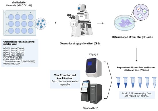
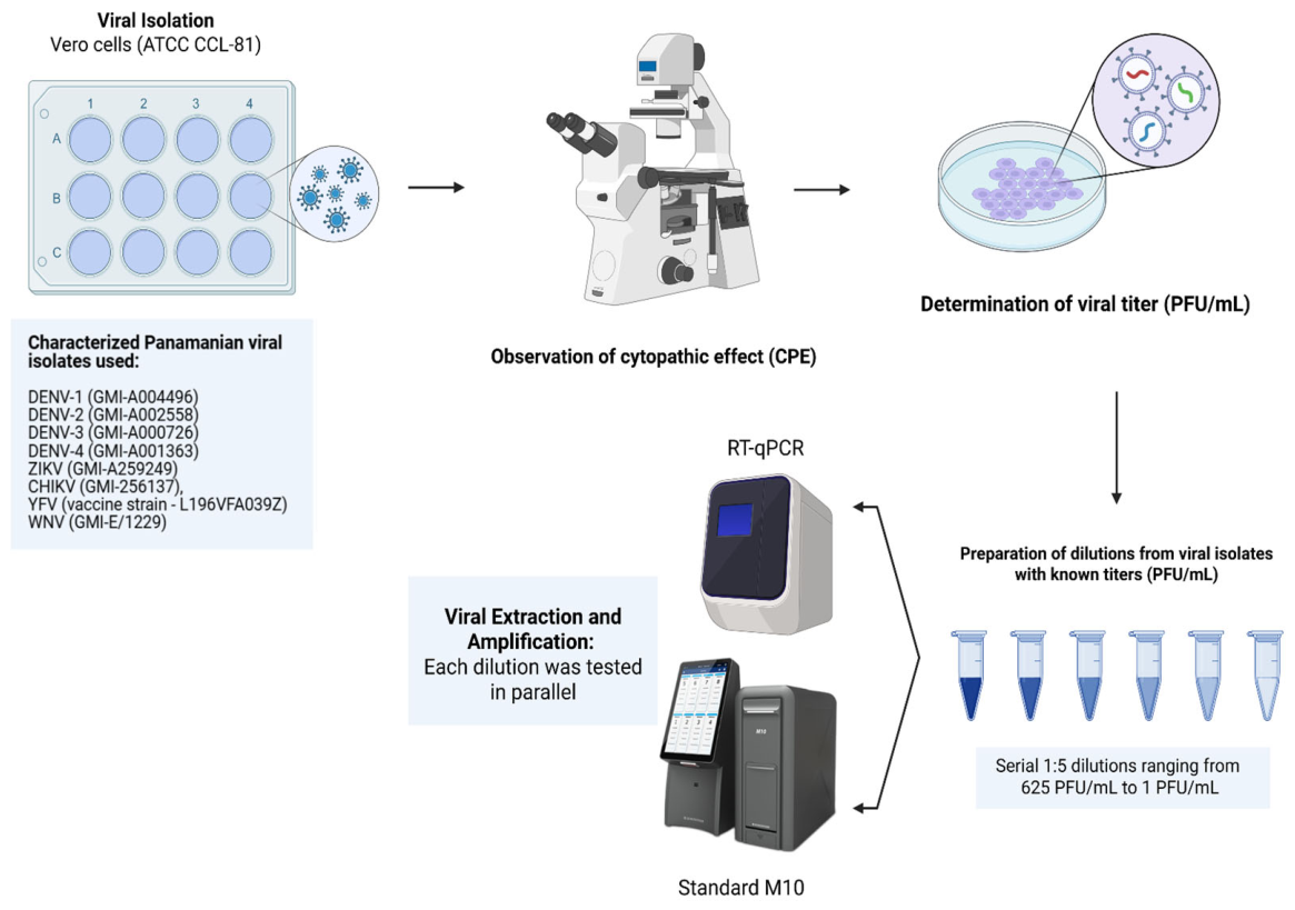
Eight characterized Panamanian viral isolates were used: DENV-1 (GMI-A004496), DENV-2 (GMI-A002558), DENV-3 (GMI-A000726), DENV-4 (GMI-A001363), ZIKV (GMI-A259249), CHIKV (GMI-256137), YFV vaccine strain (L196VFA039Z), and WNV (GMI-E/1229), all obtained from the Virology Department repository of ICGES and previously characterized by molecular methods (Figure 1).
Figure 1.
Experimental workflow for the analytical sensitivity evaluation of the STANDARD M10 Arbovirus Panel. Characterized viral isolates were propagated in Vero cells; viral stocks were harvested upon cytopathic effect and titrated by plaque assay. Fivefold (1:5) serial dilutions (625–1 PFU/mL) were prepared for each virus, with additional intermediate points for DENV-2, DENV-4, and CHIKV. Each dilution was tested in parallel: 600 µL was loaded directly into individual M10 cartridges (no prior extraction), and 200 µL underwent automated RNA extraction followed by reference RT-qPCR amplification in triplicate. Created in BioRender. Young, S. (2026) https://BioRender.com/oro6wzx (accessed on 21 April 2026).
Vero cells (ATCC CCL-81) were used for the production of viral stocks. Cells were maintained at 37 °C and 5% CO2 in Minimum Essential Medium (MEM) supplemented with 2% fetal bovine serum, 1% penicillin–streptomycin, and 0.5% amphotericin B. Viruses were harvested after 3–7 days depending on the virus and titrated to determine their plaque-forming unit concentration (PFU/mL) as previously described [58]. Cytopathic effect (CPE) was monitored daily by inverted light microscopy as an indicator of productive viral replication (Figure 1). Observed changes included progressive cell rounding, cytoplasmic vacuolation, and monolayer detachment, with onset typically between days 3–5 for DENV serotypes and ZIKV, and within 48–72 h for CHIKV, which produced more rapid and pronounced CPE. YFV (vaccine strain 17D) and WNV produced moderate cytopathic changes with focal lysis, generally apparent between days 4–7. Cultures were harvested at 70–80% CPE to balance viral yield and RNA integrity.
2.3.2. Dilutions, Viral Extraction and Amplification
Based on the known concentration of each virus (PFU/mL), serial 1:5 dilutions were prepared, ranging from 625 PFU/mL to 1 PFU/mL. For DENV-2, DENV-4, and CHIKV, additional intermediate dilution points (20 and 15 PFU/mL) were prepared to more precisely characterize the operational limit of detection in the region where signal loss was anticipated. Each dilution was tested in parallel: 600 µL was loaded directly into individual M10 cartridges without prior extraction, while 200 µL underwent automated RNA extraction using the MagMAX™ Viral/Pathogen II Nucleic Acid Isolation Kit (Thermo Fisher Scientific, Waltham, MA, USA, Cat. A48383) on the KingFisher Flex instrument, prior to RT-qPCR amplification in triplicate on an Applied Biosystems QuantStudio™ 5 Real-Time PCR System (Figure 1). Amplification was performed using individual protocols for each target [59,60,61,62] except for dengue virus, for which a single multiplex reaction was used to detect and differentiate all four serotypes. All reference RT-qPCR reactions were performed using the iTaq™ Universal Probes One-Step Kit (Cat. No. 1725141, Bio-Rad Laboratories; Berkeley, CA, USA); mean Ct values were calculated from the three replicates. For the STANDARD M10, each dilution point was tested using one cartridge, yielding a single Ct measurement per concentration, as the closed-cartridge platform does not permit within-run technical replication. Analytical sensitivity and Ct values obtained with the STANDARD M10 were compared with those generated by the reference RT-qPCR assays across all dilution points.
2.3.3. Calibration Curve Construction and Limit of Detection Determination
The analytical sensitivity analyses were performed using the Ct values obtained from fivefold (1:5) serial dilutions of cultured virus, expressed in plaque-forming units per milliliter (PFU/mL), tested in parallel by the STANDARD M10 Arbovirus Panel and the reference RT-qPCR assays. For each virus, Ct values were recorded at each dilution point and organized in a structured dataset comprising virus, dilution (PFU/mL), log10-transformed concentration, mean Ct for the RT-qPCR (mean of triplicates), and single Ct for the STANDARD M10 (one cartridge per dilution point). Detailed Ct values are provided in Supplementary Table S2.
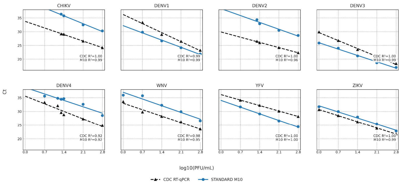
To generate the calibration curves (Figure 2), Ct values were modeled as a function of log10 PFU/mL using simple linear regression for each virus and method separately, according to the model Ct = β0 + β1 log10 (PFU/mL), where β0 represents the y-intercept and β1 the slope of the standard curve. Goodness of fit was assessed by visual inspection of residuals and by the coefficient of determination (R2). Scatter plots of Ct versus log10 PFU/mL were produced for each virus in a multi-panel faceted layout, overlaying the fitted regression lines for both methods to allow direct visual comparison of slope and intercept across analytes.
Figure 2.
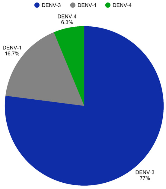
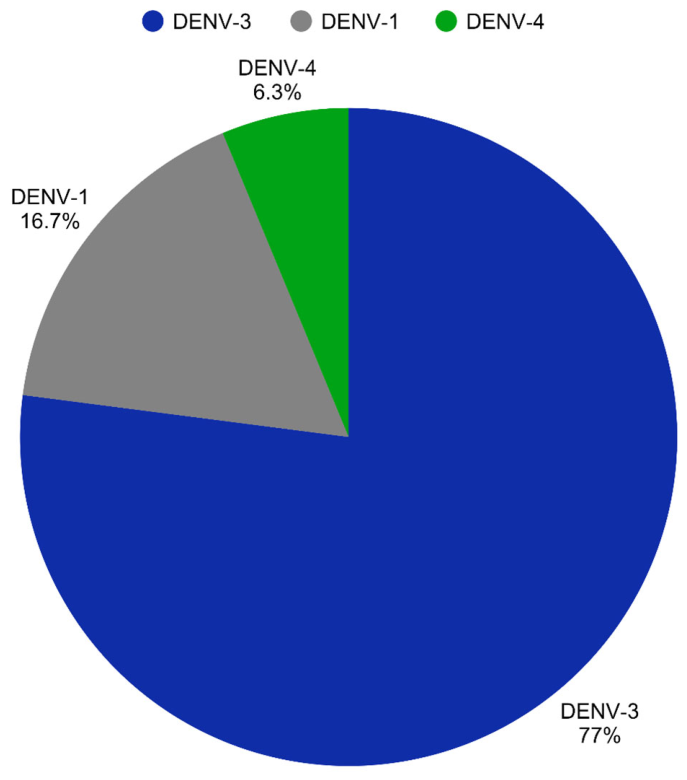
Serotype distribution among dengue-positive serum samples detected by the STANDARD M10 Arbovirus Panel. Proportional distribution of DENV serotypes among dengue reference-positive serum samples (Phase 1, January–June 2025), as determined by the STANDARD M10 target-specific channel results, which simultaneously identify DENV-1, DENV-2, DENV-3, and DENV-4 in a single reaction. DENV-3 was the predominant serotype, followed by DENV-1 and DENV-4. DENV-2 was not detected in any sample analyzed.
For each virus, the observed operational LoD was defined as the lowest concentration in the dilution series (PFU/mL) at which a Ct value was obtained: for the RT-qPCR, this required detection in all three triplicate reactions; for the STANDARD M10, detection was assessed from the single cartridge tested at each dilution point. These operational LoDs are descriptive in nature and do not correspond to LoD95 estimates derived from probit regression, which would require a minimum of 20 independent replicates at each concentration per CLSI EP17-A2 guidance and were outside the scope of this study. For the purpose of this analysis, a dilution was considered positive if the instrument generated a numeric Ct value; dilutions explicitly reported as ‘NEGATIVE’ (no amplification detected) were considered below the operational limit of detection. LoD values for both platforms are compared in Table 1 and detailed Ct values across the full dilution range are provided in Supplementary Table S2.
Table 1.
Clinical performance of the STANDARD M10 Arbovirus Panel for dengue detection against the composite reference standard (NS1 ELISA). The analysis included 163 serum samples: 100 classified as dengue reference-positive and 63 as reference-negative. The M10 correctly identified 96 of 100 positive samples and 63 of 63 negative samples (0 false positives). 95% CIs for sensitivity, specificity, PPV, and NPV were estimated using exact binomial (Clopper–Pearson) methods; CI for Cohen’s κ was estimated using the Fleiss standard error formula.
2.4. Statistical Analysis
In Phase 1, the sensitivity, specificity, and overall accuracy of the STANDARD M10 for dengue were calculated against the reference standard, with 95% confidence intervals (95% CI) estimated using exact binomial methods. In addition, an exploratory description of the DENV serotype distribution and the detection of other arboviruses within the cohort was provided.
In Phase 2, for each virus, exploratory linear regression models were fitted with Ct as the dependent variable and log10(PFU/mL), method (STANDARD M10 vs. RT qPCR) and their interaction as predictors, in order to characterize the relationship between viral concentration and Ct for both platforms. Ct values were modeled using the linear regression: Ct = β0 + β1log10(PFU/mL) + β2Method + β3log10(PFU/mL) × Method + ε where Method = 0 for the RT-qPCR and Method = 1 for the STANDARD M10 Arbovirus Panel. Regression coefficients and 95% confidence intervals based on a normal approximation are presented in Supplementary Table S4 and were interpreted as descriptive, given the limited number of dilution levels and the use of averaged Ct values per dilution.
Data management was performed in Python 3.0.1using the pandas library (v2.0) [63], and statistical modeling and graphical output were produced in R (version 4.1.0) [64]. Because both methods were evaluated across matched dilution points and the number of concentration levels per virus was limited (n = 5 for DENV-1, DENV-3, ZIKV, WNV, and YFV; n = 7 for DENV-2 and CHIKV, which included additional intermediate points; and DENV-4 was tested at n = 7 levels), the between-method Ct comparisons derived from these models are descriptive and exploratory in nature, and should not be interpreted as definitive inferential analyses.
3. Results
3.1. Phase 1: Clinical Diagnostic Performance
The STANDARD M10 identified 96 of 100 dengue reference-positive sera and 63 of 63 reference-negative sera, with no false positives. Sensitivity was 96.0% (95% CI: 89.8–98.9), specificity was 100% (95% CI: 94.3–100), overall accuracy was 97.5%, and Cohen’s κ was 0.95 (95% CI: 0.90–1.00), indicating near-perfect agreement (Table 1).
Among the dengue-positive samples identified during the first half of 2025 (January–June 2025), DENV-3 was the predominant serotype detected by the STANDARD M10 Arbovirus Panel, accounting for 74 of 96 (77.1%) positive detections. DENV-1 represented 16.7% of positive samples (16/96), while DENV-4 accounted for 6.3% (6/96). Notably, DENV-2 was not detected in any of the samples analyzed (Figure 2). This distribution aligns with PAHO surveillance alerts reporting the re-emergence of DENV-3 across the Americas [65].
Clinical specimens from patients with confirmed ZIKV, CHIKV, YFV, or WNV infection were not available in the study setting; accordingly, formal sensitivity and specificity estimate for these targets could not be derived from clinical data. The STANDARD M10 generated no discordant positive results among reference-negative samples, providing indirect evidence of specificity for non-dengue targets in the context of dengue-predominant acute febrile illness. To address the absence of confirmed clinical specimens for these pathogens, Phase 2 was designed to characterize the analytical detection limits of the panel across all eight targets under controlled conditions, providing a complementary evidence base for interpreting the platform’s potential for multiplex arboviral screening. Furthermore, analytical specificity was confirmed in controlled multiplex testing across eight viral combinations, including co-circulation scenarios; no cross-reactivity or misclassification was observed for any panel virus, and the non-panel virus Oropouche did not trigger false-positive signals on any channel (Supplementary Table S1).
3.2. Phase 2: Analytical Sensitivity and Cycle Threshold (Ct) Profiles
Observed Operational Limit of Detection: M10 vs. RT (PFU/mL)
The observed operational limit of detection (LoD) varied between platforms depending on the virus (Table 2). For DENV-1 and YFV, the M10 demonstrated superior sensitivity, with LoDs of 1 PFU/mL compared with 5 PFU/mL for the reference RT-qPCR. For DENV-3, ZIKV, WNV, and DENV-4, both platforms showed equivalent performance (LoD: 1, 1, 1, and 5 PFU/mL, respectively, for both methods). In contrast, the reference RT-qPCR demonstrated lower LoDs for DENV-2 (1 vs. 20 PFU/mL for M10) and CHIKV (1 vs. 20 PFU/mL for M10).
Table 2.
Comparison of observed operational limits of detection (LoD) between the STANDARD M10 Arbovirus Panel and the reference RT-qPCR. For the RT-qPCR, the operational LoD was defined as the lowest concentration (PFU/mL) yielding a detectable Ct value in all three triplicate reactions; for the STANDARD M10, it was defined as the lowest concentration yielding a detectable Ct from the single cartridge tested per dilution point. Serial 1:5 dilutions (625–1 PFU/mL) were tested for each virus; additional intermediate points (20 and 15 PFU/mL) were included for DENV-2, DENV-4, and CHIKV. Full Ct values are provided in Supplementary Table S2.
3.3. Calibration Curves and Comparative Ct Profiles
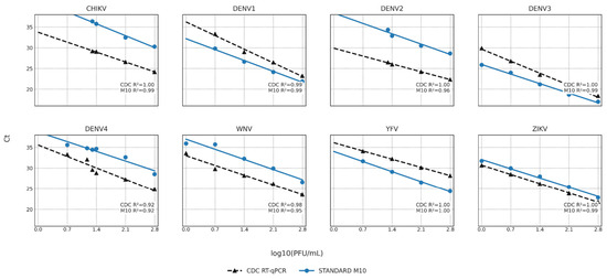
For ZIKV and WNV, both platforms produced regression lines with similar slopes and modest positive Ct shifts for the M10, indicating comparable amplification efficiency and equivalent or near-equivalent LoDs for these targets (Figure 3). For DENV-3, the M10 regression line was shifted downward relative to the RT-qPCR, reflecting superior amplification efficiency, though equivalent LoDs (1 PFU/mL) were achieved by both methods. For YFV, the M10 assay consistently yielded lower Ct values throughout the dilution series and maintained detection at 1 PFU/mL, a concentration at which the RT-qPCR assay failed to produce a positive result. For DENV-1, the M10 regression line was shifted downward (toward lower Ct values), reflecting higher amplification efficiency of the M10 for this target relative to the RT-qPCR; detection was preserved down to 1 PFU/mL, consistent with the LoD reported in Table 2. For DENV-4, both platforms showed comparable regression lines with equivalent LoDs of 5 PFU/mL, and neither method detected the target at 1 PFU/mL. In contrast, for DENV-2 and CHIKV, the M10 regression lines were shifted upward by approximately 6–7 Ct units compared with the RT-qPCR assay, indicating reduced amplification efficiency at all tested concentrations and resulting in loss of detection at low viral loads, concordant with the higher LoDs observed for these two targets (Figure 3).
Figure 3.
Comparative analytical performance and linear regression analysis of the STANDARD M10 Arbovirus Panel and RT-qPCR assays across a fivefold (1:5) serial dilution series (625–1 PFU/mL). The relationship between cycle threshold (Ct) values and viral concentration (log10 PFU/mL) is shown. Data points represent the mean Ct value of triplicate measurements for the RT-qPCR assay; for the STANDARD M10, each data point represents a single Ct measurement per cartridge, reflecting the closed-system design of the platform, which precludes within-run technical replication. Regression lines represent linear fits for the STANDARD M10 Arbovirus Panel (solid blue) and the RT-qPCR (dashed black). R2 values for each virus–method combination is annotated within each panel.
Linear regression models showed strong linearity for both platforms across all viruses (R2 range: 0.92–1.00 for all virus–method combinations), confirming a consistent relationship between Ct and viral concentration throughout the tested dilution range (625–1 PFU/mL). Exploratory regression modeling indicated systematic Ct differences between platforms across all eight targets; however, given the limited number of dilution levels and the use of averaged Ct values per concentration point, these differences should be interpreted as descriptive rather than as statistically confirmed inferences. The largest positive Ct shifts for the M10 relative to the reference RT-qPCR were observed for DENV-2 (ΔCt = +6.85) and CHIKV (ΔCt = +6.47), consistent with the higher LoDs for these two targets. For DENV-4 (ΔCt = +4.12) and WNV (ΔCt = +3.82), the M10 exhibited higher mean Ct values across the dilution range, though equivalent LoDs were achieved (5 PFU/mL and 1 PFU/mL, respectively), indicating similar analytical sensitivity at the detection threshold despite reduced amplification efficiency at higher concentrations. For ZIKV (ΔCt = +1.38), Ct values and LoDs were equivalent between platforms. Note that the mean Ct shifts reported here reflect the arithmetic mean of per-dilution Ct differences between platforms at matched concentrations where both assays produced detectable signals, and differ slightly from the regression-based intercept offsets (β2 coefficients) presented in Supplementary Table S4, which represent the modeled vertical shift of the M10 regression line relative to the RT-qPCR across the full dilution range.
4. Discussion
The present study provides an independent two-phase evaluation of the STANDARD M10 Arbovirus Panel. These results support the M10 as a viable molecular option for acute dengue diagnosis in settings where simplified, closed-cartridge workflows and rapid turnaround represent operational priorities, provided that the limitations of the reference standard and the target-dependent analytical performance described below are taken into account. In Phase 1, the assay achieved 96.0% sensitivity, 100% specificity, and 97.5% overall accuracy (Cohen’s κ = 0.95) against the composite NS1-based reference standard, consistent with recent independent assessments reporting sensitivity of approximately 93% and specificity of 100% relative to conventional RT-qPCR [39]. The use of a composite NS1 ELISA-based reference standard, rather than RT-qPCR, introduces a differential verification bias that must be acknowledged: published meta-analyses report pooled NS1 ELISA sensitivities of approximately 67–82% across primary and secondary infections, with markedly lower values in secondary infections due to immune complex formation [36,54,56]. Accordingly, NS1-negative, RT-qPCR-detectable cases, predominantly those with secondary infection or late-acute presentation, would be misclassified as reference-negative, potentially registering as M10 false positives against this reference and simultaneously deflating apparent sensitivity. The reported 96.0% sensitivity should therefore be interpreted as a conservative lower bound, and the 100% specificity as measured against an imperfect reference rather than against RT-qPCR confirmation. Future evaluations should incorporate RT-qPCR as a co-reference standard to quantify the magnitude of this bias.
In Phase 2, analytical performance was target-dependent. The M10 demonstrated limits of detection comparable to the reference RT-qPCR for six of eight targets, consistent with published data for other multiplex arboviral platforms including the TaqMan Arbovirus Triplex [33,66,67,68,69]. Analytical specificity was confirmed across all eight viral combinations with no cross-reactivity, and the non-panel virus Oropouche generated no false-positive signals (Supplementary Table S1). Manufacturer validation data similarly report high specificity and no mutual interference under competitive testing conditions [49,51].
The regression-based Ct offsets provide additional insight into the analytical performance patterns suggested by the operational LoD data. For ZIKV and WNV, the small positive Ct shifts for the STANDARD M10 (approximately +1 to +4 cycles) with slopes similar to the RT-qPCR indicate slightly later detection but preserved linearity across the tested dilution range, an effect that is unlikely to be clinically meaningful in acute-phase specimens. In contrast, for DENV-2 and CHIKV, larger positive Ct offsets indicate later detection by the M10 than by the reference assay at a given viral concentration, consistent with the higher operational LoDs observed for these targets. Conversely, negative Ct offsets for YFV and DENV-1 support the observation that the M10 achieves superior detection for these viruses, consistent with their lower LoDs relative to the reference RT-qPCR (1 vs. 5 PFU/mL for both targets). For DENV-3, the M10 exhibited lower Ct values relative to the RT-qPCR across the tested concentration range, indicating superior amplification efficiency; however, both platforms achieved equivalent operational LoDs of 1 PFU/mL, and therefore the clinical significance of this offset is limited.
The predominance of DENV-3 in this cohort (77.1%) is consistent with PAHO surveillance alerts documenting re-emergence of DENV-3 subtype III across the Americas since 2023, including Central America and Panama [65,70,71]. This resurgence reflects the accumulation of immunologically naïve populations during prolonged low-transmission intervals and underscores the epidemiological value of serotype-resolved platforms [71,72]. The absence of DENV-2 likely reflects local herd immunity and serotype oscillation dynamics during the study period rather than a detection failure, as DENV-2 was not identified by the reference standard either [12,13,73].
Several operational and analytical limitations should be acknowledged. The most substantive limitation of this study is the restriction of clinical diagnostic evaluation to dengue. Clinical specimens from patients with confirmed ZIKV, CHIKV, YFV, or WNV infection were not available in the study setting, reflecting the epidemiological dominance of dengue among acute febrile illness presentations in Panama during the study period and the operational constraints of retrospective sample collection in decentralized health facilities. Consequently, sensitivity and specificity for these non-dengue targets cannot be inferred from the present data. Phase 2 was incorporated specifically to address this gap by characterizing analytical detection thresholds across all panel targets under controlled conditions; however, analytical sensitivity data derived from cultured viral stocks do not substitute for clinical validation in patient specimens, where viral load kinetics, sample matrix effects, and host factors may influence assay performance. Prospective studies enrolling patients with confirmed non-dengue arboviral infection are therefore required to complete the clinical performance profile of the panel.
Reliable RT-qPCR performance is contingent on specimen integrity [74,75,76,77]; pre-analytical conditions account for a substantial proportion of total errors in molecular laboratory practice and can reduce RT-qPCR sensitivity even when NS1 detection is preserved [74,76]. Although strict exclusion criteria and verified storage conditions were applied, residual pre-analytical variability cannot be entirely excluded. Prompt serum separation, validated cold-chain maintenance, and minimal freeze–thaw cycles therefore remain indispensable prerequisites for reliable multiplex RT-qPCR implementation in both clinical and surveillance settings [39,74,76,77,78,79].
Additionally, positive YFV signals in individuals with recent 17D vaccine immunization may be indistinguishable from wild-type infection by multiplex RT-PCR, necessitating interpretation in conjunction with vaccination history and clinical–epidemiological data [80]. While routinely achievable in adult patients, the 600 µL cartridge input volume requirement may limit feasibility in pediatric populations. Specifically, obtaining this minimum serum volume for automated processing is often challenging in neonates and young infants [81,82].
These analytical characteristics, including serotype resolution, a sample-to-result time of approximately 60 min, reagent storage at 2–28 °C, and a closed-cartridge format, are operationally relevant in the context of decentralized arboviral testing, where infrastructure and cold-chain constraints limit implementation of conventional open-tube molecular assays [39,51]. Early virological confirmation supports timely clinical triage and informs surveillance of emergent lineages, vaccine deployment decisions, and severity risk stratification [70,83,84]. The platform’s analytical capacity to detect simultaneous arboviral targets in a single reaction, including co-infections relevant to antibody-dependent enhancement, warrants prospective clinical evaluation in cohorts specifically designed to capture co-circulating etiologies [37,46,84,85].
5. Conclusions
This study provides an independent two-phase evaluation of the STANDARD M10 Arbovirus Panel in a hyperendemic setting, combining clinical performance estimates for dengue with controlled analytical sensitivity comparisons across eight arboviral targets. In this cohort, the assay showed high agreement with a composite NS1 ELISA-based reference standard for dengue, together with serotype-level resolution and absence of cross-reactivity in the multiplex analytical experiments, supporting its use as a complementary molecular tool for acute dengue diagnosis and arboviral screening rather than as a replacement for existing reference RT-qPCR assays. The two-phase design was adopted to address a recognized operational limitation: the absence of confirmed clinical specimens for non-dengue arboviruses in the study setting. Phase 2 therefore serves as a controlled analytical complement to the clinical phase, providing reference data on detection thresholds across all eight panel targets rather than clinical performance estimates. The reduced analytical sensitivity observed for DENV-2 and CHIKV highlights that panel-wide performance is target dependent and should be interpreted cautiously, particularly at low viral loads. Further prospective, multicenter studies that include clinical specimens positive for ZIKV, CHIKV, YFV and WNV, along with formal LoD95 determinations, are needed to complete the performance profile of the panel and to robustly define its role within comprehensive arboviral syndromic surveillance strategies in the Americas.
Dengue, Zika, Chikungunya, Yellow fever, and West Nile viruses co-circulate across the Americas and cause overlapping clinical syndromes during the acute phase of infection. This study evaluated the STANDARD M10 Arbovirus Panel in Panama using a two-phase design. In the clinical phase, the assay demonstrated high agreement with the reference standard for dengue detection and provided serotype-level information, with DENV-3 predominating. In the analytical phase, operational limits of detection were comparable to reference RT-qPCR for six of eight targets; however, substantially reduced analytical sensitivity was observed for DENV-2 and Chikungunya virus. Because confirmed clinical specimens for ZIKV, CHIKV, YFV, and WNV were not available in the study setting, a controlled analytical phase was incorporated to characterize detection limits across all eight panel targets using characterized reference strains. These findings support the use of the M10 as a complementary molecular tool for dengue diagnosis and multiplex arboviral screening; prospective clinical validation of non-dengue targets remains necessary to complete the performance profile of the panel.
Supplementary Materials
The following supporting information can be downloaded at: https://www.mdpi.com/article/10.3390/diagnostics16121799/s1, Supplementary Table S1—specificity_summary_table; Supplementary Table S2—Analytical_LoD_Performance_M10_vs_CDC; Supplementary Table S3—STARD 2015 Reporting Checklist; Supplementary Table S4—Regression-based Ct offsets between STANDARD M10 and CDC RT qPCR.
Author Contributions
Conceptualization, G.A.E. and B.M.; methodology, S.Y.Y., M.C.-G., D.A., M.V., L.S., M.M.-M. and C.Y.; formal analysis, G.A.E., S.Y.Y., M.C.-G. and B.M.; investigation, S.Y.Y., M.C.-G., D.A., M.V., L.S. and M.M.-M.; writing—original draft preparation, G.A.E., S.Y.Y., M.C.-G. and B.M.; writing—review and editing, G.A.E., S.Y.Y., M.C.-G. and B.M.; supervision, G.A.E. and B.M. All authors have read and agreed to the published version of the manuscript.
Funding
SD Biosensor supplied diagnostic consumables (cartridges and reagents) and contributed to open-access publication costs. The sponsor had no influence on the study design, data collection, data analysis, data interpretation, manuscript preparation, or the decision to publish. All authors retained full scientific independence and responsibility for the content of this work.
Institutional Review Board Statement
The study was conducted in accordance with the Declaration of Helsinki, and approved by the Institutional Research Bioethics Committee of Hospital Aquilino Tejeira (IRB Min. No. 008-2025 CBI-HAT, approval date 12 March 2025).
Informed Consent Statement
Informed consent was waived by the Institutional Research Bioethics Committee of Hospital Aquilino Tejeira (IRB Min. No. 008-2025 CBI-HAT) as all specimens were residual samples collected during routine diagnostic procedures, posing minimal risk to participants and not requiring individual consent under applicable national and international regulations.
Data Availability Statement
The original contributions presented in this study are included in the article. Further inquiries can be directed to the corresponding authors.
Acknowledgments
The authors wish to thank Melanie Ortega and Jorge Cervera Dixon from the Sección de Inmunovirología, Laboratorio Central de Referencia en Salud Pública (LCRSP), Instituto Conmemorativo Gorgas para Estudios de la Salud, Panama, for their technical support during serological sample processing. The authors also acknowledge SD Biosensor for providing the arbovirus detection panels and diagnostic equipment used in this evaluation as an in-kind contribution.
Conflicts of Interest
The authors declare no conflicts of interest. The authors declare that this study received funding from SD Biosensor. The sponsor had no role in study design, data collection, statistical analysis, or interpretation of results. The decision to publish and the content of the manuscript were entirely at the discretion of the investigators.
Abbreviations
The following abbreviations are used in this manuscript:
| ATCC | American Type Culture Collection |
| CBI-HAT | Institutional Research Bioethics Committee of Hospital Aquilino Tejeira |
| CDC | Centers for Disease Control and Prevention |
| CHIKV | Chikungunya virus |
| CI | Confidence interval |
| CPE | Cytopathic effect |
| Ct | Cycle threshold |
| DENV | Dengue virus |
| ELISA | Enzyme-linked immunosorbent assay |
| IC | Internal control |
| ICGES | Gorgas Memorial Institute for Health Studies |
| IgM | Immunoglobulin M |
| LoD | Limit of detection |
| MEM | Minimum Essential Medium |
| MINSA | Ministerio de Salud de Panamá |
| NPV | Negative Predictive Value |
| NS1 | Non-structural protein 1 |
| PAHO | Pan American Health Organization |
| PFU | Plaque-forming unit |
| PPV | Positive Predictive Value |
| POCT | Point-of-care testing |
| R2 | Coefficient of determination |
| RT-qPCR | Reverse transcription quantitative polymerase chain reaction |
| WHO | World Health Organization |
| WNV | West Nile virus |
| YFV | Yellow fever virus |
| ZIKV | Zika virus |
References
- WHO. Global Arbovirus Initiative Technical Advisory Group Strengthening Global Preparedness and Response to Arboviral Disease Threats: A Call to Action. Lancet Infect. Dis. 2026, 26, 15–17. [Google Scholar] [CrossRef] [Scilit]
- Lim, A.; Shearer, F.M.; Sewalk, K.; Pigott, D.M.; Clarke, J.; Ghouse, A.; Judge, C.; Kang, H.; Messina, J.P.; Kraemer, M.U.G.; et al. The Overlapping Global Distribution of Dengue, Chikungunya, Zika and Yellow Fever. Nat. Commun. 2025, 16, 3418. [Google Scholar] [CrossRef] [Scilit]
- Haider, N.; Hasan, M.N.; Onyango, J.; Billah, M.; Khan, S.; Papakonstantinou, D.; Paudyal, P.; Asaduzzaman, M. Global Dengue Epidemic Worsens with Record 14 Million Cases and 9000 Deaths Reported in 2024. Int. J. Infect. Dis. 2025, 158, 107940. [Google Scholar] [CrossRef] [Scilit]
- Ly, H. Dengue Fever in the Americas. Virulence 2024, 15, 2375551. [Google Scholar] [CrossRef] [Scilit]
- Alissa, M.; Alsuwat, M.A.; Alzahrani, K.J. Neurological Manifestations of Flaviviridae, Togaviridae, and Peribunyaviridae as Vector-Borne Viruses. Rev. Med. Virol. 2024, 34, e2571. [Google Scholar] [CrossRef] [Scilit]
- Simmonds, P.; Becher, P.; Bukh, J.; Gould, E.A.; Meyers, G.; Monath, T.; Muerhoff, S.; Pletnev, A.; Rico-Hesse, R.; Smith, D.B.; et al. ICTV Virus Taxonomy Profile: Flaviviridae. J. Gen. Virol. 2017, 98, 2–3. [Google Scholar] [CrossRef] [Scilit]
- Abbasi, E. Global Epidemiology and Evolutionary Dynamics of Arboviruses: A Systematic Review of Surveillance, Control Strategies, and Emerging Threats. Dialogues Health 2026, 8, 100280. [Google Scholar] [CrossRef] [Scilit]
- Parums, D.V. A Review of the Changing Global Impact of Arthropod-Borne Virus Diseases and Recent Initiatives from the World Health Organization. Med. Sci. Monit. 2025, 31, e951998. [Google Scholar] [CrossRef] [Scilit]
- Guzman, M.G.; Harris, E. Dengue. Lancet 2015, 385, 453–465. [Google Scholar] [CrossRef] [Scilit] [PubMed]
- Pan American Health Organization. Integrated Management Strategy for Arboviral Disease Prevention and Control in the Americas; Pan American Health Organization: Washington, DC, USA, 2020; p. 95. [Google Scholar]
- de Almeida, M.T.; Merighi, D.G.S.; Visnardi, A.B.; Boneto Gonçalves, C.A.; Amorim, V.M.d.F.; Ferrari, A.S.d.A.; de Souza, A.S.; Guzzo, C.R. Latin America’s Dengue Outbreak Poses a Global Health Threat. Viruses 2025, 17, 57. [Google Scholar] [CrossRef] [Scilit] [PubMed]
- Pan American Health Organization; World Health Organization. Epidemiological Update: Increase in Dengue Cases in the Region of the Americas; Pan American Health Organization: Washington, DC, USA, 2024. [Google Scholar]
- Pan American Health Organization; World Health Organization. Epidemiological Alert: Increase in Dengue Cases in the Americas Region; Pan American Health Organization: Washington, DC, USA, 2024. [Google Scholar]
- World Health Organization. Disease Outbreak News; Dengue—Global Situation. Available online: https://www.who.int/emergencies/disease-outbreak-news/item/2024-DON518 (accessed on 15 April 2026).
- Pan American Health Organization; World Health Organization. Dengue Epidemiological Situation in the Region of the Americas—Epidemiological Week 48, 2025; Pan American Health Organization: Washington, DC, USA, 2025. [Google Scholar]
- Santos, L.L.M.; de Aquino, E.C.; Fernandes, S.M.; Ternes, Y.M.F.; Feres, V.C.d.R. Dengue, Chikungunya, and Zika Virus Infections in Latin America and the Caribbean: A Systematic Review. Rev. Panam. Salud Publica 2023, 47, e34. [Google Scholar] [CrossRef] [Scilit]
- Xavier, J.; Alcantara, L.C.J.; Fonseca, V.; Lima, M.; Castro, E.; Fritsch, H.; Oliveira, C.; Guimarães, N.; Adelino, T.; Evaristo, M.; et al. Increased Interregional Virus Exchange and Nucleotide Diversity Outline the Expansion of Chikungunya Virus in Brazil. Nat. Commun. 2023, 14, 4413. [Google Scholar] [CrossRef] [Scilit]
- Giovanetti, M.; Vazquez, C.; Lima, M.; Castro, E.; Rojas, A.; de la Fuente, A.G.; Aquino, C.; Cantero, C.; Fleitas, F.; Torales, J.; et al. Rapid Epidemic Expansion of Chikungunya Virus-ECSA Lineage in Paraguay. medRxiv 2023. [Google Scholar] [CrossRef] [Scilit]
- Banho, C.A.; Parra, M.C.P.; Nascimento, O.B.; Magnani, G.P.; Ferreira, M.V.M.; Lemos, A.P.; de Carvalho Marques, B.; Brancini, M.L.; Sacchetto, L.; Negri, A.F.; et al. Entomological Surveillance during a Major CHIKV Outbreak in Northwestern São Paulo: Insights from São José Do Rio Preto. medRxiv 2024. medRxiv:2024.12.04.24318429. [Google Scholar] [CrossRef] [Scilit] [PubMed]
- Barac, A.; Gupta, N.; Jokelainen, P.; Tsiodras, S.; Mora-Rillo, M.; Gkrania-Klotsas, E.; Paño-Pardo, J.R.; Bulescu, C.; Pellejero-Sagastizabal, G.; Goorhuis, A.; et al. Yellow Fever in South America in 2025: A Concerning Increase, but a True Resurgence? Clin. Microbiol. Infect. 2025, 31, 1941–1944. [Google Scholar] [CrossRef] [Scilit] [PubMed]
- Petersen, L.R.; Brault, A.C.; Nasci, R.S. West Nile Virus: Review of the Literature. JAMA 2013, 310, 308–315. [Google Scholar] [CrossRef] [Scilit]
- Grossi-Soyster, E.N.; LaBeaud, A.D. Clinical Aspects of Zika Virus. Curr. Opin. Pediatr. 2017, 29, 102–106. [Google Scholar] [CrossRef] [Scilit]
- Liang, B.; Guida, J.P.; Costa Do Nascimento, M.L.; Mysorekar, I.U. Host and Viral Mechanisms of Congenital Zika Syndrome. Virulence 2019, 10, 768–775. [Google Scholar] [CrossRef] [Scilit] [PubMed]
- Yakob, L. Zika Virus after the Public Health Emergency of International Concern Period, Brazil. Emerg. Infect. Dis. 2022, 28, 837–840. [Google Scholar] [CrossRef] [Scilit]
- Hart, J., Jr.; Tillman, G.; Kraut, M.A.; Chiang, H.-S.; Strain, J.F.; Li, Y.; Agrawal, A.G.; Jester, P.; Gnann, J.W., Jr.; Whitley, R.J.; et al. West Nile Virus Neuroinvasive Disease: Neurological Manifestations and Prospective Longitudinal Outcomes. BMC Infect. Dis. 2014, 14, 248. [Google Scholar] [CrossRef] [Scilit]
- de Lima, S.T.S.; Claro, I.M.; Hua, X.; de Jesus, R.; Serres, K.; Simões Mello, L.M.; Forato, J.; Moreira, F.R.R.; Kato, R.B.; Guimarães, G.N.; et al. Active West Nile Virus Transmission in Brazil: An Epidemiological Study. Lancet Reg. Health Am. 2025, 51, 101229. [Google Scholar] [CrossRef] [Scilit] [PubMed]
- Vargas Campos, C.A.; García-Pérez, S.; Figuerola, J.; Martínez-de la Puente, J.; Polo, I.; Rodríguez-de-Fonseca, B.; Fernández-Álvarez, S.; Galván Fraile, V.; Martín-Rey, M.; Lacasaña, M.; et al. Comprehensive Analysis of West Nile Virus Transmission: Environmental, Ecological, and Individual Factors. An Umbrella Review. One Health 2025, 20, 100984. [Google Scholar] [CrossRef] [Scilit]
- Petersen, L.R.; Jamieson, D.J.; Powers, A.M.; Honein, M.A. Zika Virus. N. Engl. J. Med. 2016, 374, 1552–1563. [Google Scholar] [CrossRef] [Scilit]
- Wilder-Smith, A.; Gubler, D.J.; Weaver, S.C.; Monath, T.P.; Heymann, D.L.; Scott, T.W. Epidemic Arboviral Diseases: Priorities for Research and Public Health. Lancet Infect. Dis. 2017, 17, e101–e106. [Google Scholar] [CrossRef] [Scilit]
- Mercado-Reyes, M.; Acosta-Reyes, J.; Navarro-Lechuga, E.; Corchuelo, S.; Rico, A.; Parra, E.; Tolosa, N.; Pardo, L.; González, M.; Martìn-Rodriguez-Hernández, J.; et al. Dengue, Chikungunya and Zika Virus Coinfection: Results of the National Surveillance during the Zika Epidemic in Colombia. Epidemiol. Infect. 2019, 147, e77. [Google Scholar] [CrossRef] [Scilit]
- Estofolete, C.F.; Terzian, A.C.B.; Colombo, T.E.; de Freitas Guimarães, G.; Ferraz, H.C., Jr.; da Silva, R.A.; Greque, G.V.; Nogueira, M.L. Co-Infection between Zika and Different Dengue Serotypes during DENV Outbreak in Brazil. J. Infect. Public Health 2019, 12, 178–181. [Google Scholar] [CrossRef] [Scilit]
- Robert, S.; Diagbouga, P.S.; Djibougou, A.D.; Guy, D.; Bagnall, R.; Ravel, F. Dengue Diagnosis and Impact on Clinical Management: A Literature Review. PLoS Negl. Trop. Dis. 2025, 19, e0013196. [Google Scholar] [CrossRef] [Scilit] [PubMed]
- Santiago, G.A.; Vergne, E.; Quiles, Y.; Cosme, J.; Vazquez, J.; Medina, J.F.; Medina, F.; Colón, C.; Margolis, H.; Muñoz-Jordán, J.L. Analytical and Clinical Performance of the CDC Real Time RT-PCR Assay for Detection and Typing of Dengue Virus. PLoS Negl. Trop. Dis. 2013, 7, e2311. [Google Scholar] [CrossRef] [Scilit]
- Varghese, J.; De Silva, I.; Millar, D.S. Latest Advances in Arbovirus Diagnostics. Microorganisms 2023, 11, 1159. [Google Scholar] [CrossRef] [Scilit]
- Hauner, A.; Aroni-Sierra, J.; Merino, X.; Villa, C.; Torres, F.; Lagatie, O.; Talledo, M.; Ariën, K.K.; Falconi-Agapito, F. Assessing the Diagnostic Performance of Clinical, Serological and Molecular Approaches to Improve Dengue Case Detection in the Peruvian Amazon. PLoS Negl. Trop. Dis. 2026, 20, e0013984. [Google Scholar] [CrossRef] [Scilit] [PubMed]
- Lima, M.d.R.Q.; Nogueira, R.M.R.; Bispo de Filippis, A.M.; dos Santos, F.B. Comparison of Two Generations of the Panbio Dengue NS1 Capture Enzyme-Linked Immunosorbent Assay. Clin. Vaccine Immunol. 2011, 18, 1031–1033. [Google Scholar] [CrossRef] [Scilit] [PubMed]
- Malik, M.R.; Walaskar, S.; Majji, R.; Sp, D.; Uppoor, S.; Thrilok Chandra, K.V.; Madhusudhan, H.N.; Balasundar, A.S.; Mishra, R.K.; Ishtiaq, F. Development of an Affordable Multiplex Quantitative RT-PCR Assay for Early Detection and Surveillance of Dengue, Chikungunya, and Co-Infections from Clinical Samples in Resource-Limited Settings. PLoS Negl. Trop. Dis. 2025, 19, e0013250. [Google Scholar] [CrossRef] [Scilit]
- Waggoner, J.J.; Gresh, L.; Mohamed-Hadley, A.; Ballesteros, G.; Davila, M.J.V.; Tellez, Y.; Sahoo, M.K.; Balmaseda, A.; Harris, E.; Pinsky, B.A. Single-Reaction Multiplex Reverse Transcription PCR for Detection of Zika, Chikungunya, and Dengue Viruses. Emerg. Infect. Dis. 2016, 22, 1295–1297. [Google Scholar] [CrossRef] [Scilit]
- Scarpaleggia, M.; Garzillo, G.; Lucente, M.; Fraccalvieri, C.; Randazzo, N.; Massaro, E.; Galano, B.; Ricucci, V.; Bruzzone, B.; Domnich, A. Diagnostic Accuracy of Five Molecular Assays for the Detection of Dengue Virus. Medicina 2024, 60, 1557. [Google Scholar] [CrossRef] [Scilit] [PubMed]
- Organización Panamericana de la Salud (OPS). Nota Técnica: Algoritmo para la Confirmación Por Laboratorio de Casos de Dengue. Diagnóstico por Laboratorio del Dengue; Pan American Health Organization: Washington, DC, USA, 2023; pp. 1–4. [Google Scholar]
- Corman, V.M.; Rasche, A.; Baronti, C.; Aldabbagh, S.; Cadar, D.; Reusken, C.B.; Pas, S.D.; Goorhuis, A.; Schinkel, J.; Molenkamp, R.; et al. Assay Optimization for Molecular Detection of Zika Virus. Bull. World Health Organ. 2016, 94, 880–892. [Google Scholar] [CrossRef] [Scilit]
- Hauner, A.; Onwuchekwa, C.; Ariën, K.K. Sample-to-Result Molecular Diagnostic Platforms and Their Suitability for Infectious Disease Testing in Low- and Middle-Income Countries. Expert Rev. Mol. Diagn. 2024, 24, 423–438. [Google Scholar] [CrossRef] [Scilit] [PubMed]
- Chen-Germán, M.; González, C.; Araúz, D.; Aguilar, C.; Vega, M.; Chavarria, O.; Moreno, A.; Santiago, E.; Franco, D.; Valdespino, E.; et al. Detection of Oropouche and Punta Toro Virus Infections by Enhanced Surveillance, Panama, 2023–2024. Emerg. Infect. Dis. 2026, 32, 114–117. [Google Scholar] [CrossRef] [Scilit]
- Pan American Health Organization. Recommendations for Laboratory Detection and Diagnosis of Arbovirus Infections in the Region of the Americas; Pan American Health Organization: Washington, DC, USA, 2023. [Google Scholar]
- Kabir, M.A.; Zilouchian, H.; Younas, M.A.; Asghar, W. Dengue Detection: Advances in Diagnostic Tools from Conventional Technology to Point of Care. Biosensors 2021, 11, 206. [Google Scholar] [CrossRef] [Scilit] [PubMed]
- World Health Organization. Dengue: Guidelines for Diagnosis, Treatment, Prevention and Control: New Edition; World Health Organization: Geneva, Switzerland, 2009. [Google Scholar]
- Centers for Disease Control and Prevention CDC. DENV-1-4 Real-Time RT-PCR Assay for Detection and Serotype Identification of Dengue Virus; Centers for Disease Control and Prevention: San Juan, PR, USA, 2025.
- SD BIOSENSOR. Available online: https://www.sdbiosensor.com/product/product_view?product_no=23001 (accessed on 3 January 2026).
- SD BIOSENSOR. Available online: https://www.sdbiosensor.com/product/product_view?product_no=125 (accessed on 3 January 2026).
- Gurajala, S. Innovations and Implementation Challenges in Dengue Diagnostics: A 21st Century Perspective on a Neglected Tropical Disease. Int. J. Trop. Dis. Health 2025, 46, 15–26. [Google Scholar] [CrossRef] [Scilit]
- SD Biosensor. STANDARD M10 Arbovirus Panel; SD Biosensor, Inc.: Suwon-si, Republic of Korea, 2023; p. 19. [Google Scholar]
- Bossuyt, P.M.; Reitsma, J.B.; Bruns, D.E.; Gatsonis, C.A.; Glasziou, P.P.; Irwig, L.; Lijmer, J.G.; Moher, D.; Rennie, D.; de Vet, H.C.W.; et al. STARD 2015: An Updated List of Essential Items for Reporting Diagnostic Accuracy Studies. BMJ 2015, 351, h5527. [Google Scholar] [CrossRef] [Scilit]
- Zhang, H.; Li, W.; Wang, J.; Peng, H.; Che, X.; Chen, X.; Zhou, Y. NS1-Based Tests with Diagnostic Utility for Confirming Dengue Infection: A Meta-Analysis. Int. J. Infect. Dis. 2014, 26, 57–66. [Google Scholar] [CrossRef] [Scilit]
- Peeling, R.W.; Artsob, H.; Pelegrino, J.L.; Buchy, P.; Cardosa, M.J.; Devi, S.; Enria, D.A.; Farrar, J.; Gubler, D.J.; Guzman, M.G.; et al. Evaluation of Diagnostic Tests: Dengue. Nat. Rev. Microbiol. 2010, 8, S30–S38. [Google Scholar] [CrossRef] [Scilit] [PubMed]
- Chen-Germán, M.; Araúz, D.; Aguilar, C.; Vega, M.; Gonzalez, C.; Gondola, J.; Moreno, L.; Cerezo, L.; Franco, L.; Mendez-Rico, J.; et al. Detection of Dengue Virus Serotype 4 in Panama after 23 Years without Circulation. Front. Cell. Infect. Microbiol. 2024, 14, 1467465. [Google Scholar] [CrossRef] [Scilit]
- Pal, S.; Dauner, A.L.; Mitra, I.; Forshey, B.M.; Garcia, P.; Morrison, A.C.; Halsey, E.S.; Kochel, T.J.; Wu, S.-J.L. Evaluation of Dengue NS1 Antigen Rapid Tests and ELISA Kits Using Clinical Samples. PLoS ONE 2014, 9, e113411. [Google Scholar] [CrossRef] [Scilit]
- Chakravarti, A.; Kumar, A.; Malik, S. Detection of Dengue Infection by Combining the Use of an NS1 Antigen Based Assay with Antibody Detection. Southeast Asian J. Trop. Med. Public Health 2011, 42, 297–302. [Google Scholar] [PubMed]
- Johnson, B.W.; Kosoy, O.; Wang, E.; Delorey, M.; Russell, B.; Bowen, R.A.; Weaver, S.C. Use of Sindbis/eastern Equine Encephalitis Chimeric Viruses in Plaque Reduction Neutralization Tests for Arboviral Disease Diagnostics. Clin. Vaccine Immunol. 2011, 18, 1486–1491. [Google Scholar] [CrossRef] [Scilit]
- Lanciotti, R.S.; Kosoy, O.L.; Laven, J.J.; Panella, A.J.; Velez, J.O.; Lambert, A.J.; Campbell, G.L. Chikungunya Virus in US Travelers Returning from India, 2006. Emerg. Infect. Dis. 2007, 13, 764–767. [Google Scholar] [CrossRef] [Scilit]
- Domingo, C.; Patel, P.; Yillah, J.; Weidmann, M.; Méndez, J.A.; Nakouné, E.R.; Niedrig, M. Advanced Yellow Fever Virus Genome Detection in Point-of-Care Facilities and Reference Laboratories. J. Clin. Microbiol. 2012, 50, 4054–4060. [Google Scholar] [CrossRef] [Scilit]
- Lanciotti, R.S.; Kerst, A.J.; Nasci, R.S.; Godsey, M.S.; Mitchell, C.J.; Savage, H.M.; Komar, N.; Panella, N.A.; Allen, B.C.; Volpe, K.E.; et al. Rapid Detection of West Nile Virus from Human Clinical Specimens, Field-Collected Mosquitoes, and Avian Samples by a TaqMan Reverse Transcriptase-PCR Assay. J. Clin. Microbiol. 2000, 38, 4066–4071. [Google Scholar] [CrossRef] [Scilit] [PubMed]
- Lanciotti, R.S.; Kosoy, O.L.; Laven, J.J.; Velez, J.O.; Lambert, A.J.; Johnson, A.J.; Stanfield, S.M.; Duffy, M.R. Genetic and Serologic Properties of Zika Virus Associated with an Epidemic, Yap State, Micronesia, 2007. Emerg. Infect. Dis. 2008, 14, 1232–1239. [Google Scholar] [CrossRef] [Scilit]
- Zenodo. The Pandas Development Team Pandas-Dev/Pandas: Pandas; Zenodo: Geneva, Switzerland, 2026. [Google Scholar]
- The R Project for Statistical Computing. Available online: https://www.R-project.org/ (accessed on 29 April 2026).
- Pan American Health Organization; World Health Organization. Epidemiological Alert Risk of Dengue Outbreaks Due to Increased Circulation of DENV-3 in the Americas Region; Pan American Health Organization: Washington, DC, USA, 2025. [Google Scholar]
- World Health Organization. Laboratory Testing for Dengue Virus: Interim Guidance, April 2025; World Health Organization: Geneva, Switzerland, 2025. [Google Scholar]
- Cantero, C.; Cardozo, F.; Waggoner, J.J.; Pinsky, B.A.; Espínola, A.; Infanzón, B.; Acosta, M.E.; Aria, L.; Arévalo de Guillén, Y.; Cuevas, T.; et al. Implementation of a Multiplex rRT-PCR for Zika, Chikungunya, and Dengue Viruses: Improving Arboviral Detection in an Endemic Region. Am. J. Trop. Med. Hyg. 2020, 102, 625–628. [Google Scholar] [CrossRef] [Scilit]
- Waggoner, J.J.; Abeynayake, J.; Sahoo, M.K.; Gresh, L.; Tellez, Y.; Gonzalez, K.; Ballesteros, G.; Pierro, A.M.; Gaibani, P.; Guo, F.P.; et al. Single-Reaction, Multiplex, Real-Time Rt-PCR for the Detection, Quantitation, and Serotyping of Dengue Viruses. PLoS Negl. Trop. Dis. 2013, 7, e2116. [Google Scholar] [CrossRef] [Scilit] [PubMed]
- Dhanoa, A.; Hassan, S.S.; Ngim, C.F.; Lau, C.F.; Chan, T.S.; Adnan, N.A.A.; Eng, W.W.H.; Gan, H.M.; Rajasekaram, G. Impact of Dengue Virus (DENV) Co-Infection on Clinical Manifestations, Disease Severity and Laboratory Parameters. BMC Infect. Dis. 2016, 16, 406. [Google Scholar] [CrossRef] [Scilit]
- Grubaugh, N.D.; Torres-Hernández, D.; Murillo-Ortiz, M.A.; Dávalos, D.M.; Lopez, P.; Hurtado, I.C.; Breban, M.I.; Bourgikos, E.; Hill, V.; López-Medina, E. Dengue Outbreak Caused by Multiple Virus Serotypes and Lineages, Colombia, 2023–2024. Emerg. Infect. Dis. 2024, 30, 2391–2395. [Google Scholar] [CrossRef] [Scilit] [PubMed]
- Pereira, J.S.; Slavov, S.N.; Brcko, I.C.; Ribeiro, G.; de Souza, V.C.; Ribeiro, I.S.; De Lima, I.T.T.; Borges, G.A.L.; Furtado, K.C.d.L.; Chagas, S.M.D.S.; et al. Unravelling Dengue Serotype 3 Transmission in Brazil: Evidence for Multiple Introductions of the 3III_B.3.2 Lineage. Virus Evol. 2025, 11, veaf034. [Google Scholar] [CrossRef] [Scilit]
- Cantero, C.; Vazquez, C.; Gonzalez, S.; Rojas, A.; Fleitas, F.; Barrios, J.; Gomez De La Fuente, A.; Villalba, S.; Torales, M.; Ijeda, A.; et al. Re-Emergence of DENV-3 in Paraguay after Two Decades: A Genomic and Epidemiological Investigation. Emerg. Microbes Infect. 2026, 15, 2623695. [Google Scholar] [CrossRef] [Scilit]
- Díaz, Y.; Chen-Germán, M.; Quiroz, E.; Carrera, J.-P.; Cisneros, J.; Moreno, B.; Cerezo, L.; Martinez-Torres, A.O.; Moreno, L.; Barahona de Mosca, I.; et al. Molecular Epidemiology of Dengue in Panama: 25 Years of Circulation. Viruses 2019, 11, 764. [Google Scholar] [CrossRef] [Scilit] [PubMed]
- Wang, N.; He, P.; Xu, B.; Wei, H.; Yu, J. Long-Term Stabilization of Dengue Virus RNA at 37 °C for 14 Months Using Silk Fibroin Films. Viruses 2025, 17, 1452. [Google Scholar] [CrossRef] [Scilit]
- Dauner, A.L.; Gilliland, T.C.; Mitra, I.; Pal, S.; Morrison, A.C.; Hontz, R.D.; Wu, S.-J.L. Evaluation of Nucleic Acid Stabilization Products for Ambient Temperature Shipping and Storage of Viral RNA and Antibody in a Dried Whole Blood Format. Am. J. Trop. Med. Hyg. 2015, 93, 46–53. [Google Scholar] [CrossRef] [Scilit]
- Iani, F.C.d.M.; Caetano, A.C.B.; Cocovich, J.C.W.; Amâncio, F.F.; Pereira, M.A.; Adelino, T.É.R.; Caldas, S.; Silva, M.V.F.; Pereira, G.d.C.; Duarte, M.M. Dengue Diagnostics: Serious Inaccuracies Are Likely to Occur If Pre-Analytical Conditions Are Not Strictly Followed. Mem. Inst. Oswaldo Cruz 2021, 115, e200287. [Google Scholar] [CrossRef] [Scilit]
- Ramachandra, S.; Randhawa, A.; Kelmani, C.; Shankar, S.M.; Doddamane, M. RNA Extraction Optimization from Dengue-Infected Clinical Residual Serum Using Laboratory Prepared Reagent. Environ. Exp. Biol. 2021, 19, 219–230. [Google Scholar] [CrossRef] [Scilit]
- Prescott, M.A.; Koesdjojo, M.T.; Mandrell, D.T.; Pastey, M.K. Development of a Rapid Point-of-Care Dengue Virus Type 2 Infection Diagnostic Assay Using Recombinase Polymerase Amplification and Lateral Flow Device. Front. Cell. Infect. Microbiol. 2025, 15, 1578549. [Google Scholar] [CrossRef] [Scilit]
- Sotoudeh Anvari, M.; Gharib, A.; Abolhasani, M.; Azari-Yam, A.; Hossieni Gharalari, F.; Safavi, M.; Zare Mirzaie, A.; Vasei, M. Pre-Analytical Practices in the Molecular Diagnostic Tests, A Concise Review. Iran. J. Pathol. 2021, 16, 1–19. [Google Scholar] [CrossRef] [Scilit] [PubMed]
- Basile, A.J.; Niedrig, M.; Lambert, A.J.; Meurant, R.; Brault, A.C.; Domingo, C.; Goodman, C.H.; Johnson, B.W.; Mossel, E.C.; Mulders, M.N.; et al. Laboratory Evaluation of RealStar Yellow Fever Virus RT-PCR Kit 1.0 for Potential Use in the Global Yellow Fever Laboratory Network. PLoS Negl. Trop. Dis. 2022, 16, e0010770. [Google Scholar] [CrossRef] [Scilit]
- Simmons, C.P.; Farrar, J.J.; van Nguyen, V.C.; Wills, B. Dengue. N. Engl. J. Med. 2012, 366, 1423–1432. [Google Scholar] [CrossRef] [Scilit] [PubMed]
- Waggoner, J.J.; Katzelnick, L.C.; Burger-Calderon, R.; Gallini, J.; Moore, R.H.; Kuan, G.; Balmaseda, A.; Pinsky, B.A.; Harris, E. Antibody-Dependent Enhancement of Severe Disease Is Mediated by Serum Viral Load in Pediatric Dengue Virus Infections. J. Infect. Dis. 2020, 221, 1846–1854. [Google Scholar] [CrossRef] [Scilit]
- Hill, V.; Cleemput, S.; Pereira, J.S.; Gifford, R.J.; Fonseca, V.; Tegally, H.; Brito, A.F.; Ribeiro, G.; de Souza, V.C.; Brcko, I.C.; et al. A New Lineage Nomenclature to Aid Genomic Surveillance of Dengue Virus. PLoS Biol. 2024, 22, e3002834. [Google Scholar] [CrossRef] [Scilit]
- Wilken, L.; Rimmelzwaan, G.F. Adaptive Immunity to Dengue Virus: Slippery Slope or Solid Ground for Rational Vaccine Design? Pathogens 2020, 9, 470. [Google Scholar] [CrossRef] [Scilit] [PubMed]
- Requena-Castro, R.; Reyes-López, M.Á.; Rodríguez-Reyna, R.E.; Palma-Nicolás, P.; Bocanegra-García, V. Molecular Detection of Mixed Infections with Multiple Dengue Virus Serotypes in Suspected Dengue Samples in Tamaulipas, Mexico. Mem. Inst. Oswaldo Cruz 2017, 112, 520–522. [Google Scholar] [CrossRef] [Scilit] [PubMed]

Disclaimer/Publisher’s Note: The statements, opinions and data contained in all publications are solely those of the individual author(s) and contributor(s) and not of MDPI and/or the editor(s). MDPI and/or the editor(s) disclaim responsibility for any injury to people or property resulting from any ideas, methods, instructions or products referred to in the content. |
© 2026 by the authors. Licensee MDPI, Basel, Switzerland. This article is an open access article distributed under the terms and conditions of the Creative Commons Attribution (CC BY) license.