


Abstract
Laboratory biomarkers influence a large proportion of clinical decision-making, yet their application is often limited by incomplete validation and context-dependent interpretability. Serum neurofilament light chain (sNfL), a biomarker of neuroaxonal injury in multiple sclerosis (MS), exemplifies this challenge. Although associated with inflammatory activity, lesion burden, and disability progression at the population level, its translation into individual patient management remains problematic. In this Perspective, we synthesise current literature on sNfL in MS and apply Bayesian diagnostic reasoning as a conceptual framework for its interpretation in individualised MS care. The need for such a framework arises from the heterogeneity of MS pathology, in which subclinical inflammation and neurodegeneration may occur as partly dissociated processes that are incompletely captured by clinical and radiological measures. Consequently, substantial uncertainty persists in disease monitoring and therapeutic decision-making. In this setting, sNfL may provide complementary information, but its interpretation is complicated by biological variability, methodological differences, confounding factors (e.g., age, body mass index, and comorbidities), and the absence of universally validated thresholds. We argue that sNfL should be interpreted within a Bayesian framework, in which biomarker results modify rather than determine the probability of disease activity. Its clinical utility is likely greatest when the pre-test probability is intermediate but remains constrained by uncertainty in both test characteristics and clinical context, leading to uncertainty propagation. Overall, sNfL should be interpreted longitudinally and within multimodal clinical decision models. Further prospective studies are needed to better define its role in individualised MS management.
1. Introduction
Laboratory investigations are an integral component of modern medicine and are estimated to influence 60–70% of clinical decisions [1]. In situations of diagnostic uncertainty, clinical expectations are frequently directed toward novel biomarkers that appear capable of resolving existing uncertainties [2,3].
However, such biomarkers are often still in phases of incomplete clinical validation, and their individual prognostic value or utility for therapeutic management in a given patient therefore remains insufficiently established [4,5,6]. This creates a tension between scientific innovation and clinical decision-making responsibility. In practice, clinicians are thus positioned between the need for objective evidence provided by innovative biomarkers and the recognition that their limited validation and interpretative ambiguity may introduce additional uncertainty into the decision-making process [7,8].
A comparable dynamic is currently emerging in routine clinical care in the context of the biomarker serum neurofilament light chain (sNfL) in patients with multiple sclerosis (MS) [9,10]. Against this background, we aim to discuss key epistemological considerations and to examine, from an analytical perspective, the following question: Can uncertainty resolve uncertainty?
2. Multiple Sclerosis
MS is a chronic immune-mediated disease of the central nervous system characterised by a heterogeneous pathological profile, including demyelination, axonal and synaptic loss, as well as neurodegenerative processes with accompanying astrogliosis [11,12,13]. Despite substantial advances in diagnostics and therapy, precise determination of disease activity in routine clinical practice remains a major challenge [14,15]. In particular, differentiating between clinical and subclinical disease activity, as well as assessing the impact of potential comorbidities on the overall clinical picture, is associated with considerable uncertainty in disease monitoring [16,17,18,19].
Traditionally, MS disease activity has been defined by clinical relapses and the accumulation of neurological disability, commonly measured using the Expanded Disability Status Scale (EDSS) [20,21]. However, with the widespread availability of magnetic resonance imaging (MRI), it has become evident that inflammatory activity frequently occurs subclinically.
On the one hand, new or contrast-enhancing lesions may arise in the absence of clinical symptoms, leading to a discrepancy between clinical manifestation and radiological activity [22,23]. This phenomenon is addressed by the concept of “no evidence of disease activity” (NEDA), which combines clinical and radiological parameters but still exhibits substantial limitations [24,25].
On the other hand, recent studies indicate that a substantial proportion of acute clinical disease activity is not necessarily associated with detectable MRI changes (26.1–43.0%) [26,27].
The growing recognition of progression independent of relapse activity (PIRA) and progression independent of relapse and MRI activity (PIRMA) further complicates the assessment of disease dynamics. Disability accumulation may occur in the absence of clinically overt relapses or new MRI lesions, suggesting neurodegenerative mechanisms beyond acute inflammatory activity [28,29,30]. These findings challenge the traditional relapse- and focal inflammation-centred model of MS and highlight that clinical stability does not necessarily equate to biological inactivity [31,32].
Taken together, these factors result in substantial clinical uncertainty regarding the true level of individual disease activity, which in turn complicates therapeutic decision-making, particularly with respect to escalation, de-escalation, or switching of disease-modifying therapy [33,34]. The resulting demand for mechanistically grounded biomarkers—along with the high expectations attached to them and external commercial influences—creates both significant opportunities and substantial risks in routine clinical practice [35,36,37].
In such clinically uncertain situations, however, the use of a test with uncertain or context-dependent interpretability itself introduces a further layer of uncertainty into clinical decision-making. This issue can currently be illustrated by the individual interpretation of sNfL levels in patients with MS in clinical practice. Here, multiple layers of uncertainty converge. These include physician uncertainty regarding clinical and subclinical disease activity, potential confounding by comorbidities, biological variability of the biomarker, and methodological uncertainties related to its validation and interpretation at the individual level.
3. NfL in Clinical Practice
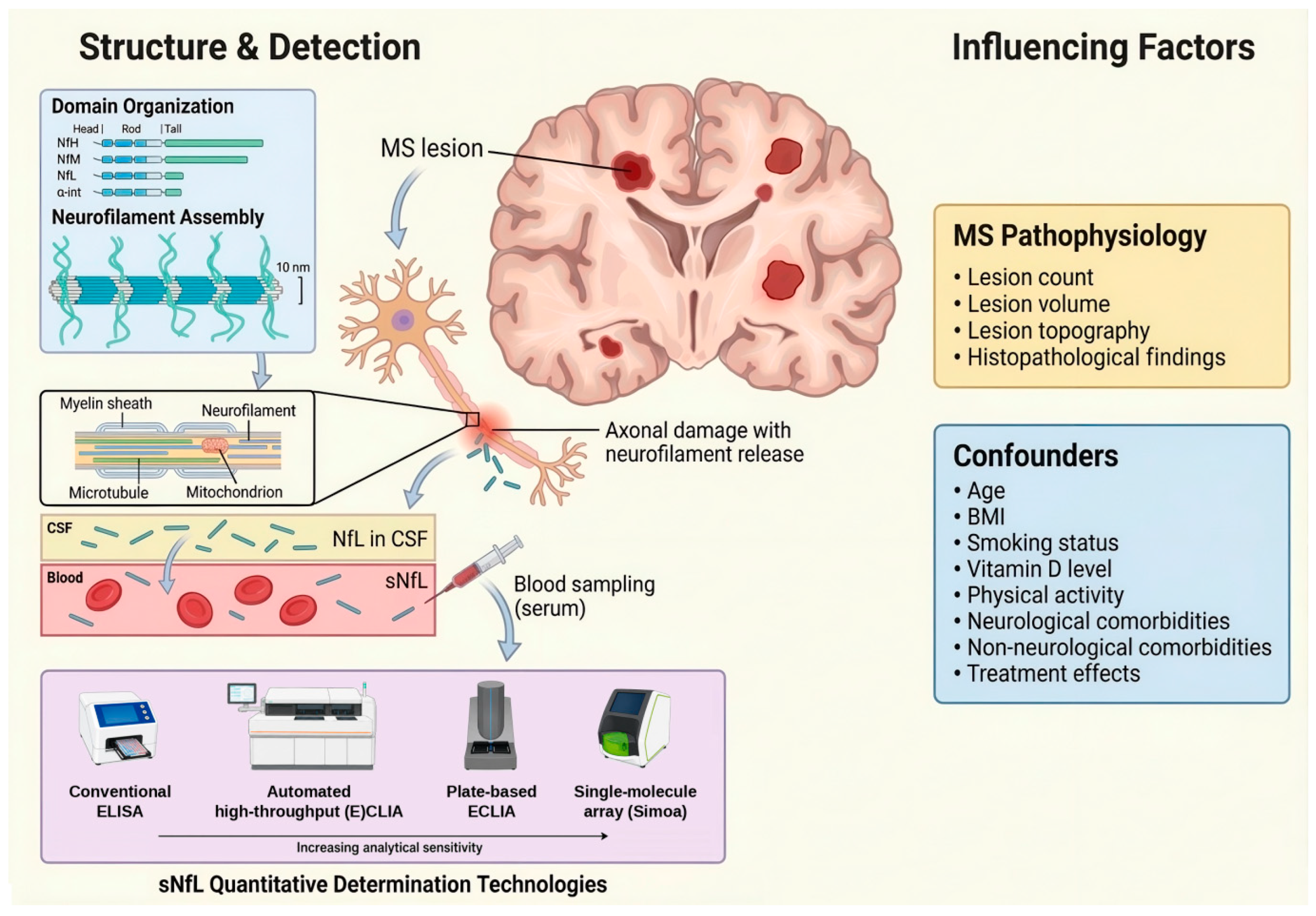
Neurofilaments are structural axonal proteins that have been studied for over 70 years and are released into the cerebrospinal fluid and blood following neuroaxonal injury [38,39,40] (Figure 1). Numerous studies in patients with MS have shown that elevated sNfL levels correlate with inflammatory activity, MRI lesion burden, and future disability progression [41,42]. However, the translation of population-level associations to the individual patient remains epistemically problematic [9]. Notably, a recent study showed that in 46 of 65 patients with MS (≈71%) who had gadolinium-enhancing MRI lesions, no increase in sNfL above the 95th percentile threshold was observed [43], underscoring the limited sensitivity of sNfL for detecting radiological disease activity.
Figure 1.
Serum NfL as a biomarker of neuroaxonal injury in MS. Schematic overview of the biological origin, detection, and interpretation of sNfL in MS. Neurofilaments, key structural components of axons, are released into the extracellular space following neuroaxonal damage associated with MS lesions. These proteins enter the CSF and subsequently the bloodstream, where sNfL can be quantified using various assay platforms that differ in analytical sensitivity. The figure also highlights key factors influencing sNfL levels, including disease-related features (e.g., lesion burden and topology) and non-specific confounders such as age, body composition, comorbid conditions, and treatment effects, which may affect the specificity of sNfL as a biomarker of disease activity. BMI, body mass index; CSF, cerebrospinal fluid; ECLIA, electrochemiluminescence immunoassay; ELISA, enzyme-linked immunosorbent assay; MS, multiple sclerosis; sNfL, serum neurofilament light chain.
To date, no universally applicable, well-validated threshold values for sNfL have been established. Measured levels are strongly influenced by age and body mass index (BMI), are highly method-dependent (e.g., assay-specific variability), and may be substantially affected by a wide range of comorbidities, including infections, vascular events, neuropathies, or trauma, as well as by metabolic factors such as diabetes, vitamin D status, lipid parameters, or changes in blood volume [39,41,44,45,46,47].
4. Bayesian Decision Logic
In situations of clinical uncertainty, the application of Bayesian decision logic is both appropriate and necessary, as test results do not provide absolute truths but instead revise probabilities [48,49]. In routine clinical practice, the key question is therefore not whether an elevated sNfL level proves that MS is currently active but rather to what extent such a finding changes the probability that inflammatory disease activity is present.
4.1. Conceptual Starting Point
Serum NfL does not establish inflammatory disease activity in MS on its own. Rather, the measured biomarker value modifies the pre-test probability that inflammatory disease activity is present.
This Bayesian relationship can be expressed conceptually as
In the context of sNfL interpretation:
- Prior probability reflects the estimated likelihood of inflammatory MS activity before biomarker testing, e.g., based on clinical symptoms and MRI findings.
- Likelihood describes how compatible an elevated sNfL value is with true inflammatory disease activity (i.e., how often it is observed in active vs. stable disease).
- Evidence represents the overall probability of observing such an sNfL value in the patient population, regardless of whether inflammatory activity is present.
- Posterior probability is the updated probability of inflammatory disease activity after considering the sNfL test result.
4.2. Classical Probabilistic Formulation
In formal notation, Bayes’ theorem can be written as
This formulation highlights that the post-test probability of inflammatory disease activity depends on the pre-test probability and on how likely the observed sNfL value is under conditions of active vs. inactive disease.
4.3. Transformation into Odds Form
For practical clinical reasoning, Bayes’ theorem can be reformulated using odds, which allows a more intuitive representation of probability updating.
Pre-test odds of inflammatory disease activity are defined as
Post-test odds are defined analogously:
Bayesian updating can then be expressed as
4.4. Likelihood Ratio and Test Characteristics
For a dichotomised biomarker interpretation (e.g., elevated vs. non-elevated sNfL), the positive and negative likelihood ratios (LR+ and LR−) can be calculated from the test characteristics, namely sensitivity and specificity:
where
- Sensitivity is the probability that sNfL is elevated in patients with true inflammatory disease activity.
- Specificity is the probability that sNfL is not elevated in patients without inflammatory disease activity.
The LR+ quantifies how strongly an elevated sNfL result shifts the probability toward disease activity, whereas the LR− quantifies how strongly a non-elevated result shifts the probability away from it.
4.5. Back-Transformation to Probability
To convert post-test odds back into a clinically interpretable probability, the following relationship is used:
This allows clinicians to translate Bayesian updating into absolute risk estimates.
4.6. Clinical Example
Assume the following illustrative scenario, based on reported diagnostic performance estimates for sNfL in MS [50]:
- Pre-test probability of inflammatory MS activity (e.g., in the context of a clinically suspected relapse) = 30%
- Sensitivity of elevated sNfL = 0.81
- Specificity = 0.70
First, calculate the positive likelihood ratio:
Next, convert pre-test probability into odds:
Update using Bayes’ rule in odds form:
Finally, convert back to probability:
Thus, in this illustrative scenario, an elevated sNfL value would increase the estimated probability of inflammatory disease activity from 30% to approximately 54%.
5. Context-Dependent Uncertainty in sNfL Interpretation
Further clinical scenarios are exemplified in Table 1, illustrating that the informative value of sNfL is greatest at intermediate pre-test probabilities—such as in the presence of clinical suspicion of disease activity or new MRI lesions—than in both clinically stable and clearly active disease. At low pre-test probability, an isolated elevation of sNfL levels may lead to an overestimation of disease activity. Thus, as outlined above, sNfL should not be conceptualised as a binary marker of disease activity but rather as a probabilistic modifier within a Bayesian decision framework. Its clinical relevance arises—at least in theory—from integration into the overall clinical context, including clinical findings, MRI, and treatment history.
Table 1.
Context-dependent interpretation of serum neurofilament light chain results in multiple sclerosis.
In practice, the clinical situation in patients with MS is frequently characterised by non-specific symptoms, heterogeneous disease courses, and variable treatment effects. In addition, the overall clinical picture may be cumulatively confounded by comorbidities. Accordingly, the sensitivity and specificity of sNfL for detecting MS disease activity are not universally fixed properties but depend on the clinical context and patient population [50,51,52,53,54]. Moreover, the individual pre-test probability of subclinical disease activity is often only crudely estimable and constitutes a key reason for clinicians to seek additional evidence. This implies that the parameters underlying the Bayesian estimation of post-test probability are subject to substantial variability—a classical problem of uncertainty propagation and, in epistemological terms, of error propagation under parameter uncertainty [7,55,56,57]. This cumulative uncertainty limits the reliability of interpreting single biomarker measurements in routine clinical care.
In addition, spectrum bias must be considered. The comparison of clearly active vs. stable clinical courses in validation studies may overestimate apparent discriminative performance and limit generalisability to clinically heterogeneous populations [50,58,59,60]. In real-world clinical practice, patients frequently present with minimal or uncertain disease activity. Within this intermediate clinical spectrum, the discriminative performance of the biomarker may be lower than in study populations with extreme contrasts, as test performance metrics are substantially influenced by disease severity, pre-test probability, comorbidities, and underlying case mix [61].
There is also a risk of partial verification bias, as not every elevation of sNfL levels in MS is systematically verified by MRI or clinical follow-up.
In summary, the use of a biologically plausible but context-dependent biomarker in a clinically uncertain setting constitutes a structural problem of iterative uncertainty propagation. An uncertain pre-test probability (individual disease activity) meets uncertain test parameters (context-dependent sensitivity and specificity and the absence of universally standardised cut-offs). Scientifically, this represents a problem of misclassification under parameter uncertainty, amplified by spectrum bias, base-rate dependence, and decision noise. Valid use of sNfL therefore requires longitudinal assessment, age-adjusted reference ranges, careful consideration of confounding factors such as BMI and comorbidities, and integration into multimodal decision models rather than interpretation of isolated values.
At present, however, a relevant evidence gap remains, as there is a lack of prospective multicentre studies systematically evaluating the role of sNfL in individualised treatment management of patients with MS in routine clinical care [9].
6. Outlook for Multimodal Biomarker Integration
Building on the framework for interpreting sNfL results discussed above, which integrates clinical and MRI-based findings with a molecular biomarker of neuroaxonal injury, Bayesian reasoning can, in principle, be extended from single-biomarker to multimarker interpretation. Future decision frameworks may incorporate additional molecular biomarkers, provided that their joint diagnostic information is validated rather than assuming that individual biomarker effects are independent.
Depending on the clinical context, further markers of tissue injury could become particularly relevant. These include GFAP as an indicator of astrocytic injury and SNAP25 as a marker of synaptic neuronal injury and degenerative processes associated with disease progression. Biomarkers reflecting immune-cell activation, such as BCMA for B-cell activation, sCD27 for T-cell activation, CHI3L1 and OPN as markers of microglial activation, and cytokines such as IL-6 and IL-17, also represent promising candidates [62,63].
However, their value for routine clinical assessment of MS remains limited. For markers of tissue destruction, this mainly reflects the lack of validation studies demonstrating added value for real-world clinical care. For immunological markers, clinical implementation is particularly constrained by substantial biological, pre-analytical, and methodological variability.
Author Contributions
Conceptualisation, U.K.Z.; Visualisation, M.H.; Writing—original draft, U.K.Z.; Writing—review and editing, M.H. All authors have read and agreed to the published version of the manuscript.
Funding
This work received no external funding.
Institutional Review Board Statement
Not applicable.
Informed Consent Statement
Not applicable.
Data Availability Statement
No new data were created for this article.
Acknowledgments
During the preparation of this manuscript, the authors used ChatGPT (OpenAI, GPT-5) for the purposes of language editing and text organisation and Gemini (Google, Nano Banana 2) to generate a figure based on author-provided input. All scientific concepts, analyses, and interpretations were developed and validated by the authors.
Conflicts of Interest
The authors declare no conflicts of interest.
Abbreviations
| BCMA | B-cell maturation antigen |
| BMI | body mass index |
| CD | cluster of differentiation |
| CHI3L1 | chitinase-3-like protein 1 |
| CSF | cerebrospinal fluid |
| ECLIA | electrochemiluminescence immunoassay |
| EDSS | Expanded Disability Status Scale |
| ELISA | enzyme-linked immunosorbent assay |
| GFAP | glial fibrillary acidic protein |
| IL | interleukin |
| LR | likelihood ratio |
| MRI | magnetic resonance imaging |
| MS | multiple sclerosis |
| NEDA | no evidence of disease activity |
| NfL | neurofilament light chain |
| OPN | osteopontin |
| PIRA | progression independent of relapse activity |
| PIRMA | progression independent of relapse and MRI activity |
| sCD27 | soluble CD27 |
| Simoa | single-molecule array |
| SNAP25 | synaptosomal-associated protein 25 |
| sNfL | serum neurofilament light chain |
References
- Olver, P.; Bohn, M.K.; Adeli, K. Central role of laboratory medicine in public health and patient care. Clin. Chem. Lab. Med. 2022, 61, 666–673. [Google Scholar] [CrossRef] [Scilit] [PubMed]
- Alzaabi, M.A.; Abdelsalam, A.; Alhammadi, M.; Bani Hani, H.; Almheiri, A.; Al Matrooshi, N.; Al Zaman, K. Evaluating Biomarkers as Tools for Early Detection and Prognosis of Heart Failure: A Comprehensive Review. Card. Fail. Rev. 2024, 10, e06. [Google Scholar] [CrossRef] [Scilit] [PubMed] [PubMed Central]
- Etzioni, R.; Gulati, R.; Patriotis, C.; Rutter, C.; Zheng, Y.; Srivastava, S.; Feng, Z. Revisiting the standard blueprint for biomarker development to address emerging cancer early detection technologies. J. Natl. Cancer Inst. 2024, 116, 189–193. [Google Scholar] [CrossRef] [Scilit] [PubMed] [PubMed Central]
- Simon, R. Clinical trial designs for evaluating the medical utility of prognostic and predictive biomarkers in oncology. Pers. Med. 2010, 7, 33–47. [Google Scholar] [CrossRef] [Scilit] [PubMed] [PubMed Central]
- FDA-NIH Biomarker Working Group. BEST (Biomarkers, EndpointS, and Other Tools) Resource [Internet]; Food and Drug Administration: Silver Spring, MD, USA, 2016. Available online: https://www.ncbi.nlm.nih.gov/books/NBK464453/ (accessed on 27 March 2026).
- Ou, F.S.; Michiels, S.; Shyr, Y.; Adjei, A.A.; Oberg, A.L. Biomarker Discovery and Validation: Statistical Considerations. J. Thorac. Oncol. 2021, 16, 537–545. [Google Scholar] [CrossRef] [Scilit] [PubMed] [PubMed Central]
- Tsaneva-Atanasova, K.; Pederzanil, G.; Laviola, M. Decoding uncertainty for clinical decision-making. Philos. Trans. A Math. Phys. Eng. Sci. 2025, 383, 20240207. [Google Scholar] [CrossRef] [Scilit] [PubMed] [PubMed Central]
- Zheng, Y.; Wagner, P.D.; Singal, A.G.; Hanash, S.M.; Srivastava, S.; Huang, Y.; Zhao, Y.Q.; Chari, S.T.; Marquez, G.; Etizioni, R.; et al. Designing Rigorous and Efficient Clinical Utility Studies for Early Detection Biomarkers. Cancer Epidemiol. Biomark. Prev. 2024, 33, 1150–1157. [Google Scholar] [CrossRef] [Scilit] [PubMed] [PubMed Central]
- Sotirchos, E.S.; Solomon, A.J. Are neurofilament light chain levels ready for routine clinical use?—No. Mult. Scler. J. 2026, 13524585261423029. [Google Scholar] [CrossRef] [Scilit] [PubMed]
- Sandgren, S.; Einsiedler, M.; Benkert, P.; Leppert, D.; Kuhle, J. Are neurofilament light chain levels ready for routine clinical use?—Yes. Mult. Scler. J. 2026, 13524585261423032. [Google Scholar] [CrossRef] [Scilit] [PubMed]
- Garton, T.; Gadani, S.P.; Gill, A.J.; Calabresi, P.A. Neurodegeneration and demyelination in multiple sclerosis. Neuron 2024, 112, 3231–3251. [Google Scholar] [CrossRef] [Scilit] [PubMed] [PubMed Central]
- Multz, R.A.; Jamshidi, P.; Ahrendsen, J.T. Multiple sclerosis: A practical review for pathologists. J. Pathol. Transl. Med. 2025, 59, 203–213. [Google Scholar] [CrossRef] [Scilit] [PubMed] [PubMed Central]
- Ullrich Gavilanes, E.M.; Bartos, L.M.; Gernert, J.A.; Carral, C.A.; Ruiz Navarro, D.; Havla, J.; Gerdes, L.A.; Gnörich, J.S.; Kunze, L.H.; Dorneich, J.S.; et al. SV2A-PET imaging uncovers cortical synapse loss in multiple sclerosis. Sci. Transl. Med. 2025, 17, eadt5585. [Google Scholar] [CrossRef] [Scilit] [PubMed]
- Solomon, A.J.; Brodersen, J.B. The 2024 revisions of the McDonald criteria pose risk of multiple sclerosis overdiagnosis. Nat. Rev. Neurol. 2025, 21, 659–660. [Google Scholar] [CrossRef] [Scilit] [PubMed]
- Ellenberger, D.; Flachenecker, P.; Fneish, F.; Frahm, N.; Hellwig, K.; Paul, F.; Stahmann, A.; Warnke, C.; Rommer, P.S.; Zettl, U.K. Aggressive multiple sclerosis: A matter of measurement and timing. Brain 2020, 143, e97. [Google Scholar] [CrossRef] [Scilit] [PubMed] [PubMed Central]
- Brieva, L.; Calles, C.; Landete, L.; Oreja-Guevara, C. Current challenges in secondary progressive multiple sclerosis: Diagnosis, activity detection and treatment. Front. Immunol. 2025, 16, 1543649. [Google Scholar] [CrossRef] [Scilit] [PubMed] [PubMed Central]
- Altintas, A.; Bulus, E. Multiple sclerosis: A disease or a syndrome? Curr. Opin. Immunol. 2026, 99, 102736. [Google Scholar] [CrossRef] [Scilit] [PubMed]
- Cárdenas-Robledo, S.; Otero-Romero, S.; García, J.D.; López, N.; Carbonell-Mirabent, P.; Rodríguez, M.; Guío-Sánchez, C.; Carvajal, R.; Passarell-Bacradit, M.A.; Sastre-Garriga, J.; et al. Clusters of Comorbidities in Multiple Sclerosis and Their Influence on Healthcare Resource Usage. Eur. J. Neurol. 2025, 32, e70386. [Google Scholar] [CrossRef] [Scilit] [PubMed] [PubMed Central]
- Zhang, Y.; Dobson, R.; Giovannoni, G. Comorbidity and modifiable risk factors in multiple sclerosis. Ther. Adv. Neurol. Disord. 2026, 19, 17562864251404455. [Google Scholar] [CrossRef] [Scilit] [PubMed] [PubMed Central]
- Kurtzke, J.F. Rating neurologic impairment in multiple sclerosis: An expanded disability status scale (EDSS). Neurology 1983, 33, 1444–1452. [Google Scholar] [CrossRef] [Scilit] [PubMed]
- Lublin, F.D.; Reingold, S.C.; Cohen, J.A.; Cutter, G.R.; Sørensen, P.S.; Thompson, A.J.; Wolinsky, J.S.; Balcer, L.J.; Banwell, B.; Barkhof, F.; et al. Defining the clinical course of multiple sclerosis: The 2013 revisions. Neurology 2014, 83, 278–286. [Google Scholar] [CrossRef] [Scilit] [PubMed] [PubMed Central]
- Chard, D.; Trip, S.A. Resolving the clinico-radiological paradox in multiple sclerosis. F1000Research 2017, 6, 1828. [Google Scholar] [CrossRef] [Scilit] [PubMed] [PubMed Central]
- Barkhof, F.; Reich, D.S.; Oh, J.; Rocca, M.A.; Li, D.K.B.; Sati, P.; Azevedo, C.J.; Bagnato, F.; Calabresi, P.A.; Ciccarelli, O.; et al. 2024 MAGNIMS-CMSC-NAIMS consensus recommendations on the use of MRI for the diagnosis of multiple sclerosis. Lancet Neurol. 2025, 24, 866–879. [Google Scholar] [CrossRef] [Scilit] [PubMed]
- Hegen, H.; Bsteh, G.; Berger, T. ‘No evidence of disease activity’—Is it an appropriate surrogate in multiple sclerosis? Eur. J. Neurol. 2018, 25, 1107-e101. [Google Scholar] [CrossRef] [Scilit] [PubMed] [PubMed Central]
- Rotstein, D.; Solomon, J.M.; Sormani, M.P.; Montalban, X.; Ye, X.Y.; Dababneh, D.; Muccilli, A.; Saab, G.; Shah, P. Association of NEDA-4 With No Long-term Disability Progression in Multiple Sclerosis and Comparison With NEDA-3: A Systematic Review and Meta-analysis. Neurol. Neuroimmunol. Neuroinflamm. 2022, 9, e200032. [Google Scholar] [CrossRef] [Scilit] [PubMed] [PubMed Central]
- Dünschede, J.; Ruschil, C.; Bender, B.; Mengel, A.; Lindig, T.; Ziemann, U.; Kowarik, M.C. Clinical-Radiological Mismatch in Multiple Sclerosis Patients during Acute Relapse: Discrepancy between Clinical Symptoms and Active, Topographically Fitting MRI Lesions. J. Clin. Med. 2023, 12, 739. [Google Scholar] [CrossRef] [Scilit] [PubMed] [PubMed Central]
- Gavoille, A.; Rollot, F.; Casey, R.; Kerbrat, A.; Le Page, E.; Bigaut, K.; Mathey, G.; Michel, L.; Ciron, J.; Ruet, A.; et al. Acute Clinical Events Identified as Relapses With Stable Magnetic Resonance Imaging in Multiple Sclerosis. JAMA Neurol. 2024, 81, 814–823. [Google Scholar] [CrossRef] [Scilit] [PubMed] [PubMed Central]
- Tur, C.; Rocca, M.A. Progression Independent of Relapse Activity in Multiple Sclerosis: Closer to Solving the Pathologic Puzzle. Neurology 2024, 102, e207936. [Google Scholar] [CrossRef] [Scilit] [PubMed]
- Müller, J.; Cagol, A.; Lorscheider, J.; Tsagkas, C.; Benkert, P.; Yaldizli, Ö.; Kuhle, J.; Derfuss, T.; Sormani, M.P.; Thompson, A.; et al. Harmonizing Definitions for Progression Independent of Relapse Activity in Multiple Sclerosis: A Systematic Review. JAMA Neurol. 2023, 80, 1232–1245. [Google Scholar] [CrossRef] [Scilit] [PubMed]
- Rollot, F.; Casey, R.; Kerbrat, A.; Edan, G.; Le Page, E.; Vukusic, S.; Mathey, G.; Maillart, E.; De Sèze, J.; Ciron, J.; et al. Progression independent of relapse and MRI activity and treatment strategies in multiple sclerosis. Brain 2026, awag060. [Google Scholar] [CrossRef] [Scilit] [PubMed]
- Sharrad, D.; Chugh, P.; Slee, M.; Bacchi, S. Defining progression independent of relapse activity (PIRA) in adult patients with relapsing multiple sclerosis: A systematic review. Mult. Scler. Relat. Disord. 2023, 78, 104899. [Google Scholar] [CrossRef] [Scilit] [PubMed]
- Zhu, C.; Zhou, Z.; Kalincik, T.; Roos, I.; Buzzard, K.; Skibina, O.; Alroughani, R.; Kuhle, J.; Girard, M.; Grammond, P.; et al. Persistent progression independent of relapse activity in multiple sclerosis. Brain Commun. 2025, 7, fcaf306. [Google Scholar] [CrossRef] [Scilit] [PubMed] [PubMed Central]
- Saposnik, G.; Andhavarapu, S.; Fernández, Ó.; Kim, H.J.; Wiendl, H.; Foss, M.; Zuo, F.; Havrdová, E.K.; Celius, E.; Caceres, F.; et al. Factors associated with treatment escalation among MS specialists and general neurologists: Results from an International cojoint study. Mult. Scler. Relat. Disord. 2022, 58, 103404. [Google Scholar] [CrossRef] [Scilit] [PubMed]
- van Reenen, E.C.; Niemeijer, A.R.; Visser, L.H.; de Beukelaar, J.W.K.; van Oosten, B.W.; Frequin, S.T.F.M.; Hoogervorst, E.L.J.; van Nistelrooij, I.A.M. Spheres of uncertainty: A phenomenological inquiry into healthcare practice surrounding the care for people with relapsing-remitting multiple sclerosis. Qual. Res. Med. Healthc. 2025, 9, 100031. [Google Scholar] [CrossRef] [Scilit] [PubMed] [PubMed Central]
- Anderhalten, L.; Wohlrab, F.; Paul, F. Emerging MRI and biofluid biomarkers in the diagnosis and prognosis of multiple sclerosis. Lancet Reg. Health–Eur. 2024, 44, 101023. [Google Scholar] [CrossRef] [Scilit] [PubMed] [PubMed Central]
- Di Filippo, M.; Gaetani, L.; Centonze, D.; Hegen, H.; Kuhle, J.; Teunissen, C.E.; Tintoré, M.; Villar, L.M.; Willemse, E.A.J.; Zetterberg, H.; et al. Fluid biomarkers in multiple sclerosis: From current to future applications. Lancet Reg. Health–Eur. 2024, 44, 101009. [Google Scholar] [CrossRef] [Scilit] [PubMed] [PubMed Central]
- Schuck, R.N.; Sekar, V. Use of Biomarkers in Drug Development for Regulatory Purposes. Clin. Transl. Sci. 2025, 18, e70377. [Google Scholar] [CrossRef] [Scilit] [PubMed] [PubMed Central]
- Yuan, A.; Rao, M.V.; Veeranna; Nixon, R.A. Neurofilaments and Neurofilament Proteins in Health and Disease. Cold Spring Harb. Perspect. Biol. 2017, 9, a018309. [Google Scholar] [CrossRef] [Scilit] [PubMed] [PubMed Central]
- Bar-Or, A.; Nicholas, J.; Feng, J.; Sorrell, F.; Cascione, M. Exploring the Clinical Utility of Neurofilament Light Chain Assays in Multiple Sclerosis Management. Neurol. Neuroimmunol. Neuroinflamm. 2025, 12, e200427. [Google Scholar] [CrossRef] [Scilit] [PubMed] [PubMed Central]
- Turner, M.R.; Thompson, A.G.; Teunissen, C.E. Blood level of neurofilament light chain as a biomarker for neurological disorders. BMJ Med. 2025, 4, e000958. [Google Scholar] [CrossRef] [Scilit] [PubMed] [PubMed Central]
- Khalil, M.; Teunissen, C.E.; Lehmann, S.; Otto, M.; Piehl, F.; Ziemssen, T.; Bittner, S.; Sormani, M.P.; Gattringer, T.; Abu-Rumeileh, S.; et al. Neurofilaments as biomarkers in neurological disorders—Towards clinical application. Nat. Rev. Neurol. 2024, 20, 269–287. [Google Scholar] [CrossRef] [Scilit] [PubMed]
- Freedman, M.S.; Gnanapavan, S.; Booth, R.A.; Calabresi, P.A.; Khalil, M.; Kuhle, J.; Lycke, J.; Olsson, T.; Consortium of Multiple Sclerosis Centers. Guidance for use of neurofilament light chain as a cerebrospinal fluid and blood biomarker in multiple sclerosis management. EBioMedicine 2024, 101, 104970. [Google Scholar] [CrossRef] [Scilit] [PubMed] [PubMed Central]
- Fox, R.J.; Cree, B.A.C.; de Sèze, J.; Gold, R.; Hartung, H.P.; Jeffery, D.; Kappos, L.; Montalban, X.; Weinstock-Guttman, B.; Singh, C.M.; et al. Temporal Relationship Between Serum Neurofilament Light Chain and Radiologic Disease Activity in Patients With Multiple Sclerosis. Neurology 2024, 102, e209357. [Google Scholar] [CrossRef] [Scilit] [PubMed] [PubMed Central]
- Beltran, T.A. Normative Values for Serum Neurofilament Light Chain in US Adults. J. Clin. Neurol. 2024, 20, 46–49. [Google Scholar] [CrossRef] [Scilit] [PubMed] [PubMed Central]
- Daponte, A.; Koros, C.; Skarlis, C.; Siozios, D.; Rentzos, M.; Papageorgiou, S.G.; Anagnostouli, M. Neurofilament Biomarkers in Neurology: From Neuroinflammation to Neurodegeneration, Bridging Established and Novel Analytical Advances with Clinical Practice. Int. J. Mol. Sci. 2025, 26, 9739. [Google Scholar] [CrossRef] [Scilit] [PubMed] [PubMed Central]
- La Civita, E.; Nicolella, V.; Fiorenza, M.; Cosimato, V.; Castaldo, G.; Morra, V.B.; Moccia, M.; Terracciano, D. Advancing Clinical Use of Neurofilament Light Chain: Translational Insights From Research to Routine Practice. Biomark. Insights 2025, 20, 11772719251364018. [Google Scholar] [CrossRef] [Scilit] [PubMed] [PubMed Central]
- Biernacki, T.; Kokas, Z.; Sandi, D.; Füvesi, J.; Fricska-Nagy, Z.; Faragó, P.; Kincses, T.Z.; Klivényi, P.; Bencsik, K.; Vécsei, L. Emerging Biomarkers of Multiple Sclerosis in the Blood and the CSF: A Focus on Neurofilaments and Therapeutic Considerations. Int. J. Mol. Sci. 2022, 23, 3383. [Google Scholar] [CrossRef] [Scilit] [PubMed] [PubMed Central]
- Bours, M.J. Bayes’ rule in diagnosis. J. Clin. Epidemiol. 2021, 131, 158–160. [Google Scholar] [CrossRef] [Scilit] [PubMed]
- Webb, M.P.K.; Sidebotham, D. Bayes’ formula: A powerful but counterintuitive tool for medical decision-making. BJA Educ. 2020, 20, 208–213. [Google Scholar] [CrossRef] [Scilit] [PubMed] [PubMed Central]
- Johnsson, M.; Stenberg, Y.T.; Farman, H.H.; Blennow, K.; Zetterberg, H.; Malmeström, C.; Sandgren, S.; Rosenstein, I.; Lycke, J.; Axelsson, M.; et al. Serum neurofilament light for detecting disease activity in individual patients in multiple sclerosis: A 48-week prospective single-center study. Mult. Scler. J. 2024, 30, 664–673. [Google Scholar] [CrossRef] [Scilit] [PubMed] [PubMed Central]
- Solís-Tarazona, L.; Raket, L.L.; Cabello-Murgui, J.; Reddam, S.; Navarro-Quevedo, S.; Gil-Perotin, S. Predictive value of individual serum neurofilament light chain levels in short-term disease activity in relapsing multiple sclerosis. Front. Neurol. 2024, 15, 1354431. [Google Scholar] [CrossRef] [Scilit] [PubMed] [PubMed Central]
- Nicolella, V.; Varelli, M.; Fasano, S.; Sirica, R.; Polito, C.; Saviano, A.; Fiorenza, M.; Novarella, F.; Ranucci, D.; Carotenuto, A.; et al. Clinical application of age-derived cut-offs for plasma neurofilament light chain in multiple sclerosis. J. Neurol. 2025, 272, 495. [Google Scholar] [CrossRef] [Scilit] [PubMed]
- Fung, W.H.; Wessels, M.H.J.; Coerver, E.M.E.; de Beukelaar, J.; Bouvy, W.H.; Canta, L.R.; Gerlach, O.H.H.; Hoitsma, E.; Hoogervorst, E.L.J.; de Jong, B.A.; et al. Reliability of serum neurofilament light and glial fibrillary acidic protein for detecting disease activity upon discontinuation of first-line disease-modifying therapy in stable multiple sclerosis (DOT-MS). J. Neurol. 2025, 272, 530. [Google Scholar] [CrossRef] [Scilit] [PubMed] [PubMed Central]
- Uher, T.; Schaedelin, S.; Srpova, B.; Barro, C.; Bergsland, N.; Dwyer, M.; Tyblova, M.; Vodehnalova, K.; Benkert, P.; Oechtering, J.; et al. Monitoring of radiologic disease activity by serum neurofilaments in MS. Neurol. Neuroimmunol. Neuroinflamm. 2020, 7, e714. [Google Scholar] [CrossRef] [Scilit] [PubMed] [PubMed Central]
- Chatzimichail, T.; Hatjimihail, A.T. A Software Tool for Estimating Uncertainty of Bayesian Posterior Probability for Disease. Diagnostics 2024, 14, 402. [Google Scholar] [CrossRef] [Scilit] [PubMed] [PubMed Central]
- Gallagher, B.D. The Post Hoc Pitfall: Rethinking Sensitivity and Specificity in Clinical Practice. J. Gen. Intern. Med. 2025, 40, 947. [Google Scholar] [CrossRef] [Scilit] [PubMed] [PubMed Central]
- Vijfschagt, N.D.; Burger, H.; Berger, M.Y.; Fanshawe, T.R.; van den Bruel, A.; Leeflang, M.M.G.; de Boer, M.R.; Holtman, G.A. Variation in sensitivity and specificity of diverse diagnostic tests across health-care settings: A meta-epidemiological study. J. Clin. Epidemiol. 2025, 184, 111816. [Google Scholar] [CrossRef] [Scilit] [PubMed]
- Novakova, L.; Zetterberg, H.; Sundström, P.; Axelsson, M.; Khademi, M.; Gunnarsson, M.; Malmeström, C.; Svenningsson, A.; Olsson, T.; Piehl, F.; et al. Monitoring disease activity in multiple sclerosis using serum neurofilament light protein. Neurology 2017, 89, 2230–2237. [Google Scholar] [CrossRef] [Scilit] [PubMed] [PubMed Central]
- Hyun, J.W.; Kim, Y.; Kim, G.; Kim, S.H.; Kim, H.J. Longitudinal analysis of serum neurofilament light chain: A potential therapeutic monitoring biomarker for multiple sclerosis. Mult. Scler. J. 2020, 26, 659–667. [Google Scholar] [CrossRef] [Scilit] [PubMed]
- Ziemssen, T.; Freedman, M.S.; Bar-Or, A.; Montalban, X.; Teunissen, C.E.; Häring, D.A.; Kukkaro, P.; Merschhemke, M.; Kieseier, B.C.; Karnik-Henry, M.; et al. Clinical validation of a novel in vitro diagnostic neurofilament light chain assay for the prognostication of disease activity in people with relapsing multiple sclerosis. Mult. Scler. J. 2025, 31, 1543–1556. [Google Scholar] [CrossRef] [Scilit] [PubMed] [PubMed Central]
- Chaturvedi, M.; Köster, D.; Bossuyt, P.M.; Gerke, O.; Jurke, A.; Kretzschmar, M.E.; Lütgehetmann, M.; Mikolajczyk, R.; Reitsma, J.B.; Schneiderhan-Marra, N.; et al. A unified framework for diagnostic test development and evaluation during outbreaks of emerging infections. Commun. Med. 2024, 4, 263. [Google Scholar] [CrossRef] [Scilit] [PubMed] [PubMed Central]
- Blok, K.M.; Klein Kranenbarg, R.A.M.; Ananth, K.; Engelenburg, H.J.; van den Bosch, A.; Giannini, L.A.A.; de Beukelaar, J.; Seelaar, H.; Huitinga, I.; Green, A.; et al. Multifaceted Biomarkers Suggest a Similar Profile of CNS Pathology in Relapsing and Progressive MS. Eur. J. Neurol. 2025, 32, e70052. [Google Scholar] [CrossRef] [Scilit] [PubMed] [PubMed Central]
- Chitnis, T.; Magliozzi, R.; Abdelhak, A.; Kuhle, J.; Leppert, D.; Bielekova, B. Blood and CSF biomarkers for multiple sclerosis: Emerging clinical applications. Lancet Neurol. 2025, 24, 1066–1078. [Google Scholar] [CrossRef] [Scilit] [PubMed]

Disclaimer/Publisher’s Note: The statements, opinions and data contained in all publications are solely those of the individual author(s) and contributor(s) and not of MDPI and/or the editor(s). MDPI and/or the editor(s) disclaim responsibility for any injury to people or property resulting from any ideas, methods, instructions or products referred to in the content. |
© 2026 by the authors. Licensee MDPI, Basel, Switzerland. This article is an open access article distributed under the terms and conditions of the Creative Commons Attribution (CC BY) license.