


Abstract
With colorectal cancer (CRC) accounting for over 1.9 million new cases and 930,000 deaths globally in 2020, there is a critical need for innovative indicators to forecast disease advancement and therapeutic outcomes. The gut microbiome has emerged as a fertile area for discovering such diagnostic and prognostic signals. This narrative review collected current evidence on intestinal microorganisms and their metabolic products as candidate markers for CRC control. Intestinal communities influence malignancy through diverse mechanisms, including metabolic shifts, immune modulation, inflammation, proliferation/apoptosis regulation, genotoxicity, and mucosal barrier disruption. Pathogenic species, such as Fusobacterium nucleatum and enterotoxigenic Bacteroides fragilis, facilitate tumorigenesis via FadA-mediated signaling and Th17/IL-17 responses. In contrast, beneficial taxa like Faecalibacterium prausnitzii and Akkermansia muciniphila provide protective effects through short chain fatty acid production. Macrophage phenotype physiological equilibrium is altered and inflammatory status fluctuates under the former. Metabolically, hydrogen sulfide damages mitochondrial DNA and secondary bile acids stimulate cellular proliferation. While 16S rRNA sequencing and shotgun metagenomics are established detection strategies, innovative platforms like organoids and gene arrays remain in the exploratory stage. Clinical data indicates that F. nucleatum aligns with advanced tumor stage, and its combined detection with colibactin-producing E. coli achieves high sensitivity for early-stage screening. Additionally, A. muciniphila levels can anticipate the efficacy of PD-1 blockade immunotherapy. Microbiota-derived tools represent a transformative direction in oncology. Future research must focus on standardizing protocols and validating multi-marker panels to enhance clinical translation.
1. Introduction
Newly diagnosed colorectal cancer (CRC) cases rose to over 150,000 and ranked third in both sexes in America, 2024 [1]. The predicted death number of CRC is about 53,000 in the USA and 153,000 in the European Union during 2024 [1,2]. CRC casts persistent heavy burden on both individual family expenditure and worldwide hygiene. One of the most powerful factors in prejudging the disease outcome is tumor stage. Most CRC developed from benign polyps over a long term of 10 to 15 years. Proper adenoma detection and excision do break the incidence of CRC [3]. In England, Duke A stage CRC reached a 5 year overall survival (OS) as high as 98%, with a gradual decrease to 85%, 63% and 7.5% for Duke B, C and D stage diseases [4]. This result was identical to another study which concluded stage III CRC 65–70% 5-year survival and a steep reduction in stage IV to 13–14% [5,6]. A large-scale study based on the Martinique Cancer Registry between 1993 and 2012 reported a median OS of 2.0 years for stage III to IV CRC, while stage I to II had not reached that outcome before the program was terminated [7]. Efficient testing is urgently needed to advance diagnosis and prognosis.
So far, the most widely accepted screening methods include colonoscopy and the fecal immunohistochemical test (FIT). Colonoscopy provides a magnified view and appended clarification based on the directly acquired sample [8]. Although colonoscopy is the golden standard test in CRC diagnosis, the trauma and high expense discourage some patients. Studies concluded FIT displayed a sensitivity of 74% to 94% and a specificity of 85% to 95% in the diagnosis of CRC [9,10]. There is still a large unknown area left for particular anticipation. For example, for patients undergoing a watch-and-wait procedure after their lesion showed complete remission with CT or colonoscopy, a novel screen test able to predict its progress instead of just waiting until, in some cases, relapse would definitely be more safe. Another occasion is therapeutic predication. These further demands call for more markers other than just 100% accurate colonoscopy alone. The circulating tumor DNA (ctDNA) test and tumor mutation burden (TMB) are both innovative and derivative tumor examination methods at this time.
About 100 trillion microorganisms reside in the human gut, of which Firmicutes takes the lead proportion of approximately 67% and Bacteroidetes the second of 21% [11,12]. Previous evidence supported their role in some tumors, such as oral and breast cancer and malignant melanoma [13,14,15,16]. Similarly, the microbiomic test has great potential in predicting CRC outcomes, therapy-related or not. Recent studies demonstrated that a high level of Fusobacterium nucleatum and Bacteroides fragilis indicates poor prognosis [17]. The former is also reported to closely relate to chemotherapy resistance and distant metastasis [18].
Based on the accumulating assays on microorganisms, we conducted this narrative review to summarize the existing evidence, their differences and limitations, and the expectations of them as CRC biomarkers and associative mechanisms.
2. The Roles Microbiomes Play in CRC Formation and Progression
Human gut microbiomes affect colorectal carcinogenesis and progression in intricate pathways. To summarize, we collected the information listed below: metabolism, immune, inflammation, proliferation or apoptosis, genetoxin and barrier.
2.1. Metabolism
Microbiomes take carbohydrates, proteins, lipid, water and other nutrition from intraluminal contents, produce substances they need and excrete metabolic waste. Some natural or metabolic intermediate sulfur-containing proteins (like disulfide bonds) discharge hydrogen sulfide along degradation under particular enzymes catalysis [19]. This process is half adjusted by intestinal microbes [20]. Hydrogen sulfide plays a dualistic role in gastrointestinal micro-ecology. It enables electric transmission in the mitochondrial electron transport chain in complex II [21]. However, its overexpression inactivated complex IV and inhibited the mitochondrial respiratory chain [22,23]. This respiratory disruption results in the depletion of adenosine triphosphate (ATP) and the accumulation of reactive oxygen (ROS) [24,25]. Furthermore, hydrogen sulfide exceeds mitochondrial permeability transition and triggers hepatocyte apoptosis [26]. An overload of ROS strain injures both mitochondrial DNA (mtDNA) and nuclear DNA, thereby contributing to the genetic instability observed in CRC [23,27]. Additionally, H2S suppresses short chain acyl-CoA dehydrogenase, which downregulates the β-oxidation of butyrate, the primary energy substrate for colonic epithelial cells [28]. Desulfovibrio, a Gram-negative obligate anaerobic bacillus within the Proteobacteria phylum and a characterized sulfate-reducing bacteria (SRB), generates H2S via membranous and soluble sulfite reductase [29]. Contrary to most other microbiomes in the bowel that utilize the assimilatory sulfate reduction pathway, Desulfovibrio operates through a dissimilatory path [30]. This process of synthesizing hydrogen sulfide is detrimental to colonocyte respiration. Following accumulated ROS stress interrupted DNA stability and acted as a genetoxin by compromising DNA stability [31]. Additionally, colonic epithelial tight junction was broken by Desulfovibrio by activating transcription factor Snail 1 and altering the cellular location of occludin [32].
Parallel to sulfur metabolism, the microbial transformation of bile acids significantly modulates the colonic microenvironment. Bile acid is a group of hepatocyte-synthesized cholesterol derivatives promoting lipid digestion and absorption. Primary bile acids (PCAs) include cholic and chenodeoxycholic acids. They are delivered into duodenum once combined with glycine or taurine. Some PCAs are modified right after secretion into secondary bile acids (SBAs). Deoxycholic acid (DCA), lithocholic acid (LCA), isoLCA, alloLCA and other derivatives comprise the SBA family [33]. Meanwhile, bile acids physiologically regulate glucolipid metabolism and enterohepatic circulation through Farnesoid X Receptor (FXR) and Takeda G Protein-Coupled Receptor 5 (TGR5). Their excessive accumulation exacerbates chronic inflammation, induces double-strand DNA breaks and triggers aberrant cellular proliferation [34]. A recent experiment elucidated that mice exposed to high levels of DCA suffered raised risk of colonic adenoma converting into adenocarcinoma [35]. DCA enhances arachidonic acid excretion and prompts its transformation into pro-angiogenic prostaglandin and ROS, injuring DNA and curbing the restoration process. It also augments cyclooxygenage 2 (COX-2) via stimulation of the epidermal growth factor receptor (EGFR), which further impels colonic carcinogenesis or distant metastasis [36].
DCA-mediated bowel dysbiosis promotes the expansion of opportunistic pathogens such as Shigella and Desulfovibri and reduces beneficial Bifidobacterium and Lactobacillus populations [37]. In high-fat-diet-fed Apc−/+ CRC rodent models, SBAs stimulated colonic cell stemness and tumorigenesis through FXR activation, which is driven in part by ROS-induced ribosomal instability [38]. Such diets typically extend the proportion of Clostridium species and increase the DCA level. The latter upregulates hepatic stellate cell caducity-related expression and pro-inflammatory phenotype, which finally advanced into hepatocellular carcinoma [39]. Similarly, Clostridium was revealed to induce CRC development by other independent studies [40,41]. A high-fat diet potentially promoted PCA-producing microorganisms, accelerating tumor formation and process [42]. SBAs impose pressure on digestive ecology in various aspects including immune, inflammatory and microbial composition. LCAs inhibit hepatic-infiltrated macrophage glycolysis and facilitate intracellular oxidative phosphorylation, reshaping macrophages from anti-tumor M1 into the immune-suppressive M2 phenotype [43]. This adaption is synergized by reinforced M2 recruitment from the activated mannose receptor in CRC [44]. M1 is more aggressive as an inflammatory biomarker maintaining immune clearance. M2-type tumor-associated macrophages (TAMs) oppositely establish an immune privilege. Pathological suppression of FXR disrupted intestinal rhythm, resulting in diminished secretory immunoglobulin A (sIgA) and elevated Enterotoxigenic Bacteroides fragilis (ETBF) colonization, ultimately causing colitis-associated CRC [45].
In contrast to the potentially deleterious effects associated with hydrogen sulfide and SBAs, short chain fatty acids (SCFAs) refer to a saturated fatty acid with a limited length of carbon chain less than six carbon atoms. Acetic acid, propionic acid and butyric acid are common SCFAs within the human body. Butyrate, in particular, acts as a critical ligand for peroxisome proliferator-activated receptor γ (PPAR-γ), which is activated by butyrate, facilitating the metabolic transition into β-oxidation. This pathway provides energy supply for colonic epithelium, and blocks some pathogenic colonization [46]. Over 90% of SCFAs are rapidly absorbed in the proximal colon [47], while only approximately 5% are later excreted in feces [48]. The progression of CRC is frequently characterized by a marked reduction in SCFA-synthesizing taxa, specifically within the Bacteroides and Firmicutes phyla. Experimental evidence suggests that SCFA-producing flora enrichment restrain the PI3K/AKT pathway, triggering apoptosis-related genes, and reduce tumor volume [47,49]. Those phenomena align with clinical data demonstrating significantly depleted butyrate levels in the colonic microenvironment of CRC patients [50]. According to animal and cellular experiments, SCFAs inhibited pro-inflammatory factors, TNF-α, IL-18, IL-1β, IL-6 and iNOS, while simultaneously raising anti-inflammatory IL-10, repolarizing macrophages into the M2 type [51,52]. At the epigenetic level, butyrate functions as a histone deacelylases (HDAC) inhibitor, enhancing the methylation of Lysine 9 on histone H3 (H3K9) and downregulating STAT6 to further facilitate M2 macrophages formation [53]. Collectively, SCFAs are a group of anti-inflammatory substances downsizing potential energy-associated damages.
In addition to the above-discussed pathways, emerging evidence underscores the significance of other microbial metabolites including aromatic amino acids and lactate in CRC progression. Dysregulated aromatic amino acid synthesis disturbs mucosal barrier integrity. Colonic tissue samples collected from volunteers and patients supported a gradual elevation of some specified amino acids, L-alanine, glycine, L-valine, and myristic acid, from healthy controls, adenoma to CRC [54]. Knockout of the monocarboxylate transporter protein 2 gene has been shown to perturb populations of Firmicutes and Bacillus spp. Subsequent lactic acid excessive gathering drives macrophages into the M2 phenotype, thus promoting a pro-tumor microenvironment in respiratory and upper digestive malignant diseases [55,56]. In short, specified or common productions are core intermediate nodes for large quantities of microorganisms contributing to pro- or anti-CRC processes. Microbial metabolite effects and mechanisms are depicted in Table 1. Figure 1 illustrates microbial metabolites and product influences on CRC.
Table 1.
Microbial metabolites effects and mechanisms on CRC.
Figure 1.
Gut microbiotic metabolites and their roles in CRC development. This picture illustrates the metabolic interplay between the intestinal microbiota and epithelium. Pro-tumorigenic signaling: Hydrogen sulfide induces mitochondrial dysfunction and oxidative DNA damage, while SBAs promote cellular stemness and chronic inflammation through FXR and TGR5 activation; Homeostatic and anti-tumorigenic signaling: SCFAs, primarily butyrate, serve as the principal energy substrate for the colonic epithelium and maintain the mucosal barrier. SCFAs exert potent anti-inflammatory effects by inhibiting histone deacetylases and inducing the differentiation of regulatory T cells to suppress tumor progression.
2.2. Immune
The gut microbiota modulates the host immune microenvironment through the secretion of various metabolic poisons or molecular modifications. Fusobacterium nucleatum, a Gram-negative staining obligate anaerobe colonizing in the human oral, gastrointestinal and urogenital tract, encodes a key virulence factor Fusobacterium adhesin A (FadA). It combines with T cell immune receptors containing immunoglobulin and tyrosine-based inhibitory motif (ITIM) domains, effectively suppressing T cells and natural killer (NK) cells via downstream signals [76]. The fusobacterial Gal-GalNAc-binding lectin Fap2 mediates F. nucleatum adherence to CRC membranous E-cadherin and propels cellular proliferation [77,78]. Concurrently, F. nucleatum induces the overexpression of toll-like receptor 4 (TLR4), actuating the failure of immunological surveillance and chemotherapeutic resistance [79]. F. nucleatum expands the population of CD11b+ myeloid-derived suppressor cells (MDSCs) including TAMs, dendritic cells (DCs), and granulocytes within the tumor immune microenvironment [80,81]. Notably, the abundance of F. nucleatum is positively correlated with CCL20 expression. The MiR-1322/CCL20 axis enhances the recruitment of TAMs and MDSCs via the NF-κB signal pathway, giving rise to M2 polarization markers, such as CD206, ARG1, IL-10 and TGF-β [82,83]. F. nucleatum reinforces suppressive T cells, TAMs, DCs and MDSCs, contributing to immune escape and promoting angiogenesis in CRC.
Bacteroides fragilis toxin (BFT), a zinc-dependent metalloproteinase synthesized by some detrimental Bacteroides fragilis, serves as a vital mediator in B. fragilis-induced colitis and CRC occurrence [84,85]. Studies found that recruited Th17 type regulatory T cells (Treg) and IL-17 are pivotal to the development of B. fragilis-associated CRC [86,87]. Distinct from the patterns observed in sporadic CRC, intensive MDSC infiltration were spotted within the BFT-constructed CRC microenvironment [88]. Specially, IL-17-aroused δγT cells attract CXCR2+ polymorphic nuclear innate myeloid cells and drive their differentiation into pro-tumor MDSCs [88,89]. Consequently, B. fragilis tends to establish an immune privilege microenvironment as soil for tumorigenesis centered on the regional enrichment of Th17 and high-level IL-17.
Beyond the representative bacterium discussed above, emerging evidence indicates immune regulation is a significant mechanism through which broader intestinal microorganisms exert influence on CRC. For instance, an experiment conducted in 2020 found that Bacteroides and Bacillus faecalis were linked with incremental Tregs infiltration in CRC [90]. A French team detected regional CD3+ T cell decline exhibited in Colibactin-Positive Escherichia coli (CoPEC)-colonized CRC, whether investigated in patients or murine animal models [91]. Compared with the control and adenoma group, colonic samples acquired from rats allocated to the colitis-associated CRC group exhibited a higher abundance of Streptococcus gallolyticus. They were shown to recruit MDSCs and lay a carcinogenetic microenvironment for CRC [92].
Conversely, probiotics exert protective influences through targeted immune intervention. Nontoxigenic Bacteroides fragilis (NTBF) secretes SCFAs that antagonize NLRP3-induced inflammatory signaling, thereby preventing excessive macrophage activation and finally suppressing colitis-associated CRC [52]. The synergistic interaction between NTBF and Akkermansia muciniphila expedites the development and maturation of DCs and upregulates IL-12 expression, which in turn enlarges the cytotoxic T cells population. This robust immune activation has been proven to multiply the therapeutic efficiency of immune checkpoint inhibitors (ICIs) in clinical practice [93,94]. Additionally, B. adolescent modulates the expression of GAS1 via the Wnt signal pathway, engaging CD143+ cancer-associated fibroblasts (CAFs) in an anti-tumor process [95]. Other Bifidobacterium strains further reinforce host defenses by increasing CD4+ and CD8+ T cells, and NK cells, while simultaneously optimizing the ratios of CD4+/Treg, CD8+/Treg, and effector CD8+/Treg to inhibit tumors [96]. To summarize, carcinogenetic flora facilitates a suppressive immune milieu while probiotics promote an anti-tumoral microenvironment.
2.3. Inflammation
Intestinal metabolic products play a fundamental role in the modulation of the colonic inflammatory landscape. Hydrogen sulfide not only breaks the mitochondrial respiratory chain, but also increases the risk of CRC via the inflammatory NF-κB pathway [23]. On the other hand, inflammatory recession on the basis of HDAC inhibition and G protein-coupled receptor expression enables SCFAs to minimize metabolic damages [97,98]. These metabolites exert potent negative regulation on pro-inflammatory factors, including TNF-α, IL-1β/6, and iNOS, while stimulating anti-inflammatory cytokine IL-10. Such molecular adaptions drive the polarization of macrophages toward the M2 phenotype and direct T cell differentiation to manage inflammation homeostasis [51,99].
The pro-carcinogenic inflammatory response associated with Bacteroides fragilis is primarily driven by its hallmark virulence factor, BFT. BFT increases the level of IL-17 and IL-23, boosts spermine oxidase (SPO) to produce ROS, disrupting regional inflammatory balance [49,100]. Subsequently promoted IL-17 and Th17 cells further trigger chemokines CXCL1, CXCL2, and CXCL5, appending secondary inflammatory cascade. Similarly, S. gallolyticus exacerbates CRC invasiveness by promoting the expansion of COX-2, IL-1 and IL-8 [101]. Investigations upon clinical samples collected from CRC or and other colorectal diseases support that the abundance of some protective species, such as Faecalibacterium prausnitzii and A. muciniphila, are significantly depleted relative to healthy controls. These taxa are thought to provide protective benefits through inflammatory restriction from Treg cells and butyrate [102,103].
In addition, emerging research indicates that colonic symbiotic fungi modulate the spleen tyrosine kinase (SYK)-caspase recruitment domain 9 (CARD9) signal axis, leading to inflammasome activation and increased IL-18 secretion. Mild uplift of inflammation heightened regional immune elimination and curbed CRC development [104]. However, extensive and long-term inflammatory infiltration will oppositely serve as a primary CRC driver.
2.4. Proliferation and Apoptosis
Cell cycle adaptation from intestinal microorganisms is put into effect under the alteration of some correlative intracellular signal pathways. NF-κB functions as a central hub in this regulatory network, consisting of a family of transcription factors that govern a broad spectrum of biological activities. Aberrant activation of NF-κB facilitates tumorigenesis by promoting cell survival and driving autonomous proliferation through the induction of key cell cycle regulators, including cyclin D1 and c-Myc [105,106]. Constitutive NF-κB signaling is a hallmark of various malignancies, such as lung and breast cancer [107,108]. Its role in CRC is closely intertwined with microbial dynamics. Specially, F. nucleatum has been observed to accelerate tumor progression in APC-deficient murine models by augmenting NF-κB signaling [82]. In E. coli enema-transplanted murine models, microbiota-stimulated secretion of cathepsin K (CTSK) via lipopolysaccharide triggers TLR4-dependent pathways and vitalizes CRC expansion and metastasis [109]. BFT similarly contributes to malignancy by promoting the Wnt, NF-κB, MAPK and STAT3 axis through Th17- and IL-17-mediated responses [72,86,110]. Peptostreptococcus anaerobius membranous signal domain PCWBR2 crosslinks with colorectal epithelial α2/β1 integrin, propelling an NF-κB and PI3K-Akt signal cascade to promote uncontrolled cellular multiplication [111].
Wnt/β-catenin is another significant proliferative pathway in CRC initiation and growth. When Wnt ligands interrupted degradation complex, stable β-catenin catabolic process came to a halt, stimulating downstream oncological targets including c-Myc and cyclin D1 and launching immoderate proliferation [112]. BFT increases the CRC proliferative rate via this pathway. The unique adhesive factor of F. nucleatum, FadA, expedites β-catenin by binding to epithelial E-cadherin to promote tumor formation [78]. In contrast, Parvimonas micra and P. anaerobius have been implicated in the inactivation of β-catenin, perturbing colonic epithelial renewal homeostasis. Such inordinate suppression of epithelial proliferation consequently brings about CRC [113]. On the contrary to these pro-proliferative influences, probiotics and their derivatives exert anti-tumor effects by inducing autophagy or apoptosis. SCFAs produced from members of Bacteroides and Firmicutes phyla effectively downregulate the PI3K/AKT signal pathway. The resulting pro-apoptosis effect facilitates the decrease in tumor mass [47]. Membranous exosomes secreted by NTBF have been found to start a non-canonical autophagy path and dispels established colitis-associated CRC [114]. Delicate homeostasis on proliferation or elimination is one of the central regulatory sites by which microorganisms impact CRC, as detailed in Table 2.
Table 2.
Proliferation- or apoptosis-associated signal pathways or targets.
2.5. Genotoxin and Mucosal Barrier
Specific pathogenic microorganisms generated special bacteriogenic toxins categorized as genotoxins, which possess the capacity to induce structural DNA strand breaks. The most common representative one is colibactin, a polyketide-peptide genotoxin synthesized by E. coli. E.coli strains are phylogenetically classified into four groups: A, B1, B2, and D [118]. Colibactin is specifically encoded in the pks gene island of the B2 group [119,120]. That group is clinically correlated with inflammatory bowel disease (IBD) and tends to force out other group strains once civilized [121,122]. In rodent models of ulcerative colitis (UC), persistent inflammatory states induced pks expression, contributing to amplified mutation frequency and consolidated colitis-associated CRC [123,124]. Whole genome sequencing further has confirmed that colibactin-induced DNA double-strand breaks are predominantly enriched within adenine and thymine base pair enriched target motifs [125]. In addition to colibactin and other previously discussed toxins like BFT, Campylobacter jejuni synthesizes cytolethal distending toxin (CDT). The catalytic dissociation of DNA mediated by CDT is dependent on CDT b subunit since this part mutation reversed carcinogenesis [126].
The structural and functional integrity of the mucosal barrier serves as the primary defense mechanism protecting the intestinal epithelium against varieties of mechanical and biochemical injures. This barrier, composed of single layer of epithelial cells interconnected by collagen IV-based tight junctions, concurrently facilitates nutrition absorption and microbiomic isolation [127,128]. BFT impairs this barrier by inducing the proteolytic cleavage of E-cadherin and the subsequent dissolution of intracellular tight junctions [129]. In human-derived colonic cell lines, this process is characterized by the concomitant secretion of IL-8, whereas in rat colonoids, it is associated with a marked reduction in tight junction protein 1 (TJP1/ZO-1) [130,131]. E. coli operon afa-1 encodes non-classical adhesive factor other than pilus, assuring its invasion on colonocytes [132]. F. nucleatum utilizes FadA binding to E-cadherin, redeploying distribution of tight junction proteins to increase mucosal permeability [133]. S. gallolyticus, a Gram-positive non-motile bacteria streptococcus, exhibits a higher binding affinity for mucosal collagen I and IV than S. bovis, a property that facilitates the degradation of these extracellular matrix proteins and the subsequent attenuation of the epithelial barrier [134]. Approximately two thirds of patients presenting with invasive S. gallolyticus infections have concomitant CRC [135,136,137]. The complex interactions between these pathogenic microbes and the host mucosal environment are illustrated in Figure 2.
Figure 2.
Mechanistic landscape of microbiomes and host interactions at the colonic epithelial barrier in CRC. This figure illustrates the divergent functional states of the colonic mucosal interface: homeostatic integrity and tumoral disruption. Green arrows represent for anti-tumor effects while red arrows for pro-tumor effects. Upper left section: Probiotic-mediated protection. A. muciniphila and NTBF reinforce anti-tumor immunity by facilitating DC maturation and IL-12-dependent cytotoxic T cell activation. SCFAs suppress tumorigenesis via inhibition of the NLRP3 inflammasome; Upper right section: Immune suppression and oncogenic signaling from F. nucleatum. FadA binds E-cadherin to activate the β-catenin/Wnt signaling axis, driving cellular proliferation. Fap2-mediated TLR4 activation contributes to chemoresistance. The recruitment of MDSCs and TAMs promotes an immunosuppressive M2 phenotype (CD206+, ARG1+, IL-10+); Lower right section: Chronic inflammation driven by ETBF. The secretion of BFT triggers a pro-carcinogenic Th17/IL-17 inflammatory cascade and recruits CXCR2+ MDSCs, ultimately inducing E-cadherin degradation and increased mucosal permeability; Lower left section: Microbial genotoxicity. Colibactin and CDT induce direct genomic instability via DNA alkylation at AT-rich regions and double-strand breaks, respectively.
3. Contemporary Applications of Microbiological Markers in CRC Diagnosis or Prognosis
The intricate pathways through which the gut microbiome influences colorectal carcinogenesis, ranging from metabolic products and immune evasion to direct mucosal barrier disruption, establish a critical biological basis for their potential as clinical indicators. Building upon these mechanistic insights, current research seeks to translate the presence and activity of specific microbial taxa into actionable diagnostic and prognostic tools that can overcome the limitations of traditional markers. The technological platforms and genetic detective methods lay the basis for their clinical implementation.
3.1. Genetic Detective Methods and Carriers
On account of the specialized genetic sequences that encode respective microbial toxins, the quantification of these regions provides a comprehensive landscape of microflora construction and some potential information to predict the status of CRC. The most prevalent methodologies for fecal microbiological assessment include 16S rRNA testing and shotgun metagenomic sequencing [138,139]. The 16S rRNA gene, a component of the small bacterial ribosomal subunit, contains highly conserved sequences alongside nine hypermutable regions. This structure facilitates robust primer design and enables precise taxonomic identification across a diverse range of microbial taxa [140,141]. Compared to FIT with a fair sensitivity of 79% according to a meta-analysis, a multi-target DNA test ameliorates sensitivity to 92.3% accompanied by a marginal increase in false positive rate [142,143]. In contrast to amplicon-based methods, shotgun metagenomic sequencing circumvents targeted amplification by performing untargeted, high-throughput sequencing of the entire environmental DNA pool. By employing randomized DNA fragmentation, this approach allows for the reconstruction of metagenome-assembled genomes, enabling high-resolution identification at the species level and providing deep functional insights together. [144,145]. Data derived from these platforms support advanced investigations into drug tolerance, virulence factor profiles, biosynthetic gene clusters and other focused points [146]. To collect these clinical research efforts, several comprehensive metagenomic sequencing databases derived from both fecal and tissue samples have been established [41,73,147].
Organoids and gene arrays are novel test vectors under investigation. These technologies leverage the concept of genetic mutational signatures’ distinctive mutational patterns left in particular DNA districts after exposure to respective inducers. This property facilitates the application of organoid models in gastrointestinal flora systematic analysis [148]. Furthermore, the practical utility of serological antibody assays has been enhanced by their inherent advantages in cost efficiency and procedural accessibility [149,150]. As for the metabolites participating in CRC development, advanced analytical techniques like nuclear magnetic resonance (NMR) spectroscopy and various chromatographic methods assure their precise authentication and quantification. Table 3 summarizes methodologies in microbiological detection and their applications in the CRC field.
Table 3.
Methodologies in microbiological test and applications in CRC.
3.2. Microbial Biomarkers
Intestinal microbial composition undergoes significant flux during the pathological progression of CRC, spanning the transition from initial adenomatous lesions to malignant transformation and distant metastasis. The most predominant human colonic microbiota include Firmicutes, Bacteroidetes, and Actinobacteria phyla [154]. The Firmicutes-to-Bacteroidetes ratio, established as a dysbiosis index to reflect host gastrointestinal health status, has been verified as a relevant prognostic indicator. Studies illustrated that a higher dysbiosis index is correlated with prolonged survival outcomes in CRC patients [155,156]. Among specific taxa, F. nucleatum has become a focal point in this field with ample evidence supporting its predictive value on worse outcomes. Fecal and colonoscopy-obtained samples confirmed its higher propagation in CRC patients than healthy cohorts [157,158], with extensive enrichment within tumor tissues compared to adjacent normal mucosa [159]. A metagenomic analysis operated in three methods consistently identified the co-enrichment of F. nucleatum and P. micra as a hallmark of the CRC landscape [160]. A higher relative abundance of F. nucleatum indicated larger tumor volume, according to a clinical trial [161]. The FadA gene from colonic adenoma and adenocarcinoma samples expressed 10 to 100 times more than that of a healthy control [78], realizing the effective discrimination of adenomas from population screening [162]. In 2019, a large-scale study enrolling 606 patients again provided the consistent conclusion that F. nucleatum abundance was positively associated with CRC clinical stage progression from initial intramucosal cancer to advanced disease [41]. Significantly, a recent study found oral-harvested F. nucleatum DNA levels were relevant to CRC. The diagnostic curve highlighted its superiority over traditional carcinoembryonic antigen and carbohydrate antigen 199 (CA199) [163].
E.coli is another research node identified as a predicative indicator of early onset CRC [99]. Specifically, some tributaries of E. coli isolated from CRC patients demonstrate an overexpression of colibactin-encoding genes according to the Wassenaar research team [164]. In accordance with that, a case–control study discovered the gradual enrichment of colibactin-producing E. coli along with the whole development of colorectal adenocarcinoma. When united with F. nucleatum, the prediction on early CRC came up to a 63.1% specificity and 84.6% sensitivity and this is consistent with other studies [68,165]. Some other microbial species including Clostridium perfringens, P. micra and Eggerthella. cordens may serve as early predicative markers [54]. Similarly, C. symbiosum is regarded as a stage-related indicator [153], while members of the Campylobacter genus are documented as potential harbingers of elevated metastasis risk, as observed in a Chinese trial with initial I- to II-stage CRC patients [166].
For patients scheduled to receive immunotherapy, microbial biomarkers manifested considerable efficiency in potency anticipation. A. muciniphila is a Gram-negative staining obligate anaerobic bacteria connected with CRC. Studies proved a higher level of A. muciniphila led to a more active therapeutic response of programmed cell death protein 1 (PD-1) blockade. Thus CRC mice that received artificial complementary A. muciniphila exhibited improved PD-1 blocker curative effects [65]. This is attributed to the promotion of DC activation, Th1 polarization and augmented cytotoxic T cell recruitment [94].
Apart from evaluating disease status and potential chemotherapeutic resistance, microbial biomarkers serve as indicators of anatomical tumor localization, too. A multi-cohort analysis completed in 2025 proposed a CRC site-associated microorganic group, from which estimation on tumor location (right colon, left colon or rectum) attained an AUC of 82.92%. This investigation further documented a stepwise increase in α-diversity as the malignancy occurred more distally along the colorectal tract from right CRC, left CRC to RC [151]. The clinical applications and mechanistic roles of these diverse microbial markers are presented in Table 4.
Table 4.
CRC-associated microbiological markers and clinical applications.
The metabolic landscape of the colonic microenvironment undergoes significant remodeling during neoplastic growth, marked by the progressive enrichment of specific animo acids. Heightened concentrations of alanine, glutamine and glycine have been substantiated across various investigations as correlates of advancing disease stages [54,60,168,169,170]. Glycine functions as a vital metabolic precursor for the biosynthesis of nucleotides, lipids and proteins, which are essential components required to meet the high synthetic demands of proliferating tumor cells. The orchestration of glycine and serine within the one-carbon metabolism network fosters an immunosuppressive microenvironment, inducing an immune-suppressive microenvironment [171,172]. Complementing these amino acid signatures, other microbial-derived products, such as DCA and lactate, have been identified as reliable indicators for monitoring the progression of CRC [56,173].
4. Discussion
It was estimated that in 2020 over 1.9 million new CRC cases presented all over the world. A total of 930,000 deaths were attributed to CRC [174]. The survival outcome is inextricably linked to the stage at diagnosis. While traditional serum markers like carcino-embryonic antigen (CEA) and CA199 are widely utilized, their clinical utility is frequently compromised by poor specificity, as levels can be elevated by unrelated conditions such as smoking, IBD, liver diseases, pancreatitis and some other pathological alteration including liver cirrhosis, acute cholangitis, diabetes mellitus, endometriosis and bronchiectasis [175]. The missed diagnoses of some later-determined while not previously exhibited CRC cases lead to uncontrollable disease. Microbiomes have been evaluated in a number of malignant diseases including oral and breast cancer and malignant melanoma, etc., as a biomarker at different degrees of validity. They are at least equally effectual in CRC as they are located in close proximity.
The intestinal landscape contains a complex array of microorganisms that either drive or inhibit oncogenesis through distinct mechanisms. On the pathogenic side, F. nucleatum stands out as a primary driver of malignancy, utilizing its FadA adhesin to activate the β-catenin pathway and accelerate tumor proliferation. Furthermore, F. nucleatum is implicated in promoting chemotherapy resistance and is a reliable indicator of advanced tumor stages. Similarly, pks+ E. coli produces the genotoxin colibactin, which induces direct DNA double-strand breaks and genetic instability. ETBF further contributes to the pro-tumor environment by releasing BFT, a toxin that collapses the mucosal barrier and triggers a Th17-mediated inflammatory cascade.
Conversely, the microbiome also harbors protective members. Faecalibacterium prausnitzii and other fiber-fermenting bacteria synthesize SCFAs like butyrate, which serve as the primary energy source for colonocytes and exert anti-inflammatory effects. A. muciniphila plays a critical role in maintaining mucosal integrity and has been identified as a key predictor of positive responses to PD-1 blockade immunotherapy. NTBF also contributes to this protective equilibrium by suppressing inflammatory pathways and promoting immune elimination.
The findings suggest that the metabolites of these microbes are as significant as the microbes themselves. While excessive hydrogen sulfide and secondary bile acids like DCA promote DNA damage and cellular stemness, SCFAs act as a metabolic shield against carcinogenesis. Regarding detection strategies, 16S rRNA sequencing offers an economical option for large-scale screening at the genus level, but shotgun metagenomic sequencing is superior for high-resolution strain identification and functional metabolic analysis. Although innovative platforms like organoids and gene arrays show promise, they remain in an exploratory phase and require standardized operating procedures before broad clinical adoption.
Beyond basic diagnosis and prognosis, microbiological markers can reveal information about the entire development process of CRC and even indicate specific tumor locations. The latest studies have switched to investigating the therapeutic efficacy predication of leading modes represented by immunocheckpoint inhibitors. They have already attained periodical achievements. This topic calls for more clinical trials in the future, as currently available evidence relies heavily on animal experiments. Current evidence indicates that the obvious potential species have been analyzed extensively. Future efforts need to be made on further data treatment and reasonable filter to deduct dimensions and simplify the clinical application process. In terms of restricted predictive efficiency, suitable combinations might provide more beneficial selections. There is a to-be-established balance between simplification and efficacy. It might be a practical attempt to concentrate more on metabolites and their combinations as so far statistic process largely depends on microbiomic automated analysis which receives statistics submitted by microorganic detectors and panels. There may be a much simpler and more convenient method waiting to be discovered. Furthermore, though fungi, viruses and phages contributed less than germs, they should not be excluded from prospective investigations. The few studies on this topic have provided limited instructions. This, together with metabolite-based explorations, has great potential in the coming new era of CRC microbiomic biomarkers. It still remains to derive more potential useful microbiomic-related biomarkers and conduct clinical translation in response to the global CRC incidence accumulation in recent years. Ongoing and future lifestyle changes along with economic growth will only highlight the CRC-correlated predictive fields even more.
5. Conclusions
The gut microbiome and its associated metabolites represent a transformative frontier for the diagnosis and prognosis of CRC. As global cases and mortality rates continue to rise, there is a critical need to move beyond traditional serum markers like CEA and CA199, which often suffer from poor specificity. The intestinal landscape is a dynamic regulatory system where pathogenic species, such as F. nucleatum, ETBF, and pks+ E. coli, drive malignancy through FadA-mediated signaling, genotoxic colibactin production, and pro-tumorigenic inflammatory cascades. Conversely, beneficial taxa like Faecalibacterium prausnitzii and A. muciniphila maintain mucosal integrity and provide protective effects through the synthesis of SCFAs like butyrate.
The clinical integration of these microbiological indicators offers the potential to not only detect early-stage disease with high sensitivity but also to predict therapeutic efficacy for immunotherapies and identify tumor locations. However, to successfully translate these findings into routine oncology, future research must prioritize the standardization of detection protocols and the validation of multi-marker panels. Furthermore, as current evidence relies heavily on animal models, large-scale clinical trials are essential to confirm the utility of these biomarkers across diverse populations and evolving lifestyles. Ultimately, leveraging the gut’s microbial and metabolic signatures will facilitate personalized therapeutic strategies and significantly improve survival outcomes for CRC patients globally.
Author Contributions
T.Y. collected research materials. L.Y. and W.M. processed materials and composed the manuscript. Y.Z. and Z.W. determined the fields and contributed to revisions. All authors have read and agreed to the published version of the manuscript.
Funding
This research was funded by West China Hospital of Sichuan University with project number 141230021 and 141120932.
Institutional Review Board Statement
Not applicable.
Informed Consent Statement
Not applicable.
Data Availability Statement
No new data were created or analyzed in this study.
Conflicts of Interest
The authors declare no conflicts of interest.
Abbreviations
CRC, colorectal cancer; FIT, fecal immunohistochemical test; ROS, reactive oxygen; SRB, sulfate-reducing bacteria; PCAs, primary bile acids; SBAs, secondary bile acids; DCA, deoxycholic acid; LCA, lithocholic acid; FXR, Farnesoid X Receptor; TGR5, Takeda G Protein-Coupled Receptor 5; TAMs, tumor associated macrophages; ETBF, Enterotoxigenic Bacteroides Fragilis; SCFAs, short chain fatty acids; HDAC, histone deacelylases; FadA, Fusobacterium adhesin A; MDSCs, myeloid-derived suppressor cells; DCs, dendritic cells; BFT, bacteroides fragilis toxin; CoPEC, Colibactin-Positive Escherichia Coli; CAFs, cancer-associated fibroblasts; IBD, inflammatory bowel disease.
References
- Siegel, R.L.; Giaquinto, A.N.; Jemal, A. Cancer statistics, 2024. CA Cancer J. Clin. 2024, 74, 12–49. [Google Scholar] [CrossRef] [Scilit]
- Santucci, C.; Mignozzi, S.; Malvezzi, M.; Boffetta, P.; Collatuzzo, G.; Levi, F.; La Vecchia, C.; Negri, E. European cancer mortality predictions for the year 2024 with focus on colorectal cancer. Ann. Oncol. 2024, 35, 308–316. [Google Scholar] [CrossRef] [Scilit]
- Helsingen, L.M.; Kalager, M. Colorectal Cancer Screening—Approach, Evidence, and Future Directions. NEJM Evid. 2022, 1, 168287. [Google Scholar] [CrossRef] [Scilit]
- NHS England. Clinical Advice for the Commissioning of the Whole Bowel Cancer Pathway. 2018. Available online: https://www.england.nhs.uk/south/wp-content/uploads/sites/6/2018/10/Clinical-Advice-for-the-Provision-of-Bowel-Cancer-Services-Jan-2018.pdf (accessed on 4 March 2026).
- Liu, Z.; Xu, Y.; Xu, G.J.; Baklaushev, V.P.; Chekhonin, V.P.; Peltzer, K.; Ma, W.J.; Wang, X.; Wang, G.W.; Zhang, C. Nomogram for predicting overall survival in colorectal cancer with distant metastasis. BMC Gastroenterol. 2021, 21, 103. [Google Scholar] [CrossRef] [Scilit] [PubMed]
- Robinson, J.R.; Newcomb, P.A.; Hardikar, S.; Cohen, S.A.; Phipps, A.I. Stage IV colorectal cancer primary site and patterns of distant metastasis. Cancer Epidemiol. 2017, 48, 92–95. [Google Scholar] [CrossRef] [Scilit]
- Joachim, C.; Macni, J.; Drame, M.; Pomier, A.; Escarmant, P.; Veronique-Baudin, J.; Vinh-Hung, V. Overall survival of colorectal cancer by stage at diagnosis Data from the Martinique Cancer Registry. Medicine 2019, 98, e16941. [Google Scholar] [CrossRef] [Scilit] [PubMed]
- Robertson, D.J.; Rex, D.K.; Ciani, O.; Drummond, M.F. Colonoscopy vs the Fecal Immunochemical Test: Which is Best? Gastroenterology 2024, 166, 758–771. [Google Scholar] [CrossRef] [Scilit]
- Castells, A.; Quintero, E.; Bujanda, L.; Castán-Cameo, S.; Cubiella, J.; Díaz-Tasende, J.; Lanas, A.; Ono, A.; Serra-Burriel, M.; Frías-Arrocha, E.; et al. Effect of invitation to colonoscopy versus faecal immunochemical test screening on colorectal cancer mortality (COLONPREV): A pragmatic, randomised, controlled, non-inferiority trial. Lancet 2025, 405, 1231–1239. [Google Scholar] [CrossRef] [Scilit]
- Laszlo, H.E.; Seward, E.; Ayling, R.M.; Lake, J.; Malhi, A.; Stephens, C.; Pritchard-Jones, K.; Chung, D.; Hackshaw, A.; Machesney, M. Faecal immunochemical test for patients with ‘high-risk’ bowel symptoms: A large prospective cohort study and updated literature review. Br. J. Cancer 2022, 126, 736–743. [Google Scholar] [CrossRef] [Scilit] [PubMed]
- Borrego-Ruiz, A.; Borrego, J.J. The Gut Microbiome in Human Obesity: A Comprehensive Review. Biomedicines 2025, 13, 2173. [Google Scholar] [CrossRef] [Scilit]
- Rinninella, E.; Tohumcu, E.; Raoul, P.; Fiorani, M.; Cintoni, M.; Mele, M.C.; Cammarota, G.; Gasbarrini, A.; Ianiro, G. The role of diet in shaping human gut microbiota. Best Pract. Res. Clin. Gastroenterol. 2023, 62–63, 101828. [Google Scholar] [CrossRef] [Scilit] [PubMed]
- Bohm, M.S.; Joseph, S.C.; Sipe, L.M.; Kim, M.; Leathem, C.T.; Mims, T.S.; Willis, N.B.; Tanveer, U.A.; Elasy, J.H.; Grey, E.W.; et al. The gut microbiome enhances breast cancer immunotherapy following bariatric surgery. JCI Insight 2025, 10, e187683. [Google Scholar] [CrossRef] [Scilit]
- Limeta, A.; Ji, B.Y.; Levin, M.; Gatto, F.; Nielsen, J. Meta-analysis of the gut microbiota in predicting response to cancer immunotherapy in metastatic melanoma. JCI Insight 2020, 5, e140940. [Google Scholar] [CrossRef] [Scilit]
- Mandal, D.P.; Mohanty, N.; Behera, P.K.; Gopinath, D.; Panda, S.; Al-Kheraif, A.A.; Divakar, D.D.; Anil, S.; Panda, S. A Plausible Proposition of CCL20-Related Mechanism in Fusobacterium nucleatum-Associated Oral Carcinogenesis. Life 2021, 11, 1218. [Google Scholar] [CrossRef] [Scilit] [PubMed]
- Pignatelli, P.; Nuccio, F.; Piattelli, A.; Curia, M.C. The Role of Fusobacterium nucleatum in Oral and Colorectal Carcinogenesis. Microorganisms 2023, 11, 2358. [Google Scholar] [CrossRef] [Scilit]
- Yu, L.L.; Zhao, G.; Wang, L.J.; Zhou, X.; Sun, J.; Li, X.X.; Zhu, Y.S.; He, Y.Z.; Kofonikolas, K.; Bogaert, D.; et al. A systematic review of microbial markers for risk prediction of colorectal neoplasia. Br. J. Cancer 2022, 126, 1318–1328. [Google Scholar] [CrossRef] [Scilit]
- Li, Z.R.; Liu, J.; Li, J.; Zhou, Z.K.; Huang, X.F.; Gopinath, D.; Luo, P.; Wang, Q.; Shan, D. Fusobacterium in the microbiome: From health to disease across the oral-gut axis and beyond. npj Biofilms Microbiomes 2025, 11, 200. [Google Scholar] [CrossRef] [Scilit]
- Myszkowska, J.; Derevenkov, I.; Makarov, S.V.; Spiekerkoetter, U.; Hannibal, L. Biosynthesis, Quantification and Genetic Diseases of the Smallest Signaling Thiol Metabolite: Hydrogen Sulfide. Antioxidants 2021, 10, 1065. [Google Scholar] [CrossRef] [Scilit]
- Birg, A.; Lin, H.C. The Role of Bacteria-Derived Hydrogen Sulfide in Multiple Axes of Disease. Int. J. Mol. Sci. 2025, 26, 3340. [Google Scholar] [CrossRef] [Scilit] [PubMed]
- Módis, K.; Coletta, C.; Erdélyi, K.; Papapetropoulos, A.; Szabo, C. Intramitochondrial hydrogen sulfide production by 3-mercaptopyruvate sulfurtransferase maintains mitochondrial electron flow and supports cellular bioenergetics. FASEB J. 2013, 27, 601–611. [Google Scholar] [CrossRef] [Scilit]
- Hill, B.C.; Woon, T.C.; Nicholls, P.; Peterson, J.; Greenwood, C.; Thomson, A.J. Interactions of sulfide and other ligands with cytochrome-c oxidase—An electron-paramagnetic-resonance study. Biochem. J. 1984, 224, 591–600. [Google Scholar] [CrossRef] [Scilit]
- Attene-Ramos, M.S.; Wagner, E.D.; Plewa, M.J.; Gaskins, H.R. Evidence that hydrogen sulfide is a genotoxic agent. Mol. Cancer Res. 2006, 4, 9–14. [Google Scholar] [CrossRef] [Scilit]
- Jiang, J.J.; Chan, A.; Ali, S.; Saha, A.; Haushalter, K.J.; Lam, W.L.M.; Glasheen, M.; Parker, J.; Brenner, M.; Mahon, S.B.; et al. Hydrogen Sulfide-Mechanisms of Toxicity and Development of an Antidote. Sci. Rep. 2016, 6, 20831. [Google Scholar] [CrossRef] [Scilit]
- Caro, A.A.; Thompson, S.; Tackett, J. Increased oxidative stress and cytotoxicity by hydrogen sulfide in HepG2 cells overexpressing cytochrome P450 2E1. Cell Biol. Toxicol. 2011, 27, 439–453. [Google Scholar] [CrossRef] [Scilit]
- Thompson, R.W.; Valentine, H.L.; Valentine, W.M. Cytotoxic mechanisms of hydrosulfide anion and cyanide anion in primary rat hepatocyte cultures. Toxicology 2003, 188, 149–159. [Google Scholar] [CrossRef] [Scilit]
- Mallardi, D.; Chimienti, G.; Maqoud, F.; Orlando, A.; Drago, S.; Malerba, E.; De Virgilio, C.; Akbarali, H.I.; Russo, F. The Dual Role of Exogenous Hydrogen Sulfide (H2S) in Intestinal Barrier Mitochondrial Function: Insights into Cytoprotection and Cytotoxicity Under Non-Stressed Conditions. Antioxidants 2025, 14, 384. [Google Scholar] [CrossRef] [Scilit] [PubMed]
- Pieper, R.; Tudela, C.V.; Taciak, M.; Bindelle, J.; Pérez, J.F.; Zentek, J. Health relevance of intestinal protein fermentation in young pigs. Anim. Health Res. Rev. 2016, 17, 137–147. [Google Scholar] [CrossRef] [Scilit] [PubMed]
- Steuber, J.; Cypionka, H.; Kroneck, P.M.H. Mechanism of dissimilatory sulfite reduction by Desulfovibrio-desulfuricans—Purification of a membrane-bound sulfite reductase and coupling with cytochrome c3 and hydrogenase. Arch. Microbiol. 1994, 162, 255–260. [Google Scholar] [CrossRef] [Scilit]
- Singh, S.B.; Carroll-Portillo, A.; Lin, H.C. Desulfovibrio in the Gut: The Enemy within? Microorganisms 2023, 11, 1772. [Google Scholar] [CrossRef] [Scilit]
- Szabo, C. Gasotransmitters in cancer: From pathophysiology to experimental therapy. Nat. Rev. Drug Discov. 2016, 15, 185–203. [Google Scholar] [CrossRef] [Scilit] [PubMed]
- Singh, S.B.; Coffman, C.N.; Varga, M.G.; Carroll-Portillo, A.; Braun, C.A.; Lin, H.C. Intestinal Alkaline Phosphatase Prevents Sulfate Reducing Bacteria-Induced Increased Tight Junction Permeability by Inhibiting Snail Pathway. Front. Cell. Infect. Microbiol. 2022, 12, 882498. [Google Scholar] [CrossRef] [Scilit]
- Fleishman, J.S.; Kumar, S. Bile acid metabolism and signaling in health and disease: Molecular mechanisms and therapeutic targets. Signal Transduct. Target. Ther. 2024, 9, 97. [Google Scholar] [CrossRef] [Scilit]
- Zhang, Y.X.; Zhou, Z.; Li, Y.S.; Xu, M.Y.; Li, Y.; Yang, Y.Q.; Hu, X.F.; Wang, X.N.; Yi, H.W.; Wu, X.D. Clostridial-dominated gut microbiota promotes 7α-dehydroxylation of obeticholic acid to lithocholic acid in cholestasis treatment. Biochem. Biophys. Res. Commun. 2025, 782, 152565. [Google Scholar] [CrossRef] [Scilit]
- Liu, Y.; Xu, J.; Ren, X.H.; Zhang, Y.; Ke, Z.L.; Zhou, J.H.; Wang, Y.; Zhang, Y.F.; Liu, Y.L. Cholecystectomy-induced secondary bile acids accumulation ameliorates colitis through inhibiting monocyte/macrophage recruitment. Gut Microbes 2022, 14, 2107387. [Google Scholar] [CrossRef] [Scilit]
- Cheng, K.R.; Raufman, J.P. Bile acid-induced proliferation of a human colon cancer cell line is mediated by transactivation of epidermal growth factor receptors. Biochem. Pharmacol. 2005, 70, 1035–1047. [Google Scholar] [CrossRef] [Scilit]
- Cao, H.L.; Xu, M.Q.; Dong, W.X.; Deng, B.R.; Wang, S.N.; Zhang, Y.J.; Wang, S.; Luo, S.H.; Wang, W.Q.; Qi, Y.R.; et al. Secondary bile acid-induced dysbiosis promotes intestinal carcinogenesis. Int. J. Cancer 2017, 140, 2545–2556. [Google Scholar] [CrossRef] [Scilit] [PubMed]
- Fu, T.; Coulter, S.; Yoshihara, E.; Oh, T.G.; Fang, S.; Cayabyab, F.; Zhu, Q.Y.; Zhang, T.; Leblanc, M.; Liu, S.H.; et al. FXR Regulates Intestinal Cancer Stem Cell Proliferation. Cell 2019, 176, 1098–1112.e18. [Google Scholar] [CrossRef] [Scilit] [PubMed]
- Yoshimoto, S.; Loo, T.M.; Atarashi, K.; Kanda, H.; Sato, S.; Oyadomari, S.; Iwakura, Y.; Oshima, K.; Morita, H.; Hattori, M.; et al. Obesity-induced gut microbial metabolite promotes liver cancer through senescence secretome. Nature 2013, 499, 97–101. [Google Scholar] [CrossRef] [Scilit]
- Wirbel, J.; Pyl, P.T.; Kartal, E.; Zych, K.; Kashani, A.; Milanese, A.; Fleck, J.S.; Voigt, A.Y.; Palleja, A.; Ponnudurai, R.; et al. Meta-analysis of fecal metagenomes reveals global microbial signatures that are specific for colorectal cancer. Nat. Med. 2019, 25, 679–689. [Google Scholar] [CrossRef] [Scilit] [PubMed]
- Yachida, S.; Mizutani, S.; Shiroma, H.; Shiba, S.; Nakajima, T.; Sakamoto, T.; Watanabe, H.; Masuda, K.; Nishimoto, Y.; Kubo, M.; et al. Metagenomic and metabolomic analyses reveal distinct stage-specific phenotypes of the gut microbiota in colorectal cancer. Nat. Med. 2019, 25, 968–976. [Google Scholar] [CrossRef] [Scilit]
- Ridlon, J.M.; Wolf, P.G.; Gaskins, H.R. Taurocholic acid metabolism by gut microbes and colon cancer. Gut Microbes 2016, 7, 201–215. [Google Scholar] [CrossRef] [Scilit]
- Shao, J.W.; Ge, T.T.; Tang, C.L.; Wang, G.; Pang, L.T.; Chen, Z. Synergistic anti-inflammatory effect of gut microbiota and lithocholic acid on liver fibrosis. Inflamm. Res. 2022, 71, 1389–1401. [Google Scholar] [CrossRef] [Scilit]
- Liu, L.; Dong, W.X.; Wang, S.N.; Zhang, Y.J.; Liu, T.Y.; Xie, R.X.; Wang, B.M.; Cao, H.L. Deoxycholic acid disrupts the intestinal mucosal barrier and promotes intestinal tumorigenesis. Food Funct. 2018, 9, 5588–5597. [Google Scholar] [CrossRef] [Scilit]
- Guo, S.H.; Peng, Y.; Lou, Y.; Cao, L.J.; Liu, J.Q.; Lin, N.M.; Cai, S.; Kang, Y.; Zeng, S.; Yu, L.S. Downregulation of the farnesoid X receptor promotes colorectal tumorigenesis by facilitating enterotoxigenic Bacteroides fragilis colonization. Pharmacol. Res. 2022, 177, 106101. [Google Scholar] [CrossRef] [Scilit] [PubMed]
- Byndloss, M.X.; Olsan, E.E.; Rivera-Chávez, F.; Tiffany, C.R.; Cevallos, S.A.; Lokken, K.L.; Torres, T.P.; Byndloss, A.J.; Faber, F.; Gao, Y.D.; et al. Microbiota-activated PPAR-γ signaling inhibits dysbiotic Enterobacteriaceae expansion. Science 2017, 357, 570–575. [Google Scholar] [CrossRef] [Scilit] [PubMed]
- Ma, H.; Yu, Y.; Wang, M.M.; Li, Z.X.; Xu, H.S.; Tian, C.; Zhang, J.; Ye, X.L.; Li, X.G. Correlation between microbes and colorectal cancer: Tumor apoptosis is induced by sitosterols through promoting gut microbiota to produce short-chain fatty acids. Apoptosis 2019, 24, 168–183. [Google Scholar] [CrossRef] [Scilit]
- Deleu, S.; Machiels, K.; Raes, J.; Verbeke, K.; Vermeire, S. Short chain fatty acids and its producing organisms: An overlooked therapy for IBD? eBioMedicine 2021, 66, 103293. [Google Scholar] [CrossRef] [Scilit]
- Louis, P.; Hold, G.L.; Flint, H.J. The gut microbiota, bacterial metabolites and colorectal cancer. Nat. Rev. Microbiol. 2014, 12, 661–672. [Google Scholar] [CrossRef] [Scilit] [PubMed]
- Lahouty, M.; Fadaee, M.; Aghaei, R.; Alizadeh, F.; Jafari, A.; Sharifi, Y. Gut microbiome and colorectal cancer: From pathogenesis to treatment. Pathol. Res. Pract. 2025, 271, 156034. [Google Scholar] [CrossRef] [Scilit]
- Liu, T.F.; Li, J.; Liu, Y.X.; Xiao, N.; Suo, H.T.; Xie, K.; Yang, C.L.; Wu, C. Short-Chain Fatty Acids Suppress Lipopolysaccharide-Induced Production of Nitric Oxide and Proinflammatory Cytokines Through Inhibition of NF-κB Pathway in RAW264.7 Cells. Inflammation 2012, 35, 1676–1684. [Google Scholar] [CrossRef] [Scilit]
- Shao, X.Y.; Sun, S.S.; Zhou, Y.Q.; Wang, H.Y.; Yu, Y.; Hu, T.; Yao, Y.Z.; Zhou, C.L. Bacteroides fragilis restricts colitis-associated cancer via negative regulation of the NLRP3 axis. Cancer Lett. 2021, 523, 170–181. [Google Scholar] [CrossRef] [Scilit]
- Ji, J.; Shu, D.M.; Zheng, M.Z.; Wang, J.; Luo, C.L.; Wang, Y.; Guo, F.Y.; Zou, X.; Lv, X.H.; Li, Y.; et al. Microbial metabolite butyrate facilitates M2 macrophage polarization and function. Sci. Rep. 2016, 6, 24838. [Google Scholar] [CrossRef] [Scilit]
- Coker, O.O.; Liu, C.; Wu, W.K.K.; Wong, S.H.; Jia, W.; Sung, J.J.Y.; Yu, J. Altered gut metabolites and microbiota interactions are implicated in colorectal carcinogenesis and can be non-invasive diagnostic biomarkers. Microbiome 2022, 10, 35. [Google Scholar] [CrossRef] [Scilit]
- Zhang, A.K.; Xu, Y.Z.; Xu, H.S.; Ren, J.; Meng, T.; Ni, Y.J.; Zhu, Q.W.; Zhang, W.B.; Pan, Y.B.; Jin, J.L.; et al. Lactate-induced M2 polarization of tumor-associated macrophages promotes the invasion of pituitary adenoma by secreting CCL17. Theranostics 2021, 11, 3839–3852. [Google Scholar] [CrossRef] [Scilit]
- Khalyfa, A.; Qiao, Z.H.; Raju, M.; Shyu, C.R.; Coghill, L.; Ericsson, A.; Gozal, D. Monocarboxylate Transporter-2 Expression Restricts Tumor Growth in a Murine Model of Lung Cancer: A Multi-Omic Analysis. Int. J. Mol. Sci. 2021, 22, 10616. [Google Scholar] [CrossRef] [Scilit]
- Pleguezuelos-Manzano, C.; Puschhof, J.; Clevers, H. Gut Microbiota in Colorectal Cancer: Associations, Mechanisms, and Clinical Approaches. Annu. Rev. Cancer Biol. 2022, 6, 65–84. [Google Scholar] [CrossRef] [Scilit]
- Zhou, D.Q.; Li, Y.S. Gut microbiota and tumor-associated macrophages: Potential in tumor diagnosis and treatment. Gut Microbes 2023, 15, 2276314. [Google Scholar] [CrossRef] [Scilit]
- Qiao, D.H.; Gaitonde, S.V.; Qi, W.Q.; Martinez, J.D. Deoxycholic acid suppresses p53 by stimulating proteasome-mediated p53 protein degradation. Carcinogenesis 2001, 22, 957–964. [Google Scholar] [CrossRef] [Scilit]
- Lin, Y.; Ma, C.C.; Liu, C.K.; Wang, Z.N.; Yang, J.R.; Liu, X.M.; Shen, Z.W.; Wu, R.H. NMR-based fecal metabolomics fingerprinting as predictors of earlier diagnosis in patients with colorectal cancer. Oncotarget 2016, 7, 29454–29464. [Google Scholar] [CrossRef] [Scilit]
- Shekar, P.; Pradeep, S.; Shivamallu, C.; Prashant, A.; Vishwanath, P. Immune-microbiota crosstalk in colorectal cancer: Mechanistic pathways, biomarkers, and translational therapeutics. Clin. Transl. Oncol. 2026, 28, 1971–1985. [Google Scholar] [CrossRef] [Scilit]
- de Souza, J.B.; Brelaz-de-Castro, M.C.A.; Cavalcanti, I.M.F. Strategies for the treatment of colorectal cancer caused by gut microbiota. Life Sci. 2022, 290, 120202. [Google Scholar] [CrossRef] [Scilit]
- Veziant, J.; Villéger, R.; Barnich, N.; Bonnet, M. Gut Microbiota as Potential Biomarker and/or Therapeutic Target to Improve the Management of Cancer: Focus on Colibactin-Producing Escherichia coli in Colorectal Cancer. Cancers 2021, 13, 2215. [Google Scholar] [CrossRef] [Scilit]
- Ren, L.L.; Ye, J.; Zhao, B.; Sun, J.B.; Cao, P.; Yang, Y. The Role of Intestinal Microbiota in Colorectal Cancer. Front. Pharmacol. 2021, 12, 674807. [Google Scholar] [CrossRef] [Scilit]
- Xu, S.Y.; Yin, W.; Zhang, Y.L.; Lv, Q.M.M.; Yang, Y.J.; He, J. Foes or Friends? Bacteria Enriched in the Tumor Microenvironment of Colorectal Cancer. Cancers 2020, 12, 372. [Google Scholar] [CrossRef] [Scilit]
- Hajishengallis, G.; Darveau, R.P.; Curtis, M.A. The keystone-pathogen hypothesis. Nat. Rev. Microbiol. 2012, 10, 717–725. [Google Scholar] [CrossRef] [Scilit]
- Dejea, C.M.; Fathi, P.; Craig, J.M.; Boleij, A.; Taddese, R.; Geis, A.L.; Wu, X.Q.; Shields, C.E.D.; Hechenbleikner, E.M.; Huso, D.L.; et al. Patients with familial adenomatous polyposis harbor colonic biofilms containing tumorigenic bacteria. Science 2018, 359, 592–597. [Google Scholar] [CrossRef] [Scilit]
- Eklöf, V.; Löfgren-Burström, A.; Zingmark, C.; Edin, S.; Larsson, P.; Karling, P.; Alexeyev, O.; Rutegård, J.; Wikberg, M.L.; Palmqvist, R. Cancer-associated fecal microbial markers in colorectal cancer detection. Int. J. Cancer 2017, 141, 2528–2536. [Google Scholar] [CrossRef] [Scilit]
- Tjalsma, H.; Boleij, A.; Marchesi, J.R.; Dutilh, B.E. A bacterial driver-passenger model for colorectal cancer: Beyond the usual suspects. Nat. Rev. Microbiol. 2012, 10, 575–582. [Google Scholar] [CrossRef] [Scilit]
- Saus, E.; Iraola-Guzmán, S.; Willis, J.R.; Brunet-Vega, A.; Gabaldón, T. Microbiome and colorectal cancer: Roles in carcinogenesis and clinical potential. Mol. Asp. Med. 2019, 69, 93–106. [Google Scholar] [CrossRef] [Scilit]
- Dan, W.Y.; Xiong, C.X.; Zhou, G.Z.; Chen, J.Z.; Pan, F. Gut microbiota as a mediator of cancer development and management: From colitis to colitis-associated dysplasia and carcinoma. Biochim. Biophys. Acta Rev. Cancer 2025, 1880, 189381. [Google Scholar] [CrossRef] [Scilit]
- Sears, C.L.; Pardoll, D.M. Perspective: Alpha-Bugs, Their Microbial Partners, and the Link to Colon Cancer. J. Infect. Dis. 2011, 203, 306–311. [Google Scholar] [CrossRef] [Scilit]
- Zeller, G.; Tap, J.; Voigt, A.Y.; Sunagawa, S.; Kultima, J.R.; Costea, P.I.; Amiot, A.; Böohm, J.; Brunetti, F.; Habermann, N.; et al. Potential of fecal microbiota for early-stage detection of colorectal cancer. Mol. Syst. Biol. 2014, 10, 766. [Google Scholar] [CrossRef] [Scilit] [PubMed]
- Tahara, T.; Yamamoto, E.; Suzuki, H.; Maruyama, R.; Chung, W.; Garriga, J.; Jelinek, J.; Yamano, H.; Sugai, T.; An, B.G.; et al. Fusobacterium in Colonic Flora and Molecular Features of Colorectal Carcinoma. Cancer Res. 2014, 74, 1311–1318. [Google Scholar] [CrossRef] [Scilit]
- Yu, T.C.; Guo, F.F.; Yu, Y.N.; Sun, T.T.; Ma, D.; Han, J.X.; Qian, Y.; Kryczek, I.; Sun, D.F.; Nagarsheth, N.; et al. Fusobacterium nucleatum Promotes Chemoresistance to Colorectal Cancer by Modulating Autophagy. Cell 2017, 170, 548–563.e16. [Google Scholar] [CrossRef] [Scilit] [PubMed]
- Gur, C.; Ibrahim, Y.; Isaacson, B.; Yamin, R.; Abed, J.; Gamliel, M.; Enk, J.; Bar-On, Y.; Stanietsky-Kaynan, N.; Coppenhagen-Glazer, S.; et al. Binding of the Fap2 Protein of Fusobacterium nucleatum to Human Inhibitory Receptor TIGIT Protects Tumors from Immune Cell Attack. Immunity 2015, 42, 344–355. [Google Scholar] [CrossRef] [Scilit]
- Abed, J.; Emgård, J.E.M.; Zamir, G.; Faroja, M.; Almogy, G.; Grenov, A.; Sol, A.; Naor, R.; Pikarsky, E.; Atlan, K.A.; et al. Fap2 Mediates Fusobacterium nucleatum Colorectal Adenocarcinoma Enrichment by Binding to Tumor-Expressed Gal-GalNAc. Cell Host Microbe 2016, 20, 215–225. [Google Scholar] [CrossRef] [Scilit]
- Rubinstein, M.R.; Wang, X.W.; Liu, W.D.; Hao, Y.J.; Cai, G.F.; Han, Y.P.W. Fusobacterium nucleatum Promotes Colorectal Carcinogenesis by Modulating E-Cadherin/β-Catenin Signaling via its FadA Adhesin. Cell Host Microbe 2013, 14, 195–206. [Google Scholar] [CrossRef] [Scilit]
- Dong, H.D.; Strome, S.E.; Salomao, D.R.; Tamura, H.; Hirano, F.; Flies, D.B.; Roche, P.C.; Lu, J.; Zhu, G.F.; Tamada, K.; et al. Tumor-associated B7-H1 promotes T-cell apoptosis: A potential mechanism of immune evasion. Nat. Med. 2002, 8, 793–800. [Google Scholar] [CrossRef] [Scilit]
- Coussens, L.M.; Pollard, J.W. Leukocytes in Mammary Development and Cancer. Cold Spring Harb. Perspect. Biol. 2011, 3, a003285. [Google Scholar] [CrossRef] [Scilit] [PubMed]
- Qian, B.Z.; Pollard, J.W. Macrophage Diversity Enhances Tumor Progression and Metastasis. Cell 2010, 141, 39–51. [Google Scholar] [CrossRef] [Scilit]
- Kostic, A.D.; Chun, E.Y.; Robertson, L.; Glickman, J.N.; Gallini, C.A.; Michaud, M.; Clancy, T.E.; Chung, D.C.; Lochhead, P.; Hold, G.L.; et al. Fusobacterium nucleatum Potentiates Intestinal Tumorigenesis and Modulates the Tumor-Immune Microenvironment. Cell Host Microbe 2013, 14, 207–215. [Google Scholar] [CrossRef] [Scilit]
- Xu, C.C.; Fan, L.N.; Lin, Y.F.; Shen, W.Y.; Qi, Y.D.; Zhang, Y.; Chen, Z.H.; Wang, L.; Long, Y.Q.; Hou, T.Y.; et al. Fusobacterium nucleatum promotes colorectal cancer metastasis through miR-1322/CCL20 axis and M2 polarization. Gut Microbes 2021, 13, 1980347. [Google Scholar] [CrossRef] [Scilit]
- Sears, C.L.; Geis, A.L.; Housseau, F. Bacteroides fragilis subverts mucosal biology: From symbiont to colon carcinogenesis. J. Clin. Investig. 2014, 124, 4166–4172. [Google Scholar] [CrossRef] [Scilit]
- Rhee, K.J.; Wu, S.G.; Wu, X.Q.; Huso, D.L.; Karim, B.; Franco, A.A.; Rabizadeh, S.; Golub, J.E.; Mathews, L.E.; Shin, J.; et al. Induction of Persistent Colitis by a Human Commensal, Enterotoxigenic Bacteroides fragilis, in Wild-Type C57BL/6 Mice. Infect. Immun. 2009, 77, 1708–1718. [Google Scholar] [CrossRef] [Scilit]
- Wu, S.G.; Rhee, K.J.; Albesiano, E.; Rabizadeh, S.; Wu, X.Q.; Yen, H.R.; Huso, D.L.; Brancati, F.L.; Wick, E.; McAllister, F.; et al. A human colonic commensal promotes colon tumorigenesis via activation of T helper type 17 T cell responses. Nat. Med. 2009, 15, 1016–1022. [Google Scholar] [CrossRef] [Scilit] [PubMed]
- Geis, A.L.; Fan, H.N.; Wu, X.Q.; Wu, S.G.; Huso, D.L.; Wolfe, J.L.; Sears, C.L.; Pardoll, D.M.; Housseau, F. Regulatory T-cell Response to Enterotoxigenic Bacteroides fragilis Colonization Triggers IL17-Dependent Colon Carcinogenesis. Cancer Discov. 2015, 5, 1098–1109. [Google Scholar] [CrossRef] [Scilit]
- Orberg, E.T.; Fan, H.; Tam, A.J.; Dejea, C.M.; Shields, C.E.D.; Wu, S.; Chung, L.; Finard, B.B.; Wu, X.; Fathi, P.; et al. The myeloid immune signature of enterotoxigenic Bacteroides fragilis-induced murine colon tumorigenesis. Mucosal Immunol. 2017, 10, 421–433. [Google Scholar] [CrossRef] [Scilit]
- Chung, L.; Orberg, E.T.; Geis, A.L.; Chan, J.L.; Fu, K.; Shields, C.E.D.; Dejea, C.M.; Fathi, P.; Chen, J.; Finard, B.B.; et al. Bacteroides fragilis Toxin Coordinates a Pro-carcinogenic Inflammatory Cascade via Targeting of Colonic Epithelial Cells. Cell Host Microbe 2018, 23, 203–214.e5. [Google Scholar] [CrossRef] [Scilit] [PubMed]
- Kikuchi, T.; Mimura, K.; Ashizawa, M.; Okayama, H.; Endo, E.; Saito, K.; Sakamoto, W.; Fujita, S.; Endo, H.; Saito, M.; et al. Characterization of tumor-infiltrating immune cells in relation to microbiota in colorectal cancers. Cancer Immunol. Immunother. 2020, 69, 23–32. [Google Scholar] [CrossRef] [Scilit]
- Lopès, A.; Billard, E.; Casse, A.; Villéger, R.; Veziant, J.; Roche, G.; Carrier, G.; Sauvanet, P.; Briat, A.; Pagès, F.; et al. Colibactin-positive Escherichia coli induce a procarcinogenic immune environment leading to immunotherapy resistance in colorectal cancer. Int. J. Cancer 2020, 146, 3147–3159. [Google Scholar] [CrossRef] [Scilit] [PubMed]
- Zhang, Y.C.; Weng, Y.J.; Gan, H.Y.; Zhao, X.M.; Zhi, F.C. Streptococcus gallolyticus conspires myeloid cells to promote tumorigenesis of inflammatory bowel disease. Biochem. Biophys. Res. Commun. 2018, 506, 907–911. [Google Scholar] [CrossRef] [Scilit]
- Furusawa, Y.; Obata, Y.; Fukuda, S.; Endo, T.A.; Nakato, G.; Takahashi, D.; Nakanishi, Y.; Uetake, C.; Kato, K.; Kato, T.; et al. Commensal microbe-derived butyrate induces the differentiation of colonic regulatory T cells. Nature 2013, 504, 446–450. [Google Scholar] [CrossRef] [Scilit]
- Singh, N.K.; Beckett, J.M.; Kalpurath, K.; Ishaq, M.; Ahmad, T.; Eri, R.D. Synbiotics as Supplemental Therapy for the Alleviation of Chemotherapy-Associated Symptoms in Patients with Solid Tumours. Nutrients 2023, 15, 1759. [Google Scholar] [CrossRef] [Scilit] [PubMed]
- Chen, S.J.; Fan, L.A.; Lin, Y.F.; Qi, Y.D.; Xu, C.C.; Ge, Q.W.; Zhang, Y.; Wang, Q.W.; Jia, D.J.C.; Wang, L.; et al. Bifidobacterium adolescentis orchestrates CD143+ cancer-associated fibroblasts to suppress colorectal tumorigenesis byWnt signaling-regulated GAS1. Cancer Commun. 2023, 43, 1027–1047. [Google Scholar] [CrossRef] [Scilit]
- Yoon, Y.; Kim, G.; Jeon, B.N.; Fang, S.; Park, H. Bifidobacterium Strain-Specific Enhances the Efficacy of Cancer Therapeutics in Tumor-Bearing Mice. Cancers 2021, 13, 957. [Google Scholar] [CrossRef] [Scilit]
- Sivaprakasam, S.; Prasad, P.D.; Singh, N. Benefits of short-chain fatty acids and their receptors in inflammation and carcinogenesis. Pharmacol. Ther. 2016, 164, 144–151. [Google Scholar] [CrossRef] [Scilit] [PubMed]
- Chang, P.V.; Hao, L.M.; Offermanns, S.; Medzhitov, R. The microbial metabolite butyrate regulates intestinal macrophage function via histone deacetylase inhibition. Proc. Natl. Acad. Sci. USA 2014, 111, 2247–2252. [Google Scholar] [CrossRef] [Scilit]
- Greten, F.R.; Grivennikov, S. Inflammation and Cancer: Triggers, Mechanisms, and Consequences. Immunity 2019, 51, 27–41. [Google Scholar] [CrossRef] [Scilit] [PubMed]
- Goodwin, A.C.; Shields, C.E.D.; Wu, S.G.; Huso, D.L.; Wu, X.Q.; Murray-Stewart, T.R.; Hacker-Prietz, A.; Rabizadeh, S.; Woster, P.M.; Sears, C.L.; et al. Polyamine catabolism contributes to enterotoxigenic Bacteroides fragilis-induced colon tumorigenesis. Proc. Natl. Acad. Sci. USA 2011, 108, 15354–15359. [Google Scholar] [CrossRef] [Scilit]
- Abdulamir, A.S.; Hafidh, R.R.; Abu Bakar, F. Molecular detection, quantification, and isolation of Streptococcus gallolyticus bacteria colonizing colorectal tumors: Inflammation-driven potential of carcinogenesis via IL-1, COX-2, and IL-8. Mol. Cancer 2010, 9, 249. [Google Scholar] [CrossRef] [Scilit]
- Miquel, S.; Martín, R.; Rossi, O.; Bermúdez-Humarán, L.G.; Chatel, J.M.; Sokol, H.; Thomas, M.; Wells, J.M.; Langella, P. Faecalibacterium prausnitzii and human intestinal health. Curr. Opin. Microbiol. 2013, 16, 255–261. [Google Scholar] [CrossRef] [Scilit]
- Atarashi, K.; Tanoue, T.; Shima, T.; Imaoka, A.; Kuwahara, T.; Momose, Y.; Cheng, G.H.; Yamasaki, S.; Saito, T.; Ohba, Y.; et al. Induction of Colonic Regulatory T Cells by Indigenous Clostridium Species. Science 2011, 331, 337–341. [Google Scholar] [CrossRef] [Scilit]
- Malik, A.; Sharma, D.; Malireddi, R.K.S.; Guy, C.S.; Chang, T.C.; Olsen, S.R.; Neale, G.; Vogel, P.; Kanneganti, T.D. SYK-CARD9 Signaling Axis Promotes Gut Fungi-Mediated Inflammasome Activation to Restrict Colitis and Colon Cancer. Immunity 2018, 49, 515–530.e5. [Google Scholar] [CrossRef] [Scilit]
- Alipourgivi, F.; Lu, T. Celebrating the 40-year milestone: NF-KB in oncoimmunity. Cancer Lett. 2026, 636, 218087. [Google Scholar] [CrossRef] [Scilit]
- Mao, H.M.; Zhao, X.C.; Sun, S.C. NF-κB in inflammation and cancer. Cell. Mol. Immunol. 2025, 22, 811–839. [Google Scholar] [CrossRef] [Scilit]
- Kim, B.Y.; Gaynor, R.B.; Song, K.; Dritschilo, A.; Jung, M. Constitutive activation of NF-κB in Ki-ras-transformed prostate epithelial cells. Oncogene 2002, 21, 4490–4497. [Google Scholar] [CrossRef] [Scilit]
- Zhang, Y.C.; Huo, F.C.; Wei, L.L.; Gong, C.C.; Pan, Y.J.; Mou, J.; Pei, D.S. PAK5-mediated phosphorylation and nuclear translocation of NF-κB-p65 promotes breast cancer cell proliferation in vitro and in vivo. J. Exp. Clin. Cancer Res. 2017, 36, 146. [Google Scholar] [CrossRef] [Scilit]
- Li, R.; Zhou, R.; Wang, H.; Li, W.D.; Pan, M.X.; Yao, X.Q.; Zhan, W.Q.; Yang, S.B.; Xu, L.J.; Ding, Y.Q.; et al. Gut microbiota-stimulated cathepsin K secretion mediates TLR4-dependent M2 macrophage polarization and promotes tumor metastasis in colorectal cancer. Cell Death Differ. 2019, 26, 2447–2463. [Google Scholar] [CrossRef] [Scilit]
- Kim, J.M.; Jung, H.Y.; Lee, J.Y.; Youn, J.; Lee, C.H.; Kim, K.H. Mitogen-activated protein kinase and activator protein-1 dependent signals are essential for Bacteroides fragilis enterotoxin-induced enteritis. Eur. J. Immunol. 2005, 35, 2648–2657. [Google Scholar] [CrossRef] [Scilit]
- Long, X.H.; Wong, C.C.; Tong, L.; Chu, E.S.H.; Szeto, C.H.; Go, M.Y.Y.; Coker, O.O.; Chan, A.W.H.; Chan, F.K.L.; Sung, J.J.Y.; et al. Peptostreptococcus anaerobius promotes colorectal carcinogenesis and modulates tumour immunity. Nat. Microbiol. 2019, 4, 2319–2330. [Google Scholar] [CrossRef] [Scilit]
- Xue, C.; Chu, Q.F.; Shi, Q.M.; Zeng, Y.F.; Lu, J.; Li, L.J. Wnt signaling pathways in biology and disease: Mechanisms and therapeutic advances. Signal Transduct. Target. Ther. 2025, 10, 106. [Google Scholar] [CrossRef] [Scilit]
- Pelka, K.; Hofree, M.; Chen, J.H.; Sarkizova, S.; Pirl, J.D.; Jorgji, V.; Bejnood, A.; Dionne, D.; Ge, W.H.; Xu, K.H.; et al. Spatially organized multicellular immune hubs in human colorectal cancer. Cell 2021, 184, 4734–4752.e20. [Google Scholar] [CrossRef] [Scilit]
- Chu, H.T.; Khosravi, A.; Kusumawardhani, I.P.; Kwon, A.H.K.; Vasconcelos, A.C.; Cunha, L.D.; Mayer, A.E.; Shen, Y.; Wu, W.L.; Kambal, A.; et al. Gene-microbiota interactions contribute to the pathogenesis of inflammatory bowel disease. Science 2016, 352, 1116–1120. [Google Scholar] [CrossRef] [Scilit]
- Wei, Z.L.; Cao, S.G.; Liu, S.L.; Yao, Z.W.; Sun, T.; Li, Y.; Li, J.T.; Zhang, D.F.; Zhou, Y.B. Could gut microbiota serve as prognostic biomarker associated with colorectal cancer patients’ survival? A pilot study on relevant mechanism. Oncotarget 2016, 7, 46158–46172. [Google Scholar] [CrossRef] [Scilit]
- Pal, P.; Shastry, R.P. Exploring the complex role of gut microbiome in the development of precision medicine strategies for targeting microbial imbalance-induced colon cancer. Folia Microbiol. 2023, 68, 691–701. [Google Scholar] [CrossRef] [Scilit]
- Yu, J.H.; Li, S.; Guo, J.; Xu, Z.S.; Zheng, J.B.; Sun, X.J. Farnesoid X receptor antagonizes Wnt/β-catenin signaling in colorectal tumorigenesis. Cell Death Dis. 2020, 11, 640. [Google Scholar] [CrossRef] [Scilit]
- Ha, J.; Kim, D.; Yeom, J.; Kim, Y.; Yoo, S.M.; Yoon, S.H. Identification of a gene cluster for D-tagatose utilization in Escherichia coli B2 phylogroup. iScience 2022, 25, 105655. [Google Scholar] [CrossRef] [Scilit]
- Tripathi, P.; Bruner, S.D. Structural Basis for the Interactions of the Colibactin Resistance Gene Product ClbS with DNA. Biochemistry 2021, 60, 1619–1625. [Google Scholar] [CrossRef] [Scilit]
- Buc, E.; Dubois, D.; Sauvanet, P.; Raisch, J.; Delmas, J.; Darfeuille-Michaud, A.; Pezet, D.; Bonnet, R. High Prevalence of Mucosa-Associated E-coli Producing Cyclomodulin and Genotoxin in Colon Cancer. PLoS ONE 2013, 8, e56964. [Google Scholar] [CrossRef] [Scilit]
- Condamine, B.; Morel-Journel, T.; Tesson, F.; Royer, G.; Magnan, M.; Bernheim, A.; Denamur, E.; Blanquart, F.; Clermont, O. Strain phylogroup and environmental constraints shape Escherichia coli dynamics and diversity over a 20-year human gut time series. ISME J. 2025, 19, wrae245. [Google Scholar] [CrossRef] [Scilit]
- Qiu, L.; Chirman, D.; Clark, J.R.; Xing, Y.K.; Santos, H.H.; Vaughan, E.E.; Maresso, A.W. Vaccines against extraintestinal pathogenic Escherichia coli (ExPEC): Progress and challenges. Gut Microbes 2024, 16, 2359691. [Google Scholar] [CrossRef] [Scilit]
- Cuevas-Ramos, G.; Petit, C.R.; Marcq, I.; Boury, M.; Oswald, E.; Nougayrède, J.P. Escherichia coli induces DNA damage in vivo and triggers genomic instability in mammalian cells. Proc. Natl. Acad. Sci. USA 2010, 107, 11537–11542. [Google Scholar] [CrossRef] [Scilit]
- Cevallos, S.A.; Lee, J.Y.; Tiffany, C.R.; Byndloss, A.J.; Johnston, L.; Byndloss, M.X.; Bäumler, A.J. Increased Epithelial Oxygenation Links Colitis to an Expansion of Tumorigenic Bacteria. mBio 2019, 10, e02244-19. [Google Scholar] [CrossRef] [Scilit]
- Dziubanska-Kusibab, P.J.; Berger, H.; Battistini, F.; Bouwman, B.A.M.; Iftekhar, A.; Katainen, R.; Cajuso, T.; Crosetto, N.; Orozco, M.; Aaltonen, L.A.; et al. Colibactin DNA-damage signature indicates mutational impact in colorectal cancer. Nat. Med. 2020, 26, 1063–1069. [Google Scholar] [CrossRef] [Scilit]
- Lara-Tejero, M.; Galán, J.E. A bacterial toxin that controls cell cycle progression as a deoxyribonuclease I-like protein. Science 2000, 290, 354–357. [Google Scholar] [CrossRef] [Scilit]
- Overstreet, A.M.C.; Burge, M.; Anderson, B.; Zhu, X.R.; Tao, Y.; Cham, C.M.; Michaud, B.; Horam, S.; Sangwan, N.; Dwidar, M.; et al. HMGB1 functions as a critical mediator of host defense at the gut mucosal barrier. Cell Host Microbe 2026, 34, 230–244.e13. [Google Scholar] [CrossRef] [Scilit]
- Qiao, Y.R.; He, C.E.; Xia, Y.X.; Ocansey, D.K.W.; Mao, F. Intestinal mucus barrier: A potential therapeutic target for IBD. Autoimmun. Rev. 2025, 24, 103717. [Google Scholar] [CrossRef] [Scilit]
- Ko, S.H.; Jeon, J.I.; Woo, H.A.; Kim, J.M. Bacteroides fragilis enterotoxin upregulates heme oxygenase-1 in dendritic cells via reactive oxygen species-, mitogen-activated protein kinase-, and Nrf2-dependent pathway. World J. Gastroenterol. 2020, 26, 291–306. [Google Scholar] [CrossRef] [Scilit]
- Kim, W.S.; Hwang, S.; Gwon, S.Y.; Jo, M.; Yoo, S.H.; Hong, J.; Jang, H.; Hong, J.E.; Kang, D.H.; Yun, M.; et al. Bacteroides fragilis Toxin Induces Sequential Proteolysis of E-Cadherin and Inflammatory Response in Mouse Intestinal Epithelial Cell Line. Microorganisms 2025, 13, 781. [Google Scholar] [CrossRef] [Scilit]
- Patterson, L.; Allen, J.; Posey, I.; Shaw, J.J.P.; Costa-Pinheiro, P.; Walker, S.J.; Gademsey, A.; Wu, X.N.; Wu, S.G.; Zachos, N.C.; et al. Glucosylceramide production maintains colon integrity in response toBacteroides fragilistoxin-induced colon epithelial cell signaling. FASEB J. 2020, 34, 15922–15945. [Google Scholar] [CrossRef] [Scilit]
- Prorok-Hamon, M.; Friswell, M.K.; Alswied, A.; Roberts, C.L.; Song, F.; Flanagan, P.K.; Knight, P.; Codling, C.; Marchesi, J.R.; Winstanley, C.; et al. Colonic mucosa-associated diffusely adherent afaC plus Escherichia coli expressing lpfA and pks are increased in inflammatory bowel disease and colon cancer. Gut 2014, 63, 761–770. [Google Scholar] [CrossRef] [Scilit]
- Liu, H.; Hong, X.L.; Sun, T.T.; Huang, X.W.; Wang, J.L.; Xiong, H. Fusobacterium nucleatumexacerbates colitis by damaging epithelial barriers and inducing aberrant inflammation. J. Dig. Dis. 2020, 21, 385–398. [Google Scholar] [CrossRef] [Scilit]
- Sillanpää, J.; Nallapareddy, S.R.; Singh, K.V.; Ferraro, M.J.; Murray, B.E. Adherence characteristics of endocarditis-derived Streptococcus gallolyticus ssp gallolyticus (Streptococcus bovis biotype I) isolates to host extracellular matrix proteins. FEMS Microbiol. Lett. 2008, 289, 104–109. [Google Scholar] [CrossRef] [Scilit]
- Intarajak, T.; Udomchaiprasertkul, W.; Khoiri, A.N.; Sutheeworapong, S.; Kusonmano, K.; Kittichotirat, W.; Thammarongtham, C.; Cheevadhanarak, S. Distinct gut microbiomes in Thai patients with colorectal polyps. World J. Gastroenterol. 2024, 30, 3336–3355. [Google Scholar] [CrossRef] [Scilit]
- Kumar, R.; Taylor, J.C.; Jain, A.; Jung, S.Y.; Garza, V.; Xu, Y. Modulation of the extracellular matrix by Streptococcus gallolyticus subsp. gallolyticus and importance in cell proliferation. PLoS Pathog. 2022, 18, e1010894. [Google Scholar] [CrossRef] [Scilit]
- Pasquereau-Kotula, E.; Martins, M.; Aymeric, L.; Dramsi, S. Significance of Streptococcus gallolyticus subsp gallolyticus Association With Colorectal Cancer. Front. Microbiol. 2018, 9, 614. [Google Scholar] [CrossRef] [Scilit]
- Villéger, R.; Lopès, A.; Carrier, G.; Veziant, J.; Billard, E.; Barnich, N.; Gagnière, J.; Vazeille, E.; Bonnet, M. Intestinal Microbiota: A Novel Target to Improve Anti-Tumor Treatment? Int. J. Mol. Sci. 2019, 20, 4584. [Google Scholar] [CrossRef] [Scilit]
- Wang, Q.; He, M.X.; Liang, J.; Tan, X.H.; Wu, Q.M.; Wang, J.; Li, X.; Qiao, M.Q.; Huang, Z.M.; Xie, Q.; et al. Chinese guidelines for integrated diagnosis and treatment of intestinal microecology technologies in tumor application (2024 Edition). J. Cancer Res. Ther. 2024, 20, 1130–1140. [Google Scholar] [CrossRef] [Scilit]
- López-Aladid, R.; Fernández-Barat, L.; Alcaraz-Serrano, V.; Bueno-Freire, L.; Vázquez, N.; Pastor-Ibáñez, R.; Palomeque, A.; Oscanoa, P.; Torres, A. Determining the most accurate 16S rRNA hypervariable region for taxonomic identification from respiratory samples. Sci. Rep. 2023, 13, 3974. [Google Scholar] [CrossRef] [Scilit]
- Clarridge, J.E. Impact of 16S rRNA gene sequence analysis for identification of bacteria on clinical microbiology and infectious diseases. Clin. Microbiol. Rev. 2004, 17, 840–862. [Google Scholar] [CrossRef] [Scilit]
- Imperiale, T.F.; Gruber, R.N.; Stump, T.E.; Emmett, T.W.; Monahan, P.O. Performance Characteristics of Fecal Immunochemical Tests for Colorectal Cancer and Advanced Adenomatous Polyps A Systematic Review and Meta-analysis. Ann. Intern. Med. 2019, 170, 319–329. [Google Scholar] [CrossRef] [Scilit]
- Kisiel, J.B.; Limburg, P.J. Colorectal Cancer Screening With the Multitarget Stool DNA Test. Am. J. Gastroenterol. 2020, 115, 1737–1740. [Google Scholar] [CrossRef] [Scilit]
- Liu, Y.X.; Qin, Y.; Chen, T.; Lu, M.P.; Qian, X.B.; Guo, X.X.; Bai, Y. A practical guide to amplicon and metagenomic analysis of microbiome data. Protein Cell 2021, 12, 315–330. [Google Scholar] [CrossRef] [Scilit]
- Quince, C.; Walker, A.W.; Simpson, J.T.; Loman, N.J.; Segata, N. Shotgun metagenomics, from sampling to analysis. Nat. Biotechnol. 2017, 35, 833–844. [Google Scholar] [CrossRef] [Scilit]
- Chen, X.; Yin, X.L.; Xu, X.Q.; Zhang, T. Species-resolved profiling of antibiotic resistance genes in complex metagenomes through long-read overlapping with Argo. Nat. Commun. 2025, 16, 1744. [Google Scholar] [CrossRef] [Scilit]
- Feng, Q.; Liang, S.S.; Jia, H.J.; Stadlmayr, A.; Tang, L.Q.; Lan, Z.; Zhang, D.Y.; Xia, H.H.; Xu, X.Y.; Jie, Z.Y.; et al. Gut microbiome development along the colorectal adenoma-carcinoma sequence. Nat. Commun. 2015, 6, 6528. [Google Scholar] [CrossRef] [Scilit]
- Pleguezuelos-Manzano, C.; Puschhof, J.; Huber, A.R.; van Hoeck, A.; Wood, H.M.; Nomburg, J.; Gurjao, C.; Manders, F.; Dalmasso, G.; Stege, P.B.; et al. Mutational signature in colorectal cancer caused by genotoxic pks+E. coli. Nature 2020, 580, 269–273. [Google Scholar] [CrossRef] [Scilit]
- Butt, J.; Blot, W.J.; Teras, L.R.; Visvanathan, K.; Le Marchand, L.; Haiman, C.A.; Chen, Y.; Bao, Y.; Sesso, H.D.; Wassertheil-Smoller, S.; et al. Antibody Responses to Streptococcus Gallolyticus Subspecies Gallolyticus Proteins in a Large Prospective Colorectal Cancer Cohort Consortium. Cancer Epidemiol. Biomark. Prev. 2018, 27, 1186–1194. [Google Scholar] [CrossRef] [Scilit]
- Wang, H.F.; Li, L.F.; Guo, S.H.; Zeng, Q.Y.; Ning, F.; Liu, W.L.; Zhang, G. Evaluation of antibody level against Fusobacterium nucleatum in the serological diagnosis of colorectal cancer. Sci. Rep. 2016, 6, 33440. [Google Scholar] [CrossRef] [Scilit]
- Lin, Y.F.; Lau, H.C.H.; Liu, C.F.; Ding, X.; Sun, Y.; Rong, J.M.; Zhang, X.; Wang, L.Y.; Yuan, K.; Miao, Y.L.; et al. Multi-cohort analysis reveals colorectal cancer tumor location-associated fecal microbiota and their clinical impact. Cell Host Microbe 2025, 33, 589–601. [Google Scholar] [CrossRef] [Scilit]
- Veziant, J.; Poirot, K.; Chevarin, C.; Cassagnes, L.; Sauvanet, P.; Chassaing, B.; Robin, F.; Godfraind, C.; Barnich, N.; Pezet, D.; et al. Prognostic value of a combination of innovative factors (gut microbiota, sarcopenia, obesity, metabolic syndrome) to predict surgical/oncologic outcomes following surgery for sporadic colorectal cancer: A prospective cohort study protocol (METABIOTE). BMJ Open 2020, 10, e031472. [Google Scholar] [CrossRef] [Scilit] [PubMed]
- Xie, Y.H.; Gao, Q.Y.; Cai, G.X.; Sun, X.M.; Zou, T.H.; Chen, H.M.; Yu, S.Y.; Qiu, Y.W.; Gu, W.Q.; Chen, X.Y.; et al. Fecal Clostridium symbiosum for Noninvasive Detection of Early and Advanced Colorectal Cancer: Test and Validation Studies. eBioMedicine 2017, 25, 32–40. [Google Scholar] [CrossRef] [Scilit] [PubMed]
- Flint, H.J.; Scott, K.P.; Louis, P.; Duncan, S.H. The role of the gut microbiota in nutrition and health. Nat. Rev. Gastroenterol. Hepatol. 2012, 9, 577–589. [Google Scholar] [CrossRef] [Scilit]
- Magne, F.; Gotteland, M.; Gauthier, L.; Zazueta, A.; Pesoa, S.; Navarrete, P.; Balamurugan, R. The Firmicutes/Bacteroidetes Ratio: A Relevant Marker of Gut Dysbiosis in Obese Patients? Nutrients 2020, 12, 1474. [Google Scholar] [CrossRef] [Scilit]
- Azad, M.B.; Konya, T.; Guttman, D.S.; Field, C.J.; Sears, M.R.; HayGlass, K.T.; Mandhane, P.J.; Turvey, S.E.; Subbarao, P.; Becker, A.B.; et al. Infant gut microbiota and food sensitization: Associations in the first year of life. Clin. Exp. Allergy 2015, 45, 632–643. [Google Scholar] [CrossRef] [Scilit]
- Xu, K.H.; Jiang, B. Analysis of Mucosa-Associated Microbiota in Colorectal Cancer. Med. Sci. Monit. 2017, 23, 4422–4430. [Google Scholar] [CrossRef] [Scilit]
- Yang, Y.Z.; Misra, B.B.; Liang, L.; Bi, D.X.; Weng, W.H.; Wu, W.; Cai, S.J.; Qin, H.L.; Goel, A.; Li, X.X.; et al. Integrated microbiome and metabolome analysis reveals a novel interplay between commensal bacteria and metabolites in colorectal cancer. Theranostics 2019, 9, 4101–4114. [Google Scholar] [CrossRef] [Scilit]
- de Carvalho, A.C.; Pereira, L.D.; Datorre, J.G.; dos Santos, W.; Berardinelli, G.N.; Matsushita, M.D.; Oliveira, M.A.; Duraes, R.O.; Guimaraes, D.P.; Reis, R.M. Microbiota Profile and Impact of Fusobacterium nucleatum in Colorectal Cancer Patients of Barretos Cancer Hospital. Front. Oncol. 2019, 9, 813. [Google Scholar] [CrossRef] [Scilit]
- Yu, J.; Feng, Q.; Wong, S.H.; Zhang, D.; Liang, Q.Y.; Qin, Y.W.; Tang, L.Q.; Zhao, H.; Stenvang, J.; Li, Y.L.; et al. Metagenomic analysis of faecal microbiome as a tool towards targeted non-invasive biomarkers for colorectal cancer. Gut 2017, 66, 70–78. [Google Scholar] [CrossRef] [Scilit]
- Guo, S.H.; Li, L.F.; Xu, B.L.; Li, M.H.; Zeng, Q.Y.; Xiao, H.; Xue, Y.; Wu, Y.X.; Wang, Y.D.; Liu, W.L.; et al. A Simple and Novel Fecal Biomarker for Colorectal Cancer: Ratio of Fusobacterium Nucleatum to Probiotics Populations, Based on Their Antagonistic Effect. Clin. Chem. 2018, 64, 1327–1337. [Google Scholar] [CrossRef] [Scilit]
- Wong, S.H.; Kwong, T.N.Y.; Chow, T.C.; Luk, A.K.C.; Dai, R.Z.W.; Nakatsu, G.; Lam, T.Y.T.; Zhang, L.; Wu, J.C.Y.; Chan, F.K.L.; et al. Quantitation of faecal Fusobacterium improves faecal immunochemical test in detecting advanced colorectal neoplasia. Gut 2017, 66, 1441–1448. [Google Scholar] [CrossRef] [Scilit]
- Zhang, X.; Zhang, Y.P.; Gui, X.R.; Zhang, Y.L.; Zhang, Z.H.; Chen, W.D.; Zhang, X.W.; Wang, Y.X.; Zhang, M.J.; Shang, Z.Q.; et al. Salivary Fusobacterium nucleatum serves as a potential biomarker for colorectal cancer. Iscience 2022, 25, 104203. [Google Scholar] [CrossRef] [Scilit] [PubMed]
- Wassenaar, T.M. E-coli and colorectal cancer: A complex relationship that deserves a critical mindset. Crit. Rev. Microbiol. 2018, 44, 619–632. [Google Scholar] [CrossRef] [Scilit] [PubMed]
- Piawah, S.; Venook, A.P. Targeted therapy for colorectal cancer metastases: A review of current methods of molecularly targeted therapy and the use of tumor biomarkers in the treatment of metastatic colorectal cancer. Cancer 2019, 125, 4139–4147. [Google Scholar] [CrossRef] [Scilit]
- He, Z.; Yu, J.; Gong, J.L.; Wu, J.J.; Zong, X.; Luo, Z.H.; He, X.W.; Cheng, W.M.; Liu, Y.G.; Liu, C.; et al. Campylobacter jejuni-derived cytolethal distending toxin colorectal cancer metastasis. Cell Host Microbe 2024, 32, 2080–2091.e6. [Google Scholar] [CrossRef] [Scilit]
- Richard, M.L.; Liguori, G.; Lamas, B.; Brandi, G.; da Costa, G.; Hoffmann, T.W.; Di Simone, M.P.; Calabrese, C.; Poggioli, G.; Langella, P.; et al. Mucosa-associated microbiota dysbiosis in colitis associated cancer. Gut Microbes 2018, 9, 131–142. [Google Scholar] [CrossRef] [Scilit]
- Li, J.Y.; Pan, J.S.; Wang, L.S.; Ji, G.; Dang, Y.Q. Colorectal Cancer: Pathogenesis and Targeted Therapy. Medcomm 2025, 6, e70127. [Google Scholar] [CrossRef] [Scilit] [PubMed]
- Zhang, R.L.; Perekatt, A.; Chen, L. Metabolic regulation of intestinal homeostasis: Molecular and cellular mechanisms and diseases. Medcomm 2024, 5, e776. [Google Scholar] [CrossRef] [Scilit]
- Monleón, D.; Morales, J.M.; Barrasa, A.; López, J.A.; Vázquez, C.; Celda, B. Metabolite profiling of fecal water extracts from human colorectal cancer. NMR Biomed. 2009, 22, 342–348. [Google Scholar] [CrossRef] [Scilit]
- Xie, Y.L.; Chen, H.M.; Fang, J.Y. Amino acid metabolism-based molecular classification of colon adenocarcinomavia in silico analysis. Front. Immunol. 2022, 13, 1018334. [Google Scholar] [CrossRef] [Scilit]
- Amelio, I.; Cutruzzolá, F.; Antonov, A.; Agostini, M.; Melino, G. Serine and glycine metabolism in cancer. Trends Biochem. Sci. 2014, 39, 191–198. [Google Scholar] [CrossRef] [Scilit]
- Dadgar-Zankbar, L.; Elahi, Z.; Shariati, A.; Khaledi, A.; Razavi, S.; Khoshbayan, A. Exploring the role of Fusobacterium nucleatum in colorectal cancer: Implications for tumor proliferation and chemoresistance. Cell Commun. Signal. 2024, 22, 547. [Google Scholar] [CrossRef] [Scilit]
- Morgan, E.; Arnold, M.; Gini, A.; Lorenzoni; Cabasag, C.J.; Laversanne, M.; Vignat, J.; Ferlay, J.; Murphy, N.; Bray, F. Global burden of colorectal cancer in 2020 and 2040: Incidence and mortality estimates from GLOBOCAN. Gut 2023, 72, 338–344. [Google Scholar] [CrossRef] [Scilit]
- Tan, E.; Gouvas, N.; Nicholls, R.J.; Ziprin, P.; Xynos, E.; Tekkis, P.P. Diagnostic precision of carcinoembryonic antigen in the detection of recurrence of colorectal cancer. Surg. Oncol. 2009, 18, 15–24. [Google Scholar] [CrossRef] [Scilit]
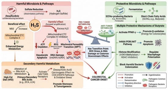
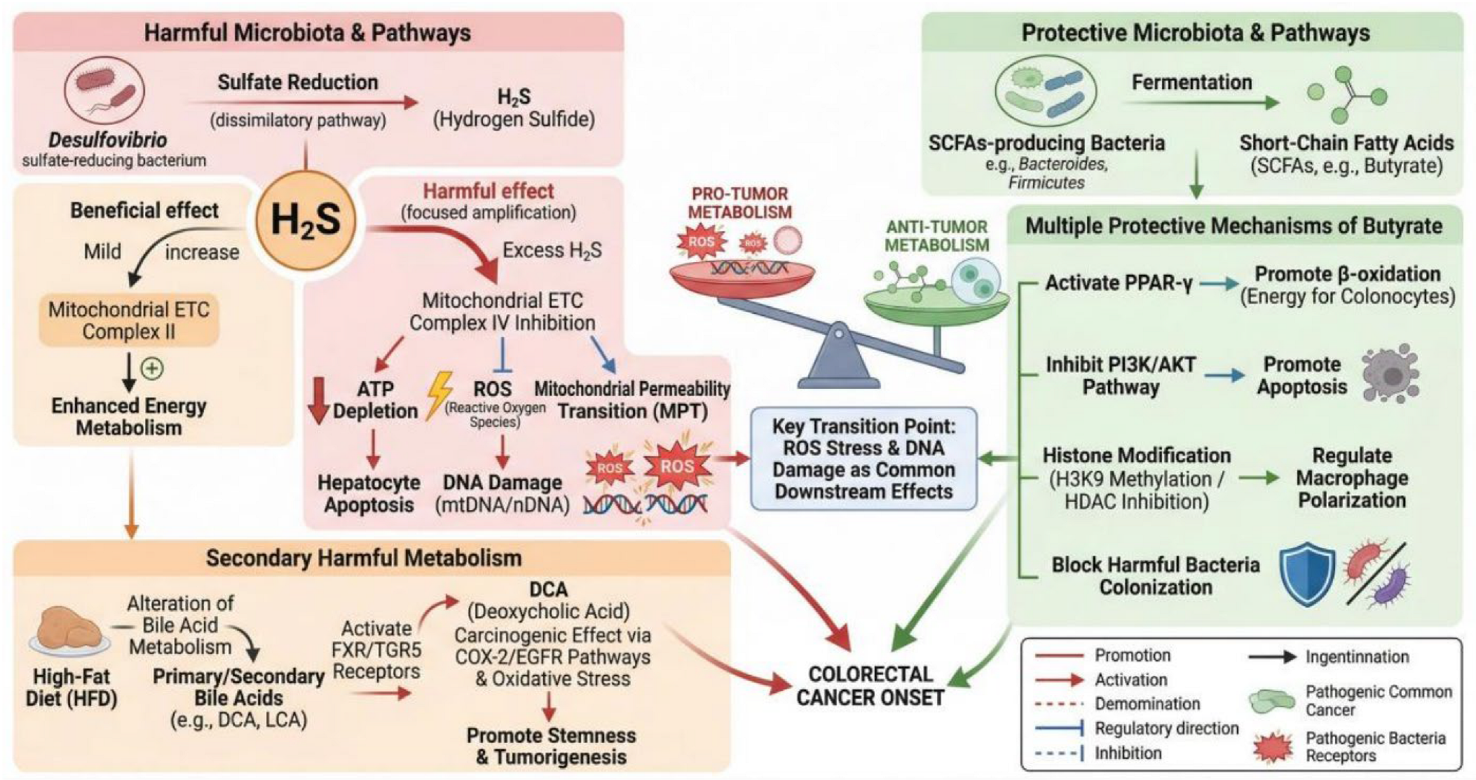
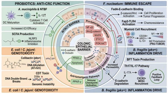
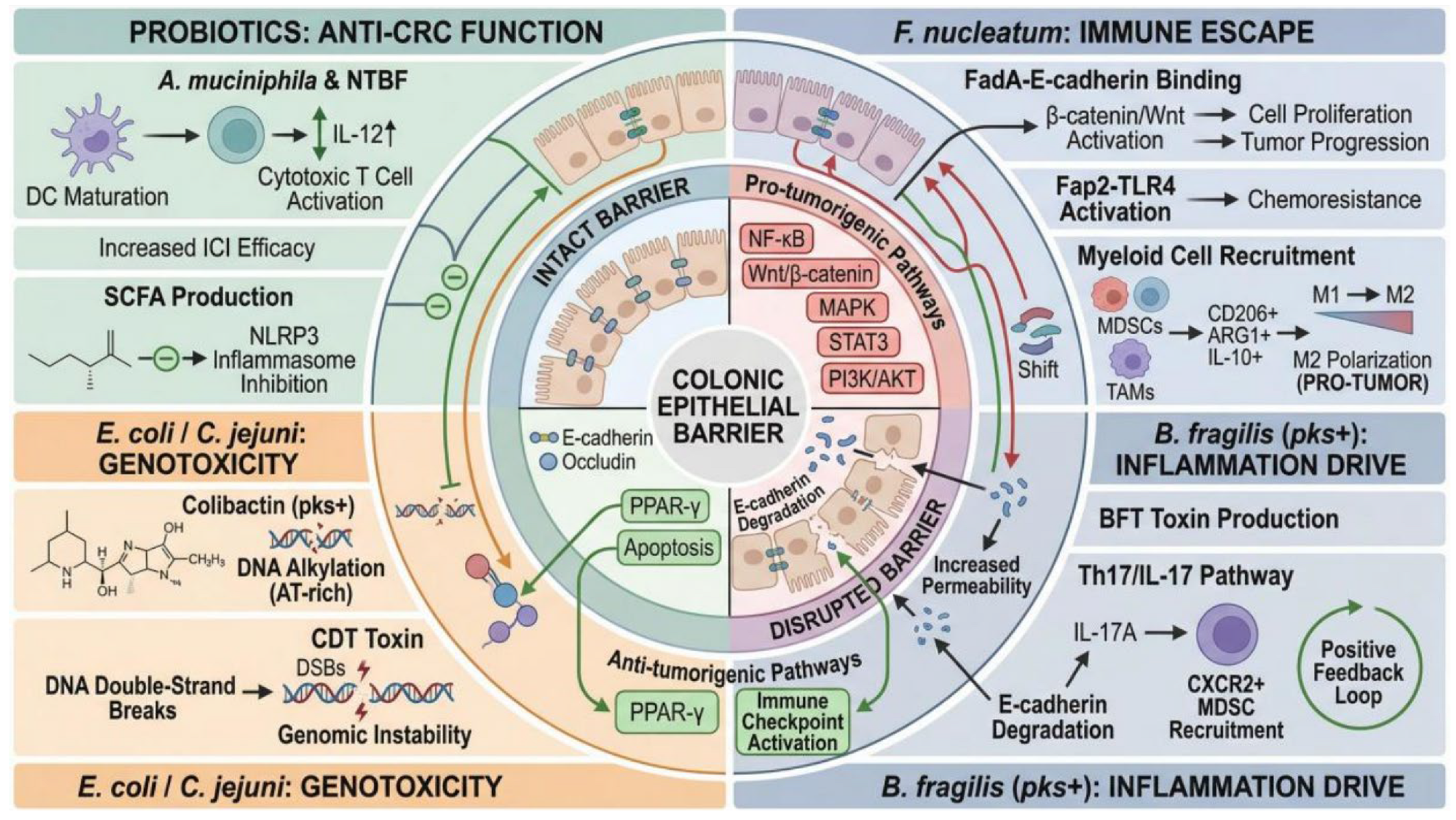

Disclaimer/Publisher’s Note: The statements, opinions and data contained in all publications are solely those of the individual author(s) and contributor(s) and not of MDPI and/or the editor(s). MDPI and/or the editor(s) disclaim responsibility for any injury to people or property resulting from any ideas, methods, instructions or products referred to in the content. |
© 2026 by the authors. Licensee MDPI, Basel, Switzerland. This article is an open access article distributed under the terms and conditions of the Creative Commons Attribution (CC BY) license.