Skip Content
You are currently on the new version of our website. Access the old version .

All Articles (748)

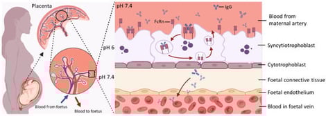
The transplacental transfer of maternal immunoglobulin G from the mother to the foetus is central for providing immunity in early life, resulting in full-term newborns having IgG repertoires and levels similar to those of their mothers. The neonatal Fc receptor is recognised as the primary transporter of IgGs across the placental epithelium. Understanding the mechanisms of transplacental antibody transfer and factors that affect them is essential in optimising maternal vaccination strategies, ultimately protecting infants from various environmental pathogens. This review first outlines the biological mechanisms governing transplacental IgG transfer, followed by a discussion of how this process may be disrupted by physiological and pathological conditions during pregnancy, including preterm birth, hypergammaglobulinemia, maternal pathogenic IgG, maternal infections, hyperglycaemia, and exposure to biological therapies. We also summarise currently available models used to study transplacental IgG transfer, highlighting existing knowledge gaps and future directions for research in this field.

6 February 2026

Key Features of Maternal–Foetal IgG Transfer via FcRn-Mediated Transcytosis. At the maternal–foetal interface, maternal IgG binds to FcRn expressed on the apical surface of SCT. The IgG–FcRn complex is internalised into endosomes, where acidic pH stabilises their interaction. This complex is then trafficked across the cell to the basal membrane. Upon exposure to physiological pH, IgG is released into the foetal stroma and subsequently enters the foetal circulation. After release, FcRn is recycled back to the apical membrane for reuse, enabling continuous transcytosis of maternal IgG during gestation. Created in https://www.BioRender.com.

Background/Objectives: Allergic features of anisakiasis, caused by ingestion of third-stage larvae of Anisakis simplex via raw or undercooked fish, manifest clinically as acute gastroallergic anisakiasis (GAA) or chronic urticaria with Anisakis sensitization (CU+). Differentiating these clinical phenotypes remains challenging. This study aimed to evaluate the maturation and avidity of specific antibodies (IgE, IgG4, IgG, and IgA) as biomarkers for discriminating between acute and chronic forms of anisakiasis. Methods: A prospective cohort of 65 patients from Madrid, Spain, was classified into three groups: GAA (n = 22), CU+ (n = 22), and chronic urticaria without sensitization (CU−, n = 21). Serum samples were analyzed for antigen-specific immunoglobulins using ELISA and Western blot. Avidity indices (AIs) were quantified through urea dissociation assays. Statistical comparisons and correlation analyses were performed to associate antibody avidity with clinical phenotype and demographic variables. Results: GAA patients exhibited significantly lower IgE avidity indices compared to CU+ individuals (mean AI: 79.9% vs. 88.5%), indicating a less mature IgE response during acute infection. Conversely, IgG4 and IgG avidity were elevated in GAA relative to CU+, reflecting an active but transient immune response. IgA antibodies were detected in both groups, although avidity differences lacked discriminatory capacity. No sex- or age-related differences in antibody avidity were observed. Longitudinal follow-up of GAA patients demonstrated an increase in IgE avidity over time. Conclusions: Quantitative assessment of antibody avidity, particularly for IgE and IgG4, enhances understanding of A. simplex immunopathogenesis and serves as a valuable biomarker for distinguishing acute from chronic clinical presentations. These findings support the use of avidity indices in the diagnosis, staging, and clinical management of anisakiasis.

6 February 2026

IgG4 immunorecognition of Anisakis simplex proteins. Frequency of IgG4-mediated band recognition in GAA, CU+, and CU− patient sera against CE antigen of A. simplex.

Background: We are developing cytotoxic immunoconjugates (CICs) to eliminate HIV-infected cells. We investigated the efficacy and kinetics of killing by different forms of CICs targeted by the same monoclonal antibody (mAb), an immunotoxin (IT), antibody-drug conjugate (ADC), and radioimmunoconjugate (RIC). Methods: We compared in vitro effects of CICs made by conjugating anti-gp41 mAb 7B2 to deglycosylated ricin A chain (7B2-dgA), the anthracycline derivative PNU-159682 (7B2-PNU), or the α-emitting isotope actinium-225 (7B2-225Ac). Kinetic analyses of cell growth were performed measuring electrical impedance every 15 min over a 7-day period using cells stably expressing the HIV envelope and Env-negative parent cells. Results: 7B2-dgA and 7B2-225Ac were more potent and acted more rapidly to kill cells than 7B2-PNU. Both the 7B2-PNU and 7B2-225Ac induced bystander-cell killing, whereas the IT did not and consequently allowed the outgrowth of Env-negative cells. Low dose or brief exposure to 7B2-PNU resulted in an increased rate of cell growth. Conclusions: An IT, ADC, and RIC showed substantial differences in the degree of specific toxicity, kinetics, and mechanisms of killing. The results of this side-by-side comparison have implications for the development of CICs to treat HIV, as well as other conditions.

28 January 2026

Env expression and cytotoxicity of CICs on 92UG and the parental 293T cells. (A) Flow cytometric analysis of cell surface Env. Cells were stained with human anti-gp120 mAb VRC01, then with anti-human IgG secondary Ab labeled with phycoerythrin (PE). The vertical axis shows side scatter on a linear scale. The horizontal axis on the left-most panel is forward scatter on a linear scale. This panel also shows the gate used for analysis of fluorescence. The remaining panels show log fluorescence intensity on the horizontal axis. The percentage of cells within the defined rectangle, i.e., Env+, is indicated. Specificity controls include unstained and only secondary Ab on 92UG cells and fully stained Env− 293T cells. (B) Cytotoxicity of CICs. Cells were incubated for three days in the presence of the indicated concentration of CIC and 500 ng/mL CD4-IgG2. Cell viability was quantified as MTS dye reduction over the final 3 h of culture. Note the 100× difference in CIC concentrations tested on 92UG versus 293T. Results show the mean and SEM of triplicate cultures. If no error bars are visible, they are smaller than the symbol.

Antibody Screening and Binding Prediction Analysis Targeting Stx2

  • Jilei Wu,
  • Chenghua Liu and
  • Jijun Yu
  • + 9 authors

Background: Shiga toxin (Stx), produced by enterohemorrhagic Escherichia coli (EHEC), is a highly potent exotoxin responsible for severe complications such as hemolytic uremic syndrome (HUS). Among its isoforms, Stx2 exhibits stronger cytotoxicity and poses greater clinical risk, yet no effective therapy currently exists. Methods: In this study, two human monoclonal antibodies, YG12-1 and YG12-2, were identified from a phage display library and systematically characterized using an integrated modeling–validation workflow. Results: Structural modeling with ImmuneBuilder and Rosetta revealed that YG12-2 possessed a longer CDRH3 topology, more short-range hydrogen bonds, and stronger electrostatic complementarity, corresponding to lower binding energy and higher apparent affinity in ELISA and SPR. Although YG12-2 had a better affinity, YG12-1 shows better protective activity in a murine model of acute peritoneal infection. This paradox highlights a non-linear relationship between structural affinity and biological efficacy, emphasizing the importance of functional epitope accessibility and pharmacokinetic behavior in determining neutralization outcomes. Conlusions: Overall, these results indicated that targeting Stx2 with YG12-1 and YG12-2 could serve as a promising protective strategy against E. coli O157:H7 infection.

27 January 2026

Schematic of antibody screening, prediction, and activity evaluation. (A) Screening and Preparation: Antibody clones were isolated by phage display panning, expressed in 293E cells, and purified for further analysis; (B,C) Binding activity of mAbs in vitro: (B) Binding affinity was assessed by ELISA. (C) Binding affinity was assessed by SPR. (D) Neutralization activity of mAbs in vivo. (E) Structural prediction and docking were performed using ImmuneBuilder (v1.1.1) and GRAMM (Web Sever); interface hydrogen-bond contacts were analyzed in PyMOL (v3.1), mutational effects (ΔΔG) were predicted using SAAMBE-3D (Web Sever), and interaction energies were finally calculated using Rosetta.

News & Conferences

Issues

Open for Submission

Editor's Choice

Get Alerted

Add your email address to receive forthcoming issues of this journal.

XFacebookLinkedIn
Antibodies - ISSN 2073-4468