Next Issue
Volume 13, October-2
Previous Issue
Volume 13, September-2


Cells, Volume 13, Issue 19 (October-1 2024) – 74 articles

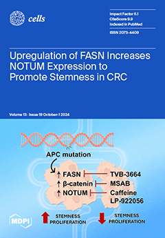
Cover Story (view full-size image): Cells (ISSN 2073-4409) is an international, peer-reviewed, open access journal which provides an advanced forum for studies related to cell biology, molecular biology and biophysics. It publishes reviews, research articles, communications and technical notes. Our aim is to encourage scientists to publish their experimental and theoretical results in as much detail as possible. The Spanish Society for Biochemistry and Molecular Biology (SEBBM), Nordic Autophagy Society (NAS), Spanish Society of Hematology and Hemotherapy (SEHH) and Society for Regenerative Medicine (Russian Federation) (RPO) are affiliated with Cells and their members receive discounts on the article processing charges.
  • Issues are regarded as officially published after their release is announced to the table of contents alert mailing list.
  • You may sign up for e-mail alerts to receive table of contents of newly released issues.
  • PDF is the official format for papers published in both, html and pdf forms. To view the papers in pdf format, click on the "PDF Full-text" link, and use the free Adobe Reader to open them.
Order results
Result details
Section
Select all
Export citation of selected articles as:
9 pages, 3790 KiB  
Communication
The Endogenous Expression of BMI1 in Adult Human Eyes
by Zhongyang Lu, Maria G. Morales, Shufeng Liu and Hema L. Ramkumar
Cells 2024, 13(19), 1672; https://doi.org/10.3390/cells13191672 - 9 Oct 2024
Viewed by 570
Abstract
BMI1, also known as B lymphoma Mo-MLV insertion region 1, is a protein in the Polycomb group that is implicated in various cellular processes, including stem cell self-renewal and the regulation of cellular senescence. BMI1 plays a role in the regulation of retinal [...] Read more.
BMI1, also known as B lymphoma Mo-MLV insertion region 1, is a protein in the Polycomb group that is implicated in various cellular processes, including stem cell self-renewal and the regulation of cellular senescence. BMI1 plays a role in the regulation of retinal progenitor cells and the renewal of adult neuronal cells. However, the presence, location, and quantification of BMI1 in the adult human eye have never previously been reported. In this study, we collected 45 frozen globes from eye banks, and ocular tissues were dissected. Protein was quantified by utilizing a custom electrochemiluminescence (ECL) assay developed to quantify the BMI1 protein. BMI1 was found in all ocular tissues at the following levels: the retina (1483.6 ± 191.7 pg/mL) and the RPE (296.4 ± 78.1 pg/mL). BMI1 expression was noted ubiquitously in the GCL (ganglion cell layer), the INL (inner nuclear layer), the ONL (outer nuclear layer), and the RPE (retinal pigment epithelium) via immunofluorescence, with higher levels in the inner than in the outer retinal layers and the RPE. These data confirm that BMI1 is expressed in the human retina. Further studies will illuminate the role that BMI1 plays in ocular cells. BMI1 levels are lower in aged retinas, possibly reflecting changes in retinal somatic and stem cell maintenance and disease susceptibility. Full article
(This article belongs to the Special Issue Retinal Pigment Epithelial Cells in Age-Related Macular Degeneration)
Show Figures

Figure 1

16 pages, 5079 KiB  
Article
Optogenetic Control of the Mitochondrial Protein Import in Mammalian Cells
by Lukas F. J. Althoff, Markus M. Kramer, Benjamin Bührer, Denise Gaspar and Gerald Radziwill
Cells 2024, 13(19), 1671; https://doi.org/10.3390/cells13191671 - 9 Oct 2024
Viewed by 988
Abstract
Mitochondria provide cells with energy and regulate the cellular metabolism. Almost all mitochondrial proteins are nuclear-encoded, translated on ribosomes in the cytoplasm, and subsequently transferred to the different subcellular compartments of mitochondria. Here, we developed OptoMitoImport, an optogenetic tool to control the import [...] Read more.
Mitochondria provide cells with energy and regulate the cellular metabolism. Almost all mitochondrial proteins are nuclear-encoded, translated on ribosomes in the cytoplasm, and subsequently transferred to the different subcellular compartments of mitochondria. Here, we developed OptoMitoImport, an optogenetic tool to control the import of proteins into the mitochondrial matrix via the presequence pathway on demand. OptoMitoImport is based on a two-step process: first, light-induced cleavage by a TEV protease cuts off a plasma membrane-anchored fusion construct in close proximity to a mitochondrial targeting sequence; second, the mitochondrial targeting sequence preceding the protein of interest recruits to the outer mitochondrial membrane and imports the protein fused to it into mitochondria. Upon reaching the mitochondrial matrix, the matrix processing peptidase cuts off the mitochondrial targeting sequence and releases the protein of interest. OptoMitoImport is available as a two-plasmid system as well as a P2A peptide or IRES sequence-based bicistronic system. Fluorescence studies demonstrate the release of the plasma membrane-anchored protein of interest through light-induced TEV protease cleavage and its localization to mitochondria. Cell fractionation experiments confirm the presence of the peptidase-cleaved protein of interest in the mitochondrial fraction. The processed product is protected from proteinase K treatment. Depletion of the membrane potential across the inner mitochondria membrane prevents the mitochondrial protein import, indicating an import of the protein of interest by the presequence pathway. These data demonstrate the functionality of OptoMitoImport as a generic system with which to control the post-translational mitochondrial import of proteins via the presequence pathway. Full article
(This article belongs to the Section Mitochondria)
Show Figures

Graphical abstract

14 pages, 2545 KiB  
Article
Investigating the p21 Ubiquitin-Independent Degron Reveals a Dual Degron Module Regulating p21 Degradation and Function
by Marianna Riutin, Pnina Erez, Julia Adler, Assaf Biran, Nadav Myers and Yosef Shaul
Cells 2024, 13(19), 1670; https://doi.org/10.3390/cells13191670 - 9 Oct 2024
Viewed by 656
Abstract
A group of intrinsically disordered proteins (IDPs) are subject to 20S proteasomal degradation in a ubiquitin-independent manner. Recently, we have reported that many IDPs/IDRs are targeted to the 20S proteasome via interaction with the C-terminus of the PSMA3 subunit, termed the PSMA3 Trapper. [...] Read more.
A group of intrinsically disordered proteins (IDPs) are subject to 20S proteasomal degradation in a ubiquitin-independent manner. Recently, we have reported that many IDPs/IDRs are targeted to the 20S proteasome via interaction with the C-terminus of the PSMA3 subunit, termed the PSMA3 Trapper. In this study, we investigated the biological significance of the IDP–Trapper interaction using the IDP p21. Using a split luciferase reporter assay and conducting detailed p21 mutagenesis, we first identified the p21 RRLIF box, localized at the C-terminus, as mediating the Trapper interaction in cells. To demonstrate the role of this box in p21 degradation, we edited the genome of HEK293 and HeLa cell lines using a CRISPR strategy. We found that the p21 half-life increased in cells with either a deleted or mutated p21 RRLIF box. The edited cell lines displayed an aberrant cell cycle pattern under normal conditions and in response to DNA damage. Remarkably, these cells highly expressed senescence hallmark genes in response to DNA damage, highlighting that the increased p21 half-life, not its actual level, regulates senescence. Our findings suggest that the p21 RRLIF box, which mediates interactions with the PSMA3 Trapper, acts as a ubiquitin-independent degron. This degron is positioned adjacent to the previously identified ubiquitin-dependent degron, forming a dual degron module that functionally regulates p21 degradation and its physiological outcomes. Full article
Show Figures

Figure 1

12 pages, 5174 KiB  
Article
Cancer-Associated Fibroblast Subtypes Reveal Distinct Gene Signatures in the Tumor Immune Microenvironment of Vestibular Schwannoma
by Ji-Yong Sung and Jung Woo Lee
Cells 2024, 13(19), 1669; https://doi.org/10.3390/cells13191669 - 9 Oct 2024
Viewed by 731
Abstract
Cancer-associated fibroblast (CAF) composition within the same organ varies across different cancer subtypes. Distinct CAF subtypes exhibit unique features due to interactions with immune cells and the tumor microenvironment. However, data on CAF subtypes in individuals with vestibular schwannoma (VS) are lacking. Therefore, [...] Read more.
Cancer-associated fibroblast (CAF) composition within the same organ varies across different cancer subtypes. Distinct CAF subtypes exhibit unique features due to interactions with immune cells and the tumor microenvironment. However, data on CAF subtypes in individuals with vestibular schwannoma (VS) are lacking. Therefore, we aimed to distinguish CAF subtypes at the single-cell level, investigate how stem-like CAF characteristics influence the tumor immune microenvironment, and identify CAF subtype-specific metabolic reprogramming pathways that contribute to tumor development. Data were analyzed from three patients with VS, encompassing 33,081 single cells, one bulk transcriptome cohort, and The Cancer Genome Atlas Pan-Cancer database (RNA sequencing and clinical data). Our findings revealed that antigen-presenting CAFs are linked to substantially heightened immune activity, supported by metabolic reprogramming, which differs from tumorigenesis. High expression of the stem-like CAF gene signature correlated with poor prognosis in low-grade gliomas within the pan-cancer database. This is the first study to classify CAF subtypes in VS patients and identify a therapeutic vulnerability biomarker by developing a stem-like CAF gene signature. Personalized treatments tailored to individual patients show promise in advancing precision medicine. Full article
Show Figures

Figure 1

21 pages, 3798 KiB  
Article
Identification of hsa_circ_0018905 as a New Potential Biomarker for Multiple Sclerosis
by Valeria Lodde, Ignazio Roberto Zarbo, Gabriele Farina, Aurora Masia, Paolo Solla, Ilaria Campesi, Giuseppe Delogu, Maria Rosaria Muroni, Dimitrios Tsitsipatis, Myriam Gorospe, Matteo Floris and Maria Laura Idda
Cells 2024, 13(19), 1668; https://doi.org/10.3390/cells13191668 - 9 Oct 2024
Viewed by 732
Abstract
Multiple sclerosis (MS) is a demyelinating autoimmune disease characterized by early onset, for which the interaction of genetic and environmental factors is crucial. Dysregulation of the immune system as well as myelinization-de-myelinization has been shown to correlate with changes in RNA, including non-coding [...] Read more.
Multiple sclerosis (MS) is a demyelinating autoimmune disease characterized by early onset, for which the interaction of genetic and environmental factors is crucial. Dysregulation of the immune system as well as myelinization-de-myelinization has been shown to correlate with changes in RNA, including non-coding RNAs. Recently, circular RNAs (circRNAs) have emerged as a key player in the complex network of gene dysregulation associated with MS. Despite several efforts, the mechanisms driving circRNA regulation and dysregulation in MS still need to be properly elucidated. Here, we explore the panorama of circRNA expression in PBMCs purified from five newly diagnosed MS patients and five healthy controls (HCs) using the Arraystar Human circRNAs microarray. Experimental validation was then carried out in a validation cohort, and a possible correlation with disease severity was tested. We identified 64 differentially expressed circRNAs, 53 of which were downregulated in PBMCs purified from MS compared to the HCs. The discovery dataset was subsequently validated using qRT-PCR with an independent cohort of 20 RRMS patients and 20 HCs. We validated seven circRNAs differentially expressed in the RRMS group versus the HC group. hsa_circ_0000518, hsa_circ_0000517, hsa_circ_0000514, and hsa_circ_0000511 were significantly upregulated in the MS group, while hsa_circ_0018905, hsa_circ_0048764, and hsa_circ_0003445 were significantly downregulated; Among them, the expression level of hsa_circ_0018905 was significantly decreased in patients showing a higher level of disability and in progressive forms of MS. We described the circRNAs expression profile of PBMCs in newly diagnosed MS patients and proposed hsa_circ_0018905 as potential MS biomarker. Full article
(This article belongs to the Special Issue Cellular and Molecular Mechanisms in Immune Regulation)
Show Figures

Figure 1

24 pages, 11478 KiB  
Article
Employing Multi-Omics Analyses to Understand Changes during Kidney Development in Perinatal Interleukin-6 Animal Model
by Ganesh Panzade, Tarak Srivastava, Daniel P. Heruth, Mohammad H. Rezaiekhaligh, Jianping Zhou, Zhen Lyu, Mukut Sharma and Trupti Joshi
Cells 2024, 13(19), 1667; https://doi.org/10.3390/cells13191667 - 9 Oct 2024
Viewed by 826
Abstract
Chronic kidney disease (CKD) is a leading cause of morbidity and mortality globally. Maternal obesity during pregnancy is linked to systemic inflammation and elevated levels of the pro-inflammatory cytokine interleukin-6 (IL-6). In our previous work, we demonstrated that increased maternal IL-6 during gestation [...] Read more.
Chronic kidney disease (CKD) is a leading cause of morbidity and mortality globally. Maternal obesity during pregnancy is linked to systemic inflammation and elevated levels of the pro-inflammatory cytokine interleukin-6 (IL-6). In our previous work, we demonstrated that increased maternal IL-6 during gestation impacts intrauterine development in mice. We hypothesized that IL-6-induced inflammation alters gene expression in the developing fetus. To test this, pregnant mice were administered IL-6 or saline during mid-gestation. Newborn mouse kidneys were analyzed using mRNA-seq, miRNA-seq and whole-genome bisulfite-seq (WGBS). A multi-omics approach was employed to quantify mRNA gene expression, miRNA expression and DNA methylation, using advanced bioinformatics and data integration techniques. Our analysis identified 19 key genes present in multiple omics datasets, regulated by epigenetics and miRNAs. We constructed a regulatory network for these genes, revealing disruptions in pathways such as Mannose type O-glycan biosynthesis, the cell cycle, apoptosis and FoxO signaling. Notably, the Atp7b gene was regulated by DNA methylation and miR-223 targeting, whereas the Man2a1 gene was controlled by DNA methylation affecting energy metabolism. These findings suggest that these genes may play a role in fetal programming, potentially leading to CKD later in life due to gestational inflammation. Full article
Show Figures

Figure 1

23 pages, 1950 KiB  
Review
Beyond Cancer Cells: How the Tumor Microenvironment Drives Cancer Progression
by Hussein Sabit, Borros Arneth, Shaimaa Abdel-Ghany, Engy F. Madyan, Ashraf H. Ghaleb, Periasamy Selvaraj, Dong M. Shin, Ramireddy Bommireddy and Ahmed Elhashash
Cells 2024, 13(19), 1666; https://doi.org/10.3390/cells13191666 - 9 Oct 2024
Viewed by 1766
Abstract
Liver cancer represents a substantial global health challenge, contributing significantly to worldwide morbidity and mortality. It has long been understood that tumors are not composed solely of cancerous cells, but also include a variety of normal cells within their structure. These tumor-associated normal [...] Read more.
Liver cancer represents a substantial global health challenge, contributing significantly to worldwide morbidity and mortality. It has long been understood that tumors are not composed solely of cancerous cells, but also include a variety of normal cells within their structure. These tumor-associated normal cells encompass vascular endothelial cells, fibroblasts, and various inflammatory cells, including neutrophils, monocytes, macrophages, mast cells, eosinophils, and lymphocytes. Additionally, tumor cells engage in complex interactions with stromal cells and elements of the extracellular matrix (ECM). Initially, the components of what is now known as the tumor microenvironment (TME) were thought to be passive bystanders in the processes of tumor proliferation and local invasion. However, recent research has significantly advanced our understanding of the TME’s active role in tumor growth and metastasis. Tumor progression is now known to be driven by an intricate imbalance of positive and negative regulatory signals, primarily influenced by specific growth factors produced by both inflammatory and neoplastic cells. This review article explores the latest developments and future directions in understanding how the TME modulates liver cancer, with the aim of informing the design of novel therapies that target critical components of the TME. Full article
(This article belongs to the Section Cell Microenvironment)
Show Figures

Figure 1

20 pages, 4665 KiB  
Article
Human Umbilical Cord-Mesenchymal Stem Cells Promote Extracellular Matrix Remodeling in Microglia
by Marta Tiffany Lombardo, Martina Gabrielli, Florence Julien-Marsollier, Valérie Faivre, Tifenn Le Charpentier, Cindy Bokobza, Deborah D’Aliberti, Nicola Pelizzi, Camilla Halimi, Silvia Spinelli, Juliette Van Steenwinckel, Elisabetta A. M. Verderio, Pierre Gressens, Rocco Piazza and Claudia Verderio
Cells 2024, 13(19), 1665; https://doi.org/10.3390/cells13191665 - 9 Oct 2024
Viewed by 978
Abstract
Human mesenchymal stem cells modulate the immune response and are good candidates for cell therapy in neuroinflammatory brain disorders affecting both adult and premature infants. Recent evidence indicates that through their secretome, mesenchymal stem cells direct microglia, brain-resident immune cells, toward pro-regenerative functions, [...] Read more.
Human mesenchymal stem cells modulate the immune response and are good candidates for cell therapy in neuroinflammatory brain disorders affecting both adult and premature infants. Recent evidence indicates that through their secretome, mesenchymal stem cells direct microglia, brain-resident immune cells, toward pro-regenerative functions, but the mechanisms underlying microglial phenotypic transition are still under investigation. Using an in vitro coculture approach combined with transcriptomic analysis, we identified the extracellular matrix as the most relevant pathway altered by the human mesenchymal stem cell secretome in the response of microglia to inflammatory cytokines. We confirmed extracellular matrix remodeling in microglia exposed to the mesenchymal stem cell secretome via immunofluorescence analysis of the matrix component fibronectin and the extracellular crosslinking enzyme transglutaminase-2. Furthermore, an analysis of hallmark microglial functions revealed that changes in the extracellular matrix enhance ruffle formation by microglia and cell motility. These findings point to extracellular matrix changes, associated plasma membrane remodeling, and enhanced microglial migration as novel mechanisms by which mesenchymal stem cells contribute to the pro-regenerative microglial transition. Full article
(This article belongs to the Special Issue Perinatal Brain Injury—from Pathophysiology to Therapy)
Show Figures

Graphical abstract

18 pages, 921 KiB  
Review
Oncological Aspects of Lysosomal Storage Diseases
by Agnieszka Ługowska
Cells 2024, 13(19), 1664; https://doi.org/10.3390/cells13191664 - 8 Oct 2024
Viewed by 628
Abstract
Lysosomal storage diseases (LSDs) are caused by the deficient activity of a lysosomal hydrolase or the lack of a functional membrane protein, transporter, activator, or other protein. Lysosomal enzymes break down macromolecular compounds, which contribute to metabolic homeostasis. Stored, undegraded materials have multiple [...] Read more.
Lysosomal storage diseases (LSDs) are caused by the deficient activity of a lysosomal hydrolase or the lack of a functional membrane protein, transporter, activator, or other protein. Lysosomal enzymes break down macromolecular compounds, which contribute to metabolic homeostasis. Stored, undegraded materials have multiple effects on cells that lead to the activation of autophagy and apoptosis, including the toxic effects of lyso-lipids, the disruption of intracellular Ca2+ ion homeostasis, the secondary storage of macromolecular compounds, the activation of signal transduction, apoptosis, inflammatory processes, deficiencies of intermediate compounds, and many other pathways. Clinical observations have shown that carriers of potentially pathogenic variants in LSD-associated genes and patients affected with some LSDs are at a higher risk of cancer, although the results of studies on the frequency of oncological diseases in LSD patients are controversial. Cancer is found in individuals affected with Gaucher disease, Fabry disease, Niemann-Pick type A and B diseases, alfa-mannosidosis, and sialidosis. Increased cancer prevalence has also been reported in carriers of a potentially pathogenic variant of an LSD gene, namely CLN3, SGSH, GUSB, NEU1, and, to a lesser extent, in other genes. In this review, LSDs in which oncological events can be observed are described. Full article
(This article belongs to the Collection The Lysosome in Cancer: From Pathogenesis to Therapy)
Show Figures

Figure 1

17 pages, 4237 KiB  
Article
Upregulation of Fatty Acid Synthase Increases Activity of β-Catenin and Expression of NOTUM to Enhance Stem-like Properties of Colorectal Cancer Cells
by Courtney O. Kelson, Josiane Weber Tessmann, Mariah E. Geisen, Daheng He, Chi Wang, Tianyan Gao, B. Mark Evers and Yekaterina Y. Zaytseva
Cells 2024, 13(19), 1663; https://doi.org/10.3390/cells13191663 - 8 Oct 2024
Viewed by 918
Abstract
Dysregulated fatty acid metabolism is an attractive therapeutic target for colorectal cancer (CRC). We previously reported that fatty acid synthase (FASN), a key enzyme of de novo synthesis, promotes the initiation and progression of CRC. However, the mechanisms of how upregulation of FASN [...] Read more.
Dysregulated fatty acid metabolism is an attractive therapeutic target for colorectal cancer (CRC). We previously reported that fatty acid synthase (FASN), a key enzyme of de novo synthesis, promotes the initiation and progression of CRC. However, the mechanisms of how upregulation of FASN promotes the initiation and progression of CRC are not completely understood. Here, using Apc/VillinCre and ApcMin mouse models, we show that upregulation of FASN is associated with an increase in activity of β-catenin and expression of multiple stem cell markers, including Notum. Genetic and pharmacological downregulation of FASN in mouse adenoma organoids decreases the activation of β-catenin and expression of Notum and significantly inhibits organoid formation and growth. Consistently, we demonstrate that NOTUM is highly expressed in human CRC and its expression positively correlates with the expression of FASN in tumor tissues. Utilizing overexpression and shRNA-mediated knockdown of FASN, we demonstrate that upregulation of FASN increases β-catenin transcriptional activity, NOTUM expression and secretion, and enhances stem-like properties of human CRC cells. Pharmacological inhibition of NOTUM decreases adenoma organoids growth and proliferation of cancer cells. In summary, upregulation of FASN enhances β-catenin signaling, increases NOTUM expression and stem-like properties of CRC cells, thus suggesting that targeting FASN upstream of the β-catenin/NOTUM axis may be an effective preventative therapeutic strategy for CRC. Full article
Show Figures

Figure 1

22 pages, 1312 KiB  
Review
Inflammatory Trajectory of Type 2 Diabetes: Novel Opportunities for Early and Late Treatment
by Valeria Pellegrini, Rosalba La Grotta, Francesca Carreras, Angelica Giuliani, Jacopo Sabbatinelli, Fabiola Olivieri, Cesare Celeste Berra, Antonio Ceriello and Francesco Prattichizzo
Cells 2024, 13(19), 1662; https://doi.org/10.3390/cells13191662 - 8 Oct 2024
Cited by 1 | Viewed by 1009
Abstract
Low-grade inflammation (LGI) represents a key driver of type 2 diabetes (T2D) and its associated cardiovascular diseases (CVDs). Indeed, inflammatory markers such as hs-CRP and IL-6 predict the development of T2D and its complications, suggesting that LGI already increases before T2D diagnosis and [...] Read more.
Low-grade inflammation (LGI) represents a key driver of type 2 diabetes (T2D) and its associated cardiovascular diseases (CVDs). Indeed, inflammatory markers such as hs-CRP and IL-6 predict the development of T2D and its complications, suggesting that LGI already increases before T2D diagnosis and remains elevated even after treatment. Overnutrition, unhealthy diets, physical inactivity, obesity, and aging are all recognized triggers of LGI, promoting insulin resistance and sustaining the pathogenesis of T2D. Once developed, and even before frank appearance, people with T2D undergo a pathological metabolic remodeling, with an alteration of multiple CVD risk factors, i.e., glycemia, lipids, blood pressure, and renal function. In turn, such variables foster a range of inflammatory pathways and mechanisms, e.g., immune cell stimulation, the accrual of senescent cells, long-lasting epigenetic changes, and trained immunity, which are held to chronically fuel LGI at the systemic and tissue levels. Targeting of CVD risk factors partially ameliorates LGI. However, some long-lasting inflammatory pathways are unaffected by common therapies, and LGI burden is still increased in many T2D patients, a phenomenon possibly underlying the residual inflammatory risk (i.e., having hs-CRP > 2 mg/dL despite optimal LDL cholesterol control). On the other hand, selected disease-modifying drugs, e.g., GLP-1RA, seem to also act on the pathogenesis of T2D, curbing the inflammatory trajectory of the disease and possibly preventing it if introduced early. In addition, selected trials demonstrated the potential of canonical anti-inflammatory therapies in reducing the rate of CVDs in patients with this condition or at high risk for it, many of whom had T2D. Since colchicine, an inhibitor of immune cell activation, is now approved for the prevention of CVDs, it might be worth exploring a possible therapeutic paradigm to identify subjects with T2D and an increased LGI burden to treat them with this drug. Upcoming studies will reveal whether disease-modifying drugs reverse early T2D by suppressing sources of LGI and whether colchicine has a broad benefit in people with this condition. Full article
(This article belongs to the Special Issue Inflammation in Target Organs)
Show Figures

Figure 1

20 pages, 5072 KiB  
Article
Protective Effects of Keratinocyte-Derived GCSF and CCL20 on UVB-Induced Melanocyte Damage
by Saowanee Jeayeng, Malinee Saelim, Phetthinee Muanjumpon, Pongsakorn Buraphat, Potjanee Kanchanapiboon, Somponnat Sampattavanich and Uraiwan Panich
Cells 2024, 13(19), 1661; https://doi.org/10.3390/cells13191661 - 8 Oct 2024
Viewed by 916
Abstract
The skin microenvironment created by keratinocytes (KC) influences the stress responses of melanocytes (MC) to UVB insults. This study employed RNA sequencing analysis as well as in vitro and in vivo models to elucidate the underlying mechanisms. Our RNA-Seq analysis revealed a statistically [...] Read more.
The skin microenvironment created by keratinocytes (KC) influences the stress responses of melanocytes (MC) to UVB insults. This study employed RNA sequencing analysis as well as in vitro and in vivo models to elucidate the underlying mechanisms. Our RNA-Seq analysis revealed a statistically significant upregulation of GCSF and CCL20 genes in UVB-irradiated KC, correlating with the protective effects of KC on MC responses to UVB exposure. Recombinant GCSF and CCL20 exhibited the most pronounced modulation of UVB-induced MC responses. These effects included the attenuation of apoptosis and reduction of ROS formation, along with the upregulation of tyrosinase and tyrosinase-related protein-1, which are involved in the melanogenic pathway. ELISA was also used to confirm that UVB could induce the secretion of GCSF and CCL20 from KC. A similar correlation between GCSF and CCL20 expression in KC and tyrosinase levels in MC was observed in UVB-irradiated mouse skin. Our study provides novel insights into the protective role of GCSF and CCL20 in the paracrine effects of KC on UVB-induced MC damage through the modulation of stress response pathways, the MITF-tyrosinase axis, and the regulation of p53. These findings have implications for the development of pharmacological strategies targeting KC-derived paracrine factors for the prevention of skin photodamage. Full article
Show Figures

Graphical abstract

3 pages, 2428 KiB  
Correction
Correction: Ahmed et al. The Barley S-Adenosylmethionine Synthetase 3 Gene HvSAMS3 Positively Regulates the Tolerance to Combined Drought and Salinity Stress in Tibetan Wild Barley. Cells 2020, 9, 1530
by Imrul Mosaddek Ahmed, Umme Aktari Nadira, Cheng-Wei Qiu, Fangbin Cao, Zhong-Hua Chen, Eva Vincze and Feibo Wu
Cells 2024, 13(19), 1660; https://doi.org/10.3390/cells13191660 - 8 Oct 2024
Viewed by 440
Abstract
In the original publication [...] Full article
(This article belongs to the Special Issue Plant Polyamines in Plant Stress Tolerance)
Show Figures

Figure 2

18 pages, 2518 KiB  
Article
RhoBTB3 Functions as a Novel Regulator of Autophagy by Suppressing AMBRA1 Stability
by Kyungho Kim, Dong-Gun Kim and Youn-Jae Kim
Cells 2024, 13(19), 1659; https://doi.org/10.3390/cells13191659 - 7 Oct 2024
Viewed by 796
Abstract
Autophagy is essential for cell survival and cellular homeostasis under various stress conditions. Therefore, autophagy dysfunction is associated with the pathogenesis of various human diseases. We explored the regulatory role of RhoBTB3 in autophagy and its interaction with activating molecules in AMBRA1. RhoBTB3 [...] Read more.
Autophagy is essential for cell survival and cellular homeostasis under various stress conditions. Therefore, autophagy dysfunction is associated with the pathogenesis of various human diseases. We explored the regulatory role of RhoBTB3 in autophagy and its interaction with activating molecules in AMBRA1. RhoBTB3 deficiency was found to induce autophagy, while its overexpression inhibited autophagy induction. Through immunoprecipitation and mass spectrometry, AMBRA1 was identified as a substrate of RhoBTB3. The study revealed that RhoBTB3 regulates AMBRA1 stability by influencing its protein levels without affecting its mRNA levels. RhoBTB3 induced the ubiquitination of AMBRA1, leading to proteasome-mediated degradation, with the ubiquitination occurring at K45 on AMBRA1 through a K27-linked ubiquitin chain. The knockdown of AMBRA1 blocked RhoBTB3 knockdown-induced autophagy, indicating the dependency of autophagy on AMBRA1. Thus, RhoBTB3 negatively regulates autophagy by mediating AMBRA1 ubiquitination and degradation, suggesting RhoBTB3 as a potential therapeutic target for autophagy-related diseases. Full article
(This article belongs to the Section Autophagy)
Show Figures

Figure 1

16 pages, 2299 KiB  
Article
Umbilical Cord Mesenchymal Stem Cell-Derived Extracellular Vesicles as Natural Nanocarriers in the Treatment of Nephrotoxic Injury In Vitro
by Márcia Bastos Convento, Andreia Silva de Oliveira, Mirian Aparecida Boim and Fernanda Teixeira Borges
Cells 2024, 13(19), 1658; https://doi.org/10.3390/cells13191658 - 7 Oct 2024
Viewed by 821
Abstract
Umbilical cord mesenchymal stem cell-derived extracellular vesicles (UC-EVs) are valuable in nanomedicine as natural nanocarriers, carrying information molecules from their parent cells and fusing with targeted cells. miRNA-126, specific to endothelial cells and derived from these vesicles, supports vascular integrity and angiogenesis and [...] Read more.
Umbilical cord mesenchymal stem cell-derived extracellular vesicles (UC-EVs) are valuable in nanomedicine as natural nanocarriers, carrying information molecules from their parent cells and fusing with targeted cells. miRNA-126, specific to endothelial cells and derived from these vesicles, supports vascular integrity and angiogenesis and has protective effects in kidney diseases. Objective: This study investigates the delivery of miRNA-126 and anti-miRNA-126 via UC-EVs as natural nanocarriers for treating nephrotoxic injury in vitro. Method: The umbilical cord-derived mesenchymal stem cell and UC-EVs were characterized according to specific guidelines. Rat kidney proximal tubular epithelial cells (tubular cells) were exposed to nephrotoxic injury through of gentamicin and simultaneously treated with UC-EVs carrying miRNA-126 or anti-miRNA-126. Specific molecules that manage cell cycle progression, proliferation cell assays, and newly synthesized DNA and DNA damage markers were evaluated. Results: We observed significant increases in the expression of cell cycle markers, including PCNA, p53, and p21, indicating a positive cell cycle regulation with newly synthesized DNA via BrDU. The treatments reduced the expression of DNA damage marker, such as H2Ax, suggesting a lower rate of cellular damage. Conclusions: The UC-EVs, acting as natural nanocarriers of miRNA-126 and anti-miRNA-126, offer nephroprotective effects in vitro. Additionally, other components in UC-EVs, such as proteins, lipids, and various RNAs, might also contribute to these effects. Full article
(This article belongs to the Section Stem Cells)
Show Figures

Figure 1

15 pages, 2415 KiB  
Article
Adenosine Metabolism Pathway Alterations in Frontal Cortical Neurons in Schizophrenia
by Smita Sahay, Emily A. Devine, Christina F.-A. Vargas, Robert E. McCullumsmith and Sinead M. O’Donovan
Cells 2024, 13(19), 1657; https://doi.org/10.3390/cells13191657 - 6 Oct 2024
Viewed by 751
Abstract
Schizophrenia is a neuropsychiatric illness characterized by altered neurotransmission, in which adenosine, a modulator of glutamate and dopamine, plays a critical role that is relatively unexplored in the human brain. In the present study, postmortem human brain tissue from the anterior cingulate cortex [...] Read more.
Schizophrenia is a neuropsychiatric illness characterized by altered neurotransmission, in which adenosine, a modulator of glutamate and dopamine, plays a critical role that is relatively unexplored in the human brain. In the present study, postmortem human brain tissue from the anterior cingulate cortex (ACC) of individuals with schizophrenia (n = 20) and sex- and age-matched control subjects without psychiatric illness (n = 20) was obtained from the Bronx–Mount Sinai NIH Brain and Tissue Repository. Enriched populations of ACC pyramidal neurons were isolated using laser microdissection (LMD). The mRNA expression levels of six key adenosine pathway components—adenosine kinase (ADK), equilibrative nucleoside transporters 1 and 2 (ENT1 and ENT2), ectonucleoside triphosphate diphosphohydrolases 1 and 3 (ENTPD1 and ENTPD3), and ecto-5′-nucleotidase (NT5E)—were quantified using real-time PCR (qPCR) in neurons from these individuals. No significant mRNA expression differences were observed between the schizophrenia and control groups (p > 0.05). However, a significant sex difference was found in ADK mRNA expression, with higher levels in male compared with female subjects (Mann–Whitney U = 86; p < 0.05), a finding significantly driven by disease (t(17) = 3.289; p < 0.05). Correlation analyses also demonstrated significant associations (n = 12) between the expression of several adenosine pathway components (p < 0.05). In our dementia severity analysis, ENTPD1 mRNA expression was significantly higher in males in the “mild” clinical dementia rating (CDR) bin compared with males in the “none” CDR bin (F(2, 13) = 5.212; p < 0.05). Lastly, antipsychotic analysis revealed no significant impact on the expression of adenosine pathway components between medicated and non-medicated schizophrenia subjects (p > 0.05). The observed sex-specific variations and inter-component correlations highlight the value of investigating sex differences in disease and contribute to the molecular basis of schizophrenia’s pathology. Full article
Show Figures

Figure 1

37 pages, 14053 KiB  
Review
Advances in Cancer Therapy: A Comprehensive Review of CDK and EGFR Inhibitors
by Mohammed Hawash
Cells 2024, 13(19), 1656; https://doi.org/10.3390/cells13191656 - 6 Oct 2024
Viewed by 1527
Abstract
Protein kinases have essential responsibilities in controlling several cellular processes, and their abnormal regulation is strongly related to the development of cancer. The implementation of protein kinase inhibitors has significantly transformed cancer therapy by modifying treatment strategies. These inhibitors have received substantial FDA [...] Read more.
Protein kinases have essential responsibilities in controlling several cellular processes, and their abnormal regulation is strongly related to the development of cancer. The implementation of protein kinase inhibitors has significantly transformed cancer therapy by modifying treatment strategies. These inhibitors have received substantial FDA clearance in recent decades. Protein kinases have emerged as primary objectives for therapeutic interventions, particularly in the context of cancer treatment. At present, 69 therapeutics have been approved by the FDA that target approximately 24 protein kinases, which are specifically prescribed for the treatment of neoplastic illnesses. These novel agents specifically inhibit certain protein kinases, such as receptor protein-tyrosine kinases, protein-serine/threonine kinases, dual-specificity kinases, nonreceptor protein-tyrosine kinases, and receptor protein-tyrosine kinases. This review presents a comprehensive overview of novel targets of kinase inhibitors, with a specific focus on cyclin-dependent kinases (CDKs) and epidermal growth factor receptor (EGFR). The majority of the reviewed studies commenced with an assessment of cancer cell lines and concluded with a comprehensive biological evaluation of individual kinase targets. The reviewed articles provide detailed information on the structural features of potent anticancer agents and their specific activity, which refers to their ability to selectively inhibit cancer-promoting kinases including CDKs and EGFR. Additionally, the latest FDA-approved anticancer agents targeting these enzymes were highlighted accordingly. Full article
Show Figures

Figure 1

12 pages, 1324 KiB  
Review
Role and Function of Peroxisomes in Neuroinflammation
by Chinmoy Sarkar and Marta M. Lipinski
Cells 2024, 13(19), 1655; https://doi.org/10.3390/cells13191655 - 5 Oct 2024
Viewed by 783
Abstract
Peroxisomes are organelles involved in many cellular metabolic functions, including the degradation of very-long-chain fatty acids (VLCFAs; C ≥ 22), the initiation of ether-phospholipid synthesis, and the metabolism of reactive oxygen species. All of these processes are essential for the maintenance of cellular [...] Read more.
Peroxisomes are organelles involved in many cellular metabolic functions, including the degradation of very-long-chain fatty acids (VLCFAs; C ≥ 22), the initiation of ether-phospholipid synthesis, and the metabolism of reactive oxygen species. All of these processes are essential for the maintenance of cellular lipid and redox homeostasis, and their perturbation can trigger inflammatory response in immune cells, including in the central nervous system (CNS) resident microglia and astrocytes. Consistently, peroxisomal disorders, a group of congenital diseases caused by a block in peroxisomal biogenesis or the impairment of one of the peroxisomal enzymes, are associated with neuroinflammation. Peroxisomal function is also dysregulated in many neurodegenerative diseases and during brain aging, both of which are associated with neuroinflammation. This suggests that deciphering the role of peroxisomes in neuroinflammation may be important for understanding both congenital and age-related brain dysfunction. In this review, we discuss the current advances in understanding the role and function of peroxisomes in neuroinflammation. Full article
(This article belongs to the Special Issue New Advances in Neuroinflammation)
Show Figures

Figure 1

16 pages, 5929 KiB  
Article
Neddylation and Its Target Cullin 3 Are Essential for Adipocyte Differentiation
by Hongyi Zhou, Vijay Patel, Robert Rice, Richard Lee, Ha Won Kim, Neal L. Weintraub, Huabo Su and Weiqin Chen
Cells 2024, 13(19), 1654; https://doi.org/10.3390/cells13191654 - 5 Oct 2024
Viewed by 845
Abstract
The ongoing obesity epidemic has raised awareness of the complex physiology of adipose tissue. Abnormal adipocyte differentiation results in the development of systemic metabolic disorders such as insulin resistance and diabetes. The conjugation of NEDD8 (neural precursor cell expressed, developmentally downregulated 8) to [...] Read more.
The ongoing obesity epidemic has raised awareness of the complex physiology of adipose tissue. Abnormal adipocyte differentiation results in the development of systemic metabolic disorders such as insulin resistance and diabetes. The conjugation of NEDD8 (neural precursor cell expressed, developmentally downregulated 8) to target protein, termed neddylation, has been shown to mediate adipogenesis. However, much remains unknown about its role in adipogenesis. Here, we demonstrated that neddylation and its targets, the cullin (CUL) family members, are differentially regulated during mouse and human adipogenesis. Inhibition of neddylation by MLN4924 significantly reduced adipogenesis of 3T3-L1 and human stromal vascular cells. Deletion of NAE1, a subunit of the only NEDD8 E1 enzyme, suppressed neddylation and impaired adipogenesis. Neddylation deficiency did not affect mitotic cell expansion. Instead, it disrupted CREB/CEBPβ/PPARγ signaling, essential for adipogenesis. Interestingly, among the neddylation-targeted CUL family members, deletion of CUL3, but not CUL1, CUL2, or CUL4A, largely replicated the adipogenic defects observed with neddylation deficiency. A PPARγ agonist minimally rescued the adipogenic defects caused by the deletion of NAE1 and CUL3. In conclusion, our study demonstrates that neddylation and its targeted CUL3 are crucial for adipogenesis. These findings provide potential targets for therapeutic intervention in obesity and metabolic disorders. Full article
(This article belongs to the Special Issue Adipose Tissue, Obesity, and Metabolic Diseases)
Show Figures

Figure 1

17 pages, 2965 KiB  
Article
Systematic Comparison of CRISPR and shRNA Screens to Identify Essential Genes Using a Graph-Based Unsupervised Learning Model
by Yulian Ding, Connor Denomy, Andrew Freywald, Yi Pan, Franco J. Vizeacoumar, Frederick S. Vizeacoumar and Fang-Xiang Wu
Cells 2024, 13(19), 1653; https://doi.org/10.3390/cells13191653 - 4 Oct 2024
Viewed by 682
Abstract
Generally, essential genes identified using shRNA and CRISPR are not always the same, raising questions about the choice between these two screening platforms. To address this, we systematically compared the performance of CRISPR and shRNA to identify essential genes across different gene expression [...] Read more.
Generally, essential genes identified using shRNA and CRISPR are not always the same, raising questions about the choice between these two screening platforms. To address this, we systematically compared the performance of CRISPR and shRNA to identify essential genes across different gene expression levels in 254 cell lines. As both platforms have a notable false positive rate, to correct this confounding factor, we first developed a graph-based unsupervised machine learning model to predict common essential genes. Furthermore, to maintain the unique characteristics of individual cell lines, we intersect essential genes derived from the biological experiment with the predicted common essential genes. Finally, we employed statistical methods to compare the ability of these two screening platforms to identify essential genes that exhibit differential expression across various cell lines. Our analysis yielded several noteworthy findings: (1) shRNA outperforms CRISPR in the identification of lowly expressed essential genes; (2) both screening methodologies demonstrate strong performance in identifying highly expressed essential genes but with limited overlap, so we suggest using a combination of these two platforms for highly expressed essential genes; (3) notably, we did not observe a single gene that becomes universally essential across all cancer cell lines. Full article
Show Figures

Figure 1

15 pages, 2588 KiB  
Article
The Neolignan Honokiol and Its Synthetic Derivative Honokiol Hexafluoro Reduce Neuroinflammation and Cellular Senescence in Microglia Cells
by Chiara Sasia, Vittoria Borgonetti, Caterina Mancini, Giulia Lori, Jack L. Arbiser, Maria Letizia Taddei and Nicoletta Galeotti
Cells 2024, 13(19), 1652; https://doi.org/10.3390/cells13191652 - 4 Oct 2024
Viewed by 900
Abstract
Microglia-mediated neuroinflammation has been linked to neurodegenerative disorders. Inflammation and aging contribute to microglial senescence. Microglial senescence promotes the development of neurodegenerative disorders, including Alzheimer’s disease (AD). In this study, we investigated the anti-neuroinflammatory and anti-senescence activity of Honokiol (HNK), a polyphenolic neolignane [...] Read more.
Microglia-mediated neuroinflammation has been linked to neurodegenerative disorders. Inflammation and aging contribute to microglial senescence. Microglial senescence promotes the development of neurodegenerative disorders, including Alzheimer’s disease (AD). In this study, we investigated the anti-neuroinflammatory and anti-senescence activity of Honokiol (HNK), a polyphenolic neolignane from Magnolia officinalis Rehder & E.H Wilson, in comparison with its synthetic analogue Honokiol Hexafluoro (CH). HNK reduced the pro-inflammatory cell morphology of LPS-stimulated BV2 microglia cells and increased the expression of the anti-inflammatory cytokine IL-10 with an efficacy comparable to CH. HNK and CH were also able to attenuate the alterations in cell morphology associated with cellular senescence in BV2 cells intermittently stimulated with LPS and significantly reduce the activity and expression of the senescence marker ß-galactosidase and the expression of p21 and pERK1/2. The treatments reduced the expression of senescence-associated secretory phenotype (SASP) factors IL-1ß and NF-kB, decreased ROS production, and abolished H2AX over phosphorylation (γ-H2AX) and acetylated H3 overexpression. Senescent microglia cells showed an increased expression of the Notch ligand Jagged1 that was reduced by HNK and CH with a comparable efficacy to the Notch inhibitor DAPT. Overall, our data illustrate a protective activity of HNK and CH on neuroinflammation and cellular senescence in microglia cells involving a Notch-signaling-mediated mechanism and suggesting a potential therapeutic contribution in aging-related neurodegenerative diseases. Full article
(This article belongs to the Special Issue Understanding Aging Mechanisms to Prevent Age-Related Diseases)
Show Figures

Figure 1

19 pages, 3059 KiB  
Article
Kisspeptin Alleviates Human Hepatic Fibrogenesis by Inhibiting TGFβ Signaling in Hepatic Stellate Cells
by Kavita Prasad, Dipankar Bhattacharya, Shams Gamal Eldin Shams, Kimberly Izarraras, Tia Hart, Brent Mayfield, Maryjka B. Blaszczyk, Zhongren Zhou, Utpal B. Pajvani, Scott L. Friedman and Moshmi Bhattacharya
Cells 2024, 13(19), 1651; https://doi.org/10.3390/cells13191651 - 4 Oct 2024
Viewed by 793
Abstract
The peptide hormone kisspeptin attenuates liver steatosis, metabolic dysfunction-associated steatohepatitis (MASH), and fibrosis in mouse models by signaling via the kisspeptin 1 receptor (KISS1R). However, whether kisspeptin impacts fibrogenesis in the human liver is not known. We investigated the impact of a potent [...] Read more.
The peptide hormone kisspeptin attenuates liver steatosis, metabolic dysfunction-associated steatohepatitis (MASH), and fibrosis in mouse models by signaling via the kisspeptin 1 receptor (KISS1R). However, whether kisspeptin impacts fibrogenesis in the human liver is not known. We investigated the impact of a potent kisspeptin analog (KPA) on fibrogenesis using human precision-cut liver slices (hPCLS) from fibrotic livers from male patients, in human hepatic stellate cells (HSCs), LX-2, and in primary mouse HSCs. In hPCLS, 48 h and 72 h of KPA (3 nM, 100 nM) treatment decreased collagen secretion and lowered the expression of fibrogenic and inflammatory markers. Immunohistochemical studies revealed that KISS1R is expressed and localized to HSCs in MASH/fibrotic livers. In HSCs, KPA treatment reduced transforming growth factor b (TGFβ)-the induced expression of fibrogenic and inflammatory markers, in addition to decreasing TGFβ-induced collagen secretion, cell migration, proliferation, and colony formation. Mechanistically, KISS1R signaling downregulated TGFβ signaling by decreasing SMAD2/3 phosphorylation via the activation of protein phosphatases, PP2A, which dephosphorylates SMAD 2/3. This study revealed for the first time that kisspeptin reverses human hepatic fibrogenesis, thus identifying it as a new therapeutic target to treat hepatic fibrosis. Full article
(This article belongs to the Section Cellular Metabolism)
Show Figures

Figure 1

27 pages, 1619 KiB  
Review
Bile Acids-Based Therapies for Primary Sclerosing Cholangitis: Current Landscape and Future Developments
by Stefano Fiorucci, Ginevra Urbani, Cristina Di Giorgio, Michele Biagioli and Eleonora Distrutti
Cells 2024, 13(19), 1650; https://doi.org/10.3390/cells13191650 - 4 Oct 2024
Viewed by 1483
Abstract
Primary sclerosing cholangitis (PSC) is a rare, chronic liver disease with no approved therapies. The ursodeoxycholic acid (UDCA) has been widely used, although there is no evidence that the use of UDCA delays the time to liver transplant or increases survival. Several candidate [...] Read more.
Primary sclerosing cholangitis (PSC) is a rare, chronic liver disease with no approved therapies. The ursodeoxycholic acid (UDCA) has been widely used, although there is no evidence that the use of UDCA delays the time to liver transplant or increases survival. Several candidate drugs are currently being developed. The largest group of these new agents is represented by FXR agonists, including obeticholic acid, cilofexor, and tropifexor. Other agents that target bile acid metabolism are ASTB/IBAP inhibitors and fibroblasts growth factor (FGF)19 analogues. Cholangiocytes, the epithelial bile duct cells, play a role in PSC development. Recent studies have revealed that these cells undergo a downregulation of GPBAR1 (TGR5), a bile acid receptor involved in bicarbonate secretion and immune regulation. Additional agents under evaluation are PPARs (elafibranor and seladelpar), anti-itching agents such as MAS-related G-protein–coupled receptors antagonists, and anti-fibrotic and immunosuppressive agents. Drugs targeting gut bacteria and bile acid pathways are also under investigation, given the strong link between PSC and gut microbiota. Full article
Show Figures

Figure 1

21 pages, 8382 KiB  
Article
Laherradurin Inhibits Colorectal Cancer Cell Growth by Induction of Mitochondrial Dysfunction and Autophagy Induction
by Izamary Delgado-Waldo, Svetlana Dokudovskaya, Yahir A. Loissell-Baltazar, Eduardo Pérez-Arteaga, Jossimar Coronel-Hernández, Mariano Martínez-Vázquez, Eloy Andrés Pérez-Yépez, Alejandro Lopez-Saavedra, Nadia Jacobo-Herrera and Carlos Pérez Plasencia
Cells 2024, 13(19), 1649; https://doi.org/10.3390/cells13191649 - 3 Oct 2024
Viewed by 1264
Abstract
LAH, an acetogenin from the Annonaceae family, has demonstrated antitumor activity in several cancer cell lines and in vivo models, where it reduced the tumor size and induced programmed cell death. We focused on the effects of LAH on mitochondrial dynamics, mTOR signaling, [...] Read more.
LAH, an acetogenin from the Annonaceae family, has demonstrated antitumor activity in several cancer cell lines and in vivo models, where it reduced the tumor size and induced programmed cell death. We focused on the effects of LAH on mitochondrial dynamics, mTOR signaling, autophagy, and apoptosis in colorectal cancer (CRC) cells to explore its anticancer potential. Methods: CRC cells were treated with LAH, and its effects on mitochondrial respiration and glycolysis were measured using Seahorse XF technology. The changes in mitochondrial dynamics were observed through fluorescent imaging, while Western blot analysis was used to examine key autophagy and apoptosis markers. Results: LAH significantly inhibited mitochondrial complex I activity, inducing ATP depletion and a compensatory increase in glycolysis. This disruption caused mitochondrial fragmentation, a trigger for autophagy, as shown by increased LC3-II expression and mTOR suppression. Apoptosis was also confirmed through the cleavage of caspase-3, contributing to reduced cancer cell viability. Conclusions: LAH’s anticancer effects in CRC cells are driven by its disruption of mitochondrial function, triggering both autophagy and apoptosis. These findings highlight its potential as a therapeutic compound for further exploration in cancer treatment. Full article
(This article belongs to the Special Issue Targeting Hallmarks of Cancer)
Show Figures

Graphical abstract

24 pages, 1463 KiB  
Review
Vulnerability of Antioxidant Drug Therapies on Targeting the Nrf2-Trp53-Jdp2 Axis in Controlling Tumorigenesis
by Ying-Chu Lin, Chia-Chen Ku, Kenly Wuputra, Deng-Chyang Wu and Kazunari K. Yokoyama
Cells 2024, 13(19), 1648; https://doi.org/10.3390/cells13191648 - 3 Oct 2024
Viewed by 764
Abstract
Control of oxidation/antioxidation homeostasis is important for cellular protective functions, and disruption of the antioxidation balance by exogenous and endogenous ligands can lead to profound pathological consequences of cancerous commitment within cells. Although cancers are sensitive to antioxidation drugs, these drugs are sometimes [...] Read more.
Control of oxidation/antioxidation homeostasis is important for cellular protective functions, and disruption of the antioxidation balance by exogenous and endogenous ligands can lead to profound pathological consequences of cancerous commitment within cells. Although cancers are sensitive to antioxidation drugs, these drugs are sometimes associated with problems including tumor resistance or dose-limiting toxicity in host animals and patients. These problems are often caused by the imbalance between the levels of oxidative stress-induced reactive oxygen species (ROS) and the redox efficacy of antioxidants. Increased ROS levels, because of abnormal function, including metabolic abnormality and signaling aberrations, can promote tumorigenesis and the progression of malignancy, which are generated by genome mutations and activation of proto-oncogene signaling. This hypothesis is supported by various experiments showing that the balance of oxidative stress and redox control is important for cancer therapy. Although many antioxidant drugs exhibit therapeutic potential, there is a heterogeneity of antioxidation functions, including cell growth, cell survival, invasion abilities, and tumor formation, as well as the expression of marker genes including tumor suppressor proteins, cell cycle regulators, nuclear factor erythroid 2-related factor 2, and Jun dimerization protein 2; their effectiveness in cancer remains unproven. Here, we summarize the rationale for the use of antioxidative drugs in preclinical and clinical antioxidant therapy of cancer, and recent advances in this area using cancer cells and their organoids, including the targeting of ROS homeostasis. Full article
(This article belongs to the Special Issue Molecular Mechanisms of Tumor Pathogenesis)
Show Figures

Figure 1

11 pages, 1116 KiB  
Communication
Effects of Tryptophan and Physical Exercise on the Modulation of Mechanical Hypersensitivity in a Fibromyalgia-like Model in Female Rats
by Rafael Marins Rezende, Roney Santos Coimbra, Markus Kohlhoff, Lukiya Silva Campos Favarato, Hércia Stampini Duarte Martino, Luciano Bernardes Leite, Leoncio Lopes Soares, Samuel Encarnação, Pedro Forte, António Miguel de Barros Monteiro, Maria do Carmo Gouveia Peluzio and Antônio José Natali
Cells 2024, 13(19), 1647; https://doi.org/10.3390/cells13191647 - 3 Oct 2024
Viewed by 733
Abstract
Though the mechanisms are not fully understood, tryptophan (Trp) and physical exercise seem to regulate mechanical hypersensitivity in fibromyalgia. Here, we tested the impact of Trp supplementation and continuous low-intensity aerobic exercise on the modulation of mechanical hypersensitivity in a fibromyalgia-like model induced [...] Read more.
Though the mechanisms are not fully understood, tryptophan (Trp) and physical exercise seem to regulate mechanical hypersensitivity in fibromyalgia. Here, we tested the impact of Trp supplementation and continuous low-intensity aerobic exercise on the modulation of mechanical hypersensitivity in a fibromyalgia-like model induced by acid saline in female rats. Twelve-month-old female Wistar rats were randomly divided into groups: [control (n = 6); acid saline (n = 6); acid saline + exercise (n = 6); acid saline + Trp (n = 6); and acid saline + exercise + Trp (n = 6)]. Hypersensitivity was caused using two intramuscular jabs of acid saline (20 μL; pH 4.0; right gastrocnemius), 3 days apart. The tryptophan-supplemented diet contained 7.6 g/hg of Trp. The three-week exercise consisted of progressive (30–45 min) treadmill running at 50 to 60% intensity, five times (Monday to Friday) per week. We found that acid saline induced contralateral mechanical hypersensitivity without changing the levels of Trp, serotonin (5-HT), and kynurenine (KYN) in the brain. Hypersensitivity was reduced by exercise (~150%), Trp (~67%), and its combination (~160%). The Trp supplementation increased the levels of Trp and KYN in the brain, and the activity of indoleamine 2,3-dioxygenase (IDO), and decreased the ratio 5-HT:KYN. Exercise did not impact the assessed metabolites. Combining the treatments reduced neither hypersensitivity nor the levels of serotonin and Trp in the brain. In conclusion, mechanical hypersensitivity induced by acid saline in a fibromyalgia-like model in female rats is modulated by Trp supplementation, which increases IDO activity and leads to improved Trp metabolism via the KYN pathway. In contrast, physical exercise does not affect mechanical hypersensitivity through brain Trp metabolism via either the KYN or serotonin pathways. Because this is a short study, generalizing its findings warrants caution. Full article
(This article belongs to the Special Issue Molecular Mechanisms of Neuropathic Pain)
Show Figures

Figure 1

13 pages, 16328 KiB  
Article
Exploring DIX-DIX Homo- and Hetero-Oligomers in Wnt Signaling with AlphaFold2
by Zehua Wen, Lei Wang, Shi-Wei Liu, Hua-Jun Shawn Fan, Jong-Won Song and Ho-Jin Lee
Cells 2024, 13(19), 1646; https://doi.org/10.3390/cells13191646 - 3 Oct 2024
Viewed by 711
Abstract
Wnt signaling is involved in embryo development and cancer. The binding between the DIX domains of Axin1/2, Dishevelled1/2/3, and Coiled-coil-DIX1 is essential for Wnt/β-catenin signaling. Structural and biological studies have revealed that DIX domains are polymerized through head-to-tail interface interactions, which are indispensable [...] Read more.
Wnt signaling is involved in embryo development and cancer. The binding between the DIX domains of Axin1/2, Dishevelled1/2/3, and Coiled-coil-DIX1 is essential for Wnt/β-catenin signaling. Structural and biological studies have revealed that DIX domains are polymerized through head-to-tail interface interactions, which are indispensable for activating β-catenin Wnt signaling. Although different isoforms of Dvl and Axin proteins display both redundant and specific functions in Wnt signaling, the specificity of DIX-mediated interactions remains unclear due to technical challenges. Using AlphaFold2(AF2), we predict the structures of 6 homodimers and 22 heterodimers of DIX domains without templates and compare them with the reported X-ray complex structures. PRODIGY is used to calculate the binding affinities of these DIX complexes. Our results show that the Axin2 DIX homodimer has a stronger binding affinity than the Axin1 DIX homodimer. Among Dishevelled (Dvl) proteins, the binding affinity of the Dvl1 DIX homodimer is stronger than that of Dvl2 and Dvl3. The Coiled-coil-DIX1(Ccd1) DIX homodimer shows weaker binding than the Axin1 DIX homodimer. Generally, heterodimer interactions tend to be stronger than those of homodimers. Our findings provide insights into the mechanism of the Wnt signaling pathway and highlight the potential of AF2 and PRODIGY for studying protein–protein interactions in signaling pathways. Full article
Show Figures

Figure 1

16 pages, 1986 KiB  
Review
Small ORFs, Big Insights: Drosophila as a Model to Unraveling Microprotein Functions
by Hélène Chanut-Delalande and Jennifer Zanet
Cells 2024, 13(19), 1645; https://doi.org/10.3390/cells13191645 - 2 Oct 2024
Viewed by 663
Abstract
Recently developed experimental and computational approaches to identify putative coding small ORFs (smORFs) in genomes have revealed thousands of smORFs localized within coding and non-coding RNAs. They can be translated into smORF peptides or microproteins, which are defined as less than 100 amino [...] Read more.
Recently developed experimental and computational approaches to identify putative coding small ORFs (smORFs) in genomes have revealed thousands of smORFs localized within coding and non-coding RNAs. They can be translated into smORF peptides or microproteins, which are defined as less than 100 amino acids in length. The identification of such a large number of potential biological regulators represents a major challenge, notably for elucidating the in vivo functions of these microproteins. Since the emergence of this field, Drosophila has proved to be a valuable model for studying the biological functions of microproteins in vivo. In this review, we outline how the smORF field emerged and the nomenclature used in this domain. We summarize the technical challenges associated with identifying putative coding smORFs in the genome and the relevant translated microproteins. Finally, recent findings on one of the best studied smORF peptides, Pri, and other microproteins studied so far in Drosophila are described. These studies highlight the diverse roles that microproteins can fulfil in the regulation of various molecular targets involved in distinct cellular processes during animal development and physiology. Given the recent emergence of the microprotein field and the associated discoveries, the microproteome represents an exquisite source of potentially bioactive molecules, whose in vivo biological functions can be explored in the Drosophila model. Full article
Show Figures

Figure 1

19 pages, 952 KiB  
Review
The Structures, Functions, and Roles of Class III HDACs (Sirtuins) in Neuropsychiatric Diseases
by Robin E. Bonomi, William Riordan and Juri G. Gelovani
Cells 2024, 13(19), 1644; https://doi.org/10.3390/cells13191644 - 2 Oct 2024
Viewed by 1058
Abstract
Over the past two decades, epigenetic regulation has become a rapidly growing and influential field in biology and medicine. One key mechanism involves the acetylation and deacetylation of lysine residues on histone core proteins and other critical proteins that regulate gene expression and [...] Read more.
Over the past two decades, epigenetic regulation has become a rapidly growing and influential field in biology and medicine. One key mechanism involves the acetylation and deacetylation of lysine residues on histone core proteins and other critical proteins that regulate gene expression and cellular signaling. Although histone deacetylases (HDACs) have received significant attention, the roles of individual HDAC isoforms in the pathogenesis of psychiatric diseases still require further research. This is particularly true with regard to the sirtuins, class III HDACs. Sirtuins have unique functional activity and significant roles in normal neurophysiology, as well as in the mechanisms of addiction, mood disorders, and other neuropsychiatric abnormalities. This review aims to elucidate the differences in catalytic structure and function of the seven sirtuins as they relate to psychiatry. Full article
Show Figures

Graphical abstract

16 pages, 1929 KiB  
Article
Liver Fibrosis Is Enhanced by a Higher Egg Burden in Younger Mice Infected with S. mansoni
by Heike Müller, Jan K. Straßmann, Anne S. Baier, Verena von Bülow, Frederik Stettler, Maximilian J. Hagen, Fabian P. Schmidt, Annette Tschuschner, Andreas R. Schmid, Daniel Zahner, Kernt Köhler, Jörn Pons-Kühnemann, Daniel Leufkens, Dieter Glebe, Surmeet Kaur, Max F. Möscheid, Simone Haeberlein, Christoph G. Grevelding, Ralf Weiskirchen, Mohamed El-Kassas, Khaled Zalata, Elke Roeb and Martin Roderfeldadd Show full author list remove Hide full author list
Cells 2024, 13(19), 1643; https://doi.org/10.3390/cells13191643 - 2 Oct 2024
Viewed by 952
Abstract
Schistosomiasis affects over 250 million people worldwide, with the highest prevalence at the age of 10–14 years. The influence of the host’s age on the severity of liver damage is unclear. We infected male 8, 14, and 20-week-old mice with S. mansoni. [...] Read more.
Schistosomiasis affects over 250 million people worldwide, with the highest prevalence at the age of 10–14 years. The influence of the host’s age on the severity of liver damage is unclear. We infected male 8, 14, and 20-week-old mice with S. mansoni. Hepatic damage, inflammation, fibrosis, and metabolism were analyzed by RT-qPCR, Western blotting, ELISA, immunohistochemistry, and mechanistic transwell chamber experiments using S. mansoni eggs and human hepatic stellate cells (HSCs) or primary mouse hepatocytes. Major results were validated in human biopsies. We found that hepatosplenomegaly, granuloma size, egg load, inflammation, fibrosis, and glycogen stores all improved with the increasing age of the host. However, serum alanine transaminase (ALT) levels were lowest in young mice infected with S. mansoni. Hepatic carbohydrate exploitation was characterized by a shift towards Warburg-like glycolysis in S. mansoni-infected animals. Notably, S. mansoni eggs stimulated hepatic stellate cells to an alternatively activated phenotype (GFAP+/desmin+/αSMA) that secretes IL-6 and MCP-1. The reduction of fibrosis in older age likely depends on the fine-tuning of regulatory and inflammatory cytokines, alternative HSC activation, and the age-dependent preservation of hepatic energy stores. The current results emphasize the significance of investigations on the clinical relevance of host age-dependent liver damage in patients with schistosomiasis. Full article
(This article belongs to the Section Tissues and Organs)
Show Figures

Graphical abstract

Previous Issue
Back to TopTop