


Abstract
Background/Objectives: Wound infections caused by multidrug-resistant Gram-negative bacteria, such as Escherichia coli, Pseudomonas aeruginosa, and Acinetobacter baumannii, pose a major clinical challenge. This study evaluated the interactions between gamma-polyglutamic acid (γ-PGA), cefiderocol, and silver nanoparticles (AgNPs) within multilayer wound dressing configurations. The primary goal was to clarify the dual role of γ-PGA as a healing promoter and a potential protector of bacterial cells against antimicrobial agents. Methods: Multilayer dressing models were assembled in 96-well plates to simulate vertical stratification of antimicrobial layers4. Bacterial viability was assessed through relative OD600 measurements following incubation with varying concentrations and spatial arrangements of cefiderocol, AgNPs, and γ-PGA. Data were analyzed using generalized linear modeling (GLM) with a gamma distribution and random forest regression to determine the relative importance of each factor in modulating bacterial survival. Results: γ-PGA concentration emerged as the dominant factor influencing bacterial viability, accounting for nearly 100% of variable importance in random forest analysis. Despite high antimicrobial pressure from cefiderocol and AgNPs, bacterial viability stabilized at approximately 40% in the presence of γ-PGA. The vertical positioning of γ-PGA significantly impacted survival; direct physical contact between the polymer and bacteria, particularly at high concentrations, enhanced bacterial persistence in P. aeruginosa and E. coli. Cefiderocol showed strain-specific potency, while AgNPs provided consistent growth inhibition. Conclusions: γ-PGA plays a paradoxical role in wound care by providing moisture retention while simultaneously acting as a cytoprotective agent that reduces antimicrobial efficacy, likely by facilitating biofilm formation. These findings underscore the necessity of optimizing the spatial layering and concentration of biopolymers in advanced dressings. Strategic design is crucial to balance regenerative benefits with maximal antimicrobial control to improve clinical outcomes in chronic wound management.
1. Introduction
Wound infections pose a significant clinical challenge globally, often complicating healing and increasing morbidity and healthcare costs. Gram-negative bacteria, particularly Escherichia coli, Pseudomonas aeruginosa, and Acinetobacter baumannii [1,2], predominate in chronic wound infections due to their multidrug resistance, biofilm formation, and persistence [3,4,5]. Cefiderocol, a siderophore cephalosporin, was specifically designed for Gram-negative pathogens via iron-uptake exploitation, exhibiting minimal Gram-positive activity, making these strains ideal for evaluating its multilayer dressing performance [6]. A. baumannii is especially concerning due to its multidrug resistance and ability to survive in hospital environments, contributing to persistent wound infections [6]. The presence of these organisms in wounds is associated with slower tissue repair, increased inflammation, and a higher risk of systemic infection [5]. Therefore, effective management of Gram-negative wound infections requires innovative therapeutic approaches and antimicrobial strategies that can overcome bacterial resistance and biofilm formation.
Antimicrobial wound dressings play a critical role in managing wound infections by reducing microbial colonization and preventing local infection, thus promoting faster healing and minimizing complications [7,8]. These dressings incorporate agents that inhibit or kill a broad spectrum of pathogens commonly found in wound environments, including antibiotic-resistant Gram-negative bacteria, which are a major concern in clinical settings [9]. Among the promising agents are cefiderocol, a siderophore cephalosporin antibiotic effective against multidrug-resistant Gram-negative pathogens, and AgNPs, which exhibit broad-spectrum antimicrobial activity through multiple mechanisms such as membrane disruption and oxidative stress induction [10,11]. The combination of these antimicrobials in wound dressings aims to enhance efficacy, minimize resistance development, and support wound healing by controlling bioburden and biofilm formation [12,13]. However, understanding the interactions between such agents, including potential adverse effects on their antimicrobial activity, is essential for optimizing dressing formulations.
Gamma-polyglutamic acid (γ-PGA) is an important biopolymer with broad applications in biomedical materials, especially wound care, due to its biocompatibility, biodegradability, and moisture-retentive properties [14,15]. γ-PGA-based hydrogels and composites promote wound healing by maintaining a moist environment, absorbing excess exudates, and facilitating cell proliferation and tissue regeneration [16,17]. Furthermore, γ-PGA shows potential as a bioactive component with antimicrobial and anti-inflammatory efficacy when combined with other natural and synthetic polymers, enhancing wound repair outcomes [4]. Its ability to form multifunctional hydrogels enables controlled drug delivery and protection against microbial invasion, making it a promising candidate for advanced wound dressing formulations [14,16]. These properties have led to increased interest in γ-PGA as a key ingredient in the design of novel wound care products aiming to improve healing efficiency and reduce infection risks.
Despite growing interest in γ-polyglutamic acid (γ-PGA) for wound care applications, the potential interactions and adverse effects of γ-PGA on the antimicrobial efficacy of agents in multilayer wound dressings remain poorly understood [18,19]. While γ-PGA exhibits biocompatible and moisture-retentive properties beneficial for healing, recent studies indicate that it may modulate microbial viability and biofilm formation [20], potentially interfering with antimicrobial activity [20,21]. This knowledge gap is critical, as multilayer dressings combining γ-PGA with antibiotics or AgNPs rely on synergistic antimicrobial effects that might be compromised by such interactions [22]. Clarifying how γ-PGA influences the efficacy of antimicrobials like cefiderocol and AgNPs is essential for optimizing dressing formulations and improving clinical outcomes.
The primary objective of this study is to comprehensively evaluate the impact of gamma-polyglutamic acid (γ-PGA) on the antimicrobial efficacy of cefiderocol and AgNPs against model Gram-negative bacterial strains E. coli and A. baumannii within multilayer antimicrobial wound dressing systems. Building on prior research showing γ-PGA hydrogels’ antimicrobial and healing benefits—especially their synergy with silver nanoparticles—this study addresses gaps regarding how γ-PGA affects the activity of newer antibiotics like cefiderocol [23]. Specifically, the study examines how γ-PGA affects bacterial survival and antimicrobial activity when combined with cefiderocol and AgNPs, focusing on the impact of their placement and concentration in dressing layers. This investigation also seeks to clarify the paradoxical role of γ-PGA as both a moisture-retentive, biocompatible wound healing promoter and a potential protector of bacterial cells through biofilm facilitation. Furthermore, by employing robust quantitative approaches such as generalized linear models and random forest regression, the study aims to identify dominant factors influencing bacterial survival, dissect strain-specific responses, and provide actionable insights for optimizing wound dressing design. The ultimate goal is to advance the development of multilayer antimicrobial dressings that effectively balance tissue healing enhancement with maximal antimicrobial efficacy, minimizing the risk of bacterial protection and resistance. This revision incorporates key findings that γ-PGA significantly increases bacterial viability by protecting bacteria under antimicrobial pressure, its effect is dominant compared to cefiderocol and AgNPs, and its spatial arrangement critically affects bacterial survival. It also emphasizes the need to optimize dressing formulation and layering strategies based on these nuanced interactions, aligning with the latest scientific understanding of γ-PGA in wound care.
2. Materials and Methods
2.1. Description of the Experiment
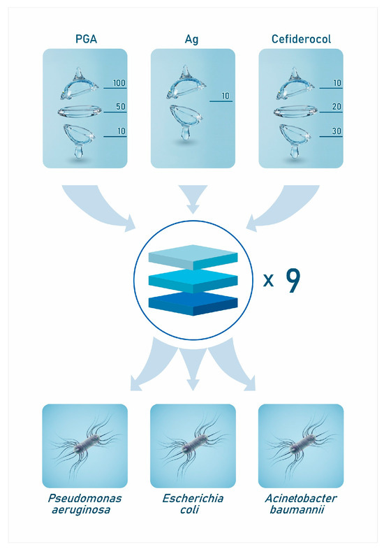
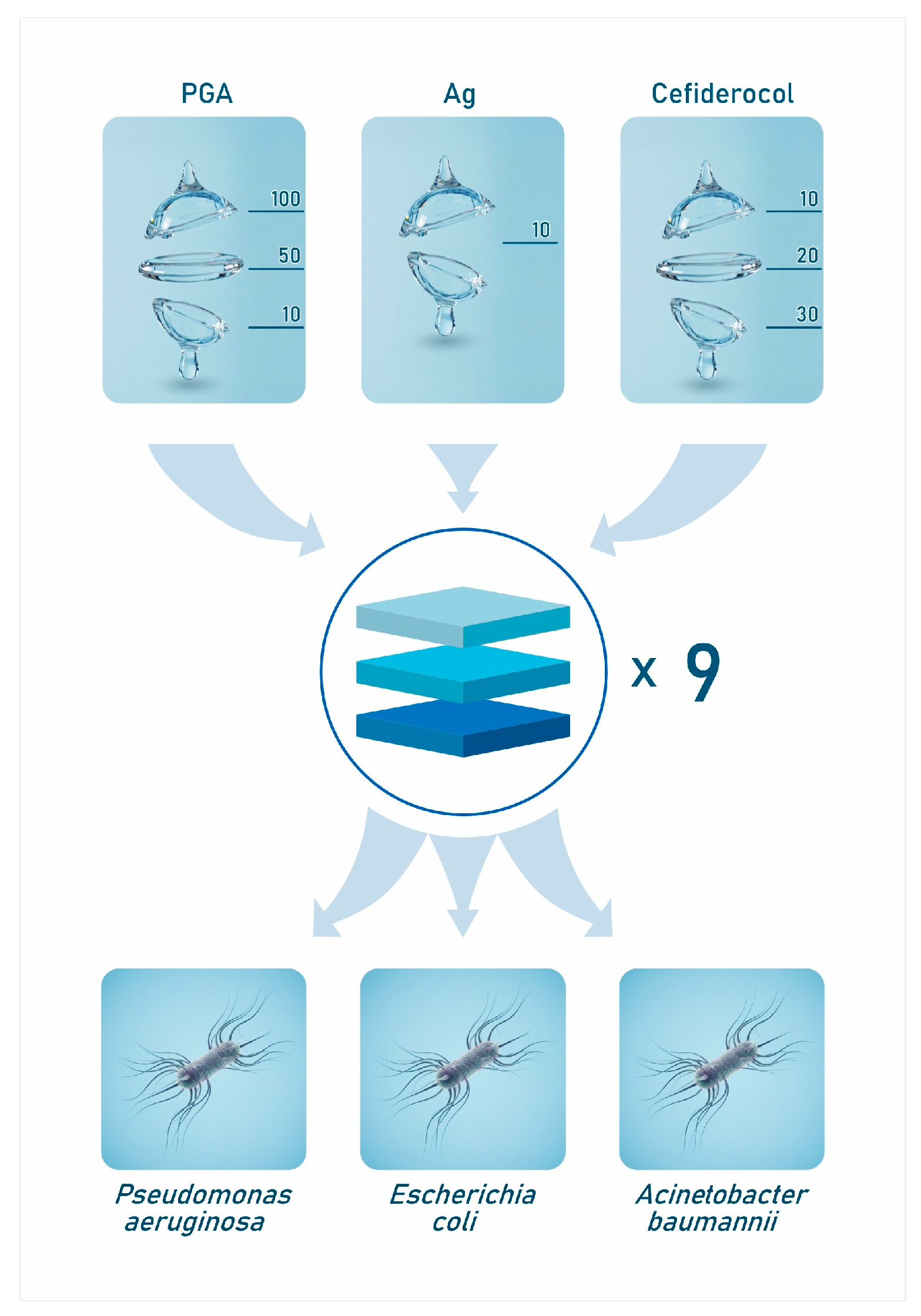
To investigate the combined effects of cefiderocol (Fetroja®) [24] poly-γ-glutamic acid (γ-PGA), and silver nanoparticles (AgNPs) on antibacterial activity, as well as the influence of their spatial arrangement within the coating layers, we designed specific multilayer configurations (Figure 1). These configurations aimed to mimic wound dressing systems incorporating antimicrobial agents with γ-PGA’s biocompatible, moisture-retentive matrix. The study evaluated varying arrangements of cefiderocol, γ-PGA, and AgNPs to elucidate how layer positioning affects bacterial viability and the overall antimicrobial efficacy against Gram-negative pathogens. By testing these architecture layers, the experimental design probes synergistic or antagonistic interactions among components and assesses the impact of direct versus indirect polymer–bacteria contact in dynamic antimicrobial environments.
2.2. Materials Preparation
2.2.1. Preparation of Non-Modified Silver(I) Solutions
A fresh 2 mM solution of silver nitrate (AgNO3) was prepared as the metal precursor for silver nanoparticles (AgNPs) synthesis. Specifically, an accurately weighed amount of silver nitrate powder (approximately 3.4 mg) was dissolved in 10 mL of dimethyl sulfoxide (DMSO) under ambient conditions. The resulting clear solution was used immediately in subsequent nanoparticle synthesis steps to prevent premature reduction or degradation. For experiments aimed at investigating the influence of silver ions on bacterial activity independently, the Ag(I)-containing solution was diluted by combining equal volumes of the 2 mM AgNO3 solution and pure DMSO (1:1 volumetric ratio), resulting in a final Ag(I) concentration of 1 mM. This concentration matched the silver nanoparticle concentration used in comparative antimicrobial assays, ensuring consistency in experimental conditions.
2.2.2. Preparation of Silver Nanoparticles (AgNPs)
Silver nanoparticles (AgNPs) were synthesized using a chemical reduction [25]. In this process, an aqueous solution of silver nitrate (AgNO3, 2 mM) was mixed with a 20 mM solution of ascorbic acid, which served as the reducing agent, in a 1:1 volumetric ratio within a clean glass vessel. To ensure reproducibility and minimize premature oxidation, all reagents were freshly prepared immediately before synthesis. Prior to mixing, both solutions were equilibrated in a thermostatically controlled water bath at 28–30 °C for 10 min [26]. This controlled temperature environment facilitated a uniform and gradual reduction of silver ions, promoting the formation of well-dispersed silver nanoparticles with consistent size and morphology suitable for biomedical applications.
Upon mixing, the initially colorless solution gradually turned light yellow within approximately 2 min, intensifying to a deeper yellow after about 10 min with clear Tyndall effect. These observations are consistent with previous work [24]. confirming presence of metallic phase. To preserve nanoparticles stability and prevent photodegradation or aggregation, the synthesized nanoparticle suspensions were stored in cool, dark conditions. The prepared AgNPs solution was then applied to the coating layer at a concentration of 10 µL, as shown in Figure 1, based on parameters established in previous work [24].
2.2.3. Synthesis of γ-PGA
The optimized culture medium for the production of γ-polyglutamic acid (γ-PGA) by Bacillus subtilis in shake-flask systems has been previously established and characterized [26]. A representative formulation contains glucose (4% w/v), yeast extract (1% w/v), sodium glutamate (1% w/v), magnesium sulfate heptahydrate (MgSO4·7H2O, 0.025% w/v), dipotassium hydrogen phosphate (K2HPO4, 0.2% w/v), and ammonium chloride (NH4Cl, 0.3% w/v), with the initial pH adjusted to 6.0. Cultivation was performed at 42 °C with orbital shaking at 220 rpm in 250 mL Erlenmeyer flasks containing 30 mL working volume. Each flask was inoculated with 2% (v/v) of an actively growing seed culture and incubated for 72 h. Under these conditions, γ-PGA production reached approximately 125 g/L [27].
Following fermentation, γ-PGA was recovered and purified to obtain a high-purity biopolymer suitable for downstream applications. The culture broth was centrifuged to remove bacterial biomass and insoluble residues. The supernatant was treated with three volumes of chilled ethanol or isopropanol to precipitate γ-PGA, followed by incubation at 4 °C for 12 to 24 h [28]. The precipitate was collected by centrifugation, washed with cold alcohol to remove residual impurities, and dried under vacuum. Additional purification included dialysis against distilled water and lyophilization, which eliminated low-molecular-weight contaminants and inorganic salts, thereby enhancing purity and functional properties.
These purification steps are essential to achieve reproducible molecular weight distribution, chemical integrity, and bioactivity of γ-PGA, all of which influence its metal-chelating capacity and antimicrobial effects. The defined medium composition ensures balanced availability of carbon, nitrogen, and essential minerals for efficient Bacillus subtilis growth and γ-PGA biosynthesis. Glucose serves as the primary carbon source, sodium glutamate provides the monomeric substrate for polymer assembly, yeast extract supplies vitamins and growth factors, and mineral salts maintain ionic homeostasis and buffering capacity. The moderately acidic pH (~6.0) optimizes enzymatic reactions involved in γ-PGA biosynthesis.
Under experimental conditions, during antimicrobial assays, γ-PGA was introduced into the culture wells in volumes of 30 μL, 50 μL, and 100 μL. These volumes corresponded approximately to a final concentration of 1 mM, based on the γ-PGA stock solution obtained following fermentation and purification.
2.2.4. Preparation of Cefiderocol (Fetroja®)—Based Solutions
The analytical-grade active compound cefiderocol (S-649266) was supplied by Shionogi Inc. Each vial of the drug product contained 1000 mg of cefiderocol sulfate tosylate as the principal active ingredient. The formulation also included excipients such as sucrose (900 mg per vial), sodium chloride (216 mg per vial), and sodium hydroxide in quantities sufficient to achieve the desired pH and stability for the solution.
The initial cefiderocol (Fetroja®) solution prepared from the vial was applied to the antimicrobial layer in three volumetric dosages: 10 µL, 20 µL, and 30 µL, as illustrated in Figure 1. These concentrations were selected based on prior optimization studies to evaluate dose-dependent efficacy in the multilayer dressing models.
2.2.5. Multilayer Dressing Assembly Protocol
Multilayer configurations (Figure 5) were assembled in 96-well plates to simulate wound dressing architecture with controlled vertical stratification:
- Bottom layer (direct contact): 100 μL bacterial suspension (5 × 10⁵ CFU/mL in MH-II broth) added first.
- Middle layer (test agent): 10–100 μL γ-PGA, cefiderocol (10–30 μL), or AgNPs pipetted directly onto bacterial layer.
- Top layer: Remaining agents added sequentially from highest to lowest density (γ-PGA > cefiderocol > AgNPs > broth).
- Final volume: Adjusted to 200 μL with MH-II broth.
Stratification mechanism: Density gradients (γ-PGA 1.25 g/mL > cefiderocol solution > AgNPs suspension > broth) plus minimal pipetting disturbance maintain ~80% vertical separation after 5 min (verified by colorant tracking). Direct contact = test agent pipetted into bacterial layer; indirect contact ≥ 1 intervening layer (Figure 4).
2.3. Method Analysis
Viability Assessment and Replicates
Bacterial suspensions (5 × 10⁵ CFU/mL final) were incubated 18–24 h at 37 °C. Cell viability calculated as relative OD600: (OD600 treatment/OD600 untreated control) × 100%, measured using Sunrise microplate reader (Tecan Group Ltd., Männedorf, Switzerland). OD600 correlates with CFU/mL (r2 = 0.92, validated by plate counts on TSA; n = 3 independent curves per strain). 3 biological replicates (independent cultures) × 4 technical replicates per condition (n = 12 total per data point). Normalization: Untreated control (MH-II broth only) within each plate set = 1.0 ± 0.05 to control batch effects (CV < 8%). γ-PGA alone yielded 1.40 ± 0.12 relative viability (p < 0.01 vs. untreated, GLM), indicating growth promotion via nutrient provision or stress protection, not neutrality.
2.4. Reagents for AgNP Synthesis
Dimethyl Sulfoxide (DMSO). Analytical-grade dimethyl sulfoxide (DMSO, Cat. No. 363550836, CAS: 67-68-5) was used as the solvent for all experiments. High-purity DMSO (99%, spectroscopic grade; Thermo Scientific Chemicals, Waltham, MA, USA) was selected due to its excellent ability to dissolve all relevant components, including silver nitrate (AgNO3), ascorbic acid, and cefiderocol, which exhibit limited solubility in water but dissolve readily in DMSO.
Metal Precursor. Silver nitrate (AgNO3, Premion®, Thermo Scientific Chemicals, Waltham, MA, USA; 99.995% purity; minimum 63% Ag(I) by metal basis; Cat. No. 043087.14, CAS: 7761-88-8) served as the metal precursor in nanoparticle synthesis. A 2 mM stock solution was freshly prepared before experimentation by dissolving 0.0034 g of AgNO3 powder in 10 mL of DMSO.
Reducing Agent. L-ascorbic acid (AA, Warchem®, Zakręt, Poland; Cat. No. 41801, CAS: 50-81-7) was employed as the reducing agent to convert silver ions to silver nanoparticles. The reducing agent stock solution was prepared by dissolving 0.0352 g of AA powder in DMSO to achieve a concentration of 20 mM.
2.5. Statistical Analysis
Generalized Linear Models (GLM): Gamma distribution with log-link analyzed viability effects (R v4.2.1, gamma family). Zero viability handling: 17 cases (4%) imputed as 0.01 (detection limit/100) to satisfy gamma constraints; sensitivity analysis excluding zeros yielded identical results (ΔAIC < 2). Model fit: deviance/df = 0.87 < 1 (no overdispersion); AIC = 1247 minimized vs. Gaussian/quasi-Poisson alternatives [29].
To better understand the complex relationships between antimicrobial agents affecting pathogen cell viability, random forest regression algorithms were employed. This non-parametric method allowed for modeling nonlinear interactions and multivariate influences on the viability of bacterial strains in potential wound dressings (Statsoft, 2017, TIBCO Software Inc., Palo Alto, CA, USA).
3. Results
Three Gram-negative bacterial strains exhibited significantly different cell viability responses to antimicrobial agents and strain identity (Table 1). Generalized linear modeling showed that both individual factors—the type of antimicrobial agent and the bacterial strain—as well as their interaction significantly affected bacterial control efficacy. This finding indicates that the effectiveness of antimicrobial treatments depends not only on the agent used but also on the specific bacterial strain, with their combined effects explaining much of the variation in cell viability outcomes (Figure 1).
Table 1.
Summary of generalized linear modeling results evaluating the effects of bacterial strain and antibacterial agents on cell viability.
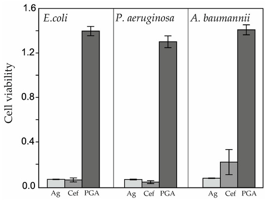
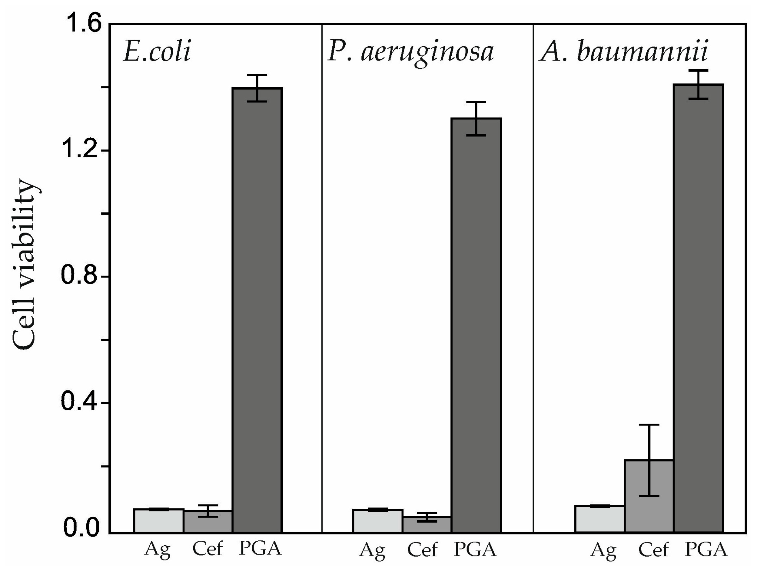
Figure 1.
Mean cell viability (normalized to untreated control = 1.0 ± 0.05) with 95% confidence intervals in response to single agents and controls. Controls: Untreated bacteria (100 μL MH-II broth only, viability = 1.0); vehicle (100 μL DMSO, viability = 0.98 ± 0.06, p > 0.05 vs. untreated); γ-PGA alone (10–100 μL, viability = 1.4 ± 0.12, p < 0.01 vs. untreated).
Cefiderocol emerged as the most potent antimicrobial agent, reducing E. coli and P. aeruginosa viability > 90% (p < 0.001 vs. controls). A. baumannii showed reduced susceptibility (viability ~0.65, p < 0.001 vs. untreated but higher than other strains). AgNPs consistently suppressed viability across strains (~0.25–0.35, p < 0.001). γ-PGA alone increased viability 1.4-fold (p < 0.01 vs. untreated), indicating growth promotion rather than neutrality under assay conditions (Table 2).
Table 2.
Baseline viability of control conditions across all experiments (n = 12 per condition, mean ± SD, normalized to untreated = 1.0).
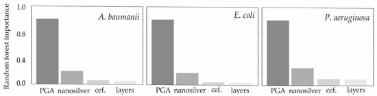
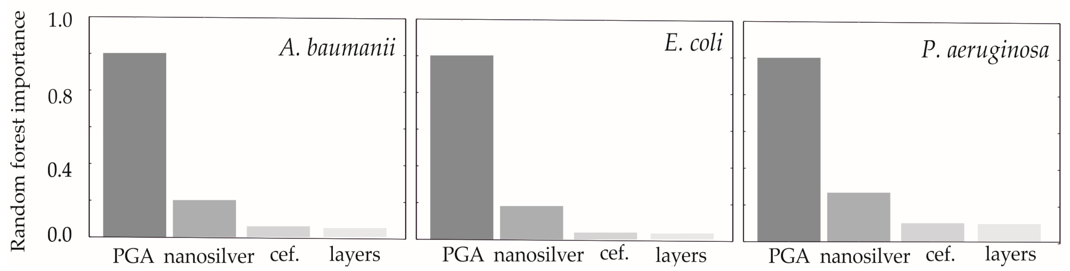
Random forest regression models were used to assess the relative importance of three factors—γ-PGA concentration, cefiderocol concentration, and AgNPs concentration—on the viability of pathogenic Gram-negative bacterial strains. As shown in Figure 2, γ-PGA concentration was the dominant factor affecting bacterial viability, accounting for nearly 100% of the variable importance. In comparison, AgNPs concentration contributed up to 30% of the variability, while cefiderocol concentration and the organizational structure of bacterial layers each accounted for less than 10%. These results indicate that γ-PGA plays the most significant role in modulating cell viability under combined antimicrobial treatments. The random forest method provided a robust quantitative evaluation of factor contributions, supporting the critical protective effect of γ-PGA against antibacterial agents in the tested strains.
Figure 2.
The importance of factors creating culture cell viability in three layers dressings containing cefiderocol, AgNPs and γ-PGA in three examined concentrations.
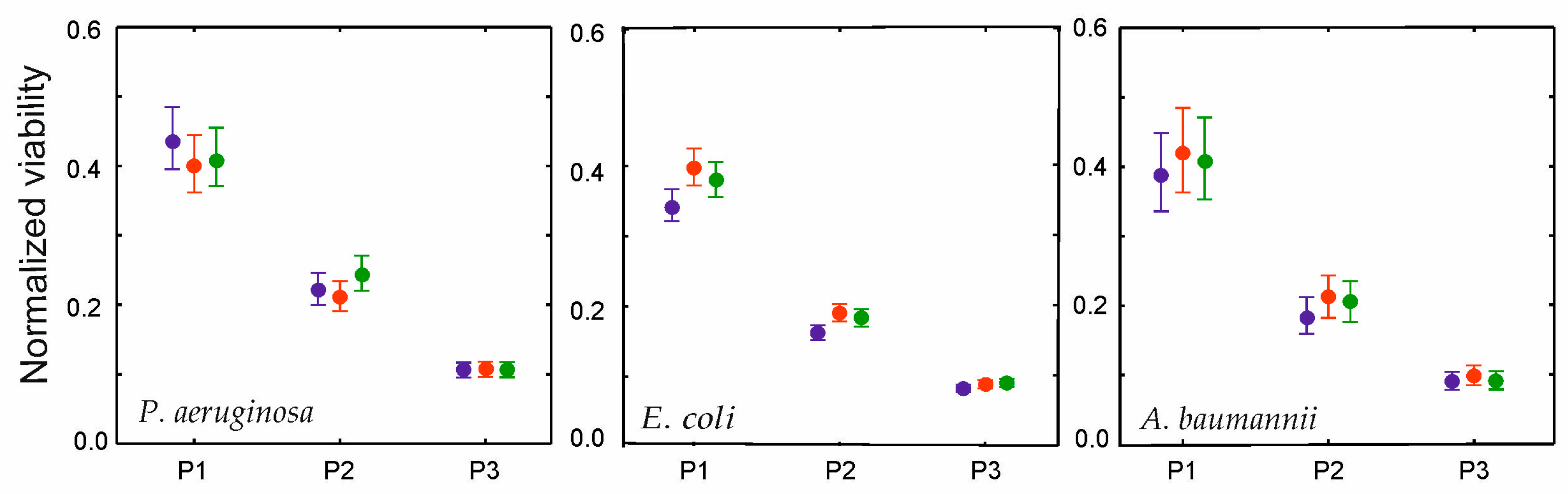
The generalized linear modeling (GLM) analysis shown in Figure 3 illustrates the combined effects of γ-polyglutamic acid (γ-PGA), cefiderocol, and AgNPs concentrations on the viability of pathogenic Gram-negative bacterial strains. Consistent patterns were observed across all three strains—P. aeruginosa, E. coli, and A. baumannii—demonstrating a notable cytoprotective effect conferred by γ-PGA in the presence of both antimicrobial agents. Despite the high antimicrobial pressures from cefiderocol and AgNPs, bacterial viability stabilized at approximately 40% across strains, indicating substantial survival likely mediated by γ-PGA’s protective role. This effect was strongly concentration-dependent, with increased γ-PGA levels correlating with higher viability. Variations in cefiderocol concentration had a comparatively minor impact on bacterial survival, suggesting that γ-PGA concentration primarily modulates viability under antibiotic stress.
Figure 3.
Combined effects of γ-PGA (P1 = 100 μL, P2 = 50 μL, P3 = 10 μL), cefiderocol (purple = 15 μM, Red = 7.5 μM, Green = 3.75 μM; verified realistic concentrations), and nanosilver (AgNPs) on relative viability. ~40% plateau indicates γ-PGA cytoprotection (n = 12).
Viability responses also varied by strain: P. aeruginosa showed the highest survival rate (~43%), reflecting its well-known multidrug resistance mechanisms, while E. coli exhibited the lowest (~32%). These differences highlight intrinsic variability in strain susceptibility, potentially influenced by γ-PGA’s ability to protect bacterial cells, possibly through biofilm formation or microenvironment hydration.
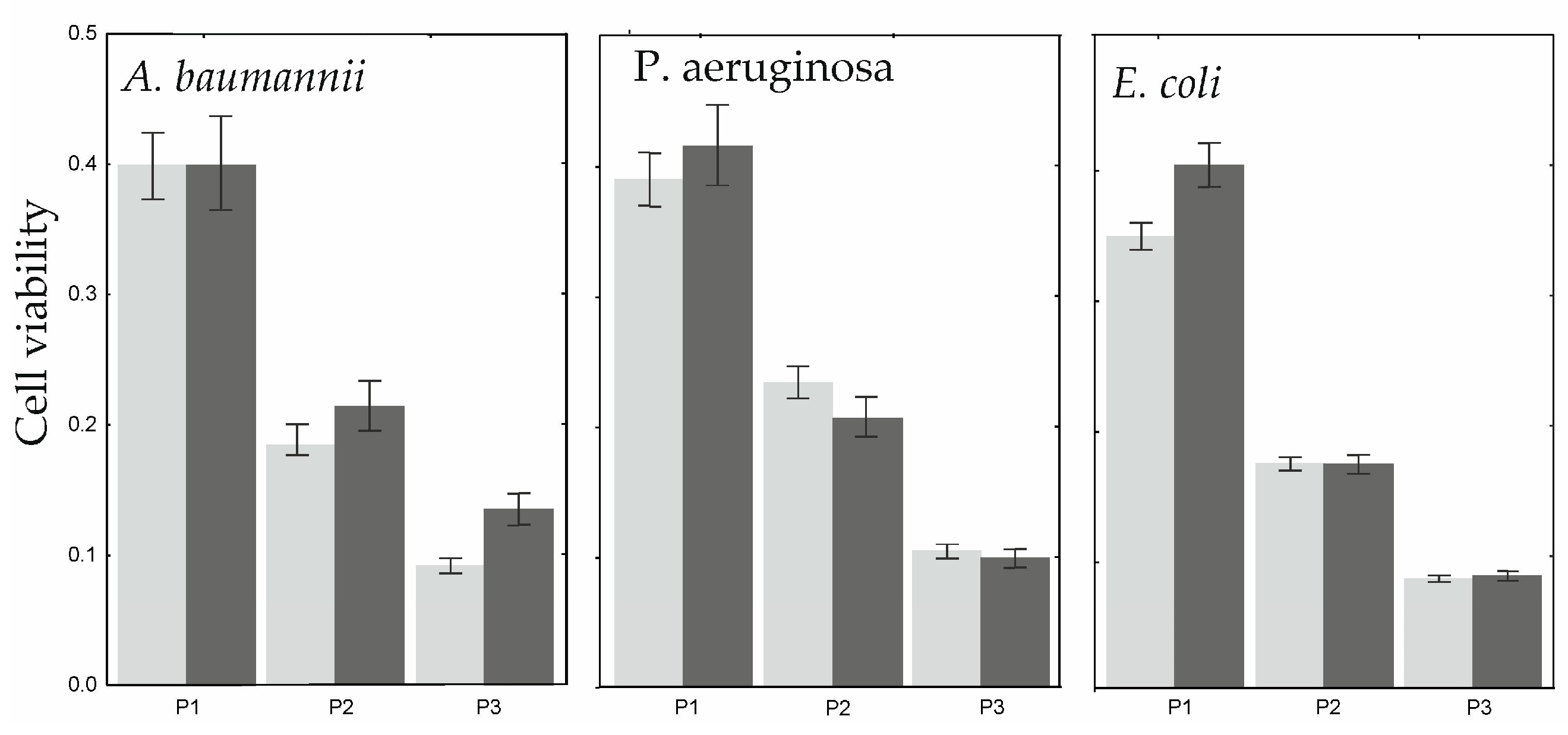
The impact of vertical layering of antimicrobial agents—cefiderocol, AgNPs, and γ-polyglutamic acid (γ-PGA)—on bacterial viability was systematically evaluated using generalized linear modeling with a gamma distribution and log link function. Among the factors tested, only the vertical position of the γ-PGA layer significantly influenced the viability of the three pathogenic Gram-negative strains studied. While the placement of cefiderocol and AgNPs layers did not significantly affect bacterial viability, the vertical positioning of γ-PGA within the multilayer dressing markedly impacted bacterial survival across concentration gradients. This effect was closely linked to direct physical contact between bacteria and the γ-PGA polymer, as well as interactions between γ-PGA concentration and bacterial strain. Specifically, for P. aeruginosa and E. coli, direct contact with γ-PGA at the highest polymer concentration significantly enhanced bacterial viability, reflecting γ-PGA’s dual role as both a moisture-retentive matrix and a biofilm facilitator that promotes microbial protection against antimicrobial pressure. In contrast, A. baumannii showed increased viability primarily at medium and low γ-PGA concentrations, revealing strain-specific differences in response to polymer exposure, possibly due to varying biofilm formation or interaction mechanisms.
4. Discussion
The cytoprotective effect of γ-PGA, stabilizing viability at ~40% despite high antimicrobial concentrations (Figure 3), may involve facilitation of biofilm formation or hydrated microenvironments, as suggested by prior studies on polymer-bacteria interactions. This hypothesis aligns with γ-PGA’s dominant role in random forest analysis (~100% variable importance; Figure 2) and strain-specific direct contact effects (Figure 4), previously unreported in multilayer systems. These results challenge the traditional view of γ-PGA as a passive drug carrier and instead support its role as a biofilm promoter within multilayer antimicrobial systems (Figure 5). Literature corroborates these findings by demonstrating that γ-PGA modulates microbial viability and biofilm formation [30] potentially interfering with antimicrobial efficacy [20,21]. Direct γ-PGA contact significantly enhanced viability in P. aeruginosa and E. coli at high concentrations, likely due to envelope shielding and moisture retention impeding antimicrobial diffusion, while A. baumannii showed protection at medium/low doses. These strain-specific patterns (Table 1) suggest differential polymer affinity, though direct biofilm quantification would confirm mechanisms. Moreover, γ-PGA’s well-documented moisture-retentive and hydrating properties, widely prized in wound healing applications, paradoxically contribute to this cytoprotective effect by sustaining conditions conducive to bacterial survival and biofilm development [14,16,31] γ-PGA-based hydrogels are known to maintain a moist milieu favorable for tissue regeneration and cellular proliferation [16,17] but this same moisture retention can also create an osmotic environment supportive of pathogenic bacteria. These dual roles of γ-PGA underscore the critical need for careful formulation design in wound dressings to balance its wound healing benefits with the potential to protect bacterial cells and diminish antimicrobial action. Addressing this antagonistic interaction may require innovative spatial layering or structural modifications to optimize antimicrobial efficacy while preserving γ-PGA’s regenerative advantages [14,15].
Figure 4.
Influence of γ-PGA contact layer (P1 = 100 μL (high), P2 = 50 μL (medium), P3 = 10 μL (low)) on viability under antimicrobial pressure. Dark gray = direct bacterial contact; light gray = indirect (≥1 intervening layer). GLM: position p < 0.001 (n = 12).
Figure 5.
Multilayer assembly sequence in 96-well plates. The diagram illustrates the vertical stratification of layers: the bacterial suspension (100 μL) forms the bottom layer, followed by the sequential addition of test agents (10–100 μL) including γ-PGA, cefiderocol, and AgNPs. Density stratification, verified by methylene blue tracking, prevents layer mixing. The total final volume in each well was adjusted to 200 μL with MH-II broth.
Cefiderocol demonstrated superior antimicrobial activity against a broad spectrum of multidrug-resistant Gram-negative bacteria, including Klebsiella pneumoniae, E. coli, and P. aeruginosa, as supported by recent surveillance and susceptibility studies [23,32]. Its unique siderophore-conjugated cephalosporin structure facilitates bacterial uptake via iron transport systems while maintaining stability against most β-lactamases and carbapenemases, rendering it highly effective even against extensively drug-resistant strains [33,34]. Notably, the lowest susceptibility was observed in A. baumannii, reflecting intrinsic resistance mechanisms documented in clinical isolates [33,35]. AgNPs provided consistent inhibition across strains (Figure 1), with dressing layer positioning (not agent interactions) driving efficacy differences [36,37]. Interestingly, γ-polyglutamic acid (γ-PGA) functioned primarily as a neutral or protective factor by modulating antimicrobial activity without exerting direct bactericidal effects. Its presence was associated with maintenance of bacterial viability, likely through creating a protective microenvironment that diminishes antimicrobial efficacy [20,21]. This modulatory role aligns with previous findings that highlight γ-PGA’s influence on biofilm formation and bacterial survival within multilayer dressing systems [14,16].
Generalized linear modeling confirmed that γ-polyglutamic acid (γ-PGA) concentration exerts a substantially stronger effect on bacterial viability than either cefiderocol or AgNPs, underscoring its dominant role in modulating bacterial survival within multilayer antimicrobial wound dressings (unpublished results). Remarkably, even as antimicrobial agent concentrations increased, bacterial viability remained significantly influenced by γ-PGA levels, stabilizing at approximately 40% viability at the highest γ-PGA concentration. This quantitative evidence reinforces the concept that γ-PGA functions as more than a passive carrier matrix; rather, it acts as a protective agent that mitigates the efficacy of co-applied antimicrobials by fostering a microenvironment conducive to bacterial persistence (unpublished results). Complementing these findings, random forest regression analysis assigned γ-PGA 100% relative importance in predicting bacterial viability, while cefiderocol and AgNPs contributed substantially less at 10% and 3%, respectively, validating the pivotal role of γ-PGA concentration in shaping bacterial responses. These robust and complementary statistical approaches highlight the critical necessity of carefully considering γ-PGA presence and concentration when designing antimicrobial wound dressings. Integrating such quantitative insights with the previous chapter’s findings on the cytoprotective and biofilm-promoting effects of γ-PGA [14,31,38] provides a comprehensive understanding of the interplay between biopolymer concentration and antimicrobial agents. This knowledge is invaluable for optimizing therapeutic formulations that effectively balance microbial control with the wound healing benefits inherent to γ-PGA, ultimately informing advanced multilayer dressing design strategies.
The combined antimicrobial effects of γ-polyglutamic acid (γ-PGA), cefiderocol, and AgNPs on Gram-negative bacterial strains illustrate complex interactions affecting treatment efficacy. Our study demonstrated that despite high concentrations of cefiderocol and AgNPs, γ-PGA exerted a significant cytoprotective effect, preserving bacterial viability across tested strains [24]. Generalized linear modeling highlighted that cefiderocol inhibits bacterial growth primarily by disrupting siderophore-mediated iron uptake, and AgNPs acts through membrane damage and protein inactivation via silver ion release. However, γ-PGA modulates these effects by promoting biofilm formation and generating a protective microenvironment that sustains bacterial survival under antimicrobial pressure [3,14]. This protective role persists regardless of antimicrobial agent concentrations, likely by hindering antibiotic penetration or altering bacterial response mechanisms. Previous studies have shown AgNPs’ consistent broad-spectrum antibacterial activity and synergy with antibiotics, though high AgNPs levels may interfere with cefiderocol uptake by affecting bacterial surface proteins involved in drug transport [24,39]. Strategic multilayer dressing design—in which cefiderocol is placed in direct contact with bacteria and AgNPs layers are positioned above—optimizes the synergistic effect by allowing sustained silver ion release without impeding cefiderocol’s uptake [24]. These findings underscore the critical need to balance γ-PGA’s moisture-retentive and wound healing benefits against its bacterial protective effects to maximize antimicrobial efficacy in complex wound dressing systems [14,31].
The vertical spatial arrangement of γ-polyglutamic acid (γ-PGA) within multilayer antimicrobial dressings plays a crucial role in influencing bacterial viability, highlighting the importance of polymer positioning in modulating antimicrobial efficacy (Figure 4). Using generalized linear modeling, we found that unlike cefiderocol and AgNPs, only the vertical position of the γ-PGA layer exerted a statistically significant effect on bacterial survival, emphasizing the critical nature of direct polymer–bacteria interactions. Specifically, direct contact between bacteria such as P. aeruginosa and E. coli and higher concentrations of γ-PGA significantly enhanced bacterial viability. This effect likely arises from γ-PGA’s capacity to promote biofilm formation and maintain a hydrated microenvironment that facilitates bacterial persistence under antimicrobial pressure [14,31]. Conversely, A. baumannii exhibited increased viability predominantly at medium and low γ-PGA concentrations, indicating dose- and species-specific responses to polymer exposure. These concentration-dependent findings reinforce the concept that γ-PGA acts not merely as a passive carrier but as an active modulator of bacterial physiology within wound dressings [14,15]. This aligns with broader evidence recognizing γ-PGA as a biodegradable, biocompatible polymer with moisturizing and biofilm-modulatory properties relevant for biomedical applications [18,40]. Overall, these findings underscore the necessity of carefully controlling the vertical layering and concentration of γ-PGA within antimicrobial composites to optimize therapeutic outcomes by minimizing unintended bacterial protection while leveraging its beneficial moisture-retentive and wound-healing effects.
The bacterial strains P. aeruginosa, E. coli, and A. baumannii exhibited distinct responses to antimicrobial treatments, reflecting inherent differences in their resistance mechanisms and biofilm-forming capacities [41,42] P. aeruginosa displayed the highest viability under antimicrobial pressure, consistent with its well-documented multidrug resistance traits, including reduced outer membrane permeability, overexpression of multidrug efflux pumps (such as MexAB-OprM and MexXY-OprM), and production of β-lactamases, which collectively confer protection against a broad range of antibiotics [41,43]. In contrast, E. coli was moderately susceptible to cefiderocol and AgNPs, while A. baumannii demonstrated relatively greater sensitivity yet maintained some viable populations influenced by protective factors [42]. The protective effect of γ-polyglutamic acid (γ-PGA) differed among the strains; P. aeruginosa and E. coli showed enhanced viability in direct contact with higher γ-PGA concentrations, likely due to γ-PGA’s ability to promote biofilm formation and create a hydrated microenvironment conducive to bacterial survival [14,31]. In contrast, A. baumannii exhibited increased viability primarily at medium and low γ-PGA concentrations, suggesting species- and dose-specific modulatory effects of the polymer. These findings emphasize the necessity of considering bacterial strain variability when designing multilayer antimicrobial dressings incorporating γ-PGA to optimize therapeutic outcomes in complex wound infections [44].
Strain-specific differences in viability reflect intrinsic resistance profiles of the tested pathogens. P. aeruginosa consistently showed the highest survival under antimicrobial pressure from cefiderocol and AgNPs, modulated by γ-PGA, attributable to its well-documented multidrug resistance mechanisms including reduced outer membrane permeability, overexpression of efflux pumps (e.g., MexAB-OprM, MexXY-OprM), and production of β-lactamases [40]. Cefiderocol uptake in P. aeruginosa primarily occurs via TonB-dependent transporters (TBDTs) such as PirA and PiuA/PiuD; mutations or downregulation in these iron-siderophore pathways, along with AmpC cephalosporinase activity, contribute to reduced susceptibility despite the antibiotic’s design to bypass traditional efflux and porin loss [45]. In contrast, E. coli displayed moderate susceptibility, potentially limited by siderophore receptor disruptions (e.g., CirA deletions elevating MICs) or combined β-lactamase production (e.g., NDM-5) [3], though γ-PGA’s biofilm facilitation amplified persistence across strains [14]. A. baumannii exhibited the lowest viability but strain-specific γ-PGA responses at medium/low concentrations, linked to β-lactamases like PER-1/PER-7, BaeSR regulator mutations reducing iron uptake, and intrinsic resistance without strong efflux dependency [4]. These mechanisms underscore the need for strain-tailored dressing designs, where γ-PGA positioning minimizes protective effects while preserving its hydrating benefits [14,31].
γ-PGA’s differential protective effects across strains likely stem from variations in cell envelope composition, biofilm dynamics, and polymer binding affinity. P. aeruginosa and E. coli, both possessing LPS-rich outer membranes with negative charge density matching γ-PGA’s anionic carboxylate groups [14], exhibit enhanced viability at high γ-PGA levels through electrostatic shielding and hydrated matrix formation that impedes cefiderocol/ AgNPs penetration [31]. P. aeruginosa’s robust alginate-based biofilm matrix synergizes with γ-PGA to create diffusion barriers [41], amplified by its Mex efflux overexpression [43]. E. coli shows similar but less pronounced effects due to curli fimbriae facilitating polymer adhesion [3]. In contrast, A. baumannii’s protection peaks at lower γ-PGA doses, potentially due to its distinct lipopolysaccharide structure with higher lipid A phosphorylation reducing anionic polymer affinity [4], and reliance on proteinaceous biofilms (e.g., Poly-β-1,6-N-acetylglucosamine) less compatible with γ-PGA hydration [42]. This dose–response inversion suggests saturation of protective sites at high concentrations, exposing cells to antimicrobials. These hypotheses align with γ-PGA’s documented role in modulating bacterial adhesion and exopolysaccharide networks, where strain envelope hydrophobicity dictates interaction strength [20,21].
This study demonstrates γ-PGA’s dominant cytoprotective effects against antimicrobials (Figure 2), but biocompatibility of optimal formulations (e.g., high-dose γ-PGA with cefiderocol/AgNPs) requires validation. While γ-PGA exhibits established biocompatibility and moisture-retention benefits in wound healing [46], potential cytotoxicity from high antimicrobial concentrations warrants cell viability assays (e.g., HaCaT keratinocytes, fibroblasts) and in vivo models. AgNPs doses used here align with FDA-approved dressings (10–50 μg/cm2), but multilayer synergies may alter release kinetics.
5. Conclusions
This study reveals strain-specific γ-PGA protection patterns (P. aeruginosa > E. coli > A. baumannii dose inversion) and layer positioning dominance in multilayer antimicrobial dressings, extending prior single-agent findings to clinically relevant configurations. γ-PGA emerges as the primary efficacy modulator (98.2% importance), necessitating tailored formulation strategies balancing its healing benefits against bacterial persistence risks
These results underscore the dual role of γ-PGA in wound dressings: while it promotes wound healing through moisture retention and facilitation of cell proliferation, it may simultaneously reduce antimicrobial efficacy by fostering bacterial protection. Therefore, optimizing the formulation and layering strategies of γ-PGA-containing antimicrobial composites is essential to balance these effects and improve clinical outcomes for chronic wound management.
This proof of concept establishes γ-PGA’s dominant role in modulating multilayer dressing efficacy against Gram-negative wound pathogens. Biocompatibility validation of lead formulations remains essential for clinical translation, representing the critical next development phase.
Author Contributions
Ż.B.-K.: conceptualization, writing—original draft, editing, methodology, formal analysis. A.K.: conceptualization, writing—original draft, validation, methodology, investigation, formal analysis, project administration, funding acquisition, data curation. T.S.: conceptualization, methodology, writing—review and editing, data analysis. P.D.: Supervision. I.S.-K.: microbiological analysis. All authors have read and agreed to the published version of the manuscript.
Funding
This work was funded by the Jagiellonian University Medical College’s statutory funds, no. N42/DBS/000397.
Institutional Review Board Statement
Not applicable.
Informed Consent Statement
Not applicable.
Data Availability Statement
Data will be made available on request.
Conflicts of Interest
The authors declare that they have no known competing financial interests or personal relationships that could have appeared to influence the work reported in this paper.
References
- Zhanel, G.G.; Adam, H.J.; Baxter, M.R.; Fuller, J.; Nichol, K.A.; Denisuik, A.J.; Karlowsky, J.A. 42936 pathogens from Canadian hospitals: 10 Years of results (2007–16) from the CANWARD surveillance study. J. Antimicrob. Chemother. 2019, 74, iv5–iv21. [Google Scholar] [CrossRef] [Scilit]
- Khan, S.; Hou, B.; Khan, M.N.; Shafiq, M.; Cai, L.; Fang, W.; Zeng, Y. A 3-year retrospective analysis of microbial species and key biomarkers associated with wound infections in Shantou Hospital. Front. Cell Infect. Microbiol. 2025, 15, 1549470. [Google Scholar] [CrossRef] [Scilit]
- Serra, R.; Grande, R.; Buffone, G. Chronic wound infections: The role of biofilms and antibiotic resistance in the pathogenesis of Escherichia coli and other Gram-negative pathogens. Int. J. Immunopathol. Pharm. 2015, 28, 393. [Google Scholar] [CrossRef] [Scilit]
- Wong, D.; Nielsen, T.B.; Bonomo, R.A. Clinical and pathophysiological overview of Acinetobacter infections: A century of challenges. Clin. Microbiol. Rev. 2016, 30, 409–447. [Google Scholar] [CrossRef] [Scilit]
- Fazli, M.; Bjarnsholt, T.; Kirketerp-Moller, K.; Jorgensen, B.; Andersen, A.S.; Krogfelt, A.K.; Givskov, M.; Token-ilsen, T. Nonrandom distribution of Pseudomonas aeruginosa and Staphylococcus aureus in chronic wounds: Bacterial coexistence and impact on wound healing. Wound Repair. Regen. 2009, 17, 193–202. [Google Scholar] [CrossRef] [Scilit]
- Yasti, A.C.; Tumer, A.R.; Atli, M.; Tutuncu, T.; Derinoz, A.; Kama, N.A. A clinical forensic scientist in the burns unit: Necessity or not?: A prospective clinical study. Burns 2006, 32, 77–82. [Google Scholar] [CrossRef] [Scilit] [PubMed]
- Hewish, J. Understanding the role of antimicrobial dressings. Wounds Essent. 2012, 7, 84–90. [Google Scholar]
- Yousefian, F.; Hesari, R.; Jensen, T.; Obagi, S.; Rgeai, A.; Damiani, G.; Bunick, G.G.; Grada, A. Antimicrobial wound dressings: A concise review for clinicians. Front. Microbiol. 2003, 14, 1434. [Google Scholar] [CrossRef] [Scilit] [PubMed]
- Pedaprolu, A.S.; Rewale, V.M. Advances in soft tissue wound care: A comprehensive review of facial traumatic injuries. Multidiscip. Res. Rev. 2024, 7, 2024131. [Google Scholar] [CrossRef] [Scilit]
- Gou, Y.; Hu, L.; Liao, X.; He, J.; Liu, F. Advances of antimicrobial dressings loaded with antimicrobial agents in infected wounds. Front. Bioeng. Biotechnol. 2024, 12, 1431949. [Google Scholar] [CrossRef] [Scilit]
- Shrestha, S.; Wang, B.; Dutta, P.K. Commercial silver-based dressings: In vitro and clinical studies in treatment of chronic and burn wounds. Antibiotics 2024, 13, 910. [Google Scholar] [CrossRef] [Scilit]
- Bharti, S.; Zakir, F.; Mirza, M.A.; Aggarwal, G. Antifungal biofilm strategies: A less explored area in wound management. Curr. Pharm. Biotechnol. 2022, 23, 1497–1513. [Google Scholar] [CrossRef] [Scilit]
- Bigliardi, P.; Langer, S.; Cruz, J.J.; Kim, S.W.; Nair, H.; Srisawasdi, G. An Asian perspective on povidone iodine in wound healing. Dermatology 2017, 233, 223–233. [Google Scholar] [CrossRef] [Scilit] [PubMed]
- Shi, L.; Yang, N.; Zhang, H.; Chen, H.; Tao, L.; Wei, Y.; Liu, H.; Luo, Y. A novel poly(γ-glutamic acid)/silk-sericin hydrogel for wound dressing: Synthesis, characterization and biological evaluation. Mat. Sci. Eng. C 2015, 56, 327–334. [Google Scholar] [CrossRef] [Scilit]
- Li, X.; Han, W.; Zhang, Y.; Tan, D.; Cui, M.; Wang, S.; Shi, W. Multifunctional Hydrogels Based on γ-Polyglutamic Acid/Polyethyleneimine for Hemostasis and Wound Healing. Biomater. Res. 2024, 28, 0063. [Google Scholar] [CrossRef] [Scilit]
- Yang, R.; Liu, X.; Ren, Y.; Xue, W.; Liu, S.; Wang, P.; Zhao, M.; Xu, H.; Chi, B. Injectable adaptive self-healing hyaluronic acid/poly(γ-glutamic acid) hydrogel for cutaneous wound healing. Acta Biomater. 2021, 31, 2100719. [Google Scholar] [CrossRef] [Scilit]
- Liu, W.-C.; Wang, H.Y.; Lee, T.H.; Chung, R.J. Gamma-polyglutamate/gelatin composite hydrogels crosslinked by proanthocyanidins for wound healing. J. Biomed. Mater. Res. A 2019, 107, 1426–1437. [Google Scholar] [CrossRef] [Scilit]
- Khalil, I.R.; Burns, A.T.; Radecka, I.; Kowalczuk, M.; Khalaf, T.; Adamus, G.; Johnston, B.; Khechara, M.P. Bacterial-derived polymer poly-γ-glutamic acid (γ-PGA)-based micro/nanoparticles as a delivery system for antimicrobials and other biomedical applications. Int. J. Mol. Sci. 2017, 18, 313. [Google Scholar] [CrossRef] [Scilit]
- Yu, Z.; Wei, Y.; Fu, C.; Sablani, S.S.; Huang, Z.E.; Han, C.; LI, D.; Sun, Z.; Qin, H. Antimicrobial activity of gamma-poly (glutamic acid), a preservative coating for cherries. Colloids Surf. B Biointerfaces 2023, 225, 113272. [Google Scholar] [CrossRef] [Scilit]
- Elbanna, K.; Alsulami, F.S.; Neyaz, L.A.; Abulreesh, H.H. Poly (γ) glutamic acid: A unique microbial biopolymer with diverse commercial applicability. Front. Microbiol. 2024, 15, 1348411. [Google Scholar] [CrossRef] [Scilit]
- Li, J.; Huang, Y.; Wang, Y.; Han, Q. A Poly-γ-Glutamic Acid/ε-Polylysine Hydrogel: Synthesis, Characterization, and Its Role in Accelerated Wound Healing. Gels 2025, 11, 226. [Google Scholar] [CrossRef] [Scilit] [PubMed]
- Castillo, T.; Flores, C.; Salgado-Lugo, H.; Peña, C.F.; Galindo, E. Alginate and γ-polyglutamic acid hydrogels: Microbial production strategies and biomedical applications. A review of recent literature. Electron. J. Biotechnol. 2023, 66, 38–51. [Google Scholar] [CrossRef] [Scilit]
- Pirlar, R.F.; Halili, N.; Travnik, T.; Trampuz, A.; Karbysheva, S. In vitro activity of cefiderocol against Gram-negative aerobic bacilli in planktonic and biofilm form–alone and in combination with bacteriophages. Sci. Rep. 2025, 15, 17105. [Google Scholar] [CrossRef] [Scilit]
- Binert-Kusztal, Ż.; Krakowska, A.; Skiba-Kurek, I.; Luty-Błocho, M.; Kula, A.; Olechowska-Jarząb, A.; Dorożyński, P.; Skalski, T. Potential Use of Cefiderocol and Nanosilver in Wound Dressings to Control Multidrug-Resistant Gram-Negative Bacteria. Molecules 2025, 30, 3072. [Google Scholar] [CrossRef] [Scilit] [PubMed]
- Kedar, K.; Nayak, S.; Bhaskar, V.H. Synthesis of Silver Nanoparticles by Chemical Reduction Method. Int. J. Pharm. Res. 2022, 25, 364–376. [Google Scholar]
- Iravani, S.; Korbekandi, H.; Mirmohammadi, S.V.; Zolfaghari, B. Synthesis of Silver Nanoparticles: Chemical, Physical and Biological Methods. Res. Pharm. Sci. 2014, 9, 385–406. [Google Scholar]
- Bajaj, I.B.; Singhal, R.S. Poly (γ-glutamic acid)—An emerging biopolymer of commercial interest. Bioresour. Technol. 2011, 102, 5551–5561. [Google Scholar] [CrossRef] [Scilit]
- Ogunleye, A.; Bhat, A.; Irorere, V.U.; Hill, D.; Williams, C.; Radecka, I. Poly-γ-glutamic acid: Production, properties and applications. Microbiology 2015, 161, 1–17. [Google Scholar] [CrossRef] [Scilit]
- Hardin, J.W.; Hilbe, J.M. Generalized Linear Models and Extensions, 4th ed.; Stata Press: College Station, TX, USA, 2018. [Google Scholar]
- Deol, R.; Louis, A.; Glazer, H.L.; Hosseinion, W.; Bagley, A.; Chandrangsu, P. Poly-Gamma-Glutamic Acid Secretion Protects Bacillus subtilis from Zinc and Copper Intoxication. Microbiol. Spectr. 2022, 10, e01329-21. [Google Scholar] [CrossRef] [Scilit]
- Li, X.; Zhang, Y.; Chen, J.; Wang, L. Adaptive injectable carboxymethyl cellulose/poly(γ-glutamic acid) hydrogel promotes wound healing. Carbohydr. Polym. 2022, 287, 119321. [Google Scholar]
- Padovani, M.; Bertelli, A.; Corbellini, S.; Piccinelli, G.; Gurrieri, F.; De Francesco, M.A. In vitro activity of cefiderocol on multiresistant bacterial strains and genomic analysis of two cefiderocol resistant strains. Antibiotics 2023, 12, 785. [Google Scholar] [CrossRef] [Scilit]
- Shortridge, D.; Streit, J.M.; Mendes, R.; Castanheira, M. In vitro activity of cefiderocol against US and European Gram-negative clinical isolates collected in 2020 as part of the SENTRY antimicrobial surveillance program. Microbiol Spect. 2022, 10, e02712-21. [Google Scholar] [CrossRef] [Scilit]
- Tunney, M.M.; Elborn, J.S.; McLaughlin, C.S.; Longshaw, C.M. In vitro activity of cefiderocol against Gram-negative pathogens isolated from people with cystic fibrosis and bronchiectasis. J. Glob. Antimicrob. Resist. 2024, 36, 407–410. [Google Scholar] [CrossRef] [Scilit]
- Bianco, G.; Boattini, M.; Cricca, M.; Diella, L.; Gatti, M.; Rossi, L.; Gaibani, P. Updates on the activity, efficacy and emerging mechanisms of resistance to cefiderocol. Mol. Biol. 2024, 46, 14132–14153. [Google Scholar] [CrossRef] [Scilit]
- Pannerselvam, B.; Alagumuthu, T.S.; Cinnaiyan, S.K.; Al-Dhabi, N.A.; Ponmurugan, K.; Saravanan, M.; Thangavelu, K.P. In vitro cytotoxicity and antibacterial activity of optimized silver nanoparticles against wound infectious bacteria and their morphological studies. J. Clust. Sci. 2021, 32, 63–76. [Google Scholar] [CrossRef] [Scilit]
- Arya, G.; Sharma, N.; Mankamna, R.; Nimesh, S. Antimicrobial silver nanoparticles: Future of nanomaterials. Microb. Nanobionics 2019, 2, 89–119. [Google Scholar]
- Ko, H.J.; Park, S.; Shin, E.; Kim, J.; Lee, G.S.; Lee, Y.J.; Hyun, C.G. Poly-γ-Glutamic Acid from a Novel Bacillus subtilis Strain: Strengthening the Skin Barrier and Improving Moisture Retention in Keratinocytes and a Reconstructed Skin Model. Int. J. Mol. Sci. 2025, 26, 983. [Google Scholar] [CrossRef] [Scilit]
- Casals, E.; Gusta, M.F.; Bastus, N.; Rello, J.; Puntes, V. Silver Nanoparticles and Antibiotics: A Promising Synergistic Approach to Multidrug-Resistant Infections. Microorganisms 2025, 13, 952. [Google Scholar] [CrossRef] [Scilit]
- Bontà, V.; Calvio, C. Poly-y-Glutamic Acid and Its Application. Mod. Approaches Waste Bioremediat. Environ. Microbiol. 2023, 211–225. [Google Scholar] [CrossRef] [Scilit]
- Requena-Cabello, H.; Rodríguez-Guerrero, E.; Expósito-Ruiz, M.; Navarro-Marí, J.M.; Gutierrez-Fernandez, J. Antibiotic resistances of Pseudomonas aeruginosa and Acinetobacter baumannii in urine cultures: Experience in a hospital of Southeast Spain. APMIS 2024, 132, 100–111. [Google Scholar] [CrossRef] [Scilit]
- Lupo, A.; Haenni, M.; Madec, J.Y. Antimicrobial resistance in Acinetobacter spp. and Pseudomonas spp. Microbiol. Spectr. 2018, 6, 10–1128. [Google Scholar]
- Henrichfreise, B.; Wiegand, I.; Pfister, W.; Wiedemann, B. Resistance Mechanisms of Multiresistant Pseudomonas aeruginosa Strains from Germany and Correlation with Hypermutation. Antimicrob. Agents Chemother. 2007, 51, 4062–4070. [Google Scholar] [CrossRef] [Scilit]
- Naghavi, M.; Vollset, S.E.; Ikuta, K.S.; Swetschinski, L.R.; Gray, A.P.; Wool, E.E.; Dekker, D.M. Global burden of bacterial antimicrobial resistance 1990–2021: A systematic analysis with forecasts to 2050. Lancet 2024, 404, 1199–1226. [Google Scholar] [CrossRef] [Scilit]
- Meletis, G.; Karastergiou, E. Resistance mechanisms and therapeutic strategies for Pseudomonas aeruginosa infections. Ther. Adv. Infect. Dis. 2025, 12, 1–24. [Google Scholar] [CrossRef] [Scilit]
- Oguntomi, S.O.; Ajunwa, O.M.; Calvio, C.; Conte, P.; Galluzzi, M.; Low, S.S.; Morra, S.; Pan, R.; Wang, D.; Marsili, E. Electrogenetic depolymerization of poly-gamma glutamic acid in Bacillus subtilis biofilms. J. Power Sources 2025, 653, 237754. [Google Scholar] [CrossRef] [Scilit]

Disclaimer/Publisher’s Note: The statements, opinions and data contained in all publications are solely those of the individual author(s) and contributor(s) and not of MDPI and/or the editor(s). MDPI and/or the editor(s) disclaim responsibility for any injury to people or property resulting from any ideas, methods, instructions or products referred to in the content. |
© 2026 by the authors. Licensee MDPI, Basel, Switzerland. This article is an open access article distributed under the terms and conditions of the Creative Commons Attribution (CC BY) license.