


Abstract
Background: Pes planus is a common foot deformity that may lead to both structural and functional consequences. This alteration in foot morphology has been associated with changes in neurosensory mechanisms such as proprioception and balance. This study aims to evaluate proprioception and balance parameters in young adults with pes planus by comparing them with healthy individuals. Methods: This case–control study, which had a quantitative and cross-sectional design, included a total of 90 volunteer university students aged 18–30. The presence of pes planus was assessed using the Navicular Drop Test. Proprioception assessment was performed using a digital goniometer to measure active and passive joint position sense, while balance assessment was performed using the Y Balance Test. Results: The groups were similar in terms of demographic and anthropometric characteristics (p > 0.05). In the proprioception assessment, a significant difference was observed in active joint position sense scores in individuals with pes planus (p = 0.032). In contrast, no significant difference was found between the two groups in terms of passive joint position sense (p = 0.769). According to the Y Balance Test results, no difference was observed in the anterior and posteromedial directions (p = 0.690 and p = 0.806). At the same time, the balance performance of individuals with pes planus was significantly lower in the posterolateral direction (p = 0.045). Conclusions: Young individuals with flexible pes planus showed differences in active joint position sense and balance performance in the posterolateral direction. This situation shows that pes planus may be associated with not only structural but also neurosensory functions. The findings highlight the importance of planning protective interventions early. In the future, there is a need for longitudinal studies conducted with larger and more heterogeneous samples, including rigid type pes planus.
1. Introduction
The foot is one of the fundamental structures located in the lower extremity that carries the entire weight of the body, both when standing and during dynamic activities involving contact with the ground. The foot, which accounts for approximately 5% of the body surface area, supports around 95% of the body weight [1]. It provides an adaptive support base for the body, distributes load-bearing forces, and contributes to energy storage during the gait cycle [2]. Sensory information from the feet plays an important role in postural control and maintaining balance [3]. The human body is biomechanically defined as a kinematic chain; the feet are located at the most distal point of this chain, serving as the support surface [4,5].
Pes planus is a foot deformity characterized by the collapse of the medial longitudinal arch. It results in the affected area of the foot coming into contact with or approaching the ground. This deformity can be rigid or flexible. In the rigid type, the arch is collapsed regardless of weight bearing; in the flexible type, the arch disappears only during weight bearing [6]. Pes planus, which is frequently detected in clinical examinations, is generally asymptomatic but can cause pain and increase the risk of injury by disrupting the biomechanics of the lower extremities and lumbar spine [2,7]. Although it typically emerges during childhood, it can develop at any stage of life [2]. It is common in infancy and early childhood; the arch of the foot develops as the child grows [8]. The prevalence of pes planus is reported to range from 3.9% to 37% [9,10]. This condition is considered a problem that can negatively affect the quality of life and individuals’ physical abilities [7].
Various studies have investigated the relationship between pes planus and proprioception and balance disorders [4,11,12]. Studies investigating the biomechanical effects of foot problems on the proprioceptive ability of lower extremity joints are limited [4]. The foot, located at the end of the kinematic chain, plays a critical role in proprioception and balance. Weakness or damage in one link of the chain can lead to functional impairments in other parts of the chain [5]. Therefore, pes planus has been suggested to be associated with differences in the static and dynamic properties of the lower extremities [11].
Balance is defined as maintaining the body’s center of gravity within the base of support. To maintain balance, the central and peripheral nervous systems must be in constant communication [4,13]. This process relies on accurate information from the visual, vestibular, and somatosensory systems [14,15]. The somatosensory system supports balance by integrating information from receptors located on the surface of the feet and in deep tissues while the individual is standing or walking [16]. It has been suggested that pes planus may be associated with balance alterations related to changes in the neuromechanical connection of the kinematic chain [4]. The number of studies conducted on pes planus, a common deformity, should be increased. The evaluation of pes planus in different geographical regions and age groups may contribute to a better understanding of the etiology of this deformity and the development of strategies to prevent possible complications [9]. In determining the lower extremity to be evaluated in this study, the use of navicular drop difference as opposed to the commonly used dominant side preference stands out as an application that adds uniqueness to the method. In addition, separate evaluation of active and passive joint position sense measurements and directional analysis of balance tests aims to reveal the neurosensory and postural effects of pes planus in greater detail. However, the fact that the sample group was selected from young adults adds an important preventive health dimension to the study, as it allows the differences in proprioception and balance to be observed even before the pes planus has caused any noticeable symptoms. In this context, the present study evaluated differences in proprioception and balance between young adults with and without pes planus.
2. Materials and Methods
Study Design: This study was designed as a quantitative, cross-sectional case–control study.
Study Setting and Duration: The study was conducted at the Faculty of Health Sciences, Mardin Artuklu University, between 1 May 2025 and 1 July 2025.
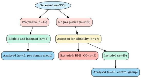
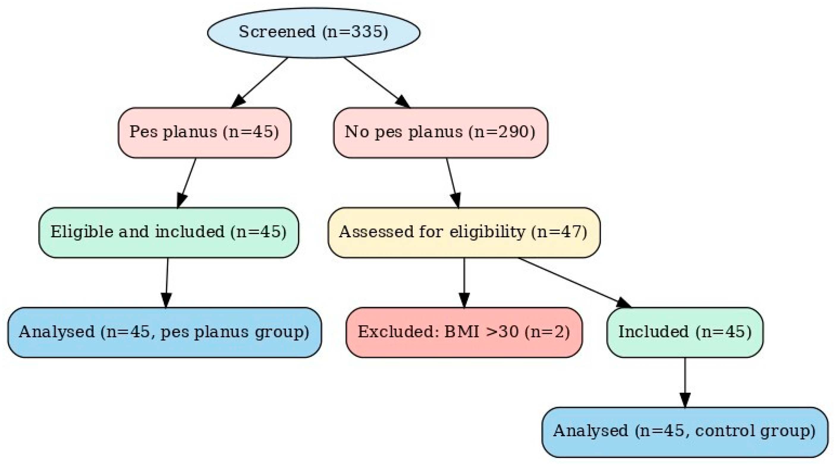
Study Sample: The study population consisted of students from the Faculty of Health Sciences, Mardin Artuklu University. Face-to-face and online announcements were made to all classes within the faculty, and participation was on a voluntary basis. All individuals with pes planus who met the inclusion/exclusion criteria for the study were included in the study, and individuals without pes planus who met the inclusion/exclusion criteria were randomly selected from among those without pes planus (Figure 1). As the study aimed to include the entire accessible target population, the sample size was not calculated a priori. However, a post hoc power analysis was performed using G*Power 3.1 software after data collection. The analysis revealed a statistical power of 90% to detect a mean difference between two independent groups (t-test) with an effect size of 0.63, α = 0.05, and a total sample size of 90 participants (45 per group).
Figure 1.
Flow chart.
Inclusion Criteria: Students from the Faculty of Health Sciences at Mardin Artuklu University, aged 18–30 years, who volunteered to participate, were included in the study.
Exclusion Criteria: Pain in the lower extremity joints, BMI ≥ 30 kg/m2, systemic disorders, neuromuscular and neurological disorders, presence of tibialis posterior tendon insufficiency, history of any lower extremity injury or surgery in the last 6 months, any orthopedic disorder other than pes planus, and having rigid pes planus for the pes planus group.
Withdrawal Criteria: Missing evaluation parameters.
Ethical Considerations: The study was approved by Mardin Artuklu University Non-Interventional Clinical Research Ethics Committee (decision date: 22.04.2025, decision no: 2025/4-7). Individuals were informed about the study verbally and in writing. Following this, the subjects who volunteered to participate in the study were included by signing an informed consent form. The study was conducted in accordance with the principles of the Declaration of Helsinki.
Data Collection: Demographic and anthropometric characteristics of the participants were recorded. Navicular Drop Test was used to evaluate the presence of pes planus, Jack’s Test was used to evaluate pes planus flexibility, active and passive joint position sense measurement with a goniometer was used for proprioception evaluation, and Y Balance Test was used for balance evaluation.
All assessments yielded quantitative outcome measures. The Navicular Drop Test was recorded in millimeters, joint position sense was expressed as angular deviation in degrees, and Y Balance Test performance was calculated as normalized reach distances based on leg length.
To minimize measurement bias, all evaluations were conducted by the same trained examiner using standardized protocols under controlled conditions.
Pes Planus Assessment: The Navicular Drop Test was performed to determine the presence and degree of pes planus. In the test, the navicular tubercle of the individuals was marked, and the distance between it and the floor was measured in the sitting position (unweighted support position) and standing position (weighted support position). Individuals with a navicular drop difference of 10 mm or more were considered to have pes planus [17].
Assessment of Pes Planus Flexibility: Jack’s Test, which is a valid method to evaluate flexibility, was used to differentiate flexible or rigid pes planus in individuals with pes planus. While the individual was in the sitting position, the first toe (hallux) was dorsiflexed by the clinician, and the arch was expected to form. In case of arch formation, the test was considered positive and interpreted as flexible pes planus [18]. Only people with positive test results were included in the study.
Proprioception Assessment: Active and passive joint position sense measurements were used to assess ankle proprioception. The tests were administered with the participants’ eyes closed. Between the active and passive tests, another evaluation was performed to eliminate the possibility of remembering the angle. Dorsiflexion angles were measured using a digital goniometer. During the measurement, the center line of the 5th metatarsal bone was aligned with the goniometer, which was positioned over the lateral malleolus. In the active joint position sense measurement, the ankle was passively positioned at 15° of dorsiflexion. The participant was asked to memorize this position before returning to the starting point. The participant was then asked to actively reproduce the same ankle angle. In the passive joint position sense measurement, the ankle was passively positioned at 15° of dorsiflexion, and the participant was instructed to memorize this position before returning to the starting point. Then the joint was moved passively, and the person was asked to stop the movement when the previously learned position was reached. The test was repeated three times. The deviation value was found by subtracting the angle made by the participant from the target angle, and the average of the three measurements was calculated [19].
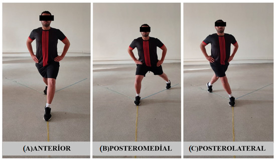
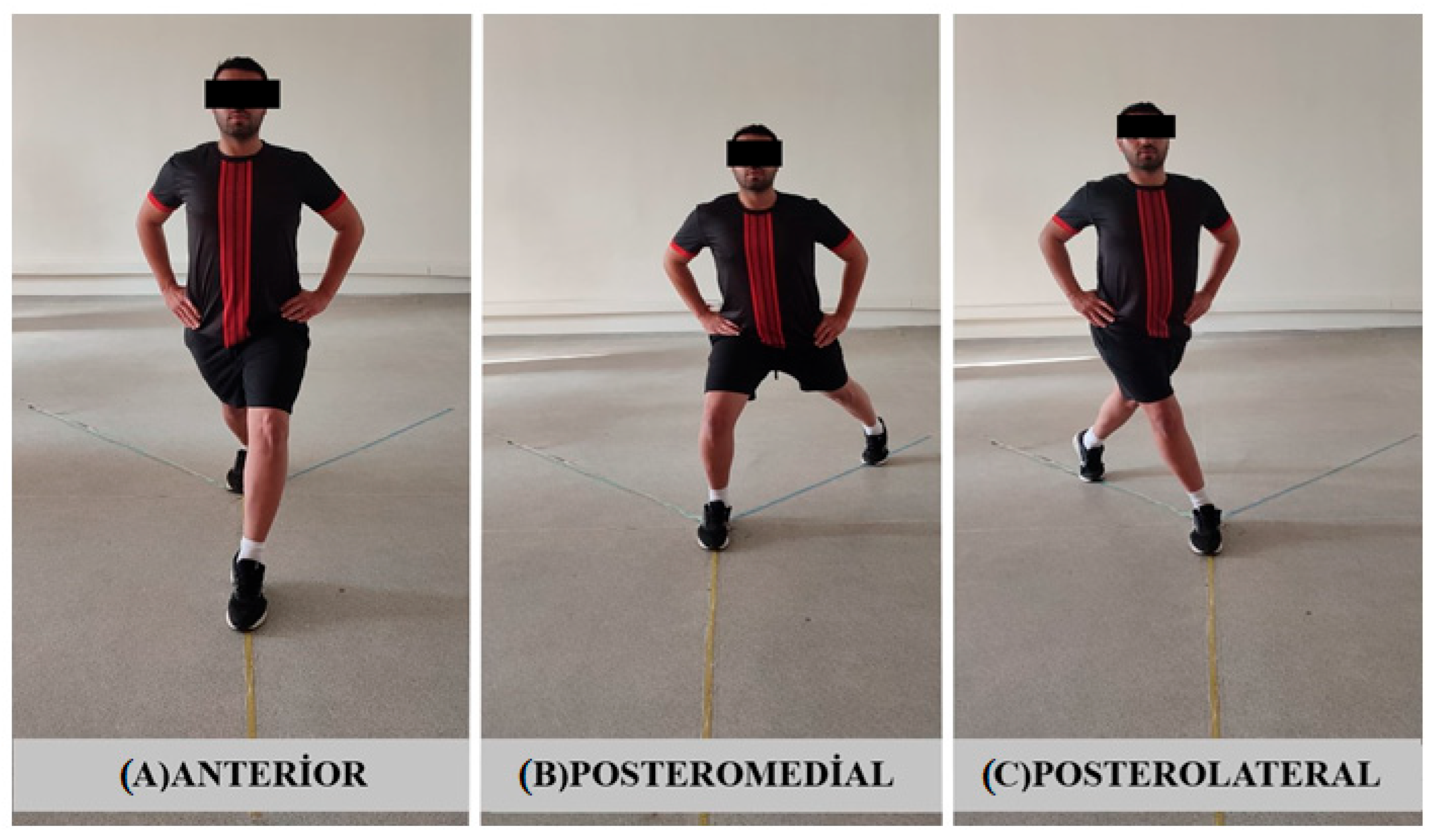
Balance Assessment: The Y Balance Test was used to assess balance. For this test, three tape lines of equal length were fixed to the floor in a Y-shaped pattern, forming angles of 90°, 135°, and 135°. The directions tested were anterior, posterolateral, and posteromedial. For the anterior reach direction, the anterior end of the foot was placed at the intersection point of the three lines, whereas for the posteromedial and posterolateral directions, the posterior end of the foot was positioned at the intersection point. The person tried to reach the farthest point in 3 different directions formed by the tape lines with the contralateral foot, and the distance from the center was measured by marking the points reached (Figure 2). The test was repeated three times until a successful reach was achieved, with 30 s rest intervals between trials and a 1 min rest when switching extremities. The data obtained were normalized according to the leg length of the person. Leg length was found by measuring the distance from the spina iliaca anterior superior to the distal end of the medial malleolus in the supine position. The ratio of the best result of the 3 successful test scores to the leg length was multiplied by 100 to obtain a normalized reach distance [20].
Figure 2.
The Y Balance Test.
Statistical Analysis: Data were analyzed using IBM SPSS Statistics version 25.0 (IBM Corp., Armonk, NY, USA). The distribution of numerical variables was assessed with the Shapiro–Wilk test. Normally distributed variables were presented as mean () ± standard deviation (SD), whereas non-normally distributed variables were presented as median (minimum–maximum). Categorical variables were expressed as frequency (n) and percentage (%). For comparisons between groups, the Independent Samples t-test was applied to normally distributed variables, and the Mann–Whitney U test was used for non-normally distributed variables. The Paired Samples t-test was performed to assess differences between two dependent measurements. The Chi-Square test or Fisher’s Exact Test was used to analyze categorical variables, as appropriate. A p-value of <0.05 was considered statistically significant for all tests.
3. Results
Mardin Artuklu University Faculty of Health Sciences has 1163 students. Among these students, 335 (28.8%) were evaluated, and 45 (13.43%) were found to have pes planus. When the demographic and anthropometric characteristics of individuals with and without pes planus were analyzed, the groups were similar in terms of age, body mass index (BMI), gender, dominant side, marital status, smoking, and alcohol use (p > 0.05) (Table 1).
Table 1.
Demographic and anthropometric characteristics of the groups.
Navicular Drop Test measurements of the assessed foot were significantly higher in the pes planus group compared with the control group (p < 0.001), as shown in Table 2A.
Table 2.
Navicular Drop Test (NDT) measurements. (A). Between-group comparison. (B). Foot-selection analysis in participants without pes planus.
In participants with unilateral pes planus, only the affected lower extremity was included in the study. In participants with bilateral pes planus, the lower extremity with the greater navicular drop was included (right: 32 [71.11%], left: 13 [28.89%]). To determine which lower extremity of participants without pes planus would be included in the study, right and left navicular drop measurements were compared, and no significant difference was found between them (p = 0.136, Table 2B). Therefore, for participants without pes planus, the lower extremity to be included in the study was randomly selected (right: 32 [71.11%], left: 13 [28.89%]).
When the groups were compared in terms of proprioception scores, a significant difference was found between the groups in active joint position sense scores (p = 0.032), but no significant difference was observed between the groups in passive joint position sense measurement (p = 0.903, Table 3).
Table 3.
Differences in the groups in terms of proprioception and balance.
When the groups were compared in terms of Y Balance Test performance, no significant differences were observed in the anterior (p = 0.249) and posteromedial (p = 0.199) directions. However, individuals with pes planus demonstrated significantly lower scores in the posterolateral direction (p = 0.045, Table 3).
4. Discussion
Pes planus is one of the most frequently reported foot deformities. Its prevalence has been reported to range from 3.9% to 37% across different populations [9,10]. In a systematic review of 12 studies involving 16,000 individuals, Salinas-Torres et al. [21] reported the prevalence of pes planus in the general population as 15.6%. Dunn et al. [9] found that this rate ranged between 3.9% and 34% across different races. Bhoir et al. [8] reported a prevalence of 11.25% among university students aged 18–25 years, and Aenumulapalli et al. [22] reported 13.6% in the 18–21 age group. In the study conducted by Eluwa et al. [23] at the University of Calabar, the prevalence of pes planus was found to be 13.4%, parallel to the rate in our study.
This variability in the prevalence of pes planus may be attributed to the anatomical variations in individuals, as well as the differences in the assessment methods used [22]. Individual variables such as age, race, and BMI have also been reported to be associated with pes planus [21]. This diversity of different populations, age groups, and assessment approaches is an important reason for inconsistencies between studies. However, the findings of this study are consistent with research on young adults.
In this study, a significant difference in active joint position sense was observed in individuals with pes planus, whereas no significant difference was found between the groups in the passive measurement. These results suggest that active and passive proprioception tests may engage different sensorimotor pathways, which may reflect the involvement of different sensorimotor pathways.
The foot structure plays a fundamental role in the posture, balance, and mobility of the human body. The structural integrity of the medial longitudinal arch is especially critical for the healthy functioning of proprioceptive feedback and balance mechanisms [16]. The foot serves as a major source of proprioceptive input due to its dense network of mechanoreceptors [24]. Pes planus, which is characterized by collapse of the medial longitudinal arch, may be associated with alterations in sensory feedback and differences in balance and movement control [25]. Yildiz & Yildiz [26] reported that ankle proprioception was impaired in individuals with pes planus, and this impairment was associated with reduced balance performance. Similarly, Ozyalvac et al. [27] also found decreased proprioceptive perception in individuals with pes planus. A study by Ghorbani et al. [4] also showed that flexible pes planus may cause loss of balance and joint position sense. These findings are consistent with the significant difference obtained during active measurement in this study.
However, in this study, no significant difference was found between the groups in passive joint position sense evaluation. In a study conducted by Yalcin et al. [12], it was shown that passive reproduction error scores in eversion were significantly higher in individuals with flexible pes planus. However, no significant difference was found in inversion. This suggests that proprioceptive outcomes may differ depending on the type of test applied and the direction of joint movement. Similarly, in this study, proprioception was assessed in the dorsiflexion direction, and a significant difference was observed only in the active measurement.
Sensory inputs are received from similar receptors in both active and passive joint position sense; however, active proprioception is based not only on peripheral sensory input but also on the integration of motor commands with their associated sensory feedback [28]. Perception during active movement is therefore formed through the integration of motor control processes and sensory feedback [29]. Accordingly, the significant difference observed in active joint position sense may be related to the more pronounced involvement of motor control pathways in individuals with pes planus. In contrast, perception during passive measurements largely depends on peripheral afferent input. For this reason, passive assessments may be more strongly influenced by methodological factors such as movement velocity, target angle, and testing position [28,29]. Detection thresholds are higher for slow movements and considerably lower for faster movements, and it has been reported that slow or small angular displacements can be difficult to detect [30]. In addition, the ability to detect ankle movements has been shown to be influenced by body position, with optimal performance observed in the standing position [31]. It is also well established that proprioceptive acuity declines with age [32]. Considering all these factors, it appears consistent with the literature that while no differences emerged between groups in passive measurements, which are more sensitive to assessments in a group of young and asymptomatic individuals, differences were detected in active measurements due to the more pronounced sensory-motor control pathways.
In proprioception evaluations, many variables such as the type of test, target angle, method of application, and age/clinical profile of the participants may affect the results. The fact that a significant difference was obtained only in active measurement in the present study suggests that proprioceptive differences in young individuals with pes planus may be subtle and not uniformly present across all sensory modalities. This finding supports the view that proprioceptive function in asymptomatic individuals may vary over time and warrants further investigation in longitudinal studies [33].
In this study, analysis of the Y Balance Test results revealed that individuals with pes planus had significantly shorter reach distances in the posterolateral direction. However, no significant difference was found between the groups in the anterior and posteromedial directions.
The structural integrity of the medial longitudinal arch plays a crucial role in postural control and balance, facilitated by mechanoreceptors located on the sole. Disruption of this structure may result in an inability to effectively absorb ground reaction forces and weakening of equilibrium mechanisms [5]. Previous studies have shown that pes planus negatively affects lower extremity physical performance and balance capacity [5,34]. Côté et al. [13] reported that individuals with pes planus exhibited poorer dynamic balance performance compared to those without pes planus. Kabak et al. [5] stated that the effects of pes planus on the dominant lower extremity may affect balance. El-Shamy & Ghait [11] observed a decrease in dynamic balance parameters in adolescent girls with flexible pes planus. Al-Abdulwahab & Kachanathu [35] reported that foot posture was associated with dynamic balance, but not with static balance. These results suggest that alterations in foot structure may be more strongly associated with dynamic balance than with static balance.
However, there are also contradictory findings among the studies on balance. According to Buyukturan et al. [36], there was no significant relationship between the degree of pes planus and balance parameters. Similarly, in a study conducted by Şahan et al. [33], no significant difference was found between static and dynamic balance scores of young individuals with and without pes planus. Another study by Gul et al. [37] showed that there was no significant correlation between pes planus and static and dynamic balance.
Dikici & Demirdel [34] found a weak negative correlation between the severity of pes planus in the dominant lower extremity and anterior and posteromedial directions, while no significant relationship was found in the posterolateral direction. On the nondominant side, a weak negative correlation was reported only in the anterior direction. Rakhmatova et al. [38] found a significant weak positive correlation in the anterior direction, but not in the posteromedial and posterolateral directions. These findings support that the relationship between pes planus and balance may vary directionally. In this study, a significant difference was found only in the posterolateral direction, which may indicate that this direction is more vulnerable in terms of balance performance in individuals with pes planus. The absence of significant differences in other directions suggests that pes planus may not yet be associated with a generalized biomechanical impairment in this sample. The young and asymptomatic profile of the participants may have limited structural changes in the foot, which may be associated with balance differences observed only in certain directions. This highlights the need for longitudinal follow-up studies to clarify the progressive effects of pes planus.
The literature indicates that the Y Balance Test requires not only balance but also components such as lower extremity strength, joint range of motion, coordination, and neuromuscular control, and that each reach direction engages different muscle groups and control strategies [39,40]. Electromyographic studies have shown that the quadriceps muscles are predominantly activated during the anterior reach, whereas the biceps femoris and tibialis anterior muscles demonstrate greater activation during posterolateral reaching [40]. Accordingly, lower extremity muscle activation during the Y Balance Test is direction-dependent. Therefore, the direction-specific differences observed in the present study may be related to the interaction between pes planus and the muscle groups predominantly involved in each reach direction. During posterolateral reaching, the backward and lateral displacement of the body’s center of mass increases torsional and rotational loads at the hip and ankle joints, thereby imposing higher demands on sensorimotor control [40]. In individuals with pes planus, insufficiency of the medial longitudinal arch and alterations in ankle control mechanisms may reduce the ability to tolerate such multiplanar loading, resulting in greater impairment of performance in the posterolateral direction. In addition, the test protocol in this study was administered sequentially in the anterior, posteromedial, and posterolateral directions. Thus, fatigue accumulated during testing may have further influenced performance, particularly in the posterolateral direction, which requires greater neuromuscular demand. Consistent with this interpretation, previous studies have reported that fatigue leads to more pronounced reductions in dynamic postural control in posterior reaching directions [41]. Taken together, the finding that a significant difference was observed only in the posterolateral direction in individuals with pes planus appears to be consistent with direction-specific biomechanical and neuromuscular demands.
This study has some limitations. Only individuals with flexible type pes planus were evaluated in the study; rigid type individuals were excluded. Therefore, it could not be concluded whether the effects on balance and proprioception differed according to the type of pes planus. In addition, since the study has a cross-sectional design, the data obtained can only be interpreted at the level of difference between the two groups; it does not allow inferences to be made about causality. Changes in the relationship between pes planus, balance, and proprioception over time could not be evaluated.
Another limitation of this study is that ankle proprioception was assessed only in dorsiflexion. Although inversion and eversion sensations may be particularly relevant in individuals with pes planus due to altered subtalar joint mechanics, the present study aimed to focus specifically on ankle joint proprioception using a single, standardized movement. Evaluating proprioception in a single plane was preferred to reduce measurement variability and improve the consistency of joint position sense assessment, as opposed to multi-planar testing. Future studies should include inversion-eversion assessment to provide a more detailed understanding of proprioceptive changes associated with pes planus.
Important variables that may affect proprioception and balance, such as physical activity level, sports history, and muscle strength, were not considered in this study. Similarly, the gender factor was not included in the subgroup analyses. It is recommended that these variables be systematically evaluated in future studies. In addition, the inclusion of rigid type deformities in the comparisons will provide important contributions to the literature. Future studies may benefit from incorporating radiological assessments and more advanced laboratory-based proprioceptive measurements, such as motion detection threshold or kinesthetic tests, as well as instrumented balance assessments (e.g., force platforms or computerized posturography), to further explore the relationship between pes planus, proprioception, and balance performance. Additionally, the direction-specific requirements of the Y Balance Test should be investigated in more detail for individuals with different foot positions.
One of the strengths of this study is the inclusion of not only individuals with pes planus but also a healthy control group with similar demographic and anthropometric characteristics. Both groups were comparable in terms of age, gender, and BMI. Evaluating neuromotor control components, such as proprioception and balance, together allowed for a multidimensional assessment of the impact of pes planus. Furthermore, selecting the foot to be evaluated based on navicular drop rather than limb dominance represents another strength of the study.
Evaluating individuals not only in terms of structural characteristics but also in terms of sensorimotor control supports a holistic approach, particularly with respect to clinical applications. In this regard, this study highlights the importance of considering functional aspects alongside anatomical characteristics in individuals with pes planus.
5. Conclusions
This study indicates that young adults with asymptomatic flexible pes planus may exhibit differences in balance performance—particularly in the posterolateral direction—and in active joint position sense. These findings suggest that structural foot disorders may be associated not only with mechanical but also neurosensory functions. The presence of significant differences only in specific directions and conditions further suggests that balance and proprioceptive differences associated with pes planus may be limited or direction-dependent.
Detecting these differences before the onset of symptoms underscores the importance of early preventive interventions. Clinicians should assess not only foot structure but also sensorimotor control in individuals with flexible pes planus, which may enhance the effectiveness of treatment strategies. In this context, exercise-based interventions aimed at maintaining balance and proprioception should be individually tailored.
Future studies involving larger and more heterogeneous samples—including individuals with rigid pes planus—and employing prospective and longitudinal designs, are needed to further explore the relationship between pes planus and neuromotor functions.
Author Contributions
Conceptualization, F.B.Y., Z.Y. and B.A.; methodology, F.B.Y. and Z.Y.; validation, F.B.Y.; formal analysis, F.B.Y.; investigation, F.B.Y. and Z.Y.; resources, F.B.Y.; data curation, F.B.Y. and Z.Y.; writing—original draft preparation, F.B.Y. and Z.Y.; writing—review and editing, F.B.Y., Z.Y. and B.A.; visualization, F.B.Y.; supervision, B.A.; project administration, F.B.Y. All authors have read and agreed to the published version of the manuscript.
Funding
This study was supported by the TÜBİTAK 2209-A University Students Research Projects Support Program (Grant No: 1919B012410793).
Institutional Review Board Statement
The study was conducted in accordance with the Declaration of Helsinki and approved by the Mardin Artuklu University Non-Interventional Clinical Research Ethics Committee (Decision No: 2025/4-7, approved on 22 April 2025).
Informed Consent Statement
Written informed consent was obtained from all subjects involved in the study.
Data Availability Statement
The datasets generated and/or analyzed during the current study are available from the corresponding author upon reasonable request.
Conflicts of Interest
The authors declare no conflicts of interest.
References
- Ramanathan, A.K.; Kiran, P.; Arnold, G.P.; Wang, W.; Abboud, R.J. Repeatability of the Pedar-X® in-shoe pressure measuring system. Foot Ankle Surg. 2010, 16, 70–73. [Google Scholar] [CrossRef] [PubMed]
- Raj, M.A.; Tafti, D.; Kiel, J. Pes planus. In StatPearls; StatPearls Publishing: Treasure Island, FL, USA, 2023. [Google Scholar]
- Hillstrom, H.J.; Song, J.; Kraszewski, A.P.; Hafer, J.F.; Mootanah, R.; Dufour, A.B.; Chow, B.S.; Deland, J.T., III. Foot type biomechanics part 1: Structure and function of the asymptomatic foot. Gait Posture 2013, 37, 445–451. [Google Scholar] [CrossRef] [PubMed]
- Ghorbani, M.; Yaali, R.; Sadeghi, H.; Luczak, T. The effect of foot posture on static balance, ankle and knee proprioception in 18-to-25-year-old female student: A cross-sectional study. BMC Musculoskelet. Disord. 2023, 24, 547. [Google Scholar] [CrossRef] [PubMed]
- Kabak, B.; Kocahan, T.; Akınoğlu, B.; Genç, A.; Hasanoğlu, A. Does pes planus influence balance performance in Athletes? Spor Hekim. Derg./Turk. J. Sports Med. 2019, 54, 195–201. [Google Scholar] [CrossRef]
- Şahin, F.N.; Ceylan, L.; Küçük, H.; Ceylan, T.; Arıkan, G.; Yiğit, S.; Sarşık, D.; Güler, Ö. Examining the relationship between pes planus degree, balance and jump performances in athletes. Int. J. Environ. Res. Public Health 2022, 19, 11602. [Google Scholar] [CrossRef] [PubMed]
- Dabholkar, T.; Agarwal, A. Quality of life in adult population with flat feet. Int. J. Health Sci. Res. 2020, 10, 193–200. [Google Scholar]
- Bhoir, T.; Anap, D.B.; Diwate, A. Prevalence of flat foot among 18–25 years old physiotherapy students: Cross sectional study. Indian. J. Basic. Appl. Med. Res. 2014, 3, 272–278. [Google Scholar]
- Dunn, J.E.; Link, C.L.; Felson, D.T.; Crincoli, M.G.; Keysor, J.J.; McKinlay, J.B. Prevalence of foot and ankle conditions in a multiethnic community sample of older adults. Am. J. Epidemiol. 2004, 159, 491–498. [Google Scholar] [CrossRef] [PubMed]
- Lauterbach, S.; Kostev, K.; Becker, R. Characteristics of diabetic patients visiting a podiatry practice in Germany. J. Wound Care. 2010, 19, 140–148. [Google Scholar] [CrossRef] [PubMed]
- El-Shamy, F.F.; Ghait, A.S. Effect of flexible pes planus on postural stability in adolescent females. Int. J. Sci. Res. 2014, 5, 6. [Google Scholar]
- Yalcin, E.; Kurtaran, A.; Selcuk, B.; Onder, B.; Yildirim, M.O.; Akyuz, M. Isokinetic measurements of ankle strength and proprioception in patients with flatfoot. Isokinet. Exerc. Sci. 2012, 20, 167–171. [Google Scholar] [CrossRef]
- Cote, K.P.; Brunet, M.E.; Gansneder, B.M.; Shultz, S.J. Effects of pronated and supinated foot postures on static and dynamic postural stability. J. Athl. Train. 2005, 40, 41. [Google Scholar] [PubMed]
- Christovão, T.C.L.; Neto, H.P.; Grecco, L.A.C.; Ferreira, L.A.B.; de Moura, R.C.F.; de Souza, M.E.; de Oliveira, L.V.F.; Oliveira, C.S. Effect of different insoles on postural balance: A systematic review. J. Phys. Ther. Sci. 2013, 25, 1353–1356. [Google Scholar] [CrossRef] [PubMed]
- Lee, C.-W.; Cho, G.-H. Effect of stationary cycle exercise on gait and balance of elderly women. J. Phys. Ther. Sci. 2014, 26, 431–433. [Google Scholar] [CrossRef] [PubMed]
- Wang, H.; Ji, Z.; Jiang, G.; Liu, W.; Jiao, X. Correlation among proprioception, muscle strength, and balance. J. Phys. Ther. Sci. 2016, 28, 3468–3472. [Google Scholar] [CrossRef] [PubMed]
- Park, D.-J.; Lee, K.-S.; Park, S.-Y. Effects of two foot-ankle interventions on foot structure, function, and balance ability in obese people with pes planus. Healthcare 2021, 9, 667. [Google Scholar] [CrossRef] [PubMed]
- Rose, G.K.; Welton, E.A.; Marshall, T. The diagnosis of flat foot in the child. J. Bone Jt. Surg. Br. Vol. 1985, 67, 71–78. [Google Scholar] [CrossRef]
- Goble, D.J. Proprioceptive acuity assessment via joint position matching: From basic science to general practice. Phys. Ther. 2010, 90, 1176–1184. [Google Scholar] [CrossRef] [PubMed]
- Lin, C.-I.; Houtenbos, S.; Lu, Y.-H.; Mayer, F.; Wippert, P.-M. The epidemiology of chronic ankle instability with perceived ankle instability-a systematic review. J. Foot Ankle Res. 2021, 14, 41. [Google Scholar] [CrossRef] [PubMed]
- Salinas-Torres, V.M.; Salinas-Torres, R.A.; Carranza-García, L.E.; Herrera-Orozco, J.; Tristán-Rodríguez, J.L. Prevalence and clinical factors associated with Pes planus among children and adults: A Population-Based synthesis and systematic review. J. Foot Ankle Surg. 2023, 62, 899–903. [Google Scholar] [CrossRef] [PubMed]
- Aenumulapalli, A.; Kulkarni, M.M.; Gandotra, A.R. Prevalence of flexible flat foot in adults: A cross-sectional study. J. Clin. Diagn. Res. JCDR. 2017, 11, AC17. [Google Scholar] [CrossRef] [PubMed]
- Eluwa, M.A.; Omini, R.B.; Kpela, T.; Ekanem, T.B.; Akpantah, A.O. The incidence of pes planus amongst Akwa Ibom State students in the University of Calabar. Internet J. Forensic Sci. 2009, 3, 1–5. [Google Scholar] [CrossRef]
- Birinci, T.; Demirbas, S.B. Relationship between the mobility of medial longitudinal arch and postural control. Acta Orthop. Traumatol. Turc. 2017, 51, 233–237. [Google Scholar] [CrossRef] [PubMed]
- Chinpeerasathian, C.; Sin Oo, P.; Siriphorn, A.; Pensri, P. Effect of foot orthoses on balance among individuals with flatfoot: A systematic review and meta-analysis. PLoS ONE 2024, 19, e0299446. [Google Scholar] [CrossRef] [PubMed]
- Yıldız, R.; Yildiz, A. The relationship between foot proprioception and balance performance in young adults with pes planus. Turk. J. Kinesiol. 2025, 11, 163–169. [Google Scholar] [CrossRef]
- Ozyalvac, O.N.; Aydin, C.G.; Akpinar, E.; Bayhan, A.I.; Yildirim, T. Isokinetic Analysis of Flexible Flatfoot: Is It a Weakness of Proprioception and Muscle Strength? J. Am. Podiatr. Med. Assoc. 2022, 112, 20045. [Google Scholar] [CrossRef]
- Proske, U.; Gandevia, S.C. The proprioceptive senses: Their roles in signaling body shape, body position and movement, and muscle force. Physiol. Rev. 2012, 92, 1651–1697. [Google Scholar] [CrossRef] [PubMed]
- Gritsenko, V.; Krouchev, N.I.; Kalaska, J.F. Afferent input, efference copy, signal noise, and biases in perception of joint angle during active versus passive elbow movements. J. Neurophysiol. 2007, 98, 1140–1154. [Google Scholar] [CrossRef] [PubMed]
- Taylor, J.L.; McCloskey, D.I. Ability to detect angular displacements of the fingers made at an imperceptibly slow speed. Brain 1990, 113, 157–166. [Google Scholar] [CrossRef] [PubMed]
- Han, J.; Anson, J.; Waddington, G.; Adams, R. Proprioceptive performance of bilateral upper and lower limb joints: Side-general and site-specific effects. Exp. Brain Res. 2013, 226, 313–323. [Google Scholar] [CrossRef] [PubMed]
- Ribeiro, F.; Oliveira, J. Effect of physical exercise and age on knee joint position sense. Arch. Gerontol. Geriatr. 2010, 51, 64–67. [Google Scholar] [CrossRef] [PubMed]
- Şahan, T.; Uğurlu, K.; Önal, B.; Sertel, M. The effect of pes planus on the biomechanics of the lower extremity, balance, fall risk, and performance. Physiother. Q. 2023, 31, 46–50. [Google Scholar] [CrossRef]
- Dikici, T.F.; Demirdel, E. The relationship between pes planus severity and lower extremity functional performance in young adults. Türk Fiz. Rehabil. Derg. 2023, 34, 304–312. [Google Scholar] [CrossRef]
- Al Abdulwahab, S.S.; Kachanathu, S.J. The effect of various degrees of foot posture on standing balance in a healthy adult population. Somatosens. Mot. Res. 2015, 32, 172–176. [Google Scholar] [CrossRef] [PubMed]
- Büyükturan, Ö.; Büyükturan, B.; Karartı, C. Sağlıklı genç yetişkinlerde pes planus ile ağrı, hipermobilite ve denge arasındaki ilişkinin incelenmesi. FÜ Sağ Bil. Tıp Derg. 2017, 31, 33–37. [Google Scholar]
- Gul, S.; ul Huda, H.N.; Raza, M.; Osama, M. Correlation of medial longitudinal arch height with postural stability, sensory integration, balance and fall risk among healthy young adults. J. Pak. Med. Assoc. 2023, 73, 2242. [Google Scholar] [CrossRef] [PubMed]
- Rakhmatova, R.; Miçooğulları, M.; Tok, M.I. How does pes planus affect jumping performance and balance ability? J. Back Musculoskelet. Rehabil. 2025, 38, 1094–1105. [Google Scholar] [CrossRef]
- Plisky, P.J.; Rauh, M.J.; Kaminski, T.W.; Underwood, F.B. Star Excursion Balance Test as a predictor of lower extremity injury in high school basketball players. J. Orthop. Sport. Phys. Ther. 2006, 36, 911–919. [Google Scholar] [CrossRef]
- Earl, J.E.; Hertel, J. Lower-extremity muscle activation during the star excursion balance tests. J. Sport. Rehabil. 2001, 10, 93–104. [Google Scholar] [CrossRef]
- Gribble, P.A.; Hertel, J.; Denegar, C.R.; Buckley, W.E. The effects of fatigue and chronic ankle instability on dynamic postural control. J. Athl. Train. 2004, 39, 321. [Google Scholar] [PubMed]

Disclaimer/Publisher’s Note: The statements, opinions and data contained in all publications are solely those of the individual author(s) and contributor(s) and not of MDPI and/or the editor(s). MDPI and/or the editor(s) disclaim responsibility for any injury to people or property resulting from any ideas, methods, instructions or products referred to in the content. |
© 2026 by the authors. Published by MDPI on behalf of the American Podiatric Medical Association (APMA). Licensee MDPI, Basel, Switzerland. This article is an open access article distributed under the terms and conditions of the Creative Commons Attribution (CC BY) license.