


Abstract
Three new, previously undescribed tanzawaic acids, steckwaic acids H–J (1–3), and twenty-three known natural products (4–26) were isolated from the marine algicolous fungus Penicillium steckii SCSIO 41040. Structurally, compound 3 underwent a rare hydration reaction at the double bond of its carboxylic acid side chain. The chemical structures and stereochemistry were determined using comprehensive spectroscopic analyses, including NMR, electronic circular dichroism (ECD) calculations, and high-resolution electrospray ionization mass spectrometry (HRESIMS), and verified by literature comparison. The protective effect of tanzawaic acids on inflammatory damage to the intestinal epithelial barrier was assessed using an LPS-stimulated Caco-2/THP-1 co-culture model. Notably, immunofluorescence and Western blotting assays showed that compound 10 significantly enhanced the fluorescence signals and protein expression of ZO-1 and occludin, alleviated lipopolysaccharide (LPS)-induced intestinal barrier damage in Caco-2 cells, and contributed to the re-establishment of intestinal barrier homeostasis. Our findings demonstrate the critical role of tanzawaic acids in maintaining intestinal barrier integrity, identifying them as promising lead compounds for UC treatment.
1. Introduction
Tanzawaic acids are a class of polyketides characterized by a trans-decalin scaffold fused with a (2E, 4E)-penta-2,4-dienoic acid side chain [1]. Their structural diversity is further enriched by variable substituents, such as acrylic acid units, hydroxyl, acetoxyl, and methoxyl groups, and even halogen atoms, as well as distinct configurations of double bonds and chiral centers [2,3,4]. Since the first report in 1993, approximately one hundred tanzawaic acids have been isolated from various Penicillium species [5]. These compounds exhibit a wide range of biological activity, such as antimicrobial, cytotoxic, antimalarial, antitubercular, and anti-inflammatory effects [2,6,7,8,9,10,11]. Notably, tanzawaic acids have exhibited significant anti-inflammatory potential. Our earlier studies demonstrated that tanzawaic acids can suppress osteoclastogenesis in vitro and protect ovariectomized (OVX) mouse models against osteoporosis in vivo [5,11]. Moreover, several studies have demonstrated that tanzawaic acids can reduce the secretion of pro-inflammatory cytokines such as tumor necrosis factor-α (TNF-α) and interleukin-1β (IL-1β) [7]. These findings suggest that tanzawaic acids are promising lead compounds for the development of anti-inflammatory agents.
UC is a chronic inflammatory bowel disease characterized by persistent mucosal inflammation and oxidative stress [12,13]. Growing evidence suggests that intestinal barrier dysfunction plays a critical role in the pathogenesis and progression of UC [14]. The intestinal barrier consists of a network of endothelial cells, with intercellular interactions mediated by tight junctions (TJs), adhesion junctions (AJs), and gap junctions (GJs). In particular, TJs, composed of proteins such as occludin, claudins, zonula occludens (ZOs), and junctional adhesion molecules (JAMs), are essential for maintaining intestinal barrier integrity [15]. Although a number of pharmaceutical agents, such as antibiotics, 5-aminosalicylic acid (5-ASA) derivatives, corticosteroids, and immunosuppressants, are presently employed in clinical settings, these therapeutic modalities exhibit restricted efficacy and induce various adverse effects [16,17]. Hence, there is an urgent requirement for UC therapeutic drugs characterized by low costs, safety, and strong efficacy. Although tanzawaic acid derivatives have shown promising anti-inflammatory properties, their potential in the treatment of UC has not been explored.
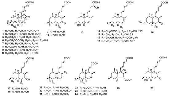
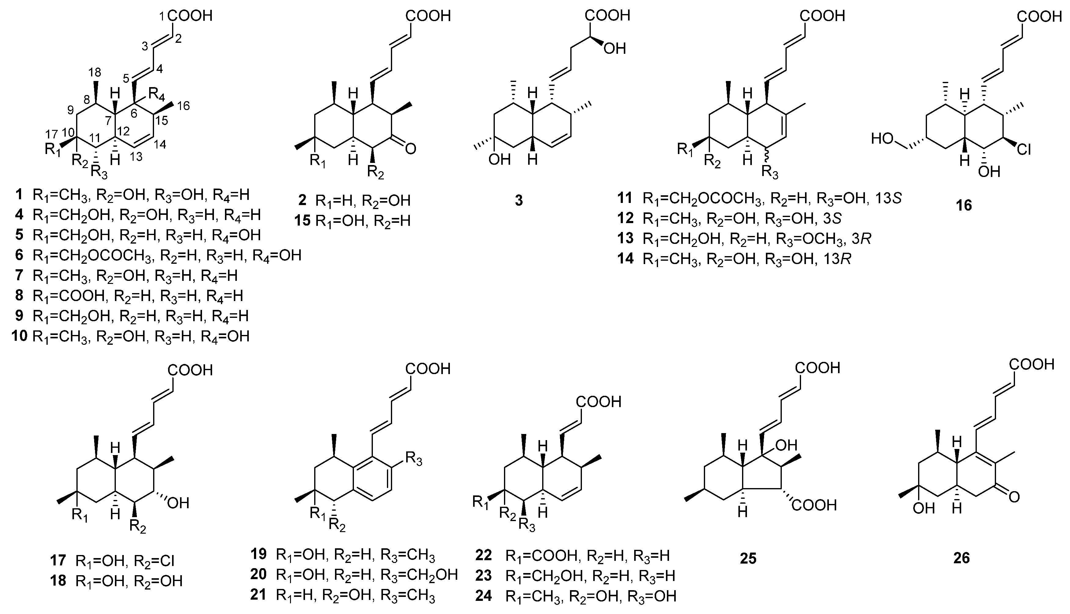
During our ongoing research on tanzawaic acids form Penicillium steckii [5,11], a series of structurally diverse tanzawaic acids were identified (Figure 1), and their anti-ulcerative colitis effects were tested. Herein, we report the isolation, structural elucidation, and inflammatory activity of tanzawaic acids.
Figure 1.
Chemical structures of compounds 1–26.
2. Results
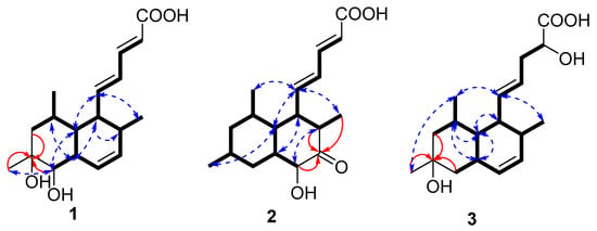
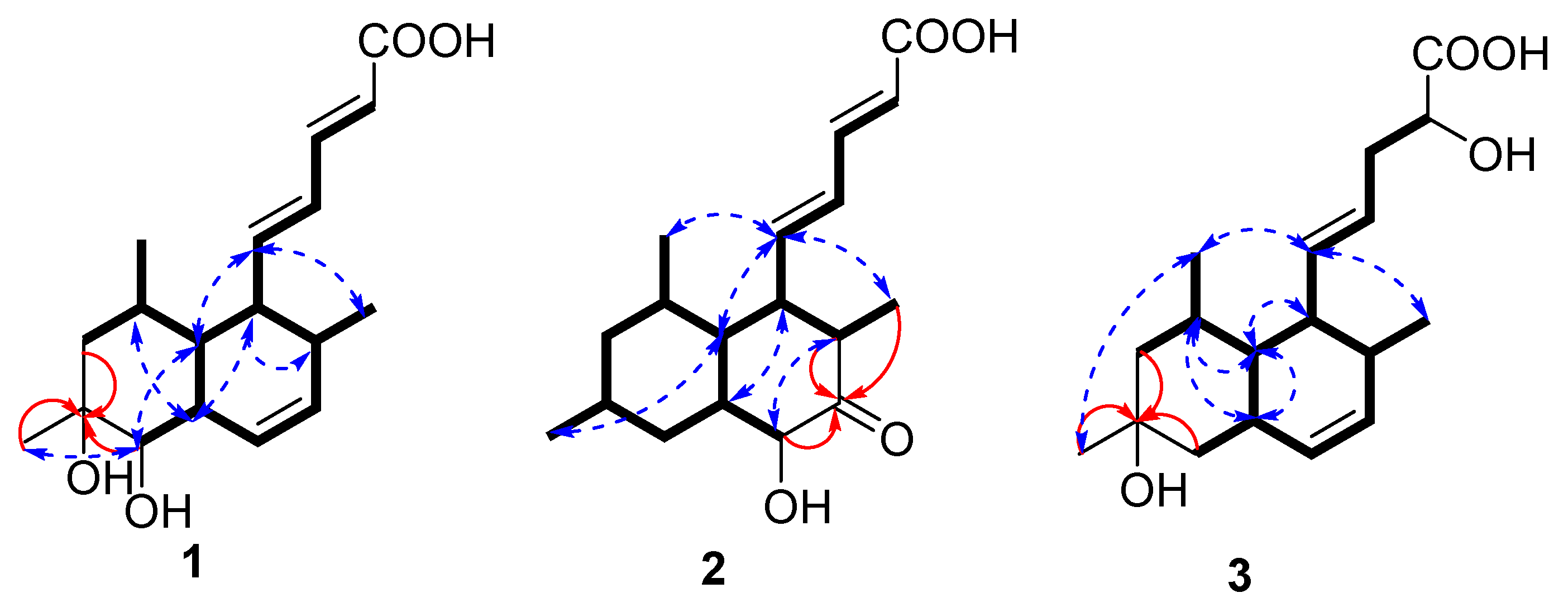
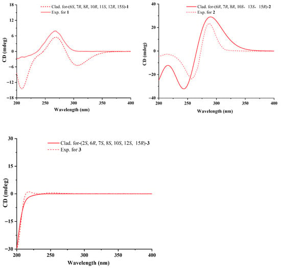
Compound 1 was isolated as a yellow powder with the molecular formula of C18H26O4 based on the HRESIMS ions detected at m/z 305.1762 [M − H]−, which required six degrees of unsaturation. The 1H NMR and 13C NMR data (Table 1) indicated six olefinic methines (δC/H 122.0/5.76, CH-2; 143.4/7.10, CH-3; 126.8/6.17, CH-4; 148.3/6.17, CH-5; 129.3/5.93, CH-13; 132.2/5.59, CH-14), five methines (δC/H 48.8/2.37, CH-6; 44.6/0.95, CH-7; 30.9/1.62, CH-8; 43.3/2.07, CH-12; 36.1/2.09, CH-15), one methylene (δC/H 48.8/1.09, 1.54, CH2-9), three methyls (δC/H 16.3/0.90, CH3-16; 27.4/1.08, CH3-17; 21.8/0.81, CH3-18), one oxygenated methine (δC/H 76.4/2.81, CH-11), and two quaternary carbonyls (δC 168.5, C-1; 70.4, C-10). Six degrees of unsaturation could be accounted for by the presence of one carboxyl, six sp2 carbon signals of three olefinic bonds, and two rings. The basic skeleton of the compound was deduced by COSY and HMBC data experiments (Figure 2). COSY correlations of H-6/H-7/H-8/H2-9, H-11/H-12/H-13/H-14/H-15, H-7/H-12 and HMBC correlations from H2-9 to C-10 and H-11 to C-10 revealed the presence of a decalin scaffold, which linked to three methyl groups at C-8, C-10, and C-15, respectively. The COSY data of H-2/H-3/H-4/H-5/H-6 pointed to a penta-2,4-dienoic acid chain connected at C-6, characteristic of tanzawaic acids. The remaining two hydroxyl groups were linked at C-10 and C-11, respectively, as determined by the HMBC signals of H2-9 to C-10 and H-11 to C-10. The planar structure was similar to those of tanzawaic acid E [18]. The relative configuration of compound 1 was determined by coupling constant data and NOESY correlations. The observed coupling constants (3JH-2-H-3 = 15.2 Hz, 3JH-4-H-5 = 15.0 Hz, and 3JH-13-H-14 = 9.6 Hz) indicated an E-configuration for the penta-2,4-dienoic acid side chain and a Z-configuration for the double bond in the ring. NOESY correlations (Figure 2) exhibited cross-peaks between H-12/H-6/H-8 and H-6/H-15 that indicated a co-facial arrangement, contrasting with those from H-5/H-7/H-11 on the opposite face. The absolute configuration of 1 was determined via the ECD spectrum (Figure 3) as 6S, 7R, 8R, 10R, 11S, 12R, 15S.
Table 1.
1H and 13C NMR spectroscopic data of 1–3 (500, 125 MHz, DMSO-d6).
Figure 2.
Key COSY (black bold lines), HMBC (red arrows), and NOESY (blue dotted line) correlations.
Figure 3.
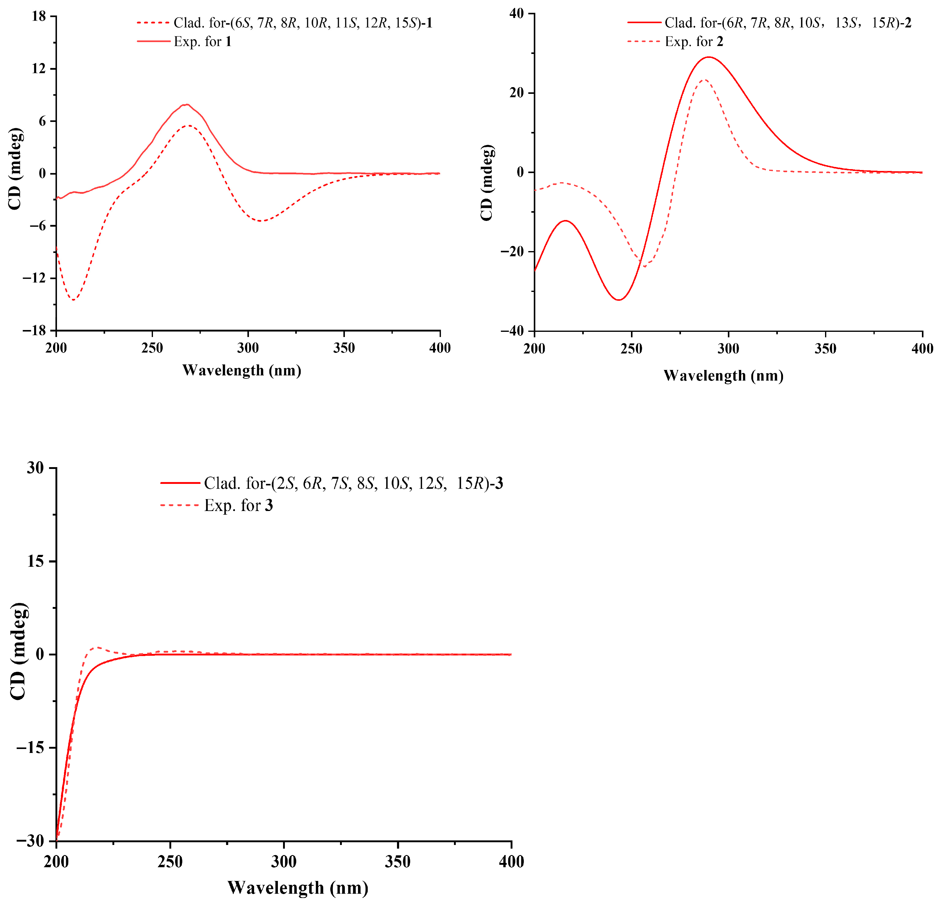
Experimental and calculated ECD spectra of compounds 1–3.
Compound 2 was obtained as a yellow oil and was assigned the molecular formula C18H26O4 via HRESIMS (m/z 305.1763 [M − H]−, calcd for C18H25O4−, 305.1758) and NMR data, with six degrees of unsaturation. Compound 2 and steckwaic acid J shared identical molecular formulas and functional groups, differing primarily in the hydroxyl group position [11]. Specifically, steckwaic acid J bore its hydroxyl group at C-10, whereas 2 carried this group at C-13 (δC 77.3), which was confirmed by COSY correlations of H-12 (δH 1.25)/H-13 (δH 3.88) and HMBC correlations from H-13 to C-14 (δC 210.4). The large coupling constants (15.4 Hz) of the double bonds Δ2 and Δ4 suggested their E-configuration. NOESY correlations (Figure 2) of H-7 (δH 1.40)/H3-17 (δH 0.87) and H-5 (δH 6.11)/H-7/H3-16 (δH 0.82)/H3-18 (δH 0.87) suggested the co-facial orientation of these groups, whereas correlations of H-12/H-6 (δH 1.99) and H-13/H-15 (δH 2.44) placed them on the opposite face. The experimental ECD spectra of 3 matched well with the calculated spectra for (6R, 7R, 8R, 10S, 12S, 13S, 15R)-2, confirming the absolute configurations of 2 as depicted.
Compound 3 was obtained as a yellow powder and has the molecular formula of C18H28O4 as established from HRESIMS analysis, indicative of five degrees of unsaturation. The NMR data of 3 were comparable with those of tanzawaic acid E [18], whereas it lacked one double-bond pair (δC 67.9 and δC 43.3). The loss of the double bond occurred in the penta-2,4-dienoic acid chain of compound 3, where an unusual addition reaction introduced a water molecule. This phenomenon was confirmed by COSY correlations observable among H-2 (δH 4.24)/H2-3 (δH 2.19/2.20)/H-4 (δH 5.32). The 3JH-4-H-5 and 3JH-13-H-14 coupling constants were 15.7 Hz and 9.5 Hz, respectively, which indicated E- and Z-configurations for the two double bonds. The NOESY correlations of H-5 (δH 5.61)/H3-16 (δH 0.86)/H3-18 (δH 0.88) and H3-18/H3-17 (δH 1.07) suggested that these two protons were co-facial, whereas correlations of H-8 (δH 1.62)/H-7 (δH 0.78)/H-12 (δH 2.19) and H-7/H-6 (δH 2.22) placed them on the opposite face. Considering the limited contribution of the flexible side chain to the ECD spectrum, and the lack of NOESY signals of H-2, a truncated model was used for the ECD calculations. The ECD calculations of the truncated model matched well with the experimental curves (Figure 3), confirming the 6R, 7S, 8S, 10S, 12S, 15R absolute configuration for 3. The absolute configuration at C-2 was determined by NMR calculations using the gauge-independent atomic orbital (GIAO) strategy at the B3LYP/6-31+G (d, p) level of theory with the PCM (DMSO-d6) solvation model [19,20]. As a result, the calculated NMR data, with a high DP4+ probability of 98.19%, indicated 2S-4 to be the most probable configuration. Therefore, the absolute configuration of 3 was established as 2S, 6R, 7S, 8S, 10S, 12S, 15R.
The known metabolites isolated from Penicillium steckii SCSIO 41040 included tanzawaic acid O (4) [2], penitanzacid A (5) [1], penicisteck acid Q (6) [5], tanzawaic acid C (7) [3], tanzawaic acid F (8) [18], tanzawaic acid M (9) [2], tanzawaic acid V (10) [3], 18-O-acetyltanzawaic acid R (11) [21], tanzawaic acid Z1 (12) [22], 13R-tanzawaic acid S (13) [21], 10-hydroxytanzawaic acid Q (14) [22], steckwaic acid J (15) [11], penicisteck acid S (16) [5], penitanzacid E (17) [1], penicisteck acid R (18) [5], tanzawaic acid D (19) [23], tanzawaic acid G (20) [11], penicisteck acid I (21) [5], steckwaic acid F (22) [21], steckwaic acid G (23) [21], penitanzacid H (24) [1], penicisteck acid P (25) [5], and penitanzacid F (26) [1]. These compounds were identified as the principal metabolites of the fungus through comparison of their spectroscopic data with previously reported values.
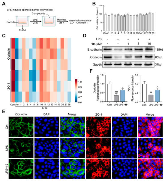
Ulcerative colitis remains a major global health burden with limited therapeutic options. The Caco-2/THP-1 co-culture system models both intestinal epithelial barrier function and immune cell-mediated inflammation, closely recapitulating ulcerative colitis’ pathogenesis for anti-UC drug evaluation. In this study, we evaluated the anti-UC potential of the tanzawaic acids isolated from the marine fungus Penicillium steckii SCSIO 41040 using an in vitro LPS-stimulated Caco-2/THP-1 co-culture model (Figure 4A) [24,25]. Initial cytotoxicity screening in Caco-2 cells revealed that the compounds maintained >90% cell viability at 10 μM, and they were further selected for subsequent pharmacological assessment (Figure 4B). Proteins such as ZO-1, claudin-1, and occludin constitute the tight junction protein family, which is essential for maintaining intestinal barrier integrity [26,27]. Therefore, this study initially screened the compounds for their ability to influence the expression of occludin and ZO-1. An immunofluorescence screening was conducted to assess the ability of each compound to restore the expression of occludin and ZO-1, and the results were visualized as a heatmap (Figure 4C). The results showed that compound 10 significantly reversed the LPS-induced downregulation of these tight junction proteins in Caco-2 cells, and it was selected for further assessment of its anti-UC potential. Western blot analysis demonstrated that 10 dose-dependently enhanced the expression of tight junction proteins (E-cadherin and occludin) (Figure 4D). Supporting these findings, immunofluorescence analysis showed that 10 effectively reversed the LPS-induced downregulation of occludin and ZO-1 in Caco-2 cells (Figure 4E,F). Conventional UC medications such as 5-ASA and corticosteroids primarily exert therapeutic effects by suppressing excessive inflammation, and they are associated with significant adverse reactions, including gastrointestinal intolerance and systemic immunosuppression. More than one patient in ten ultimately requires surgical intervention [16,17]. However, compound 10 directly targets the structural restoration of the intestinal epithelial barrier without inducing obvious cytotoxicity, representing a promising lead compound for UC treatment.
Figure 4.
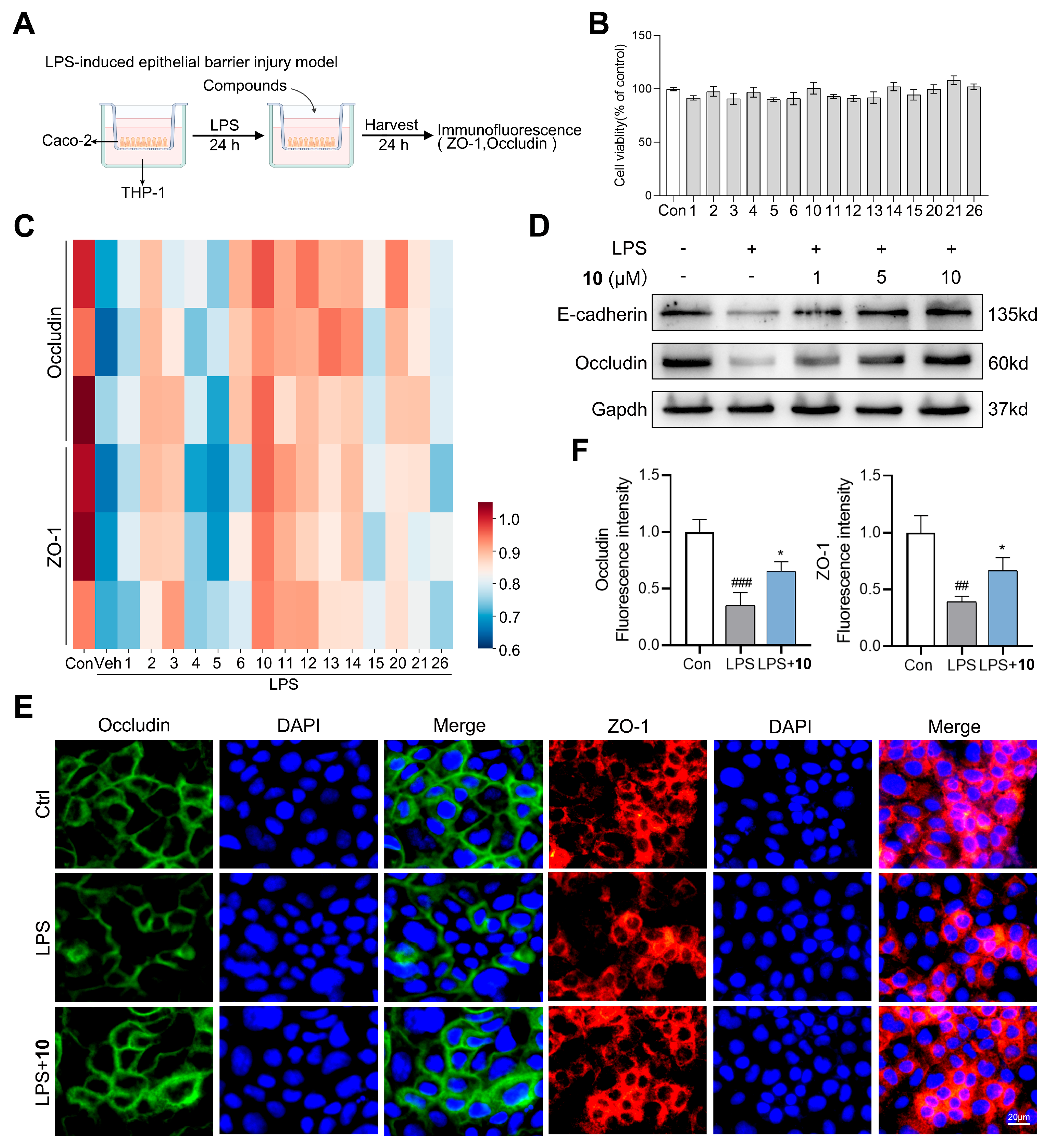
Effects of tanzawaic acids on the LPS-induced epithelial barrier injury model of Caco-2/THP-1 cells. (A) Schematic diagram of the Caco-2/THP-1 co-culture model for LPS-induced epithelial barrier injury. (B) Effects of tanzawaic acids on the cell viability of Caco-2 cells. (C) Occludin and ZO-1 expression in Caco-2 cells was determined by immunofluorescence of tanzawaic acids (10 μM). (D) Representative image of the Western blotting assay and the relative protein expression levels of E-cadherin and occludin in Caco-2 cells. (E) Occludin and ZO-1 expression in Caco-2 cells was determined by immunofluorescence of compound 10 (10 μM). (F) Quantitative analysis of (E). Values are expressed as mean ± SD. ## p < 0.01, ### p < 0.001 vs. control group; * p < 0.05 vs. LPS group.
3. Materials and Methods
3.1. General Experimental Procedures
Using a PerkinElmer 341 polarimeter (Hertford, UK), optical rotations were obtained. ECD spectra acquisition employed a Chirascan circular dichroism spectrometer (Applied Photophysics, Surrey, UK), while UV measurements utilized a Shimadzu UV-2600 UV−vis spectrophotometer (Shimadzu Corporation, Kyoto, Japan). For NMR analysis, both 1D and 2D spectra were acquired on Bruker AC 500 and 700 NMR spectrometers (Bruker, Fallanden, Switzerland), with TMS serving as the internal standard. X-ray diffraction intensity data were collected on an Agilent Xcalibur Nova single-crystal diffractometer using Cu Kα radiation (Agilent Technologies, Santa Clara, CA, USA). HRESIMS data collection occurred via a Bruker micro TOF-QII mass spectrometer (Fallanden, Switzerland), operating in either positive or negative ion mode. Thin layer chromatography (TLC) involved silica gel GF-254 (10–40 mm; Qingdao Marine Chemical Factory, Qingdao, China). Column chromatography (CC) relied upon a Sephadex LH-20 (Amersham Biosciences, Uppsala, Sweden) alongside silica gel (200–300 mesh and 100–200 mesh; Qingdao Marine Chemical Factory). HPLC separation was conducted on a Hitachi Primaide system equipped with a YMC ODS Series column (YMC-Pack ODS-A, 250 × 10 mm i.d., S-5 μm, 12 nm; YMC Co., Ltd., Kyoto, Japan). All solvents were of analytical grade (Tianjin Fuyu Chemical and Industry Factory, Tianjin, China), and both the fermentation culture medium and additional reagents originated from the Guangzhou Haili Aquarium Technology Company (Guangzhou, China).
3.2. Fungal Strain
From the South China Sea-collected green alga Botryocladia sp., the fungal strain Penicillium stecki SCSIO 41040 was isolated. This strain was subsequently preserved at 4 °C on Mueller–Hinton broth (MB) agar slants—containing malt extract (16 g), agar (18 g), sea-salt (30 g), and water (1 L) with pH 7.4–7.8—and ultimately archived at the CAS Key Laboratory of Tropical Marine Bio-Resources and Ecology, South China Sea Institute of Oceanology, Chinese Academy of Sciences (Guangzhou, China). Following PCR amplification of its ITS1-5.8S-ITS2 rDNA region (550 bp; GenBank: OP349656), DNA sequencing revealed 100% homology to Penicillium steckii (GenBank: NR111488).
3.3. Fermentation and Extraction
Into a 500 mL Erlenmeyer flask containing 150 mL seed medium (malt extract 1%, yeast extract 0.4%, glucose 0.4%, pH 7.2), several loops of strain SCSIO 41040 cells were inoculated. Subsequently, the seed culture was incubated at 28 °C for 48 h on a rotary shaker (180 rpm). For large-scale fermentation, 1 L conical flasks—each holding solid medium (300 mL/flask) prepared from KH2PO4 (0.5 g/L), MgSO4·7H2O (0.3 g/L), sorbitol (20 g/L), tryptophan (0.5 g/L), maltose (20 g/L), yeast extract (3 g/L), monosodium glutamate (10 g/L), and sea salt (32 g/L)—were inoculated and maintained at 25 °C. Following 32 days of cultivation, each flask’s fermented material underwent sequential EtOAc extraction (700 mL/flask), whereupon the combined extracts were suspended in MeOH. Petroleum ether was then employed to extract rice oil contaminants; ultimately, after concentration under reduced pressure, the MeOH solution yielded 40.0 g of reddish brown extract.
3.4. Isolation and Purification
The extract was subjected to silica gel column chromatography (CC), eluting with a gradient of CH2Cl2-MeOH (100:0–0:100) to give 15 fractions based on TLC properties. Initial purification of Fr.4, Fr.5, Fr.7, Fr.10, and Fr.11 was performed using ODS silica gel chromatography with MeOH/H2O (5–100%) as the eluent. Compound 14 (4.5 mg, tR 12.1 min) was isolated from Fr.2 by semi-preparative HPLC (70% MeCN−H2O + 0.3% TFA, 2.5 mL/min). Compound 15 (4.5 mg, tR 33.1 min) was isolated using HPLC from Fr.4.5 (25% MeCN−H2O + 0.3% TFA, 2.5 mL/min). Fr.7.3 was subjected to semi-preparative reversed-phase HPLC (62% MeOH−H2O + 0.3% TFA, 2.5 mL/min) to give four fractions. Fr.7.3.2 was processed through HPLC (60% MeOH/H2O + 0.3% TFA, 2.5 mL/min) to afford compounds 17 (48.0 mg, tR 18.0 min), 20 (11.3 mg, tR 22.0 min), and 26 (3.1 mg, tR 25.2 min). The separation of Fr.7.4 via HPLC (32% MeCN−H2O + 0.3% TFA, 2.5 mL/min) delivered compound 1 (11.5 mg, tR 15.2 min). Fr.10.5 was further purified by semi-preparative HPLC (45% MeCN−H2O + 0.3% TFA, 2.5 mL/min) to give five sub-fractions. Fr.10.5.5 was separated by semi-preparative HPLC (42% MeCN−H2O + 0.3% TFA, 2.5 mL/min) to give compounds 3 (3.3 mg, tR 15.4 min) and 19 (3.7 mg, tR 22.1 min). Then, 18 (4.0 mg, tR 19.0 min) was afforded from Fr. 10.6 by HPLC (33% MeCN−H2O + 0.3% TFA, 2.5 mL/min). Fr.11.2 was further divided into six parts (Fr.11.2.1–Fr.11.2.6) through combiflash nextgen 300+ with MeOH. Fr.11.2.5 was purified by semi-preparative HPLC (30% MeCN/H2O + 0.3% TFA, 2.5 mL/min) to yield 12 (3.9 mg, tR 15.0 min) and 24 (2.3 mg, tR 8.8 min). Fr.11.2.6 was separated by semi-preparative HPLC (32% MeCN/H2O + 0.3% TFA, 2.5 mL/min) to provide 4 (1.8 mg, tR 16.0 min) and 10 (14.0 mg, tR 8.0 min). Fr.11.3 was subjected to semi-preparative reversed-phase HPLC (60% MeOH−H2O + 0.3% TFA, 2.0 mL/min) to give 25 (3.2 mg, tR 25.0 min) and an additional four fractions (Fr.11.3.1–Fr.11.3.4). Fr.11.3.2 was further purified by semi-preparative HPLC (36% MeCN−H2O, 2.5 mL/min) to give compound 23 (5.4 mg, tR 15.3 min). The purification of Fr.11.3.3 by HPLC (36% MeCN/H2O, 2.5 mL/min) yielded compounds 11 (2.7 mg, tR 19.8 min), 5 (1.3 mg, tR 17.8 min), and 13 (3.3 mg, tR 27.5 min). Compound 21 (6.9 mg, tR 16.0 min) and five sub-fractions (Fr.11.4.1–Fr.11.4.5) of component Fr.11.4 were obtained by semi-preparative reversed-phase high-performance liquid chromatography (68% MeCN−H2O + 0.3% TFA, 2.5 mL/min). Compound 16 (2.8 mg, tR 14.0 min) was isolated using HPLC from Fr.11.4.2 (47% MeCN/H2O, 2.5 mL/min). From Fr.11.4.3, compound 6 (6.3 mg, tR 34.0 min) was acquired following HPLC (68% MeOH/H2O, 2.5 mL/min). Compounds 2 (1.8 mg, tR 18.3 min) and 22 (9.3 mg, tR 13.0 min) were isolated from Fr.11.4.5 by HPLC (62% MeOH−H2O + 0.3% TFA, 2.5 mL/min). Fr.11.5 was fractionated by HPLC (70% MeOH−H2O + 0.3% TFA, 2.5 mL/min), yielding compound 7 (20.0 mg, tR 27.0 min) and four sub-fractions (Fr.11.5.1–Fr.11.5.4). Subsequently, compounds 8 (14.0 mg, tR 29.0 min) and 9 (52.0 mg, tR 21.4 min) were isolated from Fr.11.5.3 and Fr.11.5.4 via HPLC (67% MeOH/H2O, 2.5 mL/min; 70% MeOH/H2O, 2.5 mL/min), respectively.
1: Yellow powder; + 30.9 (c, 0.1, MeOH); UV (MeOH) λmax (log ε) 263 (4.17); ECD (0.130 mM, MeOH) λmax (Δε) 268 (+7.90) nm; 1H and 13C NMR data, Table 1; HRESIMS m/z 305.1762 [M − H]− (calcd for C18H25O4−, 305.1758).
2: Yellow powder; + 22.1 (c, 0.1, MeOH); UV (MeOH) λmax (log ε) 262 (4.27); ECD (0.131 mM, MeOH) λmax (Δε) 257 (-23.79), 288 (+23.29) nm; 1H and 13C NMR data, Table 1; HRESIMS m/z 305.1763 [M − H]− (calcd for C18H25O4−, 305.1758).
3: Yellow oil; + 35.4 (c, 0.1, MeOH); UV (MeOH) λmax (log ε) 200 (3.91); ECD (0.130 mM, MeOH) λmax (Δε) 200 (−30.00) nm; 1H and 13C NMR data, Table 1; HRESIMS m/z 307.1912 [M − H]− (calcd for C18H27O4−, 307.1915).
Crystal data for C36H48O8 (M = 608.74 g/mol) (8): monoclinic, space group P21 (no. 4), a = 7.03460(10) Å, b = 12.6237(2) Å, c = 18.8721(3) Å, β = 89.1090(10)°, V = 1675.69(4) Å3, Z = 2, T = 100.00(10) K, μ(Cu Kα) = 0.680 mm−1, Dcalc = 1.206 g/cm3, 31,927 reflections measured (8.428° ≤ 2Θ ≤ 148.944°), 6691 unique (Rint = 0.0597, Rsigma = 0.0320), which were used in all calculations. The final R1 was 0.0377 (I > 2σ(I)) and wR2 was 0.1049 (all data).
Crystal data for C16H22O4 (M = 278.33 g/mol) (22): orthorhombic, space group P212121 (no. 19), a = 5.12820(10) Å, b = 11.6636(2) Å, c = 24.1513(3) Å, V = 1444.57(4) Å3, Z = 4, T = 99.98(14) K, μ(Cu Kα) = 0.739 mm−1, Dcalc = 1.280 g/cm3, 13,864 reflections measured (7.32° ≤ 2Θ ≤ 148.872°), 2896 unique (Rint = 0.0426, Rsigma = 0.0301), which were used in all calculations. The final R1 was 0.0314 (I > 2σ(I)) and wR2 was 0.0795 (all data).
Crystal data for C16H24O3 (M = 264.35 g/mol) (23): monoclinic, space group I2 (no. 5), a = 12.72599(11) Å, b = 5.50132(5) Å, c = 22.58030(18) Å, β = 101.3861(8)°, V = 1549.73(2) Å3, Z = 4, T = 99.98(12) K, μ(Cu Kα) = 0.611 mm−1, Dcalc = 1.133 g/cm3, 15,187 reflections measured (7.416° ≤ 2Θ ≤ 148.936°), 3088 unique (Rint = 0.0257, Rsigma = 0.0186), which were used in all calculations. The final R1 was 0.0263 (I > 2σ(I)) and wR2 was 0.0667 (all data).
Compounds 8, 22, and 23 were crystallized from slow evaporation in a CH3OH solution (Figure S33). A single crystal with the indicated dimensions was selected and measured on an Agilent Xcalibur Nova single-crystal diffractometer using Cu Kα radiation and refined by full-matrix least-squares calculation. The crystallographic data for structures 8, 22, and 23 have been deposited with the Cambridge Crystallographic Data Centre with supplementary publication numbers CCDC-2499249, CCDC-2499251, and CCDC-2499252, respectively.
3.5. ECD Calculation
The new compounds underwent random conformational searches using Spartan’14 (with MMFF) and Gaussian 09 (with DFT/TDDFT). The MMFF search first yielded low-energy conformers (Boltzmann population > 5%), which were then geometrically optimized via DFT (B3LYP/6-311G*) in MeOH using the IEFPCM model. For these stable conformers, ECD calculations were further performed in MeOH using TDDFT at the B3LYP/6-311G* level. Finally, Multiwfn (0.2–0.4 eV half-bandwidth) generated the ECD spectra, and the Boltzmann-weighted contributions were calculated after UV simulation.
3.6. Cytotoxic Activity Measurement
Caco-2 cells were seeded at a density of 4 × 10³ cells/well in 96-well plates. After treating the Caco-2 cells with different concentrations of tanzawaic acids for 24 h, 10 μL CCK-8 (PWL111, Bgbioscience, Dalian, China) test reagent was added to each well, following the manufacturer’s instructions [25,28].
3.7. Caco-2 and THP-1 Co-Culture Model Incubation
In brief, Caco-2 cells were seeded onto Transwell insert plates at a concentration of 1.5 × 10⁵ cells/cm2 and cultured for 21 days until cells were fully differentiated. The culture medium was changed every 2–3 days. THP-1 were seeded onto 6-well plates (4 × 10⁶ cells/well) and rested for 24 h. After replacing the media with complete DMEM, inserts with Caco-2 were added into plates containing THP-1. One microgram per milliliter of LPS was added to the basolateral side, and, after 3 h of incubation, different concentrations of tanzawaic acids (10 μM) were added to the apical side of the insert. After 24 h of incubation, Caco-2 cells from the insert were collected.
3.8. Western Blot Analysis
Caco-2 cells were lysed with ice-cold radioimmunoprecipitation assay (RIPA) lysis buffer (P0013B; Beyotime Biotechnology, Shanghai, China) supplemented with protease and phosphatase inhibitors, and the lysates were centrifuged at 14,000 r/min for 15 min. The protein concentrations were measured with a bicinchoninic acid (BCA) kit (23,227; Thermo Fisher Scientific, Waltham, MA, USA), and the protein samples were prepared using 4× sodium dodecyl sulfate–polyacrylamide gel electrophoresis (SDS-PAGE) loading buffer. Equal amounts of proteins were loaded onto 10% SDS-PAGE gels, and the separated proteins were transferred to polyvinylidene difluoride (PVDF) membranes (3010040001; Roche Diagnostics, Basel, Switzerland). The membranes were blocked with 5% skim milk in tris-buffered saline with Tween 20 (TBST) and incubated with the indicated primary antibodies at 4 °C overnight. The membranes were then washed and incubated with a horseradish peroxidase (HRP)-conjugated secondary antibody for 1 h at room temperature. Finally, the bands were detected using an enhanced chemiluminescence (ECL) kit (P0018HM; Beyotime Biotechnology, China). The antibodies used for Western blot analysis were occludin (Servicebio, GB111401-100, 1:1000, Wuhan, China), E-cadherin (Abcam, ab76055, 1:1000, Cambridge, UK), and Gapdh (Abmart, P60037; 1:3000, Shanghai, China).
3.9. Immunofluorescence
In brief, Caco-2 cells were fixed with 4% paraformaldehyde for 15 min at room temperature and washed three times with cold PBS for 3 min each. Treatment with 0.5% Triton-X 100 was performed for 20 min at room temperature, followed by three additional washes with cold PBS for 3 min each. The cells were then blocked with 10% goat serum for 1 h at room temperature. Subsequently, the blocking solution was removed, and the cells were incubated overnight at 4 °C with the occludin (Servicebio, GB111401-100, 1:200) and ZO-1 (Servicebio, GB111402-100, 1:200) primary antibody. On the following day, the antibody was removed, and the cells were washed three times with PBS for 3 min each. FITC-AffiniPuro Goat Anti-Mouse IgG (Yeasen, 33207ES60, 1:100, Shanghai, China) was added, and cells were incubated in the dark at 37 °C for 1 h, followed by three washes with PBS for 3 min each. Finally, 5 μg/mL DAPI was added, and cells were incubated at room temperature for 5 min and washed three times with PBS for 5 min each. Fluorescence imaging was conducted using a laser confocal microscope (TCSSP8 DIVE, Leica, Wetzlar, Germany).
4. Conclusions
Marine-derived fungi have emerged as an underexplored reservoir of structurally unique secondary metabolites with diverse pharmacological activity. Tanzawaic acids, a class of decalin-containing polyketides, have previously been reported to exhibit antibacterial, antifungal, and anti-inflammatory properties. However, to the best of our knowledge, this is the first systematic investigation of their intestinal barrier-protective activity, which significantly expands the therapeutic potential of this compound class. In this study, we demonstrated that tanzawaic acids, particularly compound 10, could effectively reverse the LPS-induced downregulation of ZO-1 and occludin, thereby restoring intestinal barrier integrity. However, several limitations of this study should be acknowledged. First, only in vitro activity was evaluated, and the in vivo therapeutic efficacy of compound 10 remains to be verified in animal models such as dextran sulfate sodium (DSS)-induced colitis. Second, the detailed molecular mechanisms underlying its barrier-protective effect, including specific signaling pathways, have not been fully elucidated. These unresolved questions will be the focus of our ongoing and future research efforts.
In summary, a further study of a marine-derived Penicillium steckii revealed three new (1–3) and twenty-three known (4–26) tanzawaic acids, which were obtained from the strain fermented on a liquid medium. Extensive NMR spectroscopic and HRESIMS data and ECD and NMR calculations were used to determine the structures of the new compounds. Based on the LPS-induced Caco-2/THP-1 cell injury model, the protective role of tanzawaic acids in inflammatory damage to the intestinal epithelial barrier was measured. Notably, compound 10 enhanced the expression and membrane localization of ZO-1 and occludin, which contributed to the restoration of barrier integrity. This study not only enriches the structural diversity of tanzawaic acids but also identifies compound 10 as a potential lead compound for UC treatment. Further in vivo efficacy studies, mechanistic investigations, and structural optimizations are warranted to translate these findings into clinical applications.
Supplementary Materials
The following supporting information can be downloaded at https://www.mdpi.com/article/10.3390/md24050147/s1. Figures S1–S32: 1H, 13C, DEPT135, COSY, HSQC, HMBC, NOESY, HRESIMS, UV spectra, CD spectra, and DP4+ analysis of the new compounds 1–3; Figure S33 X-ray crystal structures of 8, 22 and 23; Figure S34 The original untreated photograph of the Western blot gel.
Author Contributions
Conceptualization, J.W. (Junfeng Wang) and Y.L. (Yonghong Liu); formal analysis, Y.S., J.W. (Jiamin Wang), and Y.L. (Yuchen Lin); investigation, Y.L. (Yalin Liu), J.S., and Y.S.; project administration, C.G. and J.W. (Junfeng Wang); writing—original draft, Y.S. and X.Y.; writing—review and editing, J.W. (Junfeng Wang) and Y.L. (Yonghong Liu). All authors have read and agreed to the published version of the manuscript.
Funding
This research was funded by the Natural Science Foundation of Guangxi (2026GXNSFBA00640037), the Guangxi Key Research and Development Programme (AB24010109), and the Middle-Aged Teachers Scientific Research Project of Guangxi Universities (2025KY0396).
Institutional Review Board Statement
Not applicable.
Data Availability Statement
The original contributions presented in this study are included in the article.
Acknowledgments
We are grateful to Z.H. Xiao, A.J. Sun, X.H. Zheng, Y. Zhang, and X. Ma at the analytical facility at SCSIO for recording the spectroscopic data and to W.Z. Xiang for providing the samples of the green algae Botryocladia sp.
Conflicts of Interest
The authors declare no conflicts of interest.
References
- Wang, J.; Li, T.; Wang, P.; Ding, W.; Xu, J. Tanzawaic Acids from a Deep-Sea Derived Penicillium Species. J. Nat. Prod. 2022, 85, 1218–1228. [Google Scholar] [CrossRef] [Scilit]
- Cardoso, F.; Rosa, J.M.; Diaz, A.R.; Darias, J.; Cerella, C.; Diederich, M.; Cueto, M. Tanzawaic Acids Isolated from a Marine-Derived Fungus of the Genus Penicillium with Cytotoxic Activities. Org. Biomol. Chem. 2015, 13, 7248–7256. [Google Scholar] [CrossRef] [Scilit]
- Yu, G.; Wang, S.; Wang, L.; Che, Q.; Zhu, T.; Zhang, G.; Guo, P.; Li, D. Lipid-Lowering Polyketides from the Fungus Penicillium Steckii HDN13-279. Mar. Drugs 2018, 16, 25. [Google Scholar] [CrossRef] [Scilit]
- Sofian, F.F.; Warahapsari, F.A.; Yoshida, J.; Ito, Y.; Koseki, T.; Shiono, Y. Two New Octahydronaphthalene Derivatives, Trichodermic Acids C and D Produced by Trichoderma sp. HN-1.1. Nat. Prod. Res. 2023, 37, 484–493. [Google Scholar] [CrossRef] [Scilit]
- Chen, C.; Xiao, L.; Luo, X.; Cai, J.; Huang, L.; Tao, H.; Zhou, X.; Tan, Y.; Liu, Y. Identifying Marine-Derived Tanzawaic Acid Derivatives as Novel Inhibitors against Osteoclastogenesis and Osteoporosis via Downregulation of NF-KappaB and NFATc1 Activation. J. Med. Chem. 2024, 67, 2602–2618. [Google Scholar] [CrossRef] [Scilit]
- Sandjo, L.P.; Thines, E.; Opatz, T.; Schuffler, A. Tanzawaic acids I-L: Four New Polyketides from Penicillium sp. IBWF104-06. Beilstein J. Org. Chem. 2014, 10, 251–258. [Google Scholar] [CrossRef] [Scilit]
- Hu, X.Y.; Li, X.M.; Wang, B.G.; Meng, L.H. Tanzawaic Acid Derivatives: Fungal Polyketides from the Deep-Sea Coral-Derived Endozoic Penicillium steckii AS-324. J. Nat. Prod. 2022, 85, 1398–1406. [Google Scholar] [CrossRef] [Scilit] [PubMed]
- Tanaka, K.; Arima, S.; Lee, D.; Ohte, S.; Tomoda, H.; Uchida, R.; Ohshiro, T.; Nagamitsu, T. Structure-Activity Relationship Studies of Tanzawaic Acid A, an Antifungal Agent against Rhizopus oryzae. Bioorganic Med. Chem. Lett. 2025, 128, 130343. [Google Scholar] [CrossRef] [Scilit] [PubMed]
- Abdelwahab, M.F.; Fouad, M.A.; Kamel, M.S.; Ozkaya, F.C.; Kalscheuer, R.; Muller, W.E.G.; Lin, W.; Liu, Z.; Ebrahim, W.; Daletos, G.; et al. Tanzawaic Acid Derivatives from Freshwater Sediment-Derived Fungus Penicillium sp. Fitoterapia 2018, 128, 258–264. [Google Scholar] [CrossRef] [Scilit] [PubMed]
- Quang, T.H.; Ngan, N.T.T.; Ko, W.; Kim, D.C.; Yoon, C.S.; Sohn, J.H.; Yim, J.H.; Kim, Y.C.; Oh, H. Tanzawaic Acid Derivatives from a Marine Isolate of Penicillium sp. (SF-6013) with Anti-Inflammatory and PTP1B Inhibitory Activities. Bioorganic Med. Chem. Lett. 2014, 24, 5787–5791. [Google Scholar] [CrossRef] [Scilit]
- Song, Y.; Tan, Y.; She, J.; Chen, C.; Wang, J.; Hu, Y.; Pang, X.; Wang, J.; Liu, Y. Tanzawaic Acid Derivatives from the Marine-Derived Penicillium steckii as Inhibitors of RANKL-Induced Osteoclastogenesis. J. Nat. Prod. 2023, 86, 1171–1178. [Google Scholar] [CrossRef] [Scilit] [PubMed]
- Zhang, Y.; Ge, F.; Qu, H.; Zhao, C.; Gu, J.; Xu, Q.; Lei, H.; Liu, J.; Wang, X.; Chu, Y.; et al. Wumei Wan Ameliorates Ulcerative Colitis in rats by Modulating the Inflammation-Pyroptosis-Intestinal Stem Cell Axis. J. Ethnopharmacol. 2026, 355, 120645. [Google Scholar] [CrossRef] [Scilit] [PubMed]
- Xie, X.; Li, P.; Zhao, M.; Xu, B.; Zhang, G.; Wang, Q.; Ni, C.; Luo, X.; Zhou, L. Luteolin ameliorates ulcerative colitis in mice via reducing the depletion of NCR + ILC3 through Notch signaling pathway. Chin. J. Nat. Med. 2024, 22, 991–1002. [Google Scholar] [CrossRef] [Scilit]
- Wang, J.; Zhang, C.; Qin, J.; An, N.; Bai, M.; Du, R.H.; Shen, Y.; Wu, X.D.; Cheng, J.C.; Wu, X.F.; et al. Direct Inhibition of the TXNIP-NLRP3-GSDMD Pathway Reduces Pyroptosis in Colonocytes and Alleviates Ulcerative Colitis in Mice by the Small Compound PEITC. Acta Pharmacol. Sin. 2025, 46, 2436–2449. [Google Scholar] [CrossRef] [Scilit]
- Wan, Y.; Yang, L.; Jiang, S.; Qian, D.; Duan, J. Excessive Apoptosis in Ulcerative Colitis: Crosstalk Between Apoptosis, ROS, ER Stress, and Intestinal Homeostasis. Inflamm. Bowel Dis. 2022, 28, 639–648. [Google Scholar] [CrossRef] [Scilit]
- Sandborn, W.J.; Panes, J.; Sands, B.E.; Reinisch, W.; Su, C.; Lawendy, N.; Koram, N.; Fan, H.; Jones, T.V.; Modesto, I.; et al. Venous Thromboembolic Events in the Tofacitinib Ulcerative Colitis Clinical Development Programme. Aliment. Pharmacol. Ther. 2019, 50, 1068–1076. [Google Scholar] [CrossRef] [Scilit]
- Le Berre, C.; Honap, S.; Peyrin-Biroulet, L. Ulcerative colitis. Lancet 2023, 402, 571–584. [Google Scholar] [CrossRef] [Scilit]
- Malmstrùma, J.; Christophersena, C.; Frisvad, J.C. Secondary Metabolites Characteristic of Penicillium citrinum, Penicillium steckii and Related Species. Phytochemistry 2000, 54, 301–309. [Google Scholar] [CrossRef] [Scilit]
- Cong, M.; Li, Y.; Li, Y.; Chen, X.; Pang, X.; Yan, Y.; Tian, X.; Liu, Y.; Yang, J.; Wang, J. Spirochrains A–D, Four Caged [5,6,5] Spirocyclic Amides from an Antarctic Fungus Aspergillus ochraceopetaliformis SCSIO 05702 with Anti-RSV Activities. Org. Lett. 2025, 27, 2295–2299. [Google Scholar] [CrossRef] [Scilit]
- Chen, W.; Jiang, J.; Pang, X.; Song, Y.; Yang, Z.; Wang, J.; Liu, Y. Neuroprotective Azaphilones from a Deep-Sea Derived Fungus Penicillium sp. SCSIO41030. Org. Biomol. Chem. 2024, 22, 4521–4527. [Google Scholar] [CrossRef] [Scilit] [PubMed]
- Hu, X.Y.; Li, X.M.; Wang, B.G.; Meng, L.H. Uncommon Polyketides from Penicillium steckii AS-324, a Marine Endozoic Fungus Isolated from Deep-Sea Coral in the Magellan Seamount. Int. J. Mol. Sci. 2022, 23, 6332. [Google Scholar] [CrossRef] [Scilit]
- Dramae, A.; Intaraudom, C.; Bunbamrung, N.; Boonyuen, N.; Auncharoen, P.; Pittayakhajonwut, P. Antimicrobial Tanzawaic Acid Derivatives from the Endophytic Penicillium citrinum BCC71086. Tetrahedron 2022, 106, 132645. [Google Scholar] [CrossRef] [Scilit]
- El-Neketi, M.; Ebrahim, W.; Lin, W.; Gedara, S.; Badria, F.; Saad, H.E.; Lai, D.; Proksch, P. Alkaloids and Polyketides from Penicillium citrinum, an Endophyte Isolated from the Moroccan Plant Ceratonia siliqua. J. Nat. Prod. 2013, 76, 1099–1104. [Google Scholar] [CrossRef] [Scilit]
- de Rocha, G.H.O.; Müller, C.; Przybylski, S.; Schaller, H.; Riemschneider, S.; Lehmann, J. AhR-Induced Anti-Inflammatory Effects on a Caco-2/THP-1 Co-Culture Model of Intestinal Inflammation are Mediated by PPARγ. Int. J. Mol. Sci. 2024, 25, 13072. [Google Scholar] [CrossRef] [Scilit]
- Duan, Y.; Sun, W.; Li, Y.; Shi, Z.; Li, L.; Zhang, Y.; Huang, K.; Zhang, Z.; Qi, C.; Zhang, Y. Spirohypertones A and B as Potent Antipsoriatics: Tumor Necrosis Factor-Alpha Inhibitors with Unprecedented Chemical Architectures. Acta Pharm. Sin. B 2024, 14, 2646–2656. [Google Scholar] [CrossRef] [Scilit]
- Buckley, A.; Turner, J.R. Cell Biology of Tight Junction Barrier Regulation and Mucosal Disease. Cold Spring Harb. Perspect. Biol. 2018, 10, a029314. [Google Scholar] [CrossRef] [Scilit] [PubMed]
- Kuo, W.T.; Odenwald, M.A.; Turner, J.R.; Zuo, L. Tight Junction Proteins Occludin and ZO-1 as Regulators of Epithelial Proliferation and Survival. Ann. N. Y. Acad. Sci. 2022, 1514, 21–33. [Google Scholar] [CrossRef] [Scilit] [PubMed]
- Zheng, J.; Zhang, J.; Zhou, Y.; Zhang, D.; Guo, H.; Li, B.; Cui, S. Taurine Alleviates Experimental Colitis by Enhancing Intestinal Barrier Function and Inhibiting Inflammatory Response through TLR4/NF-kappaB Signaling. J. Agric. Food Chem. 2024, 72, 12119–12129. [Google Scholar] [CrossRef] [Scilit] [PubMed]

Disclaimer/Publisher’s Note: The statements, opinions and data contained in all publications are solely those of the individual author(s) and contributor(s) and not of MDPI and/or the editor(s). MDPI and/or the editor(s) disclaim responsibility for any injury to people or property resulting from any ideas, methods, instructions or products referred to in the content. |
© 2026 by the authors. Licensee MDPI, Basel, Switzerland. This article is an open access article distributed under the terms and conditions of the Creative Commons Attribution (CC BY) license.