


Abstract
Juglanin (kaempferol 3-O-α-L-arabinofuranoside) is a flavonol glycoside occurring in many plants, including its commercial sources Juglans regia, Polygonum aviculare and Selliguea hastata. Recent extensive studies have explored the potential of using juglanin in various pathological conditions, including cardiovascular disorders, central nervous and skeletal system disorders, metabolic syndrome, hepatic injury, and cancers. The results indicated a wide range of effects, like anti-inflammatory, anti-oxidant, anti-fibrotic, anti-thrombotic, anti-angiogenic, hepatoprotective, hypolipidemic, hypoglycemic, anti-apoptotic (normal cells), and pro-apoptotic (cancer cells). The health-promoting properties of juglanin can be attributed to its influence on many signaling pathways, associated with SIRT1, AMPK, Nrf2, STING, TLR4, MAPKs, NF-κB, AKT, JAK, and their downstream genes. This review primarily summarizes the current knowledge of molecular mechanisms, pharmacokinetics, biocompatibility, and human use safety of juglanin. In addition, the most promising new plant sources and other existing challenges and prospects have also been reviewed and discussed, aiming to provide direction and rationale for the further development and broader pharmaceutical application of juglanin.
1. Introduction
For centuries, plants have served as not just food or building materials but also as a rich base of therapeutic benefits. Today, they continue to be a subject of extensive research as potential sources of molecules for drug development. Among the critical components of plant products and leading model natural drugs are flavonoids, a vast family with over 8000 known representatives in the plant kingdom. While industrial compounds and clinically approved medicines like rutin, diosmin, hesperidin, or silybin have emerged from this group, the potential of flavonoids is still being intensely evaluated, with numerous other members showing promising biological properties [1].
Juglanin (kaempferol 3-O-α-L-arabinofuranoside, CAS 5041-67-8, Figure 1) is a member of a large group of flavonols and a compound that has garnered significant attention in recent years. It is found in a variety of plant species, including Juglans spp., e.g., Juglans regia L. (hence its common name), Polygonum aviculare L., Selliguea hastata (Thunb.) H. Ohashi and K. Ohashi (these last three being commercial sources of juglanin), Rubus spp., Prunus spp., Rosa spp., etc. [2,3,4,5]. Alongside astragalin, tiliroside, and kaempferol 3-O-rutinoside, juglanin is one of the most prevalent natural flavonols within the kaempferol glycoside group, although this group is less widespread in the plant kingdom than quercetin glycosides, such as rutin or isoquercitrin.
Figure 1.
The chemical structure of juglanin.
The exploration of juglanin’s biological activity has a rich history, dating back to 1984. However, the bulk of research has been conducted in 2020–2023, indicating a significant surge in interest. These studies, which encompass mainly in vivo animal models and in vitro cellular studies, have explored the potential of using juglanin for a wide range of conditions. The anti-inflammatory, anti-oxidative, anti-fibrotic, and anti-apoptotic (or pro-apoptotic in the case of cancer cells) properties of juglanin have been identified as its primary modes of action. The signaling pathways targeted by the compound, including those dependent on the 5′AMP-activated protein kinase (AMPK), mitogen-activated protein kinases (MAPKs), nuclear factor kappa-light-chain-enhancer of activated B cells (NF-kB), and many others, have been extensively studied, further validating its potential [6,7,8,9,10,11,12,13].
The wealth of accumulated results, pointing to the unique and promising features of juglanin, forms the foundation of this paper. The review primarily focuses on the molecular mechanisms of juglanin activity but also delves into its pharmacokinetic properties, biocompatibility, and human use safety. In addition, the known plant sources of juglanin were reviewed, with a particular emphasis on the existing and most promising industrial plant materials for its acquisition. The paper summarizes the significant scientific progress in understanding the biological properties of juglanin in recent years and discusses the current challenges and prospects of using juglanin in therapy, aiming to provide direction and rationale for further development and broader application of this undoubtedly exciting molecule.
2. Juglanin Occurrence in Plant Species
Juglanin has been detected in different plant species from divergent botanical families, such as Anacardiaceae R. Br., Annonaceae Juss., Cornaceae Bercht. ex J. Presl, Crassulaceae J. St.-Hil., Cupressaceae Gray, Dryopteridaceae Herter, Ericaceae Juss., Euphorbiaceae Juss., Fabaceae Juss., Geraniaceae Juss., Juglandaceae DC. ex Perleb, Lauraceae Juss., Lamiaceae Martinov, Myrtaceae Juss., Onagraceae Juss., Polypodiaceae J. Presl & C. Presl, Polygonaceae Juss., Rosaceae Juss., and Sapindaceae Juss.
The plant parts in which juglanin has been observed are flowers, leaves, herbs, fruits, and seeds. Table 1 presents the whole list of species where juglanin was detected (in alphabetical order), plant parts and type of extracts used in the studies, juglanin content (if determined in the study), and methods used for analysis.
Table 1.
The occurrence of juglanin in the plant kingdom.
Commercially available juglanin is obtained from Juglans regia L. (probably leaves, although the detailed information is proprietary to the manufacturer), as well as herbs of Polygonum aviculare L. (European Pharmacopoeia material) and Selliguea hastata (Thunb.) H.Ohashi and K.Ohashi (a species of traditional Chinese medicine). From these, Juglans regia seems to be the most reasonable choice because of its wide cultivation and high productivity of leaves, which are waste products of walnut acquisition and seed oil production. Although the leaves are also traditional herbal medicines, their medicinal application is limited by the presence of toxic juglone [59]. On the other hand, juglone is a powerful allelochemical, extensively tested for agricultural use as a natural insecticide, bactericide, and fungicide [60]. Therefore, the simultaneous isolation of juglanin and juglone is valuable for both agriculture and pharmaceutical industries, making the process more economically viable.
The available literature about juglanin content in plants is limited (Table 1); however, for the majority of species (including the commercial sources), it indicates relatively low levels calculated per dry weight (dw) of plant materials (0.2–1.6 mg/g dw) or extracts (1.0–3.6 mg/g dw). Moreover, numerous accompanying compounds [26,39] often make the isolation procedures difficult and expensive; consequently, the price of juglanin is about 350 euros for 5 mg. For some plant materials, the reported content of juglanin was higher, i.e., for Prunus serotina Ehrh. leaves (about 2.0–2.2 mg/g dw), Prunus spinosa L. flowers (2.6–5.7 mg/g dw), and ripe Rubus crataegifolius Bunge fruits (5.0 mg/g dw) [5,41,46]. This data translates into various isolation yields of juglanin from different species, ranging from 1–8 mg per 1 kg of respective plant material of Afgekia mahidoliae B. L. Burtt and Chermsir., Geranium platyanthum Duthie, Ludwigia adscendens (L.) H. Hara, Mallotus nanus Airy Shaw, Persea americana Mill., Polygonum aviculare L., Selliguea hastata (Thunb.) H. Ohashi and K. Ohashi, Rubus rigidus Sm., Rosa chinensis Jacq., Rosa damascene Herrm., or Prunus serrulata Lindl. [3,15,25,30,31,36,43,48,49,52,56], through 40–58 mg/kg separated from Xylopia parviflora Spruce fruits, Myrcia tomentosa (Aubl.) DC. leaves, or Prunus spinosa L. leaves [33,44,58], up to 1140–1160 mg/kg achieved form Prunus serotina Ehrh. leaves or Prunus spinosa L. flowers [42,61]. However, the reported yields come from laboratory-scale experiments, focused mainly on determining the chemical structure of isolates or their activity testing, without systematic optimization of isolation procedures. Therefore, they may not reflect the real productivity of the plant materials in terms of juglanin isolation, especially in an industrial context. Nevertheless, searching for new, efficient sources of the compound and developing performant methods of its isolation is necessary for the broader use of this compound. At present, the estimated cost of the juglanin needed to carry out the 4-week animal study on ten rats per group using the lowest effective dose (taking into account the bioavailability and activity studies described later in the manuscript and the cost of juglanin available on the market) is about 80,000 euros. Therefore, considering the potential doses needed for use in humans, it makes the clinical application of juglanin many times more expensive.
Currently, two Prunus species (i.e., Prunus serotina Ehrh. leaves and Prunus spinosa L. flowers) and Rubus crataegifolius Bunge fruits seem to be the most promising candidates for large-scale and cost-effective isolation of juglanin. Prunus serotina Ehrh., a plant native to North America but now naturalized on other continents, is known mainly in the wood industry, being used for furniture and floorboard production, or utilized as a fuel [62]. Its bark is also known in Native American ethnomedicine [63]. In this light, the leaves are a waste material that could be successfully applied for juglanin isolation. The second species, Prunus spinosa L., is a traditional medicinal plant and an architectural species widespread and cultivated in Europe. The P. spinosa flowers are valued in phytotherapy and thus easily accessible on the market as an isolation material [64]. As for Rubus crataegifolius Bunge, it is a wild fruit species in southeastern Asian countries, valued for its resistance to pests and environmental conditions, and thus widely used in breeding programs as a wellspring of desirable horticultural traits [65].
3. Pharmacokinetic Properties
3.1. Absorption, Distribution, Metabolism, and Excretion Based on the ADMETlab Webserver
Investigating the pharmacokinetic properties of a new chemical compound is an essential step in drug development. Understanding how the body interacts with administered substances is crucial for determining the correct dosage to provide the most significant benefit at the lowest risk of potential side effects.
The ADME of juglanin, i.e., absorption, distribution, metabolism, and excretion, were analyzed using the ADMETlab 2.0 web server [66]—an online platform for predicting the pharmacokinetics and toxicity properties of chemicals based on their structures, increasingly used in the design and search for new drugs.
The intestinal absorption of juglanin was estimated by calculating the predicted permeability level of Caco-2 (human colon adenocarcinoma line) and MDCK (Madin–Darby canine kidney) cells. While the results suggested poor Caco-2 permeability (−5.835 log cm/s, while for proper permeability it should be >−5.15), the MDCK permeability was considered as high (10−5 cm/s, i.e., >20 × 10−6 cm/s). Moreover, the probability of being the P-glycoprotein (efflux transporter) substrate was defined as medium (0.623, i.e., 0.3–0.7), meaning the moderate probability for juglanin to be effluxed back into the intestinal lumen and excreted out of the body instead of being transported to the bloodstream. Overall, the probability of human intestinal absorption at the level <30% was low (0.264), meaning a good absorption profile. Finally, the human oral bioavailability was estimated at between 20% and 30%, thus at the upper bioavailability range reported for different flavonols (2–44%) [67], which were, e.g., 2–20% for kaempferol (juglanin aglycone) and 12% for icariin (8-prenyl-kaempferol 3,7-O-diglucoside).
The next stage of ADME analysis was the examination of distribution parameters. The predicted value of plasma protein binding was 91.32% (optimal value for a drug should be <90%), suggesting that juglanin may have a high affinity to bind with proteins and, therefore, a low therapeutic index. On the other hand, the fraction of juglanin unbound to serum proteins was calculated within the range of 5–20% (10.7%), which can be interpreted as moderate. The volume of distribution, a theoretical concept that relates the total amount of drug in the body to the plasma concentration of the drug, was predicted as 0.988 L/kg, which can be interpreted as the proper value. The low volume of distribution (<0.04 L/kg) is observed for drugs that have a propensity to remain in the plasma, meaning a lower dose of a drug is required to achieve a given plasma concentration, but at the same time, the drug is less distributed to other tissues. The opposite is true in the case of high volume of distribution (>20 L/kg). Eventually, the probability for juglanin to cross the blood-brain barrier (BBB) was calculated as low (0.016), indicating a low chance of side effects from the central nervous system. On the other hand, based on this prediction, juglanin should not be active in the central nervous system, which is in contrast to the results of animal studies described in Section 5.6. This situation shows that computer analysis can only indicate a direction and should be confirmed by in vitro/in vivo tests.
Analysis of the interaction of juglanin with the human cytochrome P450 family suggests no inhibitory effects on CYP1A2, CYP2C19, CYP2C9, CYP2D6, CYP3A4, i.e., isoenzymes responsible for approximately 80% of the metabolism attributed to CYP450. Therefore, the potential of juglanin to impair the metabolism of other drugs seems to be low. From the tested enzymes, juglanin may be a substrate of CYP2C9 (probability at 0.792 level).
As for the excretion aspect, two parameters were calculated. The drug clearance, defined as the volume of plasma cleared of a drug over a specified time, was 4.893 mL/min/kg (clearance <5 mL/min/kg is low). Moreover, the probability of juglanin having a long half-life (>3h) was predicted as high (0.841). Therefore, eliminating juglanin from the body may take a long time, and its activity may be extended (also considering the possibility of binding to proteins). All these conclusions should be considered while determining the potential dosage for optimal activity with the lowest side effects.
3.2. Bioavailability Assessment Based on Experimental Studies
So far, only one in vivo study on the male Wistar rats model (n = 5) [68] has confirmed some of the juglanin pharmacokinetic properties described above, and the rest can only be approximated based on research on related compounds.
The oral administration of 600 mg/kg of Polygonum aviculare flavonoids from aerial parts (purified fraction of avicularin 113 mg, miricitrin 100 mg, and juglanin 41.6 mg) resulted in a concentration of juglanin in the plasma after 5 min of about 1 µg/mL. The peak concentration was after 30 min (Cmax about 3.5 µg/mL), and after 240 min it still remained at 1 µg/mL, suggesting rapid absorption and slow elimination of juglanin. It was confirmed on the cumulative excretion test of flavonoids in urine when the maximum concentration of juglanin appeared between 15 and 24 h. However, only about 5% of the juglanin dose was eliminated unchanged, suggesting extensive metabolism (juglanin metabolites were indeed visible in the chromatographic profiles of urine samples but were not marked) [68]. This follows various studies of urinary excretion of flavonoid metabolites, where the metabolites with an intact flavonoid structure were present in urine as only 0.1–3.6% of the ingested flavonols [69].
The Cmax of 3.5 µg/mL (i.e., 8.4 µM) of juglanin in rat plasma samples [68] seems to be at the upper limit reported for different flavonols (Cmax 0.3–7.6 µM), as well as other flavonoids (Cmax 0.01–6.7 µM) [69,70]. Moreover, considering the sample of plasma chromatogram after oral administration of Polygonum aviculare flavonoids, there were some peaks not identified, which again might be the products of flavonoid metabolism. Furthermore, the discussed work was only one study of juglanin bioavailability, and the number of animals was limited. Therefore, this aspect needs further investigation.
There are several ways in which juglanin may be transported across the intestine. First, as a glycoside it can be transported into epithelial cells by the sodium-dependent glucose transporter (SGLT). Some absorption studies of various glycosides suggested this route, but at the same time, it was found not to be the case in other examinations [71]. This may be because some flavonoid glycosides were proved to be SGLT inhibitors. However, in the case of kaempferol 3-O-arabinofuranoside (juglanin), kaempferol 3-O-arabinopyranoside, as well as kaempferol as an aglycone, no inhibition was noticed (in a study of 67 different flavonoids) [72]. Therefore, the considerable amounts of intact juglanin in the rat plasma shortly after its administration (see two paragraphs above) [68] may be because of SGLT transport (no other flavonoid occurring in the tested Polygonum aviculare fraction was an SGLT inhibitor). Hence, it is also essential for the absorption of flavonoids to consider a food matrix within which they are taken, and this correlation was confirmed in many previous studies [69].
The second way for juglanin to be absorbed is after its cleavage by intestinal microbiota. Arabinofuranosidases are enzymes produced by gut microbiota that may hydrolyze the L-glycosidic bond in the juglanin and to yield absorbable aglycon. Aglycones may be also further transformed by gut microbiota to phenolic acid metabolites or metabolized by, e.g., UDP-glucuronosyltransferases in the colon to glucuronides [73]. Indeed, the major metabolite identified in plasma and urine after ingesting 150 g of cooked endive (9 mg kaempferol, human study on four healthy males and four healthy females) was kaempferol-3-glucuronide. Also, free kaempferol was detected at the level of 40% of total kaempferol in the plasma [74].
Considering the above findings, the doses of juglanin selected for activity tests (discussed in Section 5), i.e., 7.5–80 mg/kg/day p.o. for animal studies, and 0.5–120 µM for cellular studies (more than half of the studies included doses ≤10 µM), appear to be reasonable. However, it is clear that pharmacokinetic properties of juglanin still hold significant potential for further research.
4. Safety of Use
4.1. The Toxicology Study Based on the ADMETlab Webserver
The potential toxicology of juglanin was analyzed with the use of the ADMETlab 2.0 webserver [66]—an online platform for the predictions of pharmacokinetic and toxicity properties of chemicals based on their structures. The probability of being hepatotoxic, carcinogenic, acting as the blocker of the hERG (human ether-a-go-go-related gene that plays a major role in the regulation of cardiac depolarization and repolarization), or causing respiratory toxicity was calculated as very low. Also, juglanin was predicted to be non-corrosive to eyes and have a medium probability (0.419 in the scale 0–1) of being an eye irritant. The rat oral toxicity was calculated as low (>500 mg/kg), and the maximum recommended daily dose (an estimate of the toxic dose threshold in humans) was defined as “excellent” (very low probability of being harmful in a dose up to 0.011 mmol/kg/day). While the Ames toxicity (a mutagenicity test based on mutation in the bacterial DNA) was calculated as highly probable, it is known that Ames-positive substances are not necessarily harmful for humans since the test may give false-positive results [75]. Finally, the high probability of skin sensitization might be a potential side effect in dermatological applications.
All in all, based on these computational studies, juglanin seems to be fairly safe to use. However, this requires an experimental confirmation in cellular in vitro and in vivo studies.
4.2. The Cellular Safety Studies
The cellular safety of juglanin was tested across a diverse range of cell lines, including lung, kidney, liver, endothelial, breast, microglial, astrocytes, bone marrow stromal, chondrocytes, dermal fibroblasts, and keratocytes (for details see Table 2). The methods employed to test cell viability, such as the MTT, MTS, or CCK-8 assays, were designed to measure cellular metabolic activity, specifically the capability of NAD(P)H-dependent cellular oxidoreductases to reduce different tetrazolium dyes. Moreover, for human liver cells (line L02), the release of LDH (lactate dehydrogenase) was tested to evaluate damage to the plasma membrane.
Table 2.
Cellular safety of juglanin measured on human or mice cells (normal or immortalized, but non-tumorigenic).
The results indicated that for the majority of cell types, juglanin was safe at a wide range of concentrations (sometimes up to 160–250 µM). Among cells that seemed to be most sensitive to the juglanin treatment were breast cells (HCC1937, HGC-27) and aortic endothelial cells, with viability levels of about 70–80% at 40 µM and 75–85% at 25–50 µM, respectively. However, these concentrations were significantly higher than those required for therapeutic activity (see Section 5, where the biological activity is discussed) and higher than observed in animal plasma after consuming flavonoids (see Section 3.2., where the bioavailability is discussed). Still, higher concentrations that can be reached locally, e.g., in the intestine, should be further tested using appropriate cell lines.
Notably, while juglanin demonstrated safety for normal breast, lung, or skin cells, it significantly disrupted the viability of corresponding cancer cells (see Section 5.8: Juglanin and anti-cancer potential) [8,77,81]. This discovery is highly promising, suggesting that juglanin could be a potential candidate for further studies as an anti-cancer compound (in addition to other activities described in the appropriate chapters), with minimal possibility of side effects on healthy cells. This potential might open up new avenues for cancer research and treatment.
4.3. Safety of Use Based on Animal Models
Considering the biosafety issue as being of great importance for potential clinical use of pharmaceuticals, some authors decided to verify the safety of juglanin prior to or simultaneously with biological activity studies (see Section 5) [6,8,9].
In the study of Ren et al. [6] on male C57BL/6N mice (n = 6; juglanin 30 mg/kg/day p.o. for 8 weeks), no significant difference in the histological features of major organs (i.e., liver; heart; kidney; spleen; lung), as well as hematological markers for hepatic, renal and cardiac toxicity (i.e., ALT, alanine aminotransferase; AST, aspartate transaminase; BUN, blood urea nitrogen; CRE, creatinine; CK-MB, creatine kinase isoenzymes) were observed between the control group and juglanin-treated group. Similarly, in the study of Sun et al. [9] (C57BL/6J mice, n = 8/group; juglanin 80 mg/kg/day p.o. for 30 days), there were no evident histological damages in liver, heart, kidney and spleen, and no changes in AST, ALT, BUN, CRE, and LDH. Finally, juglanin at 10–30 mg/kg/day p.o. for 28 days was non-toxic in liver, renal and lung tissues (histological analysis) of healthy nude mice (n = 6/group) [8]. In the case of potential blood-brain-barrier permeability, it is also essential that juglanin (30 mg/kg/day p.o. for 4–8 weeks) did not affect the memory and cognitive function of healthy rats or mice but, at the same time, improved them in case of sick individuals (for details see Section 5.6) [6,83].
However, it is important to note that the studies mentioned above primarily focus on active doses of juglanin. There still needs to be more evidence regarding its effects at higher doses, the potential toxicity of such doses, and the long-term effects of juglanin consumption. These gaps in knowledge highlight the need for further research in this area.
5. Biological Activity
This review includes 47 articles in which the biological activity of juglanin has been investigated. In Table 3, the details of all studies are presented, including the disease model, the type and parameters of the study, tested doses, the kind of activity tested, and the detailed results. Table 3 also indicates in which of the following subsections (Section 5.1, Section 5.2, Section 5.3, Section 5.4, Section 5.5, Section 5.6, Section 5.7, Section 5.8, Section 5.9, Section 5.10) it is mainly discussed (division according to diseases/body systems). The discussion part concentrates on the summary of results presented in Table 3 with special emphasis on the investigated or potential mechanisms of activity, conclusions that may be drawn from the research performed, and issues requiring further studies.
Table 3.
Biological activity of juglanin.
5.1. Juglanin and Fibrosis Treatment
Fibrosis, defined as pathological wound healing, is characterized by excessive build-up of extracellular matrix (ECM) components in various tissues of the body such as lungs, kidneys, liver, brain, or heart. While the formation of fibrotic tissue is a crucial part of the healing process, repeated injuries and chronic inflammation can result in progressive fibrosis, disrupt tissue structure, and lead to organ dysfunction [100]. Figure 2 displays a graph of fibrosis mechanisms, which includes key points related to juglanin activity [100,101,102,103,104] discussed below.
Figure 2.
The mechanism of fibrosis with potential grip points for juglanin activity. Abbreviations: collagen type I alpha 1 (COL1A1); collagen type I alpha 2 (COL1A2); connective tissue growth factor (CTGF); chemokine C-X-C motif ligand 1 (CXCL1); extracellular matrix (ECM); fibronectin 1 (FN1); interleukin 17 (IL-17); interleukin 18 (IL-18); interleukin 1β (IL-1β); interleukin 6 (IL-6); mitogen activated protein kinase (MAPK); matrix metalloproteinase 9 (MMP-9); nuclear factor-κB (NF-κB); stimulator of interferon genes (STING); transforming growth factor β1 (TGF-β1); tissue inhibitor of metalloproteinase 1 (TIMP-1); toll-like receptor 4 (TLR4); tumor necrosis factor-α (TNF-α); α-smooth muscle actin (α-SMA).
The anti-fibrotic potential of juglanin was tested in a mouse model of idiopathic pulmonary fibrosis (IPF) induced by bleomycin [9]. The histological changes in pulmonary tissues (percentage of fibrotic area, neutrophil infiltration, lung injury score) were significantly reduced both in simultaneous juglanin and bleomycin-treated groups and in delayed juglanin treatment (from the 10th day of bleomycin administration, late stage of fibrosis). This suggested the ability of juglanin to prevent both fibrosis development and fibrosis progression and in all experiments, the survival rate of laboratory animals was improved. In the presented study, juglanin acted like an anti-inflammatory agent by reducing levels of neutrophiles, macrophages, and pro-inflammatory cytokines (tumor necrosis factor-α, TNF-α; interleukin 6, IL-6) and chemokines (chemokine C-X-C motif ligand 1, CXCL1) in bronchoalveolar lavage fluid and lungs. Moreover, juglanin therapy whittled down the level of the crucial profibrotic cytokine, i.e., TGF-β1 (transforming growth factor beta 1), involved in the cascade reaction of trans-differentiation of fibroblasts to myofibroblasts, collagen deposition and fibrosis progression [100,101,102,103,104]. The reduced release of TGF-β1 was accompanied by the decreased expression of TGF-β1 down-stream proteins, including MMP-9 (matrix metalloproteinase 9), TIMP-1 (tissue inhibitor of metalloproteinase 1), α-SMA (alpha-smooth muscle actin), and finally collagen I and fibronectin (ECM components) [9]. Therefore, juglanin could be used to improve tissue repair by alleviating pulmonary inflammation and fibrosis. In vitro experiments on human lung fibroblasts and mouse epithelial cells indicated that the mechanism behind observed juglanin activity appears to involve the blockage of STING (stimulator of interferon genes) signaling; however, the relationship between STING and TGF-β1 might not be direct, and further studies are needed in this regard. What seems to also be important is that juglanin administration to healthy mice did not affect tested factors and did not interfere with physiological processes (e.g., cellular functions regulated by TGF-β family) but only diminished pathologically enhanced proteins levels. Hence, juglanin might be considered an attractive therapeutic strategy against pulmonary fibrosis and inflammatory lung injury. This conclusion was verified in a mouse model of acute lung injury caused by lipopolysaccharide [80]. Herein, the number of immune cells (neutrophils, lymphocytes, macrophages, eosinophils) and immunoglobulins (IgA, IgE) was reduced, together with the levels of numerous cytokines/chemokines (IL-1β, IL-6, IL-4, IL-17, IL-18, TNF-α, eotaxin) and fibrosis markers (TGF-β1, α-SMA, collagen type I and III). The NF-kB signaling pathway was proved to be involved in the juglanin-ameliorated inflammatory response, and the same was confirmed in vitro in human lung cells. For detailed study parameters and results, see Table 3.
The anti-fibrotic potential of juglanin was also tested in animal models of chronic kidney disease (kidney fibrosis) and hepatitis caused by fructose (liver fibrosis) [10,86]. In the first study [10], the inflammatory response was reduced by suppressing NF-κB/HDAC3 signaling in kidney (also confirmed by in vitro cellular studies, see Table 3), and the expression of fibrosis-associated genes and factors (e.g., TGF-β1; SMA-α; fibronectin 1; collagen type I alpha 1; collagen type I alpha 2) was decreased. That, together with improving lipid and carbohydrate metabolism (see Section 5.2.), resulted in improved glomerular volume and alleviated edema and exfoliation of glomerular mesangial cells, among others. The attenuation of renal injury and dysfunction was confirmed by testing the levels of kidney damage-related molecules (increased nephrin and podocin levels, decreased KIM-1—kidney damage-related molecule 1) and biochemical markers of kidney condition (decreased serum creatinine, creatinine clearance, urinary albumin-to-creatinine ratio, urinary albumin excretion ratio). In the second study (juglanin effect on liver inflammation and fibrosis) [86], the inhibition of NF-kB activation was through the direct or indirect lowering of fructose-induced TLR4 (toll-like receptor 4) activation (considering the study discussed in Section 5.4, probably indirect through Nrf2 (nuclear factor erythroid 2-related factor 2) signaling), and subsequent inactivation of the MAPK pathway. Consequently, the levels of pro-inflammatory cytokines in the serum and liver (e.g., TNF-α, IL-17, IL-10, IL-35, IL-1β, IL-6, IL-18) and the liver fibrotic score were reduced. For detailed study parameters and results, see Table 3.
Considering the promising results of the studies discussed above, juglanin emerges as a potential therapeutic agent for the prevention and treatment of lung, kidney, or liver fibrosis, with the prospect for further exploration in other organs. Its anti-inflammatory and anti-fibrotic activity is mediated through different pathways associated with NF-kB signaling, TGF-β1, and its related proteins.
5.2. Juglanin and Metabolic Syndrome Therapy
According to the National Heart, Lung and Blood Institute [105], metabolic syndrome is a cluster of three or more factors, including abdominal obesity, high blood pressure, high blood triglycerides, low HDL (high-density lipoprotein) cholesterol, and high blood sugar levels that together significantly increase the risk of stroke, diabetes, coronary heart disease, and other severe conditions. As the reports discussed below indicate, juglanin has positively affected four of these factors, with one remaining to be tested in the future (Figure 3).
Figure 3.
The effects of juglanin on metabolic syndrome. Abbreviations: acetyl-CoA carboxylase α (ACCα); AMP-activated protein kinase (AMPK); carnitine-palmitoyl transferase 1α (CPT-1); CCAAT-enhancer-binding protein α/β (C/EBP α/β); fatty acid synthase (FAS); fatty acid-binding protein 4 (FABP4); glucose transporter 4 (GLUT4); high-density lipoprotein (HDL); nuclear factor-κB (NF-κB); peroxisome proliferator-activated receptor α (PPAR-α); peroxisome proliferator-activated receptor γ (PPAR-γ); sirtuin 1 (SIRT1); stearoyl-CoA desaturase 1 (SCD1); sterol regulatory-element binding proteins 1c (SREBP-1c); triglycerides (TG); tumor necrosis factor-α (TNF-α); uncoupling protein 2 (UCP-2). ↑ increase; ↓ decrease; − not changed.
In animal models of kidney and liver diseases induced by a high-fat diet or fructose, juglanin has been revealed to reduce body fat, inhibit lipid accumulation in tested tissues, decrease the levels of serum/blood triglycerides, total cholesterol, LDL (low-density lipoprotein) cholesterol, glucose and insulin, and increase the level of HDL cholesterol [10,80,87]. The improvement of lipid metabolism was through the reduced expression of transcription factors and enzymes responsible for fatty acids synthesis and deposition (i.e., sterol regulatory element-binding factor 1, SREBF1; fatty acid synthase, FAS; stearoyl-CoA desaturase 1, SCD1; peroxisome proliferator-activated receptor γ, PPAR-γ), and enhanced expression of proteins involved in fatty acid β-oxidation, i.e., transforming of fatty acids to energy (peroxisome proliferator-activated receptor α, PPAR-α; carnitine-palmitoyl transferase 1, CPT-1; uncoupling protein 2, UCP-2) [10]. The anti-inflammatory mechanism (regulation of the NF-κB signaling pathway) was also connected with juglanin hypolipidemic activity and improvement of insulin resistance (positive effects on glucose and insulin tolerance tests) [10,86]. In another study [84], the molecular mechanism behind the inhibitory effects of juglanin on adipogenesis was tested in vitro on the 3T3-L1 preadipocytes. Juglanin significantly reduced the lipid accumulation in differentiated adipocytes (day 8 of the study), and the activation of the SIRT1/AMPK (sirtuin 1/ AMP-activated protein kinase) signaling pathway was increased. This effect was reversed in cells treated with AMPK inhibitor; hence, the inhibitory effects of juglanin on adipogenesis were proved to be mediated by AMPK. Juglanin reduced the expression of C/EBPα, C/EBPβ (CCAAT-enhancer-binding protein α/β), and SREBP-1c (sterol regulatory-element binding proteins 1c), key transcriptional factors involved in adipogenesis and lipogenesis, and decreased the levels of adipocyte markers (fatty acid-binding protein 4, FABP4; glucose transporter 4, GLUT4; adiponectin; leptin). In contrast to the earlier-described study [10], here, juglanin did not affect PPAR-α and PPAR-γ [84], which may or may not be due to different experimental conditions (in vivo/in vitro studies, various tissues and doses, etc.; detailed study parameters and results are in Table 3). Finally, juglanin was proposed to suppress appetite by the favorable binding to 5-HT2C receptor (5-hydroxytryptamine receptor 2C) outside of the known agonist binding pocket (molecular docking studies), which may be helpful in therapy for obesity [85] and warrant further studies.
In conclusion, juglanin has been shown to alleviate metabolic syndrome by regulating different aspects of lipid metabolism, and hence lower abdominal obesity and a high blood triglycerides level, and counteract a low HDL level (three out of five metabolic syndrome factors). As for the reduction of high blood glucose (fourth out of five metabolic syndrome factors), while this may involve the suppression of inflammatory response and dyslipidemia, and therefore indirectly reduce insulin resistance and normalize the glucose level [10,84,87], the exact mechanism behind glucose-lowering activity has not been fully explained yet. The previous studies only indicated that juglanin is a weak inhibitor of α-glucosidase (the enzyme responsible for carbohydrate digestion) [32], and thus juglanin activity may or may not involve glucose metabolism rather than glucose absorption. However, no activity grip point can be ruled out or confirmed so far. Additionally, there is no available data regarding the impact of juglanin on blood pressure (the last out of five metabolic syndrome factors).
5.3. Hepatoprotective Potential of Juglanin
The hepatoprotective potential of juglanin is associated with antioxidant, anti-inflammatory and anti-apoptotic activity on hepatic cells (based on in vivo animal studies and in vitro cellular models) [11,86,87]. For a graph of juglanin hepatoprotective mechanisms (discussed below), see Figure 4.
Figure 4.
The potential mechanisms of juglanin hepatoprotective activity. Abbreviations: B-cell lymphoma-extra large (Bcl-xL); bcl-2-like protein 4 (Bax); catalase (CAT); glutamate-cysteine ligase subunits (GCLC, GCLM); glutathione peroxides (GPx); interferon β (IFN-β); interleukin 1β (IL-1β); interleukin 6 (IL-6); mitogen-activated protein kinase (MAPK); NAD(P)H quinone dehydrogenase 1 (NQO-1); nuclear factor erythroid 2-related factor 2 (Nrf2); oxygenase 1 (HO-1); phosphorylated signal transducer and activator of transcription (p-STAT); phosphorylated c-Jun N-terminal kinase (p-JNK); phosphorylated extracellular signal-regulated kinase (p-ERK); phosphorylated inhibitor κB-α (p-IκBα); phosphorylated IκB kinase α (p-IKKα); phosphorylated Janus Kinase 2 gene (p-JAK2); phosphorylated nuclear factor-κB (p-NF-κB); phosphorylated TANK-binding kinase 1 (p-TBK1); toll-like receptor 4 (TLR4); reactive oxygen species (ROS); superoxide dismutase (SOD); suppressor of IKKepsilon (SIKE); tumor necrosis factor-α (TNF-α).
In a mouse model of non-alcoholic steatohepatitis-like phenotype induced by air pollution, juglanin exhibited anti-inflammatory and antioxidant effects via the activation of Nrf2/SIKE (nuclear factor erythroid 2-related factor 2/suppressor of IKKepsilon) signaling pathway [11]. As a consequence, the expression of pro-inflammatory cytokines (IL-1β, IL-6, TNF-α) and IFN-β (interferon β) was reduced, and the expression and/or activity of antioxidant enzymes (e.g., oxygenase 1, HO-1; superoxide dismutase, SOD; NAD(P)H quinone dehydrogenase 1, NQO-1; glutamate-cysteine ligase subunits, GCLC and GCLM) was increased. This mechanism was confirmed in the in vitro cellular study on human liver cells L02 [11]. Moreover, in an animal model of hepatitis caused by fructose, the anti-inflammatory and anti-apoptotic activity of juglanin was through suppression of MAPK and NF-κB signaling, as well as JAK2/STAT3-regulated (Janus kinase gene/signal transducer and activator of transcription) caspase-3 and caspase-9 release [86]. Therefore, juglanin may act as a hepatoprotective agent by affecting different signaling pathways; however, not all possibilities have been verified yet. Nevertheless, the reduction of the serum and liver levels of ALT, AST, ALP (alkaline phosphatase) demonstrated in various studies [10,11,86,87] suggests that juglanin can alleviate hepatic injury and restore liver function. For detailed study parameters and results, see Table 3.
5.4. Effects of Juglanin on UVB-Induced Skin Injury, Wound Healing and Cellular Senescence
UVB irradiation produces various harmful responses, including sunburn, edema, hyperplasia, cellular senescence and skin diseases. The simultaneous UVB irradiation and juglanin supplementation of female SKH-1 hairless mice proceeded with decreased skin injury (lower skin thickness and transepidermal water loss), as well as reduced wrinkle formation (lower total groove volume, wrinkle area, wrinkle volume and the number of wrinkles) [12]. For detailed study results, see Table 3.
Among molecular mechanisms associated with UVB-induced skin injury are oxidative stress and inflammation, which were investigated as grip points of juglanin activity. In the course of the study, it was proved that juglanin protects against skin injury by reducing the Nrf2-dependent generation of ROS (reactive oxygen species). Nrf-2 activation is followed by the enhanced activity of antioxidant enzymes (e.g., SOD; CAT, catalase; GPx, glutathione peroxides) and hence the lower level of ROS, which next prevents the MAPKs and NF-κB phosphorylation and the release of pro-inflammatory mediators. Crucially, Nrf2 knockdown (tests on human epidermal cells) did not affect the levels of MAPKs (phosphorylated mitogen-activated protein kinase 38, p-p38; phosphorylated extracellular signal-regulated kinase 1 or 2, p-ERK1/2; phosphorylated c-Jun N-terminal kinase, p-JNK) and p-NF-κB, and consequently COX-2 (cyclooxygenase 2), IL-1β and TNF-α. Therefore, the juglanin effects on MAPKs and NF-κB observed in this and probably other studies seem indirect and Nrf2-dependent [12]. On the other hand, there is still the question of whether the dependence between juglanin and Nrf2 is direct only, or also indirect? The latter possibility is encouraged by the study of Wang et al. [91], in which SIRT1 (an upstream kinase of Nrf2) was the essential factor for juglanin activity. However, at this research stage, the direct effect of juglanin on Nrf2 cannot be completely ruled out.
The ability of juglanin to lower the intracellular level of ROS was also suggested to be responsible for the inhibition of cellular senescence observed in human dermal fibroblasts [78]. In the study, juglanin exerted inhibitory activity against stress-induced (by adriamycin) premature senescence, as well as replicative senescence (when cells reach an irreversible stage of cell cycle arrest following multiple rounds of replication), and the activity of senescence-associated beta-galactosidase (SA-β-gal) was reduced in both cases. On the other hand, no effect on human umbilical vein endothelial cells was observed, suggesting that the impact of juglanin on cellular senescence might be cell-dependent with a specific regulatory target. Still, recognizing an exact mechanism of juglanin activity requires further studies. Nevertheless, juglanin may be a valuable candidate for developing herbal drugs or cosmetics to modulate tissue aging or aging-related diseases.
Finally, juglanin enhanced the percentage of wound closure in the in vitro scratch-wound healing assay on human dermal fibroblasts [15], by promoting the migration of fibroblasts. While the molecular mechanism behind this was not investigated, other studies indicated the engagement of similar pathways to those activated after UVB-induced skin injury discussed above, i.e., through MAPKs inhibition [106].
5.5. Juglanin and Cardiovascular Disorders
Cardiovascular diseases are a group of disorders of the heart and blood vessels that are the leading cause of death globally [107]. They include diseases of the blood vessels supplying the heart muscle or brain, peripheral arterial disease, rheumatic heart disease, etc. Considering the studies discussed below, juglanin may be helpful in several disorders and conditions within the cardiovascular system, and its effectiveness is based mainly on anti-inflammatory, anti-apoptotic and antioxidant activity, as well as regulation of endothelial permeability and angiogenesis (Table 3, Figure 5) [7,79,88].
Figure 5.
The potential mechanisms of juglanin activity in the cardiovascular system. Abbreviations: AMP-activated protein kinase (AMPK); endothelial nitric oxide synthase (eNOS); high mobility group box 1 (HMGB1); interleukin 1β (IL-1β); interleukin 6 (IL-6); kruppel-like factor 2 (KLF-2); mitogen-activated protein kinase (MAPK); monocyte chemoattractant protein 1 (MCP-1); NADPH oxidase 2 (NOX-2); nitrogen oxide (NO); nuclear factor erythroid 2-related factor 2 (Nrf2); nuclear factor-κB (NF-κB); oxygenase 1 (HO-1); phosphorylated c-Jun N-terminal kinase (p-JNK); phosphorylated extracellular signal-regulated kinase (p-ERK); reactive oxygen species (ROS); receptor for advanced glycation end products (RAGE); superoxide dismutase (SOD); toll-like receptor 4 (TLR4); tryptophan hydroxylase-1 (TPH-1); tumor necrosis factor-α (TNF-α); vascular cellular adhesion molecule-1 (VCAM-1); vascular endothelial growth factor (VEGF); vascular endothelial growth factor receptor 2 (VEGFR2); zonula occludens-1 (ZO-1).
In the shear stress-induced endothelial dysfunction (atherosclerosis model) tested on human aortic endothelial cells (HAECs) [79], juglanin treatment resulted in the downregulation of NOX-2 (NADPH oxidase 2) and reduction of intracellular ROS levels. This fact, and the enhancement of the expression and/or activity of antioxidant enzymes (e.g., SOD, CAT, HO-1) observed in another study [11], explains the antioxidant potential of juglanin and its diminishing effects on the harmful consequences resulting from oxidative stress. On the other hand, the multiple in vitro tests on the ROS scavenging potential of juglanin showed a relatively weak anti-radical effect, unlike that observed for, e.g., quercetin, which results from the chemical structure of juglanin (single -OH group in the flavonoid B ring) [90]. Therefore, the mechanism of juglanin activity in oxidative stress is based on antioxidant and pro-oxidant enzyme control rather than direct ROS scavenging.
Oxidative stress is closely linked with inflammation, apoptosis, and thrombosis, leading to endothelial dysfunction, atherosclerosis, myocardial injury, cerebral ischemia, etc. The inhibition of inflammatory response (decreased IL-6, IL-1β, TNF-α, and increased IL-10 levels) and apoptosis (decreased caspase-3 and caspase-9 levels) after juglanin treatment was observed, e.g., in a mouse acute myocardial infarction model, and the regulation of the MAPK signaling pathway was suggested as a responsible molecular mechanism [7]. Based on other studies (see, e.g., Section 5.4), the inhibition of MAPK is probably indirect—through Nrf2 activation and subsequent oxidative stress control [12,86]. All these effects resulted in the improved morphology of myocardial tissue (decreased loose, edematous, and necrotic cardiomyocytes, lower inflammatory cell infiltration) and the decreased CK-MB level (creatine kinase myocardial band, an enzyme released into the blood when heart muscle cells are damaged).
Similar signaling pathways may have been involved in the study of the cellular model of atherosclerosis (HAECs cells), in which the suppressed expression of HMGB1 (high-mobility group box 1) was observed [79]. HMGB1 is a nuclear DNA-binding protein that activates innate immunity, and itself can be inhibited by Nrf2-triggering. Hence, the lowering of HMGB1 secretion may be both indirect and direct (the latter needs to be further studied). Nowadays, inhibiting HMGB1 translocation from the nucleus to the cytoplasm or its direct neutralization is considered a promising approach for cardiovascular treatment. HMGB1 regulates diverse downstream effectors by interacting with its receptors, such as RAGE (receptor for advanced glycation end products) or TLR4. It leads to the release of inflammatory cytokines and chemokines, immune cell recruitment (via, e.g., monocyte chemoattractant protein 1, MCP-1, action) and their attachment to endothelial cells (via, e.g., vascular cellular adhesion molecule-1, VCAM-1, and E-selectin release) [108]. In the study of juglanin, the over-expression of the above-mentioned inflammatory mediators was successfully diminished, making juglanin a promising agent for further studies [79].
Another signaling pathway through which juglanin acts as an anti-inflammatory, antioxidant, and anti-thrombotic agent is with the participation of KLF-2 (Kruppel-like factor 2), a transcription factor engaged in the regulation of vascular endothelial cells metabolism [109]. Juglanin was able to rescue shear stress-induced reduction of KLF-2 expression in aortic endothelial cells, which enhanced eNOS (endothelial nitric oxide synthase) and NO (nitrogen oxide) levels [79]. NO plays a crucial role in regulating vascular tone, affects leukocyte adhesion to the vascular endothelium (via inhibiting NF-κB signaling), is a potent inhibitor of platelet activation, and thus exhibits an anti-thrombotic effect. While the direct test of juglanin activity on human platelets (ex vivo study) showed a weak or no effect on platelet aggregation [4], endothelial NO release seems the preferred mechanism. Moreover, juglanin was proven to prevent oxidative modifications of biomolecules, e.g., fibrinogen (ex vivo studies on human plasma or isolated protein), which also prevents endothelial damage, abnormal fibrin clot and related cardiovascular disorders [89].
Finally, in an animal model of cerebral ischemia, juglanin was found to reduce blood-brain barrier permeability, by enhancing occludin and ZO-1 (zonula occludens-1) protein levels, directly or indirectly (through, e.g., Nrf2/MAPK signaling). The inhibition of VEGF/VEGFR2 (vascular endothelial growth factor/vascular endothelial growth factor receptor 2), and hence reduction of the angiogenesis progression, was also demonstrated, which might be associated with KLF-2 activation or another, still not tested, pathway [88].
Nevertheless, juglanin’s activity in the cardiovascular system is consequential and multi-faceted, but further investigations of molecular mechanisms and pathways are still required. For detailed study parameters and results discussed above, see Table 3, and for a graph of potential molecular targets, see Figure 5.
5.6. Juglanin and Central Nervous System Disorders
According to data provided by an integrated online platform for comprehensive predictions of ADMET (adsorption, distribution, metabolism, elimination, and toxicity) properties [66] discussed in detail in Section 3.1, the probability for juglanin to cross the blood-brain barrier (BBB) is low. However, several animal studies indicated that juglanin may protect brain tissue against chemotherapy-induced toxicity and ameliorate depression-like behavior in chronic unpredictable mild stress, as well as attenuate the cognitive dysfunction in Parkinson’s disease [6,76,83]. The detailed methodology and results of these studies (described below) are included in Table 3.
The memory and cognitive function of mice or rats were tested based on different behavior tests (depending on the study), including the Morris water maze test, forced swimming test, space exploration test, Y-maze test, tail suspension test, and open-field test. They indicated an improvement in brain function after juglanin treatment in disorder-induced animals, and on the other hand, no differences were observed when juglanin was given to healthy animals. Therefore, it is apparent that juglanin does not affect the properly functioning nervous system, but only counteracts the malfunctions resulting from diseases, intoxications or other conditions. The normalization of nervous system activity was noticed as, e.g., decreased latency of escapes, immobility time, and the time needed to find a hidden platform, as well as increased time spent in the target quadrant, the frequency of appearance in the target quadrant, higher number of platform crossings, and extended swimming and climbing time [6,76,83].
Histopathological observations of the brain tissue showed that juglanin can alleviate alterations such as pyknosis, degenerated and swollen neurons or congested blood vessels, caused by, e.g., doxorubicin or stress conditions [6,83], and several mechanisms were tested for this effect.
Juglanin was proven to activate the AMPK pathway [6] and inhibit TLR4/MyD88/CD14 signaling [76] in the hippocampus, both of which result in the arresting of NF-κB signaling and enhancement of neuronal survival through the inhibition of neuroinflammation. During inflammation response, the activation of resting microglia and astrocytes occurs (evidenced by, e.g., up-regulated GFAP, glial fibrillary acidic protein, and Iba1, ionized calcium-binding adaptor molecule 1 levels) and pro-inflammatory cytokines and other mediators are released. These effects were reversed by juglanin treatment [6,76,83].
Glial activation also decreased the BDNF level (brain-derived neurotrophic factor), which is responsible for neuronal differentiation and maturation, among others [110]. Juglanin-derived AMPK triggering and the restoration of the proper BDNF level affected the PI3K/AKT (phosphoinositide 3-kinase/protein kinase B) signaling, and via inhibition of caspase and PARP cleavage, it revealed an anti-apoptotic effect [6].
The antioxidant properties of juglanin were also involved in its neuro-protective effects [83]. Oxidative stress in the brain tissue of doxorubicin-treated rats was ameliorated by juglanin, as evidenced by the decreased MDA (malondialdehyde) level. This effect was possibly related to the increased activity of antioxidant enzymes, i.e., SOD and CAT.
In an LPS-stimulated animal model of Parkinson’s disease, it was observed that juglanin treatment increased the Aβ (beta-amyloid), p-Tau (phosphorylated microtubule-associated protein), and α-synuclein expression and protein levels in the hippocampus [76]. The excessive accumulation of these proteins, considered one of the main causative factors of several neurodegenerative disorders (e.g., Alzheimer’s and Parkinson’s diseases), has been associated with toxic effects on neurons, including the induced inflammatory activation of glia, enhanced oxidative stress in neuronal cells, and pro-apoptotic effects [76,111,112]. While the exact mechanisms behind juglanin effects on Aβ, p-Tau, and α-synuclein proteins are still unknown, this type of activity is attention-worthy and a promising direction for further research.
According to the study of Ren et al. [6], juglanin may also affect the serum and hippocampal levels of depression-related neurotransmitters, i.e., increase the levels of 5-HTP (5-HTP5-hydroxytryptophan), 5-HT (5-hydroxytryptamine), DA (dopamine) and GABA (gamma-aminobutyric acid), and decrease the levels of CORT, ACTH, and glutamate. However, whether the normalization of neurotransmitter levels results from the mechanisms described above, or if juglanin may affect them directly (and the involved mechanisms), remains to be tested.
Figure 6 summarizes the pathways described above, which were tested as responsible for the action of juglanin in the central nervous system. Activity mechanisms investigated in animal models were also analyzed in vitro in cellular studies on mice primary astrocytes, murine microglial cells, and mouse hippocampal cells (see Table 3). These studies allowed the authors to confirm some of the above-mentioned mechanisms and draw more in-depth conclusions. Eventually, the effects of juglanin on pro-inflammatory mediators release were reversed in the mouse hippocampal cells with silenced AMPK, which suggests that the anti-inflammatory capacity of juglanin is AMPK-dependent [6]. Moreover, the observed inhibition of TLR4 was probably by an indirect route—through Nrf2 signaling [12].
Figure 6.
The potential pathways of juglanin activity in the central nervous system. Abbreviations: AMP-activated protein kinase (AMPK); B-cell lymphoma 2 (Bcl-2); Bcl-2-like protein 4 (Bax); beta-amyloid (Aβ); cluster of differentiation 11b (CD11b); cluster of differentiation 14 (CD14); cyclooxygenase 2 (COX-2); enhanced neurotrophic factor (BDNF); glial fibrillary acidic protein (GFAP); inducible nitric oxide synthase (iNOS); interleukin 18 (IL-18); interleukin 1β (IL-1β); interleukin 6 (IL-6); ionized calcium-binding adaptor molecule 1 (Iba1); monocyte chemoattractant protein 1 (MCP-1); myeloid differentiation primary response 88 (MyD88); nuclear factor-κB (NF-κB); phosphoinositide 3-kinase (PI3K); phosphorylated inhibitor κB-α (p-IκBα); phosphorylated IκB kinase α (p-IKKα); phosphorylated microtubule-associated protein (p-Tau); poly-ADP-ribose polymerase (PARP); protein kinase B (AKT); toll-like receptor 4 (TLR4); tumor necrosis factor-α (TNF-α).
All in all, more attention should be given to investigating the influence of juglanin on the central nervous system. While anti-inflammatory mechanisms seems to be well known for the compound, many other pathways influencing the system balance still need to be explored. For example, is it possible for juglanin to directly influence the activity of neurotransmitters and to what extent, and what is the mechanism of action of juglanin towards proteins associated with neurodegenerative disorders (e.g., amyloid-β)?
5.7. Juglanin and Skeletal System Disorders (Arthritis and Osteoporosis)
According to the National Institute of Arthritis and Musculoskeletal and Skin Diseases [113], arthritis literally means the inflammation of a joint, i.e., a place where two bones meet. There are many different types of arthritis differing in cause, location or symptoms. The most common types of arthritis are osteoarthritis (a degenerative joint disease) and rheumatoid arthritis (an autoimmune form).
Studies of adjuvant-induced arthritis (animal model), as well as in vitro cellular tests on mice chondrogenic cells and primary human osteoarthritis chondrocytes, indicated that juglanin may be used as a therapeutic agent for osteoarthritis or rheumatoid arthritis treatment, mainly based on its anti-inflammatory mechanisms [13,91,92] (Table 3, Figure 7). First, it was observed that juglanin suppressed the expression of ADAMTS (disintegrin and metalloproteinase with thrombospondin motifs 4 or 5; ADAMTS-4, ADAMTS-5) and MMPs (factor matrix metalloproteinases 1, 3 or 13; MMP-1, MMP-3, MMP-13) in IL-1β-stimulated chondrocytes [13]. Considering the ADAMTS and MMPs role in the degeneration of cartilage extracellular matrix components (aggrecan and collagen type II), this may protect cartilage against destruction during arthritis. Also, juglanin blocked the production of IL-6, TNF-α, NO, and PGE2 (prostaglandin E2) and downregulated COX-2 and iNOS (inducible nitric oxide synthase) expression, contributing to an overall reduction in inflammation. The proposed mechanism of action was by regulating NF-κB signaling [13]. In another study [91], juglanin was able to mitigate LPS-induced inflammation in chondrocytes by improving the oxidant/antioxidant ratio and inhibiting the activation of the NLRP3 (nucleotide-binding domain, leucine-rich-containing family, pyrin domain-containing-3) inflammasome—a component of the innate immune system responsible for, e.g., IL-1β and IL-18 secretion in response to infection or cellular damage. These effects were achieved through the enhancement of SIRT1 expression, while silencing SIRT1 erased the effects of juglanin on the NLRP3 inflammasome. It is also worth noticing that SIRT1, being the upstream kinase of Nrf2, may be responsible for NF-κB activation [91].
Figure 7.
The potential pathways of juglanin activity in the skeletal system. Abbreviations: apoptosis-associated Speck-like protein containing a caspase activation and recruitment domain (ASC); cysteine cathepsin K (CTSK); disintegrin and metalloproteinase with thrombospondin motifs (ADAMTS); factor matrix metalloproteinase (MMP); factor matrix metalloproteinase 9 (MMP-9); glutathione (GSH); interleukin 1 (IL-1); interleukin 1β (IL-1β); interleukin 6 (IL-6); malondialdehyde (MDA); NADPH oxidase 4 (NOX-4); nitrogen oxide (NO); nucleotide-binding domain, leucine-rich–containing family, pyrin domain–containing-3 (NLRP3); nuclear factor-κB (NF-κB); phosphorylated inhibitor κB-α (p-IκBα); phosphorylated IκB kinase (p-IKK); prostaglandin E2 (PGE2); reactive oxygen species (ROS); receptor activator for nuclear factor κ B (RANK); receptor activator for nuclear factor κ B ligand (RANKL); sirtuin 1 (SIRT1); superoxide dismutase (SOD); thioredoxin-interacting protein (TxNIP); transcription factor nuclear factor of activated T cells c1 (NFATc1); Fos proto-oncogene (c-Fos); transforming growth factor β (TGF-β); translating ribosome affinity purification (TRAP); tumor necrosis factor receptor-associated factor 6 (TRAF6); tumor necrosis factor-α (TNF-α).
The positive effect of juglanin on oxidant/antioxidant ratio, inflammatory mediators, and ADAMTS was also observed in a rat model of Freund’s complete adjuvant-induced arthritis [92]. Moreover, in this study, juglanin was also able to improve disturbed hematological parameters (increase the levels of red blood cells and hemoglobin, decrease the levels of white blood cells, platelets and erythrocyte sedimentation rate), and lipid profile and hepatic function (lower serum AST, ALT, ALP, and CRP levels), which are common features of rheumatoid arthritis (it may cause medical problems outside of joints). Histopathological analysis of the tibiotarsal joint revealed the attenuation of inflammatory cell infiltration, and cartilage and synovial destruction, and the effect of juglanin at 20–40 mg/kg/day was comparable to that observed after leflunomide treatment (anti-rheumatoid drug) at 10 mg/kg/day (juglanin at 10 mg/kg/day was inactive). Consequently, the symptoms of improvement in pain level and living comfort were observed, i.e., increased paw withdrawal latency and threshold and decreased joint diameter and paw volume.
Another skeletal disorder in which juglanin effectiveness was tested is osteoporosis. This is a bone disease that develops when bone mineral density decreases or when the structure of bone changes, leading to a decrease in bone strength and an increased risk of fractures. Among the causes of osteoporosis are changes to hormones (e.g., low estrogen levels in postmenopausal women), medications, and other medical conditions, such as the development of the autoimmune process and inflammation in the course of rheumatoid arthritis [114].
In a mouse model of bone loss induced by ovariectomy, juglanin reduced the number of osteoclasts (a type of bone cell which breaks down bone tissue), decreased trabecular separation, and increased bone volume and trabecular number [82]. Further in vitro studies on bone marrow mononuclear cells and RAW264.7 macrophages showed that juglanin inhibited RANKL-induced (receptor activator for nuclear factor κ B ligand) osteoclasts differentiation and formation (osteoclastogenesis). The expression of osteoclastogenic genes (of Fos proto-oncogene, c-Fos; tartrate-resistant acid phosphatase, TRAcP; cysteine cathepsin K, CTSK; and MMP-9) and transcription factor NFATc1 (transcription factor nuclear factor of activated T cells c1) was reduced, which was suggested to be through inhibition of the NF-κB signaling pathway. On the other hand, juglanin did not affect bone marrow stromal cell differentiation (i.e., osteoblast formation); hence, it had no effect on the formation of new bone tissue. Therefore, the accumulated research suggests that juglanin treatment may stop the development and progression of osteoporosis rather than reverse the damage already done to the bones.
5.8. Juglanin and Anti-Cancer Potential
The anti-cancer potential of juglanin and the related mechanisms were tested in animal models and cellular studies on different cell lines of skin cancer (B16F10 murine melanoma cells), breast cancer (human breast cancer cell lines MCF-7, SKBR3), and lung cancer (cell lines A549, H1975) [8,77,81]. They evidenced that juglanin may trigger cell apoptosis and autophagy and reduce cell proliferation via multiple targeting genes. For a graph of potential metabolic pathways of juglanin anti-cancer activity (discussed below), see Figure 8, and for detailed study parameters and results, see Table 3.
Figure 8.
The potential metabolic pathways of juglanin anti-cancer activity. Abbreviations: ataxia telangiectasia mutated (ATM); autophagy protein 7, 3 (ATG-7, ATG-3); B-cell lymphoma 2 (Bcl-2); B-cell lymphoma-extra large (Bcl-xL); bcl-2-like protein 4 (Bax); BH3 domain-containing protein (Bad); cyclin-dependent kinase 1 (CDK1); death receptor 4 (DR4); death receptor 5 (DR5); FAS-associated death domain protein (FADD); gap 2 phase/mitosis phase in the cell cycle (G2/M); interleukin 18 (IL-18); interleukin 1β (IL-1β); microtubule-associated protein light chain 3 (LC3); nuclear factor-κB (NF-κB); phosphatidylinositol 3-kinase catalytic subunit type 3 (PIK3C3); phosphoinositide 3-kinase (PI3K); phosphorylated cell division cycle 2 (p-Cdc2); phosphorylated cell division cycle 25C (p-Cdc25C); phosphorylated checkpoint kinase 2 (p-Chk2); phosphorylated c-Jun N-terminal kinase (p-JNK); phosphorylated extracellular signal-regulated kinase 1/2 (p-ERK 1/2); phosphorylated inhibitor κB-α (p-IκBα); phosphorylated mammalian target of rapamycin (p-mTOR); poly-ADP-ribose polymerase (PARP); protein kinase B (AKT); reactive oxygen species (ROS); TNF-related apoptosis-inducing ligand (TRAIL); tumor necrosis factor-α (TNF-α).
First, juglanin promoted apoptosis in lung cancer cells through increasing p53-dependent activation of TRAIL/DRs (TNF-related apoptosis-inducing ligand/ death receptors) signaling and, consequently, the levels of cleaved PARP and caspase-3 [8]. This effect was reversed with a p53 inhibitor, which reduced the number of apoptotic cells. Among other signaling pathways involved in juglanin pro-apoptotic effects were PI3K/AKT and MAPKs (ERK1/2, JNK, p38), also associated with anti-inflammatory potential (via the inhibition of NF-κB). Interestingly, juglanin-mediated apoptosis was enhanced using PI3K/AKT or ERK1/2 inhibitors and p38 or JNK activators, which suggested the synergistic potential of juglanin with other drugs. Both of these paths may be engaged in the reduction of anti-apoptotic members Bcl-2 (B-cell lymphoma 2) and Bcl-xL (B-cell lymphoma-extra-large), and an increase in the pro-apoptotic members Bax (bcl-2-like protein 4), Bad (BH3 domain-containing protein), and PARP observed in juglanin-treated cells and animals. Also, the expression of autophagy marker LC3 (microtubule-associated protein light chain 3) was enhanced after juglanin administration as a result of the Beclin-1 and PIK3C3 (phosphatidylinositol 3-kinase catalytic subunit type 3) over-expression. Therefore, in this study, juglanin induced both apoptosis (i.e., the process of programmed cell death) and autophagy (i.e., a controlled breakdown of intracellular organelles and chemical molecules), resulting in tumor volume and weight reduction in the mouse xenograft model. However, all possible pathways for juglanin activity still need to be discovered due to the complexity of networks between apoptosis and autophagy. What might be essential is that juglanin’s effects on cell viability and apoptosis were reversed in vitro with ROS scavengers, suggesting the importance of ROS production for juglanin effectiveness. Moreover, the toxic effects of juglanin were highly cancer-targeted, i.e., no impact on normal cells and tissues was observed in neither cellular nor animal models (for details, see Section 4.2 and Section 4.3).
Similar pathways of juglanin anti-cancer impact were observed in other studies, i.e., via JNK, p38, PI3K and AKT signaling in a mouse model of UVB-induced skin cancer [81] and JNK signaling in the breast tumor-transplanted mouse model [77]. This resulted in less intense skin vascularization, reduced epidermal hyperplasia and inflammatory cell infiltration, and enhanced apoptotic cell death in the first study, as well as lower tumor volume and a higher level of apoptotic cells in the second study. In both cases, no harmful effects of juglanin activity were observed on normal cells, while in the in vitro studies on cancerous cells, the pro-apoptotic, pro-autophagy and anti-inflammatory activities were confirmed.
Eventually, juglanin was demonstrated to inhibit the proliferation of breast cancer cells by leading to G2/M (gap 2 phase/mitosis) phase arrest, which was represented by a higher number of cells in the G0/G1 and S phases and a lower number of cells in the G2/M phase [77]. This effect came from the regulation of cell cycle-related proteins, i.e., increased phosphorylation of Chk2 (checkpoint kinase 2), Cdc25C (cell division cycle 25C), and Cdc2 (cell division cycle 2), a higher level of p27, and a lower level of cyclin D observed in cancer cells treated with juglanin [77]. The ability of juglanin to modulate the cell cycle was also confirmed in a mouse model of skin cancer induced by UVB [81].
Therefore, given the anti-cancer effects of juglanin without toxicity to normal cells and tissues, this compound may be considered a promising agent for further investigation in anti-tumor therapy, including finding optimal doses, identifying other toxicity mechanisms, verifying potential synergy with known anti-cancer drugs, and testing juglanin’s effectiveness on a wider panel of cancer cells. As for the second, the screening tests on ovarian cancer cells (SK-OV-3), skin melanoma cells (SK-MEL-2), and colon cancer cells (HCT-15) showed no toxicity after juglanin treatment (IC50 > 100 µM based on sulforhodamine B assay) [25]. However, since the same was observed for lung carcinoma epithelial cells (A549), which means a contradictory result to the above-mentioned study of apoptotic and autophagy mechanisms (at 20–40 µM) [8], the possibility of juglanin’s impact on any of the tested cells cannot be excluded. The discrepancy in the cited research may result from differences in some experimental conditions, especially parameters tested, e.g., mitochondrial activity versus protein content, or other unknown reasons.
5.9. Juglanin and Antifungal, Antiviral and Antiparasitic Potential
Compared to the diseases discussed above, the antimicrobial potential of juglanin seems to be little known. The antifungal activity of juglanin was tested on nine Candida albicans and Candida parapsilosis strains, but only for two of them was a moderate effect observed [33]. The MIC (minimal inhibitory concentration) for Candida albicans 138 was 16 μg/mL (in contrast to 8 μg/mL of fluconazole), and for Candida parapsilosis 11 it was 64 μg/mL (compared to 1 μg/mL of fluconazole). As for the antiparasitic activity, juglanin demonstrated a very weak effectiveness against Plasmodium falciparum strain 3D7 compared to an antimalarial drug, i.e., IC50 for plasmodium viability was between 24 and 120 μM for juglanin and 0.014 μM for chloroquine [35]. Finally, in the case of antiviral activity (against SARS-CoV-2, influenza virus, and dengue virus 2), most of the results, while promising, are based on computational studies (QSAR analysis and molecular docking studies) [93,94,95,96,97] and require confirmation in vitro and in vivo (for details, see Table 3).
5.10. Other Effects
Among other possible effects of juglanin on human health is its ability to inhibit rat lens aldose reductase (in vitro test); however, the lack of positive control in this study makes it impossible to comment on whether this activity is significant. Aldose reductase is an oxidoreductase enzyme that catalyzes the reduction of glucose to sorbitol as the first step in the polyol pathway of glucose metabolism. Hence, its inhibition, besides other effects of juglanin discussed in Section 5.2, like lowering blood glucose level, may be important for treating or preventing diabetes complications. The confirmation of this activity is crucial for verifying the antidiabetic potential of juglanin.
Finally, there is one study of juglanin’s protective activity against hearing loss induced by neomycin, i.e., a highly ototoxic antibiotic [36]. In a test on the wild-type larvae zebrafish, juglanin had a moderate but significant effect on the number of otic hair cells, and in the in vitro cellular study on mouse auditory cells an enhancement of cell viability was observed. While the mechanism behind this property was not tested, it may involve the lowering of ROS production and apoptotic cells death [115].
6. Materials and Methods
The literature search was performed based on the Scopus, Web of Science, and PubMed databases, searching for original articles written mainly in English. The search was conducted using the keywords “juglanin” or “kaempferol 3-O-α-L-arabinofuranoside” or “kaempferol arabinoside” or “kaempferol 3-O-arabinofuranoside” or “kaempferol 3-O-arabinoside”. The inclusion or exclusion of the articles was validated manually by reading the entire item. Papers about other compounds, e.g., juglanin C which is a diarylheptanoid and not flavonoid, were excluded from the analysis. The review includes literature dating from 1978 to May 2024. The binomial names of the species mentioned in the review were checked and revised according to World Flora Online [116], and hence they may differ from the names used in the corresponding papers.
7. Conclusions
In recent years, different aspects of juglanin’s biological properties have been evaluated, and various signaling pathways, which can influence each other in more or less cross-linked ways, have been indicated as responsible for juglanin’s activity. Juglanin has been proved to affect directly or indirectly a number of functional proteins like Nrf2, STING, NF-κB, SIRT1, AMPK, AKT, MAPK, JAK, NLRP3, TGF- β1, or KLF-2, among others, and thus have anti-inflammatory, antioxidant, anti-fibrotic, anti-thrombotic, anti-angiogenic, anti-osteoporotic, hepatoprotective, hypolipidemic, hypoglycemic, and anti-apoptotic (or in case of cancer cells, pro-apoptotic) effects (Figure 9). Juglanin was suggested to be a promising agent for the prevention or treatment of various diseases or conditions, including: fibrosis, metabolic syndrome, atherosclerosis, cerebral ischemia, myocardial reperfusion injury, skin injury, neurodegenerative disorders, osteoporosis, arthritis, and different types of cancers. Regardless of how much is known, there are still many questions about the exact mechanisms of action and molecular targets for the compound, especially within the central nervous or cardiovascular systems, in what way juglanin can lower blood glucose, and what is its real anti-cancer or antimicrobial potential. Moreover, although the available animal and cellular studies suggest no toxicity of juglanin to normal cells and tissues, this topic should also be investigated in more detail. Last but not least, in the face of promising biological properties and limited plant sources, new origins of the compound and isolation methods should be explored. The high price of juglanin available on the market is a major restriction in broader studies of juglanin and the reason for the lack of clinical trials. Currently, Prunus serotina leaves, Prunus spinosa flowers, and Rubus crataegifolius fruits seem to be the most promising candidates for large-scale and cost-effective isolation of juglanin. Hence, by making juglanin more available, further in vivo research and application studies will become more practical.
Figure 9.
Summary of the potential pathways of juglanin’s biological activity. Abbreviations: AMP-activated protein kinase (AMPK); high mobility group box 1 (HMGB1); Janus kinase gene/signal transducer and activator of transcription (JAK/STAT); Kruppel-like factor 2 (KLF-2); mitogen-activated protein kinase (MAPK); nuclear factor erythroid 2-related factor 2 (Nrf2); nuclear factor-κB (NF-κB); nucleotide-binding domain, leucine-rich-containing family, pyrin domain-containing-3 (NLRP3); phosphoinositide 3-kinase/protein kinase B (PI3K/AKT); sirtuin 1 (SIRT1); stimulator of interferon genes (STING); toll-like receptor 4 (TLR4); transforming growth factor β1 (TGF- β1).
Author Contributions
Conceptualization, M.R.; investigation, M.R. and M.W.; resources, M.A.O.; data curation, M.R.; writing—original draft preparation, M.R.; writing—review and editing, M.R., M.A.O. and M.W.; visualization, M.R.; supervision, M.R. and M.A.O.; funding acquisition, M.A.O. All authors have read and agreed to the published version of the manuscript.
Funding
This research was funded by the Medical University of Lodz, internal Grant Number 503/3-022-01/503-31-001.
Conflicts of Interest
The authors declare no conflicts of interest.
References
- Davies, N.M.; Yáñez, J.A. (Eds.) Flavonoid Pharmacokinetics: Methods of Analysis, Preclinical and Clinical Pharmacokinetics, Safety, and Toxicology, 1st ed.; Wiley: Hoboken, NJ, USA, 2012; pp. 1–338. [Google Scholar] [CrossRef] [Scilit]
- Kurkin, V.A.; Zimenkina, N.I. Study of the Component composition of leaves of species of the Genus juglans L. by HPLC. Chem. Plant Raw Mater. 2022, 4, 231–239. [Google Scholar] [CrossRef] [Scilit]
- Shin, H.; Chung, H.; Park, B.; Lee, K.Y. Identification of antioxidative constituents from Polygonum aviculare using LC-MS coupled with DPPH assay. Nat. Prod. Sci. 2016, 22, 64–69. [Google Scholar] [CrossRef] [Scilit]
- Marchelak, A.; Kolodziejczyk-Czepas, J.; Ponczek, M.B.; Liudvytska, O.; Markowicz-Piasecka, M.; Bielska, B.; Miłowska, K.; Olszewska, M.A. Flavonol and A-Type Procyanidin-Rich Extracts of Prunus spinosa L. Flower Exhibit Anticoagulant Activity through Direct Thrombin Inhibition, but Do Not Affect Platelet Aggregation in Vitro. Front. Pharmacol. 2023, 14, 1307373. [Google Scholar] [CrossRef] [Scilit]
- Kim, M.-Y.; Choi, M.-Y.; Nam, J.-H.; Park, H.-J. Quantitative Analysis of Flavonoids in the Unripe and Ripe Fruits and the Leaves of Four Korean Rubus Species. Korean J. Pharmacogn. 2008, 39, 123–126. [Google Scholar]
- Ren, Y.; Hu, S.; Pu, H.; Zhou, Y.; Jiang, M.; Li, Y.; Deng, C.; Gao, J.; Xu, M.; Ge, C. Juglanin Ameliorates Depression-like Behavior in Chronic Unpredictable Mild Stress-Induced Mice by Improving AMPK Signaling. J. Funct. Foods 2022, 98, 105263. [Google Scholar] [CrossRef] [Scilit]
- Sun, J.; Song, L. Juglanin Alleviates Myocardial Injury in Rats with Acute Myocardial Infarction through Modulating MAPK Signaling Pathway. Qual. Assur. Saf. Crops Foods 2021, 13, 116–122. [Google Scholar] [CrossRef] [Scilit]
- Chen, L.; Xiong, Y.-Q.; Xu, J.; Wang, J.-P.; Meng, Z.-L.; Hong, Y.-Q. Juglanin Inhibits Lung Cancer by Regulation of Apoptosis, ROS and Autophagy Induction. Oncotarget 2017, 8, 93878–93898. [Google Scholar] [CrossRef] [Scilit]
- Sun, S.-C.; Han, R.; Hou, S.-S.; Yi, H.-Q.; Chi, S.-J.; Zhang, A.-H. Juglanin Alleviates Bleomycin-Induced Lung Injury by Suppressing Inflammation and Fibrosis via Targeting Sting Signaling. Biomed. Pharmacother. 2020, 127, 110119. [Google Scholar] [CrossRef] [Scilit]
- Li, Q.; Ge, C.; Tan, J.; Sun, Y.; Kuang, Q.; Dai, X.; Zhong, S.; Yi, C.; Hu, L.-F.; Lou, D.-S.; et al. Juglanin Protects against High Fat Diet-Induced Renal Injury by Suppressing Inflammation and Dyslipidemia via Regulating NF-κB/HDAC3 Signaling. Int. Immunopharmacol. 2021, 95, 107340. [Google Scholar] [CrossRef] [Scilit]
- Ge, C.; Tan, J.; Zhong, S.; Lai, L.; Chen, G.; Zhao, J.; Yi, C.; Wang, L.; Zhou, L.; Tang, T.; et al. Nrf2 Mitigates Prolonged PM2.5 Exposure-Triggered Liver Inflammation by Positively Regulating SIKE Activity: Protection by Juglanin. Redox Biol. 2020, 36, 101645. [Google Scholar] [CrossRef] [Scilit]
- Kong, Y.; Xu, S. Juglanin Administration Protects Skin against UVB-induced Injury by Reducing Nrf2-dependent ROS Generation. Int. J. Mol. Med. 2020, 46, 67–82. [Google Scholar] [CrossRef] [Scilit]
- Chen, X.; Zhang, C.; Wang, X.; Huo, S. Juglanin Inhibits IL-1β-Induced Inflammation in Human Chondrocytes. Artif. Cells Nanomed. Biotechnol. 2019, 47, 3614–3620. [Google Scholar] [CrossRef] [Scilit]
- Owczarek, A.; Kołodziejczyk-Czepas, J.; Marczuk, P.; Siwek, J.; Wąsowicz, K.; Olszewska, M. Bioactivity Potential of Aesculus hippocastanum L. Flower: Phytochemical Profile, Antiradical Capacity and Protective Effects on Human Plasma Components under Oxidative/Nitrative Stress In Vitro. Pharmaceuticals 2021, 14, 1301. [Google Scholar] [CrossRef] [Scilit]
- Suktap, C.; Lee, H.K.; Amnuaypol, S.; Suttisri, R.; Sukrong, S. Wound Healing Effect of Flavonoid Glycosides from Afgekia mahidolae B.L.Burtt & Chermsir. Leaves. Rec. Nat. Prod. 2018, 12, 391–396. [Google Scholar] [CrossRef] [Scilit]
- Nenadis, N.; Llorens, L.; Koufogianni, A.; Díaz, L.; Font, J.; Gonzalez, J.A.; Verdaguer, D. Interactive Effects of UV Radiation and Reduced Precipitation on the Seasonal Leaf Phenolic Content/Composition and the Antioxidant Activity of Naturally Growing Arbutus unedo Plants. J. Photochem. Photobiol. B 2015, 153, 435–444. [Google Scholar] [CrossRef] [Scilit]
- Aquino, A.J.; Da, C.; Alves, T.; Oliveira, R.V.; Ferreira, A.G.; Cass, Q.B. Chemical Secondary Metabolite Profiling of Bauhinia longifolia Ethanolic Leaves Extracts. Ind. Crops Prod. 2019, 132, 59–68. [Google Scholar] [CrossRef] [Scilit]
- Lavoie, S.; Côté, I.; Pichette, A.; Gauthier, C.; Ouellet, M.; Nagau-Lavoie, F.; Mshvildadze, V.; Legault, J. Chemical Composition and Anti-Herpes Simplex Virus Type 1 (HSV-1) Activity of Extracts from Cornus canadensis. BMC Complement. Altern. Med. 2017, 17, 123. [Google Scholar] [CrossRef] [Scilit]
- Xu, J.-F.; Tan, N.-H. Studies on the Chemical Constituents from the Branches and Leaves of Cupressus duolouxiana. J. Chin. Med. Mater. 2007, 30, 669–671. [Google Scholar]
- Kim, Y.; Lim, D.J.; Song, J.-S.; Kim, J.-A.; Lee, B.-H.; Son, Y.K. Identification and Comparison of Bioactive Components of Two Dryopteris sp. Extract Using LC-QTOF-MS. Plants 2022, 11, 3233. [Google Scholar] [CrossRef] [Scilit]
- Hong, H.K.; Kwak, J.H.; Kang, S.C.; Lee, J.W.; Park, J.H.; Ahn, J.W.; Kang, H.S.; Choung, E.S.; Zee, O.P. Antioxidative Constituents from the Whole Plants of Euphorbia supina. Korean J. Pharmacogn. 2008, 39, 260–264. [Google Scholar]
- Chae, H.-S.; Kim, Y.-M.; Lee, E.J.; Song, H.H.; Oh, S.-R.; Choi, Y.H.; Chin, Y.-W. Corilagin with Inhibitory Activity against NO Production from Euphorbia supina. Nat Prod. Sci. 2014, 20, 126–129. [Google Scholar]
- An, R.B.; Kwon, J.W.; Kwon, T.O.; Chung, W.T.; Lee, H.S.; Kim, Y.C. Chemical Constituents from the Whole Plants of Euphorbia supina Rafin. Korean J. Pharmacogn. 2007, 38, 291–295. [Google Scholar]
- Nugroho, A.; Rhim, T.-J.; Choi, M.-Y.; Choi, J.S.; Kim, Y.-C.; Kim, M.-S.; Park, H.-J. Simultaneous Analysis and Peroxynitrite-Scavenging Activity of Galloylated Flavonoid Glycosides and Ellagic Acid in Euphorbia supina. Arch. Pharm. Res. 2014, 37, 890–898. [Google Scholar] [CrossRef] [Scilit]
- Chang, S.W.; Kim, K.H.; Lee, I.K.; Choi, S.U.; Lee, K.R. Phytochemical Constituents of Geranium eriostemon. Nat. Prod. Sci. 2009, 15, 151–155. [Google Scholar]
- Żurek, N.; Pycia, K.; Pawłowska, A.; Potocki, L.; Kapusta, I.T. Chemical Profiling, Bioactive Properties, and Anticancer and Antimicrobial Potential of Juglans regia L. Leaves. Molecules 2023, 28, 1989. [Google Scholar] [CrossRef] [Scilit]
- Yan, M.; Chen, M.; Zhou, F.; Cai, D.; Bai, H.; Wang, P.; Lei, H.; Ma, Q. Separation and Analysis of Flavonoid Chemical Constituents in Flowers of Juglans regia L. by Ultra-High-Performance Liquid Chromatography-Hybrid Quadrupole Time-of-Flight Mass Spectrometry. J. Pharm. Biomed. Anal. 2019, 164, 734–741. [Google Scholar] [CrossRef] [Scilit]
- Ramon, P.; Bergmann, D.; Abdulla, H.; Sparks, J.; Omoruyi, F. Bioactive Ingredients in K. pinnata Extract and Synergistic Effects of Combined K. pinnata and Metformin Preparations on Antioxidant Activities in Diabetic and Non-Diabetic Skeletal Muscle Cells. Int. J. Mol. Sci. 2023, 24, 6211. [Google Scholar] [CrossRef] [Scilit]
- Matsuura, S.; Iinuma, M.; Ito, E.; Takami, H.; Kagei, K. Studies on the Constituents of the Useful Plants. VIII. The Constituents of Lespedeza cuneata G. Don. Yakugaku Zasshi 1978, 98, 1542–1544. [Google Scholar]
- Marzouk, M.S.; Soliman, F.M.; Shehata, I.A.; Rabee, M.; Fawzy, G.A. Flavonoids and Biological Activities of Jussiaea repens. Nat. Prod. Res. 2007, 21, 436–443. [Google Scholar] [CrossRef] [Scilit]
- Van Kiem, P.; Mai, N.T.; Van Minh, C.; Khoi, N.H.; Dang, N.H.; Thao, N.P.; Cuong, N.X.; Nam, N.H.; Nhiem, N.X.; Heyden, Y.V.; et al. Two New C-Glucosyl Benzoic Acids and Flavonoids from Mallotus nanus and Their Antioxidant Activity. Arch. Pharm. Res. 2010, 33, 203–208. [Google Scholar] [CrossRef] [Scilit]
- Feng, F.; Yin, Z.; Wang, P.; Zhang, Q.; Kang, W. A-Glucosidase Inhibitory Compounds in Malus halliana Koehne Tea. Curr. Top. Nutraceutical Res. 2019, 17, 236–241. [Google Scholar]
- Da Silva Sa, F.; De Paula, J.; Dos Santos, P.; De Almeida Ribeiro Oliveira, L.; De Almeida Ribeiro Oliveira, G.; Liao, L.; De Paula, J.; Do Rosario Rodrigues Silva, M. Phytochemical Analysis and Antimicrobial Activity of Myrcia tomentosa (Aubl.) DC. Leaves. Molecules 2017, 22, 1100. [Google Scholar] [CrossRef] [Scilit]
- Imatomi, M.; Novaes, P.; Matos, A.P.; Gualtieri, S.C.J.; Molinillo, J.M.G.; Lacret, R.; Varela, R.M.; Macías, F.A. Phytotoxic Effect of Bioactive Compounds Isolated from Myrcia tomentosa (Myrtaceae) Leaves. Biochem. Syst. Ecol. 2013, 46, 29–35. [Google Scholar] [CrossRef] [Scilit]
- Tajuddeen, N.; Swart, T.; Hoppe, H.C.; Van Heerden, F.R. Antiplasmodial and Cytotoxic Activities of Extract and Compounds from Ozoroa obovata (Oliv.) R. & A. Fern. Var. Obovata. Chem. Biodivers. 2021, 18, e2100240. [Google Scholar] [CrossRef] [Scilit]
- Park, S.; Nam, Y.H.; Rodriguez, I.; Park, J.H.; Kwak, H.J.; Oh, Y.; Oh, M.; Park, M.S.; Lee, K.W.; Lee, J.S.; et al. Chemical Constituents of Leaves of Persea americana (Avocado) and Their Protective Effects against Neomycin-Induced Hair Cell Damage. Rev. Bras. Farmacogn. 2019, 29, 739–743. [Google Scholar] [CrossRef] [Scilit]
- Kim, H.J.; Woo, E.-R.; Park, H. A Novel Lignan and Flavonoids from Polygonum aviculare. J. Nat. Prod. 1994, 57, 581–586. [Google Scholar] [CrossRef] [Scilit]
- Granica, S.; Piwowarski, J.P.; Popławska, M.; Jakubowska, M.; Borzym, J.; Kiss, A.K. Novel Insight into Qualitative Standardization of Polygoni avicularis Herba (Ph. Eur.). J. Pharm. Biomed. Anal. 2013, 72, 216–222. [Google Scholar] [CrossRef] [Scilit]
- Nugroho, A.; Kim, E.J.; Choi, J.S.; Park, H.-J. Simultaneous Quantification and Peroxynitrite-Scavenging Activities of Flavonoids in Polygonum aviculare L. Herb. J. Pharm. Biomed. Anal. 2014, 89, 93–98. [Google Scholar] [CrossRef] [Scilit]
- Central Siberian Botanical Garden SB RAS; Petruk, A.A.; Vysochina, G.I. Central Siberian Botanical Garden SB RAS Polygonum aviculare L. (Polygonaceae) phenol compounds in geographically distant populations. Proc. Univ. Appl. Chem. Biotechnol. 2019, 9, 95–101. [Google Scholar] [CrossRef] [Scilit]
- Olszewska, M. Quantitative HPLC Analysis of Flavonoids and Chlorogenic Acid in the Leaves and Inflorescences of Prunus serotina Ehrh. Acta Chromatogr. 2007, 19, 253–269. [Google Scholar]
- Olszewska, M. High-Performance Liquid Chromatographic Identification of Flavonoid Monoglycosides from Prunus serotina Ehrh. Acta Pol. Pharm. 2005, 62, 435–441. [Google Scholar]
- Jung, H.A.; Kim, A.R.; Chung, H.Y.; Choi, J.S. In Vitro Antioxidant Activity of Some Selected Prunus Species in Korea. Arch. Pharm. Res. 2002, 25, 865–872. [Google Scholar] [CrossRef] [Scilit]
- Olszewska, M.; Wolbis, M. Flavonoids from the Leaves of Prunus spinosa L. ChemInform 2002, 33, 232. [Google Scholar] [CrossRef] [Scilit]
- Sakar, M.; Kolodziej, H. Flavonoid Glycosides from the Flowers of Prunus spinosa. Fitoterapia 1993, 64, 180–181. [Google Scholar]
- Marchelak, A.; Olszewska, M.A.; Owczarek, A. Data on the Optimization and Validation of HPLC-PDA Method for Quantification of Thirty Polyphenols in Blackthorn Flowers and Dry Extracts Prepared Thereof. Data Brief 2020, 29, 105319. [Google Scholar] [CrossRef] [Scilit]
- Kim GiBeom, K.G.; Shin KwanSeog, S.K.; Kim ChangMin, K.C.; Kwon YongSoo, K.Y. Flavonoids from the Leaves of Rhododendron schlippenbachii. Korean J. Pharmacogn. 2006, 37, 177–183. [Google Scholar]
- Wang, P.; Hu, M.; Wang, L.; Qu, J.; Liu, Y.; Li, C.; Liu, Z.; Ma, C.; Kang, W. Chemical Constituents and Coagulation Effects of the Flowers of Rosa chinensis Jacq. J. Future Foods 2023, 3, 155–162. [Google Scholar] [CrossRef] [Scilit]
- Kumar, N.; Singh, B.; Kaul, V.K. Flavonoids from Rosa damascena Mill. Nat. Prod. Commun. 2006, 1, 623–626. [Google Scholar] [CrossRef] [Scilit]
- Nijat, D.; Lu, C.; Lu, J.; Abdulla, R.; Hasan, A.; Aidarhan, N.; Aisa, H.A. Spectrum-Effect Relationship between UPLC Fingerprints and Antidiabetic and Antioxidant Activities of Rosa rugosa. J. Chromatogr. B 2021, 1179, 122843. [Google Scholar] [CrossRef] [Scilit]
- Nijat, D.; Abdulla, R.; Liu, G.; Luo, Y.; Aisa, H.A. Identification and Quantification of Meiguihua Oral Solution Using Liquid Chromatography Combined with Hybrid Quadrupole-Orbitrap and Triple Quadrupole Mass Spectrometers. J. Chromatogr. B 2020, 1139, 121992. [Google Scholar] [CrossRef] [Scilit]
- Nguelefack, T.B.; Mbakam, F.H.K.; Tapondjou, L.A.; Watcho, P.; Nguelefack-Mbuyo, E.P.; Ponou, B.K.; Kamanyi, A.; Park, H.-J. A Dimeric Triterpenoid Glycoside and Flavonoid Glycosides with Free Radical-Scavenging Activity Isolated from Rubus rigidus var. camerunensis. Arch. Pharm. Res. 2011, 34, 543–550. [Google Scholar] [CrossRef] [Scilit]
- Rahman, A.; Ibrahim, M.; Siddiqui, J.I.; Ansari, S.A.; Ahmad, S. Metabolomic Profiling and Antidiabetic Potential of Rumex vesicarius Seed Extract in High-Fat Diet and Streptozotocin-Induced Diabetic Rat. Braz. J. Pharm. Sci. 2022, 58, e21032. [Google Scholar] [CrossRef] [Scilit]
- Shepeli, D.F. Studying the Composition of Salmus Preparation. Pharm. Chem. J. 2005, 39, 428–432. [Google Scholar] [CrossRef] [Scilit]
- Duan, S.; Tang, S.; Qin, N.; Duan, H. Chemical Constituents of Phymatopteris hastate and Their Antioxidant Activity. Zhongguo Zhong Yao Za Zhi Zhongguo Zhongyao Zazhi China J. Chin. Mater. Medica 2012, 37, 1402–1407. [Google Scholar]
- Yao, H.; Duan, J.; Ai, F.; Li, Y. Chemical Constituents from a Chinese Fern Polypodium hastatum Thunb. Biochem. Syst. Ecol. 2012, 44, 275–278. [Google Scholar] [CrossRef] [Scilit]
- Matsuura, S.; Yoshioka, S.; Iinuma, M. Studies on the Constituents of Useful Plants. VII. The Constituents of the Leaves of Cassia obtusifolia L. Yakugaku Zasshi 1978, 98, 1288–1291. [Google Scholar] [CrossRef] [Scilit]
- Tchinda, A.; Agbor, G.; Tsala, D.; Yaya, A.; Nga, E.; Talla, E.; Wauters, J.-N.; Frédérich, M. Antioxidant Activity of Flavonoids Isolated From the Fruits of Xylopia parviflora (A. Rich.) Benth. Int. J. Pharm. Sci. Res. 2014, 6, 323–328. [Google Scholar]
- Matławska, I.; Bylka, W.; Widy-Tyszkiewicz, E.; Stanisz, B. Determination of the Juglone Content of Juglans regia Leaves by GC/MS. Nat. Prod. Commun. 2015, 10, 1934578X1501000. [Google Scholar] [CrossRef] [Scilit]
- Islam, A.K.M.M.; Widhalm, J.R. Agricultural Uses of Juglone: Opportunities and Challenges. Agronomy 2020, 10, 1500. [Google Scholar] [CrossRef] [Scilit]
- Olszewska, M.; Wolbis, M. Flavonoids from the Flowers of Prunus spinosa L. Acta Pol. Pharm. 2001, 58, 367–372. [Google Scholar]
- Marciszewska, K.; Szczepkowski, A.; Lachowicz, H.; Antczak, A.; Szadkowska, D.; Suchodolski, J. The Physical, Mechanical, and Chemical Properties of Black Cherry Tree Wood (Prunus serotina Ehrh.) and Its Susceptibility to Fungal Decomposition in Areas Where It Is Secondary and Invasive: A Case Study in the Kampinos National Park (Poland). Eur. J. Wood Wood Prod. 2024, 82, 683–701. [Google Scholar] [CrossRef] [Scilit]
- Moerman, D.E. Native American Ethnobotany; Timber Press: Portland, Oregon, 1998; ISBN 0-88192-453-9. [Google Scholar]
- Marchelak, A.; Owczarek, A.; Matczak, M.; Pawlak, A.; Kolodziejczyk-Czepas, J.; Nowak, P.; Olszewska, M.A. Bioactivity Potential of Prunus spinosa L. Flower Extracts: Phytochemical Profiling, Cellular Safety, pro-Inflammatory Enzymes Inhibition and Protective Effects against Oxidative Stress in Vitro. Front. Pharmacol. 2017, 8, 680. [Google Scholar] [CrossRef] [Scilit]
- Miyashita, T.; Kunitake, H.; Yotsukura, N.; Hoshino, Y. Assessment of Genetic Relationships among Cultivated and Wild Rubus accessions Using AFLP Markers. Sci. Hortic. 2015, 193, 165–173. [Google Scholar] [CrossRef] [Scilit]
- ADMETlab 2.0. Available online: https://admetmesh.scbdd.com/ (accessed on 30 April 2024).
- Naeem, A.; Ming, Y.; Pengyi, H.; Jie, K.Y.; Yali, L.; Haiyan, Z.; Shuai, X.; Wenjing, L.; Ling, W.; Xia, Z.M.; et al. The Fate of Flavonoids after Oral Administration: A Comprehensive Overview of Its Bioavailability. Crit. Rev. Food Sci. Nutr. 2022, 62, 6169–6186. [Google Scholar] [CrossRef] [Scilit]
- Xu, F.; Guan, H.; Li, G.; Liu, H. LC Method for Analysis of Three Flavonols in Rat Plasma and Urine after Oral Administration of Polygonum aviculare Extract. Chromatographia 2009, 69, 1251–1258. [Google Scholar] [CrossRef] [Scilit]
- Hollman, P.C.H. Absorption, Bioavailability, and Metabolism of Flavonoids. Pharm. Biol. 2004, 42, 74–83. [Google Scholar] [CrossRef] [Scilit]
- Manach, C.; Donovan, J.L. Pharmacokinetics and Metabolism of Dietary Flavonoids in Humans. Free Radic. Res. 2004, 38, 771–786. [Google Scholar] [CrossRef] [Scilit]
- Slámová, K.; Kapešová, J.; Valentová, K. “Sweet Flavonoids”: Glycosidase-Catalyzed Modifications. Int. J. Mol. Sci. 2018, 19, 2126. [Google Scholar] [CrossRef] [Scilit]
- Kao, T.-Y.; Wu, H.-W.; Lee, S.-S.; Liang, P.-H.; Guh, J.-H.; Hsu, L.-C. Characterization of a Fluorescent Glucose Derivative 1-NBDG and Its Application in the Identification of Natural SGLT1/2 Inhibitors. J. Food Drug Anal. 2021, 29, 521–532. [Google Scholar] [CrossRef] [Scilit]
- Yang, N.; Sun, R.; Liao, X.; Aa, J.; Wang, G. UDP-Glucuronosyltransferases (UGTs) and Their Related Metabolic Cross-Talk with Internal Homeostasis: A Systematic Review of UGT Isoforms for Precision Medicine. Pharmacol. Res. 2017, 121, 169–183. [Google Scholar] [CrossRef] [Scilit]
- DuPont, M.S.; Day, A.J.; Bennett, R.N.; Mellon, F.A.; Kroon, P.A. Absorption of Kaempferol from Endive, a Source of Kaempferol-3-Glucuronide, in Humans. Eur. J. Clin. Nutr. 2004, 58, 947–954. [Google Scholar] [CrossRef] [Scilit]
- Föllmann, W.; Degen, G.; Oesch, F.; Hengstler, J.G. Ames Test. In Brenner’s Encyclopedia of Genetics; Elsevier: Amsterdam, The Netherlands, 2013; pp. 104–107. ISBN 978-0-08-096156-9. [Google Scholar]
- Zhang, F.-X.; Xu, R.-S. Juglanin Ameliorates LPS-Induced Neuroinflammation in Animal Models of Parkinson’s Disease and Cell Culture via Inactivating TLR4/NF-κB Pathway. Biomed. Pharmacother. 2018, 97, 1011–1019. [Google Scholar] [CrossRef] [Scilit]
- Sun, Z.-L.; Dong, J.-L.; Wu, J. Juglanin Induces Apoptosis and Autophagy in Human Breast Cancer Progression via ROS/JNK Promotion. Biomed. Pharmacother. 2017, 85, 303–312. [Google Scholar] [CrossRef] [Scilit]
- Yang, H.H.; Hwangbo, K.; Zheng, M.S.; Son, J.-K.; Kim, H.Y.; Baek, S.H.; Choi, H.C.; Park, S.Y.; Kim, J.-R. Inhibitory Effects of Juglanin on Cellular Senescence in Human Dermal Fibroblasts. J. Nat. Med. 2014, 68, 473–480. [Google Scholar] [CrossRef] [Scilit]
- Zhao, J.; Quan, X.; Xie, Z.; Zhang, L.; Ding, Z. Juglanin Suppresses Oscillatory Shear Stress-Induced Endothelial Dysfunction: An Implication in Atherosclerosis. Int. Immunopharmacol. 2020, 89, 107048. [Google Scholar] [CrossRef] [Scilit]
- Dong, Z.-W.; Yuan, Y.-F. Juglanin Suppresses Fibrosis and Inflammation Response Caused by LPS in Acute Lung Injury. Int. J. Mol. Med. 2018, 41, 3353–3365. [Google Scholar] [CrossRef] [Scilit]
- Hou, G.; Zeng, K.; Lan, H.; Wang, Q. Juglanin Ameliorates UVB-induced Skin Carcinogenesis via Anti-inflammatory and Proapoptotic Effects in Vivo and in Vitro. Int. J. Mol. Med. 2018, 42, 41–52. [Google Scholar] [CrossRef] [Scilit]
- Zhang, F.; Huang, X.; Qi, Y.; Qian, Z.; Ni, S.; Zhong, Z.; Zhang, X.; Li, D.; Yu, B. Juglanin Inhibits Osteoclastogenesis in Ovariectomized Mice via the Suppression of NF-κB Signaling Pathways. Front. Pharmacol. 2021, 11, 596230. [Google Scholar] [CrossRef] [Scilit]
- Wei, T.; Wang, L.; Tang, J.; Ashaolu, T.J.; Olatunji, O.J. Protective Effect of Juglanin against Doxorubicin-Induced Cognitive Impairment in Rats: Effect on Oxidative, Inflammatory and Apoptotic Machineries. Metab. Brain Dis. 2022, 37, 1185–1195. [Google Scholar] [CrossRef] [Scilit]
- Wang, G.; Wu, B.; Xu, W.; Jin, X.; Wang, K.; Wang, H. The Inhibitory Effects of Juglanin on Adipogenesis in 3T3-L1 Adipocytes. Drug Des. Devel. Ther. 2020, 14, 5349–5357. [Google Scholar] [CrossRef] [Scilit]
- Yuen, H.; Hung, A.; Yang, A.W.H.; Lenon, G.B. Mechanisms of Action of Cassiae Semen for Weight Management: A Computational Molecular Docking Study of Serotonin Receptor 5-HT2C. Int. J. Mol. Sci. 2020, 21, 1326. [Google Scholar] [CrossRef] [Scilit]
- Zhou, G.-Y.; Yi, Y.-X.; Jin, L.-X.; Lin, W.; Fang, P.-P.; Lin, X.-Z.; Zheng, Y.; Pan, C.-W. The Protective Effect of Juglanin on Fructose-Induced Hepatitis by Inhibiting Inflammation and Apoptosis through TLR4 and JAK2/STAT3 Signaling Pathways in Fructose-Fed Rats. Biomed. Pharmacother. 2016, 81, 318–328. [Google Scholar] [CrossRef] [Scilit]
- Yu, L.-Y.; Li, W.-Y.; Lin, J. Juglanin Improves Lipid Metabolism Disorder, Liver Injury, and Intestinal Integrity in Nonalcoholic Fatty Liver Mice. World Chin. J. Dig. 2020, 28, 113–121. [Google Scholar] [CrossRef] [Scilit]
- Liu, J.; Chen, L.; Zhang, X.; Pan, L.; Jiang, L. The Protective Effects of Juglanin in Cerebral Ischemia Reduce Blood–Brain Barrier Permeability via Inhibition of VEGF/VEGFR2 Signaling. Drug Des. Devel. Ther. 2020, 14, 3165–3175. [Google Scholar] [CrossRef] [Scilit]
- Marchelak, A.; Kolodziejczyk-Czepas, J.; Wasielewska, P.; Nowak, P.; Olszewska, M.A. The Effects of Prunus spinosa L. Flower Extracts, Model Polyphenols and Phenolic Metabolites on Oxidative/Nitrative Modifications of Human Plasma Components with Particular Emphasis on Fibrinogen in Vitro. Antioxidants 2021, 10, 581. [Google Scholar] [CrossRef] [Scilit]
- Marchelak, A.; Owczarek, A.; Rutkowska, M.; Michel, P.; Kolodziejczyk-Czepas, J.; Nowak, P.; Olszewska, M.A. New Insights into Antioxidant Activity of Prunus spinosa Flowers: Extracts, Model Polyphenols and Their Phenolic Metabolites in Plasma towards Multiple in Vivo-Relevant Oxidants. Phytochem. Lett. 2019, 30, 288–295. [Google Scholar] [CrossRef] [Scilit]
- Wang, T.; Wang, J.; Sun, T.; Li, Y. Amelioration of Juglanin against LPS-Induced Activation of NLRP3 Inflammasome in Chondrocytes Mediated by SIRT1. Inflammation 2021, 44, 1119–1129. [Google Scholar] [CrossRef] [Scilit]
- Ning, C.; Ning, S.; Luo, H.; Yan, L. Juglanin Attenuated Adjuvant-Induced Arthritis via Inactivating NF-κB/IκBα and a Disintegrin-like and Metalloproteinase Domain with Thrombospondin-1 Repeat Pathways in Experimental Rats. Pharmacogn. Mag. 2020, 16, 803–811. [Google Scholar] [CrossRef] [Scilit]
- Schwarz, S.; Sauter, D.; Wang, K.; Zhang, R.; Sun, B.; Karioti, A.; Bilia, A.; Efferth, T.; Schwarz, W. Kaempferol Derivatives as Antiviral Drugs against the 3a Channel Protein of Coronavirus. Planta Med. 2014, 80, 177–182. [Google Scholar] [CrossRef] [Scilit]
- Adisurja, G.P.; Parikesit, A.A. Virtual Screening of the Flavonoids Compounds with the SARS-CoV-2 3C-like Protease as the Lead Compounds for the COVID-19. Coronaviruses 2021, 2, 13–21. [Google Scholar] [CrossRef] [Scilit]
- Hossain, R.; Sarkar, C.; Hassan, S.M.H.; Khan, R.A.; Arman, M.; Ray, P.; Islam, M.T.; Daştan, S.D.; Sharifi-Rad, J.; Almarhoon, Z.M.; et al. In Silico Screening of Natural Products as Potential Inhibitors of SARS-CoV-2 Using Molecular Docking Simulation. Chin. J. Integr. Med. 2022, 28, 249–256. [Google Scholar] [CrossRef] [Scilit]
- Yang, Y.; Yang, Z.; Liu, J.; Wu, F.; Yuan, X. Screening of Potential Anti-Influenza Agents from Juglants mandshurica Maxim. by Docking and MD Simulations. Dig. J. Nanomater. Biostructures DJNB 2015, 10, 43–57. [Google Scholar]
- Renantha, R.R.; Liga, A.R.; Tanugroho, C.B.; Denovian, L.X.; Budiyanto, S.L.A.Z.; Parikesit, A.A. Flavonoids as Potential Inhibitors of Dengue Virus 2 (DENV2) Envelope Protein. J. Pharm. Pharmacogn. Res. 2022, 10, 660–675. [Google Scholar] [CrossRef] [Scilit]
- Anbazhakan, K.; Praveena, R.; Sadasivam, K.; Salgado, G.; Cardona, W.; Gerli, L.; Alvarado-Soto, L.; Ramirez-Tagle, R. Theoretical Insight on Structural Activities and Targets of Kaempferol Glycosides. Afinidad 2021, 78, 236–239. [Google Scholar]
- Shimizu, M.; Ito, T.; Terashima, S.; Hayashi, T.; Arisawa, M.; Morita, N.; Kurokawa, S.; Ito, K.; Hashimoto, Y. Inhibition of Lens Aldose Reductase by Flavonoids. Phytochemistry 1984, 23, 1885–1888. [Google Scholar] [CrossRef] [Scilit]
- Henderson, N.C.; Rieder, F.; Wynn, T.A. Fibrosis: From Mechanisms to Medicines. Nature 2020, 587, 555–566. [Google Scholar] [CrossRef] [Scilit]
- Jiang, F.; Liu, G.-S.; Dusting, G.J.; Chan, E.C. NADPH Oxidase-Dependent Redox Signaling in TGF-β-Mediated Fibrotic Responses. Redox Biol. 2014, 2, 267–272. [Google Scholar] [CrossRef] [Scilit]
- Ishida, Y.; Kuninaka, Y.; Mukaida, N.; Kondo, T. Immune Mechanisms of Pulmonary Fibrosis with Bleomycin. Int. J. Mol. Sci. 2023, 24, 3149. [Google Scholar] [CrossRef] [Scilit]
- Verrecchia, F.; Mauviel, A. Transforming Growth Factor-β and Fibrosis. World J. Gastroenterol. 2007, 13, 3056–3062. [Google Scholar] [CrossRef] [Scilit]
- Antar, S.A.; Ashour, N.A.; Marawan, M.E.; Al-Karmalawy, A.A. Fibrosis: Types, Effects, Markers, Mechanisms for Disease Progression, and Its Relation with Oxidative Stress, Immunity, and Inflammation. Int. J. Mol. Sci. 2023, 24, 4004. [Google Scholar] [CrossRef] [Scilit]
- Advancing Heart, Lung, Blood, and Sleep Research & Innovation|NHLBI, NIH. Available online: https://www.nhlbi.nih.gov/ (accessed on 6 May 2024).
- Cen, R.; Wang, L.; He, Y.; Yue, C.; Tan, Y.; Li, L.; Lei, X. Dermal Fibroblast Migration and Proliferation Upon Wounding or Lipopolysaccharide Exposure Is Mediated by Stathmin. Front. Pharmacol. 2022, 12, 781282. [Google Scholar] [CrossRef] [Scilit] [PubMed]
- World Health Organization (WHO). Available online: https://www.who.int (accessed on 6 May 2024).
- Wahid, A.; Chen, W.; Wang, X.; Tang, X. High-Mobility Group Box 1 Serves as an Inflammation Driver of Cardiovascular Disease. Biomed. Pharmacother. 2021, 139, 111555. [Google Scholar] [CrossRef] [Scilit] [PubMed]
- Dabravolski, S.A.; Sukhorukov, V.N.; Kalmykov, V.A.; Grechko, A.V.; Shakhpazyan, N.K.; Orekhov, A.N. The Role of KLF2 in the Regulation of Atherosclerosis Development and Potential Use of KLF2-Targeted Therapy. Biomedicines 2022, 10, 254. [Google Scholar] [CrossRef] [Scilit] [PubMed]
- Jin, Y.; Sun, L.H.; Yang, W.; Cui, R.J.; Xu, S.B. The Role of BDNF in the Neuroimmune Axis Regulation of Mood Disorders. Front. Neurol. 2019, 10, 515. [Google Scholar] [CrossRef] [Scilit]
- Wang, P.; Ye, Y. Astrocytes in Neurodegenerative Diseases: A Perspective from Tauopathy and α-Synucleinopathy. Life 2021, 11, 938. [Google Scholar] [CrossRef] [Scilit]
- Murgas, P.; Godoy, B.; Von Bernhardi, R. Aβ Potentiates Inflammatory Activation of Glial Cells Induced by Scavenger Receptor Ligands and Inflammatory Mediators in Culture. Neurotox. Res. 2012, 22, 69–78. [Google Scholar] [CrossRef] [Scilit]
- Branch, N.S.C.; Arthritis, O. Available online: https://www.niams.nih.gov/health-topics/arthritis (accessed on 6 May 2024).
- Branch, N.S.C.; Osteoporosis, O. Available online: https://www.niams.nih.gov/health-topics/osteoporosis (accessed on 6 May 2024).
- Cheng, H.; Lee, S.; Chang-Chien, J.; Su, T.; Yang, J.; Su, C. Protective Mechanism of Ferulic Acid against Neomycin-induced Ototoxicity in Zebrafish. Environ. Toxicol. 2023, 38, 604–614. [Google Scholar] [CrossRef] [Scilit]
- The WFO Plant List|World Flora Online. Available online: https://wfoplantlist.org/ (accessed on 30 April 2024).
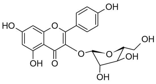
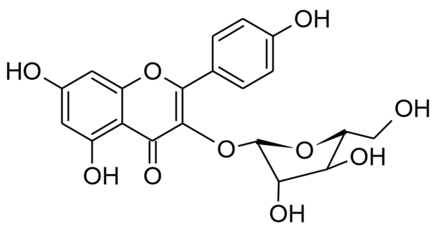
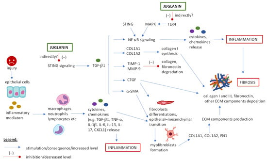
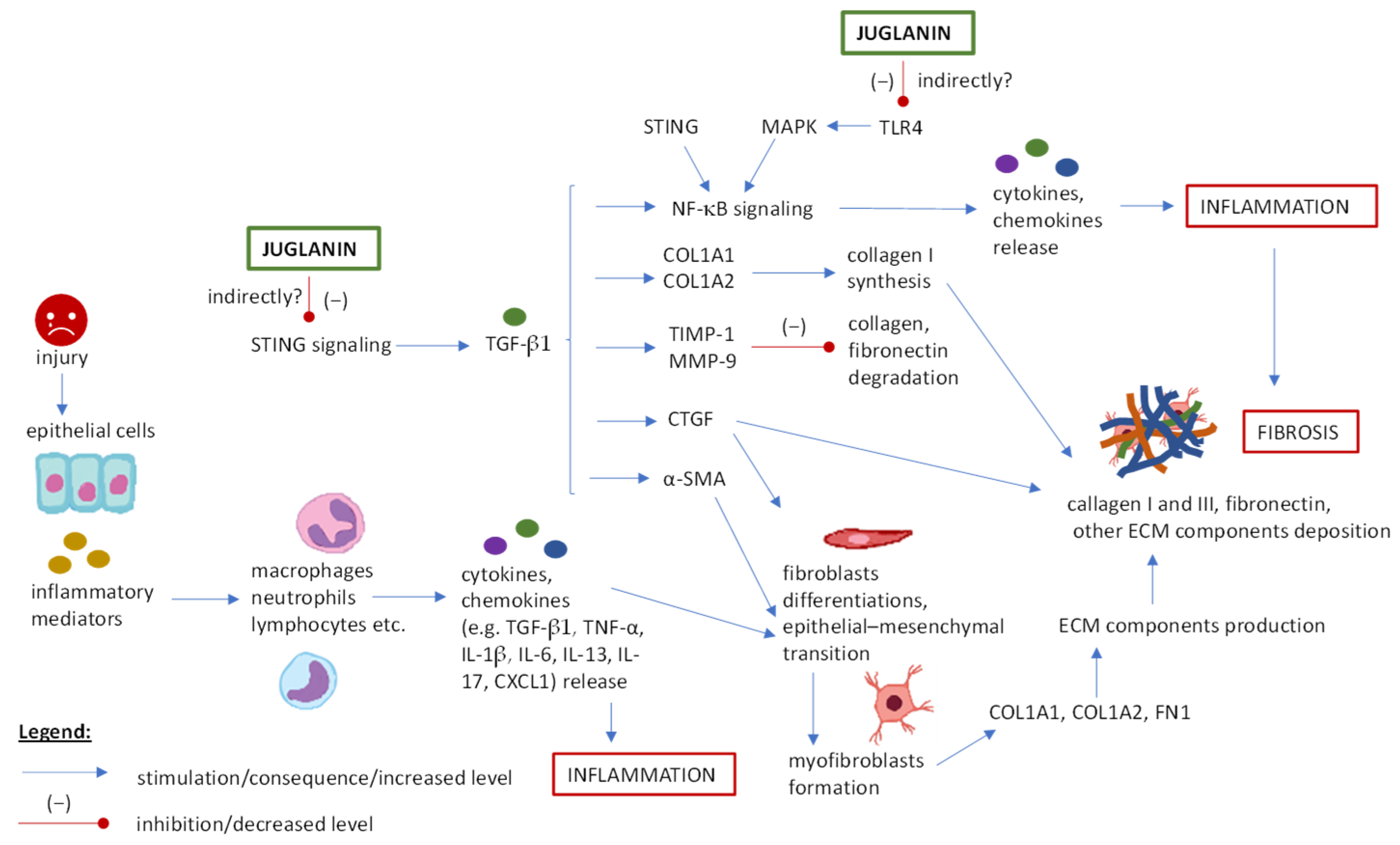
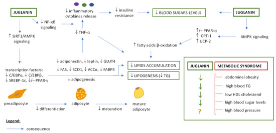
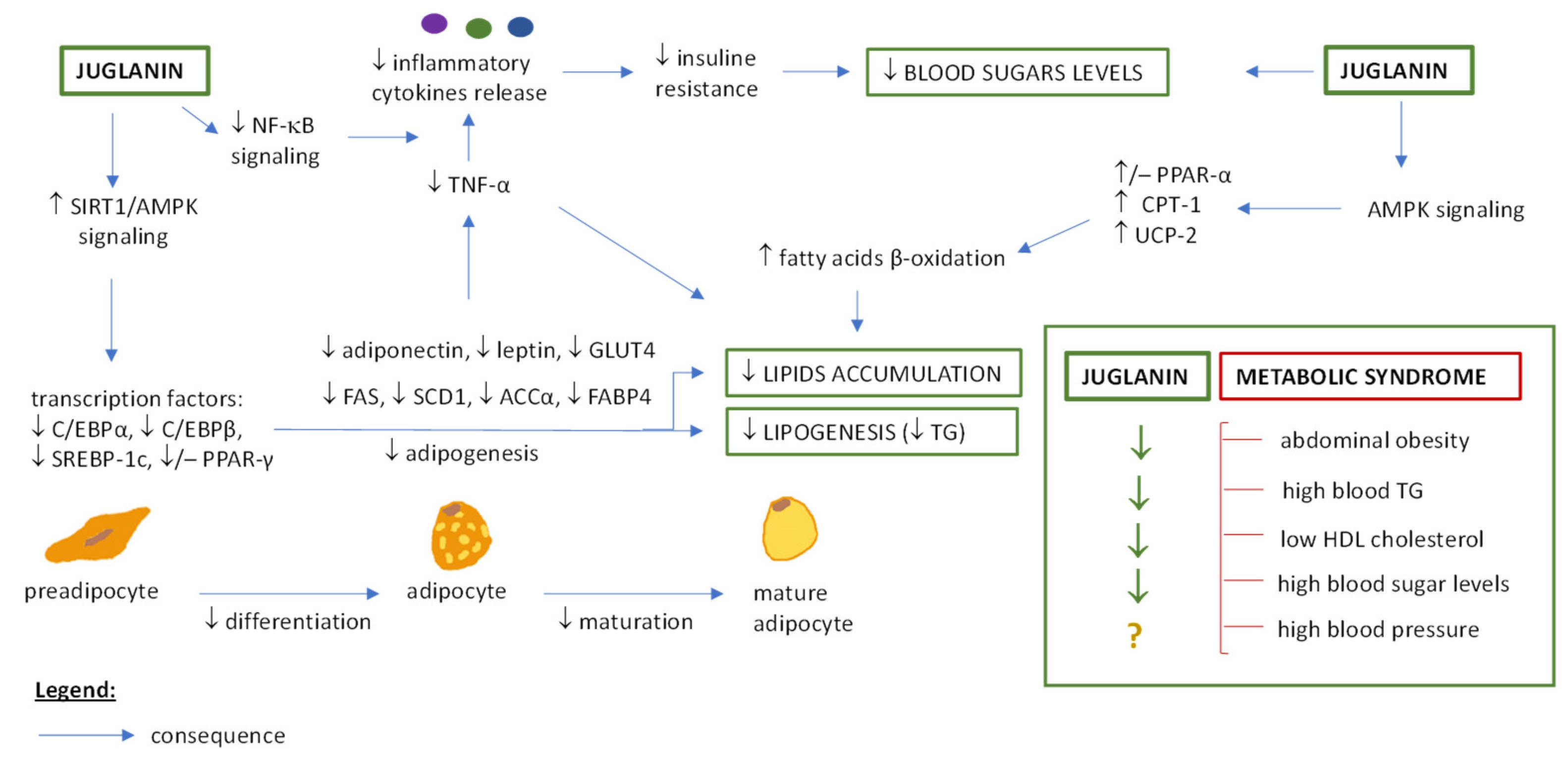
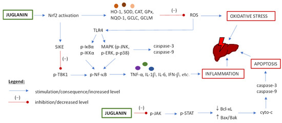
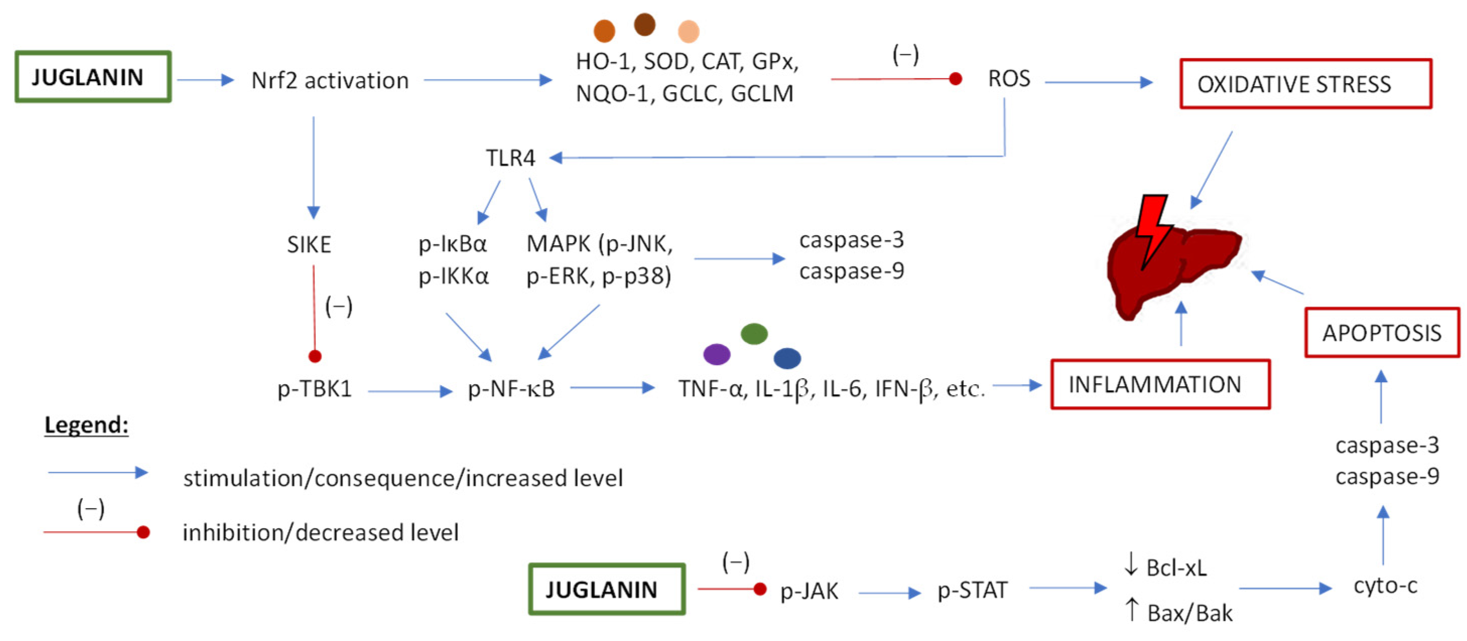
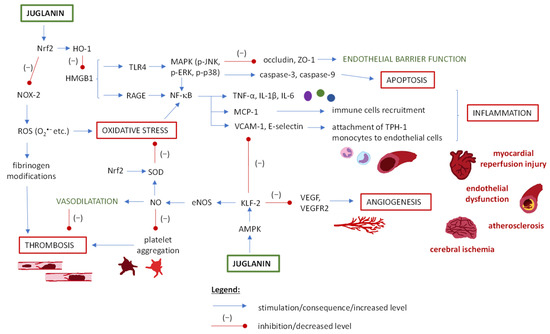
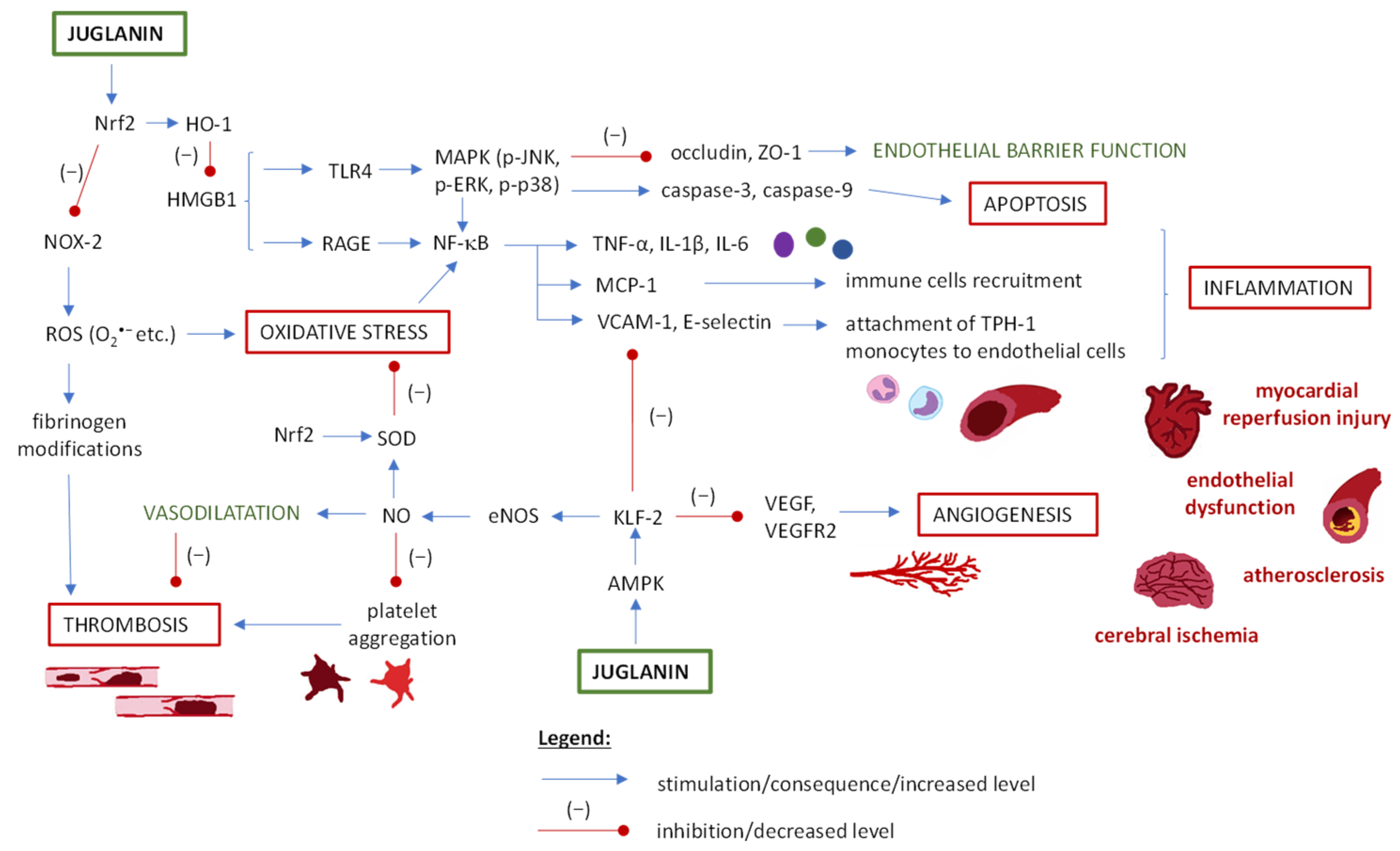
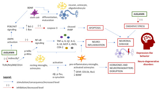
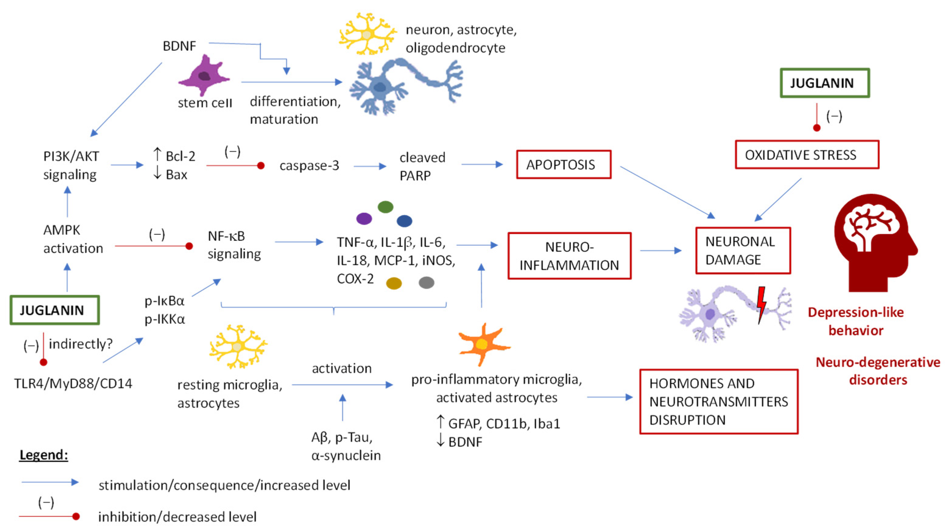
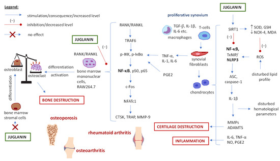
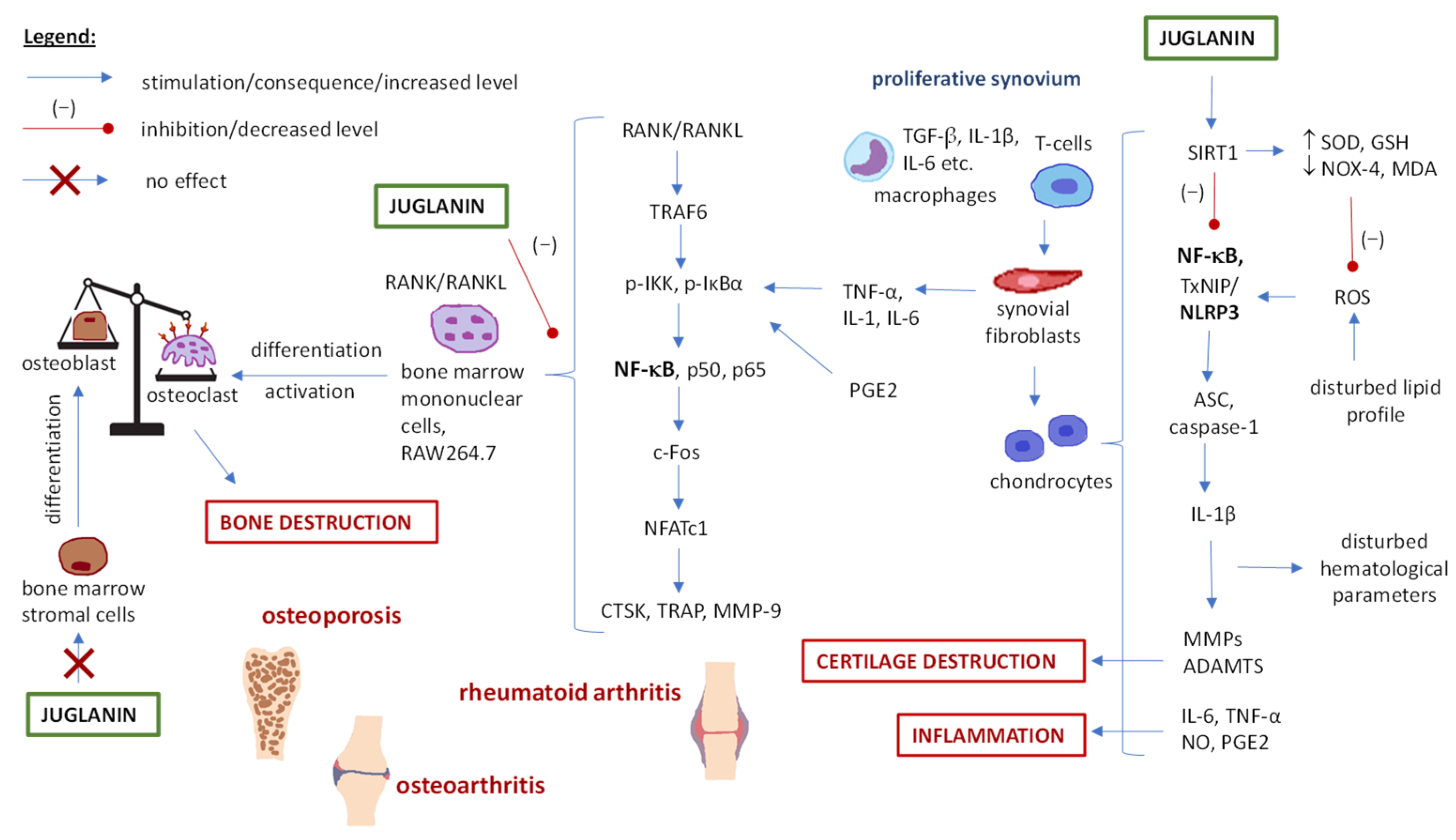
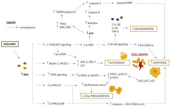
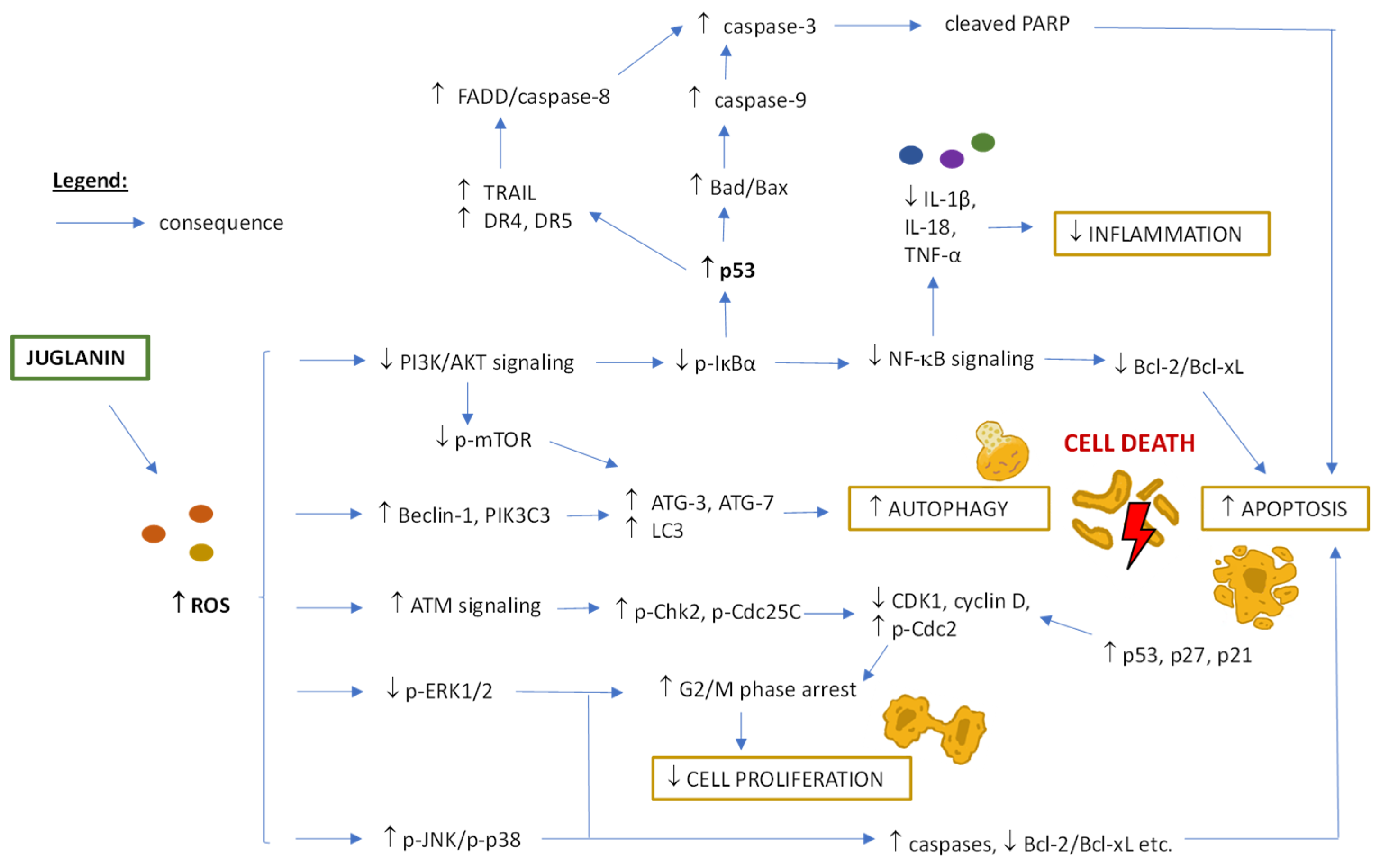
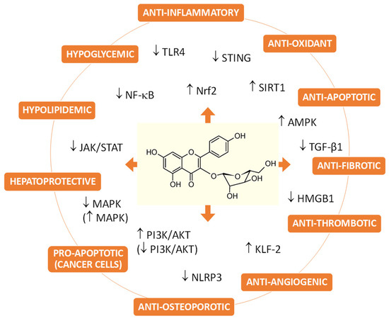
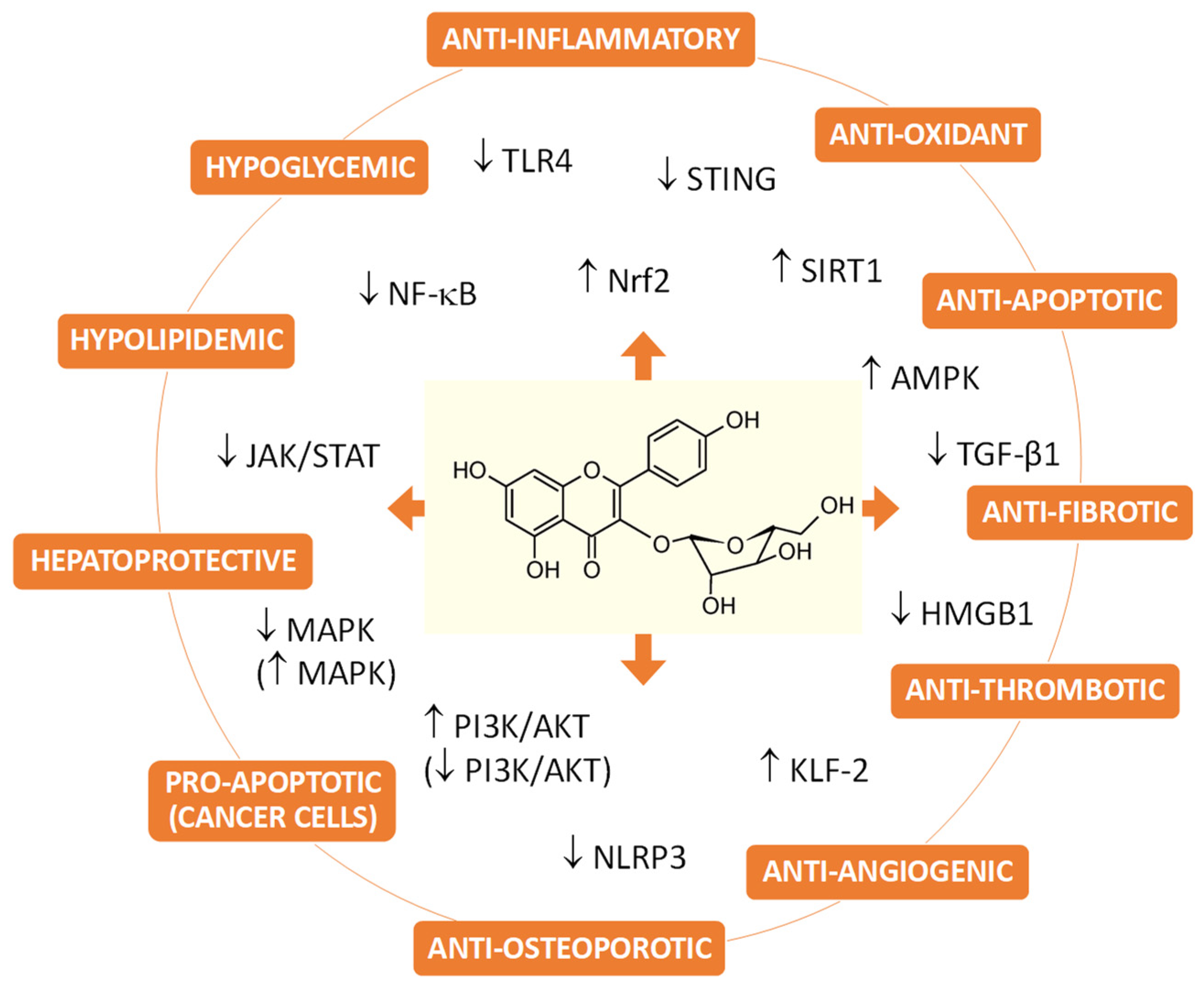

Disclaimer/Publisher’s Note: The statements, opinions and data contained in all publications are solely those of the individual author(s) and contributor(s) and not of MDPI and/or the editor(s). MDPI and/or the editor(s) disclaim responsibility for any injury to people or property resulting from any ideas, methods, instructions or products referred to in the content. |
© 2024 by the authors. Licensee MDPI, Basel, Switzerland. This article is an open access article distributed under the terms and conditions of the Creative Commons Attribution (CC BY) license (https://creativecommons.org/licenses/by/4.0/).